men with low vitamin a stores respond adequately to primary yellow fever and secondary tetanus...
TRANSCRIPT
The Journal of Nutrition
Nutritional Immunology
Men with Low Vitamin A Stores RespondAdequately to Primary Yellow Fever andSecondary Tetanus Toxoid Vaccination1,2
Shaikh M. Ahmad,3,5 Marjorie J. Haskell,3 Rubhana Raqib,5 and Charles B. Stephensen3,4*
3Program in International and Community Nutrition, Department of Nutrition and 4USDA Western Human Nutrition Research
Center, University of California Davis, CA 95616 and 5Immunology, Laboratory Sciences Division, International Centre for Diarrhoeal
Disease Research, Bangladesh, Mohakhali 1212, Dhaka, Bangladesh
Abstract
Current recommendations for vitamin A intake and liver stores (0.07 mmol/g) are based on maintaining normal vision. Higher
levels may be required for maintaining normal immune function. The objective of this study was to assess the relationship
between total body vitamin A stores in adult men and measures of adaptive immune function. We conducted an 8-wk
residential study among 36 healthy Bangladeshi men with low vitamin A stores. Subjects received a standard diet and were
randomized in a double-blind fashion to receive vitamin A (240 mg) or placebo during wk 2 and 3. Subjects received Yellow
Fever Virus (YFV) and tetanus toxoid (TT) vaccines during wk 5. Vitamin A stores were estimated by isotopic dilution during
wk 8. Vaccine-specific lymphocyte proliferation, cytokine production, and serum antibody responses were evaluated before
and after vaccination. Vitamin A supplementation increased YFV- and TT-specific lymphocyte proliferation and YFV-specific
interleukin (IL)-5, IL-10, and tumor necrosis factor-a production but inhibited development of a TT-specific IL-10 response.
Both groups developed protective antibody responses to both vaccines. Some responses correlated positively with vitamin
A stores. These findings indicate that the currently recommended vitamin A intake is sufficient to sustain a protective
response to YFV and TT vaccination. However, YFV-specific lymphocyte proliferation, some cytokine responses, and
neutralizing antibody were positively associated with liver vitamin A stores . 0.084 mmol/g. Such increases may enhance
vaccine protection but raise the question of whether immune-mediated chronic diseases may by exacerbated by high-level
dietary vitamin A. J. Nutr. 138: 2276–2283, 2008.
Introduction
Several aspects of adaptive immunity are compromised by vitaminA deficiency (1). In particular, vitamin A deficiency impairs T cell-mediated responses (2,3) by impairing aspects of both T helpertype 1 (Th1)6 and Th2 function (4,5) and by impairing B-cellfunction (6). Impaired T-cell–mediated antibody responses havebeen demonstrated in several animal models (2,3,7,8). Human
studies have examined the effect of vitamin A supplementation onthe antibody response to childhood vaccines. Most studies foundno enhancement (9), although 1 study reported an increased serumantibody response to Tetanus Toxoid (TT) (10).
To our knowledge, no studies have used immune function as anindex to evaluate dietary vitamin A requirements. The estimatedaverage requirement (EAR) for vitamin A is based on mainte-nance of liver stores . 0.07 mmol retinol/g liver, which preventsdevelopment of eye signs of deficiency (an abnormal electroreti-nogram) for 4 mo while consuming a vitamin A-free diet (11).However, vitamin A supplementation reduces child mortality frominfectious diseases even when children with eye signs (xerophthal-mia) are excluded from such studies (12–14). This observationsuggests that the maintenance of immune function associated withprotection from infection may require higher liver stores than areneeded to prevent eye signs of deficiency.
We hypothesized that vitamin A supplementation to increaseliver vitamin A stores . 0.07 mmol/g would enhance the primaryresponse to the live-virus Yellow Fever Virus (YFV) vaccine andthe recall response to the inactivated TT vaccine. We alsohypothesized that these responses would correlate positivelywith vitamin A stores. To test these hypotheses, we conducted a
1 Supported by the USDA Current Research Information System Project no.
5306-51530-013-00D and Specific Cooperative Agreement 58-5306-4-034F with
International Centre for Diarrheal Disease Research, Bangladesh, Dhaka,
Bangladesh. On-campus doctoral training of S.M.A. at UC Davis was supported
by NIH research grant D43 TW01267, funded by the Fogarty International Center
and the National Institute of Child Health and Human Development.2 Author disclosures: S. Ahmad, M. Haskell, R. Raqib, and C. Stephensen, no
conflicts of interest.
* To whom correspondence should be addressed. E-mail: charles.stephensen@
ars.usda.gov.
6 Abbreviations used: EAR, estimated average requirement; ICDDR,B,
International Centre for Diarrheal Disease Research, Bangladesh; IFNg,
interferon-g; IL, interleukin; PBMC, peripheral blood mononuclear cell; PRNT,
plaque reduction neutralization test; RDA, recommended dietary allowance; SI,
stimulation index; Th1, T helper type 1; TNFa, tumor necrosis factor-a; TT,
tetanus toxoid; VA, vitamin A group; YFV, yellow fever virus.
2276 0022-3166/08 $8.00 ª 2008 American Society for Nutrition.
Manuscript received 29 April 2008. Initial review completed 6 June 2008. Revision accepted 28 August 2008.
doi:10.3945/jn.108.092056.
by guest on June 16, 2016jn.nutrition.org
Dow
nloaded from
randomized, placebo-controlled intervention trial in Bangla-deshi men with marginal vitamin A stores receiving a controlleddiet low in vitamin A. Vitamin A stores were measured with thedeuterated retinol dilution technique (15).
Materials and Methods
Study site and subjects. This study was carried out at the International
Centre for Diarrheal Disease Research, Bangladesh (ICDDR,B), locatedin Dhaka. The Institutional Review Board of the University of
California, Davis and the Ethical Review Committee of ICDDR,B both
approved the study procedures. All participants provided informed
written consent. A total of 157 adult men (20–30 y of age) were screenedfor admission and 36 were recruited. Two weeks before collecting blood
for screening, all subjects were treated with albendazole to clear
intestinal helminth infections. The inclusion criteria were: serum retinol
,1.22 mmol/L, a normal C-reactive protein concentration (,5mg/L),normal clinical chemistry and hematology values, and freedom from any
infectious or chronic disease by medical history and physician exam. A
previous study in Bangladesh by one of us (M. J. H.) (15) demonstratedthat men meeting these serum retinol and C-reactive protein criteria had
low liver vitamin A stores (,0.07 mmol/g). Subjects enrolled in the study
lived near ICDDR,B during the study period and came to the ICDDR,B
grounds to stay at the Advanced Biomedical Research Unit for 12 h/d,7 d/wk, where they received and consumed all meals during the 2-mo
residential study period. Study subjects were under the regular care of a
physician who monitored them 3 times weekly for general health status
and eye signs of vitamin A deficiency.
Study diet. The diet was modeled on a traditional Bangladeshi diet and
provided ;40 mg/d retinol equivalents (6.4% of the EAR). The diet
consisted of rice, wheat flat bread, lentils, small amounts of curriedchicken or mutton, and pale fruits and vegetables, such as cauliflower,
cabbage, white potatoes, white squash, and banana. Subjects consumed
the diet ad libitum. These low, constant levels of dietary vitamin Astabilized the serum retinol concentration and eliminated any diet-
induced variation among subjects during the isotope dilution period and
during the assay of immunological parameters. The diet provided the
recommended dietary allowance (RDA) for total energy and other majorvitamins and minerals were based on food composition tables from the
USDA and a Bangladeshi nutrient database (16). All subjects received a
single dose (60 mg) of vitamin A at the end of the study.
Study design. The study was a placebo-controlled, double-blind
intervention trial using a randomized block design (with a block size of
6) among 36 men conducted in 2 phases (18 subjects per phase) betweenMay and October 2005. Following a 1-wk stabilization on the study diet
(d 1–7), subjects received either 4 vitamin A capsules (containing 60 mg
retinol equivalents of retinyl palmitate per capsule) or 4 identical placebo
capsules (containing corn oil), 1 capsule per day at 5-d intervals (d 7, 12,17, and 22). One week after supplementation (d 29), all subjects were
vaccinated with the YFV vaccine (5.04 Log10 plaque-forming units in a
0.5-mL dose; YF-VAX, Aventis Pasteur) subcutaneously in one arm and
the TT vaccine ($40 IU in a 0.5-mL dose; TETAVAX, Aventis Pasteur SA)i.m. in the other arm. One week after vaccination (d 36), a single dose
(10mg)of stable isotope-labeledvitaminA([2H4]-retinyl acetate;Cambridge
Isotope Laboratories) was given orally. Venous blood was obtained before(d 29), 1 wk (d 36), and 1 mo (d 57) after vaccination. Blood was collected
after overnight food deprivation of ;10 h between 0700 and 0800. The
vitamin A and placebo groups will be referred to as the high Vitamin A
(VA) and low VA groups, respectively.
Serum retinol and vitamin A reserves. Serum was stored at 280�Cfor measurement of retinol by HPLC as described previously (17). Total
body vitamin A pool size was estimated with the deuterated retinoldilution technique (18) using the equation of Furr et al. (19) on the day
the isotopic dose was given (this calculation did not include the mass of
the tracer dose), 1 wk after vaccination, and again 4 wk after vaccination
at the final blood draw (when the 10-mg mass of the tracer dose was
included in estimating vitamin A pool size). The liver vitamin A
concentration was estimated by assuming that liver weight was 2.4% of
body weight (20) and that 90% of vitamin A is found in the liver. In well-
nourished persons, the liver contains .90% of total-body vitamin A
(20,21), but in vitamin A-deficient individuals, liver vitamin A concen-
trations are lower, ;50–90% of total stores, although vitamin A in other
tissues remains relatively constant (21). In this study, we have used 90%
as the percentage of total body vitamin A present in liver, because all
subjects had serum retinol . 0.70 mmol/L and are thus not considered
deficient using a standard definition (22). Whole-body and liver vitamin
A stores at 1 mo postvaccination were estimated using the prediction
equation with a modification to include the contribution of mass of
tracer dose to the total body vitamin A pool size, because 4 hydrogen
atoms in the isotopic retinol were replaced with 4 deuterium atoms
probably retain the same biological activity as nonisotopic retinol.
Peripheral blood mononuclear cells proliferation and cytokine
assay. Peripheral blood mononuclear cells (PBMC) were separated fromfresh heparinaized blood using a standard density gradient method
(Ficoll-Paque-PLUS, Amersham Biosciences) cultured in standard Russ-
10 media (3) supplemented with 10% heat-inactivated (56�C for 30 min)autologous plasma. Triplicate cultures (2 3 105 PBMC in a 96-well
U-bottom plate) were stimulated with 3 levels of YFV vaccine (plaque-
forming units) and 3 levels of limits of flocculation of TT antigen
(02/126; NIBSC) for 6 d at 37�C and 5% CO2. Cell proliferation wasmeasured by incorporation of [3H]-thymidine. Results were expressed as
total counts or as a stimulation index (SI), the ratio of antigen-
stimulated:unstimulated wells. An additional well of the highest antigen
dose was collected at 6 d for analysis of supernatant levels of interleukin(IL)-2, IL-4, IL-10, interferon-g (IFNg), tumor necrosis factor-a (TNFa),
and IL-5 by Luminex assay (Bio-Plex system, Bio-Rad Laboratories).
Serum and lymphocyte supernatant anti-TT IgG assay. Unstimu-
lated PBMC were cultured for 3 d to identify antibody-secreting
lymphocytes essentially as described (23). Culture supernatants and
plasma were both assayed for TT-specific IgG using EIA kits (TheBinding Site). Results are expressed as mIU/L and levels above the 0.15
mIU/L threshold are considered protective (24).
Serum YFV specific plaque reduction neutralization test 50 assay.
The plaque reduction neutralization test (PRNT) to detect neutralizing
antibody for the YFV 17D vaccine strain was carried out by Dr. Sutee
Yoksan at the Center for Vaccine Development, Mahidol University,
Thailand as described (25).
Statistical analysis. Statistical analyses were conducted using Sigma-
Stat 3.1 (Systat Software) and SPSS 13.0 for windows (SPSS). Distribu-tions of study variables were analyzed to identify outliers and test
assumptions of normality and equal variance. Data were transformed to
produce normal distributions as needed.
Two subjects in the placebo group had implausibly high vitamin Apool size estimates (0.42 and 0.74 mmol). These levels are inconsistent
with their reported dietary intakes and with pool size data from other
subjects recruited in the same fashion in the same setting (M. J. Haskell,
unpublished observations). Such overestimates of pool size are likely dueto poor absorption of the tracer dose. One subject in the vitamin A group
also demonstrated an unreasonably high estimate, 0.54 mmol, which
was .3 SD higher than the mean value of this group. It is also likely thatthis subject had poor absorption of the tracer dose. Data from these 3
subjects were not used in the correlation analyses involving pool size
estimates. Data from these subjects was used in the comparisons between
low and high VA groups, because these comparisons do not depend onthe pool size estimates.
Low-level prevaccination YFV antibody titers were detected in 2
placebo subjects, indicating previous exposure to a related virus (YFV is
not found in Bangladesh). Because this previous exposure produced amemory response that was cross-reactive with YFV, their response to YFV
immunization would be considered a secondary rather than a primary
response (26). Hence, these subjects were not included in the analysis ofthe YFV response but were included in the analysis of the TT response. In
Vitamin A stores and vaccine responses 2277
by guest on June 16, 2016jn.nutrition.org
Dow
nloaded from
addition, 1 subject in the vitamin A group did not respond with a
measurable serum antibody titer or with an increased proliferative
response to YFV 1 mo after YFV vaccination. This suggests that thesubject was not vaccinated, although study records indicate the vaccina-
tion did occur. This subject was not included in analysis of the YFV data,
because either a rare vaccine failure or failure to vaccinate would
invalidate this subject in analyzing the effect of vitamin A on YFVimmunity. This subject responded well to the TT vaccination and his data
were included in that portion of the analysis.
Two-way repeated-measures ANOVA using the Holm-Sidak post hoc
comparison proceure was used to compare vaccine-specific immuneresponses. The post hoc test was applied when significant differences
were found between study groups, between study days, or their interaction.
Between-group comparisons at a single time point were conducted usingStudent’s t test.Theoverall significancelevelof these testswassetatP,0.05.
Spearman rho correlation analysis was used to determine whether
individual immune response variables were associated with vitamin A
stores. Two-phase segmental linear regression was used to identify possiblebreak points at which a linear association between stores and immune
response variables might reach a plateau. Groups of similar immune
response variables were converted to Z-scores (mean of 0, SD of 1) and
analyzed together for the break-point analysis. For each vaccine, clusters ofimmune response variables measured at 1 wk or at 1 mo postvaccination
were used. These clusters included: 1) the 3 levels of antigen-stimulated
PBMC proliferation and serum antibody titer, because lymphocyteproliferation is a necessary prerequisite for producing a T-cell–mediated
serum antibody response;2) vaccine-specificTh1 cytokines (IL2, IFNg, and
TNFa), which stimulate cytotoxicity T cell responses and macrophage
activation; and 3) vaccine-specific Th2 cytokines (IL-4, IL-5, and IL-10),which stimulate B cells for antibody production. GraphPad Prism 5 for
Windows (GraphPad Software) was used for this analysis.
Results
Baseline characteristics. Study subjects had normal anthro-pometric and hematologic measurements at baseline. The2 groups did not differ in these characteristics (Table 1).
Vitamin A status. Following supplementation, the mean serumretinol concentration in the high VA group increased signif-icantly, whereas that of the low VA group did not change (Table
2). As expected, the estimated total body vitamin A pool size inthe high VA group was significantly higher than the pool size inthe low VA group (Table 2). All subjects in the high VA grouphad liver stores .0.07 mmol/g, the target level used for the RDAdetermination, whereas in the low VA group, all but 2 subjects(0.077 and 0.084 mmol/g) had liver stores ,0.07 mmol/g. Nosubjects developed signs of clinical vitamin A deficiency.
Primary YFV vaccine response in low and high VA groups.
Vaccination induces development of antigen-specific T-cells thatcirculate in peripheral blood. Thus, assessment of antigen-specificlymphocyte proliferation in PBMC cultures is a useful index ofresponse to immunization. In the present study, YFV-specificlymphocyte proliferation was assessed using 3 antigen doses at1 wk and 1 mo after immunization. Proliferation was negligiblebefore immunization and increased significantly in both groups atboth 1 wk and 1 mo after immunization using at the 2 highestantigen doses (Fig. 1A,B). At the lowest antigen dose, the increasein SI was significant only in the high VA group and only 1 mo afterimmunization (Fig. 1C). The SI tended to differ between the lowand high VA groups at 1 mo (P ¼ 0.08). When proliferation wasexpressed as total Bq of 3H-thymidine incorporation, postvaccineincorporation increased significantly over the prevaccine levelsfor the high VA group but not for the low VA group at all antigendoses at both time points (Fig. 1D,F). Thus, the postvaccineincrease in the YFV-specific proliferative response was greater inthe high VA group than in the low VA group, although when thegroups were compared directly, they did not significantly differfrom one another (Fig. 1A–F).
Another important component of the vaccine-specific T-cellresponse is the production of growth (IL-2) and effector (IL-4,IL-5, IL-10, IFNg, and TNFa) cytokines. Postvaccine increasesin IL-2 concentrations ocurred 1 mo after immunization in boththe low and high VA groups, although IL-2 levels were lowerthan baseline 1 wk after immunization (Fig. 1H).
YFV is a live virus vaccine and induces a predominant Th1response. Two effector cytokines produced by Th1 cells weremeasured in this study, IFNg and TNFa. Production of IFNg
increased significantly in both the high and low VA groupsfollowing YFV immunization and the increases were of similarmagnitude (Fig. 1K). However, TNFa production increasedsignificantly after immunization in the high VA group but not inthe low VA group (Fig. 1L). The difference in the response trendswas marginally significant (P ¼ 0.06).
Il-4 and IL-5 are signature cytokines of Th2 cells. Both the highand low VA groups had significant postvaccine increases in IL-4that were of similar magnitude (Fig. 1I). The postvaccine increasein IL-5 was significant in the low VA group 1 mo but not 1 wk afterimmunization, whereas this response was significant at both timepoints in the high VA group (Fig. 1M). When the IL-5:IFNg ratiowas evaluated as an index of the relative magnitude of the Th2
TABLE 1 Baseline characteristics of the study subjectsin the 2 groups1
Study groups
Characteristics Low VA High VA
Age, y 24.3 6 2.70 23.5 6 3.05
Height, cm 164 6 7.71 161 6 8.26
Weight, kg 53.1 6 5.50 51.9 6 7.49
BMI, kg/m2 19.7 6 1.60 19.7 6 1.79
WBC, 3109/L 9.45 6 1.92 9.00 6 1.92
Hemoglobin, g/L 152 6 10.9 152 6 9.70
MCV, limits of flocculation 85.8 6 5.01 84.4 6 3.46
MCH, pg/cell 29.1 6 3.06 29.6 6 1.32
MCHC, g/L 335 6 20.6 341 6 16.0
1 Values are means 6 SD, n ¼ 18. MCV, mean corpuscular volume; MCH, mean
corpuscular hemoglobin; MCHC, mean corpuscular hemoglobin concentration; WBC,
white blood cell.
TABLE 2 Serum retinol concentration, estimated totalbody vitamin A pool size, and liver vitamin Aconcentration in low and high VA groups vaccinatedwith YFV and TT1
Study groups
Low VA (n ¼ 16) High VA (n ¼ 17)
Serum retinol, mmol/L
Presupplementation 1.52 6 0.39 1.49 6 0.37
Postsupplementation2 1.42 6 0.32 1.85 6 0.34*#
Whole-body vitamin A,3 mmol 0.06 6 0.04 0.23 6 0.06*
Liver vitamin A,3 mmol/g 0.04 6 0.03 0.16 6 0.04*
Whole-body vitamin A,4 mmol 0.07 6 0.04 0.24 6 0.06*
Liver vitamin A,4 mmol/g 0.05 6 0.03 0.17 6 0.04*
1 Values are means 6 SD. *Different from low VA, P , 0.001; #different from
presupplementation, P , 0.01.2 Prevaccination (1-wk after the end of placebo/vitamin A supplementations).3 1 wk postvaccination.4 1 mo postvaccination.
2278 Ahmad et al.
by guest on June 16, 2016jn.nutrition.org
Dow
nloaded from
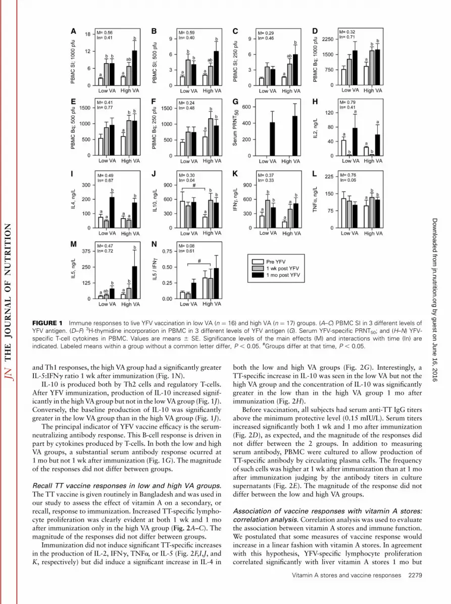
and Th1 responses, the high VA group had a significantly greaterIL-5:IFNg ratio 1 wk after immunization (Fig. 1N).
IL-10 is produced both by Th2 cells and regulatory T-cells.After YFV immunization, production of IL-10 increased signif-icantly in the high VA group but not in the low VA group (Fig. 1J).Conversely, the baseline production of IL-10 was significantlygreater in the low VA group than in the high VA group (Fig. 1J).
The principal indicator of YFV vaccine efficacy is the serum-neutralizing antibody response. This B-cell response is driven inpart by cytokines produced by T-cells. In both the low and highVA groups, a substantial serum antibody response ocurred at1 mo but not 1 wk after immunization (Fig. 1G). The magnitudeof the responses did not differ between groups.
Recall TT vaccine responses in low and high VA groups.
The TT vaccine is given routinely in Bangladesh and was used inour study to assess the effect of vitamin A on a secondary, orrecall, response to immunization. Increased TT-specific lympho-cyte proliferation was clearly evident at both 1 wk and 1 moafter immunization only in the high VA group (Fig. 2A–C). Themagnitude of the responses did not differ between groups.
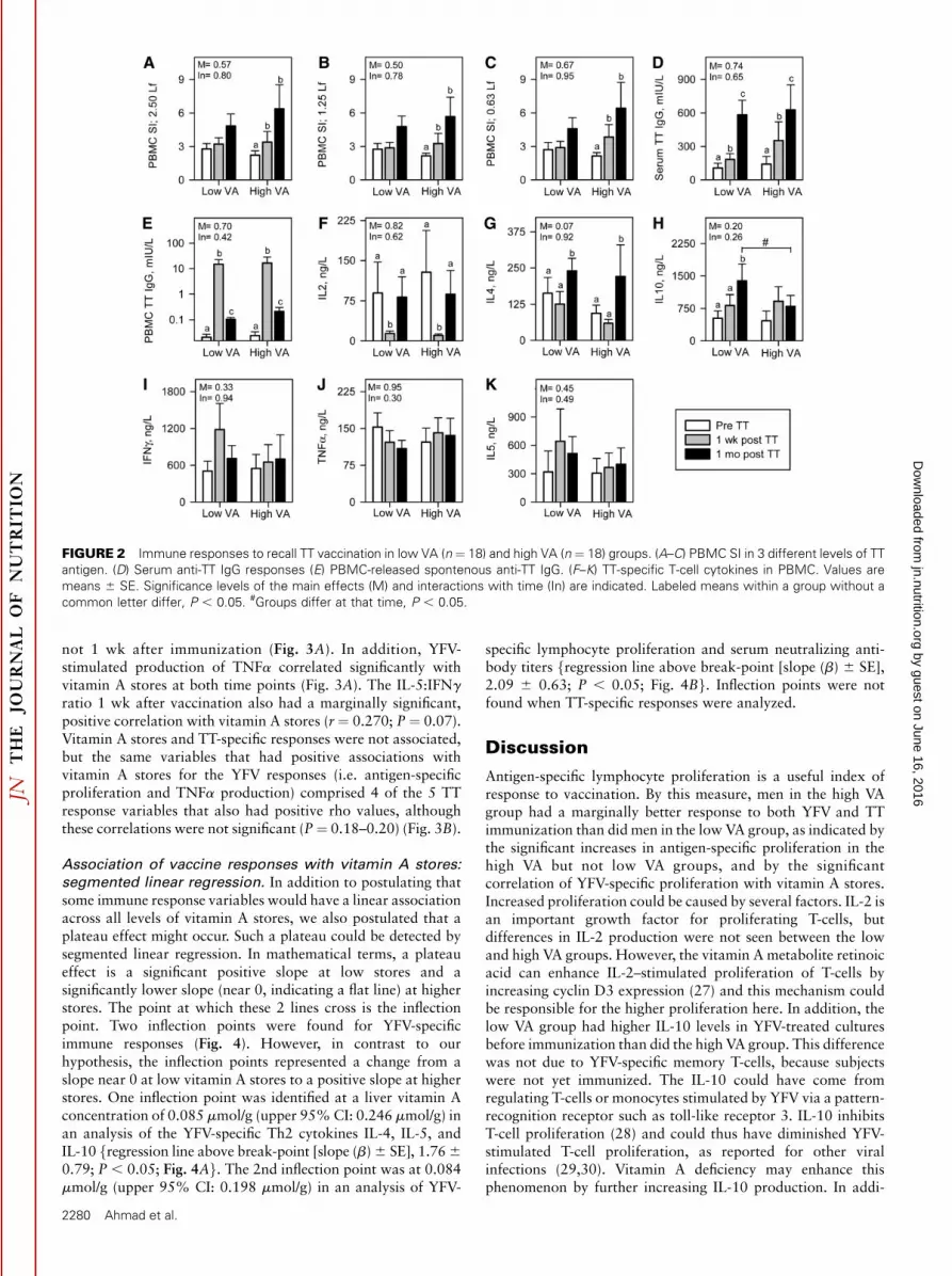
Immunization did not induce significant TT-specific increasesin the production of IL-2, IFNg, TNFa, or IL-5 (Fig. 2F,I,J, andK, respectively) but did induce a significant increase in IL-4 in
both the low and high VA groups (Fig. 2G). Interestingly, aTT-specific increase in IL-10 was seen in the low VA but not thehigh VA group and the concentration of IL-10 was significantlygreater in the low than in the high VA group 1 mo afterimmunization (Fig. 2H).
Before vaccination, all subjects had serum anti-TT IgG titersabove the minimum protective level (0.15 mIU/L). Serum titersincreased significantly both 1 wk and 1 mo after immunization(Fig. 2D), as expected, and the magnitude of the responses didnot differ between the 2 groups. In addition to measuringserum antibody, PBMC were cultured to allow production ofTT-specific antibody by circulating plasma cells. The frequencyof such cells was higher at 1 wk after immunization than at 1 moafter immunization judging by the antibody titers in culturesupernatants (Fig. 2E). The magnitude of the response did notdiffer between the low and high VA groups.
Association of vaccine responses with vitamin A stores:
correlation analysis. Correlation analysis was used to evaluatethe association between vitamin A stores and immune function.We postulated that some measures of vaccine response wouldincrease in a linear fashion with vitamin A stores. In agreementwith this hypothesis, YFV-specific lymphocyte proliferationcorrelated significantly with liver vitamin A stores 1 mo but
FIGURE 1 Immune responses to live YFV vaccination in low VA (n ¼ 16) and high VA (n ¼ 17) groups. (A–C) PBMC SI in 3 different levels of
YFV antigen. (D–F) 3H-thymidine incorporation in PBMC in 3 different levels of YFV antigen (G). Serum YFV-specific PRNT50; and (H–N) YFV-
specific T-cell cytokines in PBMC. Values are means 6 SE. Significance levels of the main effects (M) and interactions with time (In) are
indicated. Labeled means within a group without a common letter differ, P , 0.05. #Groups differ at that time, P , 0.05.
Vitamin A stores and vaccine responses 2279
by guest on June 16, 2016jn.nutrition.org
Dow
nloaded from
not 1 wk after immunization (Fig. 3A). In addition, YFV-stimulated production of TNFa correlated significantly withvitamin A stores at both time points (Fig. 3A). The IL-5:IFNg
ratio 1 wk after vaccination also had a marginally significant,positive correlation with vitamin A stores (r ¼ 0.270; P ¼ 0.07).Vitamin A stores and TT-specific responses were not associated,but the same variables that had positive associations withvitamin A stores for the YFV responses (i.e. antigen-specificproliferation and TNFa production) comprised 4 of the 5 TTresponse variables that also had positive rho values, althoughthese correlations were not significant (P¼ 0.18–0.20) (Fig. 3B).
Association of vaccine responses with vitamin A stores:
segmented linear regression. In addition to postulating thatsome immune response variables would have a linear associationacross all levels of vitamin A stores, we also postulated that aplateau effect might occur. Such a plateau could be detected bysegmented linear regression. In mathematical terms, a plateaueffect is a significant positive slope at low stores and asignificantly lower slope (near 0, indicating a flat line) at higherstores. The point at which these 2 lines cross is the inflectionpoint. Two inflection points were found for YFV-specificimmune responses (Fig. 4). However, in contrast to ourhypothesis, the inflection points represented a change from aslope near 0 at low vitamin A stores to a positive slope at higherstores. One inflection point was identified at a liver vitamin Aconcentration of 0.085 mmol/g (upper 95% CI: 0.246 mmol/g) inan analysis of the YFV-specific Th2 cytokines IL-4, IL-5, andIL-10 fregression line above break-point [slope (b) 6 SE], 1.76 6
0.79; P , 0.05; Fig. 4Ag. The 2nd inflection point was at 0.084mmol/g (upper 95% CI: 0.198 mmol/g) in an analysis of YFV-
specific lymphocyte proliferation and serum neutralizing anti-body titers fregression line above break-point [slope (b) 6 SE],2.09 6 0.63; P , 0.05; Fig. 4Bg. Inflection points were notfound when TT-specific responses were analyzed.
Discussion
Antigen-specific lymphocyte proliferation is a useful index ofresponse to vaccination. By this measure, men in the high VAgroup had a marginally better response to both YFV and TTimmunization than did men in the low VA group, as indicated bythe significant increases in antigen-specific proliferation in thehigh VA but not low VA groups, and by the significantcorrelation of YFV-specific proliferation with vitamin A stores.Increased proliferation could be caused by several factors. IL-2 isan important growth factor for proliferating T-cells, butdifferences in IL-2 production were not seen between the lowand high VA groups. However, the vitamin A metabolite retinoicacid can enhance IL-2–stimulated proliferation of T-cells byincreasing cyclin D3 expression (27) and this mechanism couldbe responsible for the higher proliferation here. In addition, thelow VA group had higher IL-10 levels in YFV-treated culturesbefore immunization than did the high VA group. This differencewas not due to YFV-specific memory T-cells, because subjectswere not yet immunized. The IL-10 could have come fromregulating T-cells or monocytes stimulated by YFV via a pattern-recognition receptor such as toll-like receptor 3. IL-10 inhibitsT-cell proliferation (28) and could thus have diminished YFV-stimulated T-cell proliferation, as reported for other viralinfections (29,30). Vitamin A deficiency may enhance thisphenomenon by further increasing IL-10 production. In addi-
FIGURE 2 Immune responses to recall TT vaccination in low VA (n ¼ 18) and high VA (n ¼ 18) groups. (A–C) PBMC SI in 3 different levels of TT
antigen. (D) Serum anti-TT IgG responses (E) PBMC-released spontenous anti-TT IgG. (F–K) TT-specific T-cell cytokines in PBMC. Values are
means 6 SE. Significance levels of the main effects (M) and interactions with time (In) are indicated. Labeled means within a group without a
common letter differ, P , 0.05. #Groups differ at that time, P , 0.05.
2280 Ahmad et al.
by guest on June 16, 2016jn.nutrition.org
Dow
nloaded from
tion, YFV-specific TNFa production was higher in the high VAgroup and can enhance proliferation by acting directly on T-cells(31,32) or indirectly by enhancing differentiation of antigen-presenting cells (33). Thus, the changes in YFV-specific IL-10 andTNFa production in these subjects could be responsible for theincreased YFV-specific T-cell proliferation in the high VA group.
In these experiments, vaccine-specific IL-2 levels decreasedsignificantly in both groups 1 wk after vaccination. This decreasepresumably resulted from the rapid utilization of IL-2 byactivated T-cells (34), thus triggering proliferation and differen-tiation into effector cells (35). In this experiment, we measuredcytokine 6 d after stimulation. However, studies with otherflavivirus-infected peripheral blood leukocyte cultures indicatethe highest IL-2 release levels occur after 2 d and then diminish by6 d (whereas the highest IL-10 response is detected at 6 d) (36).One month after immunization, an antigen-specific IL-2 responsewas seen in 6-d cultures, perhaps resulting from a greater numberof IL-2–secreting memory cells at the later time point.
The YFV-specific IFNg response did not differ between the2 groups, but based on animal studies, we had predicted adecreased Th1 response in the high VA group (7). However, a
modest shift toward a Th2 response occurred in the high VAgroup, which had a significantly higher YFV-specific IL-5(particularly when evaluated as a ratio with IFNg) and IL-10responses than did the low VA group. This relative increase inIL-5 could also promote a greater serum antibody response.
We used TT immunization in this study to evaluate the effectof vitamin A on response to secondary immunization. As withYFV, the TT-specific increase in lymphocyte proliferation inresponse to immunization was significant in the high VA groupbut not in the low VA group. This finding is consistent withearlier results in an animal model where vitamin A enhanced
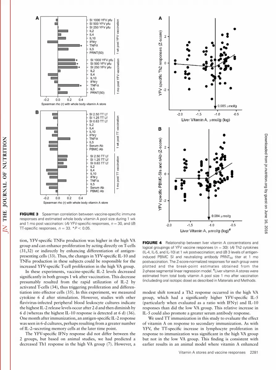
FIGURE 4 Relationship between liver vitamin A concentrations and
logical groupings of YFV vaccine responses (n ¼ 30): (A) Th2 cytokines
(IL-4, IL-5, and IL-10) at 1 wk postvaccination; and (B) 3 levels of antigen-
induced PBMC SI and neutralizing antibody PRNT50 titer at 1 mo
postvaccination. The Z-score-normalized responses for each group were
plotted and the break-point estimates obtained from the
2-phase segmental linear regression model. #Liver vitamin A stores were
estimated from total body vitamin A pool size 1 mo after vaccination
(includeding oral isotopic dose) as described in Materials and Methods.
FIGURE 3 Spearman correlation between vaccine-specific immune
responses and estimated whole body vitamin A pool size during 1 wk
and 1 mo post vaccination: (A) YFV-specific responses, n ¼ 30, and (B)
TT-specific responses, n ¼ 33. *P , 0.05.
Vitamin A stores and vaccine responses 2281
by guest on June 16, 2016jn.nutrition.org
Dow
nloaded from

TT-specific lymphocyte proliferation (37), although in 1 humanstudy, there was no difference at 6 mo after vaccination (38).
Four of the 6 cytokines examined did not have TT-specificincreases, including IL-2, IL-4, IL-5, IFNg, and TNFa, althougha significant TT-specific increase was seen for IL-4 1 mo afterimmunization for both groups. Interestingly, only the low VAgroup had a significant increase in TT-specific IL-10. Thisobservation is reminiscent of a previous study in which a greaterovalbumin-specific IL-10 response was seen in vitaminA-deficient mice compared with mice receiving a control diet(4). These studies are similar in that both used a purified proteinantigen given with a relatively noninflammatory adjuvant (alumin the case of the present study and incomplete Freund’s in theprevious study), suggesting that vitamin A deficiency may lead toan increased frequency of IL-10–producing T-cells in such cases.
The serum antibody response to TT immunization was notaffected by vitamin A status in this study. Both groups mountedsignificant recall responses. Five other studies reported similarfindings (39–43), although 1 study among children withconfirmed vitamin A deficiency did report enhancement of thememory response to TT immunization (10). Subjects in the latterstudy had more profound vitamin A deficiency than did thesubjects in the present study, as indicated by lower serum retinolconcentrations and the presence of xerophthalmia in 1 of thestudy groups.
Segmented linear regression analysis was used to identifypositive associations between vitamin A stores and 2 clusters ofYFV-specific immune response variables 1): YFV-specific Th2cytokine production (IL-4, IL-5, and IL-10); and 2) YFV-specificlymphocyte proliferation and serum antibody. These associa-tions may be mechanistically related, because Th2 cytokines canpromote both proliferation and antibody production. We hadanticipated such responses would be positively correlated withvitamin A at the lowest storage levels and then might plateau athigher levels. However, the opposite trend occurred, in that apositive association was not seen at low storage levels but wasseen at .0.084 mmol retinol/g liver. This observation suggeststhat the current 0.07 mmol/g index level for setting the EAR andRDA is adequate to maintain protective vaccine responses,particularly because all subjects developed protective antibodytiters in response to both vaccines. However, increasing vitaminA stores above the 0.084 mmol/g level was associated withfurther increases in some measures of vaccine responsiveness.This observation raises the question of what immune functionchanges might be seen by further increasing stores to the evenhigher levels present in adults in the US and other industrializedcountries (19,21,22,44–46). Might these high levels of vitamin Aenhance chronic diseases mediated by antibody, Th2 cytokines,or TNFa? This possibility suggests that future evaluation of thetolerable upper intake for vitamin A intake might consider riskof adverse immune reactivity in addition to teratogenicity,hepatotoxicity, and fracture risk, which were used in setting thecurrent upper intake (11).
This study had several strengths, including a double-blind,placebo-controlled design, a healthy and homogeneous group ofsubjects with low baseline vitamin A stores, a residential design tominimize environmental influences, and provision of a nutrition-ally balanced diet to eliminate dietary differences among subjects.In addition, a unique aspect of the study was the use of isotopedilution to provide a quantitative estimate of vitamin A stores.This allowed association of individual stores with immunefunction endpoints rather than relying solely on group compar-isons. Such individualized data allowed the use of more powerfulstatistical techniques that revealed significant associations be-
tween pool size and immune function. Shortcomings of the studyincluded the relatively small sample size and the lack of womenamong study subjects.
In summary, subjects with and without vitamin A deficiency(as defined by liver vitamin A stores , 0.07 mmol/g) respondedadequately to immunization with primary YFV and secondaryTT immunization in that all subjects developed protectiveantibody titers and these titers did not differ between the groups.However, vitamin A deficiency diminished vaccine-specificlymphocyte proliferation and the production of some cytokines,including IL-5, IL-10, and TNFa. The effect of vitamin A statuson IL-10 differed by vaccine and may depend on the underlyinglevel and type of immune stimulation produced by the vaccineadjuvant. Because these changes in immune response correlateddirectly with liver vitamin A stores, it would be prudent toconsider if such ‘‘enhancement ’’ of immune function by high-level vitamin A intake might increase the risk of inflammatorydiseases in subjects with genetic or other environmental riskfactors that affect immune function.
Acknowledgments
We thank the following for their help and expertise: XiaowenJiang for estimating cytokines by multiplexed luminex assay,Alina Wettstein for measuring serum retinol, EmmanuelAklamati for carrying out isotopic retinol measurements, JanetM. Peerson for advice on statistical analysis, and Proteim Sarkerfor conducting flow analysis and helping during in vitro methoddevelopment. We also appreciate Dr. M. Jobayer Chisti andDr. Kazi Jamil for monitoring health of the study subjects atICDDR,B. Dhaka, Bangladesh.
Literature Cited
1. Stephensen CB. Vitamin A, infection, and immune function. Annu RevNutr. 2001;21:167–92.
2. Smith SM, Hayes CE. Contrasting impairments in IgM and IgGresponses of vitamin A-deficient mice. Proc Natl Acad Sci USA. 1987;84:5878–82.
3. Pasatiempo AM, Kinoshita M, Taylor CE, Ross AC. Antibody produc-tion in vitamin A-depleted rats is impaired after immunization withbacterial polysaccharide or protein antigens. FASEB J. 1990;4:2518–27.
4. Stephensen CB, Jiang X, Freytag T. Vitamin A deficiency increases the invivo development of IL10-positive Th2 cells and decreases developmentof Th1 cells in mice. J Nutr. 2004;134:2660–6.
5. Cantorna MT, Nashold FE, Hayes CE. In vitamin A deficiency multiplemechanisms establish a regulatory T helper cell imbalance with excessTh1 and insufficient Th2 function. J Immunol. 1994;152:1515–22.
6. Chen Q, Ross AC. Vitamin A and immune function: retinoic acidmodulates population dynamics in antigen receptor and CD38-stimulatedsplenic B cells. Proc Natl Acad Sci USA. 2005;102:14142–9.
7. Stephensen CB, Blount SR, Schoeb TR, Park JY. Vitamin A deficiencyimpairs some aspects of the host response to influenza A virus infectionin BALB/c mice. J Nutr. 1993;123:823–33.
8. Wiedermann U, Hanson LA, Kahu H, Dahlgren UI. Aberrant T-cellfunction in vitro and impaired T-cell dependent antibody response invivo in vitamin A-deficient rats. Immunology. 1993;80:581–6.
9. Villamor E, Fawzi WW. Effects of vitamin a supplementation onimmune responses and correlation with clinical outcomes. Clin Micro-biol Rev. 2005;18:446–64.
10. Semba RD, Muhilal, Scott AL, Natadisastra G, Wirasasmita S, Mele L,Ridwan E, West KP Jr, Sommer A. Depressed immune response totetanus in children with vitamin A deficiency. J Nutr. 1992;122:101–7.
11. Panel on Micronutrients, Food and Nutrition Board, Institute of Medicine.Dietary reference intakes for vitamin A, vitamin K, arsenic, boron,chromium, copper, iodine, iron, manganese, molybdenum, nickel, silicon,vanadium, and zinc. Washington, DC: National Academy Press; 2001.
2282 Ahmad et al.
by guest on June 16, 2016jn.nutrition.org
Dow
nloaded from
12. Fawzi WW, Chalmers TC, Herrera MG, Mosteller F. Vitamin Asupplementation and child mortality. A meta-analysis. JAMA. 1993;269:898–903.
13. Glasziou PP, Mackerras DE. Vitamin A supplementation in infectiousdiseases: a meta-analysis. BMJ. 1993;306:366–70.
14. Bates CJ. Vitamin A. Lancet. 1995;345:31–5.
15. Haskell MJ, Mazumder RN, Peerson JM, Jones AD, Wahed MA,Mahalanabis D, Brown KH. Use of the deuterated-retinol-dilutiontechnique to assess total-body vitamin A stores of adult volunteersconsuming different amounts of vitamin A. Am J Clin Nutr. 1999;70:874–80.
16. Darnton-Hill I, Hassan N, Karim R, Duthie MR. Tables of nutrientcomposition of Bangladesh foods (particular emphasis on vitamin Acontent). Dhaka: Helen Keller International, Bangladesh and WorldFood Programme; 1988.
17. Stephensen CB, Alvarez JO, Kohatsu J, Hardmeier R, Kennedy JI Jr,Gammon RB Jr. Vitamin A is excreted in the urine during acuteinfection. Am J Clin Nutr. 1994;60:388–92.
18. Haskell MJ, Jamil KM, Hassan F, Peerson JM, Hossain MI, Fuchs GJ,Brown KH. Daily consumption of Indian spinach (Basella alba) or sweetpotatoes has a positive effect on total-body vitamin A stores inBangladeshi men. Am J Clin Nutr. 2004;80:705–14.
19. Furr HC, Amedee-Manesme O, Clifford AJ, Bergen HR III, Jones AD,Anderson DP, Olson JA. Vitamin A concentrations in liver determinedby isotope dilution assay with tetradeuterated vitamin A and by biopsyin generally healthy adult humans. Am J Clin Nutr. 1989;49:713–6.
20. Olson JA. Recommended dietary intakes (RDI) of vitamin A in humans.Am J Clin Nutr. 1987;45:704–16.
21. Raica N Jr, Scott J, Lowry L, Sauberlich HE. Vitamin A concentration inhuman tissues collected from five areas in the United States. Am J ClinNutr. 1972;25:291–6.
22. Underwood BA. Hypovitaminosis A: international programmatic issues.J Nutr. 1994;124:S1467–72.
23. Chang HS, Sack DA. Development of a novel in vitro assay (ALS assay)for evaluation of vaccine-induced antibody secretion from circulatingmucosal lymphocytes. Clin Diagn Lab Immunol. 2001;8:482–8.
24. WHO. The immunological basis for immunization series. Module 3:tetanus. Geneva: WHO; 1993.
25. Russell PK, Nisalak A. Dengue virus identification by the plaquereduction neutralization test. J Immunol. 1967;99:291–6.
26. Martin NC, Pardo J, Simmons M, Tjaden JA, Widjaja S, Marovich MA,Sun W, Porter KR, Burgess TH. An immunocytometric assay based ondengue infection via DC-SIGN permits rapid measurement of anti-dengue neutralizing antibodies. J Virol Methods. 2006;134:74–85.
27. Engedal N, Gjevik T, Blomhoff R, Blomhoff HK. All-trans retinoic acidstimulates IL2-mediated proliferation of human T lymphocytes: earlyinduction of cyclin D3. J Immunol. 2006;177:2851–61.
28. Taga K, Mostowski H, Tosato G. Human interleukin-10 can directlyinhibit T-cell growth. Blood. 1993;81:2964–71.
29. Brady MT, MacDonald AJ, Rowan AG, Mills KH. Hepatitis C virusnon-structural protein 4 suppresses Th1 responses by stimulating IL10production from monocytes. Eur J Immunol. 2003;33:3448–57.
30. Granelli-Piperno A, Golebiowska A, Trumpfheller C, Siegal FP,Steinman RM. HIV-1-infected monocyte-derived dendritic cells do not
undergo maturation but can elicit IL10 production and T cellregulation. Proc Natl Acad Sci USA. 2004;101:7669–74.
31. Shalaby MR, Espevik T, Rice GC, Ammann AJ, Figari IS, Ranges GE,Palladino MA Jr. The involvement of human tumor necrosis factors-alphaand -beta in themixed lymphocyte reaction. J Immunol. 1988;141:499–503.
32. Yokota S, Geppert TD, Lipsky PE. Enhancement of antigen- andmitogen-induced human T lymphocyte proliferation by tumor necrosisfactor-alpha. J Immunol. 1988;140:531–6.
33. Geissmann F, Revy P, Brousse N, Lepelletier Y, Folli C, Durandy A,Chambon P, Dy M. Retinoids regulate survival and antigen presentationby immature dendritic cells. J Exp Med. 2003;198:623–34.
34. Fujii M, Sugamura K, Sano K, Nakai M, Sugita K, Hinuma Y. High-affinity receptor-mediated internalization and degradation of interleu-kin 2 in human T cells. J Exp Med. 1986;163:550–62.
35. Malek TR, Bayer AL. Tolerance, not immunity, crucially depends onIL2. Nat Rev Immunol. 2004;4:665–74.
36. Chaturvedi UC, Elbishbishi EA, Agarwal R, Raghupathy R, Nagar R,Tandon R, Pacsa AS, Younis OI, Azizieh F. Sequential production ofcytokines by dengue virus-infected human peripheral blood leukocytecultures. J Med Virol. 1999;59:335–40.
37. Ma Y, Ross AC. The anti-tetanus immune response of neonatal mice isaugmented by retinoic acid combined with polyriboinosinic:polyribo-cytidylic acid. Proc Natl Acad Sci USA. 2005;102:13556–61.
38. Kramer TR, Udomkesmalee E, Dhanamitta S, Sirisinha S, CharoenkiatkulS, Tuntipopipat S, Banjong O, Rojroongwasinkul N, Smith JC Jr.Lymphocyte responsiveness of children supplemented with vitamin Aand zinc. Am J Clin Nutr. 1993;58:566–70.
39. Newton S, Cousens S, Owusu-Agyei S, Filteau S, Stanley C, Linsell L,Kirkwood B. Vitamin a supplementation does not affect infants’immune responses to polio and tetanus vaccines. J Nutr. 2005;135:2669–73.
40. Brown KH, Rajan MM, Chakraborty J, Aziz KM. Failure of a largedose of vitamin A to enhance the antibody response to tetanus toxoid inchildren. Am J Clin Nutr. 1980;33:212–7.
41. Kutukculer N, Akil T, Egemen A, Kurugol Z, Aksit S, Ozmen D, TurganN, Bayindir O, Caglayan S. Adequate immune response to tetanustoxoid and failure of vitamin A and E supplementation to enhanceantibody response in healthy children. Vaccine. 2000;18:2979–84.
42. Rahman MM, Mahalanabis D, Hossain S, Wahed MA, Alvarez JO,Siber GR, Thompson C, Santosham M, Fuchs GJ. Simultaneous vitaminA administration at routine immunization contact enhances antibodyresponse to diphtheria vaccine in infants younger than six months.J Nutr. 1999;129:2192–5.
43. Bhaskaram P, Jyothi SA, Rao KV, Rao BSN. Effects of subclinicalvitamin a deficiency and administration of vitamin-A as a single largedose on immune function in children. Nutr Res. 1989;9:1017–25.
44. Hoppner K, Phillips WE, Erdody P, Murray TK, Perrin DE. Vitamin Areserves of Canadians. Can Med Assoc J. 1969;101:84–6.
45. Mitchell GV, Young M, Seward CR. Vitamin A and carotene levels of aselected population in metropolitan Washington, D. C. Am J Clin Nutr.1973;26:992–7.
46. Schindler R, Friedrich DH, Kramer M, Wacker HH, Feldheim W. Sizeand composition of liver vitamin A reserves of human beings who diedof various causes. Int J Vitam Nutr Res. 1988;58:146–54.
Vitamin A stores and vaccine responses 2283
by guest on June 16, 2016jn.nutrition.org
Dow
nloaded from