controlled release microparticles as a single dose diphtheria toxoid vaccine: immunogenicity in...
TRANSCRIPT
Elsevier
ELSEVIER
Controlled release microparticles as a single dose hepatitis B vaccine:
Vaccine, Vol. 15, No. 5, pp. 475-481, 1997 Copyright 0 1997 Elsevier Science Ltd. All rights reserved
Printed in Great Britain PII: SO264-41OX(97)00225-3 0264-410X/97 $17+0.00
evaluation of immunogenicity in mice
Manmohan Singh*j$, Xuan-Mao Li*, J.P. McGee*f, Tim Zamb*, Wayne Koff*, Chang Yi Wang* and D.T. O’Hagan*t
Hepatitis B surface antigen (HBsAg) was encapsulated in microparticles prepared from polylactide-co-glycolide (PLG) andpolylactide (PLA) polymers using a solvent evapor- ation process. The immunoreactivity of the entrapped antigen was investigated by SDS-PAGE and Western blot. The microencapsulation process was modiJied to obtain both small (~10 pm) and large microparticles (10-100~ pm), 80% of the antigen was encapsulated. Various combinations of small and large microparticles with controlled release characteristics were investigated in CD1 mice. Groups of animals u’ere immunized with 30 pug equivalent of HBsAg in microparticles per animal. The control group received three injections of 10 pug of HBsAg on alum at 0, 1 and 6 months. Results indicated that a single injection of HBsAg in microparticles could maintain the antibody response at a level comparable to the three-injection alum schedule for at least 1 year. An in vitro inhibition assay was developed to demonstrate that antigen-antibodv reactivity were comparable for the microparticle immunized mice and the alum immunized mice. A competition assay with a monoclonal antibody specific for the neutralizing epitope of HBsAg demonstrated comparable binding for the sera from the microparticle and alum immunized mice. {Q 1997 Elsevier Science Ltd.
Keywords: controlled relrase: hepatitis B vaccine; microparticles; vaccine delivery
Hepatitis B infection is a major worldwide health prob- lem, causing progressive liver cirrhosis and hepato- cellular carcinoma’-‘. The WHO estimates that over 300 million people worldwide are carriers of the virus3.4. In the United States, it is estimated that there are a quarter of a million cases of hepatitis B infection annu- ally. In some areas of Asia and in sub-saharan Africa, primary hepatocellular carcinoma attributable to hepa- titis B infection ranks as the leading cause of cancer deaths among males5.6. It is known that nearly 7-10% of acute infection cases become chronic. The WHO’s glo- bal advisory group of the expanded programme on immunization (EPI) in 1991 issued a recommendation to include hepatitis B vaccination in national immunization programmes in all countries by 1997. Immunization represents the only known method to prevent the spread of the virus. Any attempts to reduce the incidence of hepatitis B infection on a global scale requires the availability of large quantities of a suitable and
*United Biomedical, Inc., 25 Davids Drive, Hauppauge, NY 11788, USA: tPresent address: Chiron Corporation, 4560 Horton Street, Emeryville, CA 94608, USA; SPresent address: Core Technologies Ltd, Hannah Research Insti- tute, Kirkhill, Ayr KA6 5HL, UK. $To whom correspondence should be addressed. (Received 18 June 1996; revised 5 September 1996; accepted 8 October 1996)
affordable vaccine. Two approaches to produce hepatitis B vaccine (HBV) on a commercial scale have proven successful, the purification of hepatitis B surface antigen (HBsAg) obtained from the plasma of chronic carriers of the virus and the production of HBsAg using re- combinant DNA techniques. The application of re- combinant DNA technology to produce a HBV with expression of HBsAg in yeast offers a promising alter- native to the previously used plasma-derived vaccines in terms of cost, supply and safety14. Many field trials and controlled clinical studies have demonstrated the safety, immunogenicity and efficacy of HBVs comprising alum- adsorbed HBsAg. The commonly used immunization schedule for HBV is three injections given at 0, 1 and 6 months at either a 10 or 20 pug dose.
Despite the availability of an effective vaccine, the high cost of the vaccine, the geographical location of the population at risk and the multiple injection schedule of the currently available vaccines have resulted in under utilization of the vaccine. The HBV is now being used as a component of EPI along with diphtheria-tetanus- pertussis (DTP) in certain geographical regions. Efforts are also underway to include HBV in the worldwide EPI schedule to control the disease on a global scale’. The development of a delivery system for the HBV which could induce the desired antibody response from a single injection would be of enormous benefit. The last few
Vaccine 1997 Volume 15 Number 5 475
Controlled release microparticles as a HBV: M. Singh et al.
years have seen a rapid growth in the development of alternative “vaccine delivery systems”. Several groups have demonstrated the potential of biodegradable microparticles prepared from polylactide-co-glycolide (PLG) polymers for vaccine delivery8p’2. These polymers are biocompatible and have been used in humans for over three decades as surgical sutures and prosthetic devices13. Several long-term release formulations pre- pared from these polymers are currently licensed for human use.
An earlier report looked at the short-term immuno- genicity of the hepatitis vaccine in poly(D,L-lactide) (PLA)-PLG microparticles’4. The present study is con- cerned with the potential development of a “single- dose” vaccine for HBsAg using controlled release biodegradable microparticles. This study compares the antibody response generated with a single injection of the microparticles to that observed with three injections on alum. In the absence of a small animal model to carry out challenge studies with hepatitis B virus, this study correlates protection with the controlled release formu- lations through the induction of antibody responses and by specific inhibition and competition assays. These assays would demonstrate comparable binding of the sera against HBsAg in the microparticle immunized and alum immunized groups. Also the binding capacities of the sera to the native HBsAg would be compared to that of a monoclonal antibody.
MATERIALS AND METHODS
Antigen The yeast derived recombinant HBsAg vaccine was
obtained from the Biological Products Institute of Lan Zhou, China in an unadsorbed form. This recombinant HBsAg vaccine is a licensed product in China and is currently being used in humans as a prophylactic vaccine to protect against infection with hepatitis B virus. The recombinant vaccine was concentrated by amicon ultrafiltration and assayed by micro-BCA assay for protein content before being subjected to micro- encapsulation.
MATERIALS
The PLG and PLA biodegradable polymers were ob- tained from Boehringer Ingelheim, USA. The Resomers used in this study were RG505 (PLG 50:50), RG858 (PLG 85:15), R208 (PLA loo), R210 (PLA loo), L210 (L isomer of PLA 100). Anti-HBsAg ELISA kits (Hepanos- tika anti-HBs) were obtained from Organon Teknika B.V., The Netherlands. Inhibition and competition assay reagents were obtained from KPL, Inc., Gaithersburg, MD, USA. All other reagents were obtained from Sigma Chemicals, St. Louis and used as shipped. Aluminium hydroxide gel (alhydrogel) was purchased from Superfos, Denmark.
Antigen characterization The antigen was characterized by determination of its
protein content and by estimating the extent of aggre- gation by SDS-PAGE. These parameters were later
476 Vaccine 1997 Volume 15 Number 5
utilized for estimating the antigen integrity and the extent of HBsAg aggregation after microencapsulation.
Preparation of microparticles The microparticles were prepared by a solvent evapo-
ration technique as previously reported9.“.‘5. Briefly, a 2.0% w/w loading level of the vaccine in the larger microparticles, was prepared by dissolving the requisite volume of the antigen in 2 ml of distilled water and this aqueous solution was then emulsified at high speed with a silverson homogenizer with 10 ml of a 10% w/v polymer solution in methylene chloride A primary w/o emulsion was formed. The primary emulsion was then added to 50 ml of distilled water containing polyvinyl alcohol (10% w/v). This resulted in the formation of a w/o/w emulsion which was stirred at 1000 revs min ~ ’ for 12 h at room temperature and the methylene chlor- ide allowed to evaporate. The resulting microparticles were filtered, washed twice with distilled water and dried in a dessicator.
Smaller sized microparticles were prepared in a simi- lar manner, but the polymer concentration and stirring speeds were varied to obtain uniform microparticles of ~10 pm. Both the preparation methods for small and large microparticles had earlier been standardized using ovalbumin (OVA) as a model protein to obtain uniform and reproducible batches of microparticles”.
Particle size and surface morphology The size distribution of the vaccine entrapped in the
microparticles was determined using a particle size analyser (Malvern Instruments, UK). The surface morphology was determined by scanning electron microscopy (35 JEOL SEM) with a 1OOA gold- palladium coating.
Loading efficiency studies The loading level of the antigen within the biodegrad-
able microparticles was determined by dissolving 20 mg of the microparticles in 2 ml of 5% SDS-O. 1 M sodium hydroxide solution’6. The amount of vaccine was determined by BCA protein assay.
Assessment of immunoreactivity of entrapped HBsAg The immunoreactivity of the linear epitopes of HB-
sAg was investigated using Western blotting. Antigen samples were analysed before microencapsulation and after release from the microparticles using a 12% gel and the Mini-Protean system from Bio-Rad. The immuno- blot assay kit (Bio-Rad) was used for testing the samples. The immunoreactivity of the antigen before and after microencapsulation were determined by Western blotting with a monoclonal HBsAg antibody (Immunopure@ Anti-HBsAg 37101, Pierce). The blots were visualized by 4-chloro-1-napthol.
Animals CD1 mice aged 68 weeks and weighing ca 40-60 g
were used for the study and maintained in standard
Controlled release microparticles as a HBK M. Singh et al.
housing and diet at HRP Inc., Denver, PA, throughout the course of the study. Ten animals were used in each group set.
Immunization and bleeding The first study looked at the immunogenicity of two
candidate single dose microencapsulated HBsAg vac- cines and compared the responses obtained to those obtained with the conventional three injection schedule with HBsAg adsorbed to alum. This study utilized two size ranges of microparticles with entrapped HBsAg in order to obtain good priming of the immune system (< 10 pm) followed by long-term controlled release of the antigen (>lO pm). In this study, each microparticle dose consisted of a combination of microparticles pre- pared from three different polymers (PLG505, PLG858 and PLA208) and a total dose of 30 ,ug of HBsAg was administered which was equally divided between the three polymers (10 pug per polymer type). One group received PLG505 and PLG858 in small sized micro- particles (< 10 pm) and PLA208 in large sized micropar- titles (10 pm) suspended in normal saline (Formulation A). A second microparticle group received PLG505 in small sized microparticles (< 10 pm) and PLG858 and PLA208 in large sized microparticles (> 10 pm) sus- pended in normal saline (Formulation B). A third group, the alum immunized control group, received 10 pg of HBsAg on alum at 0, 1 and 6 months intra- muscularly. The volume of injection was 100 ~1 injected intramuscularly as a single dose for the microparticles. Sera was obtained by tail vein bleeding at periodic intervals.
It was considered likely that priming the mice with HBsAg on alum might lead to a greater initial response than immunization with microparticles alone. Therefore, a second study was designed in which the initial dose of HBsAg was administered on alum, along with the remainder of the dose encapsulated in microparticles as a single injection. In this study one group received 30 pug of HBsAg as a single injection equally divided among alum-PLG858-PLA208-PLA210 microparticles (For- mulation C). The PLG858 microparticles were small in size (~10 pm) whereas the PLA208 and PLA210 were large in size (> 10 ,um). The 30 pg HBsAg dose was thus divided as 7.5 ,ug on alum and 7.5 ,ug each in micropar- titles PLG858, PLA208 and PLA210. The alum group from the first study was used as a control group for this study. Sera samples were obtained at periodic intervals for 1 year.
In continuation with the objectives of the second study, it was desirable to know whether injecting the alum component of the vaccine separately from the total microparticle dose had any effect on the immune re- sponse profile for a given formulation. Therefore, a third study was designed which looked at the effects on the immune response in CD1 mice if the two components of the total dose (Alum+Microparticles) was injected sep- arately at two sites rather than being injected together as a single injection at a one site. Two groups of ten animals each were injected with 30 pug of HBsAg divided in alum-microparticle mixtures. The HBsAg dose was divided as 15 pg on alum and 15 pg in microparticles (L210). The first group was injected at a single site with the alum-microparticle mixture (Formulation D), whereas in the second group (Formulation E), the alum
dose (15 pg) was injected on one limb and the micro- particle dose (15 pug) in the normal saline on the other limb of the animals. The animals were bled at periodic intervals until one year.
Immunoassay Anti-HBsAg units in the serum samples were deter-
mined by a commercial ELISA kit from Organon Tech- nika B.V., The Netherlands (Hepanostika’fi’ anti-HBs microELISA system). The determination was based on a sandwich assay in which coated HBsAg plates were used to bind anti-HBsAg antibodies and evaluated with a HBsAg-HRP conjugate. The detection limits of the assay are 10 IU I-’
Inhibition assay to determine antibody binding specificity To determine if the specificity of the anti-HBsAg
antibodies generated by the microparticle formulations for native HBsAg was comparable to the specificity of the anti-HBsAg antibodies generated by the group re- ceiving three divided doses of HBsAg on alum, we developed an inhibition assay, which was similar to the assay reported for tetanus toxoid by Men et al. (1995)“. The assay was based on the estimation of % inhibition of the two test sera calculated by performing an immuno- assay with and without native antigen, which competed for antibody binding. Briefly, a 96-well flat-bottom Nunc immunoplate was coated with a 1 lug per well concentration of HBsAg and incubated overnight at 4°C. The plate was washed with washing buffer thrice and incubated with BSA blocking buffer for 2 h. Dilu- tions of an antigen-antibody mixture that contained anti-HBsAg serum at a dilution to obtain a 50% binding in the presence of different concentrations of native HBsAg, were added to each well and the plate incubated at 37°C for 2 h. After three washes, 50 ,ul of horseradish peroxidase-conjugated goat anti-mouse Ig (KPL 74- 1806) was added to each well and the plate incubated for another 2 h. The plates were washed thrice once again. Finally, the plates were read at 450 nm after addition of tetra-methyl benzidine (TMB) microwell peroxidase substrate and TMB stop solution at requisite intervals. Percent inhibition was calculated by the following equation:
% inhibition= WD.,,,, - O.D.background)/fD.D.max -O.D.t,ackgrounJ
O.D.,,, represents value obtained in absence of competitor antigen.
Competition ELISA Since there is no small animal model available to carry
out direct challenge studies with hepatitis B virus to demonstrate protection following immunization, a com- petition ELISA which utilizes a murine monoclonal antibody specific to the neutralizing region of the HB- sAg (Immunopure anti-HBsAg 37 10 1, Pierce) was devel- oped. This assay was established to demonstrate that the polyclonal sera generated in mice with the microencap- sulated HBsAg formulation was able to compete for the same region in the native HBsAg as the specific mono- clonal antibody. If both of these antibodies compete for
Vaccine 1997 Volume 15 Number 5 477
Controlled release microparticles as a HBV: M. Singh et al.
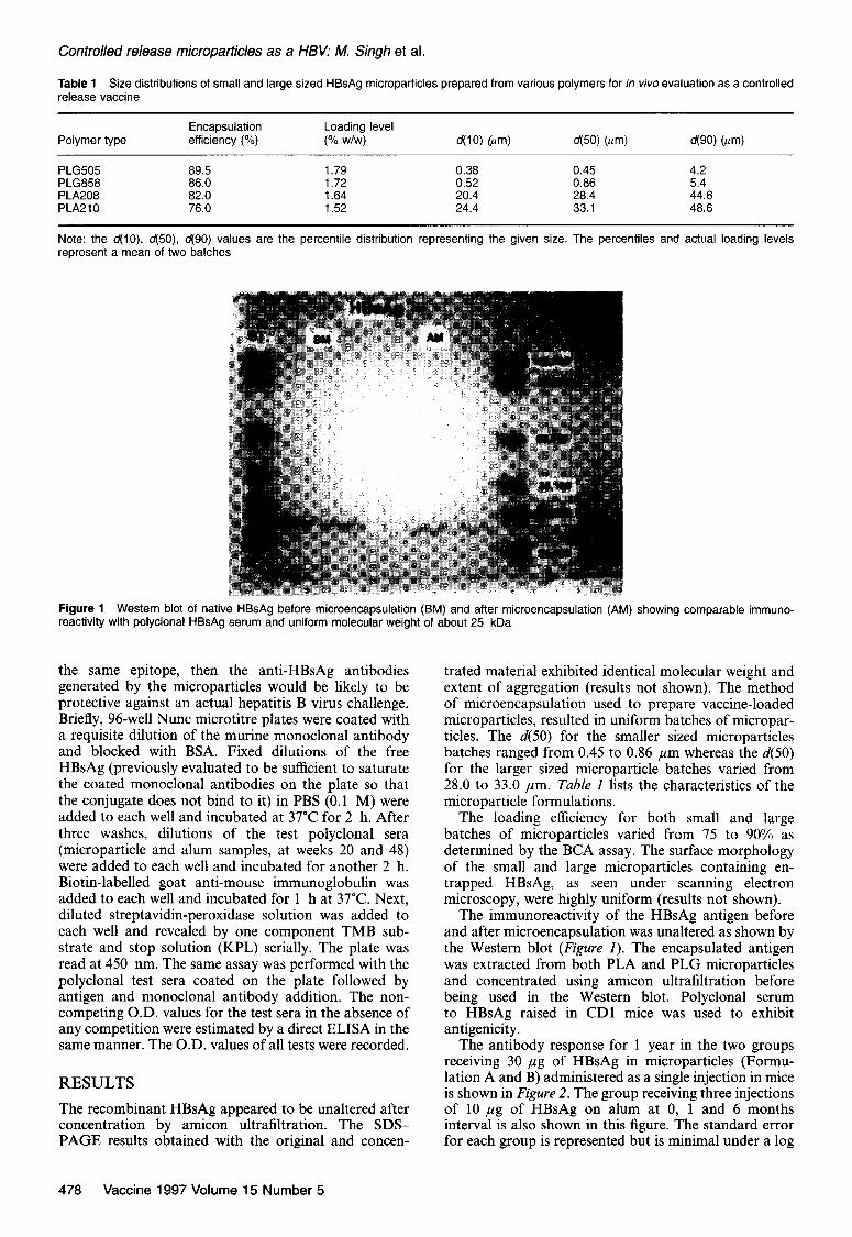
Table 1 Size distributions of small and large sized HBsAg microparticles prepared from various polymers for in vivo evaluation as a controlled release vaccine
Polymer type Encapsulation Loading level efficiency (%) (% w/w) d(f0) lum) d(50) Olm) d(90) Olm)
PLG505 89.5 1.79 0.38 0.45 4.2 PLG858 86.0 1.72 0.52 0.86 5.4 PLA208 82.0 1.64 20.4 28.4 44.6 PLA210 76.0 1.52 24.4 33.1 48.6
Note: the ~$10) d(50), 490) values are the percentile distribution representing the given size. The percentiles and actual loading levels represent a mean of two batches
Figure 1 Western blot of native HBsAg before microencapsulation (BM) and after microencapsulation (AM) reactivity with polyclonal HBsAg serum and uniform molecular weight of about 25 kDa
showing comparable immuno-
the same epitope, then the anti-HBsAg antibodies generated by the microparticles would be likely to be protective against an actual hepatitis B virus challenge. Briefly, 96-well Nunc microtitre plates were coated with a requisite dilution of the murine monoclonal antibody and blocked with BSA. Fixed dilutions of the free HBsAg (previously evaluated to be sufficient to saturate the coated monoclonal antibodies on the plate so that the conjugate does not bind to it) in PBS (0.1 M) were added to each well and incubated at 37°C for 2 h. After three washes, dilutions of the test polyclonal sera (microparticle and alum samples, at weeks 20 and 48) were added to each well and incubated for another 2 h. Biotin-labelled goat anti-mouse immunoglobulin was added to each well and incubated for 1 h at 37°C. Next, diluted streptavidin-peroxidase solution was added to each well and revealed by one component TMB sub- strate and stop solution (KPL) serially. The plate was read at 450 nm. The same assay was performed with the polyclonal test sera coated on the plate followed by antigen and monoclonal antibody addition. The non- competing O.D. values for the test sera in the absence of any competition were estimated by a direct ELISA in the same manner. The O.D. values of all tests were recorded.
RESULTS
The recombinant HBsAg appeared to be unaltered after concentration by amicon ultrafiltration. The SDS- PAGE results obtained with the original and concen-
trated material exhibited identical molecular weight and extent of aggregation (results not shown). The method of microencapsulation used to prepare vaccine-loaded microparticles, resulted in uniform batches of micropar- titles. The d(50) for the smaller sized microparticles batches ranged from 0.45 to 0.86 pm whereas the d(50) for the larger sized microparticle batches varied from 28.0 to 33.0 pm. Table I lists the characteristics of the microparticle formulations.
The loading efficiency for both small and large batches of microparticles varied from 75 to 90% as determined by the BCA assay. The surface morphology of the small and large microparticles containing en- trapped HBsAg, as seen under scanning electron microscopy, were highly uniform (results not shown).
The immunoreactivity of the HBsAg antigen before and after microencapsulation was unaltered as shown by the Western blot (Figure I). The encapsulated antigen was extracted from both PLA and PLG microparticles and concentrated using amicon ultrafiltration before being used in the Western blot. Polyclonal serum to HBsAg raised in CD1 mice was used to exhibit antigenicity.
The antibody response for 1 year in the two groups receiving 30 pug of HBsAg in microparticles (Formu- lation A and B) administered as a single injection in mice is shown in Figure 2. The group receiving three injections of 10 pug of HBsAg on alum at 0, 1 and 6 months interval is also shown in this figure. The standard error for each group is represented but is minimal under a log
478 Vaccine 1997 Volume 15 Number 5
Controlled release microparticles as a HBV: M. Singh et al.
=
v) loo00 .t:
S 1000
! + Al+l X 3 INJS
0 10 20 30 40 SO 60
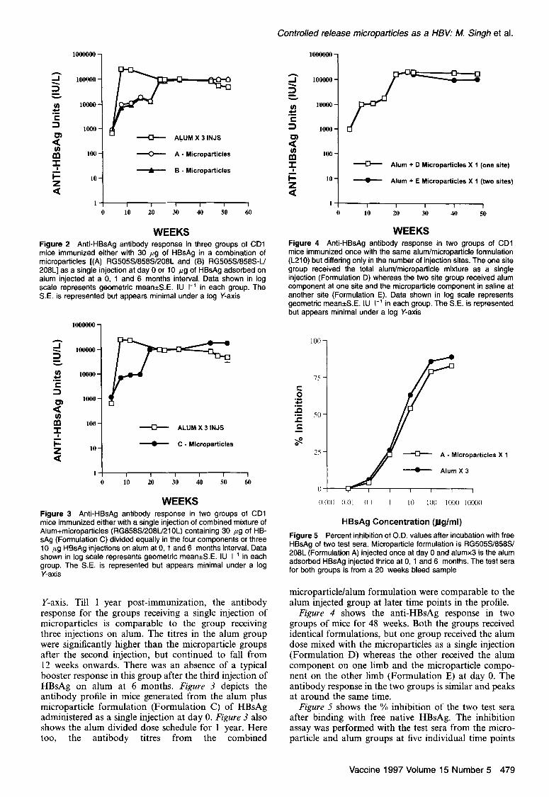
WEEKS Figure 2 Anti-HBsAg antibody response in three groups of CD1 mice immunized either with 36 ,ug of HBsAg in a combination of microoarticles f(A) RG505S/858S/208L and (BI RG505S/858S-U 208Ljas a sing& injection at day 0 or 10 pg of HBsAg adsorbed on alum injected at a 0, 1 and 6 months interval. Data shown in log scale represents geometric mean&.E. IU I-’ in each group. The SE. is represented but appears minimal under a log Y-axis
lOOOOOO-
2
5 10wOO =
v) .tl
IOOOO-
S
z
lOOO-
!E IOO-
e
2
lo-
Ip” -D- ALUM X 3 INJS
_ C - Microparticles
WEEKS Figure 3 Anti-HBsAg antibody response in two groups of CD1 mk!e immunized eitherwith a single injection of combined mixture of Alum+microoarticles (RG858S/208U210L) containina 30 ua of HB- sAg (Formulation C) divided equally in the’four comp&ents>r three 10 ,ug HBsAg injections on alum at 0, 1 and 6 months interval. Data shown in log scale represents geometric mean*S.E. IU I-’ in each group. The S.E. is represented but appears minimal under a log Y-axis
Y-axis. Till 1 year post-immunization, the antibody response for the groups receiving a single injection of microparticles is comparable to the group receiving three injections on alum. The titres in the alum group were significantly higher than the microparticle groups after the second injection, but continued to fall from 12 weeks onwards. There was an absence of a typical booster response in this group after the third injection of HBsAg on alum at 6 months. Figure 3 depicts the antibody profile in mice generated from the alum plus microparticle formulation (Formulation C) of HBsAg administered as a single injection at day 0. Figure 3 also shows the alum divided dose schedule for 1 year. Here too, the antibody titres from the combined
tl?
!F
100
1
- Alum + Cl Microparticles X 1 (one site)
k 10 --C- Alum + E Microparticles X 1 (two sites)
Figure 4 Anti-HBsAg antibody response in two groups of CD1 mice immunized once with the same alum/microparticle formulation
WEEKS
(L210) but differing only in the number of injection sites. The one site group received the total alumlmicroparticle mixture as a single injection (Formulation 0) whereas the two site group received alum component at one site and the microparticle component in saline at another site (Formulation E). Data shown in log scale represents geometric mean*S.E. IU I-’ in each group. The S.E. is represented but appears minimal under a log Y-axis
HBsAg Concentration (flglml)
Figure 5 Percent inhibition of O.D. values after incubation with free HBsAg of two test sera. Microparticle formulation is RG505S/858S/ 208L (Formulation A) injected once at day 0 and alumx3 is the alum adsorbed HBsAg injected thrice at 0, 1 and 6 months. The test sera for both groups is from a 20 weeks bleed sample
microparticle/alum formulation were comparable to the alum injected group at later time points in the profile.
Figure 4 shows the anti-HBsAg response in two groups of mice for 48 weeks. Both the groups received identical formulations, but one group received the alum dose mixed with the microparticles as a single injection (Formulation D) whereas the other received the alum component on one limb and the microparticle compo- nent on the other limb (Formulation E) at day 0. The antibody response in the two groups is similar and peaks at around the same time.
Figure 5 shows the % inhibition of the two test sera after binding with free native HBsAg. The inhibition assay was performed with the test sera from the micro- particle and alum groups at five individual time points
Vaccine 1997 Volume 15 Number 5 479
Controlled release microparticles as a HBV: M. Singh et al.
Table 2 Competition assay results between the microparticle and alum test sera in mice at weeks 20 and 48, with the monoclonal antibody
Sample
O.D. with O.D. with competing competing
O.D. without antibody antibody competing as first as a second antibody antibody antibody
M/particle-20 weeks 1.36 0.62 0.45 Alum-20 weeks 1.94 0.94 0.90 M/particle-48 weeks 1.82 0.82 0.84 Alum48 weeks 1.74 0.92 0.96
The O.D. values represent a mean of triplicate readings with and without competing antibody at 450 nm. Suppression of O.D. values upon competition with the monoclonal suggests similar binding regions
from the in vivo study. The results shown in Figure 5 are from the test sera at week 20. The inhibition profiles for both the alum and microparticle formulations were similar at all time points.
Table 2 shows the O.D. values obtained from the competition assay with various sera dilutions. Two sets of results are presented with the competing antibody, one with the competing monoclonal as the first antibody and another as a second antibody. The suppression of the O.D. values in both sets of results indicate compe- tition for the same region in free HBsAg by the test and monoclonal antibodies. Thus the specificity of the sera from the alum and microparticle groups is similar to that of the monoclonal antibody.
DISCUSSION
The solvent evaporation method of microencapsulation utilized in this study resulted in the formation of uni- form sized biodegradable microparticles containing entrapped HBsAg. The surfaces of these microparticles as seen under scanning electron microscopy (results not shown) were free from any pores or cracks which could enhance burst or initial antigen release. The inimuno- reactivity of the HBsAg within the microparticles was also unaltered as shown by Western blotting of the antigen before and after microencapsulation (Figure I) with a polyclonal HBsAg antibody. The maintenance of immunoreactivity was probably a major factor con- tributing to the microparticle formulation being able to generate high levels of HBsAg specific antibodies. Earlier studies conducted with OVA as a model protein, showed that the release of the protein from micro- particles prepared with the same polymers as used here, was over several months (results not shown). These controlled release HBsAg microparticles follow the same long-term release kinetics.
The results obtained in these studies demonstrate the potential of delivering the required doses of HBV as a single injection using biodegradable microparticles. Both small and large sized HBsAg microparticles were equally effective in potentiating the immune response. It is believed that the low molecular weight biodegradable polymer, formulated as small sized microparticles repre- sents the major priming component of the microparticle formulation. After the small sized microparticles have contributed significantly in generating an initial re- sponse, the antigen release from the large microparticles
prepared from relatively higher molecular weight poly- mer, probably induce a more potent and sustained response. The results obtained indicate an in vivo “auto- boost” from the combined microparticle formu- lation occurring around weeks 1620, which serves to potentiate the antibody response. Thus an in vivo biphasic antigen release profile from the microparticles is probably helpful in mimicking the conventional vaccination schedule.
An addition of 7.5 pg of the total 30 ,ug HBsAg dose, on alum along with the remaining 22.5 ,ug in micro- particles in the second study (Formulation C), did not significantly alter the cumulative antibody profile in mice from immunization with microparticles alone. It is only after the second dose on alum at week 4 that the antibody titres rise significantly in the control alum immunized group. The addition of a portion of the total HBsAg dose on alum along with the microparticles was not essential for generation of a potent antibody response. Clearly a single microparticle HBsAg formu- lation serves the same purpose. Splitting the alum and the microparticle component into two sites on an animal did not seem to alter the antibody profile in mice as is seen in Figure 4. Therefore a single injection site is more convenient for a microparticle formulation.
Anti-HBsAg antibodies from the microparticle group exhibited similar binding capacity as the alum group (Figure 5) suggesting that the process of microencapsu- lation did not alter the immunogenicity of the HBsAg. This is a critical observation, since the anti-HBsAg antibodies from the microparticle injected group should neutralize the virus to the same extent as the convention- ally approved alum-HBsAg vaccine. The competition of the test sera (microparticle and alum) with the specific monoclonal antibody demonstrated that both the test sera were specific to the relevant epitopes on the HBsAg surface. This result, although highly qualitative in nature, in the absence of a relevant small animal chal- lenge model for hepatitis B virus, suggests potential for protection with a microparticle formulation in vivo.
The antibody titres generated with a single injection of the microparticles containing 30 ,ug of HBsAg in all the studies were significantly higher than the minimum titres required in humans for protection (10 IU l- ‘) against hepatitis B infection. These preliminary studies indicate the feasibility of converting the present three-injection schedule for HBsAg into a “single-shot” therapy. Sub- sequent experiments in alternative animal models should lead to a greater understanding about the duration of protection with a single controlled release vaccine. Based on the current findings the controlled release vaccine should be evaluated in primates.
ACKNOWLEDGEMENTS
We wish to acknowledge our thanks to the Biological Products Institute of Lan Zhou in China for provision of the HBsAg. Thanks are also due to Hanying Wang and Joanne DeSteffano for help in the animal experiments.
REFERENCES
1 Stephenne, J. Recombinant versus plasma-derived hepatitis B vaccines: issues of safety, immunogenicity and cost- effectiveness. Vaccine 1988, 6,299
480 Vaccine 1997 Volume 15 Number 5
Controlled release microparticles as a HBV: M. Singh et al.
2
3
4
5
6
7
8
9
10
11
Jonsson, 6. Cost-benefit analysis of hepatitis B vaccination. Post Grad. Med. J. 1987, 632, 27 Scheiermann, N., Gesemann, K.M., Kreuzfelder, E. and Paar, D. Effects of a recombinant yeast-derived hepatitis B vaccine in healthy adults. Post Grad. Med. J. 1987, 632, 115 Scolnick, E.M., McLean, A.A., West, D.J., McAleer, W.J., Miller, W.J. and Buynak, E.B. Clinical evaluation in healthy adults of a hepatitis B vaccine made by recombinant DNA. J. Am. Med. Assoc. 1984, 251, 2812 Aristegui, J., Muniz, J. and Legorburu, A.P. et al. Newborn universal immunization against hepatitis B: immunogenicity and reactogenicity of simultaneous administration of diphtheria/ tetanus/pedussis (DTP) and oral polio vaccines with hepatitis B vaccine at 0, 2 and 6 months of age. Vaccine 1995, 13, 973 Bryan, J.P., Craig, P.G. and Reyes, L. et al. Randomized comparison of 5 and 10 pg doses of two recombinant hepatitis B vaccines. Vaccine 1995, 13, 978 Kane, M. Global programme for control of hepatitis B infection. Vaccine 1995, 13, S47 Aguado, M.T. Future approaches to vaccine development: single-dose vaccines using controlled-release delivery systems. Vaccine 1993, 11, 596 O’Hagan, D.T., Rahman, D. and McGee, J.P. et a/. Biodegrad- able microparticles as controlled release antigen delivery system. immunology 1991,73,239 Eldridge, J.H., Stass, J.K., Meulbroek, J.A., McGhee, JR., Tice, T.R. and Gilley, R.M. Biodegradable microspheres as a vaccine delivery system. Molec. Immunol. 1991, 28, 287 Singh, M., Singh, 0. and Talwar, G.P. Biodegradable delivery system for a birth control vaccine: immunogenicity studies in rats and monkeys. Phafm. Res. 1995, 12, 1796
12
13
14
15
16
17
18
Alonso, M.J., Gupta, R.K., Caroline, M., Siber, G.R. and Langer, R. Biodegradable microspheres as controlled-release tetanus toxoid delivery systems. Vaccine 1994, 12, 299
Yamaguchi, K. and Anderson, J.M. In vivo biocompatibility studies of medisorb 65/35 D I-lactide/glycolide copolymer microspheres. J. Conk Release 1993, 24, 81 Nellore, R.V., Pande, P.G., Young, D. and Bhagat, H.R. Evalu- ation of biodegradable microspheres as vaccine adjuvant for hepatitis B surface antigen. J. Parent. Sci. Tecbnol. 1992, 46, 176
Jeffery, H., Davis, S.S. and O’Hagan, D.T. The preparation and characterization of poly(lactide-co-glycolide) microparticles: II. The entrapment of a model protein using a (water-in-oil)-in water emulsion solvent evaporation technique. Pharm. Res. 1993, 10,362
Sharif, S. and O’Hagan, D.T. A comparison of alternative methods for the determination of the levels of protein entrapped in poly (lactide-co-glycolide) microparticles. ht. J. Pharm. 1995, 115,259
McGee, J.P., Singh, M., Li, X.M., Qiu, H. and O’Hagan, D. The encapsulation of a model protein in poly (d,l lactide-co- glycolide) microparticles of various sizes-an evaluation of process reproducibility. J. Microencapsulation (in press)
Men, Y., Thomasin, C., Merkle, H.P., Gander, 8. and Corradin, G. A single administration of tetanus toxoid in biodegradable microspheres elicits T cell and antibody responses similar or superior to those obtained with aluminium hydroxide. Vaccine 1995, 13,683
Vaccine 1997 Volume 15 Number 5 481