gene expression after vaccination of mice with formulations of diphtheria toxoid or tetanus toxoid...
TRANSCRIPT
Vaccine 21 (2003) 2307–2317
Gene expression after vaccination of mice with formulations of diphtheriatoxoid or tetanus toxoid and different adjuvants: identification of shared
and vaccine-specific genes in spleen lymphocytes
Karin Regnström∗, Eva Ragnarsson, Per ArturssonDepartment of Pharmacy, Uppsala University, P.O. Box 580, SE-751 23 Uppsala, Sweden
Received 13 May 2002; received in revised form 19 December 2002; accepted 31 January 2003
Abstract
We immunized mice with four different combinations of diphtheria toxoid or tetanus toxoid with aluminum phosphate or Freund’sadjuvant and studied the resulting gene expression profiles in spleen lymphocytes. Genes, which are unique for each combination or sharedin several combinations, were found activated, with functions in immune response but also in other cellular processes like apoptosis or signaltransduction. Using bioinformatic tools we show, that some of the genes may serve as indicators for adverse reactions, while other genesmay be new immune response markers. The results also suggest that adjuvant participates in the formation of an immunological memory.© 2003 Elsevier Science Ltd. All rights reserved.
Keywords:Gene expression; Vaccine; Biomarkers
1. Introduction
Tetanus and diphtheria toxoid formulated with aluminumphosphate are clinically used vaccines, given to childrenfrom the age of 3 months. However, because they were de-veloped at the beginning of this century, when immunologymethods were not yet sophisticated, not much is knownabout their mechanism of action on the molecular level. Wetherefore compared the gene expression patterns of fourdifferent antigen-adjuvant combinations (AACs) in spleenlymphocytes of mice. They consisted of two clinically rele-vant vaccine antigens, tetanus toxoid and diphtheria toxoid,each formulated with different adjuvants, the clinically usedaluminum phosphate (TA and DA) or Freund’s adjuvant(TF and DF), which is only used preclinically.
Tetanus toxoid and diphtheria toxoid are derived frommodified toxins, with retained receptor binding activity, butlost toxicity [1]. Both antigens are similar in that they elicitan IgG production for protective immunity, and containseveral T-cell epitopes because of their large size. They areusually administered together with pertussis as a trivalentvaccine to children at very low age.
Two of the most used adjuvants are aluminum phosphateand Freund’s complete and incomplete adjuvant. Freund’s
∗ Corresponding author. Tel.:+46-18-471-4628; fax:+46-18-471-4223.E-mail address:[email protected] (K. Regnström).
complete adjuvant is the most potent adjuvant known, caus-ing strong delayed-type hypersensitivity reactions to thesmallest amounts of simple proteins. This adjuvant is usedas a standard for adjuvant strength. The Freund’s-basedvaccines are administered i.p., because of the ability ofFreund’s adjuvant to form granulomas at the injectionsite, when administered i.m. While aluminum phosphatehas been used in vaccines in man since the 1930s, theuse of Freund’s adjuvant has been restricted to preclini-cal work in experimental animals because of its toxicityand the suspicion that it evokes tumors[2]. It has beenproposed that both aluminum phosphate and the water/oilemulsion in Freund’s adjuvant function as a slow-releasedepot for the antigen and that they enhance the antigenuptake by macrophages[2]. However, the exact cellularand biochemical reactions induced are poorly understood[2].
We report here changes in the expression of genes in-volved in systemic immune response, and in other cellularresponses, in the four different AACs. We show using clus-tering methods that each AAC changes the expression of aspecific set of genes, indicating that adjuvant has a moreimportant role in immune response reactions than previ-ously recognized. Furthermore, we identified a group of 61genes, which are commonly activated in all AACs. Severalof these genes have not been reported earlier to be involvedin immune response-related processes, but their importance
0264-410X/03/$ – see front matter © 2003 Elsevier Science Ltd. All rights reserved.doi:10.1016/S0264-410X(03)00103-8

2308 K. Regnström et al. / Vaccine 21 (2003) 2307–2317
was validated using a literature network tool and the MeSHbrowser.
2. Materials and methods
2.1. Animal experiments
Female Balb/c mice, 10 weeks old (Charles River), wererandomly separated into groups of five animals. The micewere given an i.m. dose of vaccine (manufactured by SBLVaccin AB) containing 1 Lf tetanus toxoid or 1.3 Lf diphthe-ria toxoid in aluminum phosphate adjuvant at pH 6.0 (1:1)(TA and DA, respectively) i.m. in the left hind leg quadri-ceps. Other groups of mice were given the same doses ofeach antigen (SBL Vaccin AB) in Freund’s complete adju-vant (TF and DF, respectively) i.p. The same doses wereadministered 3 weeks later, but Freund’s complete adju-vant was substituted by Freund’s incomplete adjuvant. Twoweeks after the booster, blood was collected from all miceby tail vein puncture for measurement of the systemic an-tibody response by the toxin-binding inhibition assay[3].Spleens were isolated, pooled for each AAC to compensatefor individual genetic variation according to[8], and washedin cold PBS.
2.2. Cell culture experiments
A single-cell suspension from the spleens was pre-pared and erythrocytes were removed by incubation in1 M Tris buffer, pH 7.2 on ice for 10 min. The cells werewashed twice with RPMI medium (Dulbecco) supple-mented with 292�g/ml L-Gln, 50�M 2-mercaptoethanol,100 IU/ml penicillin, 100�g/ml streptomycin, and 10%heat-inactivated FCS (Gibco). Aliquots of the cells(1.25 × 10⁶ cells/ml) were plated into cell culture flasksand incubated for 4 or 24 h, at 37◦C in 5% CO2 at approxi-mately 90% humidity with the respective antigen at a dosethat corresponded to that used during immunization. Afterincubation, the supernatants were recovered and used forthe cytokine assay. The cells were washed with cold PBSand used for RNA isolation.
2.3. Analysis of gene expression by array technology
Total RNA was purified from splenocyte cells usingAmbion’s Totally RNATM kit. DNA contamination wasremoved using DNAse I (Promega). Atlas mouse cDNAexpression arrays with 588 genes (Clontech) were usedaccording to the manufacturer’s guidelines and the repro-ducibility was assessed as reported earlier[4]. The level ofexpression was quantified by phosphorimaging (Packard)and the image data was processed using the AtlasImagesoftware version 1.01 (Clontech). Signals lower than theaverage background signal were filtered out. The adjustedintensity of each gene was obtained by subtracting the av-
erage background signal from the signal intensity. The rawdata are available athttp://www.farmaci.uu.se. To comparetwo or more arrays, the adjusted signal intensities of allgenes on the arrays were normalized to the data derivedfrom non-treated mice using the global mode and the summethod. Briefly, the ratio of the adjusted signal intensity ofgene Z on array 1 to the adjusted intensity of the same geneZ on array 2 was calculated for all the genes on the arrayand averaged. In order to filter for genes with significantgene expression changes, we used the following criteria:only genes with a difference in signal strength of morethan 100 and a more than two-fold expression difference ascompared to non-treated control mice in at least one samplewere included[4,5]. The Spotfire Pro software (version 4.2)was used to visualize global expression changes. Geneswere sorted by the program Excel, clustered by the programsoftware GeneCluster[6] and visualized by Treeview[7].
2.4. Analysis of mRNA expression by RT-PCR
Total RNA was extracted as described above. RT-PCR re-actions were performed using the Qiagen one-step RT-PCRkit and 1�g total RNA according to the manufacturer’sguidelines with an annealing temperature of 55◦C and 45 cy-cles. The following primers were used: PCNA forward-GCT-TGGCAATGGGAACATTA, and PCNA reverse-TCATCT-TCAATCTTGGGAGC; PAI-1 forward-TTAGTGCAACC-CTGGCCGACTTCAC and PAI-1 reverse-CTGTCAAGGC-TCCATCACTTGCCC; myeloblastin forward-CCCGTGT-GCTGCAGGAACTG-AACG and myeloblastin reverse-CTGGT-CGCCCCAGCTTT-AGGCTG; BRF-1 forward-CCGATGACCTCTTGGGCTCACC and BRF-1 reverse-ATGGGCAGGCGTC-TTGAGT-TGTCC; TTF-1 forward-AAACCGCCCATATACTGCT-GTCTACG and TTF-1reverse-CTTGTTACAGATGGTTGTGAGCCACC; Pax-5forward-GACATGGAGGA-GTGAATCAGC and Pax-5reverse- GCCACTGATGGAGTATGAGGA.
3. Results and discussion
Our experimental approach was the following. Two clin-ically relevant and commercially available antigens, tetanustoxoid and diphtheria toxoid were used and formulated eitherwith aluminum phosphate (TA and DA) or with Freund’s ad-juvant (TF and DF). The four different AACs were adminis-tered to mice according to a standard immunization protocol.After a booster dose, spleen cells were harvested and pooledfor each AAC (n = 5 mice) to diminish inter-individualvariation effects[4,8]. The spleen lymphocytes were isolatedand re-stimulated in vitro with the respective antigen for 4and 24 h before RNA purification. These time-points werechosen because they make it possible to measure the highestgene expression levels of many Th1 and Th2 markers[4].
All immunized mice developed strong serological titersagainst the antigen given, and showed protein levels for the

K. Regnström et al. / Vaccine 21 (2003) 2307–2317 2309
cytokines interferon�, interleukin 2, 4 and 5 that were com-parable to those reported earlier[4] (data not shown). Thegene expression levels of immune response markers for theTh1 and Th2 response were assessed from the array resultsas reported earlier[4] and contained levels, which agreedwell with the corresponding cytokine protein levels (data notshown). RT-PCR was performed to confirm the expressionlevels of highly up-regulated genes in the different samples,as indicated in the different figures.
3.1. Pair-wise comparison of gene expression profiles
We were interested to study the specific effects of eachAAC on the expression level of all genes represented onthe array in spleen lymphocytes. Therefore the Spotfiresoftware was used to compare the expression profiles of the
Fig. 1. Global gene expression analysis. RNA was isolated and transformed to radioactively labeled cDNA. The levels of cDNA of the different geneswere measured and processed by Clontech Atlas technology and by Spotfire software. The plots compare the expression levels of all genes of each pair ofarrays, expressed as adjusted intensity scores. The lines represent gene expression differences by a factor of two between the two samples. Comparisonsof AACs as indicated at 4 h (A–D) and 24 h (E–H) after in vitro re-stimulation with the corresponding antigen. Genes with significant changes inexpression levels exceeding the scale of the figure by at least a factor of two are indicated with their names only in kursiv letters. The expression levelsof the following genes were confirmed by RT-PCR in all AACs shown: Pax-5, myeloblastin, BRF-1 and TTF-1 (data not shown).
different AACs two by two at each time-point after the invitro re-stimulation with antigen, at 4 and 24 h (Fig. 1A–F).This provided an opportunity to compare vaccine samplesthat differed either in their adjuvant or in their antigencomposition. This approach was also used to assess thenumber of genes with significant gene expression changesas defined inSection 2.3. The samples at 4 h after in vitrore-stimulation with antigen (Fig. 1A–D) generally showeda much larger number of genes with high expression levelsthan the samples obtained at 24 h after in vitro re-stimulationwith antigen (Fig. 1E–H).
3.1.1. Adjuvant-associated differences in the numberof expressed genes
Comparing samples with the same antigen, but differ-ent adjuvant, we observed that a greater number of genes

2310 K. Regnström et al. / Vaccine 21 (2003) 2307–2317
Fig. 1. (Continued).
had higher gene expression levels in vaccines with alu-minum phosphate as adjuvant than was the case when us-ing Freund’s adjuvant (Fig. 1A, B, E and F). However, thenumber of genes with high expression levels in the samplederived from immunization with TF at 24 h after in vitrore-stimulation (TF24) was higher than that obtained fromother 24 h samples (Fig. 1F). This indicates different kineticsin gene activation induced by Freund’s adjuvant. The over-all lower number of activated genes in the samples derivedfrom Freund’s adjuvant-containing vaccines was surprising,since we had observed earlier that systemic gene expressionchanges reminiscent for potential side-effects can be moni-tored in spleen lymphocytes[4] and many side-effects havebeen reported for the Freund’s adjuvant[2]. However, theadjuvants differ in their administration route, i.m. for alu-minum phosphate and i.p. for Freund’s adjuvant. Since westudied the systemic immune response in spleen lympho-cytes, it can be speculated that Freund’s adjuvant evokesmore local responses, which is supported by the fact thatFreund’s adjuvant typically forms granulomas at the admin-
istration site. Different kinetics in gene expression causedby different antigen-release kinetics could also account forthis finding.
3.1.2. Antigen-associated differences in the numberof expressed genes
Comparing samples derived from vaccines with the sameadjuvant but different antigens, we found that diphtheriatoxoid activated far more genes than tetanus toxoid (Fig. 1C,D and G). The overall largest number of activated genes wasobserved in DA at 4 h after in vitro re-stimulation (DA4)(Fig. 1A and C). Furthermore, we found a weak relationshipbetween the number of genes expressed by a specific AACand the corresponding IgG levels (data not shown).
3.1.3. Expression of genes unique for anantigen-adjuvant combination
We were also interested to find out if each AAC could af-fect unique genes. Using the pair-wise comparisons betweenthe different samples by the Spotfire software, we found

K. Regnström et al. / Vaccine 21 (2003) 2307–2317 2311
genes in the DA samples, which had similar expression levelsindependent of which sample it was plotted against. Thesegenes are unique for the adjuvant-antigen combination at acertain time-point after in vitro re-stimulation (seeFig. 1Aand C: prothymosin�, Csk, Pim-1 and BRF-1;Fig. 1E andG: GADPH, vimentin and FGF-7). Some of the genes didnot have an obvious connection to the immune responseprocess but were classified as involved in oncogenesis orcontrol of the cell cycle by the array manufacturer, and sowe selected some of these genes and searched the litera-ture and the GeneCards database at the Weizmann Instituteof Science for their reported functions. We also performedRT-PCR to confirm the expression levels found by arraytechnology.
In DA4 the Pim-1 proto-oncogene and the butyrate re-sponse factor (BRF-1) (Fig. 1A and C) were chosen, whichwere 23–28 times more activated in DA4 than in any othersample.
Pim-1 proto-oncogene is an apoptosis inhibitor similar toBcl-2 and, as such, significantly involved in the early B-celllymphopoiesis in mice[9,10].
The butyrate response factor (BRF-1) is a regulatory pro-tein involved in growth factor regulatory pathways. Reportsthat butyrate significantly enhanced the expression of HLAmolecules and ICAM-1 in the colonic epithelial HT-29 cellline [11], stimulates cytokine expression in naı̈ve T cells[12], and has anti-inflammatory characteristics[13] suggestthat the butyrate response factor is important for the immuneresponse to DA in mice.
Myeloblastin (also called proteinase 3) was highlyup-regulated in TF24 (10 times higher than the lowest levelin all samples) (Fig. 1F and H). This protein has beenascribed antibacterial properties[14], but has also beenidentified as an antigen for auto-antibodies in Wegener’sgranulomatosis patients[15]. Its mechanism of action isonly slowly becoming clear[16]. It is co-cited with IL-8,which is important in directing neutrophil migration to thesite of infection, in 64 PubMed articles. Since it is knownthat complete Freund’s adjuvant can induce autoimmunedisease[17] we suggest that myeloblastin is a suitablemarker for such auto-immune reactions.
The transcription terminator factor TTF-1 was found inTF 4 h after in vitro re-stimulation at a level that was 20 timeshigher than the lowest level in the other samples (Fig. 1Band D), while a level that was four to five times higher wasfound in each of the AACs at one of the time-points. TTF-1is a transcriptional activator[18] and an 18 bp sequence mo-tif, called the “Sal-box” has been identified, to which TTF1binds for regulation[19]. No references to involvement inthe immune response were found in the literature. We there-fore performed a DNA sequence similarity search with the“Sal-box” motif using the BLASTN 2.1.3 version by NCBI,and found a matching sequence of 17 bp and 100% identitywith the MHC class I SLA genomic region inSus scrofa(data not shown). We speculate that TTF1 is involved in thetranscriptional regulation of MHC class I genes. We have
recently found TTF1 also up-regulated in spleen lympho-cytes of mice after administration of a gene vaccine, con-sisting of polyethyleneimine and a plasmid coding forLacZ(manuscript under revision in Gene Therapy). However, fur-ther research will be needed to confirm this finding.
In summary, these examples indicate that it is the specificcombination between the antigen and the adjuvant that gov-erns the quantity and the type of genes that will be affected,and to what extent. Interestingly, the importance of many ofthese genes for the immune response is not yet understoodand therefore not stated in the scientific literature.
3.2. Comparison of the gene expression profiles of alladjuvant-antigen combinations at each time-point
In order to identify gene expression differences betweenall AACs at each time-point after in vitro re-stimulation,cluster analysis[6] was performed. Six clusters were ob-tained that contained genes with significant gene expressionchanges in one of the vaccines at each time point whencompared to the other vaccines (Fig. 2A and B). Further-more, the number and type of genes with high expressionlevels differed between the two time-points.
3.2.1. Number of activated genesThe total number of genes with significant changes in
gene expression of all the clusters at each time-point afterin vitro re-stimulation with antigen was nearly three timeshigher for the 4 h samples (325 genes,Fig. 2A) than it wasfor the 24 h samples (126 genes,Fig. 2B). This confirmsthat there is a burst of gene expression early during theimmune response, as stated above, and is in agreement withour earlier observations[4].
Counting the genes for each AAC at each time-point, thediphtheria containing vaccine samples showed the highestnumber of affected genes with DA4 at the top with 264 genes(clusters c0, c1 and c3:Fig. 2A). This coincides with a levelof expression that is two times higher that all known immuneresponse markers on the array (data not shown). The numberof genes expressed in the remaining AACs decreased in thefollowing order (see alsoFig. 4):
DA4 > DF4 > TF24> DA24 > TA4 > TA24
> TF4 > DF24.
3.2.2. Type of activated genesTo assess the type of genes that was activated in the differ-
ent AACs, the genes were assigned to functional categoriesas specified by the array manufacturer (Fig. 3). Fig. 3Ashows that DF4 (50 genes in c4 and c5:Fig. 2A) and TA4(c0 and c4 inFig. 2A) mostly showed an activation ofgenes involved in transcriptional regulation, and in cell cy-cle control, oncogenesis and tumor suppression. Genes acti-vated in the TF4 sample (c2 inFig. 2A), on the other hand,were involved in cell signaling, proteases or part of the cy-toskeleton. Genes activated in the DA4 sample were of all

2312 K. Regnström et al. / Vaccine 21 (2003) 2307–2317
Fig. 2. Gene expression differences in spleen cells between mice treated with DA, DF, TA and TF after in vitro re-stimulation with the correspondingantigen for 4 h (A) and 24 h (B). The differences are shown as self-organizing map clusters according to Tamayo’s algorithm[6] using the criteria forsignificant gene expression described inSection 2.3. (A) 325 genes were found in six clusters (c0–c5) containing between 11 and 167 genes at 4 h and(B) 126 genes in six clusters (c0–c5) containing between 8 and 37 genes at 24 h: (+) up-regulated; (-) no change in expression; (- -) down-regulated.Lines connecting the dots indicate the mean expression profiles; the two outer lines indicate the S.D. A complete list of the genes in each cluster canbe obtained in the supplementary material athttp://www.farmaci.uu.se.

K. Regnström et al. / Vaccine 21 (2003) 2307–2317 2313
Fig. 3. Functional categories of genes significantly activated in the different AACs. The information obtained by clustering (Fig. 2) was used to assessthe involvement of the genes in different cellular processes as specified by the array manufacturer. Thex-axis indicates the number of activated genes ineach functional category for each AAC: (A) 4 h and (B) 24 h after in vitro re-stimulation with the corresponding antigen.
the categories listed above, with a pronounced activation ofgenes involved in apoptosis and DNA repair, and heat-shock,stress-response and intracellular communication. The impor-tance of this type of gene for immunostimulation has beencited in the “danger theory” as described by Matzinger[20],and agrees with our earlier observations for the tetanus tox-oid aluminum phosphate vaccine[4].
Clustering of the data from the 24 h samples (Fig. 2B)resulted in a more even distribution between the differentcombinations (Fig. 3B): all samples, except DF, activatedgenes of all categories. In the samples containing Freund’sadjuvant, TF24 (total of 48 genes in c4 and c5:Fig. 2B),the number of genes involved in oncogenesis, tumor sup-pression and cell cycle control was significantly higher thanit was in the other categories. This agrees with Freund’sknown ability to evoke tumors[2]. A complete list of allgenes contained in the different clusters can be found athttp://www.farmaci.uu.se.
In summary, we conclude that a different set of geneswith different functions is activated by each AAC at eachtime-point after in vitro re-stimulation with antigen. Invitro re-stimulation mimics protective immunity, in whichthe encounter with antigen leads to the proliferation ofantigen-specific T effector cells and memory B cells. No ad-juvant is present during in vitro re-stimulation of the spleenlymphocytes in our study. Why, therefore, do lymphocytesderived from immunization with formulations containingthe same antigen but different adjuvant show differentgene expression profiles when re-stimulated with antigen?Several explanations are possible: (1) adjuvant contributesdirectly to the differentiation of lymphocytes during innateimmunity by, e.g. expression of different co-stimulatory
molecules, resulting in an adjuvant-specific memory inthese cells. Indications for an adjuvant-specific memorywere already found in our earlier publication[4]; (2) ad-juvant contributes indirectly to the differentiation processbecause different formulation types make the APCs presentdifferent peptides; and (3) differences are observed becauseof the different administration routes. A combination ofall these factors is also probable. Further experiments areneeded to support this hypothesis.
3.3. Genes shared by expression profiles independentof time-point
3.3.1. Number of shared genesThe cluster analyses were performed separately for each
time-point after in vitro antigen re-stimulation without in-formation, if some of the highly activated genes in the 4 hsamples were also expressed in the 24 h samples. Thereforethe output files from the GeneCluster program obtained at4 and 24 h were sorted and compared (Fig. 4). 93 genes(the total of the number of genes found in the light partsof the columns inFig. 4A and B), or about 28% of thetotal number of activated genes in the 4 h samples, werealso found in the 24 h sample, where they formed 74% ofall genes found at this time-point. However, 32 of thesegenes were only found in the same AACs at the differenttime-points after antigen re-stimulation, but not in otherAACs. These genes are unique for a specific AAC andwere excluded, leaving a total of 61 genes. These 61 geneswere expressed in more than one AAC at the differenttime-points and represent genes, which are shared by AACsstudied.

2314 K. Regnström et al. / Vaccine 21 (2003) 2307–2317
Fig. 4. Numbers of activated genes found in other AACs at (A) 4 h and (B) 24 h after in vitro re-stimulation with the corresponding antigen. The darkparts of the columns represent the number of genes unique for the given AAC at the respective time-point. The light parts of the columns represent thenumber of genes found in other AACs (percentage of total number of genes with significant gene expression change at a given time-point). Both partstogether represent the total number of genes activated in the specific AAC at each time-point.
3.3.2. Type of shared genesA list of these genes is given inTable 1. Many genes
known to be involved in the immune response werefound, such as CD3, the IL-2 receptor gamma, PAX-5 andSTAT5A [4]. The list also contains the genes for TTF1 andmyeloblastin, supporting the hypothesis that they are in-volved in the immune response (see alsoSection 3.1.3). Ithas been reported that aluminum alters the butyrate receptorfunction [21] and calcium binding[22]. We found elevatedlevels of the butyrate response factor and of the calciumbinding protein (CAB45) in DA4 and TA24. We thereforesuggest that the aluminum phosphate adjuvant is responsi-ble for this up-regulation. Furthermore, heat-shock proteinsand cathepsins are found inTable 1. However, some of thegenes in the list have not previously been reported to beinvolved in immune response processes and were thereforeanalyzed further.
3.3.3. Validation of the significance of the sharedgenes for the immune response
We subjected the list of these 61 shared genes to theliterature association tool PubGene[23], which extractsgene-to-gene co-citations from the over 10 million Medlinerecords available and presents their relationship as a net-work. Since this tool is only available for human genes, wetranslated the mouse gene symbols to human gene symbols.The literature network obtained from PubGene contained 26of the submitted genes (34%) (Figs. 4 and 5). Surprisingly,the highest score was obtained for the proliferating cell nu-clear antigen (PCNA). We therefore confirmed the highermRNA level of PCNA in DA4 and TF1 obtained by arrayanalysis using RT-PCR (data not shown). The PCNA proteinis reported to be involved in DNA synthesis, DNA replica-tion, DNA repair and cell cycle progression and has not hith-erto been identified as active in immune response reactions.
Submitting the list of these 26 genes to PubGene’sMeSh Map tool (http://www.nlm.nih.gov/mesh), the re-
turned term associations for these genes agreed wellwith immune response functions reflecting the activationof T-lymphocytes: the three highest-scoring terms were“antigen” (score of 11,754), “mice” (7000) and “antibodies”(3958). The remaining terms contained “T-lymphocytesimmunology” (1190), “cell adhesion” (1040), “cell division”(1002), “tumor necrosis factor pharmacology” (908),“PCNA analysis” (733), “interferon type II pharmacology”(549), “lymphocyte function associated antigen-1 (LFA-1)physiology” (523), “interleukin-1 pharmacology” (351) and“interleukin-2 pharmacology” (332). The total score list maybe searched at www. . . (to be announced by the publisher).
The high score for “LFA-1 physiology” shows thatcytolytic T-helper cell formation is prominent. The IL-1 in-volvement is important for the potentiation of the T-cell re-sponse. The high score for “interferon type II pharmacology”is an important indication of the expression of MHC classII antigens, which are known to be important in autoim-mune diseases[17]. These genes might, therefore, be usedas markers for autoimmune disease in the future.
In summary, a group of 61 shared genes were observedthat are activated by all AACs studied, but with differentkinetics. It is possible, however, that other genes were acti-vated in all samples, but were not found by the clusteringmethods used, since the software filtered the expression datato exclude small changes in level of expression. Some ofthese genes may be involved in the immune response. The re-maining group of activated genes comprises those that werespecifically affected in each combination of antigen and ad-juvant. This is surprising and either reflects the complexityof the immune response or shows that a variety of other cel-lular processes are induced during immune responses.
We conclude that we have found an indication for animmunological memory for both antigen- and adjuvant-affected genes, giving the adjuvant a much more central rolein the immune response than previously recognized. Thisshould have considerable implications for future vaccine

K. Regnström et al. / Vaccine 21 (2003) 2307–2317 2315
Table 1List of 61 genes with significant activation in at least two different AACs as assessed by the self-organizing map clustering method
Clusternumber 4 h
Clusternumber 24 h
GeneBankaccession number
Gene name
1 0 U51196 EB1 APC-binding protein1 1 X01023 c-myc proto-oncogene1 1 U10440 p27kip1; G1 cyclin-Cdk protein kinase inhibitor; p21-related1 1 U05247 Csk; c-Src-kinase and negative regulator1 1 X92411 RAD23 UV excision repair protein homolog B (MHR23B; RAD23B); xeroderma
pigmentosum group C repair complementing 58 kDa protein (XP-C repair complementing58 kDa protein)
1 1 M58566 Butyrate response factor 11 1 U25096 Lung Kruppel-like factor (LKLF)1 1 M33158 CD3 antigen delta polypeptide1 1 M14220 Glucose-6-phosphate isomerase (GPI); phosphoglucose isomerase (PGI);
phosphohexoseisomerase (PHI); neuroleukin (NLK)1 1 M32599 Glyceraldehyde-3-phosphate dehydrogenase (G3PDH; GADPH)1 1 J05186 Ca2+ binding protein (CAB45)1 3 M12056 Proto-oncogene tyrosine-protein kinase lck; lsk1 3 M36829 84 kDa heat shock protein (HSP84); HSP 90-beta; tumor-specific transplanation 84 kDa
antigen (TSTA); HSPCB1 3 M36830 HSP86; heat shock 86 kDa protein1 3 U06924 Signal transducer and activator of transcription 1 (STAT1)1 3 M97013 Paired box protein PAX5; B-cell specific transcription factor; BSAP1 3 U51037 Transcription factor CTCF (11 zinc fingers)1 3 L20048 Cytokine receptor common gamma subunit precursor (gamma-C); interleukin-2 receptor
gamma subunit1 3 L31609 40S ribosomal protein S29 (RPS29)1 3 X51703 Ubiquitin; UBA52; UBB; UBC; UBCEP11 5 U19119 Interferon inducible protein 11 5 E37230 Phospholipase A22 2 X81584 Insulin-like growth factor binding protein -6 (IGFBP 6)2 2 M11434 7S nerve growth factor alpha subunit (alpha-NGF; NGFA); KLK43 0 M16449 c-myb proto-oncogene protein3 1 J04115 Transcription factor AP-1; c-jun proto-oncogene; AH1193 1 M21065 Interferon regulatory factor 1 (IRF1)3 1 X14951 Cell surface adhesion glycoproteins LFA-1/CR3/p150,95 beta subunit precursor; integrin
beta 2 (ITGB2); CD18 antigen; complement receptor C3 beta subunit3 1 X53337 Cathepsin D (CTSD)3 1 U06119 Cathepsin H3 2 X83974 Transcription termination factor 1 (TTF1)3 3 X07640 Cell surface glycoprotein MAC-1 alpha subunit precursor; CR-3 alpha subunit; CD11B
antigen; leukocyte adhesion receptor MO1; integrin alpha-M (ITGAM)4 0 X52264 Intercellular adhesion molecule 1 precursor (ICAM1); MALA24 1 X57796 Tumor necrosis factor receptor 1 precursor (TNFR1); TNFRSF1A4 2 U14752 Ephrin A2 precursor; eph-related receptor tyrosine kinase ligand 6 (EPLG6; LERK6);
ELF1; CEK7 ligand (CEK7-L)4 3 D78382 Tob antiproliferative factor; interacts with p 185erbB24 3 D87747 LCR-1; CXCR-4; CXC (SDF-1)chemokine receptor 4; HIV coreceptor (fusin); G
protein-coupled receptor LCR1 homolog4 3 M29855 Interleukin-3 receptor4 3 M33960 Plasminogen activator inhibitor4 4 L39770 Gbx 24 4 M34381 Octamer binding transcription factor 3 (OCT3; OTF3); OCT4; NF-A3; POU5F15 1 L27105 NF2; merlin (moesin-ezrin-radixin-like protein); shwannomin; murine neurofibromatosis
type 2 susceptibility protein5 1 X83971 fos-related antigen 2 (FRA2); fos-L25 1 Z48538 Stat5a; mammary gland factor5 1 M16506 B-cell lymphoma protein 2 (BCL2)5 1 M83649 Fas I receptor; Fas antigen (Apo-1 antigen)5 1 U12273 DNA-(apurinic/apyrimidinic) lyase; AP endonuclease 1 (APEX nuclease; APEN; APEX)5 1 Z21848 DNA polymerase delta catalytic subunit (POLD1)5 1 X53068 Proliferating cell nuclear antigen (PCNA); cyclin5 1 X51975 Interleukin-6 receptor alpha subunit precursor (IL-6R alpha; IL6RA)5 1 D26077 Kinesin like protein KIF 3B5 2 X63615 Cam K II; Ca2+/calmodulin-dependent protein kinase II (beta subunit)
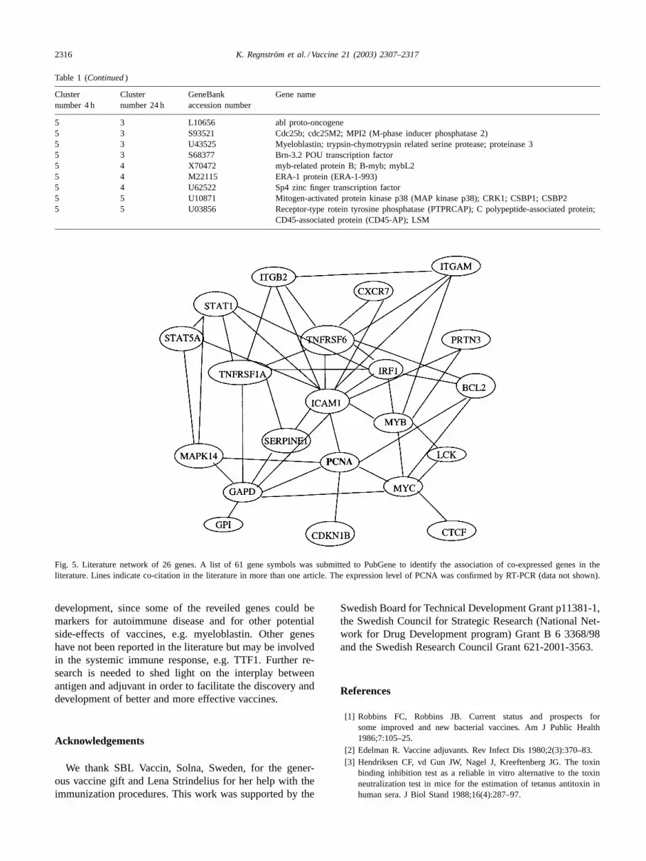
2316 K. Regnström et al. / Vaccine 21 (2003) 2307–2317
Table 1 (Continued)
Clusternumber 4 h
Clusternumber 24 h
GeneBankaccession number
Gene name
5 3 L10656 abl proto-oncogene5 3 S93521 Cdc25b; cdc25M2; MPI2 (M-phase inducer phosphatase 2)5 3 U43525 Myeloblastin; trypsin-chymotrypsin related serine protease; proteinase 35 3 S68377 Brn-3.2 POU transcription factor5 4 X70472 myb-related protein B; B-myb; mybL25 4 M22115 ERA-1 protein (ERA-1-993)5 4 U62522 Sp4 zinc finger transcription factor5 5 U10871 Mitogen-activated protein kinase p38 (MAP kinase p38); CRK1; CSBP1; CSBP25 5 U03856 Receptor-type rotein tyrosine phosphatase (PTPRCAP); C polypeptide-associated protein;
CD45-associated protein (CD45-AP); LSM
Fig. 5. Literature network of 26 genes. A list of 61 gene symbols was submitted to PubGene to identify the association of co-expressed genes in theliterature. Lines indicate co-citation in the literature in more than one article. The expression level of PCNA was confirmed by RT-PCR (data not shown).
development, since some of the reveiled genes could bemarkers for autoimmune disease and for other potentialside-effects of vaccines, e.g. myeloblastin. Other geneshave not been reported in the literature but may be involvedin the systemic immune response, e.g. TTF1. Further re-search is needed to shed light on the interplay betweenantigen and adjuvant in order to facilitate the discovery anddevelopment of better and more effective vaccines.
Acknowledgements
We thank SBL Vaccin, Solna, Sweden, for the gener-ous vaccine gift and Lena Strindelius for her help with theimmunization procedures. This work was supported by the
Swedish Board for Technical Development Grant p11381-1,the Swedish Council for Strategic Research (National Net-work for Drug Development program) Grant B 6 3368/98and the Swedish Research Council Grant 621-2001-3563.
References
[1] Robbins FC, Robbins JB. Current status and prospects forsome improved and new bacterial vaccines. Am J Public Health1986;7:105–25.
[2] Edelman R. Vaccine adjuvants. Rev Infect Dis 1980;2(3):370–83.[3] Hendriksen CF, vd Gun JW, Nagel J, Kreeftenberg JG. The toxin
binding inhibition test as a reliable in vitro alternative to the toxinneutralization test in mice for the estimation of tetanus antitoxin inhuman sera. J Biol Stand 1988;16(4):287–97.
K. Regnström et al. / Vaccine 21 (2003) 2307–2317 2317
[4] Regnström K, Ragnarsson EGE, Rydell N, Sjöholm I, Artursson P.Tetanus antigen modulates the gene expression profile of aluminumphosphate in spleen lymphocytes in vivo. Pharmacogenomics J2002;2:57–64.
[5] Teague TK, Hildeman D, Kedl RM, Mitchell T, Rees W, SchaeferBC, et al. Activation changes the spectrum but not the diversity ofgenes expressed by T cells. Proc Natl Acad Sci USA 1999;96(22):12691–6.
[6] Tamayo P, Slonim D, Mesirov J, Zhu Q, Kitareewan S, DmitrovskyE, et al. Interpreting patterns of gene expression with self-organizingmaps: methods and application to hematopoietic differentiation. ProcNatl Acad Sci USA 1999;96(6):2907–12.
[7] Eisen MB, Spellman PT, Brown PO, Botstein D. Cluster analysisand display of genome-wide expression patterns. Proc Natl Acad SciUSA 1998;95(25):14863–8.
[8] Lazaridis M. Course in statistical analysis of microarray studies.In: Proceedings of the 9th International Conference on IntelligentSystems for Molecular Biology, 2001 July; Copenhagen.
[9] Möröy T, Grzeschiczek A, Petzold S, Hartmann KU. Expressionof a Pim-1 transgene accelerates lymphoproliferation and inhibitsapoptosis inlpr/lpr mice. Proc Natl Acad Sci USA 1993;90:10734–8.
[10] Domen J, vd Lugt NM, Acton D, Laird PW, Linders K, Berns A.Pim-1 levels determine the size of early B lymphoid compartmentsin bone marrow. J Exp Med 1993;178(5):1665–73.
[11] Kvale D, Brandtzaeg P. Constitutive and cytokine induced expressionof HLA molecules, secretory compononent, and intracellular adhesionmolecule-1 is modulated by butyrate in the colonic epithelia cell lineHT-29. Gut 1995;36(5):737–42.
[12] Avni O, Rao A. T cell differentiation: a mechanistic view. Curr OpinImmunol 2000;12:654–9.
[13] Saemann MD, Bohmig GA, Osterreicher CH, Burtscher H, ParoliniO, Diakos C, et al. Anti-inflammatory effects of sodium butyrate on
human monocytes: potent inhibition of IL-12 and up-regulation ofIL-10 production. FASEB J 2000;14(15):2380–2.
[14] Campanelli DPA, Detmers CF, Nathan, Gabay JE. J Clin Invest1998;85:904–15.
[15] Falk RJ, Jenette JC. Wegener’s granulomatosis, systemic vasculitisand antineutrophil cytoplasmic autoantibodies. Ann Rev Med1991;42:459–69.
[16] Kurowsawa S, Esmon CT, Stearns-Kurowsawa DJ. The solubleendothelial protein C receptor binds to activated neutrophils:involvement of proteinase-3 and CD11b/CD18. J Immunol 2000;165:4697–703.
[17] Rook GAW, Ristori G, Salvetti M, Giovannoni G, Thompson E,Stanford JL. Bacterial vaccines for the treatment of multiple sclerosisand other autoimmune diseases. Immunol Today 2000;21(10):503–8.
[18] Langst G, Blank TA, Becker PB, Grummt I. RNA polymeraseI transcription on nucleosomal templates: the transcriptiontermination factor TTF-1 induces chromatin remodeling and relievestranscriptional repression. EMBO J 1997;16(4):760–8.
[19] Evers R, Grummt I. Molecular co-evolution of mammalian ribosomalgene terminator sequences and the transcription termination factorTTF-I. Proc Natl Acad Sci USA 1995;92:5827–31.
[20] Matzinger P. An innate sense of danger. Semin Immunol1998;10(5):399–415.
[21] Banks WA, Kastin AJ, Fasold MB. Differential effect of aluminumon the blood–brain barrier transport of peptides. J Pharm Exp Ther1988;244:579–85.
[22] Deleers M. Cationic atmosphere and cation competition binding atnegative charged membranes. Pathological implications of aluminum.Res Commun Chem Pharm 1985;49:277–94.
[23] Jensen TK, Laegreid A, Komorowski J, Hovig E. A literature networkof human genes for high-throughput analysis of gene expression.Nat Genet 2001;28:21–8.