development and evaluation of chitosan microspheres for tetanus, diphtheria and divalent vaccines: a...
TRANSCRIPT

1175
Introduction
Vaccines and large molecule proteins’ delivery using polymeric microspheres as a carrier system has received increased attention in the recent years. These systems represent a promising delivery system for these large macromolecules across mucosal barriers i.e. oral and nasal routes.[1,2]
Tetanus and diphtheria toxoids can cause infections in unvaccinated or inadequately immunized individu-als. The infection caused by these toxoids can easily be eliminated by immunization. Active immunization with diphtheria and tetanus toxoids is part of the current rec-ommended childhood immunization schedule which would require booster doses for effective immunization. However, compliance to the administration of vaccines
taken in multiple divided doses is minimal especially in developed countries which have high dropout rates from individuals receiving the first dose. These individu-als will not have effective immunization unless they take two or three booster doses. Therefore, the conversion of multiple doses vaccines into controlled release single shot may represent an important advance which leads to improve vaccination coverage, eliminate requirements for multiple injections and be more cost effective.[3]
Microencapsulation of vaccines has received much attention recently due to their ability to co-encapsulate multiple antigens, generate good immune responses and be stable enough to be administered orally, nasally or parenterally.[4,5]
Controlled release vaccines are most frequently for-mulated using the multiple emulsion solvent evaporation
ReseaRch aRtIcle
Development and evaluation of chitosan microspheres for tetanus, diphtheria and divalent vaccines: a comparative study of subcutaneous and intranasal administration in mice
Fahima M. Hashem1, Sahar A. Fahmy1,Aly M. El-Sayed2, and Majid M. Al-Sawahli2
1Department of Pharmaceutics and Industrial Pharmacy, Faculty of Pharmacy, Helwan University, Cairo, Egypt and 2Holding Company for Biological Products & Vaccines (VACSERA), Giza, Egypt
abstractContext: There is a need to use the new technologies to induce immunity with minimum number of vaccination sessions to ensure compliance with reducing cost. Objectives: To develop single shot vaccines of tetanus, diphtheria and divalent toxoids microsphere’s formulations and to induce their immune response after intranasal and subcutaneous administration in mice. Materials and methods: The microspheres were prepared using different concentrations of chitosan. Microsphere’s morphology, particle size analysis, encapsulation efficiency and antigen integrity were performed and the best formulations were selected for in vitro and in vivo testing in mice. Results: The developed microspheres have a yield percent of 70.3–91.5%. In vitro release of antigens indicated that tetanus release was increased up to 75 and 81% post T5 and TD5 formulations respectively, whereas diphtheria cumulative release increased up to 74 and 69% post D3 and TD5, respectively. Discussion: Antibody levels produced were lower than that obtained from alum adsorbed vaccine but higher than the minimum level required to induce immunogenicity (>0.01 IU/mL). The subcutaneous route of administration was superior over the intranasal route in producing higher antibody levels. Conclusion: Chitosan microspheres were developed successfully and prove that chitosan represents a good candidate for vaccines delivery.
Keywords: Microparticles, vaccine delivery, controlled release, immunogenicity in mice, single shot vaccine
Address for Correspondence: Sahar A. Fahmy, Assistant professor, Department of Pharmaceutics and Industrial Pharmacy, Faculty of Pharmacy, Helwan University, Cairo, Egypt, 11790. Tel: 002 0224321562; Mobile: +971503279016. Fax: 002 0237223039. E-mail: [email protected]; [email protected]
(Received 15 June 2011; revised 19 August 2011; accepted 20 August 2011)
Pharmaceutical Development and Technology, 2013; 18(5): 1175–1185© 2013 Informa Healthcare USA, Inc.ISSN 1388-0209 print/ISSN 1744-5116 onlineDOI: 10.3109/10837450.2011.618949
Pharmaceutical Development and Technology
2013
18
5
1175
1185
15 June 2011
19 August 2011
20 August 2011
1388-0209
1744-5116
© 2013 Informa Healthcare USA, Inc.
10.3109/10837450.2011.618949
NPHB
618949
Phar
mac
eutic
al D
evel
opm
ent a
nd T
echn
olog
y D
ownl
oade
d fr
om in
form
ahea
lthca
re.c
om b
y T
he U
nive
rsity
of
Man
ches
ter
on 1
1/11
/14
For
pers
onal
use
onl
y.

1176 F.M. Hashem et al.
Pharmaceutical Development and Technology
method based on the biodegradable and Food and Drug Administration approved poly-(lactic acid) (PLA) and poly-(lactic-co-glycolic acid) (PLGA) polymers.[6] These polymers have already been formulated and available for use in the market as injectable sustained release drug products such as Decapeptyltm (Ipsen, UK), Zoladextm (AstraZeneca, UK), Enantonetm (Takeda, Germany) and Risperdal Constatm (Janssen Cilag, Australia).
Microsphere particle size alteration has demonstrated to significantly affect antigen release rate and conse-quently its immunogenicity. Particles larger than 10 µm will act as a depot after injection, producing a controlled release of the antigen; whereas particles smaller than 10 µm may be phagocytosed by macrophages.[7–9] It was demonstrated that microspheres with sizes less than 10 µm will be able to enter into lymph nodes and provide a high level concentration over an extended period of time.[8]
Chitosan microspheres are used to provide controlled release of many drugs and to improve the bioavailability of degradable substances such as proteins, as well as to improve the uptake of hydrophilic substances across the epithelial layers.[10] Chitosan is one of the most promis-ing polymers because it is enzymatically degradable polymer, biocompatible and has low toxicity. In addi-tion, it has mucoadhesive properties and permeation-enhancing properties which make it a good candidate for the preparation of mucosal delivery systems. Its degradation products have been shown to be nontoxic, non-immunogenic, and non-carcinogenic which make it a good candidate for the development of single dose vaccines.[11,12] Developed chitosan microspheres cross-linked with glutaraldehyde were shown to be long-acting biodegradable carriers suitable for controlled delivery of many antigens and proteins.[13]
Challenges may be faced during the development of such delivery systems. The main challenges are repre-sented in low encapsulation efficiency, protein inactiva-tion during the encapsulation process and difficulties in controlling active protein releases.[14–16]The use of protein stabilizers such as trehalose is very important during the formulation of such delivery systems which results in increasing the stability of vaccines microspheres.[17]
Administration of drugs via nasal route was proven to be very efficient for systemic delivery especially for low molecular weight polar drugs, peptides and proteins that are not easily administered via other routes than by injec-tion or where a rapid onset of action is required.[18,19]
Chitosan-based formulations can greatly improve the absorption of drugs from the nasal cavity.[20] Studies dem-onstrated that chitosan promoted the absorption of pro-tein and high molecular weight drugs by improving the adhesion between the formulation and nasal tissues.[21] In addition, the interaction of the positively charged chi-tosan with the negatively charged mucin layer and the tight junctions facilitates the paracellular transport of hydrophilic macromolecules by opening the tight junc-tions of the mucosal barriers.[22]
Chitosan nasal delivery system has been tested with vaccines, namely for influenza, pertussis and diphthe-ria in various animal models and in man. It has been shown that chitosan significantly enhances the immune response obtained from influenza vaccine when given nasally to mice with IgG levels similar to those obtained after parenteral application and significant IgA levels both in the nasal fluid and in the lungs. Similarly, in stud-ies in naïve guinea pigs, it was shown that diphtheria vaccine (cross-reacting material (CRM197) administered nasally in a chitosan formulation provided significantly higher serum IgG titers than the levels obtained for the nasal formulation without chitosan. For the nasal chito-san solution formulation, the IgG titers were similar to those obtained after an adjuvant intraperitoneal injec-tion of CRM197, whereas for the nasal chitosan powder formulation the levels were significantly higher.[23,24]
The current study was performed to develop a single dose of chitosan microsphere’s formulations loaded with bacterial vaccines i.e. tetanus, diphtheria and divalent vaccines to obtain a good immune response with mini-mum side effects and prolonged efficacy. In addition, the induction of systemic immune response after subcu-taneous (SC) and intranasal (IN) administrations of the developed formulations in mice was also investigated.
Materials and methods
MaterialsPurified concentrated tetanus toxoid (MW 150 kDa; 1500 Lf/mL), purified concentrated diphtheria toxoid (MW 60 kDa; 2700 Lf/ml) and water for injection were obtained from VACSERA, Egypt. Chitosan (minimum 85% deacetylated with MW 200,000), span 80, tween 80, tris (tri hydroxy methyl aminomethane), TEMED (N, N, N’, N’-tetramethylethylenediamine), and bisacrylamide were obtained from Sigma Aldrich, USA. Glutaraldehyde solution 25% in water (2.6M) was obtained from Fluka Chemika, Switzerland, and D (+) trehalose >99.5% High-performance liquid Chromatography grade from Fluka Biochemical, USA. Concentrated hydrochloric acid 37%, toluene 80, glycine, ammonium persulfate and glacial acetic acid were obtained from BDH, England. Phosphate buffer saline (PBS) tablets from “PBS”, Amaresco, USA; Acrylamide and sodium dodecyl sulfate; Bio-Rad Laboratories, California, USA. Biorad protein std. at 1 µg/µL. Bradford reagent: was prepared by dissolving 100 mg coomassie brilliant blue G-250 in 50 mL 95% ethanol and 100 mL 85% (w/v) phosphoric acid, then diluted to 1 L and filtered through Whatman #1 paper just before use. Concentrated coomassie dye diluted to 1:5 with distiller water.
Preparation of chitosan microspheresChitosan microspheres were prepared by the method described by Jaganathan et al. aseptically under laminar flow cabinet at room temperature.[8] Briefly, selected chi-tosan concentration in aqueous solution was prepared in 3% (w/v) acetic acid containing 2% (w/v) sodium
Phar
mac
eutic
al D
evel
opm
ent a
nd T
echn
olog
y D
ownl
oade
d fr
om in
form
ahea
lthca
re.c
om b
y T
he U
nive
rsity
of
Man
ches
ter
on 1
1/11
/14
For
pers
onal
use
onl
y.

Development of bacterial vaccine delivery systems 1177
© 2013 Informa Healthcare USA, Inc.
chloride. The solution was stirred for 2 hrs at 1000 rpm to form a gel which was kept overnight for stabilization. The gel was filtered on porous membrane of 0.45 µm.
A dispersion phase was prepared by mixing toluene (50 mL) and span-80 (5 mL) at 1000 rpm for 10 min. With continuous stirring, 4 mL of selected chitosan gel with 1 mL of (0.01%) hydrochloric acid and 2 mL of the bacte-rial vaccine model containing selected concentrations of trehalose were added to the dispersion phase. At the end of second hour of stirring, 5 mL of glutaraldehyde satu-rated toluene (GST) was added drop wise using sterile syringe. Then, another 5 mL of GST were added and stir-ring continued for another 2 hrs.
At the end of 6 hrs stirring, the resultant suspension was incubated in 15 mL sterile plastic centrifuge tubes, and fixed in cold centrifuge adjusted at 1000 rpm for 10 min at 4°C. The supernatant was removed, and the settled microspheres were collected. The separated microspheres obtained were washed five times with 5 mL of toluene followed by 5 mL of acetone three times using cold centrifuge at 1000 rpm for 10 min. The microspheres were re-suspended in 10 mL acetone and dried at room temperature. Dried microspheres were stored in sterile dry labeled vials at 4°C till use.
A total of 18 different formulations were prepared for the three bacterial models as illustrated in Table 1.
In vitro evaluation of the prepared microspheresThe prepared formulations were evaluated for their anti-gen integrity, yield percent and protein content. The best formulae were selected to be evaluated further for encap-sulation efficiency, scanning electron microscope (SEM) analysis, microsphere’s morphology, infrared spectroscopy (IR) and in vitro release of antigen from the microspheres.
Determination of antigen integrityThis procedure was done as reported by Jaganathan et al where 10 mg of the selected formula were powdered in a sterile mortar.[8] The antigen was extracted in 3 mL PBS (pH 7.4) in sterile plastic tubes by fixing the tubes at angle on orbital shaker for 7 days. Samples were centri-fuged and the supernatant was separated. The extracted antigen solution was preceded in sodium dodecyl sul-phate polyacrylamide gel electrophoresis (SDS-PAGE) to evaluate the effect of the encapsulation process on proteins’ stability.[25] Standard tetanus and diphtheria antigens without microencapsulation were used as con-trol groups. Electrophoresis was carried out at a constant
current 15 mAmp/gel. When the bromophenol blue dye was approximately 1 cm from the bottom of the gel, the power was turned off and the gel was carefully removed for fixation and staining with coomassie blue dye.
Determination of the yield percentThe prepared microspheres were collected and weighed. The percent yield was determined by dividing the weight of the collected microspheres by the weight of the total amount of all the components used in the formulation.[26]
Determination of antigen content in chitosan microspheresProtein content was determined according to the method described by Bradford et al. using the commercially available Bio-Rad kit (Bio-Rad Laboratories, Richmond, CA, USA) after antigen extraction.[27] In a micro-titer plate 96-well, 3 µL of standard or sample in triplicates mixed with 145 µL diluted dye. The whole plate was shook on mini-orbital shaker for 15 min at 5 rpm speed. The plate was read at 595 nm. Antigen content was determined three times for each formulation on three different days and the mean values were calculated.
Encapsulation efficiencyThe formulations with the highest protein content were selected to be tested for its encapsulation efficiency which was determined by:
Encapsulation efficiency
Actual protein
content
The=
ooretical
content
100×
Microspheres morphology and particle sizeSEM (JEOL JXA-840A, Japan) was utilized to examine the morphology of the developed microspheres as well as to measure its particle size. Placebo microspheres and selected formulae were deposited onto sided tapes and then coated with gold and examined. Microspheres were re-dispersed in distilled water; air dried; fixed on aluminum stubs under ambient conditions with a con-ductive copper tape, then covered with a gold layer with a Sputter Coater Edwards S 150 A (Edwards High Vacuum International, Crawley, UK).
Infrared spectroscopic analysis (IR)To detect any interaction occurred between the vaccines and any component of the microspheres during prepara-tion, samples of chitosan, tetanus and diphtheria toxoids, physical mixture of vaccine and chitosan and the prepared microspheres were subjected to infrared spectroscopic analysis. The resulted spectra obtained were compared with that of the unloaded chitosan and native vaccines.
In vitro release of antigen from microspheresThe release of antigens from chitosan microspheres was done in PBS (pH 7.4) containing 0.02% (w/v) tween 80. Several vials containing 10 mg microspheres and 5 mL of
Table 1. Formulations of chitosan microspheres loaded with tetanus (T), diphtheria (D) and divalent vaccines (TD).
Formulae Chitosan (%w/v)Trehalose
(%w/v)T1, D1, TD1 1 1.5T2, D2, TD2 1 2T3, D3, TD3 1.5 1.5T4, D4, TD4 1.5 2T5, D5, TD5 2 1.5T6, D6, TD6 2 2
Phar
mac
eutic
al D
evel
opm
ent a
nd T
echn
olog
y D
ownl
oade
d fr
om in
form
ahea
lthca
re.c
om b
y T
he U
nive
rsity
of
Man
ches
ter
on 1
1/11
/14
For
pers
onal
use
onl
y.
1178 F.M. Hashem et al.
Pharmaceutical Development and Technology
dissolution media were incubated at 37°C on an orbital shaker. Antigen release was estimated at 0.5, 1, 2, 3, 4, 5 and 6 days. The microsphere suspension was centrifuged at 8000 rpm for 10 min and the supernatant was collected to estimate protein content by enzyme-linked immuno-sorbent assay (ELISA).[28] Blank microspheres containing no antigen were used as control.
Cumulative release of the test toxoid was determined according to the following equation:
Cumulative release of test OD test Reference conc.
OD r= ×
eeference
Where, OD is the optical density measured for both the test and the reference
Stability studyStability study was done to the selected formulae, T5, D3 and TD5 after 12 and 18 months post preparation kept at 4°C. Protein content was evaluated by Bradford assay and was also evaluated for their morphology and particle size analysis using SEM.
Immunogenicity studiesImmunogenicity studies were done using Swiss mice 2–3 weeks old, weighing 14–16 g, males (kindly supplied by animal house, VACSERA, Egypt). Animals were grouped in eight (n = 10) and were allowed to acclimatize for 1 week before proceeding of the experiment. They were deprived of food 3 hrs prior to immunization. Animals were housed in plastic cages, bred randomly, with free access to food and water. The animal study was conducted with the approval of the animal care committee at VASCERA.
The prepared microspheres were diluted with 1.0% sterile solution of sodium carboxymethyl cellulose in 0.9% normal saline individually to get a single dose of 0.5 limit of flocculation (Lf)/0.5 mL (1/10 of single human dose) to be injected subcutaneously or to get a single dose 0.5 Lf/6 µL (equivalent to the same total Lf as in SC administration) to be administered intranasally in mice. Lf defines the quan-tity of toxoid as assessed by flocculation. For IN adminis-tration, the selected formulations were incorporated in 1.0 w/v chitosan gel and were administered in a total volume of 6 µL (i.e. 3 µL to each nostril using a micropipette in con-scious mice). Two control groups received the equivalent
dose of either alum adsorbed tetanus or diphtheria and booster dose was given after 4 weeks of primary immuni-zation. One week after administration of the booster dose, blood samples were collected via the retro orbital plexus at 7, 15, 35, 60, 90 and 120 days. Blood samples were then centrifuged for 20 min at 3500 rpm.[28] Samples were stored at −20°C until the determination of systemic immune response and IgG antibody level using ELISA assay.
Statistical analysisThe analysis was performed using Minitab® version 16 statistical software (Minitab, Inc., USA). Data were ana-lyzed using ANOVA test at 5% level with p value <0.05 representing significant difference.
Results
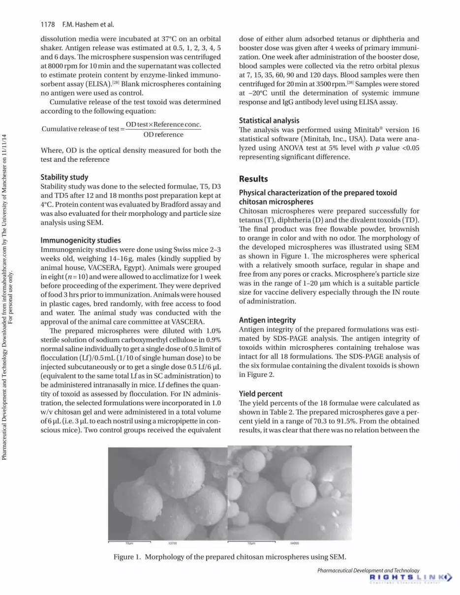
Physical characterization of the prepared toxoid chitosan microspheresChitosan microspheres were prepared successfully for tetanus (T), diphtheria (D) and the divalent toxoids (TD). The final product was free flowable powder, brownish to orange in color and with no odor. The morphology of the developed microspheres was illustrated using SEM as shown in Figure 1. The microspheres were spherical with a relatively smooth surface, regular in shape and free from any pores or cracks. Microsphere’s particle size was in the range of 1–20 µm which is a suitable particle size for vaccine delivery especially through the IN route of administration.
Antigen integrityAntigen integrity of the prepared formulations was esti-mated by SDS-PAGE analysis. The antigen integrity of toxoids within microspheres containing trehalose was intact for all 18 formulations. The SDS-PAGE analysis of the six formulae containing the divalent toxoids is shown in Figure 2.
Yield percentThe yield percents of the 18 formulae were calculated as shown in Table 2. The prepared microspheres gave a per-cent yield in a range of 70.3 to 91.5%. From the obtained results, it was clear that there was no relation between the
Figure 1. Morphology of the prepared chitosan microspheres using SEM.
Phar
mac
eutic
al D
evel
opm
ent a
nd T
echn
olog
y D
ownl
oade
d fr
om in
form
ahea
lthca
re.c
om b
y T
he U
nive
rsity
of
Man
ches
ter
on 1
1/11
/14
For
pers
onal
use
onl
y.

Development of bacterial vaccine delivery systems 1179
© 2013 Informa Healthcare USA, Inc.
yield percent and the model of bacterial vaccine incorpo-rated in the microspheres. There was also no significant correlation between the yield percent and either chitosan concentrations or trehalose concentrations (p > 0.05) for tetanus, diphtheria and divalent formulae.
Antigen contentAntigen content was determined using Bradford assay and the results are presented in Table 3. The effect of using different concentrations of chitosan and treha-lose on microspheres loading was estimated to have no significant effect on protein content (p > 0.05). For chitosan concentration, the difference was found to be non significant (P = 0.357, 0.848, 0.102) for between tetanus, diphtheria and divalent formulae, respec-tively. For trehalose concentration, the difference was found to be non significant (P = 0.81, 0.632, 0.249) for between tetanus, diphtheria and divalent formulae, respectively.
It is worth to mention that, for the divalent vaccine loaded chitosan microspheres, the antigen content assessed was for both titanus and diphtheria because Bradford assay detects only total protein content and cannot differentiate different types of proteins.
The tested formulations which produced the high-est antigen contents from the 18 tested formulae were selected for further investigations. The selected formulae were T5 (2% w/v chitosan, 1.5% w/v trehalose), D3 (1.5% w/v chitosan, 1.5% w/v trehalose) and TD5 (2% w/v chi-tosan, 1.5% w/v trehalose).
Encapsulation efficiencyAll selected formulae produced low percentage encapsu-lation efficiency in the range of 18–20%. This low value could be due to the several washing and separation steps during microsphere’s preparations. In addition, the use of organic solvents may have an impact on the encapsu-lation process.
Infrared spectroscopic analysis (IR)Infrared spectra of the chitosan polymer, tetanus, diph-theria, physical mixture of vaccine and chitosan polymer and the spectra of the selected formulae revealed that there was no interaction between the vaccine, chitosan and any component incorporated during microsphere’s preparation. The characteristic peaks of tetanus vaccine appeared at the same wave length in the spectra of T5 and TD5 formulae. In addition, the characteristic peaks of diphtheria vaccine appeared at the same wave length in the spectra of D3 and TD5 formulae.
In vitro release of antigens from the microspheresThe tetanus and diphtheria release pattern from the prepared chitosan microspheres stabilized with treha-lose was compared to blank microspheres as a control. Figure 3 illustrates the tetanus and diphtheria toxoid release patterns from the selected formulae with time. Tetanus cumulative release after 6 days was increased up to 75 and 81% post for T5 and TD5, respectively. Diphtheria cumulative release increased up to 74 and 69% post D3 and TD5, respectively after 6 days. These
Table 2. Yield percent of the prepared formulations.Formula Yield % (SD) Formula Yield % (SD) Formula Yield % (SD)
T1 85.5 (8.1) D1 91.5(17.4) TD1 71 (16.8)T2 90.7 (13.2) D2 83.5(6.58) TD2 81.5 (7.9)T3 88.1 (11.7) D3 77.3(15.7) TD3 78.4 (14.6)T4 79.5 (8.7) D4 87(9.6) TD4 70.3 (14.6)T5 88.1 (9.1) D5 71.4(19.3) TD5 78.6 (18.3)T6 81.2 (14.2) D6 83.2(11.6) TD6 87.8 (9.4)
Figure 2. SDS-PAGE analysis of six formulae of the divalent vaccine-chitosan microspheres compared to the control group C; standard tetanus and diphtheria antigens.
Phar
mac
eutic
al D
evel
opm
ent a
nd T
echn
olog
y D
ownl
oade
d fr
om in
form
ahea
lthca
re.c
om b
y T
he U
nive
rsity
of
Man
ches
ter
on 1
1/11
/14
For
pers
onal
use
onl
y.

1180 F.M. Hashem et al.
Pharmaceutical Development and Technology
data indicated the controlled release of bacterial vaccine from chitosan microspheres over time.
Stability studySEM analysis for the selected formulations T5, D3 and TD5 were done after 18 months of storage at 4°C which
revealed that the microsphere’s morphology did not change upon storage for up to 18 months. Microsphere’s shape was spherical with a relatively smooth surface and free from any pores or cracks. The actual size of individ-ual microspheres was in the range of 1–20 µm and few microspheres measured of size up to 50 µm. Analyzing the protein content of the stored microspheres was done and the results presented in Table 4. The protein content of the selected formulae showed no significant difference between microspheres after the storage period at 4°C and the freshly prepared microspheres.
Immunogenicity studiesImmune response was examined by immunizing mice with the selected formulae T5, D3 and TD5 via both IN and SC route of administrations. The average particle size of the prepared formulation was measured to be 8.52 ± 4.31 µm, 11.35 ± 8.24 µm and 9.35 ± 6.82 for T5, D3 and TD5, respectively. Large particle size bigger than 20 µm were not excluded from the in vivo study. Mice received a SC injection of 0.5 Lf/0.5 mL or IN administra-tion to get a single dose 0.5 Lf/6 µL and the antibody lev-els were measured 1 week after the booster dose. The IgG levels produced from the developed formulations were compared to that produced after the immunization upon the SC administration of alum adsorbed vaccine.
The immune response was measured at 7, 15, 35, 60, 90 and 120 days post vaccination (DPV). Serum tetanus antibody level could be detected on the 7th DPV after T5 administration recording 1.51 IU/mL upon SC admin-istration and then a slight progressive elevation was detected relatively with time. The peak reached on the 90th DPV recording 2.00 IU/mL. Serum antibody level post IN administration of T5 showed that antibody level were comparatively low to that of SC after 15 days DPV recording 0.356 IU/mL and the peak was reached 1.868 IU/mL on the 90th DPV as shown in Figure 4.
Figure 3. Tetanus and diphtheria toxoid in vitro release profiles of T5, D3 and TD5 formulae.
Table 3. Protein contents for the prepared formulations.
Formula Protein content
µg/mL (SD) Lf/mL Lf/mg
T1 0.195 (0.08) 8.5 2.55
T2 0.225 (0.12) 9.8 2.94
T3 0.193 (0.14) 8.4 2.52
T4 0.124 (0.11) 5.4 1.62
T5 0.241 (0.06) 10.4 3.12
T6 0.225 (0.05) 9.8 2.94
Formula Protein content
(µg/mL) Lf/mL Lf/mg
D1 0.215 (0.19) 7.4 2.22
D2 0.233 (0.09) 8 2.4
D3 0.296 (0.1) 10.1 3.03
D4 0.262 (0.13) 9 2.7
D5 0.235 (0.15) 8.1 2.43
D6 0.198 (0.07) 6.8 2.04
Formula Protein content
(µg/mL) Lf/mL (µg/mL)
TD1 0.262 (0.12) 11.4 3.42
TD2 0.227 (0.06) 9.9 2.97
TD3 0.274 (0.19) 11.9 3.57
TD4 0.233 (0.12) 10.1 3.03
TD5 0.286 (0.09) 12.4 3.72
TD6 0.234 (0.15) 10.2 3.06
Phar
mac
eutic
al D
evel
opm
ent a
nd T
echn
olog
y D
ownl
oade
d fr
om in
form
ahea
lthca
re.c
om b
y T
he U
nive
rsity
of
Man
ches
ter
on 1
1/11
/14
For
pers
onal
use
onl
y.
Development of bacterial vaccine delivery systems 1181
© 2013 Informa Healthcare USA, Inc.
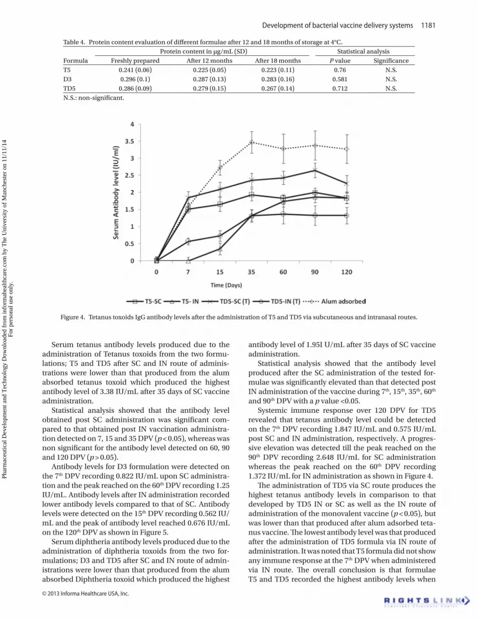
Figure 4. Tetanus toxoids IgG antibody levels after the administration of T5 and TD5 via subcutaneous and intranasal routes.
Table 4. Protein content evaluation of different formulae after 12 and 18 months of storage at 4°C.
FormulaProtein content in µg/mL (SD) Statistical analysis
Freshly prepared After 12 months After 18 months P value SignificanceT5 0.241 (0.06) 0.225 (0.05) 0.223 (0.11) 0.76 N.S.D3 0.296 (0.1) 0.287 (0.13) 0.283 (0.16) 0.581 N.S.TD5 0.286 (0.09) 0.279 (0.15) 0.267 (0.14) 0.712 N.S.N.S.: non-significant.
Serum tetanus antibody levels produced due to the administration of Tetanus toxoids from the two formu-lations; T5 and TD5 after SC and IN route of adminis-trations were lower than that produced from the alum absorbed tetanus toxoid which produced the highest antibody level of 3.38 IU/mL after 35 days of SC vaccine administration.
Statistical analysis showed that the antibody level obtained post SC administration was significant com-pared to that obtained post IN vaccination administra-tion detected on 7, 15 and 35 DPV (p < 0.05), whereas was non significant for the antibody level detected on 60, 90 and 120 DPV (p > 0.05).
Antibody levels for D3 formulation were detected on the 7th DPV recording 0.822 IU/mL upon SC administra-tion and the peak reached on the 60th DPV recording 1.25 IU/mL. Antibody levels after IN administration recorded lower antibody levels compared to that of SC. Antibody levels were detected on the 15th DPV recording 0.562 IU/mL and the peak of antibody level reached 0.676 IU/mL on the 120th DPV as shown in Figure 5.
Serum diphtheria antibody levels produced due to the administration of diphtheria toxoids from the two for-mulations; D3 and TD5 after SC and IN route of admin-istrations were lower than that produced from the alum absorbed Diphtheria toxoid which produced the highest
antibody level of 1.95I U/mL after 35 days of SC vaccine administration.
Statistical analysis showed that the antibody level produced after the SC administration of the tested for-mulae was significantly elevated than that detected post IN administration of the vaccine during 7th, 15th, 35th, 60th
and 90th DPV with a p value <0.05.Systemic immune response over 120 DPV for TD5
revealed that tetanus antibody level could be detected on the 7th DPV recording 1.847 IU/mL and 0.575 IU/mL post SC and IN administration, respectively. A progres-sive elevation was detected till the peak reached on the 90th DPV recording 2.648 IU/mL for SC administration whereas the peak reached on the 60th DPV recording 1.372 IU/mL for IN administration as shown in Figure 4.
The administration of TD5 via SC route produces the highest tetanus antibody levels in comparison to that developed by TD5 IN or SC as well as the IN route of administration of the monovalent vaccine (p < 0.05), but was lower than that produced after alum adsorbed teta-nus vaccine. The lowest antibody level was that produced after the administration of TD5 formula via IN route of administration. It was noted that T5 formula did not show any immune response at the 7th DPV when administered via IN route. The overall conclusion is that formulae T5 and TD5 recorded the highest antibody levels when
Phar
mac
eutic
al D
evel
opm
ent a
nd T
echn
olog
y D
ownl
oade
d fr
om in
form
ahea
lthca
re.c
om b
y T
he U
nive
rsity
of
Man
ches
ter
on 1
1/11
/14
For
pers
onal
use
onl
y.

1182 F.M. Hashem et al.
Pharmaceutical Development and Technology
administered subcutaneously than administered via IN route of administration.
Diphtheria antibody level was detected from the 7th DPV of TD5 recording 1.21 IU/mL and 0.518 IU/mL in case of SC and IN administration, respectively. A pro-gressive elevation was seen till the peak reached on the 90th DPV recording 1.216 IU/mL upon SC administration whereas the peak was delayed and reached on the 15th
DPV recording 0.738 IU/mL as shown in Figure 5. It was noted that D3 formula is the only formula that did not show immune response at the 7th day post immunization when administered via IN route.
Discussion
The developed vaccine formulations were prepared suc-cessfully using the cross linking method. The prepared microspheres were spherical with a relatively smooth surface. This may be due to the use of glutaraldehyde in the preparation process of the microspheres. It was reported that microspheres cross-linked with glutaral-dehyde have shown smooth surface morphology in com-parison to non cross-linked chitosan microspheres.[13,29] The size of prepared microspheres was in the range of 1–20 µm which is small enough to deliver vaccines via IN route. Smaller particles are normally taken up by the APC (macrophages and dendritic cells) through phago-cytosis and subsequently degraded and thus considered to be the most suitable size to get optimum absorption. Microspheres greater than 20 µm in size are not phagocy-tosed by macrophages and the antigen will be degraded by hydrolysis.[8] These larger particle size acts as a depot
after injection which prolongs the release of the antigen over time.
The yield percent of chitosan microspheres in the current study was found to be within the range of 70.3 to 91.5%. This range is similar to what were reported in other studies which showed that chitosan loaded tetanus vaccine yield percent was found to be 70–84% using limes flocculation test[30] and was reported to be 78.1% using for tetanus chitosan microspheres prepared by supercritical fluid encapsulation technology.[31]
The integrity of the antigen was proven to be not affected by the microencapsulation process. The use of trehalose as a stabilizing agent prevented antigen aggre-gation at the w/o interface. In addition, it prevented antigen inactivation due to the exposure to the deleteri-ous solvents during the process of microencapsulation. This is done by shielding the antigen from the organic solvent via a preferential hydration of their surfaces and preventing protein-interface contact. Moreover, trehal-soe has adequate aqueous solubility which will leave the microsphere’s matrix with pores which increases antigen release from the developed formulations.[8] In addition, trehalose has appreciable solubility in aqueous media and it is reported that it can preserve the integrity of the microspheres following several centrifugation/re-sus-pension processes.[32]
The obtained results were in line with similar studies that reported the necessity of adding protein stabiliz-ers such as trehalose, albumin, heparin, pluronic F68, bovine serum albumin to maintain antigen stability.[17] Antigen integrity was performed using SDS-PAGE analy-sis and was proven to be intact for all 18 formulations
Figure 5. Diphtheria toxoids IgG antibody levels after the administration of D3 and TD5 via subcutaneous and intranasal routes.
Phar
mac
eutic
al D
evel
opm
ent a
nd T
echn
olog
y D
ownl
oade
d fr
om in
form
ahea
lthca
re.c
om b
y T
he U
nive
rsity
of
Man
ches
ter
on 1
1/11
/14
For
pers
onal
use
onl
y.
Development of bacterial vaccine delivery systems 1183
© 2013 Informa Healthcare USA, Inc.
indicating that the encapsulation process had no effect on the integrity of the extracted protein. Antigen content analysis confirmed that neither chitosan nor trehalose has an effect on microsphere loading which is in accor-dance with other studies.[33]
T5 (2% w/v chitosan, 1.5% w/v trehalose), D3 (1.5% w/v chitosan, 1.5% w/v trehalose) and TD5 (2% w/v chi-tosan, 1.5% w/v trehalose) produced the highest antigen contents from the 18 tested formulations. This indicates that Chitosan conc. of 1.5% or 2% is the best concen-tration for the preparation of vaccine loaded chitosan microspheres. This is in agreement with another study done by Dineshkumar B. el al., where the study proved that among the three tested chitosan concentrations (1, 2 and 3%), the 2% chitosan microspheres showed better consistency and sustained release of tetanus toxoid for 50 days.[34]
The antigens release profiles from the selected micro-spheres formulae T5, D3 and TD5 were measured for up to 6 days and compared with blank microspheres with no antigen. Jaganthan et al reported that there is a need to use a protein stabilizer during the preparation of pro-tein containing microspheres to avoid antigen aggrega-tion or inactivation during the process of microspheres preparation where tetanus–chitosan microspheres prepared without trehalose released only 4.5% of the loaded tetanus up to 35 days.[8] In the present study, it is demonstrated that antigen release was obtained from the prepared microspheres formulation over 6 days indicat-ing that trehalose was proven to increase the stability of the prepared chitosan toxoid microspheres. The present study demonstrated the ability of chitosan microspheres cross-linked by glutaraldehyde to be a good candidate as long-acting biodegradable carriers suitable for controlled delivery of vaccines which was also reported in previous studies.[35,36]
Stability studies for the prepared formulations done for 18 months post preparation showed that there was no significant change in the protein content, microsphere’s morphology or and particle size for the selected formu-lae (T5, D3 and TD5) which indicated high stability of the prepared formulations. This is in agreement with other studies where diphtheria loaded chitosan microspheres did not show any morphological alterations observed by light microscopy when they were stored for at least 3 months at either ambient temperature or at 4°C.[37]
Antibody levels generated post immunization in mice after the administration of the selected formulae by SC routes of administrations were detected up to 120 DPV which could indicate that the toxoid microspheres act as antigen depot from where the toxoid may gradu-ally released at the site of injection. These results are in agreement with these reported by Jaganathan et al., where tetanus chitosan microspheres single injec-tion produced persisting antibody level for more than 150 days.[8] Another study reported that the systemic immune response against diphtheria (D) was strongly elevated after association of the vaccine with chitosan
microparticles, where (D) in PBS only induces small immune responses.[19]
Single injection of antigen chitosan microspheres resulted into an immune response somewhat less than that produced from the multiple injection of alum adsorbed (T) or (D) toxoids administered via SC route of administration. This may be attributed to the low encap-sulation efficiency of the prepared vaccine microspheres. This is in accordance with other studies where immu-nization by I.M. injection of tetanus solution resulted in the lowest IgA and highest IgG titers in rabbits com-pared with liposomal tetanus vaccine both coated and uncoated with chitosan.[38] Another study also showed the same results where tetanus chitosan microspheres were able to produce the antibodies to the required level mentioned in the Indian official monograph, but was lesser than the antibody level produced from Pasteur Institute Vaccine used as a control by 50%.[30] The anti-body level produced from the control was 2–4 IU/mL which is the same range obtained in the current study from the multiple injection of alum adsorbed vaccine used as a control.
The same results were also obtained in another study where the antibody levels produced by the administra-tion of tetanus vaccine encapsulated with poly-lactic acid (PLA) using a low temperature solvent free encapsula-tion technology using supercritical fluids in mice showed a prolonged antibodies titer for 5 months, but was lower than the antibody levels produced by the alum adsorbed tetanus vaccine.[31]
However, the produced antibodies’ levels from the developed formulations in the current study were sev-eral orders of magnitude higher than the minimum level required for protection. Protection against the disease is due to the development of neutralizing antibodies to the related toxins and it was reported that a serum antitoxin level of 0.01 IU/mL is considered the minimum protec-tive level. This supports the success of the developed formulations in providing the needed level of protection by only single injection and eliminating multiple vaccine injections.[39]
Although antibody titer remains a non definitive measure of disease protection, it is still being used as a qualitative measure of vaccine’s efficacy. A challenge test which definitively tests the efficacy of a vaccine to pro-vide disease protection should be considered, but only when patient safety can be assured. In many instances a vaccine challenge test is not feasible due to the disease being vaccinated for and antibody titers become the only appropriate measure of the vaccines efficacy.[40]
From the results obtained in the current study, it is indicated that chitosan microspheres are capable of inducing required antibody titer, but it is necessary to study the several factors affecting the method of prepa-ration, particle size, the speed of agitation used during the manufacturing process and other factors which can improve the microsphere’s system for eliciting the anti-body level equivalent to that obtained from the alum
Phar
mac
eutic
al D
evel
opm
ent a
nd T
echn
olog
y D
ownl
oade
d fr
om in
form
ahea
lthca
re.c
om b
y T
he U
nive
rsity
of
Man
ches
ter
on 1
1/11
/14
For
pers
onal
use
onl
y.
1184 F.M. Hashem et al.
Pharmaceutical Development and Technology
adsorbed vaccine multiple injection and also for a longer antibody induction period.
Recently, the mucosal vaccine delivery has got great interest due to the fact that mucosal surfaces represent the major site of entry for many pathogens. Nasal delivery is especially attractive for immunization mainly due to the nature of nasal epithelium which is characterized by high permeability, low enzymatic activity and the presence of an important number of immunocompetent cells. This also is in addition to offering simplified and more cost-effective protocols for vaccination with improved patient compliance. The availability of vaccine nanoparticles provides a suitable way for the nasal delivery of antigenic molecules. It has several advantages of improving protec-tion and facilitating the transport antigens. In addition, nanoparticles’ delivery systems could improve effective antigen recognition by immune cells which subsequently develop a suitable immune response.[41]
Chitosan and starsh are the two most widely used bioadhesive polymers for the preparation of nasal drug delivery systems. Chitosan has several advantages due to its nontoxic, biocompatible and biodegradable nature. In addition, it has mucoadhesive and permeation-enhanc-ing properties. The strong mucoadhesive property of chitosan is most important for drug delivery through the mucosal routes. The interaction of the positively charged chitosan with the negatively charged mucin layer and the tight junctions will open the tight junctions of the mucosal barriers allowing the paracellular transport of hydrophilic macromolecules.[22]
It has been reported that the clearance half-life of chi-tosan microspheres was 25% greater than that for starch microspheres. This may be attributed to the difference in the surface charge, molecular contact and flexibility of two polymers. In addition, chitosan shows a transient inhibitory effect on mucociliary clearance of the bioad-hesive formulations.[42]
Based on the prepared formulations and the prepara-tion methods described in the current study, the results showed the superiority of the SC route immunization over the IN administration providing that parenteral vaccination induced a much more effective neutralizing antibody defense than mucosal vaccination. This also has been reported in other studies where a good systemic and local mucosal immune response was obtained with the oral immunization with polyacryl starch micropar-ticles conjugated to diphtheria toxin CRM197 and dem-onstrated that no immune response was obtained after IN immunization.[43] In the present study, there was considerable uptake of the microparticles through the nasal route but induced immune response lesser than that obtained from the SC administration. This could be explained by the low uptake of the microspheres through the nasal mucosa to significant extent.[43,44]
The results of divalent formula antibody levels of the present study was in accordance with other stud-ies which showed that all divalent microspheres were strongly immunogenic irrespective of particle size and
hydrophobicity along 16 weeks of testing.[38] Surprisingly, the antibodies’ level produced in mice by the divalent formulation was higher than that of the monovalent formulations. This is in contrast to the previous studies done in rats where monovalent diphtheria or tetanus microspheres produced stronger immune responses than the divalent vaccines which may be attributed to the antigenic competition resulting in reduction in the antigen presentation.[42,45]
conclusion
From the present study, it is evident that chitosan micro-spheres cross-linked by glutaraldehyde is considered to be a good candidate as long-acting biodegradable carriers suitable for delivery of vaccines for both SC and IN route of administrations. The microspheres formulations were developed successfully using trehalose as a protein stabi-lizer. A single shot injection of the chitosan microspheres of diphtheria, tetanus and the divalent toxoids resulted in good antibody levels which lasts for 120 days post vac-cination. The antibodies level produced from all formula-tions administered by SC and IN routes of administration was higher than the minimum level required for protec-tion. SC route of administration in the current study was proven to be superior over the IN route of administration for the developed formulations. In addition, the present study demonstrated appropriate immunogenicity and potency of the divalent vaccines which would reduce the number of vaccination required during early childhood, with considerable financial and economic benefits.
Declaration of interest
The authors declare no conflicts of interest.
References1. Vila A, Sánchez A, Tobío M, Calvo P, Alonso MJ. Design of
biodegradable particles for protein delivery. J Control Release 2002;78:15–24.
2. Almeida AJ, Alpar HO, Brown MR. Immune response to nasal delivery of antigenically intact tetanus toxoid associated with poly(l-lactic acid) microspheres in rats, rabbits and guinea-pigs. J Pharm Pharmacol 1993;45:198–203.
3. Aguado MT. Future approaches to vaccine development: Single-dose vaccines using controlled-release delivery systems. Vaccine 1993;11:596–597.
4. Mohanan D, Slütter B, Henriksen-Lacey M, Jiskoot W, Bouwstra JA, Perrie Y et al. Administration routes affect the quality of immune responses: A cross-sectional evaluation of particulate antigen-delivery systems. J Control Release 2010;147:342–349.
5. Bowey K, Neufeld RJ. Systemic and mucosal delivery of drugs within polymeric microparticles produced by spray drying. BioDrugs 2010;24:359–377.
6. Shi L, Caulfield MJ, Chern RT, Wilson RA, Sanyal G, Volkin DB. Pharmaceutical and immunological evaluation of a single shot hepatitis B vaccine formulated with PLGA microspheres. J Pharm Sci 2002;91:1019–1035.
7. Singh M, Chakrapani A, O’Hagan D. Nanoparticles and microparticles as vaccine-delivery systems. Expert Rev Vaccines 2007;6:797–808.
Phar
mac
eutic
al D
evel
opm
ent a
nd T
echn
olog
y D
ownl
oade
d fr
om in
form
ahea
lthca
re.c
om b
y T
he U
nive
rsity
of
Man
ches
ter
on 1
1/11
/14
For
pers
onal
use
onl
y.
Development of bacterial vaccine delivery systems 1185
© 2013 Informa Healthcare USA, Inc.
8. Jaganathan KS, Rao YU, Singh P, Prabakaran D, Gupta S, Jain A et al. Development of a single dose tetanus toxoid formulation based on polymeric microspheres: a comparative study of poly(d,l-lactic-co-glycolic acid) versus chitosan microspheres. Int J Pharm 2005;294:23–32.
9. Singh M, Li XM, Wang H, McGee JP, Zamb T, Koff W et al. Controlled release microparticles as a single dose diphtheria toxoid vaccine: Immunogenicity in small animal models. Vaccine 1998;16:346–352.
10. Dani BA, DeLuca PP. Preparation, characterization, and in vivo evaluation of salmon calcitonin microspheres. AAPS PharmSciTech 2001;2:22.
11. Kofuji K, Akamine H, Oshirabe H, Maeda Y, Murata Y, Kawashima S. Retention and release behavior of insulin in chitosan gel beads. J Biomater Sci Polym Ed 2003;14:1243–1253.
12. Kofuji K, Murata Y, Kawashima S. Sustained insulin release with biodegradation of chitosan gel beads prepared by copper ions. Int J Pharm 2005;303:95–103.
13. Ge YB, Chen DW, Xie LP, Zhang RQ. Optimized preparation of daidzein-loaded chitosan microspheres and in vivo evaluation after intramuscular injection in rats. Int J Pharm 2007;338:142–151.
14. Alonso MJ, Cohen S, Park TG, Gupta RK, Siber GR, Langer R. Determinants of release rate of tetanus vaccine from polyester microspheres. Pharm Res 1993;10:945–953.
15. Schwendeman SP, Costantino HR, Gupta RK, Tobio M, Chang AC, Alonso MJ et al. Strategies for stabilising tetanus toxoid towards the development of a single-dose tetanus vaccine. Dev Biol Stand 1996;87:293–306.
16. Zhu G, Mallery SR, Schwendeman SP. Stabilization of proteins encapsulated in injectable poly (lactide- co-glycolide). Nat Biotechnol 2000;18:52–57.
17. Johansen P, Men Y, Audran R, Corradin G, Merkle HP, Gander B. Improving stability and release kinetics of microencapsulated tetanus toxoid by co-encapsulation of additives. Pharm Res 1998;15:1103–1110.
18. Illum L. (1998). Bioadhesive formulations for nasal peptide delivery. In: Mathiowitz E, Lehr CM, Chickering D (eds). Drug delivery-issues in fundamentals− Novel approaches and development. New York: Marcel Dekker, pp. 507–539.
19. van der Lubben IM, Verhoef JC, Borchard G, Junginger HE. Chitosan for mucosal vaccination. Adv Drug Deliv Rev 2001;52:139–144.
20. Illum L, Farraj NF, Davis SS. Chitosan as a novel nasal delivery system for peptide drugs. Pharm Res 1994;11:1186–1189.
21. Soane RJ, Hinchcliffe M, Davis SS, Illum L. Clearance characteristics of chitosan based formulations in the sheep nasal cavity. Int J Pharm 2001;217:183–191.
22. Illum L, Watts P, Fisher AN, Jabbal-Gill I, Davis SS. Novel chitosan based delivery systems for nasal administration of a LHRH-analogue. STP Pharma 2000;10:89–94.
23. Soane RJ, Frier M, Perkins AC, Jones NS, Davis SS, Illum L. Evaluation of the clearance characteristics of bioadhesive systems in humans. Int J Pharm 1999;178:55–65.
24. Artursson P, Lindmark T, Davis SS, Illum L. Effect of chitosan on the permeability of monolayers of intestinal epithelial cells (Caco-2). Pharm Res 1994;11:1358–1361.
25. Hames BD, Rickwood D. (1991). Gel Electrophoresis of Proteins: A Practical Approach. New York, USA: Oxford University Press.
26. Lee JH, Park TG, Lee YB, Shin SC, Choi HK. Effect of adding non-volatile oil as a core material for the floating microspheres prepared by emulsion solvent diffusion method. J Microencapsul 2001;18:65–75.
27. Bradford MM. A rapid and sensitive method for the quantitation of microgram quantities of protein utilizing the principle of protein-dye binding. Anal Biochem 1976;72:248–254.
28. el-Karamany RM. Production in Vero cells of an inactivated rabies vaccine from strain FRV/K for animal and human use. Acta Virol 1987;31:321–328.
29. Gohel MC, Sheth MN, Jani GK, Patel H. Design of chitosan microspheres containing diclofenac sodium. Ind J Pharm Sci 1994;56:210–214.
30. Manivannan R, Dhanaraj SA, Rao YU, Balasubramaniam A, Gowrishankar NL, Jawahar N et al. In vivo evaluation of single dose tetanus toxoid vaccine formulation with chitosan microspheres. Indian J Pharm Sci 2008;70:11–15.
31. Baxendalea AJ, Hooffa PV, Durrantb LG, Spendloveb I, Howdlea SM, Woods HM et al. Single shot tetanus vaccine manufactured by a supercritical fluid encapsulation technology. Int J Pharm 2011;413:147–154.
32. Gupta KC, Jabrail FH. Glutaraldehyde and glyoxal cross-linked chitosan microspheres for controlled delivery of centchroman. Carbohydr Res 2006;341:744–756.
33. Chang AC, Gupta RK. Stabilization of tetanus toxoid in poly(dl-lactic-co-glycolic acid) microspheres for the controlled release of antigen. J Pharm Sci 1996;85:129–132.
34. Dineshkumar B, Dhanaraj SA, Santhi K Vijayan P, Chandrasekhar R. Single dose vaccine delivery system of tetanus toxoid formulation based on chitosan microspheres. Int J Adv Pharm Sci 2010;1:42–49.
35. Jameela SR, Kumary TV, Lal AV, Jayakrishnan A. Progesterone-loaded chitosan microspheres: A long acting biodegradable controlled delivery system. J Control Release 1998;52:17–24.
36. Prabaharan M, Mano JF. Chitosan-based particles as controlled drug delivery systems. Drug Deliv 2005;12:41–57.
37. Ponchel G, Touchard F, Duchene D, Pepas NA. Bioadhesive analysis of controlled-release systems, I: Fracture and interpenetration analysis in poly (acrylic acid)-containing systems. J Control Release 1987;5:129–141.
38. Amin M, Jaafari MR, Tafaghodi M. Impact of chitosan coating of anionic liposomes on clearance rate, mucosal and systemic immune responses following nasal administration in rabbits. Colloids Surf B Biointerfaces 2009;74:225–229.
39. Department of Health and Human Services, Food and Drug Administration. Biological products; bacterial vaccines and toxoids; implementation of efficacy review; proposed rule. Federal Register 1985;50:51002–51117.
40. Abrahamian FM, Pollack CV Jr, LoVecchio F, Nanda R, Carlson RW. Fatal tetanus in a drug abuser with “protective” antitetanus antibodies. J Emerg Med 2000;18:189–193.
41. Csaba N, Garcia-Fuentes M, Alonso MJ. Nanoparticles for nasal vaccination. Adv Drug Deliv Rev 2009;61:140–157.
42. Chaudhari M, Jadhav KR, Kadam VJ. An over view: Microspheres as a nasal drug delivery system. Int J Pharm Sci Rev Res 2010;5:8–17.
43. Niclas R, Ingvar S. Mucosal vaccination against diphtheria using starch microparticles as adjuvant for cross-reacting material (CRM197) of diphtheria toxin. Vaccine 2005;23:2775–2783.
44. Osth K, Strindelius L, Larhed A, Ahlander A, Roomans GM, Sjöholm I et al. Uptake of ovalbumin-conjugated starch microparticles by pig respiratory nasal mucosa in vitro. J Drug Target 2003;11:75–82.
45. Singh M, Li XM, Wang H, McGee JP, Zamb T, Koff W et al. Immunogenicity and protection in small-animal models with controlled-release tetanus toxoid microparticles as a single-dose vaccine. Infect Immun 1997;65:1716–1721.
Phar
mac
eutic
al D
evel
opm
ent a
nd T
echn
olog
y D
ownl
oade
d fr
om in
form
ahea
lthca
re.c
om b
y T
he U
nive
rsity
of
Man
ches
ter
on 1
1/11
/14
For
pers
onal
use
onl
y.