limiting angiotensin ii signaling with a cell-penetrating peptide mimicking the second intracellular...
TRANSCRIPT

Limiting angiotensin II signaling with a cell penetrating peptidemimicking the second intracellular loop of the angiotensin IItype I receptor
Jun Yu1, Linda Taylor1, Dale Mierke2, Eric Berg3, Michael Shia3, Jordan Fishman3,Christine Sallum1, and Peter Polgar1,*1 Department of Biochemistry, Boston University School of Medicine, Boston, MA 021182 Department of Chemistry, Dartmouth College, Hanover, NH 037553 21stCentury Biochemicals, Marlboro, MA 01752
AbstractA cell-penetrating peptide consisting of the second intracellular loop (IC2) of the Angiotensin II(AngII) type I receptor (AT1) linked to the HIV transactivating regulatory protein (TAT) domainwas used to identify the role of this motif for intracellular signal transduction. HEK-293 cellsstably transfected with AT1R cDNA and primary cultures of human pulmonary artery smoothmuscle cells expressing endogenous AT1 receptor were exposed to the cell-penetrating peptideconstruct and the effect on angiotensin II signaling determined. The AT1 IC2 peptide effectivelyinhibited AngII stimulated phosphatidylinositol turnover and calcium influx. It also limited theactivation of Akt/PKB as determined by an inhibition of phosphorylation of Akt at Ser473 andcompletely abolished the AngII dependent activation of the transcriptional factor NFκB. Incontrast, the AT1 IC2 peptide had no effect on AngII/AT1 receptor activation of ERK. Theseresults illustrate the potential of using cell penetrating peptides to both delineate receptor-mediatedsignal transduction as well as to selectively regulate G protein coupled receptor signaling.
IntroductionCell-penetrating peptides (CPPs) are typically short cationic amino acid sequences that havebeen demonstrated to mediate intracellular delivery of a range of biological cargos (1).These peptides facilitate the movement of a variety of molecules into various compartmentsof intact cells (2,3). The CPPs are now being used to transport proteins into cells in cultureand into in situ organs. For example, Jasmin and coworkers coupled a cell penetratingpeptide, antennapedia (AP), to a peptide corresponding to the scaffolding domain ofcaveolin-1 to ameliorate the development of monocrotaline-induced pulmonaryhypertension in rats (4). In another study, a peptide corresponding to the last seven aminoacids of the carboxyl terminus of the glutamate receptor 2 (GluR2) coupled to a modifiedtetrapeptide (Phe-Arg-Phe-Lys) previously shown to cross cell membranes (5) was able toreverse cocaine addiction in rats (6). Thus the cell penetrating peptides have potential inboth experimental approaches and in therapeutic procedures.
Hypertension and its complications are major contributors to mortality and morbidity bothnationally and globally. Angiotensin II (AngII), a vasoactive peptide, acting through its type
*Corresponding author: Peter Polgar, Dept. of Biochemistry, Boston University School of Medicine, 72 East Concord Street K212,Boston, MA 02118, USA, Tel: 617-638-4717 Fax: 617-638-5339, [email protected].
NIH Public AccessAuthor ManuscriptChem Biol Drug Des. Author manuscript; available in PMC 2010 November 30.
Published in final edited form as:Chem Biol Drug Des. 2010 July ; 76(1): 70–76. doi:10.1111/j.1747-0285.2010.00985.x.
NIH
-PA Author Manuscript
NIH
-PA Author Manuscript
NIH
-PA Author Manuscript
I receptor (AT1), has been shown to have far reaching effects on vascular tone, structure,growth and fibrosis, and is a key regulator of vascular remodeling. Treatments whichameliorate the pathologic effects of AngII have been shown to limit organ damage inhypertension and limit morbidity and mortality (7,8). In fact, ACE (angiotensin convertingenzyme) inhibitors, which block the production of AngII from angiotensin I, are veryeffective in the treatment of hypertension.
The AT1 receptor for AngII is seven transmembrane G-protein coupled. The detailed signaltransduction pathway of AT1 receptor in cardiovascular system was thoroughly reviewed byMehta and Griendling (9). Our laboratory has been actively identifying motifs and theirinteractions within the intracellular face of the angiotensin II AT1 receptor which regulatesspecific signaling actions. Our mutagenesis studies suggest that the second intracellular loop(IC2) of the AT1 receptor is important in both G-protein and kinase related signaltransmission (10). In the series of experiments described herein we covalently attach a HIVtransactivation regulatory protein (TAT) domain, a cell penetrating peptide (11), to a peptideconsisting of the IC2 sequence of the AT1R to determine whether with this approachangiotensin II regulated signaling can be controlled. Our results show that these cellpenetrating peptides are a potentially powerful tool for elucidation of angiotensin II/AT1receptor signaling and for limiting the signaling ability of the AT1 receptor.
Materials and MethodsMaterials
[3H] AngII (52.5 Ci/mmol), and myo-[1,2-3H] inositol (60 Ci/mmol) were obtained fromPerkin Elmer Life Sciences (Boston, MA). Antibodies for detection of phospho-ERK1/2(Thr202/Tyr204), ERK1/2, phospho-Akt (Ser473), Akt, NFκB p65 and phosphor-NFκB p65 (Ser536) were purchased from Cell Signaling Technologies (Beverly, MA).Protease inhibitor cocktail was from Roche Diagnostics (Indianapolis, IN). BCA ProteinAssay Kit was from Pierce (Rockford, IL). ECL Western Blotting Detection Reagents werefrom Amersham Biosciences (Piscataway, NJ). The cDNA for AT1 receptor (pcDNA3.1-AGTR1) was purchased from Missouri S&T cDNA Resource Center (Rolla, MO 65409).
Cell CultureCell culture and transfections in HEK-293 cells were performed as described by Yu et al(10). HEK-293 cells were cultured in Dulbecco’s modified Eagle’s medium (DMEM),containing 10% fetal bovine serum supplemented with 50 units/ml penicillin and 50 ug/mlstreptomycin at 37 °C in a humidified CO2 (5%) incubator. HEK-293 cells were transfectedusing Lipofectamine 2000 according to the protocol of the manufacturer (Invitrogen,Carlsbad, CA) and selected in the presence of 0.5 mg/ml G418. The G418 resistant cellculture was tested for specific binding to [3H] Ang II.
Smooth muscle cells isolated from the pulmonary arteries obtained from lung transplantswere a generous gift from Dr. Serpil Erzurum (Cleveland Clinic, Cleveland, OH). Cells weremaintained in 15 mM Hepes buffered DMEM/F12 medium (Invitrogen) supplemented with10% fetal bovine serum (Lonza) and 250 units/ml penicillin, 250 ug/ml streptomycin and0.625 ug/ml amphotericin B (Invitrogen) in an atmosphere of 95% air/5% CO2 at 37°C. Inour studies, cells were used at passages 5 to 9.
[3H]AngII Ligand BindingReceptor binding studies of the AT1 receptors in intact HEK293 and in human pulmonaryartery smooth muscle cells were carried out as described by Yu et al.(10). Briefly, confluentcell monolayers in 24-well plates were incubated in binding buffer (50 mmol/L Tris, 120
Yu et al. Page 2
Chem Biol Drug Des. Author manuscript; available in PMC 2010 November 30.
NIH
-PA Author Manuscript
NIH
-PA Author Manuscript
NIH
-PA Author Manuscript
mmol/L NaCl, 4 mmol/L KCl, 10 μg/ml bacitracin, 10 mmol/L glucose, 0.1% BSA, 1 mmol/L CaCl2, 5 mmol/L MgCl2, 10 mmol/L HEPES pH7.35) containing various concentrationsof [3H]-AngII ranging from 0.04 to 10nM in the absence (total binding) or presence of100nM unlabeled AngII (nonspecific binding) for 2 hours at 4°C. After the incubation thecells were washed three times with ice-cold buffer and then solubilized with 0.2% SDS.Radioactivity was determined in a PACKARD 1900 TR β counter after addition of 2ml ofEcolite scintillation fluid. Equilibrium binding data (Kd and Bmax) were analyzed by bestfit to a single site model using the SigmaPlot® 8 program (SPSS Inc.).
Peptide SynthesisThe cell penetrating peptides were synthesized by 21st Century Biochemicals, Marlboro,MA, using Fmoc/t-Bu solid-phase peptide chemistry. The crude products were purified tohomogeneity by preparative high-performance liquid chromatography and converted to anacetate salt to avoid the exposure of cells to TFA (trifluoroacetate). The final peptides werecharacterized by analytical high-performance liquid chromatography (purity >95%),nanospray mass spectrometry and the sequence confirmed via collision-inducedfragmentation. The peptide sequences consisted of the AT1 receptor IC2 (underlined) andthe TAT sequence (not underlined). The sequence is illustrated in Fig. 1.GRKKRRQRRRPPQGGVHPMKSRLRRT and a negative control peptide which wastargeted against the IC2 of the human bradykinin B2 receptor also attached distally to theTAT sequence (negative control), GRKKRRQRRRPPQGGVKTMSMGRMRG). A 5-FAM(5-fluorescein coupled via an amide linkage) fluorophore linked to the N-terminal of TATpeptide (5-FAM-Ahx-GRKKRRQRRRPPQ-amide) was also synthesized. The peptides weredissolved in endotoxin-free water to obtain 5mM stock solutions.
Determination of Peptide Cell Penetration by Fluorescence Activated Cell Sorting (FACS)5-FAM tagged TAT peptide (10 μM) in serum free media was added to either HEK-293 orhuman pulmonary smooth muscle cell cultures grown in 6 well plates. After 30 min at 37°C,to cleave adhering CPPs from the cell membranes and to detach the cells from the wells, thecells were washed twice with PBS and then trypsinized and resuspended in PBS as a singlecell suspension. The single cell suspension was transferred into FACS tubes at aconcentration of 1.5 × 10⁶ cells per ml. Cells were analyzed by FACS on a FacsCalibur(Becton Dickinson, Franklin Lakes, NJ) within 1 h after trypsinization. A total of 8,000gated cells per sample were counted. Data were analyzed using Cytomation Summitsoftware (Cytomation Inc., Fort Collins, USA). As controls, HEK or HPASMCs wereincubated with peptide-free medium.
Phosphoinositide (PI) TurnoverHEK-293 cells were incubated with 1μCi/ml myo-[3H] inositol in 1ml of growth mediumand the levels of inositolphosphates (IPs) determined 24 hours later as described by Prado etal. (12). Briefly, ten minutes prior to ligand stimulation, cells were exposed to DMEMcontaining 20mM LiCl2 and 20 mM HEPES, pH 7.4. The cells were then exposed to 10 nMAngII for 30 minutes at 37°C, and the incubations were terminated by removal of the mediaand addition of 0.5ml of 10mM ice-cold formic acid. The cells were scraped and the formicacid soluble material isolated by centrifugation and neutralized by the addition of 10ml5mM sodium tetraborate. Total [3H]-IPs were extracted using a Dowex AG 1-X8 formateresin in an anion exchange column and eluted with 2M ammonium formate, pH 5.0, asdescribed. Radioactivity was determined in a Packard liquid scintillation counter.
Yu et al. Page 3
Chem Biol Drug Des. Author manuscript; available in PMC 2010 November 30.
NIH
-PA Author Manuscript
NIH
-PA Author Manuscript
NIH
-PA Author Manuscript

Calcium MobilizationMobilization of Ca2+ was determined as reported previously with some modifications (13).The HEK-293 cells were trypsinized and washed two times in physiological buffer solution(140 mmol/L NaCl, 5 mmol/L KCl, 1 mmol/L MgCl2, 10 mmol/L glucose, 0.9 mmol/LCaCl2, 15 mmol/L HEPES, 0.1% BSA). The HEK-293 cells were resuspended at 1.5 × 10⁷
cells/ml and incubated with Fura-2/AM for 30 min (2 μmol/L final concentration). After 30min, the cell suspension was diluted 5 times with physiological buffer solution andincubated for another 15 min. Cells were pelleted and resuspended at 1 × 10⁶ cells/ml. Ca2+
mobilization experiments were performed using a Hitachi F-2500 FluorescenceSpectrophotometer.
Western Blot AnalysisHuman pulmonary smooth muscle cells were incubated with 10nM AngII for 5 min. Thecells were then washed twice with ice-cold PBS. Cell lysates were prepared by addition ofice-cold RIPA buffer, 150 mM NaCl, 1.0% Igepal CA-630, 0.5% sodium deoxycholate,0.1% SDS, 50 mM Tris, pH 8.0 (Sigma, St Louis, MO) and 1× complete protease inhibitorcocktail (Roche Applied Science, Indianapolis, IN) and sedimented at 12,000 rpm in amicrocentrifuge at 4°C for 20 minutes. The proteins were fractionated on 10% SDS-PAGEgels and western blots were carried out using antibodies against phosphorylated orunphosphorylated ERK1/2, NFκB (p65 subunit) or Akt. Proteins were detected bychemiluminescence and the film scanned with an Epson Perfection 3170 scanner usingEpson Scan (version 1.22A) software. The image was then analyzed using Sigma Scan10(Jandel Scientific, San Rafael, CA) to determine the intensity of each band.
Statistical analysis and data analysisStatistical evaluation of the data was carried out using the Student t-test. Probability valuesless than 0.05 were considered significant.
ResultsExpression of the AT1 Receptor
The human AT1 receptor cDNA (AGTR1) was stably transfected into the HEK-293 cells.The human pulmonary artery smooth muscle cells (HPASMCs) express the AT1 receptorendogenously. Real time PCR showed the expression of AT1 mRNA in both the HEK-293and the HPASMCs. Binding studies showed 738,800 receptors/cell in the HEK-293 cellsand a much smaller number, 7461 receptors/cell in the HPASMCs.
Cell Penetration of the TAT Cell Penetrating PeptideThe cell penetrating TAT sequence was distally attached to a fluorescent FAM molecule. Itspenetration of HEK-293 and HPASMCs was determined using Fluorescence Activated CellSorting (FACS). 10 μM FAM tagged TAT peptide in serum free media was added to eitherHEK-293 or human pulmonary smooth muscle cell cultures. Results are shown in Fig. 2Aand 2B. 96% of the HEK-293 and 92% of the HPASMCs contained the fluorescent TATpeptide. To insure that no extracellular, adhering CPPs remained at the cell surface thecells were treated with 0.5% trypsin after a 30 minutes incubation with the peptides at 37°C.
Effect of the Peptide on AT1R Activated Gαq SignalingTo study the role of the secondary intracellular domain of the AT1 receptor on its signaltransduction, we synthesized a HIV-Tat conjugated peptide as shown in Fig. 1. The AT1receptor is known to couple to the G protein Gαq. The activation of phospholipase C (PLC)by AngII occurs through this mechanism. PLC in turn catalyzes the PI turnover reaction. To
Yu et al. Page 4
Chem Biol Drug Des. Author manuscript; available in PMC 2010 November 30.
NIH
-PA Author Manuscript
NIH
-PA Author Manuscript
NIH
-PA Author Manuscript

obtain a robust result the PI turnover in response to AngII was measured in the HEK-293cells. As shown in Fig. 3 AngII induced a marked PI turnover. This was unhindered in thepresence of a control peptide (10 μM), which contains the IC2 of another G protein coupledreceptor, bradykinin B2 receptor. On the contrary, the AT1 IC2 mimicking peptide (10 μM)inhibited AngII promoted PI turnover by 94%.
Effect of Peptide on Intracellular Calcium MobilizationAs shown in Fig. 4 AngII caused a strong calcium influx in the AT1 transfected HEK-293cells. The peptide targeted against the AT1 IC2 (10 μM) inhibited the AngII mobilizedinflux by 81%. The 10 μM control peptide, which did not contain the AT1 IC2 amino acidsequence, had no effect on AngII promoted Ca2+ entry.
Effect of Peptide on the AngII Signaling in Human Pulmonary Artery Smooth Muscle CellsWe proceeded to determine the effect of the AT1 IC2 peptide on AngII signaling through itsendogenous AT1 receptor in primary human smooth muscle cells obtained from pulmonaryarteries of lung transplants. A sizable activation of Akt/PKB in these cells by AngII isdemonstrated in Fig. 5A. Quantization of the western blot shows that the IC2 peptide (10μM) reduced this phosphorylation by 61%. The control peptide (10 μM) had no effect.
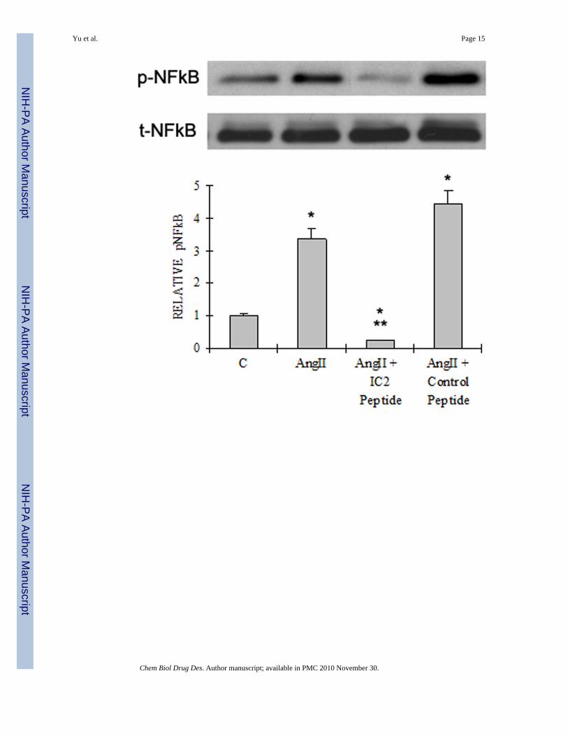
Phosphorylation of the p65 subunit of nuclear factor κB (NFκB) is an event required formaximal transcriptional activity of NFκB (14). As illustrated, AngII clearly enhanced theSer536 phosphorylation of p65 (Fig. 5B). This phosphorylation was inhibited approximately93% by the AT1 IC2 peptide at 10 μM, whereas the control peptide (10 μM) has noinhibitory effect.
With regard to the activation of ERK1/2, we previously showed that alternation within theIC2 of AT1 receptor had no effect on AngII activated ERK phosphorylation (10). Consistentwith this observation, neither the control peptide nor the IC2 peptide at 10 μM affected theactivation of this MAP kinase by AngII (Fig. 5C).
DiscussionAngiotensin II through its AT1 receptor participates in the etiology of a number of diseases,such as hypertension and fibrotic diseases. Although its overall actions can be regulated bypreventing its formation with ACE inhibitors or blocking its overall action with AT1receptor blockers, the mechanisms of it actions are essentially unknown. A very proficientway to study the signaling by the AT1 receptor is through its signal motif manipulation bymutagenesis. This approach provides information as to the actions and interactions of themotifs but does not provide an opportunity to regulate the action of the wild type receptor.This communication is an initial attempt to make use of cell penetrating peptides to bothdelineate and regulate the action(s) of the AT1 receptor.
In previous studies we demonstrated that AngII, through the AT1 receptor, increases Aktphosphorylation in AT1 cDNA tranfected HEK-293 cells while bradykinin decreases it inbradykinin B2 receptor cDNA transfected cells (10). In HEK-293 cells transfected with amutant AT1 receptor containing the IC2 of the BKB2R receptor AngII decreased pAkt (10)thus assigning regulation of Akt phosphorylation. In contrast, these receptor manipulationsat the level of the IC2 of the AT1R had no effect on the activation of ERK by the mutantreceptor as compared to the WT AT1 receptor. Thus the IC2 of the AT1R is an importantsignaling motif with respect to both G-protein associated signaling as exemplified by PIturnover and Akt activation, whereas IC2 seems not involved in ERK activation. It has beenshown that AngII activates ERK by a Gαq-independent action (15). Studies using carboxylterminal-truncated AT1 receptors indicate that the amino acid sequence between 312 and
Yu et al. Page 5
Chem Biol Drug Des. Author manuscript; available in PMC 2010 November 30.
NIH
-PA Author Manuscript
NIH
-PA Author Manuscript
NIH
-PA Author Manuscript
337 is required for the activation of ERK. The YIPP motif within this sequence appears tobe the important motif in this action (16). Mutations in the highly conserved D125R126Y127
sequence of the AT1R result in a receptor that is not coupled to Gαq, but still activates ERK(17).
In the past few years there has been a progressive development of membrane-penetratingpeptides which has allowed for the delivery of otherwise impermeable bioactive moleculesacross the plasma membrane. Numerous studies using 10–100μM CPP concentrations havedemonstrated that once accumulated in specific compartments (i.e., the cytosol, nucleus andmitochondria) the specific cargo can regulate targeted cellular activities through protein-protein interactions within the intact cells (18). The results obtained here utilizing the AT1-IC2/HIV TAT cell penetrating peptide confirmed our previous observations and illustratedthat the introduction of competing peptide into the cytosol mimicking a receptor motif (IC2)can indeed alter receptor signaling ability. In predominant number of reports 10–100 μMCPP concentrations have been used to demonstrate the effect of these peptides. We chosethe lowest concentration of this span in order to limit as much as possible any non specificeffects of TAT. TAT was shown to rapidly transduce large cargos such as CRE recombinaseinto primary cells (19). Thus the size of the cargo does not limit the cell penetrating abilityof TAT. As shown by the 5-FAM/FACS experiment carried out as part of these studies theTAT peptide clearly crossed into the cytosol of both the HEK-293 and the pulmonarysmooth muscle cells. The AT1 IC2 peptide effectively inhibited Gαq related signaling suchas PI turnover and Ca2+ influx in HEK-293 cells. In addition, it limited the AngII dependentactivation of Akt/PKB and NFκB, but had no effect on ERK activation in HPASMCs. Weused the TAT-linked second intracellular loop of the bradykinin B2 receptor as the controlpeptide. This peptide had no effect on AngII induced signaling actions. Thompson et alshowed that cotransfecting second and third loops inhibit AT1 receptor activation of PLC inHEK-293 cells (20). Our peptide approach fits their results. Vazquez et al reported that thesynthesized third intracellular loop of the AT1 receptor linked to HIV-TAT was able to betransduced into the hypothalamus and brainstem neurons and increased neuronal firing rate,an effect similar to that observed with angiotensin II stimulation of the neuronal AT1R (21).A more recent study demonstrated that a peptide based on the C-terminus of the AT1receptor with native cell-penetrating sequences elicits G-protein mediated blood vesselcontraction (22), though our IC2 peptide seems evoking an inhibitory effect. Therefore, theactivating or inhibiting effects of the intracellular motifs in GPCRs are subtle andcomplicated. Further structural studies such as molecular modeling or NMR could elucidatethe accurate mechanisms of those effects including possible “off target” effects of thepeptides.
In summary, our results with cell/tissue penetrating peptides are very encouraging, and areleading us to anticipate that these peptides will contribute to the control of receptor initiatedsignaling and blood vessel function.
AcknowledgmentsThis work was supported in part by NIH, NHLBI Grant HL25776.
The smooth muscle cells isolated from the pulmonary arteries obtained from human lung transplants were agenerous gift from Dr. Serpil Erzurum (Cleveland Clinic, Cleveland, OH).
References1. Frederic H, May Catherine M, Gilles D. Twenty years of cell-penetrating peptides: from molecular
mechanisms to therapeutics. British Journal of Pharmacology 2009;157(2):195–206. [PubMed:19309362]
Yu et al. Page 6
Chem Biol Drug Des. Author manuscript; available in PMC 2010 November 30.
NIH
-PA Author Manuscript
NIH
-PA Author Manuscript
NIH
-PA Author Manuscript

2. Schwarze SR, Hruska KA, Dowdy SF. Protein transduction: unrestricted delivery into all cells?Trends in Cell Biology 2000;10(7):290. [PubMed: 10856932]
3. Murriel CL, Dowdy SF. Influence of protein transduction domains on intracellular delivery ofmacromolecules. Expert Opinion on Drug Delivery 2006;3(6):739–46. [PubMed: 17076596]
4. Jasmin J-F, Mercier I, Dupuis J, Tanowitz HB, Lisanti MP. Short-Term Administration of a Cell-Permeable Caveolin-1 Peptide Prevents the Development of Monocrotaline-Induced PulmonaryHypertension and Right Ventricular Hypertrophy. Circulation 2006;114(9):912–20. [PubMed:16940204]
5. Zhao K, Luo G, Zhao G-M, Schiller PW, Szeto HH. Transcellular Transport of a Highly Polar 3+Net Charge Opioid Tetrapeptide. J Pharmacol Exp Ther 2003;304(1):425–32. [PubMed: 12490619]
6. Famous KR, Kumaresan V, Sadri-Vakili G, Schmidt HD, Mierke DF, Cha J-HJ, et al.Phosphorylation-Dependent Trafficking of GluR2-Containing AMPA Receptors in the NucleusAccumbens Plays a Critical Role in the Reinstatement of Cocaine Seeking. J Neurosci 2008;28(43):11061–70. [PubMed: 18945913]
7. Larkin JE, Frank BC, Gaspard RM, Duka I, Gavras H, Quackenbush J. Cardiac transcriptionalresponse to acute and chronic angiotensin II treatments. Physiol Genomics 2004;18(2):152–66.[PubMed: 15126644]
8. Duprez DA. Role of the renin-angiotensin-aldosterone system in vascular remodeling andinflammation: a clinical review. J Hypertens 2006;24(6):983–91. [PubMed: 16685192]
9. Mehta PK, Griendling KK. Angiotensin II cell signaling: physiological and pathological effects inthe cardiovascular system. Am J Physiol Cell Physiol 2007;292(1):C82–97. [PubMed: 16870827]
10. Yu J, Lubinsky D, Tsomaia N, Huang Z, Taylor L, Mierke D, et al. Activation of ERK, JNK, Akt,and G-protein coupled signaling by hybrid angiotensin II AT1/bradykinin B2 receptors expressedin HEK-293 cells. J Cell Biochem 2007;101(1):192–204. [PubMed: 17212359]
11. Mann DA, Frankel AD. Endocytosis and targeting of exogenous HIV-1 Tat protein. EMBO J1991;10(7):1733–9. [PubMed: 2050110]
12. Prado GN, Taylor L, Polgar P. Effects of intracellular tyrosine residue mutation and carboxylterminus truncation on signal transduction and internalization of the rat bradykinin B2 receptor. JBiol Chem 1997;272(23):14638–42. [PubMed: 9169425]
13. Prado GN, Mierke DF, Pellegrini M, Taylor L, Polgar P. Motif mutation of bradykinin B2 receptorsecond intracellular loop and proximal C terminus is critical for signal transduction,internalization, and resensitization. J Biol Chem 1998;273(50):33548–55. [PubMed: 9837936]
14. Strassheim D, Asehnoune K, Park J-S, Kim J-Y, He Q, Richter D, et al. Phosphoinositide 3-Kinaseand Akt Occupy Central Roles in Inflammatory Responses of Toll-Like Receptor 2-StimulatedNeutrophils. J Immunol 2004;172(9):5727–33. [PubMed: 15100319]
15. Miura, S-i; Zhang, J.; Matsuo, Y.; Saku, KS.; Karnik, S. Activation of Extracellular Signal-Activated Kinase by Angiotensin II-Induced Gq-Independent Epidermal Growth Factor ReceptorTransactivation. Hypertension Research 2004;27(10):765–70. [PubMed: 15785012]
16. Seta K, Sadoshima J. Phosphorylation of tyrosine 319 of the angiotensin II type 1 receptormediates angiotensin II-induced trans-activation of the epidermal growth factor receptor. J BiolChem 2003;278(11):9019–26. [PubMed: 12522132]
17. Seta K, Nanamori M, Modrall JG, Neubig RR, Sadoshima J. AT1 receptor mutant lackingheterotrimeric G protein coupling activates the Src-Ras-ERK pathway without nucleartranslocation of ERKs. Journal of Biological Chemistry 2002;277(11):9268–77. [PubMed:11777928]
18. Costa-Junior HM, Suetsugu MJ, Krieger JE, Schechtman D. Specific modulation of protein kinaseactivity via small peptides. Regulatory Peptides 2009;153(1–3):11–8. [PubMed: 19135095]
19. Peitz M, Pfannkuche K, Rajewsky K, Edenhofer F. Ability of the hydrophobic FGF and basic TATpeptides to promote cellular uptake of recombinant Cre recombinase: A tool for efficient geneticengineering of mammalian genomes. Proceedings of the National Academy of Sciences of theUnited States of America 2002;99(7):4489–94. [PubMed: 11904364]
20. Thompson JB, Wade SM, Harrison JK, Salafranca MN, Neubig RR. Cotransfection of Second andThird Intracellular Loop Fragments Inhibit Angiotensin AT1a Receptor Activation of
Yu et al. Page 7
Chem Biol Drug Des. Author manuscript; available in PMC 2010 November 30.
NIH
-PA Author Manuscript
NIH
-PA Author Manuscript
NIH
-PA Author Manuscript

Phospholipase C in HEK-293 Cells. Journal of Pharmacology and Experimental Therapeutics1998;285(1):216–22. [PubMed: 9536013]
21. Vazquez J, Sun C, Du J, Fuentes L, Sumners C, Raizada MK. Transduction of a FunctionalDomain of the AT1 Receptor in Neurons by HIV-Tat PTD. Hypertension 2003;41(3):751–6.[PubMed: 12623991]
22. Östlund P, Kilk K, Lindgren M, Hällbrink M, Jiang Y, Budihna M, et al. Cell-Penetrating Mimicsof Agonist-Activated G-Protein Coupled Receptors. International Journal of Peptide Research andTherapeutics 2005;11(4):237.
Yu et al. Page 8
Chem Biol Drug Des. Author manuscript; available in PMC 2010 November 30.
NIH
-PA Author Manuscript
NIH
-PA Author Manuscript
NIH
-PA Author Manuscript
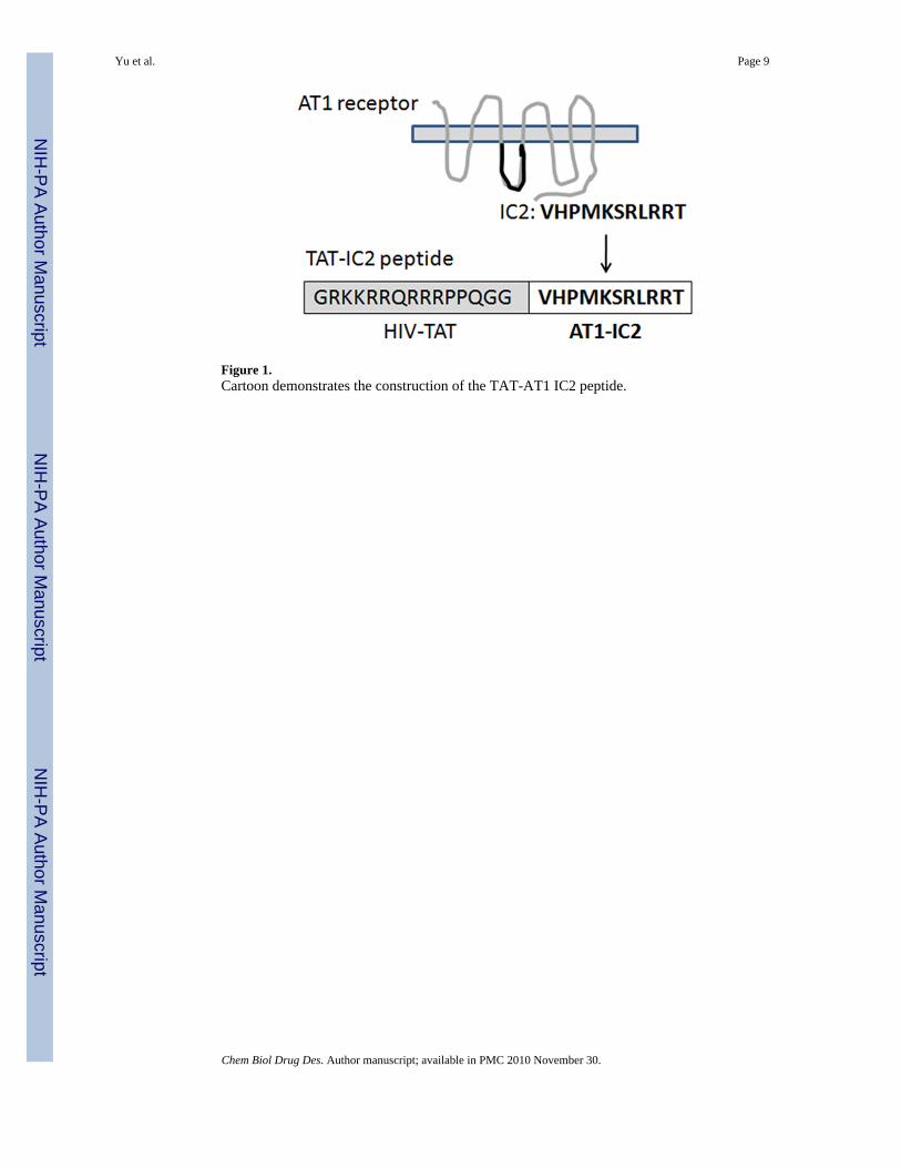
Figure 1.Cartoon demonstrates the construction of the TAT-AT1 IC2 peptide.
Yu et al. Page 9
Chem Biol Drug Des. Author manuscript; available in PMC 2010 November 30.
NIH
-PA Author Manuscript
NIH
-PA Author Manuscript
NIH
-PA Author Manuscript
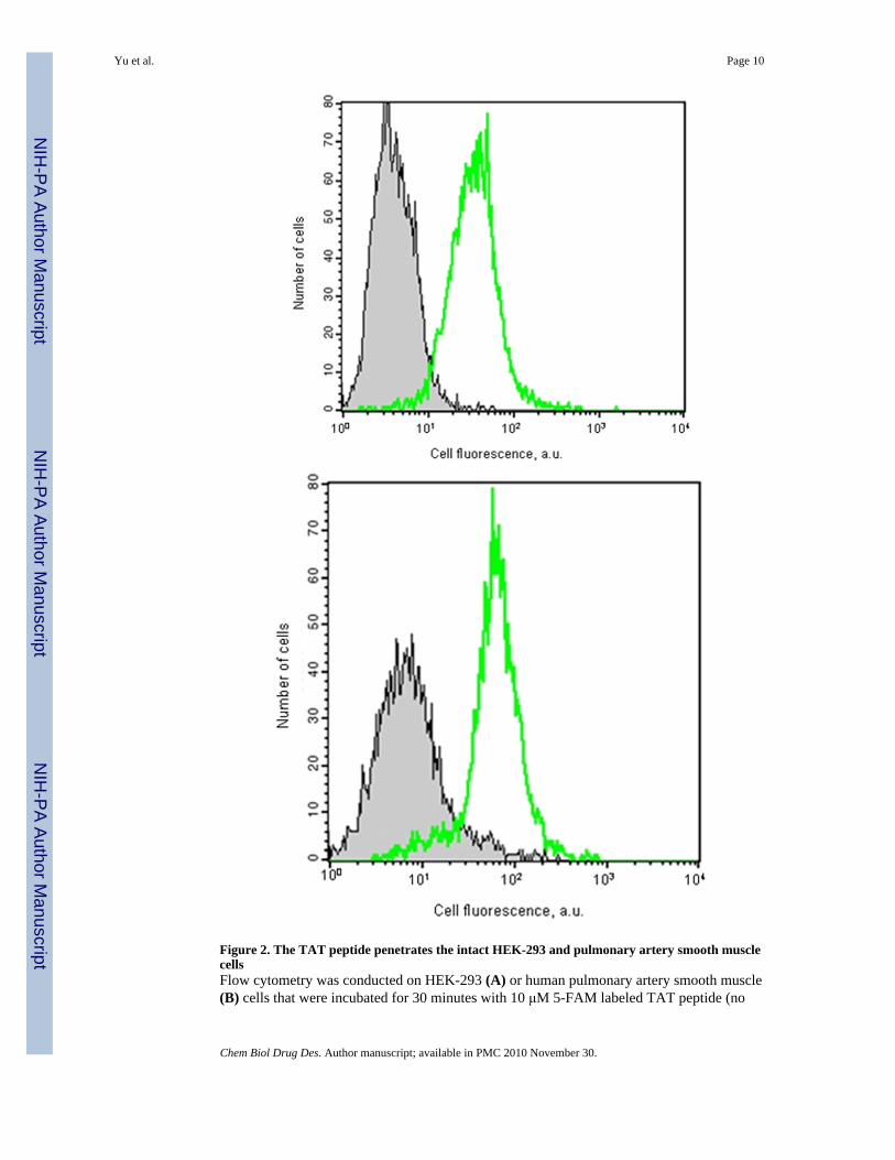
Figure 2. The TAT peptide penetrates the intact HEK-293 and pulmonary artery smooth musclecellsFlow cytometry was conducted on HEK-293 (A) or human pulmonary artery smooth muscle(B) cells that were incubated for 30 minutes with 10 μM 5-FAM labeled TAT peptide (no
Yu et al. Page 10
Chem Biol Drug Des. Author manuscript; available in PMC 2010 November 30.
NIH
-PA Author Manuscript
NIH
-PA Author Manuscript
NIH
-PA Author Manuscript

filling under the curve) or peptide free medium (grey filling under the curve). Cells werethen trypsinized and resuspended in PBS as a single cell suspension before flow cytometry.
Yu et al. Page 11
Chem Biol Drug Des. Author manuscript; available in PMC 2010 November 30.
NIH
-PA Author Manuscript
NIH
-PA Author Manuscript
NIH
-PA Author Manuscript
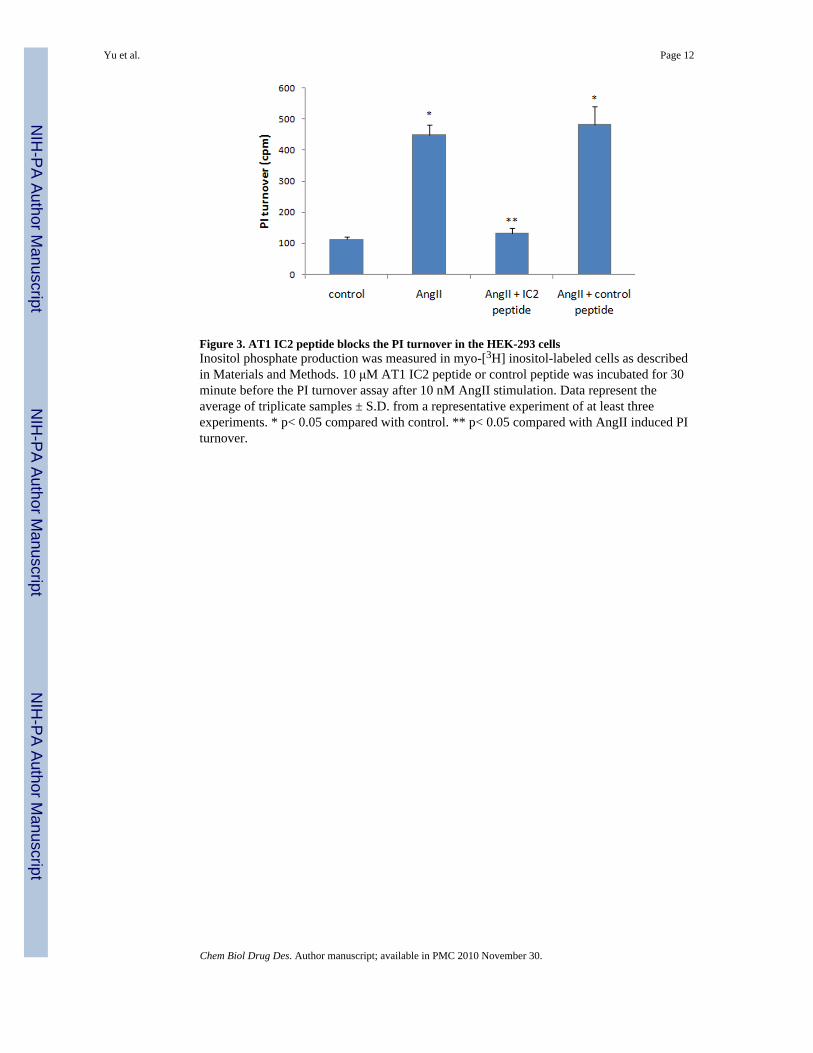
Figure 3. AT1 IC2 peptide blocks the PI turnover in the HEK-293 cellsInositol phosphate production was measured in myo-[3H] inositol-labeled cells as describedin Materials and Methods. 10 μM AT1 IC2 peptide or control peptide was incubated for 30minute before the PI turnover assay after 10 nM AngII stimulation. Data represent theaverage of triplicate samples ± S.D. from a representative experiment of at least threeexperiments. * p< 0.05 compared with control. ** p< 0.05 compared with AngII induced PIturnover.
Yu et al. Page 12
Chem Biol Drug Des. Author manuscript; available in PMC 2010 November 30.
NIH
-PA Author Manuscript
NIH
-PA Author Manuscript
NIH
-PA Author Manuscript
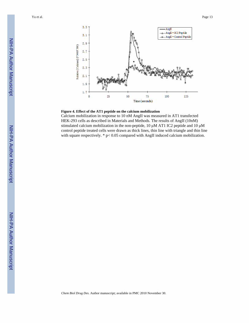
Figure 4. Effect of the AT1 peptide on the calcium mobilizationCalcium mobilization in response to 10 nM AngII was measured in AT1 transfectedHEK-293 cells as described in Materials and Methods. The results of AngII (10nM)stimulated calcium mobilization in the non-peptide, 10 μM AT1 IC2 peptide and 10 μMcontrol peptide treated cells were drawn as thick lines, thin line with triangle and thin linewith square respectively. * p< 0.05 compared with AngII induced calcium mobilization.
Yu et al. Page 13
Chem Biol Drug Des. Author manuscript; available in PMC 2010 November 30.
NIH
-PA Author Manuscript
NIH
-PA Author Manuscript
NIH
-PA Author Manuscript

Yu et al. Page 14
Chem Biol Drug Des. Author manuscript; available in PMC 2010 November 30.
NIH
-PA Author Manuscript
NIH
-PA Author Manuscript
NIH
-PA Author Manuscript
Yu et al. Page 15
Chem Biol Drug Des. Author manuscript; available in PMC 2010 November 30.
NIH
-PA Author Manuscript
NIH
-PA Author Manuscript
NIH
-PA Author Manuscript
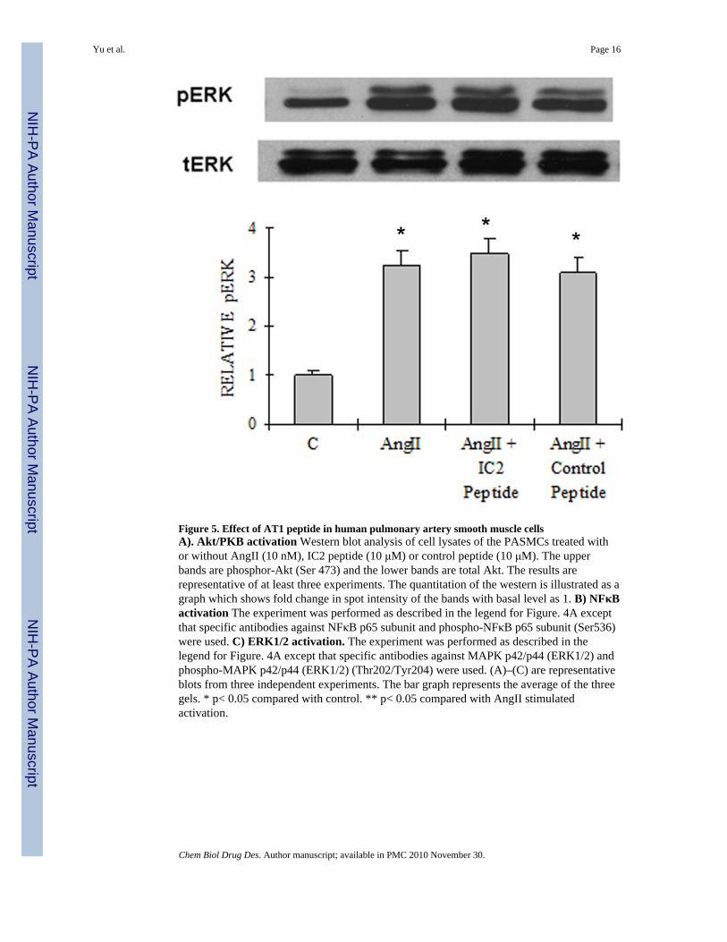
Figure 5. Effect of AT1 peptide in human pulmonary artery smooth muscle cellsA). Akt/PKB activation Western blot analysis of cell lysates of the PASMCs treated withor without AngII (10 nM), IC2 peptide (10 μM) or control peptide (10 μM). The upperbands are phosphor-Akt (Ser 473) and the lower bands are total Akt. The results arerepresentative of at least three experiments. The quantitation of the western is illustrated as agraph which shows fold change in spot intensity of the bands with basal level as 1. B) NFκBactivation The experiment was performed as described in the legend for Figure. 4A exceptthat specific antibodies against NFκB p65 subunit and phospho-NFκB p65 subunit (Ser536)were used. C) ERK1/2 activation. The experiment was performed as described in thelegend for Figure. 4A except that specific antibodies against MAPK p42/p44 (ERK1/2) andphospho-MAPK p42/p44 (ERK1/2) (Thr202/Tyr204) were used. (A)–(C) are representativeblots from three independent experiments. The bar graph represents the average of the threegels. * p< 0.05 compared with control. ** p< 0.05 compared with AngII stimulatedactivation.
Yu et al. Page 16
Chem Biol Drug Des. Author manuscript; available in PMC 2010 November 30.
NIH
-PA Author Manuscript
NIH
-PA Author Manuscript
NIH
-PA Author Manuscript