cardiovascular effects of angiotensin a: a novel peptide of the renin-angiotensin system
TRANSCRIPT

Hindawi Publishing CorporationInternational Journal of HypertensionVolume 2012, Article ID 147825, 13 pagesdoi:10.1155/2012/147825
Review Article
New Cardiovascular and Pulmonary TherapeuticStrategies Based on the Angiotensin-Converting Enzyme2/Angiotensin-(1–7)/Mas Receptor Axis
Anderson J. Ferreira,1 Tatiane M. Murca,1 Rodrigo A. Fraga-Silva,2
Carlos Henrique Castro,3 Mohan K. Raizada,4 and Robson A. S. Santos2
1 Department of Morphology, Institute of Biological Sciences, Federal University of Minas Gerais, 31.270-901 Belo Horizonte,MG, Brazil
2 Department of Physiology and Biophysics, Institute of Biological Sciences, Federal University of Minas Gerais,31.270-901 Belo Horizonte, MG, Brazil
3 Department of Physiology Sciences, Federal University of Goias, 74.001-970 Goiania, GO, Brazil4 Department of Physiology and Functional Genomics, College of Medicine, University of Florida, 32.610 Gainesville, FL, USA
Correspondence should be addressed to Anderson J. Ferreira, [email protected]
Received 31 August 2011; Accepted 12 October 2011
Academic Editor: Roberto Pontremoli
Copyright © 2012 Anderson J. Ferreira et al. This is an open access article distributed under the Creative Commons AttributionLicense, which permits unrestricted use, distribution, and reproduction in any medium, provided the original work is properlycited.
Angiotensin (Ang)-(1–7) is now recognized as a biologically active component of the renin-angiotensin system (RAS). Thediscovery of the angiotensin-converting enzyme homologue ACE2 revealed important metabolic pathways involved in the Ang-(1–7) synthesis. This enzyme can form Ang-(1–7) from Ang II or less efficiently through hydrolysis of Ang I to Ang-(1–9) withsubsequent Ang-(1–7) formation. Additionally, it is well established that the G protein-coupled receptor Mas is a functional ligandsite for Ang-(1–7). The axis formed by ACE2/Ang-(1–7)/Mas represents an endogenous counter regulatory pathway within theRAS whose actions are opposite to the vasoconstrictor/proliferative arm of the RAS constituted by ACE/Ang II/AT1 receptor. Inthis review we will discuss recent findings concerning the biological role of the ACE2/Ang-(1–7)/Mas arm in the cardiovascularand pulmonary system. Also, we will highlight the initiatives to develop potential therapeutic strategies based on this axis.
1. Introduction
The renin-angiotensin system (RAS) plays a key role inseveral target organs, such as heart, blood vessels, and lungs,exerting a powerful control in the maintenance of the home-ostasis [1–4]. This system is activated by the conversion of theangiotensinogen to the inactive peptide angiotensin (Ang) Ithrough the renin action [5]. Subsequently, Ang I is cleavedby the angiotensin-converting enzyme (ACE) generatingAng II [6], the main angiotensin peptide, whose actionsare mediated by two G protein-coupled receptors (GPCR),AT1 and AT2 [7, 8] (Figure 1). The major physiologicalfunctions of Ang II are mediated by AT1 receptor [9, 10]. Inpathological conditions, activation of this receptor inducesdeleterious effects, such as vasoconstriction, fibrosis, cellular
growth and migration, and fluid retention [11, 12]. On theother hand, Ang II binding to the AT2 receptor generallycauses opposite effects when compared with those actionsmediated by the AT1 receptor [13, 14].
Recently, it has been proposed that, in addition to theACE/Ang II/AT1 receptor axis, the RAS possesses a counterregulatory axis composed by ACE2, Ang-(1–7), and Masreceptor (Figure 1). Ang-(1–7) is a biologically active com-ponent of the RAS which binds to Mas inducing manybeneficial actions, such as vasodilatation, antifibrosis, andantihypertrophic and antiproliferative effects [15–23]. Thispeptide is produced mainly through the action of ACE2,which has approximately 400-fold less affinity to Ang I thanto Ang II [24–26]; thereby, Ang II is the major substratefor Ang-(1–7) synthesis. In fact, the conversion of Ang II to

2 International Journal of Hypertension
Angiotensinogen
Asp-Arg-Val-Tyr-Ile-His-Pro-Phe-His-Leu-Val-Ile-. . .
Angiotensin I
Asp-Arg-Val-Tyr-Ile-His-Pro-Phe-His-Leu
Renin
ACE2
Angiotensin-(1–9)Asp-Arg-Val-Tyr-Ile-His-Pro-Phe-His
ACE, NEPPEP, NEP
ACE
Angiotensin-(1–5)
Asp-Arg-Val-Tyr-Ile
Tonin, Catepsin G
Catepsin A, Chymase
Angiotensin II
Asp-Arg-Val-Tyr-Ile-His-Pro-Phe
Angiotensin-(1–7)
Asp-Arg-Val-Tyr-Ile-His-Pro
Prorenin
Inactive kinin
Bradykinin
Arg-Pro-Pro-Gly-Phe-Ser-Pro-Phe-Arg
ACE
Proliferation
Hypertrothy
Fibrosis
Infammation
Endothelial dysfunction
Oxidative stress
Apoptosis
AT1 AT2 Mas
ACE2
ACE
ffCounterregulatory e ects
Figure 1: Schematic representation of the renin-angiotensin system (RAS) cascade. The counterregulatory axes of the RAS are composed byACE/Ang II/AT1 and ACE2/Ang-(1–7)/Mas. ACE: angiotensin-converting enzyme; Ang: angiotensin; AT1: Ang II type 1 receptor; AT2: AngII type 2 receptor; Mas: Ang-(1–7) receptor; PCP: prolylcarboxypeptidase; PEP: prolyl-endopeptidase; NEP: neutral-endopeptidase 24.11.
Ang-(1–7) by ACE2 is important to regulate the RAS activitysince Ang-(1–7) induces opposite effects to those elicited byAng II [16–24]. Additionally, ACE2 can form Ang-(1–7) lessefficiently through hydrolysis of Ang I to Ang-(1–9) withsubsequent Ang-(1–7) formation [24].
The relevance of the RAS is highlighted by the successobtained in therapeutic strategies based on the pharma-cological inhibition of this system in cardiovascular andrespiratory diseases [27–32]. Blockade of the RAS withACE inhibitors (ACEi) or AT1 receptor antagonists (ARBs)improves the outcomes of patients with hypertension, acutemyocardial infarction, and chronic systolic heart failure [33–35]. Furthermore, based on the involvement of the ACE/AngII/AT1 axis in respiratory diseases and the crucial role of thelungs in the RAS metabolism, several studies have reportedthe contribution of the RAS in lung pathophysiology [28, 30,31, 36–40]. Importantly, it has been shown that administra-tion of ACEi and ARBs causes substantial increases in plasmaAng-(1–7) levels, leading to the assumption that part of their
clinical effects might be mediated by this heptapeptide [41–43]. Indeed, some effects of ACEi and ARBs can be blockedor attenuated by A-779, a Mas antagonist, confirming therole of Ang-(1–7) in the actions of these compounds [44].The beneficial effects of Ang-(1–7), as well as its likelyparticipation in the effects of the ACEi and ARBs, representevidences for the potential of the ACE2/Ang-(1-7)/Mas axisas a therapeutic target.
In this review, we will focus on the recent findings relatedto the pathophysiology actions of the ACE2/Ang-(1–7)/Masaxis in the cardiovascular and respiratory system. Also,we will discuss the promising initiatives to develop newtherapeutic strategies based on this axis to treat pathologicalconditions.
2. Cardiac ACE2/Ang-(1–7)/Mas Axis
The heart is one of the most important targets for theactions of the ACE2/Ang-(1–7)/Mas axis. In the heart, ACE2

International Journal of Hypertension 3
is expressed in the endothelium [45], myofibroblasts [46],cardiomyocytes, and fibroblasts [47, 48]. Classical pharma-cotherapeutic agents used to treat heart failure, includingACEi, ARBs, and aldosterone receptor blockers, increaseACE2 activity and/or expression, indicating its importance inthe cardiac diseases establishment and progression [49–51].
Additionally, pharmacological and genetic (transgenicanimals and gene transfer) approaches have evidenced thesignificance of ACE2 in cardiac pathologies. Despite somecontroversies concerning the consequences of the ACE2deficiency, in general, evidences indicate a protective role ofACE2 in the heart [48, 52–57]. Crackower and colleagues[52] were the first to demonstrate that genetic ablation ofACE2 results in severe blood-pressure-independent systolicimpairment. Also, disruption of ACE2 was able to acceleratecardiac hypertrophy and shortened the transition period toheart failure in response to pressure overload by increasinglocal Ang II [54]. Recently, it has been demonstrated that lossof ACE2 enhances the susceptibility to myocardial infarction,with increased mortality, infarct expansion and adverseventricular remodeling [56]. In keeping with these geneticfindings, pharmacological inhibition of ACE2 exacerbatedcardiac hypertrophy and fibrosis in Ren-2 hypertensiverats [58]. On the other hand, cardiac overexpression ofACE2 prevented hypertension-induced cardiac hypertrophyand fibrosis in spontaneously hypertensive rats (SHR) andin Ang-II-infused rats [59, 60]. Indeed, transfection ofLenti-ACE2 (lentivirus containing ACE2 cDNA) or Ad-ACE2 (recombinant adenovirus carrying the murine ACE2)into the surrounding area of the infarcted myocardiumwas protective against pathological remodeling and cardiacsystolic dysfunction in a rat model of myocardial infarction[61, 62]. This effect was associated with decreased expressionof ACE and Ang II and increased expression of Ang-(1–7) [62]. Collectively, these observations reveal that ACE2effectively plays a protective role in the cardiac structure andfunction.
Since the discovery of Ang-(1–7) in the late 1980s [63,64], several studies have demonstrated important effectsof this peptide in hearts. The presence of Ang-(1–7) andits receptor Mas in the heart [65, 66] and the ability ofthis organ to produce Ang-(1–7) [55, 67] are evidencesof the role of this peptide in cardiac tissues. Functionally,Ang-(1–7) induces an antiarrhythmogenic effect againstischemia/reperfusion injuries in rats [17, 68] as well asprevents atrial tachycardia and fibrillation in rats and dogs[69, 70]. Treatment with Ang-(1–7) improved the coronaryperfusion and cardiac function in rats after myocardialinfarction [71] and after ischemia/reperfusion injury [72].Increases in circulating Ang-(1–7) levels in transgenic ratsreduced the cardiac hypertrophy [17] and fibrosis [20,22] induced by isoproterenol administration. These effectsare apparently independent of changes in blood pressuresince Grobe and colleagues [18] have demonstrated thatthe antifibrotic and antihypertrophic actions of Ang-(1–7) are still observed in Ang-II-infused hypertensive rats.Local overexpression of Ang-(1–7) in hearts of mice andrats improved the myocardial contractility and prevented the
isoproterenol- and hypertension-induced cardiac remodel-ing [19, 21]. Altogether, these findings support a direct effectof Ang-(1-7) in the heart.
Further evidence for the role of Ang-(1–7)/Mas in thepathophysiology of the heart came from experimental pro-tocols utilizing mice with genetic deficiency of Mas. Theyrevealed that the cardiac function is impaired in Mas knock-out mice likely due to the increased extracellular matrixproteins deposition in the heart [66, 73]. This profibroticphenotype may be related to changes in matrix metallopro-teinases (MMPs) and tissue inhibitors of metalloproteinases(TIMPs) levels and/or activities [74, 75].
Although further elucidations regarding the signalingpathways involved in Mas activation are necessary, somemechanisms have been proposed. Overexpression of Ang-(1–7) in hearts of rats causes an improvement in the [Ca2+]handling in cardiomyocytes and increases the expression ofSERCA2a [21]. In keeping with these results, cardiomyocytesfrom Mas-deficient mice present slower [Ca2+]i transientsaccompanied by a lower Ca2+ ATPase expression in thesarcoplasmic reticulum [66, 76]. Although acute Ang-(1–7) treatment failed to alter Ca2+ handling in ventricularmyocytes of rats [76], these findings suggest an importantrole of the Ang-(1–7)/Mas in the long-term maintenance ofthe Ca2+ homeostasis in the heart.
One of the mechanisms by which Ang-(1–7) plays itseffects in the heart is stimulating the nitric oxide (NO) pro-duction. Indeed, it has been demonstrated that Ang-(1–7)via Mas increases the synthesis of NO through a mechanisminvolving the activation of the endothelial NO synthase(eNOS). These effects were abolished by A-779 and areabsent in cardiomyocytes from Mas-deficient mice [76].Recently, Gomes et al. [77] found that the treatment ofisolated cardiomyocytes of rats with Ang-(1–7) efficientlyprevents the Ang-II-induced hypertrophy by modulatingthe calcineurin/NFAT signaling cascade. These effects wereblocked by NO synthase inhibition and by guanylyl cyclaseinhibitors, indicating that these effects are mediated by theNO/cGMP pathway.
Also, Ang-(1–7) inhibits serum-stimulated mitogen-ac-tivated protein kinase (MAPK) activation in cardiac my-ocytes [78] and prevents the Ang-II-mediated phospho-rylation of ERK1/2 and Rho kinase in hearts in a dose-dependent manner [79]. In line with these data, activation ofendogenous ACE2 significantly reduced the phosphorylationof ERK1/2 in hearts of hypertensive rats (SHRs) [48].However, Mercure et al. [19] reported that overexpressionof Ang-(1–7) in hearts of rats decreases the Ang-II-inducedphosphorylation of c-Src and p38 kinase, whereas theincrease in ERK1/2 phosphorylation was unaffected by theexpression of the transgene, thereby suggesting a selectiveeffect of Ang-(1–7) on intracellular signaling pathwaysrelated to cardiac remodeling.
Overall, these data reveal a key role of the ACE2/Ang-(1–7)/Mas axis in the pathophysiology of the cardiac structureand function. Activation of this axis might be an importantstrategy to develop a new generation of cardiovascular ther-apeutic agents against cardiac dysfunction and pathologicalremodeling of the heart.

4 International Journal of Hypertension
3. Vascular ACE2/Ang-(1–7)/Mas Axis
Early studies have reported the endothelium as the majorsite for generation [67] and metabolism [41] of Ang-(1–7). In addition to Ang-(1–7), endothelial cells also expressACE2 and Mas [80, 81]. Thus, now it is recognized that theACE2/Ang-(1–7)/Mas axis is present in vascular endothelialcells and modulates its function promoting vasorelaxation[82], reduction of the oxidative stress [83, 84], and antipro-liferative effects [85, 86].
The vasodilatory actions of Ang-(1–7) have been re-ported in many studies in several vascular beds and prepa-rations, including mouse [16, 23] and rat [15] aortic rings,canine [87] and porcine [88] coronary arteries, caninemiddle cerebral artery [89], porcine piglet pial arterioles[90], feline mesenteric vascular bed [91], rabbit renal afferentarterioles [92], and mesenteric microvessels of normotensive[93] and hypertensive [94] rats. Vascular Ang-(1–7) actionsare still controversial in human. For example, it has beenshown that Ang-(1–7) causes vasodilation in forearm circu-lation of normotensive subjects and patients with essentialhypertension [95] while other studies were unable to reportany significant effect of Ang-(1–7) in the same vascularterritory in ACEi-treated patients [43].
The Mas receptor is critically involved in the vasculareffects of Ang-(1–7). In fact, many of these actions are com-pletely abolished by A-779 or partially blocked by this antag-onist [3, 86, 96]. Importantly, the endothelium-dependentrelaxation induced by Ang-(1–7) in mouse aortic rings isabsent in vessels derived from Mas-knockout mice [16].However, other studies have shown that Ang-(1–7) alsointeracts with ACE, AT1, and AT2-like receptors, suggestingthe existence of additional sites of interaction for Ang-(1–7)[3, 97, 98]. Indeed, Silva et al. [99] reported evidence for thepresence of a distinct subtype of Ang-(1–7) receptor sensibleto D-pro7-Ang-(1–7), a second Mas antagonist, but not toA-779 in aortas of Sprague-Dawley rats.
The vascular effects of Ang-(1–7) are endotheliumdependent and involve the production of vasodilator prod-ucts, such as prostanoids, NO, and endothelium-derivedhyperpolarizing factor (EDHF) [16, 81, 100]. Pinheiroand coworkers [101] found that Ang-(1–7) promotes anincrease in NO release in Mas-transfected chinese hamsterovary (CHO) cells [101]. Furthermore, short-term infu-sion of Ang-(1–7) improved the endothelial function bya mechanism involving NO release in rats [102]. Masdeletion resulted in endothelial dysfunction associated withan unbalance between NO and oxidative stress [83]. Also,Mas activation by Ang-(1–7) in human endothelial cellsstimulated eNOS phosphorylation/activation via the Akt-dependent pathway [81]. Other mechanisms appear to beinvolved in the Ang-(1–7) vascular actions. Roks et al. [103]have shown that Ang-(1–7) inhibits the vasoconstrictioninduced by Ang II in human internal mammary arteries,thereby suggesting that Ang-(1–7) can regulate the Ang IIeffects [103]. In fact, Ang-(1–7) negatively modulates theAng II type 1 receptor-mediated activation of c-Src, and itsdownstream targets ERK1/2 and NAD(P)H oxidase [104].The counterregulatory action of Ang-(1–7) on Ang II
signaling has been also observed in cardiomyocytes [77],vascular smooth muscle cells [105], and fibroblasts [106].Additionally, an interaction between Mas and bradykinin(Bk) type 2 (B2) receptors may modulate some of the Ang-(1–7) effects in blood vessels [107]. Indeed, it has beendemonstrated that Ang-(1–7) potentiates the vasodilator andhypotensive effects of Bk in several vascular beds [93, 108–110].
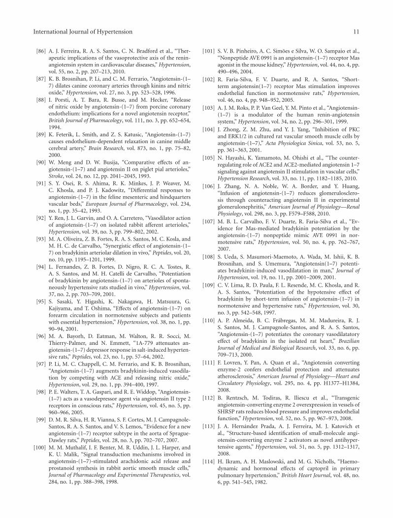
As the major enzyme involved in Ang-(1–7) formation,ACE2 has also a crucial role in vessels. Lovren et al. [111]have demonstrated that ACE2 ameliorates the endothelialhomeostasis via a mechanism involving reduction of thereactive oxygen species production [111]. Of note, this effectwas attenuated by A-779 [111]. Moreover, overexpression ofACE2 in vessels of hypertensive rats resulted in reductionin the arterial blood pressure and improvement of theendothelial function associated with increased circulatingAng-(1–7) levels [112]. Overall, these data indicate that thebeneficial effects of ACE2 are, at least in part, mediated byAng-(1–7). Recently, we have demonstrated that activationof endogenous ACE2 causes a dose-dependent hypotensiveeffect in normotensive and hypertensive rats [113]. Also,the response to Bk administration was augmented in ratschronically treated with XNT, an ACE2 activator [113]. How-ever, we were unable to demonstrate any significant effect ofXNT on blood pressure in response to the administrationof Ang II or Losartan in normotensive and hypertensive rats(Figure 2).
4. Pulmonary ACE2/Ang-(1–7)/Mas Axis
In the past few years, the participation of the ACE2/Ang-(1–7)/Mas axis in the establishment and progression of pul-monary diseases has become evident. Indeed, the importantrole of the RAS in the lung pathophysiology and the sideeffects and pulmonary toxicity induced by the ACEi raisedthe interest to evaluate the activation of the ACE2/Ang-(1–7)/Mas axis as an alternative target to treat pulmonarypathologies. Thus, it has been reported beneficial outcomesinduced by the activation of this axis in animal modelsof acute respiratory distress syndrome (ARDS), pulmonaryhypertension (PH), fibrosis, and lung cancer [31, 37, 114–117]. These studies pointed out that the imbalance betweenthe ACE/Ang II/AT1 and the ACE2/Ang-(1–7)/Mas axes ofthe RAS might be relevant in lung diseases. Taking intoaccount that systemic hypotension is an important limitationto the use of ACEi and ARBs in pulmonary patients, therapiesbased on the ACE2/Ang-(1–7)/Mas axis emerge as a safeand efficient approach since studies using the ACE2 activatorXNT or ACE2 gene transfer have shown that these strategiesinduce beneficial pulmonary outcome without changes insystemic blood pressure in rats and mice [39, 117, 118].
Imai and colleagues [37] demonstrated the role of ACE2in ARDS pathogenesis. They found that a more severe ARDSwas reached in ACE2 knockout mice, and this phenotypewas reversed by double genetic deletion of the ACE2 andACE genes or by the treatment with recombinant humanACE2 (rhACE2). Furthermore, Ang II levels were related

International Journal of Hypertension 5
0
10
20
30
40
0 0.025 0.05 0.075 0.1 0.125 0.15 0.175
Dose (μg/Kg)
Ch
ange
s in
blo
od p
ress
ure
(m
mH
g)
(a)
0
10
20
30
40
0 0.025 0.05 0.075 0.1 0.125 0.15 0.175
Dose (μg/Kg)
Ch
ange
s in
blo
od p
ress
ure
(m
mH
g)
(b)
−10
−5
0
5
10
10 20 30 40 50 60 70
Time (min)
WKY vehicle, n = 8WKY XNT, n = 8
Ch
ange
s in
blo
od p
ress
ure
(m
mH
g)
(c)
−10
−5
0
5
10
10 20 30 40 50 60 70
Time (min)
SHR vehicle, n = 5SHR XNT, n = 6
Ch
ange
s in
blo
od p
ress
ure
(m
mH
g)
(d)
Figure 2: Effects of Ang II and Losartan on arterial blood pressure of rats chronically treated with XNT. The responses to increasing dosesof Ang II were similar in vehicle- and XNT-treated (a) normotensive (Wistar-Kyoto rats—WKY) and (b) hypertensive (spontaneouslyhypertensive rats—SHR) rats. Likewise, the response to Losartan (0.25 mg/kg) was similar in vehicle- and XNT-treated (c) normotensive(WKY) and (d) hypertensive (SHRs) rats. The blood pressure was measured through a catheter inserted into the carotid artery and Ang IIand Losartan were administrated in bolus using the jugular vein.
to the severity of the lung injury. Of note, ACE2 is widelyexpressed in the pulmonary endothelium, vasculature, andpneumocytes [119, 120]. Also, rhACE2 inhibited the increaseof Ang II and TNF-α levels, attenuated the arterial hypoxemiaand PH, and ameliorated the distribution of the pulmonaryblood flow in lipopolysaccharide-induced lung injury inpiglets [121]. Therefore, these studies suggest that ACE2 is asuitable target to arrest the development of ARDS in patientsat risk.
The stimulation of the ACE2/Ang-(1–7)/Mas axis hasbeen successful used to prevent and reverse PH and fibrosisin animals. ACE2 activation using the compound XNT orinduction of ACE2 overexpression by gene transfer efficientlyprevented and, more importantly, reversed the increase ofthe right systolic ventricular pressure (RSVP), pulmonaryfibrosis, imbalance of the RAS, and inflammation in animals(rats and mice) with PH induced by monocrotaline (MCT)or in rats with pulmonary fibrosis caused by bleomycin treat-ment [39, 117, 118]. In keeping with these findings, Ang-(1–7) gene transfer into the lungs triggered similar protective
actions in MCT-treated rats [39]. In addition, Ang-(1–7)via Mas prevented the apoptosis of alveolar epithelial cellsand the Jun N-terminal kinase (JNK) activation induced bybleomycin [122]. The involvement of the Ang-(1–7)/Mas inPH was further evidenced by the observation that the XNTeffects are blocked by A-779 [117]. Furthermore, in both lungspecimens from patients with idiopathic pulmonary fibrosisand from animals with bleomycin-induced pulmonary fibro-sis were reported a reduction in mRNA, protein, and activityof ACE2 with a reciprocal increase in Ang II level [116].
A growing body of studies has focused on the relevanceof the ACE2/Ang-(1–7)/Mas axis in the pulmonary cancerpathophysiology. The protein expression of ACE2 is reducedin non-small-cell lung carcinoma (NSCLC) along with anincrease in Ang II levels. Moreover, overexpression of ACE2in cultured A549 lung cancer cells and in human lungcancer xenografs inhibited the cell growth and the vascularendothelial growth factor-a (VEGFa) expression induced byAng II [123, 124]. Gallagher and Tallant [125] evaluated the

6 International Journal of Hypertension
effects of several angiotensin peptides [Ang I, Ang II, Ang-(2–8), Ang-(3–8), and Ang-(3–7)] in SK-LU-1 cancer cellsgrowth, and only Ang-(1–7) showed significant attenuationof the DNA synthesis and proliferation. The antiproliferativeeffect of Ang-(1–7) was mediated by its receptor Mas andinhibition of the ERK1/2 pathway. Neither the blockage ofAT1 nor AT2 succeeded in inhibiting the action of Ang-(1–7).In keeping with these data, the antiproliferative effect of Ang-(1–7) was observed in human A549 lung tumor xenograftgrowth along with a marked decrease in the vessel densityin mice through a mechanism involving cyclooxygenase-2(COX-2) [126, 127]. Of note, in a nonrandomized phase Iclinical trial conducted by Petty and colleagues [38], sub-cutaneous injections of Ang-(1–7) were administered in 18patients with advanced solid tumors refractory to standardtherapy. Despite the mild adverse effects observed with theAng-(1–7) treatment, generally it was well tolerated. Therewere no treatment-related deaths. Clinical benefits wereobserved in 27% of the patients. Altogether, these studiesprovide insights into the involvement of the ACE2/Ang-(1–7)/Mas axis in lung cancer.
5. Pharmacological Therapeutic StrategiesBased on the ACE2/Ang-(1–7)/Mas Axis
Many advances have been achieved regarding the ther-apeutic regulation of the RAS. Current therapies basedon the modulation of the RAS include the ACEi, ARBs,and renin inhibitors. In general, these drugs prevent orreverse endothelial dysfunction and atherosclerosis, reducecardiovascular mortality and morbidity of patients withcoronary artery disease, and hold antihypertensive effects[128].
Classically, the mechanisms of action of the ACEi andARBs involve the blockade of the synthesis and actions ofAng II, respectively. However, the RAS is a complex hor-monal system and, consequently, other mechanisms arelikely implicated in the actions of these drugs [42, 86, 129].They cause substantial increase in plasma levels of Ang-(1–7), leading to the assumption that their clinical effects mightbe partly mediated by this heptapeptide [42, 130]. Indeed, avariety of effects of the ACEi and ARBs can be abolished orattenuated by Mas antagonism, confirming the role of Ang-(1–7) in the actions of these compounds [129, 131]. Thebeneficial effects of Ang-(1–7) as well as its likely involvementin the effects of the ACEi and ARBs represent a strongevidence for the therapeutic potential of the activation of theACE2/Ang-(1–7)/Mas axis (Figure 3).
5.1. Ang-(1–7) Formulations. The beneficial effects of Ang-(1–7) are well known; however, the therapeutic utilization ofthis peptide is limited due to its unfavorable pharmacokineticproperties. Ang-(1–7) has a short half-life (approximately10 seconds) since it is rapidly cleaved by peptidases [132].Furthermore, Ang-(1–7) is degraded during its passagethrough the gastrointestinal tract when orally administrated.Thus, new strategies are crucial to make feasible the clinicalapplication of Ang-(1–7).
Recently, a formulation based on the Ang-(1–7) includedinto hydroxypropyl β-cyclodextrin [HPβCD/Ang-(1–7)] wasdeveloped by Lula and colleagues [133]. Cyclodextrins arepharmaceutical tools used for design and evaluation ofdrug formulations, and they enhance the drug stabilityand absorption across biological barriers and offer gastricprotection [134]. The amphiphilic character of cyclodex-trins allows the possibility of formation of supramolecularinclusion complexes stabilized by noncovalent interactionswith a variety of guest molecules [133, 134]. In this regard,the formulation HPβCD/Ang-(1–7) allowed the oral admin-istration of Ang-(1–7). Pharmacokinetic and functionalstudies showed that oral HPβCD/Ang-(1–7) administrationsignificantly increases plasma Ang-(1–7) levels and pro-motes an antithrombotic effect that was blunted in Masdeficient mice [135]. Marques and colleagues [136] havefound that chronic oral administration of HPβCD/Ang-(1–7) significantly attenuates the heart function impairment andcardiac remodeling induced by isoproterenol treatment andmyocardial infarction in rats [136].
In addition, liposomal delivery systems represent analternative method to administer Ang-(1–7) [137]. Admin-istration of liposomes containing Ang-(1–7) in rats led toprolonged hypotensive effect for several days in contrastto the response observed when the free peptide was used[137, 138].
A strategy used to protect the Ang-(1–7) against prote-olytic degradation was proposed by Kluskens and coworkers[139]. Using the ability of prokaryotes to cyclize peptides,they synthesized a cyclic Ang-(1–7) derivative [thioether-bridged Ang-(1–7)] which presented an increased stability inhomogenates of different organs and plasma and enhancedthe Ang-(1–7) bioavailability in rats [139]. Furthermore,cyclized Ang-(1–7) induced a relaxation in precontractedaorta rings of rats which was blocked by the Ang-(1–7)receptor antagonist D-Pro7-Ang-(1–7), providing evidencethat cyclized Ang-(1–7) also interacts with Mas [139].
5.2. Synthetic Mas Receptor Agonists. AVE 0991 was the firstnonpeptide synthetic compound developed with the inten-tion of stimulating the Mas receptor. This compound mimicsthe Ang-(1–7) effects in several organs such as vessels [140,141], kidney [101], and heart [142, 143]. Similar to Ang-(1–7), AVE 0991 induced a vasodilation effect which wasabsent in aortic rings of Mas-deficient mice [140]. Moreover,its effects in aortic rings were blocked by the two Ang-(1–7) receptor antagonists, A-779 and D-Pro7-Ang-(1–7) [140].AVE 0991 potentiated the acetylcholine-induced vasodilationin conscious normotensive rats, and this effect was abolishedby A-779 and L-NAME [102]. Similarly, it was able toincrease the hypotensive effect of Bk in normotensive rats,and A-779 also blocked this effect [107]. Ferreira et al.[142, 143] reported that AVE 0991 protects the heart againstcardiac dysfunction and remodeling caused by isoproterenoltreatment or by myocardial infarction in rats [142, 143]. InMas-transfected cells, AVE 0991 induced NO release whichwas blunted by A-779 and not by AT2 or AT1 antagonists[101]. All these data support the concept that AVE 0991 is

International Journal of Hypertension 7
AngiotensinogenAsp-Arg-Val-Tyr-Ile-His-Pro-Phe-His-Leu-Val-Ile-. . .
Angiotensin IAsp-Arg-Val-Tyr-Ile-His-Pro-Phe-His-Leu
Renin
ACE2 Angiotensin-(1–9)Asp-Arg-Val-Tyr-Ile-His-Pro-Phe-His
NEP, ACE
ACE
ACE
Angiotensin-(1–5)Asp-Arg-Val-Tyr-Ile
Angiotensin II
Asp-Arg-Val-Tyr-Ile-His-Pro-Phe
Angiotensin-(1–7)
Asp-Arg-Val-Tyr-Ile-His-Pro
Inactive kinin
BradykininArg-Pro-Pro-Gly-Phe-Ser-Pro-Phe-Arg
ACE2 activators
ACE2 activators
ACE2
Ang-(1–7) formulations
Mas receptorsynthetic agonists
AT1 blockers
AT1 AT2 Mas
Renin inhibitors
ACE inhibitors
ACE inhibitors
ACE inhibitors
ACE
Figure 3: Schematic diagram showing the therapeutic strategies to modulate the activity of the renin-angiotensin system (RAS). In additionto the classical RAS blockers, that is, ACE inhibitors and AT1 receptor blockers, the figure highlights the renin inhibitors, the Ang-(1–7)formulations [HPβCD/Ang-(1-7) and cyclic Ang-(1-7)], the synthetic Mas receptor agonists (AVE 0991 and CGEN-856S), and the ACE2activator (XNT). ACE: angiotensin-converting enzyme; AT1: Ang II type 1 receptor; AT2: Ang II type 2 receptor; Mas: Ang-(1–7) receptor;NEP: neutral-endopeptidase 24.11.
an Ang-(1–7) mimetic and that its actions are mediated bythe interaction with Mas.
Using a computational discovery platform for predictingnovel naturally occurring peptides that may activate GPCR,two novel peptides, designated as CGEN-856 and CGEN-857, with amino acid sequence unrelated to angiotensinpeptides, were found to display high specificity for Mas [23].These peptides elicited Ca+2 influx in CHO cells overex-pressing Mas without any activity in AT1 or AT2 receptors[144]. CGEN-856S, a derivative of the CGEN-856 peptide,induced beneficial cardiovascular effects similar to thosecaused by Ang-(1–7) [23]. This compound competes withAng-(1–7) for the same bind site in Mas-transfected cells.Furthermore, similar to Ang-(1–7), CGEN-856S produced avasodilation effect which was absence in Mas-deficient mice,indicating that this compound also acts via Mas [23]. Thiswas confirmed by the inhibition of the CGEN-856S effects
by the Mas antagonist A-779. Importantly, Savergnini et al.[23] showed that CGEN-856S promotes antiarrhythmogeniceffects and produces a small dose-dependent decrease inarterial pressure of conscious SHR [23].
5.3. ACE2 Activators. A new approach addressing the ther-apeutic potential of the activation of the ACE2/Ang-(1–7)/Mas axis was proposed by Hernandez Prada et al.[113]. Based on the crystal structure of ACE2 and using avirtual screening strategy, it was identified small moleculesthat may interact with this enzyme leading to changes inits conformation and, consequently, enhancing its activity[113]. Thus, the ACE2 activator, namely XNT, was identifiedand its administration in SHR decreased blood pressure,induced an improvement in cardiac function, and reversedthe myocardial and perivascular fibrosis observed in theseanimals [48, 113]. The beneficial effects of XNT were also
8 International Journal of Hypertension
observed in rats with PH induced by MCT [117]. Further-more, this compound attenuated the thrombus formationand reduced the platelet attachment to vessels in hypertensiverats [145].
It appears that the pharmacological activation of ACE2promotes its beneficial effects due to an increased Ang-(1–7)production with concomitant degradation of Ang II. In fact,coadministration of A-779 abolished the protective effectsof XNT on PH [117]. In addition, the antifibrotic effect ofXNT observed in hearts of SHR was associated with increasesin cardiac Ang-(1–7) expression [48]. However, it is alsopertinent to point out that off-target effects of XNT on thesebeneficial outcomes cannot be ruled out at the present time.
6. Conclusions
The complexity of the RAS is far beyond we could suspectfew years ago. There is growing evidence that changes inthe novel components of the RAS [Ang-(1–7), ACE2, andMas] may take part of the establishment and progression ofcardiovascular and respiratory diseases. Importantly, thesenew components of the RAS, due to their counter regulatoryactions, are candidates to serve as a concept to develop newcardiovascular and respiratory drugs.
References
[1] J. E. Hall, A. C. Guyton, and H. L. Mizelle, “Role of the renin-angiotensin system in control of sodium excretion and arte-rial pressure,” Acta Physiologica Scandinavica, Supplement,vol. 139, no. 591, pp. 48–62, 1990.
[2] A. C. Guyton, “Kidneys and fluids in pressure regulation:small volume but large pressure changes,” Hypertension, vol.19, no. 1, pp. I2–I8, 1992.
[3] R. A. S. Santos, M. J. Campagnole-Santos, and S. P. Andrade,“Angiotensin-(1–7): an update,” Regulatory Peptides, vol. 91,no. 1–3, pp. 45–62, 2000.
[4] R. P. Marshall, “The pulmonary renin-angiotensin system,”Current Pharmaceutical Design, vol. 9, no. 9, pp. 715–722,2003.
[5] C. M. Ferrario and W. B. Strawn, “Role of the renin-an-giotensin-aldosterone system and proinflammatory medi-ators in cardiovascular disease,” The American Journal ofCardiology, vol. 98, no. 1, pp. 121–128, 2006.
[6] T. Kokubu, E. Ueda, T. Joh, and K. Nishimura, “Purificationand properties of angiotensin I-converting enzyme in humanlung and its role on the metabolism of vasoactive peptides inpulmonary circulation,” Advances in Experimental Medicineand Biology B, vol. 120, pp. 467–475, 1979.
[7] T. Inagami, “A memorial to Robert Tiegerstedt: the centen-nial of renin discovery,” Hypertension, vol. 32, no. 6, pp. 953–957, 1998.
[8] R. M. Touyz and C. Berry, “Recent advances in angiotensinII signaling,” Brazilian Journal of Medical and BiologicalResearch, vol. 35, no. 9, pp. 1001–1015, 2002.
[9] T. Matsusaka and I. Ichikawa, “Biological functions ofangiotensin and its receptors,” Annual Review of Physiology,vol. 59, pp. 395–412, 1997.
[10] A. M. Allen, J. Zhuo, and F. A. O. Mendelsohn, “Localizationand function of angiotensin AT1 receptors,” American Journalof Hypertension, vol. 13, no. 1, pp. 31S–38S, 2000.
[11] S. Kim and H. Iwao, “Molecular and cellular mechanisms ofangiotensin II-mediated cardiovascular and renal diseases,”Pharmacological Reviews, vol. 52, no. 1, pp. 11–34, 2000.
[12] P. K. Mehta and K. K. Griendling, “Angiotensin II cell sig-naling: physiological and pathological effects in the cardi-ovascular system,” American Journal of Physiology—CellPhysiology, vol. 292, no. 1, pp. C82–C97, 2007.
[13] M. Horiuchi, W. Hayashida, T. Kambe, T. Yamada, and V. J.Dzau, “Angiotensin type 2 receptor dephosphorylates Bcl-2by activating mitogen-activated protein kinase phosphatase-1 and induces apoptosis,” Journal of Biological Chemistry, vol.272, no. 30, pp. 19022–19026, 1997.
[14] R. M. Touyz, D. Endemann, G. He, J. S. Li, and E. L.Schiffrin, “Role of AT2 receptors in angiotensin II-stimulatedcontraction of small mesenteric arteries in young SHR,”Hypertension, vol. 33, no. 1, pp. 366–372, 1999.
[15] Y. le Tran and C. Forster, “Angiotensin-(1–7) and the rataorta: modulation by the endothelium,” Journal of Cardio-vascular Pharmacology, vol. 30, no. 5, pp. 676–682, 1997.
[16] R. A. S. Santos, A. C. Simoes e Silva, C. Maric et al., “An-giotensin-(1–7) is an endogenous ligand for the G protein-coupled receptor Mas,” Proceedings of the National Academyof Sciences of the United States of America, vol. 100, no. 14, pp.8258–8263, 2003.
[17] R. A. S. Santos, A. J. Ferreira, A. P. Nadu et al., “Expressionof an angiotensin-(1–7)-producing fusion protein producescardioprotective effects in rats,” Physiological Genomics, vol.17, pp. 292–299, 2004.
[18] J. L. Grobe, A. P. Mecca, M. Lingis et al., “Prevention ofangiotensin II-induced cardiac remodeling by angiotensin-(1–7),” American Journal of Physiology—Heart and Circula-tory Physiology, vol. 292, no. 2, pp. H736–H742, 2007.
[19] C. Mercure, A. Yogi, G. E. Callera et al., “Angiotensin(1–7)blunts hypertensive cardiac remodeling by a direct effect onthe heart,” Circulation Research, vol. 103, no. 11, pp. 1319–1326, 2008.
[20] A. P. Nadu, A. J. Ferreira, T. L. Reudelhuber, M. Bader,and R. A. S. Santos, “Reduced isoproterenol-induced renin-angiotensin changes and extracellular matrix deposition inhearts of TGR(A1–7)3292 rats,” Journal of the AmericanSociety of Hypertension, vol. 2, no. 5, pp. 341–348, 2008.
[21] A. J. Ferreira, C. H. Castro, S. Guatimosim et al., “Attenuationof isoproterenol-induced cardiac fibrosis in transgenic ratsharboring an angiotensin-(1–7)-producing fusion protein inthe heart,” Therapeutic Advances in Cardiovascular Disease,vol. 4, no. 2, pp. 83–96, 2010.
[22] N. M. Santiago, P. S. Guimaraes, R. A. Sirvente et al.,“Lifetime overproduction of circulating angiotensin-(1–7)attenuates deoxycorticosterone acetate-salt hypertension-induced cardiac dysfunction and remodeling,” Hypertension,vol. 55, no. 4, pp. 889–896, 2010.
[23] S. Q. Savergnini, M. Beiman, R. Q. Lautner et al., “Vascularrelaxation, antihypertensive effect, and cardioprotection of anovel peptide agonist of the Mas receptor,” Hypertension, vol.56, no. 1, pp. 112–120, 2010.
[24] C. Vickers, P. Hales, V. Kaushik et al., “Hydrolysis of bio-logical peptides by human angiotensin-converting enzyme-related carboxypeptidase,” Journal of Biological Chemistry,vol. 277, no. 17, pp. 14838–14843, 2002.
[25] S. R. Tipnis, N. M. Hooper, R. Hyde, E. Karran, G. Christie,and A. J. Turner, “A human homolog of angiotensin-converting enzyme: cloning and functional expression as acaptopril-insensitive carboxypeptidase,” Journal of BiologicalChemistry, vol. 275, no. 43, pp. 33238–33243, 2000.

International Journal of Hypertension 9
[26] M. Donoghue, F. Hsieh, E. Baronas et al., “A novelangiotensin-converting enzyme-related carboxypeptidase(ACE2) converts angiotensin I to angiotensin 1–9,” Cir-culation Research, vol. 87, no. 5, pp. E1–E9, 2000.
[27] C. M. Ferrario, “The renin-angiotensin system: importancein physiology and pathology,” Journal of CardiovascularPharmacology, vol. 15, supplement 3, pp. S1–S5, 1990.
[28] R. I. Cargill and B. J. Lipworth, “Lisinopril attenuates acutehypoxic pulmonary vasoconstriction in humans,” Chest, vol.109, no. 2, pp. 424–429, 1996.
[29] M. G. Nicholls, A. M. Richards, and M. Agarwal, “The im-portance of the renin-angiotensin system in cardiovasculardisease,” Journal of Human Hypertension, vol. 12, no. 5, pp.295–299, 1998.
[30] R. P. Marshall, R. J. McAnulty, and G. J. Laurent, “An-giotensin II is mitogenic for human lung fibroblasts via acti-vation of the type 1 receptor,” American Journal of Respiratoryand Critical Care Medicine, vol. 161, no. 6, pp. 1999–2004,2000.
[31] S. E. Orfanos, A. Armaganidis, C. Glynos et al., “Pulmonarycapillary endothelium-bound angiotensin-converting en-zyme activity in acute lung injury,” Circulation, vol. 102, no.16, pp. 2011–2018, 2000.
[32] I. Fleming, K. Kohlstedt, and R. Busse, “The tissue renin-angiotensin system and intracellular signalling,” CurrentOpinion in Nephrology and Hypertension, vol. 15, no. 1, pp.8–13, 2006.
[33] E. L. Schiffrin, “Vascular and cardiac benefits of angiotensinreceptor blockers,” The American Journal of Medicine, vol.113, no. 5, pp. 409–418, 2002.
[34] T. K. W. Ma, K. K. H. Kam, B. P. Yan, and Y. Y. Lam, “Renin-angiotensin-aldosterone system blockade for cardiovasculardiseases: current status,” British Journal of Pharmacology, vol.160, no. 6, pp. 1273–1292, 2010.
[35] K. Vijayaraghavan and P. Deedwania, “Renin-angiotensin-aldosterone blockade for cardiovascular disease prevention,”Cardiology Clinics, vol. 29, no. 1, pp. 137–156, 2011.
[36] F. Fourrier, C. Chopin, B. Wallaert et al., “Comparedevolution of plasma fibronectin and angiotensin-convertingenzyme levels in septic ARDS,” Chest, vol. 87, no. 2, pp. 191–195, 1985.
[37] Y. Imai, K. Kuba, S. Rao et al., “Angiotensin-convertingenzyme 2 protects from severe acute lung failure,” Nature,vol. 436, no. 7047, pp. 112–116, 2005.
[38] W. J. Petty, A. A. Miller, T. P. McCoy, P. E. Gallagher, E.A. Tallant, and F. M. Torti, “Phase I and pharmacokineticstudy of angiotensin-(1–7), an endogenous antiangiogenichormone,” Clinical Cancer Research, vol. 15, no. 23, pp. 7398–7404, 2009.
[39] V. Shenoy, A. J. Ferreira, R. A. Fraga-Silva et al., “Theangiotensin-converting enzyme 2/angiogenesis-(1–7)/Masaxis confers cardiopulmonary protection against lung fibrosisand pulmonary hypertension,” American Journal of Respira-tory and Critical Care Medicine, vol. 182, no. 8, pp. 1065–1072, 2010.
[40] B. D. Uhal, X. Li, A. Xue, X. Gao, and A. Abdul-Haf-ez, “Regulation of alveolar epithelial cell survival by theACE-2/angiotensin 1–7/Mas axis,” American Journal of Phys-iology—Lung Cellular and Molecular Physiology, vol. 301, no.3, pp. L269–L274, 2011.
[41] M. C. Chappell, N. T. Pirro, A. Sykes, and C. M. Fer-rario, “Metabolism of angiotensin-(1–7) by angiotensin-con-verting enzyme,” Hypertension, vol. 31, no. 1, pp. 362–367,1998.
[42] S. N. Iyer, C. M. Ferrario, and M. C. Chappell, “Angiotensin-(1–7) contributes to the antihypertensive effects of blockadeof the renin-angiotensin system,” Hypertension, vol. 31, no. 1,pp. 356–361, 1998.
[43] A. P. Davie and J. J. V. McMurray, “Effect of angiotensin-(1–7) and bradykinin in patients with heart failure treated withan ACE inhibitor,” Hypertension, vol. 34, no. 3, pp. 457–460,1999.
[44] R. R. Britto, R. A. S. Santos, C. R. Fagundes-Moura, M. C.Khosla, and M. J. Campagnole-Santos, “Role of angiotensin-(1–7) in the modulation of the baroreflex in renovascularhypertensive rats,” Hypertension, vol. 30, no. 3, pp. 549–556,1997.
[45] G. Y. Oudit, M. A. Crackower, P. H. Backx, and J. M.Penninger, “The role of ACE2 in cardiovascular physiology,”Trends in Cardiovascular Medicine, vol. 13, no. 3, pp. 93–101,2003.
[46] J. L. Guy, D. W. Lambert, A. J. Turner, and K. E. Porter,“Functional angiotensin-converting enzyme 2 is expressed inhuman cardiac myofibroblasts,” Experimental Physiology, vol.93, no. 5, pp. 579–588, 2008.
[47] P. E. Gallagher, C. M. Ferrario, and E. A. Tallant, “Regulationof ACE2 in cardiac myocytes and fibroblasts,” AmericanJournal of Physiology—Heart and Circulatory Physiology, vol.295, no. 6, pp. H2373–H2379, 2008.
[48] A. J. Ferreira, V. Shenoy, Y. Qi et al., “Angiotensin-convertingenzyme 2 activation protects against hypertension-inducedcardiac fibrosis involving extracellular signal-regulatedkinases,” Experimental Physiology, vol. 96, no. 3, pp. 287–294,2011.
[49] C. M. Ferrario, J. Jessup, M. C. Chappell et al., “Effect ofangiotensin-converting enzyme inhibition and angiotensin IIreceptor blockers on cardiac angiotensin-converting enzyme2,” Circulation, vol. 111, no. 20, pp. 2605–2610, 2005.
[50] S. Keidar, A. Gamliel-Lazarovich, M. Kaplan et al.,“Mineralocorticoid receptor blocker increases angiotensin-converting enzyme 2 activity in congestive heart failurepatients,” Circulation Research, vol. 97, no. 9, pp. 946–953,2005.
[51] Kaiqiang Ji, M. Minakawa, K. Fukui, Y. Suzuki, and I.Fukuda, “Olmesartan improves left ventricular functionin pressure-overload hypertrophied rat heart by blockingangiotensin II receptor with synergic effects of upregulationof angiotensin converting enzyme 2,” Therapeutic Advancesin Cardiovascular Disease, vol. 3, no. 2, pp. 103–111, 2009.
[52] M. A. Crackower, R. Sarao, G. Y. Oudit et al., “Angiotensin-converting enzyme 2 is an essential regulator of heartfunction,” Nature, vol. 417, no. 6891, pp. 822–828, 2002.
[53] S. B. Gurley, A. Allred, T. H. Le et al., “Altered blood pressureresponses and normal cardiac phenotype in ACE2-nullmice,” Journal of Clinical Investigation, vol. 116, no. 8, pp.2218–2225, 2006.
[54] K. Yamamoto, M. Ohishi, T. Katsuya et al., “Deletionof angiotensin-converting enzyme 2 accelerates pressureoverload-induced cardiac dysfunction by increasing localangiotensin II,” Hypertension, vol. 47, no. 4, pp. 718–726,2006.
[55] A. J. Trask, D. B. Averill, D. Ganten, M. C. Chappell, andC. M. Ferrario, “Primary role of angiotensin-convertingenzyme-2 in cardiac production of angiotensin-(1–7) intransgenic Ren-2 hypertensive rats,” American Journal ofPhysiology—Heart and Circulatory Physiology, vol. 292, no. 6,pp. H3019–H3024, 2007.
10 International Journal of Hypertension
[56] Z. Kassiri, J. Zhong, D. Guo et al., “Loss of angiotensin-converting enzyme 2 accelerates maladaptive left ventricularremodeling in response to myocardial infarction,” Circula-tion: Heart Failure, vol. 2, no. 5, pp. 446–455, 2009.
[57] J. Zhong, D. Guo, C. B. Chen et al., “Prevention of an-giotensin II-mediated renal oxidative stress, inflammation,and fibrosis by angiotensin-converting enzyme 2,” Hyperten-sion, vol. 57, pp. 314–322, 2011.
[58] A. J. Trask, L. Groban, B. M. Westwood et al., “Inhibition ofangiotensin-converting enzyme 2 exacerbates cardiac hyper-trophy and fibrosis in ren-2 hypertensive rats,” AmericanJournal of Hypertension, vol. 23, no. 6, pp. 687–693, 2010.
[59] M. J. Huentelman, J. L. Grobe, J. Vazquez et al., “Protec-tion from angiotensin II-induced cardiac hypertrophy andfibrosis by systemic lentiviral delivery of ACE2 in rats,”Experimental Physiology, vol. 90, no. 5, pp. 783–790, 2005.
[60] C. Dıez-Freire, J. Vazquez, M. F. Correa de Adjounian et al.,“ACE2 gene transfer attenuates hypertension-linked patho-physiological changes in the SHR,” Physiological Genomics,vol. 27, no. 1, pp. 12–19, 2006.
[61] S. Der Sarkissian, J. L. Grobe, L. Yuan et al., “Cardiacoverexpression of angiotensin converting enzyme 2 protectsthe heart from ischemia-induced pathophysiology,” Hyper-tension, vol. 51, no. 3, pp. 712–718, 2008.
[62] Y. X. Zhao, H. Q. Yin, Q. T. Yu et al., “ACE2 overexpressionameliorates left ventricular remodeling and dysfunction in arat model of myocardial infarction,” Human Gene Therapy,vol. 21, no. 11, pp. 1545–1554, 2010.
[63] R. A. S. Santos, K. B. Brosnihan, M. C. Chappell et al.,“Converting enzyme activity and angiotensin metabolism inthe dog brainstem,” Hypertension, vol. 11, no. 2, pp. I153–I157, 1988.
[64] M. T. Schiavone, R. A. S. Santos, K. B. Brosnihan, M. C.Khosla, and C. M. Ferrario, “Release of vasopressin from therat hypothalamo-neurohypophysial system by angiotensin-(1–7) heptapeptide,” Proceedings of the National Academy ofSciences of the United States of America, vol. 85, no. 11, pp.4095–4098, 1988.
[65] D. B. Averill, Y. Ishiyama, M. C. Chappell, and C. M. Ferrario,“Cardiac angiotensin-(1–7) in ischemic cardiomyopathy,”Circulation, vol. 108, no. 17, pp. 2141–2146, 2003.
[66] R. A. S. Santos, C. H. Castro, E. Gava et al., “Impairmentof in vitro and in vivo heart function in angiotensin-(1–7)receptor Mas knockout mice,” Hypertension, vol. 47, no. 5,pp. 996–1002, 2006.
[67] R. A. S. Santos, K. B. Brosnihan, D. W. Jacobsen, P. E. Di-Corleto, and C. M. Ferrario, “Production of angiotensin-(1–7) by human vascular endothelium,” Hypertension, vol. 19,no. 2, pp. II56–II61, 1992.
[68] A. J. Ferreira, R. A. Santos, and A. P. Almeida, “Angiotensin-(1–7): cardioprotective effect in myocardial ischemia/reperfusion,” Hypertension, vol. 38, no. 3, pp. 665–668, 2001.
[69] A. J. Ferreira, P. L. Moraes, G. Foureaux, A. B. Andrade, R.A.S. Santos, and A. P. Almeida, “The angiotensin-(1–7)/Masreceptor axis is expressed in sinoatrial node cells of rats,”Journal of Histochemistry and Cytochemistry, vol. 59, no. 8,pp. 761–768, 2011.
[70] E. Liu, Z. Xu, J. Li, S. Yang, W. Yang, and G. Li, “Enalapril,irbesartan, and angiotensin-(1–7) prevent atrial tachycardia-induced ionic remodeling,” International Journal of Cardiol-ogy, vol. 146, no. 3, pp. 364–370, 2011.
[71] A. E. Loot, A. J. M. Roks, R. H. Henning et al., “Angiotensin-(1–7) attenuates the development of heart failure after
myocardial infarction in rats,” Circulation, vol. 105, no. 13,pp. 1548–1550, 2002.
[72] A. J. Ferreira, R. A. S. Santos, and A. P. Almeida, “An-giotensin-(1–7) improves the post-ischemic function inisolated perfused rat hearts,” Brazilian Journal of Medical andBiological Research, vol. 35, no. 9, pp. 1083–1090, 2002.
[73] C. H. Castro, R. A. S. Santos, A. J. Ferreira, M. Bader, N.Alenina, and A. P. Almeida, “Effects of genetic deletion ofangiotensin-(1–7) receptor Mas on cardiac function duringischemia/reperfusion in the isolated perfused mouse heart,”Life Sciences, vol. 80, no. 3, pp. 264–268, 2006.
[74] C. H. Pan, C. H. Wen, and C. S. Lin, “Interplay of angiotensinII and angiotensin(1–7) in the regulation of matrix metallo-proteinases of human cardiocytes,” Experimental Physiology,vol. 93, no. 5, pp. 599–612, 2008.
[75] Z. Pei, R. Meng, G. Li et al., “Angiotensin-(1–7) ame-liorates myocardial remodeling and interstitial fibrosis inspontaneous hypertension: role of MMPs/TIMPs,” ToxicologyLetters, vol. 199, no. 2, pp. 173–181, 2010.
[76] M. F. Dias-Peixoto, R. A. S. Santos, E. R. M. Gomes etal., “Molecular mechanisms involved in the angiotensin-(1–7)/Mas signaling pathway in cardiomyocytes,” Hypertension,vol. 52, no. 3, pp. 542–548, 2008.
[77] E. R. M. Gomes, A. A. Lara, P. W. M. Almeida et al.,“Angiotensin-(1–7) prevents cardiomyocyte pathologicalremodeling through a nitric oxide/guanosine 3′,5′-cyclicmonophosphate-dependent pathway,” Hypertension, vol. 55,no. 1, pp. 153–160, 2010.
[78] E. A. Tallant, C. M. Ferrario, and P. E. Gallagher, “An-giotensin-(1–7) inhibits growth of cardiac myocytes throughactivation of the Mas receptor,” American Journal ofPhysiology—Heart and Circulatory Physiology, vol. 289, no. 4,pp. H1560–H1566, 2005.
[79] J. F. Giani, M. M. Gironacci, M. C. Munoz, D. Turyn,and F. P. Dominici, “Angiotensin-(1–7) has a dual role ongrowth-promoting signalling pathways in rat heart in vivoby stimulating STAT3 and STAT5a/b phosphorylation andinhibiting angiotensin II-stimulated ERK1/2 and Rho kinaseactivity,” Experimental Physiology, vol. 93, no. 5, pp. 570–578,2008.
[80] L. M. Burrell, C. I. Johnston, C. Tikellis, and M. E. Cooper,“ACE2, a new regulator of the renin-angiotensin system,”Trends in Endocrinology and Metabolism, vol. 15, no. 4, pp.166–169, 2004.
[81] W. O. Sampaio, R. A. S. Santos, R. Faria-Silva, L. T. Da MataMachado, E. L. Schiffrin, and R. M. Touyz, “Angiotensin-(1–7) through receptor Mas mediates endothelial nitric oxidesynthase activation via Akt-dependent pathways,” Hyperten-sion, vol. 49, no. 1, pp. 185–192, 2007.
[82] R. A. S. Santos, A. J. Ferreira, S. V. B. Pinheiro, W. O. Sampaio,R. Touyz, and M. J. Campagnole-Santos, “Angiotensin-(1–7)and its receptor as a potential targets for new cardiovasculardrugs,” Expert Opinion on Investigational Drugs, vol. 14, no.8, pp. 1019–1031, 2005.
[83] P. Xu, A. C. Costa-Goncalves, M. Todiras et al., “Endothelialdysfunction and elevated blood pressure in Mas gene-deletedmice,” Hypertension, vol. 51, no. 2, pp. 574–580, 2008.
[84] L. A. Rabelo, N. Alenina, and M. Bader, “ACE2-angiotensin-(1–7)-Mas axis and oxidative stress in cardiovascular dis-ease,” Hypertension Research, vol. 34, no. 2, pp. 154–160,2011.
[85] E. A. Tallant and M. A. Clark, “Molecular mechanismsof inhibition of vascular growth by angiotensin-(1–7),”Hypertension, vol. 42, no. 4, pp. 574–579, 2003.
International Journal of Hypertension 11
[86] A. J. Ferreira, R. A. S. Santos, C. N. Bradford et al., “Ther-apeutic implications of the vasoprotective axis of the renin-angiotensin system in cardiovascular diseases,” Hypertension,vol. 55, no. 2, pp. 207–213, 2010.
[87] K. B. Brosnihan, P. Li, and C. M. Ferrario, “Angiotensin-(1–7) dilates canine coronary arteries through kinins and nitricoxide,” Hypertension, vol. 27, no. 3, pp. 523–528, 1996.
[88] I. Porsti, A. T. Bara, R. Busse, and M. Hecker, “Releaseof nitric oxide by angiotensin-(1–7) from porcine coronaryendothelium: implications for a novel angiotensin receptor,”British Journal of Pharmacology, vol. 111, no. 3, pp. 652–654,1994.
[89] K. Feterik, L. Smith, and Z. S. Katusic, “Angiotensin-(1–7)causes endothelium-dependent relaxation in canine middlecerebral artery,” Brain Research, vol. 873, no. 1, pp. 75–82,2000.
[90] W. Meng and D. W. Busija, “Comparative effects of an-giotensin-(1–7) and angiotensin II on piglet pial arterioles,”Stroke, vol. 24, no. 12, pp. 2041–2045, 1993.
[91] S. Y. Osei, R. S. Ahima, R. K. Minkes, J. P. Weaver, M.C. Khosla, and P. J. Kadowitz, “Differential responses toangiotensin-(1–7) in the feline mesenteric and hindquartersvascular beds,” European Journal of Pharmacology, vol. 234,no. 1, pp. 35–42, 1993.
[92] Y. Ren, J. L. Garvin, and O. A. Carretero, “Vasodilator actionof angiotensin-(1–7) on isolated rabbit afferent arterioles,”Hypertension, vol. 39, no. 3, pp. 799–802, 2002.
[93] M. A. Oliveira, Z. B. Fortes, R. A. S. Santos, M. C. Kosla, andM. H. C. de Carvalho, “Synergistic effect of angiotensin-(1–7) on bradykinin arteriolar dilation in vivo,” Peptides, vol. 20,no. 10, pp. 1195–1201, 1999.
[94] L. Fernandes, Z. B. Fortes, D. Nigro, R. C. A. Tostes, R.A. S. Santos, and M. H. Catelli de Carvalho, “Potentiationof bradykinin by angiotensin-(1–7) on arterioles of sponta-neously hypertensive rats studied in vivo,” Hypertension, vol.37, no. 2, pp. 703–709, 2001.
[95] S. Sasaki, Y. Higashi, K. Nakagawa, H. Matsuura, G.Kajiyama, and T. Oshima, “Effects of angiotensin-(1–7) onforearm circulation in normotensive subjects and patientswith essential hypertension,” Hypertension, vol. 38, no. 1, pp.90–94, 2001.
[96] M. A. Bayorh, D. Eatman, M. Walton, R. R. Socci, M.Thierry-Palmer, and N. Emmett, “1A-779 attenuates an-giotensin-(1–7) depressor response in salt-induced hyperten-sive rats,” Peptides, vol. 23, no. 1, pp. 57–64, 2002.
[97] P. Li, M. C. Chappell, C. M. Ferrario, and K. B. Brosnihan,“Angiotensin-(1–7) augments bradykinin-induced vasodila-tion by competing with ACE and releasing nitric oxide,”Hypertension, vol. 29, no. 1, pp. 394–400, 1997.
[98] P. E. Walters, T. A. Gaspari, and R. E. Widdop, “Angiotensin-(1–7) acts as a vasodepressor agent via angiotensin II type 2receptors in conscious rats,” Hypertension, vol. 45, no. 5, pp.960–966, 2005.
[99] D. M. R. Silva, H. R. Vianna, S. F. Cortes, M. J. Campagnole-Santos, R. A. S. Santos, and V. S. Lemos, “Evidence for a newangiotensin-(1–7) receptor subtype in the aorta of Sprague-Dawley rats,” Peptides, vol. 28, no. 3, pp. 702–707, 2007.
[100] M. M. Muthalif, I. F. Benter, M. R. Uddin, J. L. Harper, andK. U. Malik, “Signal transduction mechanisms involved inangiotensin-(1–7)-stimulated arachidonic acid release andprostanoid synthesis in rabbit aortic smooth muscle cells,”Journal of Pharmacology and Experimental Therapeutics, vol.284, no. 1, pp. 388–398, 1998.
[101] S. V. B. Pinheiro, A. C. Simoes e Silva, W. O. Sampaio et al.,“Nonpeptide AVE 0991 is an angiotensin-(1–7) receptor Masagonist in the mouse kidney,” Hypertension, vol. 44, no. 4, pp.490–496, 2004.
[102] R. Faria-Silva, F. V. Duarte, and R. A. Santos, “Short-term angiotensin(1–7) receptor Mas stimulation improvesendothelial function in normotensive rats,” Hypertension,vol. 46, no. 4, pp. 948–952, 2005.
[103] A. J. M. Roks, P. P. Van Geel, Y. M. Pinto et al., “Angiotensin-(1–7) is a modulator of the human renin-angiotensinsystem,” Hypertension, vol. 34, no. 2, pp. 296–301, 1999.
[104] J. Zhong, Z. M. Zhu, and Y. J. Yang, “Inhibition of PKCand ERK1/2 in cultured rat vascular smooth muscle cells byangiotensin-(1–7),” Acta Physiologica Sinica, vol. 53, no. 5,pp. 361–363, 2001.
[105] N. Hayashi, K. Yamamoto, M. Ohishi et al., “The counter-regulating role of ACE2 and ACE2-mediated angiotensin 1–7signaling against angiotensin II stimulation in vascular cells,”Hypertension Research, vol. 33, no. 11, pp. 1182–1185, 2010.
[106] J. Zhang, N. A. Noble, W. A. Border, and Y. Huang,“Infusion of angiotensin-(1–7) reduces glomerulosclero-sis through counteracting angiotensin II in experimentalglomerulonephritis,” American Journal of Physiology—RenalPhysiology, vol. 298, no. 3, pp. F579–F588, 2010.
[107] M. B. L. Carvalho, F. V. Duarte, R. Faria-Silva et al., “Ev-idence for Mas-mediated bradykinin potentiation by theangiotensin-(1–7) nonpeptide mimic AVE 0991 in nor-motensive rats,” Hypertension, vol. 50, no. 4, pp. 762–767,2007.
[108] S. Ueda, S. Masumori-Maemoto, A. Wada, M. Ishii, K. B.Brosnihan, and S. Umemura, “Angiotensin(1–7) potenti-ates bradykinin-induced vasodilatation in man,” Journal ofHypertension, vol. 19, no. 11, pp. 2001–2009, 2001.
[109] C. V. Lima, R. D. Paula, F. L. Resende, M. C. Khosla, and R.A. S. Santos, “Potentiation of the hypotensive effect ofbradykinin by short-term infusion of angiotensin-(1–7) innormotensive and hypertensive rats,” Hypertension, vol. 30,no. 3, pp. 542–548, 1997.
[110] A. P. Almeida, B. C. Frabregas, M. M. Madureira, R. J.S. Santos, M. J. Campagnole-Santos, and R. A. S. Santos,“Angiotensin-(1–7) potentiates the coronary vasodilatatoryeffect of bradykinin in the isolated rat heart,” BrazilianJournal of Medical and Biological Research, vol. 33, no. 6, pp.709–713, 2000.
[111] F. Lovren, Y. Pan, A. Quan et al., “Angiotensin convertingenzyme-2 confers endothelial protection and attenuatesatherosclerosis,” American Journal of Physiology—Heart andCirculatory Physiology, vol. 295, no. 4, pp. H1377–H1384,2008.
[112] B. Rentzsch, M. Todiras, R. Iliescu et al., “Transgenicangiotensin-converting enzyme 2 overexpression in vessels ofSHRSP rats reduces blood pressure and improves endothelialfunction,” Hypertension, vol. 52, no. 5, pp. 967–973, 2008.
[113] J. A. Hernandez Prada, A. J. Ferreira, M. J. Katovich etal., “Structure-based identification of small-molecule angi-otensin-converting enzyme 2 activators as novel antihyper-tensive agents,” Hypertension, vol. 51, no. 5, pp. 1312–1317,2008.
[114] H. Ikram, A. H. Maslowski, and M. G. Nicholls, “Haemo-dynamic and hormonal effects of captopril in primarypulmonary hypertension,” British Heart Journal, vol. 48, no.6, pp. 541–545, 1982.

12 International Journal of Hypertension
[115] M. Ghazi-Khansari, A. Mohammadi-Karakani, M. Sotoudeh,P. Mokhtary, E. Pour-Esmaeil, and S. Maghsoud, “Antifi-brotic effect of captopril and enalapril on paraquat-inducedlung fibrosis in rats,” Journal of Applied Toxicology, vol. 27,no. 4, pp. 342–349, 2007.
[116] X. Li, M. Molina-Molina, A. Abdul-Hafez, V. Uhal, A.Xaubet, and B. D. Uhal, “Angiotensin converting enzyme-2is protective but downregulated in human and experimentallung fibrosis,” American Journal of Physiology—Lung Cellularand Molecular Physiology, vol. 295, no. 1, pp. L178–L185,2008.
[117] A. J. Ferreira, V. Shenoy, Y. Yamazato et al., “Evidence forangiotensin-converting enzyme 2 as a therapeutic targetfor the prevention of pulmonary hypertension,” AmericanJournal of Respiratory and Critical Care Medicine, vol. 179, no.11, pp. 1048–1054, 2009.
[118] Y. Yamazato, A. J. Ferreira, K.-H. Hong et al., “Prevention ofpulmonary hypertension by angiotensin-converting enzyme2 gene transfer,” Hypertension, vol. 54, no. 2, pp. 365–371,2009.
[119] I. Hamming, W. Timens, M. L.C. Bulthuis, A. T. Lely, G.J. Navis, and H. van Goor, “Tissue distribution of ACE2protein, the functional receptor for SARS coronavirus. Afirst step in understanding SARS pathogenesis,” Journal ofPathology, vol. 203, no. 2, pp. 631–637, 2004.
[120] L. Baginski, G. Tachon, F. Falson, J. S. Patton, U. Bakowsky,and C. Ehrhardt, “Reverse Transcription Polymerase ChainReaction (RT-PCR) analysis of proteolytic enzymes in cul-tures of human respiratory epithelial cells,” Journal of AerosolMedicine and Pulmonary Drug Delivery, vol. 24, no. 2, pp. 89–101, 2011.
[121] B. Treml, N. Neu, A. Kleinsasser et al., “Recombinant an-giotensin-converting enzyme 2 improves pulmonary bloodflow and oxygenation in lipopolysaccharide-induced lunginjury in piglets,” Critical Care Medicine, vol. 38, no. 2, pp.596–601, 2010.
[122] B. D. Uhal, X. Li, A. Xue, X. Gao, and A. Abdul-Haf-ez, “Regulation of alveolar epithelial cell survival by theACE-2/angiotensin 1–7/ Mas axis,” American Journal ofPhysiology—Lung Cellular and Molecular Physiology, vol. 301,no. 3, pp. L269–L274, 2011.
[123] Y. Feng, H. Wan, J. Liu et al., “The angiotensin-convertingenzyme 2 in tumor growth and tumor-associated angiogen-esis in non-small cell lung cancer,” Oncology Reports, vol. 23,no. 4, pp. 941–948, 2010.
[124] Y. Feng, L. Ni, H. Wan et al., “Overexpression of ACE2produces antitumor effects via inhibition of angiogenesis andtumor cell invasion in vivo and in vitro,” Oncology Reports,vol. 26, no. 5, pp. 1157–1164, 2011.
[125] P. E. Gallagher and E. A. Tallant, “Inhibition of human lungcancer cell growth by angiotensin-(1–7),” Carcinogenesis, vol.25, no. 11, pp. 2045–2052, 2004.
[126] J. Menon, D. R. Soto-Pantoja, M. F. Callahan et al.,“Angiotensin-(1–7) inhibits growth of human lung adeno-carcinoma xenografts in nude mice through a reduction incyclooxygenase-2,” Cancer Research, vol. 67, no. 6, pp. 2809–2815, 2007.
[127] D. R. Soto-Pantoja, J. Menon, P. E. Gallagher, and E. A.Tallant, “Angiotensin-(1–7) inhibits tumor angiogenesis inhuman lung cancer xenografts with a reduction in vascularendothelial growth factor,” Molecular Cancer Therapeutics,vol. 8, no. 6, pp. 1676–1683, 2009.
[128] T. Unger, “The role of the renin-angiotensin system inthe development of cardiovascular disease,” The AmericanJournal of Cardiology, vol. 89, no. 2, pp. 3A–9A, 2002.
[129] I. Kucharewicz, R. Pawlak, T. Matys, D. Pawlak, and W.Buczko, “Antithrombotic effect of captopril and losartan ismediated by angiotensin-(1–7),” Hypertension, vol. 40, no. 5,pp. 774–779, 2002.
[130] S. N. Iyer, M. C. Chappell, D. B. Averill, D. I. Diz, andC. M. Ferrario, “Vasodepressor actions of angiotensin-(1–7) unmasked during combined treatment with lisinopril andlosartan,” Hypertension, vol. 31, no. 2, pp. 699–705, 1998.
[131] J. P. Collister and M. D. Hendel, “The role of Ang (1–7)in mediating the chronic hypotensive effects of losartan innormal rats,” Journal of the Renin-Angiotensin-AldosteroneSystem, vol. 4, no. 3, pp. 176–179, 2003.
[132] K. Yamada, S. N. Iyer, M. C. Chappell, D. Ganten, and C. M.Ferrario, “Converting enzyme determines plasma clearanceof angiotensin-(1–7),” Hypertension, vol. 32, no. 3, pp. 496–502, 1998.
[133] I. Lula, A. L. Denadai, J. M. Resende et al., “Study ofangiotensin-(1–7) vasoactive peptide and its β-cyclodextrininclusion complexes: complete sequence-specific NMRassignments and structural studies,” Peptides, vol. 28, no. 11,pp. 2199–2210, 2007.
[134] K. Uekama, “Design and evaluation of cyclodextrin-baseddrug formulation,” Chemical and Pharmaceutical Bulletin,vol. 52, no. 8, pp. 900–915, 2004.
[135] R. A. Fraga-Silva, F. P. Costa-Fraga, N. Alenina et al., “Anorally active formulation of angiotensin-(1–7) produces anantithrombotic effect,” Clinics, vol. 66, no. 5, pp. 837–841,2011.
[136] F. D. Marques, A. J. Ferreira, R. Sinisterra et al., “An oralformulation of angiotensin-(1–7) produces cardioprotectiveeffects in infarcted and isoproterenol-treated rats,” Hyperten-sion, vol. 57, no. 3, pp. 477–483, 2011.
[137] N. M. Silva-Barcellos, S. Caligiorne, R. A. S. Santos, and F.Frezard, “Site-specific microinjection of liposomes into thebrain for local infusion of a short-lived peptide,” Journal ofControlled Release, vol. 95, no. 2, pp. 301–307, 2004.
[138] N. M. Silva-Barcellos, F. Frezard, S. Caligiorne, and R. A.S. Santos, “Long-lasting cardiovascular effects of liposome-entrapped angiotensin-(1–7) at the rostral ventrolateralmedulla,” Hypertension, vol. 38, no. 6, pp. 1266–1271, 2001.
[139] L. D. Kluskens, S. A. Nelemans, R. Rink et al., “Angiotensin-(1–7) with thioether bridge: an angiotensin-convertingenzyme-resistant, potent angiotensin-(1–7) analog,” Journalof Pharmacology and Experimental Therapeutics, vol. 328, no.3, pp. 849–855, 2009.
[140] V. S. Lemos, D. M.R. Silva, T. Walther, N. Alenina, M.Bader, and R. A.S. Santos, “The endothelium-dependentvasodilator effect of the nonpeptide Ang(1–7) mimic AVE0991 is abolished in the aorta of Mas-knockout mice,” Journalof Cardiovascular Pharmacology, vol. 46, no. 3, pp. 274–279,2005.
[141] G. Wiemer, L. W. Dobrucki, F. R. Louka, T. Malinski, andH. Heitsch, “AVE 0991, a nonpeptide mimic of the effectsof angiotensin-(1–7) on the endothelium,” Hypertension, vol.40, no. 6, pp. 847–852, 2002.
[142] A. J. Ferreira, B. A. Jacoby, C. A. A. Araujo et al., “Thenonpeptide angiotensin-(1–7) receptor Mas agonist AVE-0991 attenuates heart failure induced by myocardial infarc-tion,” American Journal of Physiology—Heart and CirculatoryPhysiology, vol. 292, no. 2, pp. H1113–H1119, 2007.
International Journal of Hypertension 13
[143] A. J. Ferreira, T. L. Oliveira, M. C. M. Castro et al., “Iso-proterenol-induced impairment of heart function and re-modeling are attenuated by the nonpeptide angiotensin-(1–7) analogue AVE 0991,” Life Sciences, vol. 81, no. 11, pp. 916–923, 2007.
[144] R. Shemesh, A. Toporik, Z. Levine et al., “Discovery andvalidation of novel peptide agonists for G-protein-coupledreceptors,” Journal of Biological Chemistry, vol. 283, no. 50,pp. 34643–34649, 2008.
[145] R. A. Fraga-Silva, B. S. Sorg, M. Wankhede et al., “ACE2activation promotes antithrombotic activity,” MolecularMedicine, vol. 16, no. 5-6, pp. 210–215, 2010.