human t and natural killer cells possess a functional renin-angiotensin system: further mechanisms...
TRANSCRIPT
Human T and Natural Killer Cells Possess a FunctionalRenin-Angiotensin System: Further Mechanisms ofAngiotensin II–Induced Inflammation
Mollie Jurewicz,* David H. McDermott,† Joan M. Sechler,† Kathryn Tinckam,*Ayumi Takakura,* Charles B. Carpenter,* Edgar Milford,* and Reza Abdi**Transplantation Research Center, Renal Division, Brigham and Women’s Hospital, Boston, Massachusetts; and†Laboratory of Molecular Immunology, National Institute of Allergy and Infectious Diseases, National Institutes ofHealth, Bethesda, Maryland
The renin-angiotensin system (RAS) plays an important role in the regulation of inflammation and in the progression ofchronic kidney disease. Accumulation of inflammatory cells into the renal parenchyma has been a hallmark of chronic kidneydisease; however, little is known concerning the presence and the function of RAS elements in T and natural killer (NK) cells.Here is reported a co-stimulatory effect of angiotensin II (AngII) by showing an augmentation of mitogen and anti–CD3-stimulated T and NK cell proliferation with AngII treatment. Angiotensinogen and AngI also generated the same effect,suggesting that NK and T cells have functional renin and angiotensin-converting enzyme activity. Indeed, they express renin,the renin receptor, angiotensinogen, and angiotensin-converting enzyme by mRNA analysis. Flow cytometric analysis andWestern blot revealed angiotensin receptor 2 (AT2) expression in T and NK cells, whereas AT1 expression was found in T andNK cells and monocytes by Western blot. These receptors were shown to be functional in calcium signaling, chemotaxis, andproliferation. However, AT1 and AT2 antagonists alone or in combination were unable to abrogate completely the effects ofAngII, suggesting that another AngII receptor may also be functional in leukocytes. This is the first study to show that T andNK cells are fully equipped with RAS elements and are potentially capable of producing and delivering AngII to sites ofinflammation. Because their chemotaxis is enhanced by AngII, this creates a potential inflammatory amplification system.
J Am Soc Nephrol 18: 1093–1102, 2007. doi: 10.1681/ASN.2006070707
B ecause of its hemodynamic effects, angiotensin II (AngII)plays a central role in the progression of chronic kidneydiseases (CKD) and ischemic heart disease (1,2). AngII
has been shown to be a potent proinflammatory molecule, andthe beneficial effects of renin-angiotensin system (RAS) block-ade are due not only to lowering BP but also to a reduction ininflammation (3). One of the main features of CKD is theaccumulation of inflammatory cells, which plays a crucial rolein disease progression (4), and recruitment of macrophages tothe kidney through AngII infusion has been reported in variousrodent models (5,6). In both diabetic nephropathy and athero-sclerosis, monocytes/macrophages have been reported to playa key role (7–9). Monocytes have also been the primary focus ofstudies that have examined the interaction of AngII and inflam-matory cells (10). However, the importance of T, natural killer(NK), and dendritic cells (DC) in inflammation and vasculardisease has only recently begun to be appreciated. DC have
been shown to present oxidized LDL to T cells, generatingautoreactive T cells and promoting arterial injury (11). NK cellsparticipate through the production of proatherogenic cytokinessuch as IFN-� (12). Previous studies on AngII-induced inflam-mation and its role in kidney disease primarily focused on theinduction of inflammatory molecules and the paracrine effectsof AngII in vascular remodeling and tissue fibrosis (13,14).Despite these studies having examined the impact of AngII onvarious steps of inflammation, the direct regulatory effect ofAngII on the function and cellular localization of specific im-mune cells remains to be fully explored in humans. The role ofa mobile RAS present on the leukocyte is an important avenueof research that will contribute to a better understanding of thefactors that control leukocyte recruitment and will potentiallyidentify important therapeutic targets. In this report, we exam-ine the status of the RAS at the cellular level and the effect ofAngII on the function of T and NK cells.
Materials and MethodsProliferation Assay
Cells were isolated from healthy volunteers and were cultured asdescribed previously (15).
Cytotoxicity AssayThe effect of AngII on NK cell function was tested using a cytotox-
icity assay.
Received July 6, 2006. Accepted January 18, 2007.
Published online ahead of print. Publication date available at www.jasn.org.
M.J. and D.H.M. contributed equally to this work.
We dedicate this article to the memory of Joan Sechler, who died on July 5, 2006.
Address correspondence to: Dr. Reza Abdi, Transplantation Research Center, RenalDivision, Brigham and Women’s Hospital, 221 Longwood Avenue, Boston, MA02115. Phone: 617-732-5259; Fax: 617-732-5254; E-mail: [email protected]
Copyright © 2007 by the American Society of Nephrology ISSN: 1046-6673/1804-1093
Culture Supernatant AnalysisThe Beadlyte Human Multi-Cytokine Beadmaster Kit (Upstate, Lake
Placid, NY) was used according to the manufacturer’s protocol.
Real-Time PCR AnalysisTotal RNA extraction, reverse transcription, and quantitative PCR
(QPCR) were performed as described previously (16).
Western BlotA Western blot was performed using detection antibodies for AngII
type 1 (AT1) and type 2 (AT2) receptors.
Flow Cytometric AnalysisExperiments were performed with different antibodies to attempt to
show effectively AT1 and AT2 surface receptor expression.
Calcium FluxFluorescence and calcium flux were measured in leukocytes as de-
scribed previously (17).
Chemotaxis to AngIIChemotaxis of specific cell subsets to AngII was assessed. Please refer
to the online supplementary material for more details of Materials andMethods.
ResultsAngII and Its Precursors Augment Mitogenic T and NKCell Proliferation
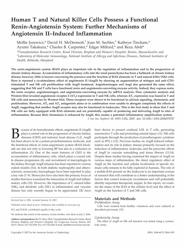
To study the mitogenic effects of AngII on human T and NKcells, we used nylon wool–purified cells that were cultured inthe presence or absence of AngII. After isolation, cells were onaverage 87% CD3� (T cells), 8% CD56� (NK cells), and 2%CD11c� (DC) by flow cytometric analysis. In all subjects stud-ied, AngII alone was unable to stimulate cells in the absence ofmitogen; however, AngII significantly increased phytohemag-glutinin (PHA)-induced cell proliferation in a dosage-depen-dent manner (Figure 1A). T cell receptor (TCR)-specific prolif-eration by plate-bound anti-CD3 was also shown to increase inresponse to AngII, indicating that AngII can provide co-stim-ulatory signals when the TCR is specifically engaged (Figure1B). To determine whether RAS enzymatic components arefunctionally present in our system, we also added AngI andangiotensinogen to mitogen-stimulated lymphocyte cultures;cells that were cultured with PHA in the presence of 10�6
mol/L AngI as well as angiotensinogen increased proliferationto a degree similar to AngII (data not shown), suggesting thatthese cells are able to convert RAS substrates. To determinewhether the observed proliferative effect of AngII could besuppressed, we pretreated cells with 10�5 mol/L losartan andPD123319, antagonists for the AT1 and AT2 receptors, respec-tively, for 1 h before culturing them as described. Comparedwith 10�6 mol/L AngII treatment, both antagonists individu-ally blocked proliferation significantly in response to AngII(Figure 1C), although the level of proliferation was higher thanthat of PHA. Interestingly, when both antagonists were added,proliferation was not significantly reduced compared with 10�6
mol/L AngII treatment and in fact increased when comparedwith either antagonist alone. This suggests that although AngII
acts via both AT1 and AT2, AngII may exert its co-stimulatoryeffect through another AngII receptor. Cells were also labeledwith carboxy fluorescein succinimidyl ester (CFSE) and cul-tured as described to determine which subsets proliferate inresponse to AngII. As shown in Figure 1, D and E, CD8� T cellsand NK cells showed a statistically significant, although �20%,increase in proliferation in response to AngII.
We also performed a proliferation assay in which purifiedcells were depleted of NK cells by bead-mediated removal ofCD56� cells. Under these conditions, the proliferative effect ofAngII was blocked (Figure 1F), indicating that AngII may exertits function through NK cells. Upon examination of the directeffect of AngII on T cell proliferation by depleting NK andlabeling the remaining T cell population with CFSE, as shownin Figure 1G, we show that CD8� T cell proliferation in re-sponse to AngII is substantially diminished in the absence ofNK. To address the direct effect of AngII on NK cell function,we assessed the cytotoxicity of NK cells in the presence andabsence of AngII. No difference was noted in cell death whenNK cells were treated with AngII (data not shown).
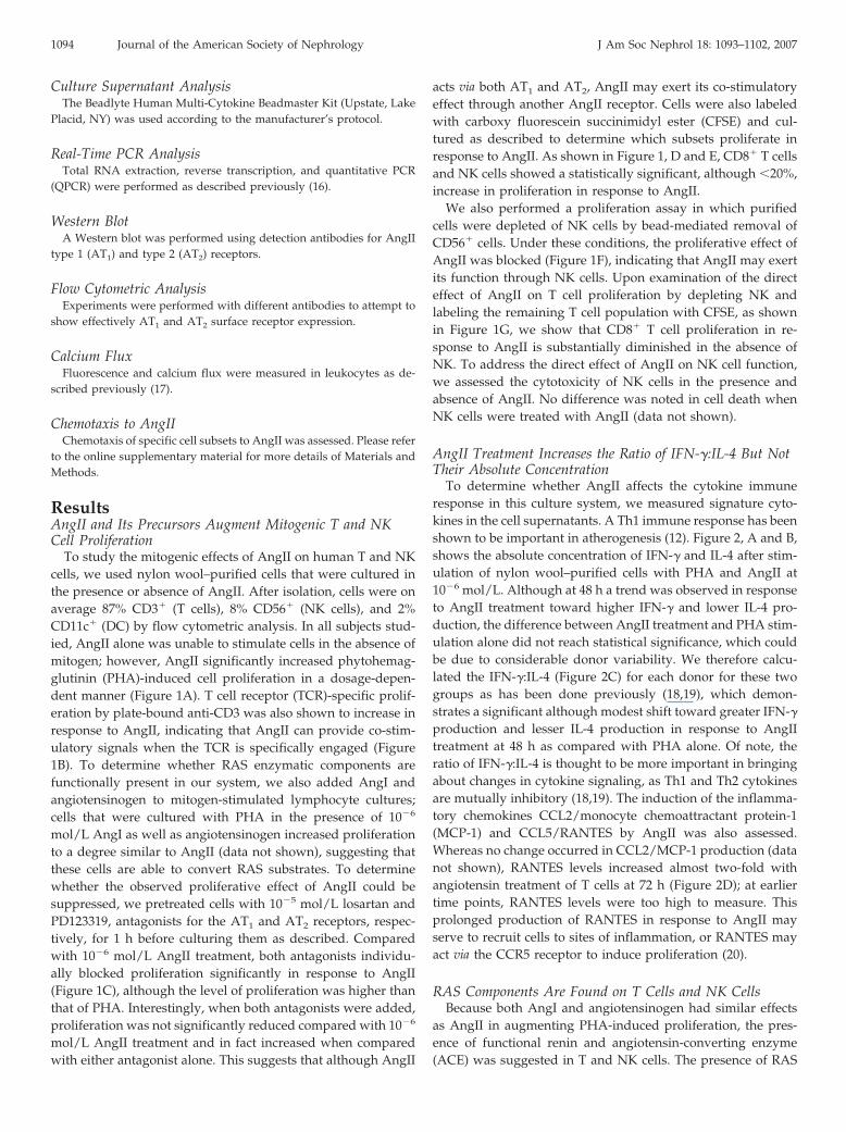
AngII Treatment Increases the Ratio of IFN-�:IL-4 But NotTheir Absolute Concentration
To determine whether AngII affects the cytokine immuneresponse in this culture system, we measured signature cyto-kines in the cell supernatants. A Th1 immune response has beenshown to be important in atherogenesis (12). Figure 2, A and B,shows the absolute concentration of IFN-� and IL-4 after stim-ulation of nylon wool–purified cells with PHA and AngII at10�6 mol/L. Although at 48 h a trend was observed in responseto AngII treatment toward higher IFN-� and lower IL-4 pro-duction, the difference between AngII treatment and PHA stim-ulation alone did not reach statistical significance, which couldbe due to considerable donor variability. We therefore calcu-lated the IFN-�:IL-4 (Figure 2C) for each donor for these twogroups as has been done previously (18,19), which demon-strates a significant although modest shift toward greater IFN-�production and lesser IL-4 production in response to AngIItreatment at 48 h as compared with PHA alone. Of note, theratio of IFN-�:IL-4 is thought to be more important in bringingabout changes in cytokine signaling, as Th1 and Th2 cytokinesare mutually inhibitory (18,19). The induction of the inflamma-tory chemokines CCL2/monocyte chemoattractant protein-1(MCP-1) and CCL5/RANTES by AngII was also assessed.Whereas no change occurred in CCL2/MCP-1 production (datanot shown), RANTES levels increased almost two-fold withangiotensin treatment of T cells at 72 h (Figure 2D); at earliertime points, RANTES levels were too high to measure. Thisprolonged production of RANTES in response to AngII mayserve to recruit cells to sites of inflammation, or RANTES mayact via the CCR5 receptor to induce proliferation (20).
RAS Components Are Found on T Cells and NK CellsBecause both AngI and angiotensinogen had similar effects
as AngII in augmenting PHA-induced proliferation, the pres-ence of functional renin and angiotensin-converting enzyme(ACE) was suggested in T and NK cells. The presence of RAS
1094 Journal of the American Society of Nephrology J Am Soc Nephrol 18: 1093–1102, 2007
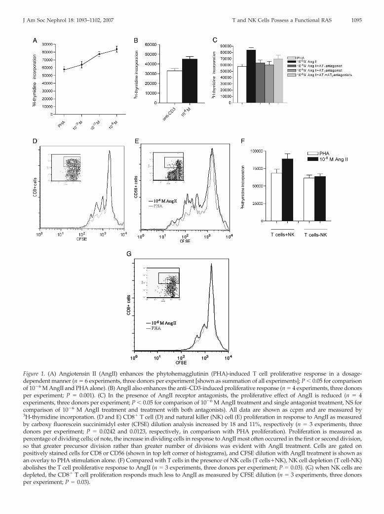
Figure 1. (A) Angiotensin II (AngII) enhances the phytohemagglutinin (PHA)-induced T cell proliferative response in a dosage-dependent manner (n � 6 experiments, three donors per experiment [shown as summation of all experiments]; P � 0.05 for comparisonof 10�6 M AngII and PHA alone). (B) AngII also enhances the anti–CD3-induced proliferative response (n � 4 experiments, three donorsper experiment; P � 0.001). (C) In the presence of AngII receptor antagonists, the proliferative effect of AngII is reduced (n � 4experiments, three donors per experiment; P � 0.05 for comparison of 10�6 M AngII treatment and single antagonist treatment, NS forcomparison of 10�6 M AngII treatment and treatment with both antagonists). All data are shown as ccpm and are measured by3H-thymidine incorporation. (D and E) CD8� T cell (D) and natural killer (NK) cell (E) proliferation in response to AngII as measuredby carboxy fluorescein succinimidyl ester (CFSE) dilution analysis increased by 18 and 11%, respectively (n � 3 experiments, threedonors per experiment; P � 0.0242 and 0.0123, respectively, in comparison with PHA proliferation). Proliferation is measured aspercentage of dividing cells; of note, the increase in dividing cells in response to AngII most often occurred in the first or second division,so that greater precursor division rather than greater number of divisions was evident with AngII treatment. Cells are gated onpositively stained cells for CD8 or CD56 (shown in top left corner of histograms), and CFSE dilution with AngII treatment is shown asan overlay to PHA stimulation alone. (F) Compared with T cells in the presence of NK cells (T cells�NK), NK cell depletion (T cell-NK)abolishes the T cell proliferative response to AngII (n � 3 experiments, three donors per experiment; P � 0.03). (G) when NK cells aredepleted, the CD8� T cell proliferation responds much less to AngII as measured by CFSE dilution (n � 3 experiments, three donorsper experiment; P � 0.03).
J Am Soc Nephrol 18: 1093–1102, 2007 T and NK Cells Possess a Functional RAS 1095
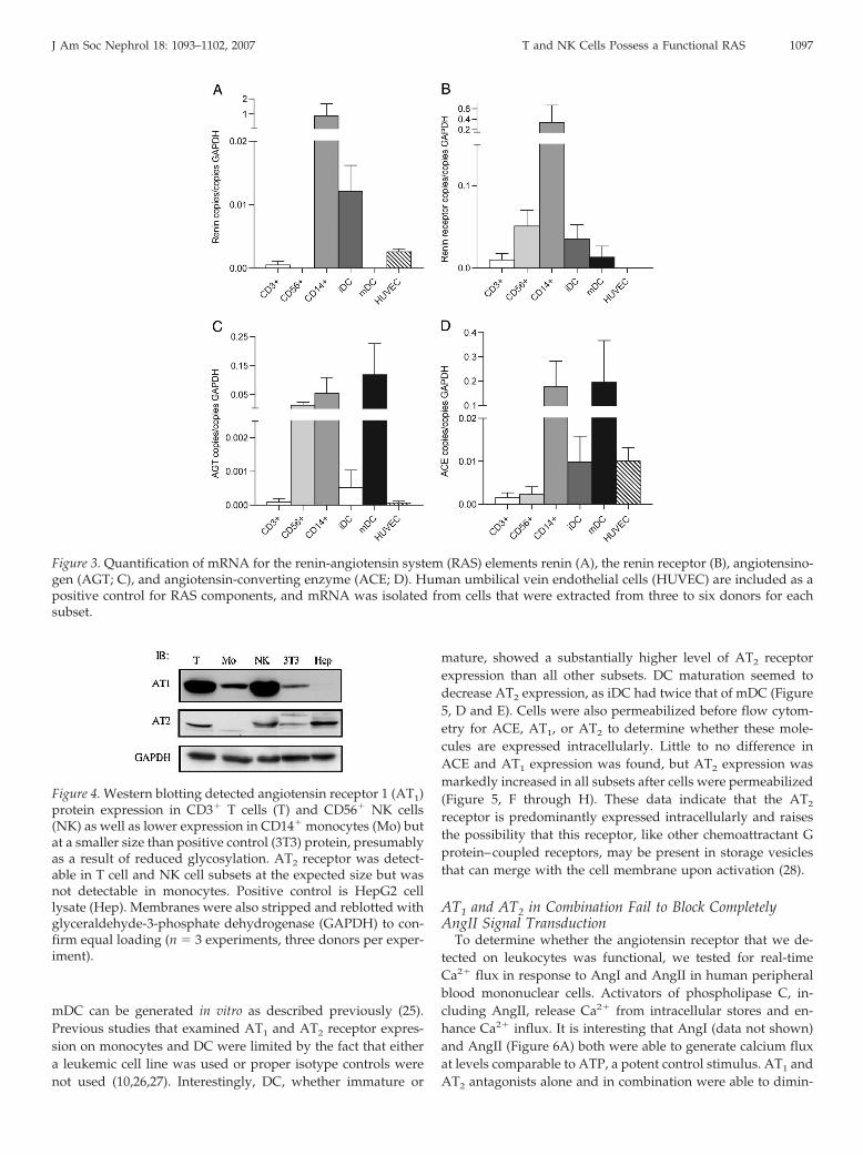
elements on T and NK cells has not been comprehensivelyexplored. mRNA was isolated from purified T and NK cells aswell as from monocytes and DC, and QPCR was used toquantify the expression of angiotensinogen, ACE, renin, andthe renin receptor. Of note, the renin receptor was recentlyreported to bind both circulatory renin and prorenin, promot-ing conversion of angiotensinogen to AngI (21). DC were de-rived from peripheral blood monocytes; their maturity wasassessed by studying surface expression for CCR7, HLA-DR,and CD80. T and NK cells both were found to express ACE andthe renin receptor, whereas only T cells expressed detectablelevels of renin and only NK cells were found to express angio-tensinogen (Figure 3). Monocyte and DC RAS expression waspreviously reported (22,23), and results of these studies areconsistent with our data; expression for the renin receptor,however, is novel, and monocytes, immature DC (iDC), andmature DC (mDC) all were determined to express the reninreceptor. Expression for renin and its receptor was highest inmonocytes, with iDC expressing approximately one tenth thecopy number of monocytes (Figure 3, A and B). Monocytesagain showed substantially higher expression of ACE (Figure3D), whereas monocytes and mDC expressed considerablemRNA levels of angiotensinogen (Figure 3C).
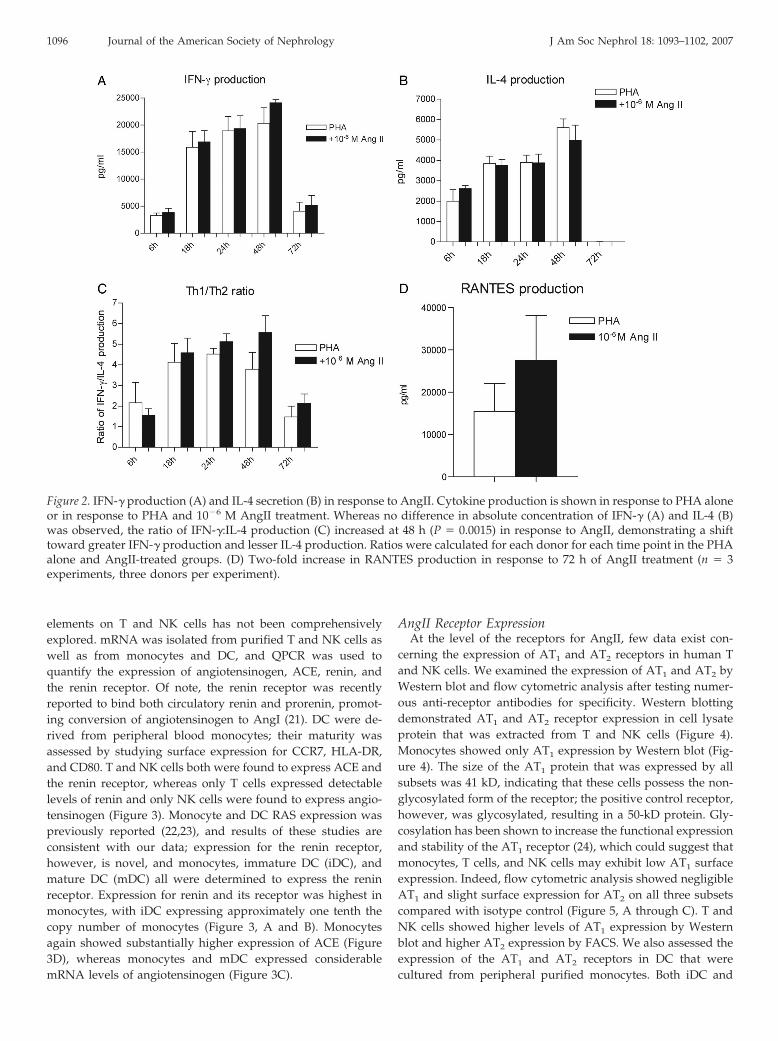
AngII Receptor ExpressionAt the level of the receptors for AngII, few data exist con-
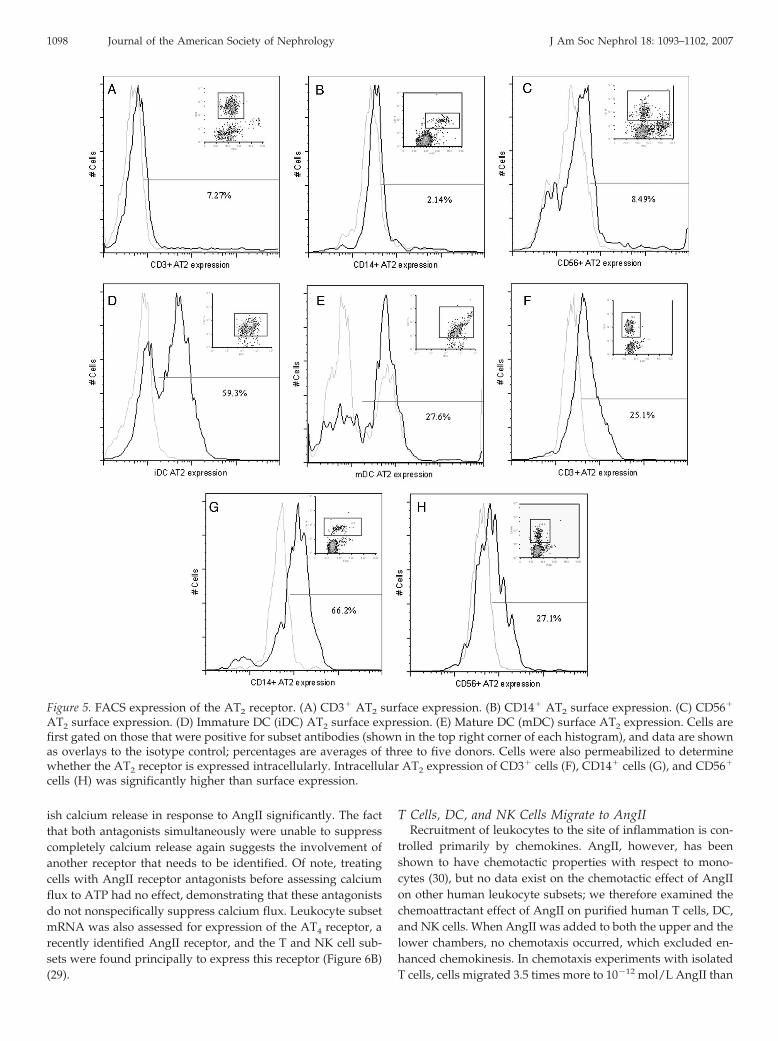
cerning the expression of AT1 and AT2 receptors in human Tand NK cells. We examined the expression of AT1 and AT2 byWestern blot and flow cytometric analysis after testing numer-ous anti-receptor antibodies for specificity. Western blottingdemonstrated AT1 and AT2 receptor expression in cell lysateprotein that was extracted from T and NK cells (Figure 4).Monocytes showed only AT1 expression by Western blot (Fig-ure 4). The size of the AT1 protein that was expressed by allsubsets was 41 kD, indicating that these cells possess the non-glycosylated form of the receptor; the positive control receptor,however, was glycosylated, resulting in a 50-kD protein. Gly-cosylation has been shown to increase the functional expressionand stability of the AT1 receptor (24), which could suggest thatmonocytes, T cells, and NK cells may exhibit low AT1 surfaceexpression. Indeed, flow cytometric analysis showed negligibleAT1 and slight surface expression for AT2 on all three subsetscompared with isotype control (Figure 5, A through C). T andNK cells showed higher levels of AT1 expression by Westernblot and higher AT2 expression by FACS. We also assessed theexpression of the AT1 and AT2 receptors in DC that werecultured from peripheral purified monocytes. Both iDC and
Figure 2. IFN-� production (A) and IL-4 secretion (B) in response to AngII. Cytokine production is shown in response to PHA aloneor in response to PHA and 10�6 M AngII treatment. Whereas no difference in absolute concentration of IFN-� (A) and IL-4 (B)was observed, the ratio of IFN-�:IL-4 production (C) increased at 48 h (P � 0.0015) in response to AngII, demonstrating a shifttoward greater IFN-� production and lesser IL-4 production. Ratios were calculated for each donor for each time point in the PHAalone and AngII-treated groups. (D) Two-fold increase in RANTES production in response to 72 h of AngII treatment (n � 3experiments, three donors per experiment).
1096 Journal of the American Society of Nephrology J Am Soc Nephrol 18: 1093–1102, 2007
mDC can be generated in vitro as described previously (25).Previous studies that examined AT1 and AT2 receptor expres-sion on monocytes and DC were limited by the fact that eithera leukemic cell line was used or proper isotype controls werenot used (10,26,27). Interestingly, DC, whether immature or
mature, showed a substantially higher level of AT2 receptorexpression than all other subsets. DC maturation seemed todecrease AT2 expression, as iDC had twice that of mDC (Figure5, D and E). Cells were also permeabilized before flow cytom-etry for ACE, AT1, or AT2 to determine whether these mole-cules are expressed intracellularly. Little to no difference inACE and AT1 expression was found, but AT2 expression wasmarkedly increased in all subsets after cells were permeabilized(Figure 5, F through H). These data indicate that the AT2
receptor is predominantly expressed intracellularly and raisesthe possibility that this receptor, like other chemoattractant Gprotein–coupled receptors, may be present in storage vesiclesthat can merge with the cell membrane upon activation (28).
AT1 and AT2 in Combination Fail to Block CompletelyAngII Signal Transduction
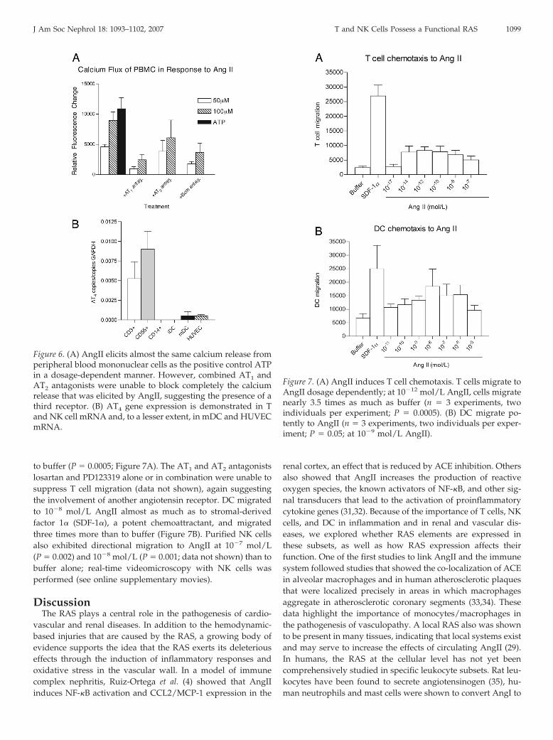
To determine whether the angiotensin receptor that we de-tected on leukocytes was functional, we tested for real-timeCa2� flux in response to AngI and AngII in human peripheralblood mononuclear cells. Activators of phospholipase C, in-cluding AngII, release Ca2� from intracellular stores and en-hance Ca2� influx. It is interesting that AngI (data not shown)and AngII (Figure 6A) both were able to generate calcium fluxat levels comparable to ATP, a potent control stimulus. AT1 andAT2 antagonists alone and in combination were able to dimin-
Figure 3. Quantification of mRNA for the renin-angiotensin system (RAS) elements renin (A), the renin receptor (B), angiotensino-gen (AGT; C), and angiotensin-converting enzyme (ACE; D). Human umbilical vein endothelial cells (HUVEC) are included as apositive control for RAS components, and mRNA was isolated from cells that were extracted from three to six donors for eachsubset.
Figure 4. Western blotting detected angiotensin receptor 1 (AT1)protein expression in CD3� T cells (T) and CD56� NK cells(NK) as well as lower expression in CD14� monocytes (Mo) butat a smaller size than positive control (3T3) protein, presumablyas a result of reduced glycosylation. AT2 receptor was detect-able in T cell and NK cell subsets at the expected size but wasnot detectable in monocytes. Positive control is HepG2 celllysate (Hep). Membranes were also stripped and reblotted withglyceraldehyde-3-phosphate dehydrogenase (GAPDH) to con-firm equal loading (n � 3 experiments, three donors per exper-iment).
J Am Soc Nephrol 18: 1093–1102, 2007 T and NK Cells Possess a Functional RAS 1097
ish calcium release in response to AngII significantly. The factthat both antagonists simultaneously were unable to suppresscompletely calcium release again suggests the involvement ofanother receptor that needs to be identified. Of note, treatingcells with AngII receptor antagonists before assessing calciumflux to ATP had no effect, demonstrating that these antagonistsdo not nonspecifically suppress calcium flux. Leukocyte subsetmRNA was also assessed for expression of the AT4 receptor, arecently identified AngII receptor, and the T and NK cell sub-sets were found principally to express this receptor (Figure 6B)(29).
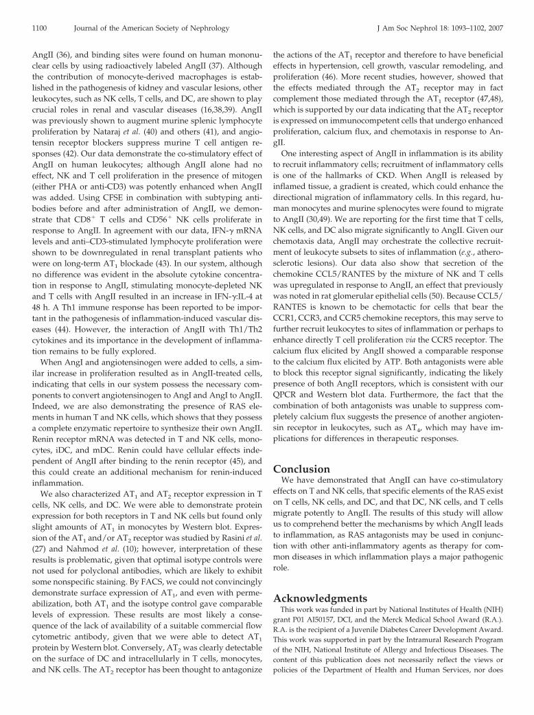
T Cells, DC, and NK Cells Migrate to AngIIRecruitment of leukocytes to the site of inflammation is con-
trolled primarily by chemokines. AngII, however, has beenshown to have chemotactic properties with respect to mono-cytes (30), but no data exist on the chemotactic effect of AngIIon other human leukocyte subsets; we therefore examined thechemoattractant effect of AngII on purified human T cells, DC,and NK cells. When AngII was added to both the upper and thelower chambers, no chemotaxis occurred, which excluded en-hanced chemokinesis. In chemotaxis experiments with isolatedT cells, cells migrated 3.5 times more to 10�12 mol/L AngII than
Figure 5. FACS expression of the AT2 receptor. (A) CD3� AT2 surface expression. (B) CD14� AT2 surface expression. (C) CD56�
AT2 surface expression. (D) Immature DC (iDC) AT2 surface expression. (E) Mature DC (mDC) surface AT2 expression. Cells arefirst gated on those that were positive for subset antibodies (shown in the top right corner of each histogram), and data are shownas overlays to the isotype control; percentages are averages of three to five donors. Cells were also permeabilized to determinewhether the AT2 receptor is expressed intracellularly. Intracellular AT2 expression of CD3� cells (F), CD14� cells (G), and CD56�
cells (H) was significantly higher than surface expression.
1098 Journal of the American Society of Nephrology J Am Soc Nephrol 18: 1093–1102, 2007
to buffer (P � 0.0005; Figure 7A). The AT1 and AT2 antagonistslosartan and PD123319 alone or in combination were unable tosuppress T cell migration (data not shown), again suggestingthe involvement of another angiotensin receptor. DC migratedto 10�8 mol/L AngII almost as much as to stromal-derivedfactor 1� (SDF-1�), a potent chemoattractant, and migratedthree times more than to buffer (Figure 7B). Purified NK cellsalso exhibited directional migration to AngII at 10�7 mol/L(P � 0.002) and 10�8 mol/L (P � 0.001; data not shown) than tobuffer alone; real-time videomicroscopy with NK cells wasperformed (see online supplementary movies).
DiscussionThe RAS plays a central role in the pathogenesis of cardio-
vascular and renal diseases. In addition to the hemodynamic-based injuries that are caused by the RAS, a growing body ofevidence supports the idea that the RAS exerts its deleteriouseffects through the induction of inflammatory responses andoxidative stress in the vascular wall. In a model of immunecomplex nephritis, Ruiz-Ortega et al. (4) showed that AngIIinduces NF-�B activation and CCL2/MCP-1 expression in the
renal cortex, an effect that is reduced by ACE inhibition. Othersalso showed that AngII increases the production of reactiveoxygen species, the known activators of NF-�B, and other sig-nal transducers that lead to the activation of proinflammatorycytokine genes (31,32). Because of the importance of T cells, NKcells, and DC in inflammation and in renal and vascular dis-eases, we explored whether RAS elements are expressed inthese subsets, as well as how RAS expression affects theirfunction. One of the first studies to link AngII and the immunesystem followed studies that showed the co-localization of ACEin alveolar macrophages and in human atherosclerotic plaquesthat were localized precisely in areas in which macrophagesaggregate in atherosclerotic coronary segments (33,34). Thesedata highlight the importance of monocytes/macrophages inthe pathogenesis of vasculopathy. A local RAS also was shownto be present in many tissues, indicating that local systems existand may serve to increase the effects of circulating AngII (29).In humans, the RAS at the cellular level has not yet beencomprehensively studied in specific leukocyte subsets. Rat leu-kocytes have been found to secrete angiotensinogen (35), hu-man neutrophils and mast cells were shown to convert AngI to
Figure 6. (A) AngII elicits almost the same calcium release fromperipheral blood mononuclear cells as the positive control ATPin a dosage-dependent manner. However, combined AT1 andAT2 antagonists were unable to block completely the calciumrelease that was elicited by AngII, suggesting the presence of athird receptor. (B) AT4 gene expression is demonstrated in Tand NK cell mRNA and, to a lesser extent, in mDC and HUVECmRNA.
Figure 7. (A) AngII induces T cell chemotaxis. T cells migrate toAngII dosage dependently; at 10�12 mol/L AngII, cells migratenearly 3.5 times as much as buffer (n � 3 experiments, twoindividuals per experiment; P � 0.0005). (B) DC migrate po-tently to AngII (n � 3 experiments, two individuals per exper-iment; P � 0.05; at 10�9 mol/L AngII).
J Am Soc Nephrol 18: 1093–1102, 2007 T and NK Cells Possess a Functional RAS 1099
AngII (36), and binding sites were found on human mononu-clear cells by using radioactively labeled AngII (37). Althoughthe contribution of monocyte-derived macrophages is estab-lished in the pathogenesis of kidney and vascular lesions, otherleukocytes, such as NK cells, T cells, and DC, are shown to playcrucial roles in renal and vascular diseases (16,38,39). AngIIwas previously shown to augment murine splenic lymphocyteproliferation by Nataraj et al. (40) and others (41), and angio-tensin receptor blockers suppress murine T cell antigen re-sponses (42). Our data demonstrate the co-stimulatory effect ofAngII on human leukocytes; although AngII alone had noeffect, NK and T cell proliferation in the presence of mitogen(either PHA or anti-CD3) was potently enhanced when AngIIwas added. Using CFSE in combination with subtyping anti-bodies before and after administration of AngII, we demon-strate that CD8� T cells and CD56� NK cells proliferate inresponse to AngII. In agreement with our data, IFN-� mRNAlevels and anti–CD3-stimulated lymphocyte proliferation wereshown to be downregulated in renal transplant patients whowere on long-term AT1 blockade (43). In our system, althoughno difference was evident in the absolute cytokine concentra-tion in response to AngII, stimulating monocyte-depleted NKand T cells with AngII resulted in an increase in IFN-�:IL-4 at48 h. A Th1 immune response has been reported to be impor-tant in the pathogenesis of inflammation-induced vascular dis-eases (44). However, the interaction of AngII with Th1/Th2cytokines and its importance in the development of inflamma-tion remains to be fully explored.
When AngI and angiotensinogen were added to cells, a sim-ilar increase in proliferation resulted as in AngII-treated cells,indicating that cells in our system possess the necessary com-ponents to convert angiotensinogen to AngI and AngI to AngII.Indeed, we are also demonstrating the presence of RAS ele-ments in human T and NK cells, which shows that they possessa complete enzymatic repertoire to synthesize their own AngII.Renin receptor mRNA was detected in T and NK cells, mono-cytes, iDC, and mDC. Renin could have cellular effects inde-pendent of AngII after binding to the renin receptor (45), andthis could create an additional mechanism for renin-inducedinflammation.
We also characterized AT1 and AT2 receptor expression in Tcells, NK cells, and DC. We were able to demonstrate proteinexpression for both receptors in T and NK cells but found onlyslight amounts of AT1 in monocytes by Western blot. Expres-sion of the AT1 and/or AT2 receptor was studied by Rasini et al.(27) and Nahmod et al. (10); however, interpretation of theseresults is problematic, given that optimal isotype controls werenot used for polyclonal antibodies, which are likely to exhibitsome nonspecific staining. By FACS, we could not convincinglydemonstrate surface expression of AT1, and even with perme-abilization, both AT1 and the isotype control gave comparablelevels of expression. These results are most likely a conse-quence of the lack of availability of a suitable commercial flowcytometric antibody, given that we were able to detect AT1
protein by Western blot. Conversely, AT2 was clearly detectableon the surface of DC and intracellularly in T cells, monocytes,and NK cells. The AT2 receptor has been thought to antagonize
the actions of the AT1 receptor and therefore to have beneficialeffects in hypertension, cell growth, vascular remodeling, andproliferation (46). More recent studies, however, showed thatthe effects mediated through the AT2 receptor may in factcomplement those mediated through the AT1 receptor (47,48),which is supported by our data indicating that the AT2 receptoris expressed on immunocompetent cells that undergo enhancedproliferation, calcium flux, and chemotaxis in response to An-gII.
One interesting aspect of AngII in inflammation is its abilityto recruit inflammatory cells; recruitment of inflammatory cellsis one of the hallmarks of CKD. When AngII is released byinflamed tissue, a gradient is created, which could enhance thedirectional migration of inflammatory cells. In this regard, hu-man monocytes and murine splenocytes were found to migrateto AngII (30,49). We are reporting for the first time that T cells,NK cells, and DC also migrate significantly to AngII. Given ourchemotaxis data, AngII may orchestrate the collective recruit-ment of leukocyte subsets to sites of inflammation (e.g., athero-sclerotic lesions). Our data also show that secretion of thechemokine CCL5/RANTES by the mixture of NK and T cellswas upregulated in response to AngII, an effect that previouslywas noted in rat glomerular epithelial cells (50). Because CCL5/RANTES is known to be chemotactic for cells that bear theCCR1, CCR3, and CCR5 chemokine receptors, this may serve tofurther recruit leukocytes to sites of inflammation or perhaps toenhance directly T cell proliferation via the CCR5 receptor. Thecalcium flux elicited by AngII showed a comparable responseto the calcium flux elicited by ATP. Both antagonists were ableto block this receptor signal significantly, indicating the likelypresence of both AngII receptors, which is consistent with ourQPCR and Western blot data. Furthermore, the fact that thecombination of both antagonists was unable to suppress com-pletely calcium flux suggests the presence of another angioten-sin receptor in leukocytes, such as AT4, which may have im-plications for differences in therapeutic responses.
ConclusionWe have demonstrated that AngII can have co-stimulatory
effects on T and NK cells, that specific elements of the RAS existon T cells, NK cells, and DC, and that DC, NK cells, and T cellsmigrate potently to AngII. The results of this study will allowus to comprehend better the mechanisms by which AngII leadsto inflammation, as RAS antagonists may be used in conjunc-tion with other anti-inflammatory agents as therapy for com-mon diseases in which inflammation plays a major pathogenicrole.
AcknowledgmentsThis work was funded in part by National Institutes of Health (NIH)
grant P01 AI50157, DCI, and the Merck Medical School Award (R.A.).R.A. is the recipient of a Juvenile Diabetes Career Development Award.This work was supported in part by the Intramural Research Programof the NIH, National Institute of Allergy and Infectious Diseases. Thecontent of this publication does not necessarily reflect the views orpolicies of the Department of Health and Human Services, nor does
1100 Journal of the American Society of Nephrology J Am Soc Nephrol 18: 1093–1102, 2007
mention of trade names, commercial products, or organizations implyendorsement by the US government.
DisclosuresNone.
References1. Ruiz-Ortega M, Lorenzo O, Suzuki Y, Ruperez M, Egido J:
Proinflammatory actions of angiotensins. Curr Opin Neph-rol Hypertens 10: 321–329, 2001
2. Long DA, Price KL, Herrera-Acosta J, Johnson RJ: Howdoes angiotensin II cause renal injury? Hypertension 43:722–723, 2004
3. Cooper ME, Tikellis C, Thomas MC: Preventing diabetes inpatients with hypertension: One more reason to block therenin-angiotensin system. J Hypertens Suppl 24: S57–S63,2006
4. Ruiz-Ortega M, Bustos C, Hernandez-Presa MA, LorenzoO, Plaza JJ, Egido J: Angiotensin II participates in mono-nuclear cell recruitment in experimental immune complexnephritis through nuclear factor-kappa B activation andmonocyte chemoattractant protein-1 synthesis. J Immunol161: 430–439, 1998
5. Anders HJ, Ninichuk V, Schlondorff D: Progression ofkidney disease: Blocking leukocyte recruitment with che-mokine receptor CCR1 antagonists. Kidney Int 69: 29–32,2006
6. Lombardi DM, Viswanathan M, Vio CP, Saavedra JM,Schwartz SM, Johnson RJ: Renal and vascular injury in-duced by exogenous angiotensin II is AT1 receptor-depen-dent. Nephron 87: 66–74, 2001
7. Cattell V: Macrophages in acute glomerular inflammation.Kidney Int 45: 945–952, 1994
8. Chow F, Ozols E, Nikolic-Paterson DJ, Atkins RC, TeschGH: Macrophages in mouse type 2 diabetic nephropathy:Correlation with diabetic state and progressive renal in-jury. Kidney Int 65: 116–128, 2004
9. Nikolic-Paterson DJ, Lan HY, Hill PA, Atkins RC: Macro-phages in renal injury. Kidney Int Suppl 45: S79–S82, 1994
10. Nahmod KA, Vermeulen ME, Raiden S, Salamone G, Gam-berale R, Fernandez-Calotti P, Alvarez A, Nahmod V,Giordano M, Geffner JR: Control of dendritic cell differen-tiation by angiotensin II. FASEB J 17: 491–493, 2003
11. Angeli V, Llodra J, Rong JX, Satoh K, Ishii S, Shimizu T,Fisher EA, Randolph GJ: Dyslipidemia associated withatherosclerotic disease systemically alters dendritic cellmobilization. Immunity 21:5 61–74, 2004
12. Farag SS, Caligiuri MA: Human natural killer cell devel-opment and biology. Blood Rev 20: 123–137, 2005
13. Remuzzi G, Perico N, Macia M, Ruggenenti P: The role ofrenin-angiotensin-aldosterone system in the progression ofchronic kidney disease. Kidney Int Suppl 99: S57–S65, 2005
14. Vaziri ND, Xu ZG, Shahkarami A, Huang KT, Rodriguez-Iturbe B, Natarajan R: Role of AT-1 receptor in regulationof vascular MCP-1, IL-6, PAI-1, MAP kinase, and matrixexpressions in obesity. Kidney Int 68: 2787–2793, 2005
15. Voshol H, Dullens HF, Den Otter W, Vliegenthart JF: Hu-man natural killer cells: A convenient purification proce-dure and the influence of cryopreservation on cytotoxicactivity. J Immunol Methods 165: 21–30, 1993
16. Fiorina P, Ansari MJ, Jurewicz M, Barry M, Ricchiuti V,
Smith RN, Shea S, Means TK, Auchincloss H Jr, Luster AD,Sayegh MH, Abdi R: Role of CXC chemokine receptor 3pathway in renal ischemic injury. J Am Soc Nephrol 17:716–723, 2006
17. McDermott DH, Fong AM, Yang Q, Sechler JM, CupplesLA, Merrell MN, Wilson PW, D’Agostino RB, O’DonnellCJ, Patel DD, Murphy PM: Chemokine receptor mutantCX3CR1–M280 has impaired adhesive function and corre-lates with protection from cardiovascular disease in hu-mans. J Clin Invest 111: 1241–1250, 2003
18. Makhseed M, Raghupathy R, El-Shazly S, Azizieh F, Al-Harmi JA, Al-Azemi MM: Pro-inflammatory maternal cy-tokine profile in preterm delivery. Am J Reprod Immunol 49:308–318, 2003
19. Panitsas FP, Theodoropoulou M, Kouraklis A, KarakantzaM, Theodorou GL, Zoumbos NC, Maniatis A, Mouzaki A:Adult chronic idiopathic thrombocytopenic purpura (ITP)is the manifestation of a type-1 polarized immune re-sponse. Blood 103: 2645–2647, 2004
20. Taub DD, Turcovski-Corrales SM, Key ML, Longo DL,Murphy WJ: Chemokines and T lymphocyte activation: I.Beta chemokines costimulate human T lymphocyte activa-tion in vitro. J Immunol 156: 2095–2103, 1996
21. Nguyen G, Delarue F, Burckle C, Bouzhir L, Giller T, SraerJD: Pivotal role of the renin/prorenin receptor in angioten-sin II production and cellular responses to renin. J ClinInvest 109: 1417–1427, 2002
22. Costerousse O, Allegrini J, Lopez M, Alhenc-Gelas F: An-giotensin I-converting enzyme in human circulating mono-nuclear cells: Genetic polymorphism of expression in T-lymphocytes. Biochem J 290: 33–40, 1993
23. Lapteva N, Nieda M, Ando Y, Ide K, Hatta-Ohashi Y,Dymshits G, Ishikawa Y, Juji T, Tokunaga K: Expression ofrenin-angiotensin system genes in immature and maturedendritic cells identified using human cDNA microarray.Biochem Biophys Res Commun 285: 1059–1065, 2001
24. Lanctot PM, Leclerc PC, Clement M, Auger-Messier M,Escher E, Leduc R, Guillemette G: Importance of N-glyco-sylation positioning for cell-surface expression, targeting,affinity and quality control of the human AT1 receptor.Biochem J 390: 367–376, 2005
25. Elkord E, Williams PE, Kynaston H, Rowbottom AW: Hu-man monocyte isolation methods influence cytokine pro-duction from in vitro generated dendritic cells. Immunology114: 204–212, 2005
26. Okamura A, Rakugi H, Ohishi M, Yanagitani Y, Takiuchi S,Moriguchi K, Fennessy PA, Higaki J, Ogihara T: Upregu-lation of renin-angiotensin system during differentiation ofmonocytes to macrophages. J Hypertens 17: 537–545, 1999
27. Rasini E, Cosentino M, Marino F, Legnaro M, Ferrari M,Guasti L, Venco A, Lecchini S: Angiotensin II type 1 recep-tor expression on human leukocyte subsets: A flow cyto-metric and RT-PCR study. Regul Pept 134: 69–74, 2006
28. Gasser O, Missiou A, Eken C, Hess C: Human CD8� Tcells store CXCR1 in a distinct intracellular compartmentand upregulate it rapidly to the cell surface upon activa-tion. Blood 106: 3718–3724, 2005
29. Chai SY, Fernando R, Peck G, Ye SY, Mendelsohn FA,Jenkins TA, Albiston AL: The angiotensin IV/AT4 recep-tor. Cell Mol Life Sci 61: 2728–2737, 2004
30. Kintscher U, Wakino S, Kim S, Fleck E, Hsueh WA, LawRE: Angiotensin II induces migration and Pyk2/paxillin
J Am Soc Nephrol 18: 1093–1102, 2007 T and NK Cells Possess a Functional RAS 1101
phosphorylation of human monocytes. Hypertension 37:587–593, 2001
31. Vaziri ND: Roles of oxidative stress and antioxidant ther-apy in chronic kidney disease and hypertension. Curr OpinNephrol Hypertens 13: 93–99, 2004
32. Remuzzi G, Benigni A, Remuzzi A: Mechanisms of pro-gression and regression of renal lesions of chronic ne-phropathies and diabetes. J Clin Invest 116: 288–296, 2006
33. Friedland J, Setton C, Silverstein E: Angiotensin convertingenzyme: Induction by steroids in rabbit alveolar macro-phages in culture. Science 197: 64–65, 1977
34. Diet F, Pratt RE, Berry GJ, Momose N, Gibbons GH, DzauVJ: Increased accumulation of tissue ACE in human ath-erosclerotic coronary artery disease. Circulation 94: 2756–2767, 1996
35. Gomez RA, Norling LL, Wilfong N, Isakson P, Lynch KR,Hock R, Quesenberry P: Leukocytes synthesize angio-tensinogen. Hypertension 21: 470–475, 1993
36. Reilly CF, Tewksbury DA, Schechter NM, Travis J: Rapidconversion of angiotensin I to angiotensin II by neutrophiland mast cell proteinases. J Biol Chem 257: 8619–8622, 1982
37. Shimada K, Yazaki Y: Binding sites for angiotensin II inhuman mononuclear leucocytes. J Biochem 84: 1013–1015,1978
38. Bobryshev YV, Lord RS: Co-accumulation of dendritic cellsand natural killer T cells within rupture-prone regions inhuman atherosclerotic plaques. J Histochem Cytochem 53:781–785, 2005
39. Burne-Taney MJ, Liu M, Ascon D, Molls RR, Racusen L,Rabb H: Transfer of lymphocytes from mice with renalischemia induce albuminuria in naive mice: A possiblemechanism linking early injury to progressive renal dis-ease? Am J Physiol Renal Physiol 291: F981–F986, 2006
40. Nataraj C, Oliverio MI, Mannon RB, Mannon PJ, AudolyLP, Amuchastegui CS, Ruiz P, Smithies O, Coffman TM:Angiotensin II regulates cellular immune responsesthrough a calcineurin-dependent pathway. J Clin Invest104: 1693–1701, 1999
41. Kunert-Radek J, Stepien H, Komorowski J, Pawlikowski M:Stimulatory effect of angiotensin II on the proliferation ofmouse spleen lymphocytes in vitro is mediated via bothtypes of angiotensin II receptors. Biochem Biophys Res Com-mun 198: 1034–1039, 1994
42. Sagawa K, Nagatani K, Komagata Y, Yamamoto K: Angio-tensin receptor blockers suppress antigen-specific T cellresponses and ameliorate collagen-induced arthritis inmice. Arthritis Rheum 52: 1920–1928, 2005
43. Weidanz JA, Jacobson LM, Muehrer RJ, Djamali A, HullettDA, Sprague J, Chiriva-Internati M, Wittman V, Thek-kumkara TJ, Becker BN: ATR blockade reduces IFN-gamma production in lymphocytes in vivo and in vitro.Kidney Int 67: 2134–2142, 2005
44. Ranjbaran H, Sokol SI, Gallo A, Eid RE, Iakimov AO,D’Alessio A, Kapoor JR, Akhtar S, Howes CJ, Aslan M,Pfau S, Pober JS, Tellides G: An inflammatory pathway ofIFN-gamma production in coronary atherosclerosis. J Im-munol 178: 592–604, 2007
45. Nguyen G: Renin/prorenin receptors. Kidney Int 69: 1503–1506, 2006
46. AbdAlla S, Lother H, Abdel-tawab AM, Quitterer U: Theangiotensin II AT2 receptor is an AT1 receptor antagonist.J Biol Chem 276: 39721–39726, 2001
47. Steckelings UM, Kaschina E, Unger T: The AT2 receptor: Amatter of love and hate. Peptides 26: 1401–1409, 2005
48. Reudelhuber TL: The continuing saga of the AT2 receptor:A case of the good, the bad, and the innocuous. Hyperten-sion 46: 1261–1262, 2005
49. Weinstock JV, Kassab J: Chemotactic response of splenicmononuclear cells to angiotensin II in murine schistosomi-asis. J Immunol 137: 2020–2024, 1986
50. Wolf G, Ziyadeh FN, Thaiss F, Tomaszewski J, Caron RJ,Wenzel U, Zahner G, Helmchen U, Stahl RA: AngiotensinII stimulates expression of the chemokine RANTES in ratglomerular endothelial cells. Role of the angiotensin type 2receptor. J Clin Invest 100: 1047–1058, 1997
Supplemental information for this article is available online at http://www.jasn.org/.
1102 Journal of the American Society of Nephrology J Am Soc Nephrol 18: 1093–1102, 2007