regulation of renin-angiotensin system in unilateral ureteral obstruction
TRANSCRIPT
Kidney International, Vol. 44 (1993), pp. 390—400
Regulation of renin-angiotensin system in unilateral ureteralobstruction
J. Luis PIMENTEL JR., MANUEL MARTINEZ-MALDONADO, JOSIAH N. WILcox,SUSHENG WANG, and CHUYING Luo
Atlanta Veterans Affairs Medical Center, Decatur, and Renal Division, Department of Medicine, Emory University School of Medicine,Atlanta, Georgia, USA
Regulation of renin-angiotensin system in unilateral ureteral obstruc-tion. The effect of 24-hour unilateral ureteral obstruction (UUO) on theexpression and regulation of the renin-angiotensin system (RAS) in ratsand of pretreatment with lisinopril (5 mg/kg/day) or the AT,-R inhibitor,losartan, (10 mg/kg/day) on renal hemodynamics was evaluated. Bothdrugs improved the post-obstructed kidney (P0K) renal hemodynam-ics, lowered MAP, and normalized eicosanoid excretion by the P0K.Cortex and medulla POK:CK ratio of relative density R mRNA was—3.5 for both. Sham, P0K, and CK showed renin immunoreactivityand R mRNA exclusively in juxtaglomerular position. In addition, inP0K renin was expressed in mesangial cells, along greater lengths ofafferent arterioles and in dilated distal tubules and ioops of Henle. InSitu hybridization revealed that —20% more glomeruli in P0K than CKoverexpressed R mRNA. Blood vessels of P0K consistently showedgreater ACE and Ao mRNA expression than CK. Overexpression ofthe genes coding for members of the RAS is possibly responsible forlocal Ang II production which, in view of the response to CEI andAT,-R inhibitors, is at least partly responsible for the severe hemody-namic changes in UUO.
Complete obstruction of one ureter (UUO) results in pro-found hormonal and hemodynamic changes in the obstructedkidney as well as in the contralateral kidney [1—3].' Followingthe obstruction, a transient but sharp surge in renal plasma flow(RPF) occurs that gives way in a few hours to a drop in renalperfusion and glomerular filtration rate (GFR) [4—6]. The falls inRPF and GFR are the consequence of intense renal vasocon-striction which is multifactorial. The renin-angiotensin system(RAS), altered synthesis of prostanoids including thromboxane,nitric oxide (or endothelium-derived relaxing factor) produc-
'Abbreviations used in this article are: Ang II, angiotensin II; ACE,angiotensin-converting enzyme; Ao, angiotensinogen; AT,-R, angioten-sin II receptor; CEI, converting enzyme inhibitors; CK, contralateralkidney; GFR, glomerular filtration rate; JGA, juxtaglomerular appara-tus; MAP, mean arterial pressure; PAH, para-aminohippurate; PBS,phosphate buffered saline; PG, prostaglandin; P0K, post-obstructedkidney; PRA, plasma renin activity; renin, R; RAS, renin-angiotensinsystem; RPF, renal plasma flow; UUO, unilateral ureteral obstruction;V, urine flow.
Received for publication January 13, 1993and in revised form April 5, 1993Accepted for publication April 5, 1993
© 1993 by the International Society of Nephrology
tion, and other vasoactive agents have been implicated in thehemodynamic resetting in UUO [7, 8].
Evidence has been marshalled that activation of the RAS, asindicated by increases in plasma renin (R) and angiotensin II(Ang II) occurs early in UUO [9] and that Ang II may beresponsible for the synthesis of thromboxane [10]. Thus, di-rectly or indirectly, Ang II is a major factor in the renalvasoconstriction of ureteral obstruction; in the case of bilateralureteral obstruction Ang II and thromboxane may account forapproximately 90% of the hemodynamic changes observed [11].Moreover, angiotensin-converting enzyme inhibitors (CEI) re-verse or ameliorate UUO-induced renal vasoconstriction [12,13].
Most studies on the pathophysiology of the hemodynamicconsequences of ureteral obstruction have been conducted inanesthetized rats with either bilateral or unilateral ureteralobstruction. Thus, it is not possible to eliminate an effect ofanesthesia per se on renal function. Furthermore, despite theevidence for a role of the renin-angiotensin system in theresponse to UUO, little information exists on the regulation andexpression of the genes that encode for the components of theRAS in this model. In one study, altered R mRNA expression inadult rats could not be found after chronic UUO [14]. Thepresent experiments were designed to investigate the effect ofUUO on the expression of RAS genes by both Northernanalysis and in situ hybridization, and of angiotensin-convertingenzyme (ACE) and angiotensinogen (Ao) by the latter tech-nique. In addition, to examine the specific role of Ang II in therenal hemodynamic recovery after UUO, we have examined inawake rats the action of both the converting enzyme inhibitor(CE!) lisinopril and the non-peptide, specific Ang II receptorantagonist DuP 753 (hereinafter called losartan) which, incontrast to CE!, does not alter production of bradykinin orprostaglandins (PGs) [15]. We felt that comparisons betweenthese two agents (DuP753 and CEI) might permit inferences tobe drawn relating to the role of Ang II as well as of bradykininin UUO.
Methods
General procedures and surgery
Thirty-six male Wistar Munich rats weighing 150 to 200 g(Charles River Laboratories, Wilmington, Massachusetts,
390
Pimentel et a!: Regulation of renin-angiotensin system 391
USA) were used. Animals were housed individually in a roomproviding 12-hour light and dark cycles, and were fed a standardrat chow (Purina 5010, Purina Mills, Richmond, Indiana, USA)containing 23% protein, 0.28% sodium, and 0.92% potassium.All procedures followed the guidelines for the humanitariancare of animals and were approved by the local animal carecommittee. To create a state of UUO, 32 rats were anesthetizedwith ketamine (10 mg/l50 g body wt) and through a smallsuprapubic incision the left ureter was identified, dissected freefrom its retroperitoneal site, and completely occluded with asquare knot using 4-0 silk. The incision was closed and animalswere allowed free access to tap water and food until clearancesstudies were performed. Four rats were sham-operated controls(ureter dissection but no ligature). At the end of 24-hour UUO,another group of rats (N = 4) was decapitated, the headcollected and snap-frozen in liquid nitrogen, and blood obtainedfor measurement of plasma renin activity (PRA). Immediatelythereafter, the kidneys and liver were removed and snap-frozenin liquid nitrogen; in most instances kidneys were separatedinto cortex and medulla for extraction of RNA. A right and leftkidney of each rat provided enough cortical material for RNAextraction and comparison of sides, but two right and two leftkidneys were needed to isolate the outer and inner medulla.Frozen tissues were chiseled into small pieces, placed inguanidium isothiocyanate (GTC), and homogenized immedi-ately. Samples could then be stored at 4°C until analysis.
Drugs used
Lisinopril, an angiotensin I converting enzyme inhibitor, andlosartan, a nonpeptide subtype 1-selective angiotensin II antag-onist (previously known as DuP753) were provided by MerckSharp & Dohme (West Point, Pennsylvania, USA) and B. I.DuPont de Nemours & Co. (Wilmington, Delaware, USA),respectively.
Clearance studies
To study the effect of Ang II blockade, two groups of rats(N = 7 each) received either losartan (10 mg/kg body wt) orlisinopril (5 mg/kg body wt) by gavage for five days prior to andincluding the day of UUO. Control rats (N = 7) received anequal volume of vehicle for the same period of time. On the dayof study, animals were anesthetized with ketamine (10 mg/iSO gbody wt) and cannulas placed in the right external jugular veinfor infusion of 3H-inulin and '4C-PAH, and in the right carotidartery for blood sampling and blood pressure monitoring. Thesuprapubic suture was removed and the left ureter was cannu-lated proximal to the ligature placed 24 hours earlier. Thebladder was catheterized for right kidney urine collection. Ratswere placed in plexiglass holders and allowed to awaken. Primedoses of 3H-inulin (2 tCi) and '4C PAH (1 Ci) were given i.v.in 0.2 ml normal saline and a constant infusion of normal salineat 0.013 mllmin delivering 0.1 pCi 3H and 0.05 pCi PAH/minwas continued throughout the experiment.
Following an equilibration period of 60 minutes and approx-imately three hours after UUO release, four consecutive 20-minute urine collections were obtained for measurement ofGFR by inulin clearance, effective RPF by PAH clearance, andurine sodium and potassium excretion. At the end of theexperiment, rats were euthanized with an anesthetic overdoseand the kidneys were rapidly removed and bisected by a sagittal
cut; one-half was used for immunohistochemistry and in situhybridization, the other half for RNA isolation (see below).
RNA extraction and Northern hybridization analysis
Total RNA was extracted by the guanidine thiocyanatemethod, as previously described from this and other laborato-ries [16]. A total of 15 to 20 pg RNA was run on agarose gelsusing formaldehyde. RNA was transferred by capillary blottingusing standard procedures and Immobilon-N membranes(charge-modified PVDF transfer membranes, Millipore Corp.,Bedford, Massachusetts, USA). Blots were washed in 6xSSCfor 5 to 10 minutes, then baked under vacuum for one hour at80°C.
Renin and Ao cDNA
Ao eDNA (pRang 6) was cloned from adult male Sprague-Dawley rat kidney [17]. It was essentially full length (1634 bp).The cDNA insert was removed from the cloning vector Agtl0 byEcoRl digestion, made blunt-ended, and subeloned into theSmal site of the plasmid pGEM-4 (Promega Biotec, Madison,Wisconsin, USA). The insert can be removed by double diges-tion with Hindlil and EcoRl. Transcription from the T7 pro-moter yields antisense cRNA. R cDNA was isolated from a ratkidney AgtlO library [18]. It was initially subcloned into theEcoRl site of the plasmid Bluescript, then excised with BamHland Hindill and subcloned into the same sites in the plasmidpGEM4 (Promega). The insert is —1425 bp; transcription fromthe T7 promoter yields antisense cRNA. Both probes wereobtained as a courtesy of Dr. K. Lynch, University of Virginia.
ACE cRNA
ACE was initially cloned into EcoRl site of plasmid Blue-script [19]. To reduce the distance between insert and pro-moter, the cDNA, a gift from Dr. K. Bernstein, Emory Univer-sity School of Medicine, was excised with BamHl and Hindliland subcloned into the same sites in the plasmid pGEM3Z(Promega). Linearized plasmid with Hindlil using T7 promoteryields antisense cRNA. A full-length antisense transcript wasused for hybridization.
Preparation of riboprobesLabeled riboprobes were prepared using 0.5 to 1 g linear-
ized plasmid and a-32P(UTP) or 35S(UTP) for Northern blots orin situ hybridization, respectively. Purified riboprobes werestored in 70% ethanol at —20°C.
Prehybridization and hybridization
Blots were prehybridized for six hours at 60°C with hybrid-ization buffer (50% formamide, 5 x SSC (1 X SSC: 0.15 MNaC1, 0.015 M Na citrate, pH 7), 8 x Denhardt's solution, 1 mMEDTA, 0.1% sodium pyrophosphate, and 0.5% SDS, 200 g/mldenatured salmon sperm DNA). A mixture of 0.5 to 1.0 x l0CPM probe in 0.2 ml hybridization buffer was heated to 85°C forfive minutes, then added to 4 ml hybridization buffer and 1 ml50% dextran sulfate and mixed well. Hybridization was carriedout overnight (16 to 18 hr) at 60°C. Membranes were washed for30 minutes first in 2 x SSC + 0.2% SDS at 65°C and finally in0.2 x SSC + 0.2% SDS at 70°C. Dry blots were exposedovernight with intensifying screen on Kodak film XRP-5.
392 Pimentel et a!: Regulation of renin-angiotensin system
Membrane reprobingFor reprobing with GAPDH eRNA, membranes that had
been previously hybridized were washed in 0.1 N NaOH for 30minutes at room temperature. Geiger counter check was carriedout until there was no detectable radioactivity. Then the mem-brane was neutralized by immersion in 0.0254 Tris-HC1, pH 8 +0.1 x SSC + 0.5% SDS for 45 to 60 minutes. The membraneswere then ready to be reprobed. Prehybridization and hybrid-ization for GAPDH mRNA were done as described above. Incontrast to what has been observed with insulin in other cells[20], GAPDH mRNA has been shown to be unaltered by UUO[21].
Densitornetry analysis
The autoradiographs were scanned using the Image Quant°Mdensitometer. Results are expressed as relative density. In allcases, the relative density of GAPDH mRNA was used ascomparison. This was accomplished by factoring the relativedensity of the various mRNAs' autoradiograms to that of theGAPDH mRNA autoradiogram which was arbitrarily assigneda value of one.
Renin immunohistochemistry
This was performed as described by Gomez et al [22, 231.One-half of the obstructed and control kidneys were fixed in 4%paraformaldehyde buffered with 0.1 M NaPO4 (pH 7.4) for threeto four hours at 4°C, cryoprotected in 15% sucrose-PBS over-night, embedded in optimal cutting temperature compound(OCT. Miles Laboratories, Elkhart, Indiana, USA), frozen inliquid nitrogen, and stored at -70°C. Cryosections (7 to 10 m)were thaw-mounted onto poly-L-lysine (Sigma, St. Louis,Missouri, USA) coated slides, refrozen, and stored at —70°Cwith desiccant until use. Sections were incubated with a specificprimary polyclonal rabbit anti-rat R antibody diluted 1:1000(provided by Dr. T. Inagami, Dept. of Biochemistry, VanderbiltUniversity); then, a secondary biotinylated goat anti-rabbitantibody added followed by incubation with ABC-AP complexand substrate kit 1 (Vector Laboratories, Burlingame, Califor-nia, USA). Sections were counterstained with hematoxylin. Asnegative controls, the primary antiserum was replaced by 1%BSA in PBS and the same secondary antibody was used. Anaverage of two sections per kidney were examined by lightmicroscopy. A total of 100 cortical and 50 juxtamedullaryJGA were counted per slide. We chose these numbers becausein the obstructed kidney identifiable glomeruli were not muchsuperior to these numbers. The slides were not coded sinceP0K could be readily identified by dilated tubular structures.Only animals that were not treated with Ang II inhibitors wereused for this purpose. The intensity of staining of each JGA wasconsidered either mild (less than 40 grains; score 1), moderate(40 to 100 grains; score 2) or intense (grains >100; score 3); JGAwithout detectable stain were given a score of 0. The meanSEM scores obtained were used for statistical comparison.
In situ hybridization
R, Ao, and ACE probes were full-length antisense cRNAlabeled with 35S-UTP (Amersham, Arlington Heights, Illinois,USA; specific activity 1200 Ci/mmol). The experiments werecontrolled by hybridizing serial sections with the same cRNA
probe transcribed in the sense orientation. In situ hybridizationwas performed as previously described [24, 25]. Briefly, cryo-sections were pretreated with paraformaldehyde, proteinase K(Sigma), and prehybridized in 100 ml hybridization buffer (50%formamide, 0.3 M NaCl, 20 mri Iris pH 8.0, 5 mM EDTA,0.02% polyvinylpyrrolidine, 0.02% Ficoll, 0.02% bovine serumalbumin, 10% dextran sulfate, and 10 mrsi dithiothreitol) at 42°C.The hybridizations were performed using 600,000 cpm of S-riboprobe at 55°C. Mter hybridization, the sections werewashed with 2 x SSC (1 x SSC = 150 mM NaC1, 15 m Nacitrate, pH 7.0) with 10 m f3-mercaptoethanol and 1 mMEDTA, treated with RNase (Sigma), again washed in the samebuffer, followed by a high stringency wash in 0.1 x SSC with 10mM p-mercaptoethanol and 1 mM EDTA, at 52°C. The slideswere then washed in 0.5 x SSC and dehydrated in gradedalcohols containing 0.3 M NH4Ac. The sections were dehy-drated, dipped in Kodak NTB2 nuclear emulsion, dried in thedark at 4°C with desiccant for two to eight weeks, and devel-oped at 15°C. Slides were counterstained with hematoxylin andeosine.
Analytical proceduresPRA was measured by RIA as previously reported [26, 27].
Eicosanoid levels were measured by acetyicholinesterease en-zyme immunoassay (Cayman Chemical Company, Ann Arbor,Michigan, USA). Urine and plasma were analyzed for 3H and"C in a beta scintillation counter. Blank counts were <1% ofthe samples. Plasma and urine electrolytes were determined byan automatic multichannel CX3 (Beckman Synchron, Colum-bia, Maryland, USA) using ion-selective electrodes. Fractionalexcretions of sodium, potassium, and water were calculatedusing standard formulas.
Statistical methods
Values are expressed as mean SEM. Student's unpaired1-test, one-way analysis of variance (ANOVA), or the nonpa-rametric Wilcoxon signed-rank test were used when appropri-ate. When each animal served as its own control (P0K vs. CK),significance was obtained by paired 1-test. A value of P < 0.05was accepted as significant.
Results
Hemodynamic effects of UUO and response to losartan andlisinopril
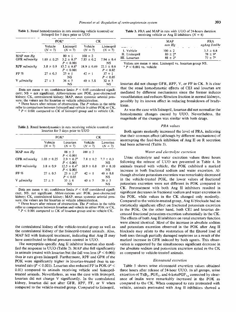
UUO in rats receiving vehicle for five days prior to UUOexhibited a profound reduction in GFR (—86%), RPF (—78%),FF (—36%), and V (—36%) upon release of the obstruction(P0K) without changes in MAP (Table 1).
As can also be seen in Table 1, RPF and GFR in thepost-obstructed as compared to the contralateral kidney werestill markedly reduced even after lisinopril treatment. Whencompared to the effects of vehicle treatment on the control(contralateral) kidney, lisinopril produced no change in GFR,an increase in RPF (18%), and a 13% decrease in FF (P < 0.05).
By contrast, in the P0K lisinopril increased GFR and RPFsignificantly when compared to the P0K of vehicle-treated rats.Filtration fraction and V were not significantly altered. It shouldbe noted that, despite the improvement in renal hemodynamicsinduced by lisinopril in P0K, these values were still lower than
Pimentel et a!: Regulation of renin-angiolensin system 393
Table 1. Renal hemodynamics in rats receiving vehicle (control) orlisinopril for 5 days prior to UUO
POKE CK
Vehicle Lisinopril Vehicle Lisinopril(N=7) (N=7) (N=7) (N=7)
MAP mm HgGFR mi/mm/kg 1.03 0.25
80 1
3.2 0,3bP < 0.001
100 27.85 0.2 7.94 0.4
NSRPF mI/mm/kg 3.8 0.9 13.2 0.8"
P < 0.00118.9 0.69 22.1 0.8
P < 0.02FF% 27±0.5 25±1
NS42±1 37±1
P<0.05V pJ/mmnlkg 27 3 36 5
NS40 5.8 32 3
NS
Data are mean SE; confidence limits P < 0.05 considered signifi-cant; NS = not significant. Abbreviations are: P0K, post-obstructedkidney; CK, contralateral kidney; MAP, mean systemic arterial pres-sure; the values are for losartan or vehicle administration.
a Three hours after release of obstruction. The P values in the tablerefer to comparison between lisinopril and vehicle in either P0K or CK.'P < 0.001 compared to CK of lisinopril group and to vehicle CK.
Table 2. Renal hemodynamics in rats receiving vehicle (control) orlosartan for 5 days prior to UUO
P0Ka CK
Vehicle(N=7)
Losartan(N=7)
Vehicle(N=7)
Losartan(N=7)
MAP mm Hg
GFR mI/mm/kg
RPF mI/mm/kg
FF%V pJ/min/kg
1,03 0.25
3.8 0.9
27 0.5
27 3
88 2P < 0.0012.9 0.2"P < 0.00112.5 0.4"P < 0.00123 1.3"P<0.0535 6
NS
100 2
7.8 0.2
18.9 0.8
42 1
40 5
7.7 0.3NS
19 0.10NS
40 0.8NS
39 5NS
Data are mean sa; confidence limits P < 0.05 considered signifi-cant, NS, not significant. Abbreviations are: P0K, post-obstructedkidney; CK, contralateral kidney; MAP, mean systemic arterial pres-sure; the values are for losartan or vehicle administration.
aThree hours after release of obstruction. The P values in the tablerefer to comparison between losartan and vehicle in either P0K or CK.
b P < 0.001 compared to CK of losartan group and to vehicle CK,
the contralateral kidney of the vehicle-treated group as well asthe contralateral kidney of the lisinopril-treated animals. Also,MAP fell with lisinopril treatment, indicating that Ang II mayhave contributed to blood pressure control in UUO.
The nonpeptide-specific Ang II inhibitor losartan also modi-fied the response to UUO (Table 2). MAP also fell significantlyin animals treated with losartan but the fall was less (P < 0.001)than in rats given lisinopril. Furthermore, RPF and GFR of theP0K were significantly higher in losartan-treated than in un-treated rats (P <0.001). Losartan also reduced FF in P0K (P <0.01) compared to animals receiving vehicle and lisinopril-treated animals. Nevertheless, as was the case with lisinopril,losartan did not change V significantly. In the contralateralkidney, losartan did not alter GFR, RPF, FF, or V whencompared to the vehicle-treated group. Compared to lisinopril,
Table 3. PRA and MAP in rats with UUO of 24-hours durationreceiving vehicle or Ang II inhibitors (N = 6)
MAPmm Hg
PRAngAng I/mi/hr
I. Vehicle 100 2 3.5 0.8II. Lisinopril 80 2 78 9
III. Losartan 88 2 77 7a
Values are mean SEM. Lisinopril vs. losartan group NS.a P < 0.001 vs. vehicle
losartan did not change GFR, RPF, V, or FF in CK. It is clearthat the renal hemodynamic effects of CEI and losartan aremediated by different mechanisms since the former inducesvasodilatation and reduces filtration fraction in normal kidneys,possibly by its known effect in reducing breakdown of brady-kinin.
As was the case with lisinopril, losartan did not normalize thehemodynamic changes caused by UUO. Nevertheless, themagnitude of the changes was similar with both drugs.
PRA values
Both agents markedly increased the level of PRA, indicatingthat their common effect (although by different mechanisms) ofinterrupting the feed-back inhibition of Ang II on R secretionhad been achieved (Table 3).
Water and electrolyte excretionUrine electrolyte and water excretion values three hours
following the release of UUO are presented in Table 4. Inanimals treated with vehicle, the P0K exhibited a markedincrease in both fractional sodium and water excretion. Al-though absolute potassium excretion was remarkably decreasedin the vehicle-treated P0K, the lower values of fractionalpotassium excretion were not different in P0K compared toCK. Pretreatment with both Ang II inhibitors resulted insignificant decreases in fractional sodium and water excretion inthe P0K, while values in the CK changed only modestly.Compared to the vehicle-treated group, Ang II blockade had nostatistically significant effect on fractional potassium excretionin the P0K. On the other hand, both CEI and losartan de-creased fractional potassium excretion substantially in the CK.The effects of both Ang II inhibitors on renal excretory functionwere almost identical. Most of the increase in absolute sodiumand potassium excretion observed in the P0K after Ang IIblockade may relate to the restoration of the filtered load ofboth ions through partially damaged nephrons as a result of themarked increase in GFR induced by both agents. This obser-vation is supported by the simultaneous significant decrease inthe absolute sodium and potassium excretion noted in the CKas compared to vehicle-treated animals.
Eicosanoid excretionTable 5 shows urine eicosonoid excretion values obtained
three hours after release of 24-hour UUO. In all groups, urineexcretion of TxB2, PGE2, and 6-ketoPGF1,, corrected by clear-ance of inulin were remarkably increased in the P0K ascompared to the CK. When compared to rats pretreated withvehicle, animals pretreated with Ang II inhibitors showed a
60
30
20 -
10-
0-Cortical Juxtarnedullary
394 Pimentel et a!: Regulation of renin-angiotensin system
Table 4. Effect of Ang II inhibitors on electrolyte and water excretion following release of 24-hour UU0
UNaV p.Eq/min/kg UKY p.Eq/min/kgP0K CK
FENa % FEK % FEH20 %P0K CK P0K CK P0K CK P0K CK
I 2.87 0.39 2,47 0.2 0.86 0.1 5.86 0.7 2.38 0.65 0.22 0.03 13.88 1.6 15.10 1.8 3.12 0.7 0.55 0.04P MS <0.001 <0.001 NS <0.007
II 3,54 0.3 1.47 0.36 1.99 0.2 3.40 0.63 0.89 0.2 0.35 0.1 11.47 2.3 7.55 1.7 1.22 0.2 0.40 0.02p <0.03 <0.05 <0.04 NS <0.001
III 3.61 0.5 1.27 0.4 1.60 0.2 3.65 0.7 0.89 0.1 0.25 0.06 10.8 1.1 8.9 1.4 1.12 0.2 0.50 0.06p <0.005 <0.02 <0.001 MS <0.02
PT vs. II NS < 0.05 <0.01 <0.02 <0.02 NS NS < 0.02 <0.02 <0.006P I vs. III NS <0.04 <0.02 <0.02 <0.02 MS NS <0.02 <0.01 NSP11 vs. III NS MS MS NS NS MS NS NS NS NS
Values are mean standard error of the mean in 7 rats. Groups I, II, III are as in Table 3.Abbreviations are: UNaV, absolute sodium excretion; UKV, absolute potassium excretion; FENa, fractional sodium excretion; FEK, fractional
potassium excretion; FEH20, fractional H20 excretion; P0K, post-obstructive kidney; CK, contralateral normal kidney.
Table 5. Urine eicosanoid excretion three hours after release of 24-hour UUO in rats receiving vehicle or Ang II inhibitors
NPOE2 6-keto-PGF1a TxB2
pg/mi Gin
Postobstructive kidneyVehicle (I) 6 2753 43S' 1500 334 155 39Lisinopril (II) 7 320 66a,d 175 3lC 22 3Losartan (III) 7 98 13a,b 195 31C 30P1 vs. II <0.001 <0.004 <0.003P1 vs. III <0.001 <0.004 <0.004P11 vs. III <0.008 MS NS
Contralateral kidneyVehicle(I) 6 54±5 149±37 9±2Lisinopril (II) 7 55 3 148 47 10 2Losartan (III) 7 50 16 144 35 14 5PIvs. H NS NS MSPIvs. III NS MS NSP II vs. III MS MS MS
Values are mean 5EM. Abbreviations are: MS, nonsignificant;POE2, prostaglandin E2; TxB2, thromboxane B2; 6-keto-POF1a, 6-ke-toprostaglandin Fin.
a MS vs. CK of all groupsb P < 0.001 vs. CK of all groupsc P < 0.05 vs. CK of all groupsd P < 0.004 vs. CK of vehicle-treated rats
CC11)
a)2C)(Ua)0C=FF
CC-D
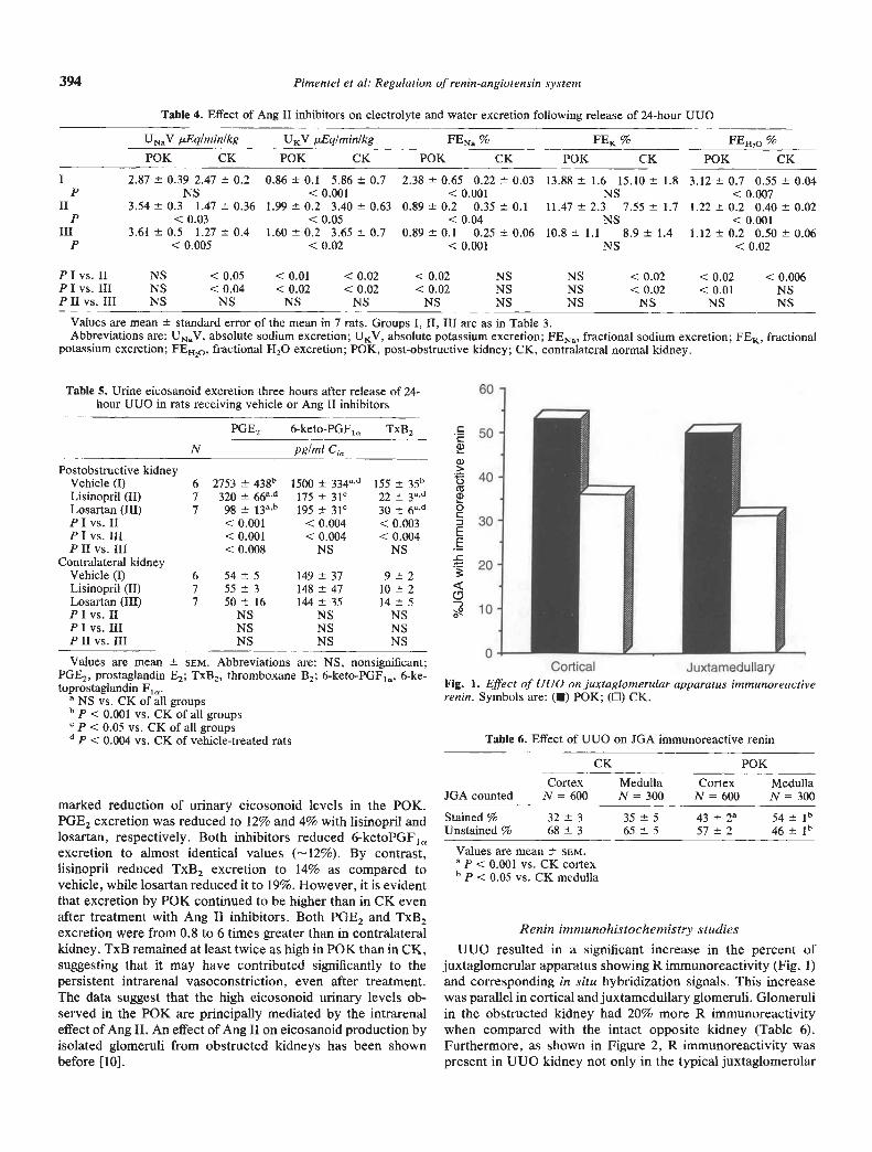
Fig. 1. Effect of UUO on juxtaglomerular apparatus immunoreactiverenin. Symbols are: (R) P0K; (D) CK.
CK P0KCortex Medulla Cortex Medulla
JOAcounted N=600 N= 300 N= 600 N= 300
Stained% 32±3 35±5 43±2a 541bUnstained% 68±3 65±5 57±2 461b
Table 6. Effect of UUO on JGA immunoreactive renin
marked reduction of urinary eicosonoid levels in the P0K.POE2 excretion was reduced to 12% and 4% with lisinopril andlosartan, respectively. Both inhibitors reduced 6-ketoPOF1excretion to almost identical values (—12%). By contrast,lisinopril reduced TxB2 excretion to 14% as compared tovehicle, while losartan reduced it to 19%. However, it is evidentthat excretion by P0K continued to be higher than in CK evenafter treatment with Ang II inhibitors. Both POE2 and TxB2excretion were from 0.8 to 6 times greater than in contralateralkidney. TxB remained at least twice as high in P0K than in CK,suggesting that it may have contributed significantly to thepersistent intrarenal vasoconstriction, even after treatment.The data suggest that the high eicosonoid urinary levels ob-served in the P0K are principally mediated by the intrarenaleffect of Ang II. An effect of Ang II on eicosanoid production byisolated glomeruli from obstructed kidneys has been shownbefore [10].
Values are mean SCM.a P < 0.001 vs. CK cortexb p < 0.05 vs. CK medulla
Renin immunohistochemistry studiesUUO resulted in a significant increase in the percent of
juxtaglomerular apparatus showing R immunoreactivity (Fig. 1)and corresponding in situ hybridization signals. This increasewas parallel in cortical and juxtamedullary glomeruli. Olomeruliin the obstructed kidney had 20% more R immunoreactivitywhen compared with the intact opposite kidney (Table 6).Furthermore, as shown in Figure 2, R immunoreactivity waspresent in UUO kidney not only in the typical juxtaglomerular
S: '- 4a -- 1-
• - — at --
•4
aa -'• --
4
— • -
• t 4
• a
i.
4
a!
• I—
4
-.4.I — • '1 ••'
-'I.
Pimentel et a!: Regulation of renin-angiotensin system 395
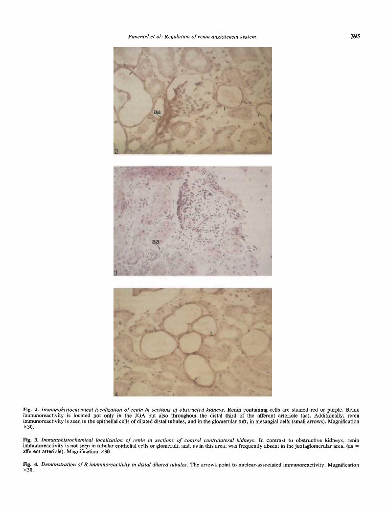
Fig. 2. Immunohistochemical localization of renin in sections of obstructed kidneys. Renin containing cells are stained red or purple. Reninimmunoreactivity is located not oniy in the JGA but also throughout the distal third of the afferent arteriole (aa). Additionally, reninimmunoreactivity is seen in the epithelial cells of dilated distal tubules, and in the glomerular tuft, in mesangial cells (small arrows). Magnificationx 30.
Fig. 3. Immunohistochemical localization of renin in sections of control contralateral kidneys. In contrast to obstructive kidneys, reninimmunoreactivity is not seen in tubular epithelial cells or glomeruli, and, as in this area, was frequently absent in the juxtaglomerular area. (aa =afferent arteriole). Magnification x30.
Fig. 4. Demonstration of R immunoreactivity in distal dilated tubules. The arrows point to nuclear-associated immunoreactivity. Magnificationx30.
396 Pimentel et al: Regulation of renin-angiotensin system
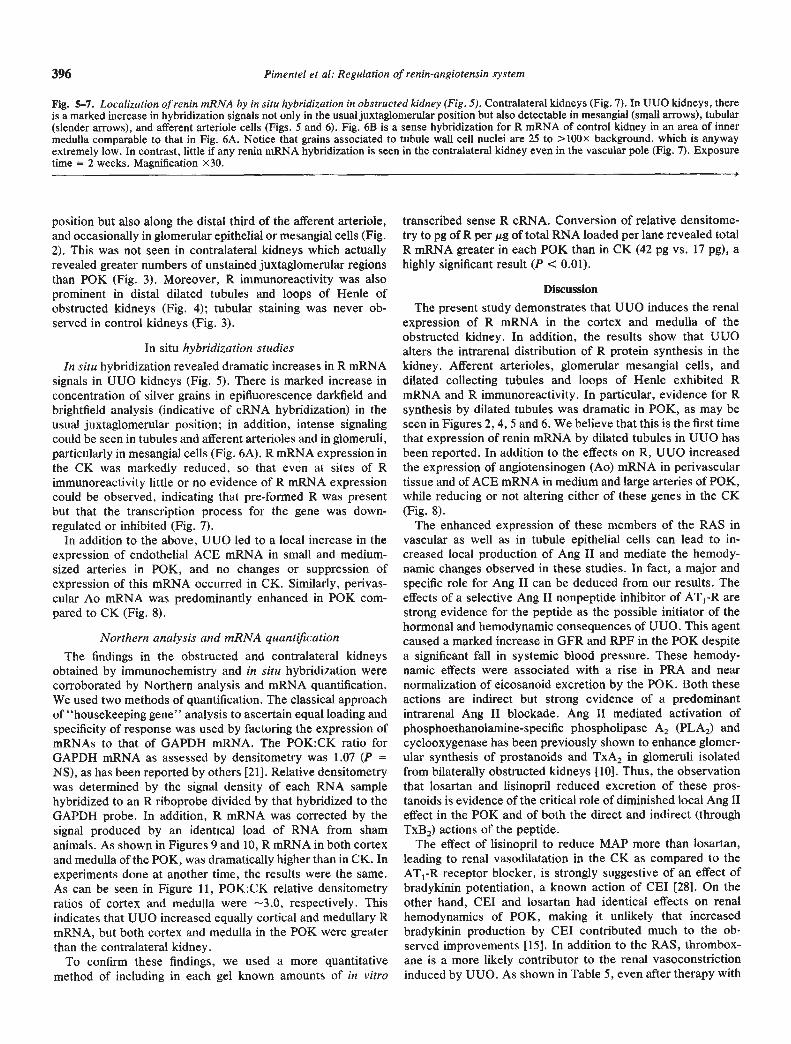
Fig. 5—7. Localization of renin mRNA by in situ hybridization in obstructed kidney (Fig. 5). Contralateral kidneys (Fig. 7). In UUO kidneys, thereis a marked increase in hybridization signals not only in the usual juxtaglomerular position but also detectable in mesangial (small arrows), tubular(slender arrows), and afferent arteriole cells (Figs. 5 and 6). Fig. 6B is a sense hybridization for R mRNA of control kidney in an area of innermedulla comparable to that in Fig. 6A. Notice that grains associated to tubule wall cell nuclei are 25 to > bOx background, which is anywayextremely low. In contrast, little if any renin mRNA hybridization is seen in the contralateral kidney even in the vascular pole (Fig. 7). Exposuretime = 2 weeks. Magnification x30.
position but also along the distal third of the afferent arteriole,and occasionally in glomerular epithelial or mesangial cells (Fig.2). This was not seen in contralateral kidneys which actuallyrevealed greater numbers of unstained juxtaglomerular regionsthan P0K (Fig. 3). Moreover, R immunoreactivity was alsoprominent in distal dilated tubules and loops of Henle ofobstructed kidneys (Fig. 4); tubular staining was never ob-served in control kidneys (Fig. 3).
In situ hybridization studiesIn situ hybridization revealed dramatic increases in R mRNA
signals in UUO kidneys (Fig. 5). There is marked increase inconcentration of silver grains in epifluorescence darkfield andbrightfield analysis (indicative of cRNA hybridization) in theusual juxtaglomerular position; in addition, intense signalingcould be seen in tubules and afferent arterioles and in glomeruli,particularly in mesangial cells (Fig. 6A). R mRNA expression inthe CK was markedly reduced, so that even at sites of Rimmunoreactivity little or no evidence of R mRNA expressioncould be observed, indicating that pre-formed R was presentbut that the transcription process for the gene was down-regulated or inhibited (Fig. 7).
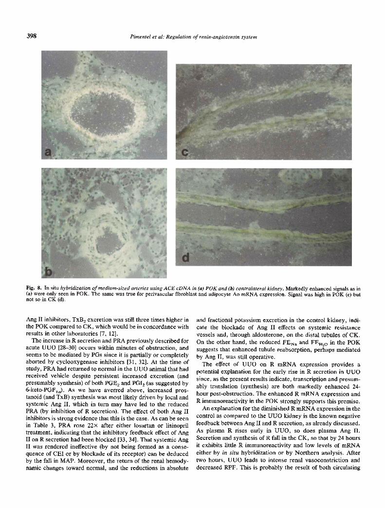
In addition to the above, UUO led to a local increase in theexpression of endothelial ACE mRNA in small and medium-sized arteries in P0K, and no changes or suppression ofexpression of this mRNA occurred in CK. Similarly, perivas-cular Ao mRNA was predominantly enhanced in P0K com-pared to CK (Fig, 8).
Northern analysis and mRNA quantjficationThe findings in the obstructed and contralateral kidneys
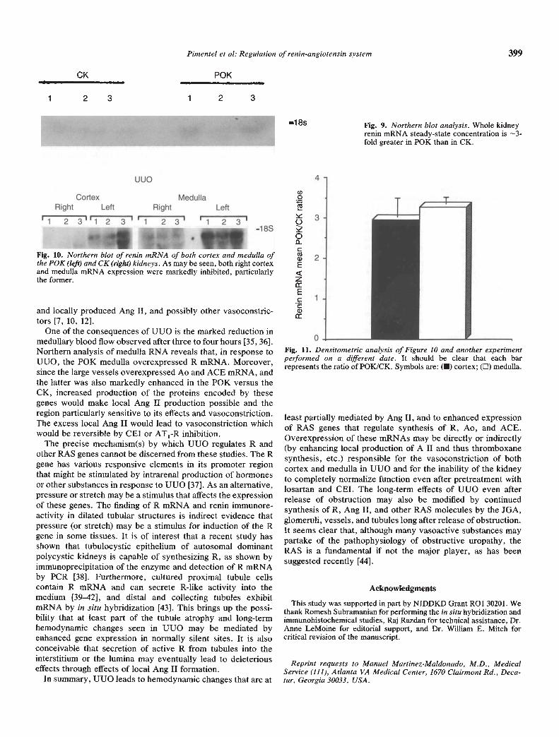
obtained by immunochemistry and in situ hybridization werecorroborated by Northern analysis and mRNA quantification.We used two methods of quantification. The classical approachof "housekeeping gene" analysis to ascertain equal loading andspecificity of response was used by factoring the expression ofmRNAs to that of GAPDH mRNA. The POK:CK ratio forGAPDH mRNA as assessed by densitometry was 1.07 (P =NS), as has been reported by others [211. Relative densitometrywas determined by the signal density of each RNA samplehybridized to an R riboprobe divided by that hybridized to theGAPDH probe. In addition, R mRNA was corrected by thesignal produced by an identical load of RNA from shamanimals. As shown in Figures 9 and 10, R mRNA in both cortexand medulla of the P0K, was dramatically higher than in CK. Inexperiments done at another time, the results were the same.As can be seen in Figure 11, POK:CK relative densitometryratios of cortex and medulla were --3.0, respectively. Thisindicates that UUO increased equally cortical and medullary RmRNA, but both cortex and medulla in the P0K were greaterthan the contralateral kidney.
To confirm these findings, we used a more quantitativemethod of including in each gel known amounts of in vitro
transcribed sense R cRNA. Conversion of relative densitome-try to pg of R per g of total RNA loaded per lane revealed totalR mRNA greater in each P0K than in CK (42 pg vs. 17 pg), ahighly significant result (P < 0.01).
Discussion
The present study demonstrates that UUO induces the renalexpression of R mRNA in the cortex and medulla of theobstructed kidney. In addition, the results show that UUOalters the intrarenal distribution of R protein synthesis in thekidney. Afferent arterioles, glomerular mesangial cells, anddilated collecting tubules and loops of Henle exhibited RmRNA and R immunoreactivity. In particular, evidence for Rsynthesis by dilated tubules was dramatic in P0K, as may beseen in Figures 2, 4, 5 and 6. We believe that this is the first timethat expression of renin mRNA by dilated tubules in UUO hasbeen reported. In addition to the effects on R, UUO increasedthe expression of angiotensinogen (Ao) mRNA in perivasculartissue and of ACE mRNA in medium and large arteries of P0K,while reducing or not altering either of these genes in the CK(Fig. 8).
The enhanced expression of these members of the RAS invascular as well as in tubule epithelial cells can lead to in-creased local production of Ang II and mediate the hemody-namic changes observed in these studies. In fact, a major andspecific role for Ang II can be deduced from our results. Theeffects of a selective Ang II nonpeptide inhibitor of AT1-R arestrong evidence for the peptide as the possible initiator of thehormonal and hemodynamic consequences of UUO. This agentcaused a marked increase in GFR and RPF in the P0K despitea significant fall in systemic blood pressure. These hemody-namic effects were associated with a rise in PRA and nearnormalization of eicosanoid excretion by the P0K. Both theseactions are indirect but strong evidence of a predominantintrarenal Ang II blockade. Ang II mediated activation ofphosphoethanolamine-specific phospholipase A2 (PLA2) andcyclooxygenase has been previously shown to enhance glomer-ular synthesis of prostanoids and TxA2 in glomeruli isolatedfrom bilaterally obstructed kidneys [10]. Thus, the observationthat losartan and lisinopril reduced excretion of these pros-tanoids is evidence of the critical role of diminished local Ang IIeffect in the P0K and of both the direct and indirect (throughTxB2) actions of the peptide.
The effect of lisinopril to reduce MAP more than losartan,leading to renal vasodilatation in the CK as compared to theAT1-R receptor blocker, is strongly suggestive of an effect ofbradykinin potentiation, a known action of CEI [281. On theother hand, CEI and losartan had identical effects on renalhemodynamics of P0K, making it unlikely that increasedbradykinin production by CEI contributed much to the ob-served improvements [15]. In addition to the RAS, thrombox-ane is a more likely contributor to the renal vasoconstrictioninduced by UUO. As shown in Table 5, even after therapy with
1•
a—
644
a-. a.
—,
398 Pimentel a a!: Regulation of renin-angiotensin system
Fig. 8. In situ hybridization of medium-sized arteries using ACE cDNA in (a) P0K and (b) contralateral kidney. Markedly enhanced signals as in(a) were only seen in P0K. The same was true for perivascular fibroblast and adipocyte Ao mRNA expression. Signal was high in P0K (c) butnot so in CK (d).
Ang II inhibitors, TxB2 excretion was still three times higher inthe P0K compared to CK, which would be in concordance withresults in other laboratories [7, 12].
The increase in R secretion and PRA previously described foracute UUO [28—30] occurs within minutes of obstruction, andseems to be mediated by PGs since it is partially or completelyaborted by cyclooxygenase inhibitors [31, 32]. At the time ofstudy, PRA had returned to normal in the UUO animal that hadreceived vehicle despite persistent increased excretion (andpresumably synthesis) of both PGE2 and PGI2 (as suggested by6-keto-PGF1a). As we have averred above, increased pros-tanoid (and TxB) synthesis was most likely driven by local andsystemic Ang II, which in turn may have led to the reducedPRA (by inhibition of R secretion). The effect of both Ang IIinhibitors is strong evidence that this is the case. As can be seenin Table 3, PRA rose 22x after either losartan or lisinopriltreatment, indicating that the inhibitory feedback effect of AngII on R secretion had been blocked [33, 34]. That systemic AngII was rendered ineffective (by not being formed as a conse-quence of CEI or by blockade of its receptor) can be deducedby the fall in MAP. Moreover, the return of the renal hemody-namic changes toward normal, and the reductions in absolute
and fractional potassium excretion in the control kidney, indi-cate the blockade of Ang II effects on systemic resistancevessels and, through aldosterone, on the distal tubules of CK.On the other hand, the reduced FENa and FEB20 in the P0Ksuggests that enhanced tubule reabsorption, perhaps mediatedby Ang II, was still operative.
The effect of UUO on R mRNA expression provides apotential explanation for the early rise in R secretion in UUOsince, as the present results indicate, transcription and presum-ably translation (synthesis) are both markedly enhanced 24-hour post-obstruction. The enhanced R mRNA expression andR immunoreactivity in the P0K strongly supports this premise.
An explanation for the diminished R mRNA expression in thecontrol as compared to the UUO kidney is the known negativefeedback between Ang II and R secretion, as already discussed.As plasma R rises early in UUO, so does plasma Ang II.Secretion and synthesis of R fall in the CK, so that by 24 hoursit exhibits little R immunoreactivity and low levels of mRNAeither by in situ hybridization or by Northern analysis. Aftertwo hours, UUO leads to intense renal vasoconstriction anddecreased RPF. This is probably the result of both circulating
uuo
Cortex MedullaRight Left Right Left
'1 2 3''l 2 3''l 23' '1 2 3'48S
CK
Pimentel et a!: Regulation of renin-angiotensin system 399
P0K
1 2 3 1 2 3
—1 8s Fig. 9. Northern blot analysis. Whole kidneyrenin mRNA steady-state concentration is —3-fold greater in P0K than in CK.
and locally produced Ang II, and possibly other vasoconstric-tors [7, 10, 12].
One of the consequences of UUO is the marked reduction inmedullary blood flow observed after three to four hours [35, 36].Northern analysis of medulla RNA reveals that, in response toUUO, the P0K medulla overexpressed R mRNA. Moreover,since the large vessels overexpressed Ao and ACE mRNA, andthe latter was also markedly enhanced in the P0K versus theCK, increased production of the proteins encoded by thesegenes would make local Ang II production possible and theregion particularly sensitive to its effects and vasoconstriction.The excess local Ang II would lead to vasoconstriction whichwould be reversible by CEI or AT1-R inhibition.
The precise mechanism(s) by which UUO regulates R andother RAS genes cannot be discerned from these studies. The Rgene has various responsive elements in its promoter regionthat might be stimulated by intrarenal production of hormonesor other substances in response to UUO [37]. As an alternative,pressure or stretch may be a stimulus that affects the expressionof these genes. The finding of R mRNA and renin immunore-activity in dilated tubular structures is indirect evidence thatpressure (or stretch) may be a stimulus for induction of the Rgene in some tissues. It is of interest that a recent study hasshown that tubulocystic epithelium of autosomal dominantpolycystic kidneys is capable of synthesizing R, as shown byimmunoprecipitation of the enzyme and detection of R mRNAby PCR [38]. Furthermore, cultured proximal tubule cellscontain R mRNA and can secrete R-like activity into themedium [39—42], and distal and collecting tubules exhibitmRNA by in situ hybridization [43]. This brings up the possi-bility that at least part of the tubule atrophy and long-termhemodynamic changes seen in UUO may be mediated byenhanced gene expression in normally silent sites. It is alsoconceivable that secretion of active R from tubules into theinterstitium or the lumina may eventually lead to deleteriouseffects through effects of local Ang II formation.
In summary, UUO leads to hemodynamic changes that are at
least partially mediated by Ang II, and to enhanced expressionof RAS genes that regulate synthesis of R, Ao, and ACE.Overexpression of these mRNAs may be directly or indirectly(by enhancing local production of A II and thus thromboxanesynthesis, etc.) responsible for the vasoconstriction of bothcortex and medulla in UUO and for the inability of the kidneyto completely normalize function even after pretreatment withlosartan and CEI. The long-term effects of UUO even afterrelease of obstruction may also be modified by continuedsynthesis of R, Ang II, and other RAS molecules by the JGA,glomeruli, vessels, and tubules long after release of obstruction.It seems clear that, although many vasoactive substances maypartake of the pathophysiology of obstructive uropathy, theRAS is a fundamental if not the major player, as has beensuggested recently [44].
Fig. 10. Northern blot of renin mRNA of both cortex and medulla ofthe P0K (left) and CK (right) kidneys. As may be seen, both right cortexand medulla mRNA expression were markedly inhibited, particularlythe former.
0
Fig. 11. Densitometric analysis of Figure 10 and another experimentperformed on a different date. It should be clear that each barrepresents the ratio of POK/CK. Symbols are: (U) cortex; (EJ) medulla.
Acknowledgments
This study was supported in part by NIDDKD Grant ROl 30201. Wethank Romesh Subramanian for performing the in situ hybridization andimmunohistochemical studies, Raj Razdan for technical assistance, Dr.Anne LeMoine for editorial support, and Dr. William E. Mitch forcritical revision of the manuscript.
Reprint requests to Manuel Martinez-Maldonado, M.D., MedicalService (111), Atlanta VA Medical Center, 1670 Clairmont Rd., Deca-tur, Georgia 30033, USA.
400 Pimentel et a!: Regulation of renin-angiotensin system
References
1. SIEGEL NJ, UPADHAYA K, KASHGARIAN M: Inhibition by indo-methacin of adaptive changes in the contralateral kidney afterrelease of unilateral ureteral occlusion. Kidney mt 20:691—694, 1981
2. EL-DAHR SS, SAMIR-SAYEM R, GOMEZ RA, Gitv MS, PEACH MJ,CAREY RM, CHEVALIER RL: In situ localization of renin and itsmRNA in neonatal ureteral obstruction. Am J Physiol 258:F854—F862, 1990
3. CHEVALIER RL, GOMEZ RA: Response of the renin-angiotensinsystem to relief of neonatal ureteral obstruction. Am J Physiol255:F1070—1077, 1988
4. SUKI WN, GUTHRIE AG, MARTINEZ-MALDONADO M, EKNOYAN0: Effects of ureteral pressure elevation on renal hemodynamicsand urine concentration. Am J Physiol 220:38—44, 1971
5. NASH PD, SELKURT FE: Effects of elevated ureteral pressure onrenal blood flow. (abstract) Circ Res 14:1142, 1964
6. MOODY TE, VAUGHAN ED, GILLENWATER JY Relationship be-tween renal blood flow and urereteral pressure during 18 hours oftotal unilateral ureteral occlusion: Implications for changing sites ofincreased renal resistance. Invest Urol 13:246—251, 1975
7. KLAHR 5: Pathophysiology of obstructive nephropathy: A 1990update. Semin Nephrol 11:156—168, 1991
8. REYES AA, MARTIN D, SETTLE 5, KLAHR S: EDRF: Role in renalfunction and blood pressure of normal rats and rats with obstructiveuropathy. Kidney mt 41:403—403, 1992
9. VAUGHAN ED, SWEET RC, GILLENwATER JY: Peripheral reninand blood pressure changes following complete unilateral ureteralocclusion. (abstract) J Urol 104:89, 1970
10. YANAGISAWA H, MORRISSEY J, MORRISON AR, PURKERSON ML,KLAHR 5: Role of Ang II in eicosanoid production by isolatedglomeruli from rats with bilateral ureteral obstruction. Am J Physiol258:F85—F93, 1990
11. KLAHR 5, HAsuus K, PURKERSON M: Effects of obstruction onrenal functions. Pediatr Nephrol 2:34—42, 1988
12. YARGER WE, SCHOCKEN DD, HARRIS RH: Obstructive nephropa-thy in the rat: Possible roles for the renin-angiotensin system,prostaglandins, and TBs in postobstructive renal function. J ClinInvest 65:400—412, 1980
13. ICHIKAWA I, PURKERSON ML, YATES J, KLAHR S: Dietary proteinintake conditions the degree of renal vasoconstriction in acute renalfailure caused by ureteral obstruction. Am J Physiol F54—F61, 1985
14. EL-DAHR SS, GOMEZ RA, KHARE 0, PEACH MJ, CAREY RM,CHEVALIER RL: Expression of renin and its mRNA in the adult ratkidney with chronic ureteral obstruction. Am J Kidney Dis 15:575—582, 1990
15. WONG PC, PRICE WA, CHIU AT, DUNCIA JV, CARINI DJ, WEXLERRR, JOHNSON AL, TIMMERMANS PB: Nonpeptide angiotensin IIreceptor antagonists. IX: Antihypertensive activity in rats ofDuP753, an orally active antihypertensive agent. J Pharmacol ExpTher 252:726—732, 1990
16. CHIRGWIN JJ, PRZBYLA AE, MACDONALD RJ, RUTTER WJ: Isola-tion of biologically active ribonucleic acid from sources enriched inribonuclease. (abstract) Biochemistry 18:5294, 1979
17. LYNCH KR, SIMNAD VI, LEN-ARI ET, GARRISON JC: Localizationof preangiotenlinogen mRNA sequences in the rat brain. Hyper-tension 8:540—543, 1986
18, FIELD Li, MCGOWAN RA, DICKINSON DP, GROSS KW: Tissue andgene specificity of mouse renin expression. Hypertension 6:597—603, 1984
19, BERNSTEIN KE, MARTIN BM, BERNSTEIN EA, LINTON J, STRIKERL, STRIKER 0: The isolation of angiotensin-converting enzymecDNA. JBiol Chem 263:11021—11024, 1988
20. ALEXANDER M, LOMANTO M, NASRIN N, RAMAIKA C: InsulinStimulates glyceraldehyde-3-phosphate dehydrogenase gene ex-pression through cis-acting DNA sequences. Proc Nat! Acad SciUSA 85:5092—5096, 1988
21. STORCH 5, SAGGI 5, MEGYSEI J, PRICE PM, SAFIRSTEIN R:Ureteral obstruction decreases renal preproepidermal growth fac-tor and Tamm-Horsfall expression. Kidney mt 42:89—94, 1992
22. GOMEZ RA, LYNCH KR, STURGILL BC, ELWOOD JP, CHEVALIERRL, CAREY RM, PEACH MJ: Distribution of renin mRNA and itsprotein in the developing kidney. Am J Physiol 257:F850—F858,1989
23. GOMEZ RA, LYNCH KR, CHEVALIER RL, EVERETT AD, JOHNS
DW, WILFONG N, PEACH Mi, CAREYRM: Reninand angiotensino-gen gene expression and intrarenal renin distribution during ACEinhibition. Am J Physiol 254:F900—F906, 1988
24. WILCoX JN, SMITH KM, WILLIAMS LT, SCHWARTZ 5, GORDON D:Platelet-derived growth factor mRNA detection in human athero-sclerotic plaques by in situ hybridization. J Clin Invest 82:1134—1143, 1988
25. WILCOX iN, SMITH KM, SCHWARTZSM, GORDOND: Localizationof tissue factor in the normal vessel wall and in the atheroscleroticplaque. Proc Nat! Acad Sci USA 86:2839—2843, 1989
26. FERNANDEZ-REPOLLET E, TAPIA E, MARTINEZ-MALDONADO M:Effects of aagiotensin-converting enzyme inhibition on alteredrenal hemodynamics induced by low protein diet in the rat. J C/inInvest 80:1045—1049, 1987
27. HABER E, KOERNER T, PAGE LB, KLIMAN B, PURNODE A:Applications of radioimmunoassay for angiotensin Ito the physio-logic measurement of plasma renin activity in normal humansubjects. J C/in Endocrino! Metab 29:1349—1355, 1969
28. CARLSON EL, SPARKS HV: Intrarenal distribution of blood flowduring elevation of ureteral pressure in dogs. (abstract) Circ Res27:601, 1970
29. VANDER AJ: Renin secretion during mannitol diuresis and ureteralocclusion. (abstract) Proc Soc Exp Biol Med 128:518, 1968
30. KAL0YANIDES GJ, BASTRON RD, DIBONA OF: Effect of ureteralclamping and increased renal arterial pressure on renin release. AmJ Physio! 225:95—99, 1973
31. BLACKSHEAR JL, WATHEN RL: Effects of indomethacin on renalblood flow and renin Secretory responses to ureteral occlusion inthe dog. Miner E!ectro! Metab 1:271—278, 1978
32. CADNAPAPHORNCHAI P, AISENBREY0, MCDONALD KM, BURKETJ, SCHRIER RW: Prostaglandin-mediated hyperemia and ream-mediated hypertension during acute ureteral obstruction. Pros-taglandins 16:965—971, 1978
33. NAKAMURA A, IWAO H, FUKUIK, KIMURA5, TAMAKI T, NAKAN-ISHI 5, ABE Y: Regulation of liver angiotensinogen and kidney reninmRNA levels by angiotenSin II. Am J Physio! 21 :E1—E6, 1990
34. DZAU Vi, BURT DW, PRAn RE: Molecular biology of the renin-angiotensin system. Am J Physiol 255(Renal Fluid Electrol PhySiol24):F563—F573, 1988
35. SOLEZ K, PONCHAK 5, BUONO RA, VERNON N, FINER PM,MILLERM, HEPTINSTALL RH: Inner medullary plasma flow in thekidney with ureteral obstruction. Am J Physia! 231:1315—1321, 1976
36. WRIGHT FS: Effects of urinary tract obstruction on glomerularfiltration rate and renal blood flow. Semin Nephro! 2:5—16, 1982
37. BAXTER JD, JAMES MNG, CHU W, DUNCAN K, HAIDAR M,CARILLI C, REUDELHUBER T: The molecular biology of humanrenin and its gene. Yale J Rio! Med 62:493—501, 1989
38. TORRES VE, DONOVAN KA, SCICLI 0, HOLLEY KE, THIBODEAUSN, CARRETERO OA, INAGAMI T, MCATEER JA, JOHNSON CM:Synthesis of renin by tubuloCyStic epithelium in autosomal-domi-nant polycystic kidney disease. Kidney Int 42:364—373, 1992
39. YANAGAWA N, CAPPARELLI AW, Jo OD, FRIEDAL A, BARRI JD,EGGENA P: Production of angiotensinogen and renin-like activityby rabbit proximal tubular cells in culture. (abstract) Kidney Int39:941, 1991
40. WIDELL J, ALPERN RJ, HENRICH W: Renin synthesis by culturedproximal tubule cells: Regulation by isoproterenol and forskolin.(abstract) J Am Soc Nephro! 1:672, 1990
41. SHEIKH-HAMAD D, MURRAY 5, EGGENA P.CLEGG K, YANAGAYN: Modulation of renin and angiotensinogen mRNA in culturedrabbit proximal tubular cells (PTC) by Sodium (Na). (abstract) JAmSoc Nephro! 1:744, 1990
42. INGELFINGER JR, BOUYOUNES B, DZAU Vi: Cultured rat proximalrenal tubule (PT) cells express a local angiotensin System. (abstract)JAm Soc Nephro! 1:741, 1990
43. RAJARAMAN 5, GRAVES K, KUNAPULI 5: Identification of the siteof synthesis of angiotensinogen and renin in the kidney by in situhybridization. (abstract) Kidney Int 33:69, 1988
44. REYES AA, KLAHR 5: Renal function after release of ureteralobstruction: Role of endothelin and the renal artery endothelium.Kidney hit 42:632—638, 1992