impaired hierarchical control within the lateral prefrontal cortex in schizophrenia
TRANSCRIPT

i
Impaired Hierarchical Control Within the LateralPrefrontal Cortex in SchizophreniaGuillaume Barbalat, Valerian Chambon, Philippe J.D. Domenech, Chrystèle Ody, Etienne Koechlin,Nicolas Franck, and Chlöé Farrer
Background: In schizophrenia, disturbances of cognitive control have been associated with impaired functional specialization within the lateralprefrontal cortex (LPFC), but little is known about the functional interactions between specialized LPFC subregions. Here, we addressed thisquestion with a recent model that describes the LPFC functioning as a cascade of control processes along a rostrocaudal axis, whereby anteriorfrontal regions influence the processing in posterior frontal regions to guide action selection on the basis of the temporal structure of information.
Methods: We assessed effective connectivity within the rostrocaudal axis of the LPFC by means of functional magnetic resonance imagingin 15 schizophrenic patients and 14 matched healthy control subjects with structural equation modeling and psychophysiologicalinteractions.
Results: In healthy subjects, activity in the left caudal LPFC regions was under the influence of left rostral LPFC regions when controllinginformation conveyed by past events. By contrast, schizophrenic patients failed to demonstrate significant effective connectivity fromrostral to caudal LPFC regions in both hemispheres.
Conclusions: The hierarchical control along the rostrocaudal axis of the LPFC is impaired in schizophrenia. This provides the first evidenceof a top-down functional disconnection within the LPFC in this disorder. This disruption of top-down connectivity from rostral to caudal LPFCregions observed in patients might affect their ability to select the appropriate sets of stimulus-response associations in the caudal LPFC onthe basis of information conveyed by past events. This impaired hierarchical control within the LPFC could result from poorly encoded
contextual information due to abnormal computations in the caudal LPFC.tdmmrscttclsds(
sthscitcp“
sitegs
Key Words: Effective connectivity, functional magnetic resonanceimaging, hierarchical control, lateral prefrontal cortex, rostrocaudalaxis, schizophrenia
I n schizophrenia, disturbances of cognitive control, the ability tocoordinate thoughts and actions in relation to internal goals,have been robustly associated with impaired functional special-
zation within the lateral prefrontal cortex (LPFC) (1– 6). Recentmodels suggest that cognitive control is constructed as a set ofhierarchical modules that involve selecting and maintaining goalsat multiple levels of abstraction, from general task goals at higherlevels (such as watching a movie in the cinema) to concrete motorresponses at the lowest levels (such as taking transport to go to thecinema, buying a ticket at the box office, or sitting comfortably infront of the screen) (7). Such a behavioral hierarchy has been shownto be subserved by a hierarchical organization along the rostrocau-dal axis of the LPFC, where more anterior regions are associatedwith progressively more abstract action control, whereas more pos-terior regions process more concrete information about action (i.e.,action that is closer to the actual motor output) (8). Furthermore,there seems to be a dominance relationship whereby more anteriorregions that process abstract, superordinate, domain-general rules,modulate domain-specific, subordinate, posterior regions (9).
From the Centre de Neuroscience Cognitive, (GB, VC, PD, NF), UMR 5229CNRS, Université Claude Bernard Lyon1, Lyon, France; Centre Hospitalierle Vinatier (NF), Lyon; Institut National de la Sante Et de la RechercheMedicale (CO, EK), Université Pierre et Marie Curie and École NormaleSupérieure, Paris; and Université de Toulouse (CF), CerCo, UPS andUMR5549 CNRS, Faculté de Médecine de Rangueil, Toulouse, France.
Authors VC and PJDD contributed equally to this work.Address correspondence to Guillaume Barbalat, M.D., Ph.D. Centre deNeu-
roscience Cognitive Universite Claude Bernard Lyon, 67, bd Pinel, 69 675BRON, Cedex, France; E-mail: [email protected].
nReceived Dec 30, 2009; revised Jan 31, 2011; accepted Feb 1, 2011.
0006-3223/$36.00doi:10.1016/j.biopsych.2011.02.009
We previously investigated the overall organization of cognitive con-rol within the LPFC in schizophrenia with an influential model (10) thatescribes the architecture of cognitive control as a cascade of executiveodulesrangingfrompremotortomoreanteriorLPFCregions(3,11).Thisodel includes a sensory control level involved in selecting the motor
esponses that are the most appropriate to stimuli that occur and sub-ervedbythelateralpremotorregions(typically,BrodmannArea[BA]6).Aontextualcontrol level istheninvolvedinselectingpremotorrepresenta-ions (i.e., stimulus-response associations) according to contextual signalshataccompanytheoccurrenceofstimuli.Thiscontrol issubservedbytheaudalpartoftheLPFC(typically,BAs9/44/45).Finally,theepisodiccontrol
evel is involvedinselectingcaudalLPFCrepresentations(task-setsorcon-istent sets of stimulus-response associations evoked in the same imme-iate, perceptual context) according to the temporal episode in whichtimuli occur. This control is subserved by the rostral part of the LPFCtypically, BAs 46/10).
We demonstrated that, although the lower-order, less abstract,ensory level of cognitive control was spared in schizophrenia, con-extual control was significantly impaired (11), which was related toypoactivation in the caudal LPFC regions (3). With regard to epi-odic control, we found mixed but consistent findings. When noontextual signals were involved in the task, there was no behav-
oral disturbance of episodic control in schizophrenia (11). By con-rast, adding contextual signals in the task reduced this level ofognitive control. In other words, this impaired episodic controlrocess refers in fact to a dysfunctional interaction between the
episodic” and the “contextual” modules (3,11).At the neural level, this disturbed episodic control process in
chizophrenic patients was not reflected by any hypoactivationn the rostral LPFC. By contrast, we found a hyperactivation inhis region, which we interpreted as a consequence of the addedffort that patients might expend to retrieve the poorly inte-rated contextual information (2,3,12,13). However, the neuralubstrates underlying this dysfunctional control of episodic sig-
als remain unknown.BIOL PSYCHIATRY 2011;70:73–80© 2011 Society of Biological Psychiatry
abdS
E
iElb
iprsd
eeeat
bocbctlsh(F
icaqasactFlgic
74 BIOL PSYCHIATRY 2011;70:73–80 G. Barbalat et al.
w
According to the functional disconnection hypothesis proposedby Friston (14), such a dysfunctional interaction between two cog-nitive processes should result from dysfunctional interaction in thedynamics of the brain regions subserving these processes ratherthan dysfunctional specialization within a specific region. The fram-ing of the cascade model further predicts that this impairment inepisodic control would depend on the way rostral LPFC exerts itsinfluence on the caudal LPFC regions (9). However, until now, stud-ies that have investigated the interaction between specialized neu-ral systems related to executive dysfunctions in schizophrenia havedemonstrated altered LPFC connectivity with other cortical struc-tures such as the inferior parietal lobule (5), the hippocampus (15),or the anterior cingulate cortex (16) but have not directly studiedthe functional integration of the different cognitive control mod-ules within the LPFC itself.
The goal of this follow-up study was to test whether the per-turbed control of temporal episodic signals in patients reflects adysfunction in the top-down selection of caudal LPFC representa-tions by rostral LPFC. For this purpose, we based our analysis ondata collected in our previously published study (3) and measuredeffective connectivity between LPFC regions involved in control-ling episodic and contextual signals in both groups with structuralequation modeling (SEM) and psychophysiological interactions (PPIs).
Methods and Materials
SubjectsThis analysis initially involved 15 schizophrenic patients (n � 15)
nd 15 matched healthy control subjects, 1 of whom was excludedecause of excessive motion in the scanner (n � 14) (3). For moreetails about the description of the participants, please refer toupplement 1 (see also Table 1).
xperimental ParadigmThe experiment included eight scanning sessions, each consist-
ng of eight separate blocks presented in a counterbalanced order.ach block comprised a series of 12 successive stimuli (colored
etters; duration: 500 msec; onset asynchrony: 3500 msec) precededy an instruction cue lasting 4200 msec (Figure 1). Each instruction
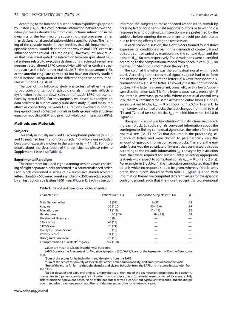
Table 1. Clinical and Demographic Characteristics
Characteristic Patients
Male Gender, n (%) 8 (Age, yrs 35 (Education, yrs 11 (Handedness .8Duration of Illness, yrs 10 (SANS Score 43 (SAPS Score 23 (Reality Distortion Scorea 8 (Poverty Scoreb 34 (Disorganization Scorec 23 (Chlorpromazine-Equivalentd, mg/day 247 (
Values are mean � SD, unless otherwise indicated.SANS, Scale for the Assessment for Negative Sympto
(51).aSum of the scores for hallucinations and delusions fbSum of the scores for poverty of speech, flat affect,cSum of the scores for formal thought disorder and bi
the SANS.dDepot doses of and daily-oral atypical antipsychot
olanzapine in 3 patients, amilsupride in 3 patients, and
chlorpromazine-equivalent doses. None of the patients receiagent, sedative treatment, mood stabilizer, antidepressant, oww.sobp.org/journal
nformed the subjects to make speeded responses to stimuli byressing left or right hand-held response buttons or to withhold a
esponse to a no-go stimulus. Instructions were prelearned by theubjects before running the experiment to avoid possible biasesue to learning effects during the test session.
In each scanning session, the eight blocks formed four distinctxperimental conditions crossing the demands of contextual andpisodic control varied by manipulating the context (Icon) and thepisode (Iepi) factors, respectively. These variations were quantifiedccording to the computational model from Koechlin et al. (10), onhe basis of Shannon’s information theory (17).
The color of the letter was the contextual signal within eachlock. According to the contextual signal, subjects had to performne of three tasks: 1) ignore the letters; 2) a vowel/consonant dis-rimination task (T1: if the letter is a vowel, press the right responseutton; if the letter is a consonant, press left); or 3) a lower/upper-ase discrimination task (T2: if the letter is uppercase, press right; ifhe letter is lowercase, press left). Where contextual control wasow, the task remained the same across the entire block (T1 or T2,ingle-task-set blocks, Icon � 0 bit; block no. 1,2,5,6 in Figure 1). Inigh contextual control blocks, the task changed from trial to trial
T1 and T2, dual task-set blocks, Icon � 1 bit; blocks no. 3,4,7,8 inigure 1).
The episodic signal was by definition the instruction cue preced-ng each block. Episodic signals conveyed information about theontingencies linking contextual signals (i.e., the color of the letter)nd task-sets (i.e., T1 or T2) that occurred in the proceeding se-uence of letters and were chosen to parametrically vary themount of episodic information across blocks. Therefore, the epi-ode factor was the covariate of interest that contrasted episodesccording to the episodic information Iepi conveyed by instructionues that were required for subsequently selecting appropriateask-sets with respect to contextual signals (Iepi � 0 to 1 and 2 bits).or example, in Block No. 1, the instruction cue indicated that, if the
etter is white, no response should be given, whereas if the letter isreen, the subjects should perform task T1 (Figure 1). Then, with
nformation theory, we computed different values for the episodicontrol demand, such that the more frequent the crosstemporal
15) Comparison Subjects (n � 14) p
8 (57) .6836 (10.6) .7911 (1.9) .82
) .84 (.11) .50— —— —— —— —— —— —— —
). SAPS, Scale for the Assessment of Positive Symptoms
he SAPS.onia/asociality, and amotivation from the SANS.
behavior from the SAPS and the score for attention from
the time of the examination (risperidone in 6 patients,prazole in 2 patients) were converted to average daily
(n �
53)10.5)1.3)6 (.099)19)21)10)18)13)190)
ms (50
rom tanhedzarre
ics ataripi
ved a concurrent typical antipsychotic, anticholinergicr other psychotropic agent.

aw
faic8
ac
irS
E
ntrg
S
riameat
cccwsasmp(tiw
P
iL
tn(lmruefLfgIar
R
w
G. Barbalat et al. BIOL PSYCHIATRY 2011;70:73–80 75
contingencies between contextual signals and task-sets, the lowerthe amount of episodic information—thus the lower the demand ofepisodic control. More specifically, the episodic control demanddepended on the proportion f of episodes involving congruent
ssociations between contextual signals and task-sets over thehole experiment. When this proportion was maximal (f � 1, such
as in blocks no. 1,2,3,4 where green always denoted “T1,” red alwaysdenoted “T2,” and white was always “no-go”), the demand of epi-sodic control was low (Iepi � 0 bit). By contrast, the decrease of thisrequency (f � 1, such as in blocks no. 5,6,7,8 where blue, purple,nd yellow could all denote “T1,” “T2,” or “no-go”) led to an increase
n the episodic control demand (Iepi � 0 bit). Because the samerosstemporal contingencies were involved in blocks no.7 and no., these two blocks had a lower episodic control demand (Iepi � 1
bit) than that in blocks no. 5 or no. 6, which contained differentcrosstemporal contingencies (Iepi � 2 bits).
In each block, the proportion of letters to be ignored was 33%. Indual task-set blocks, the ratio of trials associated with task-set T1versus task-set T2 was equal to 1. Finally, in each block, the ratio ofleft versus right responses was equal to 1, and the ratio of congru-
Figure 1. Experimental design. (A) Rounded boxes represent behavioralepisodes (numbered from no. 1 to no. 8) with related stimuli (letters) andinstructions. Episodes formed four distinct experimental conditions cross-ing the episodic factor with the context factor. According to the color of theletter (contextual signal), subjects either ignored the letter or performed avowel/consonant (T1) or lower/uppercase (T2) discrimination task on theletters. Block no. 1: contextual signals were either green or white. Whitesignals indicated that subjects should ignore the letter. Green signals indi-cated that subjects should perform task T1 (single task-set episode). Blockno. 2: contextual signals were either red or white. White signals indicatedthat subjects should ignore the letter. Red signals indicated that subjectsshould perform task T2 (single task-set episode). Blocks no. 3 and no. 4:contextual signals were green, red, or white. Subjects responded to lettersas described for blocks no. 1 and no. 2 (dual task-set episode). Blocks no. 5:contextual signals were yellow, blue, or purple. Blue signals instructed sub-jects to ignore the letters. Yellow and purple signals instructed subjects toperform task T1 (single task-set episode). Block no. 6: contextual signalswere yellow, blue, or purple. Yellow signals instructed subjects to ignore theletters. Blue and purple signals instructed subjects to perform task T2 (singletask-set episode). Blocks no. 7 and no. 8: contextual signals were yellow,blue, or purple. Purple signals instructed subjects to ignore the letters. Blueand yellow signals instructed subjects to perform tasks T1 and T2, respec-tively (dual task-set episode). Dashed lines connect episodes involving con-gruent associations between contextual signals and task-sets. (B) Typicalepisode.
ent versus incongruent letters (same vs. different responses for T1 B
nd T2) was equal to 1. Accordingly, sensorimotor control wasonstant across the experiment.
The methods for the behavioral analyses, magnetic resonancemaging (MRI) procedures and preprocessing, delimitation of theegions of interest, and regions of interest analyses are reported inupplement 1.
ffective Connectivity AnalysesWe investigated, on the basis of anatomical and functional con-
ections in the frontal lobes described previously (10,18), the exis-ence of a top-down control system from rostral to caudal LPFCegions (identified by the exploratory analyses in each of the tworoups, see Supplement 1).
tructural Equation ModelingThe structural equation model included top-down paths from
ostral to caudal regions as well as additional reciprocal paths link-ng the same regions located in the left and right hemispheres toccount for callosal interhemispheric connections. The functionalodel was therefore reformulated as a model of structural linear
quations with path coefficients quantifying effective connectivitys partial temporal correlations between related regional activa-ions.
We sought to test the prediction of the cascade model that pathoefficients from rostral to caudal LPFC regions significantly in-rease with the demand of episodic control rather than contextualontrol (10). Subject-specific time series of functional MRI signalsere obtained at activation peaks, averaged over subjects, and
tandardized in each condition (mean and variance were equatedcross conditions). The resulting time series were then used fortructural model estimation and statistical inference on the basis of
aximum-likelihood statistics. We assessed significant variations ofath coefficients within each group with a nested model approach
19) (see also Supplement 1). Variations of path coefficients relatedo the episode and context factors were estimated from variationsn interregional correlation matrices observed between all episodes
ith Iepi � 0 versus Iepi � 0 and Icon � 0 versus Icon � 1, respectively.
PIsTo account for between-subject variability and to make a statistical
nference about group differences in effective connectivity within thePFC, we computed pair-wise PPI between LPFC regions (20).
Here, we specifically sought to test whether substantial varia-ions from rostral to caudal LPFC activity resulted from underlyingeuronal interactions with the episodic factor in both hemispheres
i.e., from the condition where the episodic control demand wasow—Iepi� 0 bit—to the condition where the episodic control de-
and was high—Iepi� 2 bits—with Icon � 0 bit). For each of theegions identified by the exploratory voxel-wise contrasts, individ-al time-series were extracted at the peak voxel and standardized inach condition. Then, treating intersubject variability as a randomactor, we tested whether the slopes (�) of the regression of caudalPFC activity against rostral LPFC activity significantly increased as aunction of the episodic factor within each group and betweenroups (from �low, the slope when Iepi � 0, to �high, the slope when
epi � 2 bits) (see Supplement 1 for more details). Note that these PPInalyses are orthogonal with the ones issued from our previouseport (3).
esults
Patients made significantly more errors than control subjectsith regard to both the context and the episode factors (Figure 2).
ecause this poor performance in patients might confoundwww.sobp.org/journal

pt
co1[t
S
t(nt[
tmecrpetpf1
P
fccoc(ir8
r
r
76 BIOL PSYCHIATRY 2011;70:73–80 G. Barbalat et al.
w
changes in functional brain activation, we matched groups for ac-curacy by removing from the analyses blocks in which performancewas unsatisfactory (i.e., accuracy � .65) (see Tan et al. [4] for the useof a similar threshold) (see also Supplement 1 for more details).After applying this criterion, there were no behavioral differencesbetween the two groups with regard to both the episode and thecontext factors [F (1,81) � .21, p � .05].
In each of the two groups, we first identified rostral and caudalLPFC as the LPFC regions involved in controlling episodic and con-textual signals, respectively (Table 2). Caudal LPFC regions demon-strated a group � context interaction [F (1,81) � 3.76, p � .05], with
atients showing no modulation of activation related to the con-extual factor in these regions (Figure S1 in Supplement 1). By
Figure 2. Behavioral results. Error rates (%, mean � SE across participants)across experimental conditions. Open circles and squares indicate singletask-set episodes in control subjects and schizophrenic patients, respec-tively. Solid circles and squares indicate dual task-sets episodes in controlsubjects and schizophrenic patients, respectively.
Table 2. Within-Group Localization of the LPFC Regions Displaying Episod
Group, Effect, and Lateral FrontalCortex Region
EstimatedBA
Healthy SubjectsContext effectd
Left middle frontal gyrus, caudal PFC BA 9Right middle frontal gyrus, caudal PFC BA 9
Episode effect (excluding context effect)Left superior frontal gyrus, rostral PFC BA 10Right middle frontal gyrus, rostral PFC BA 10
Schizophrenia PatientsContext effectd
Left middle frontal gyrus, caudal PFC BA 9Right middle frontal gyrus, caudal PFC BA 9
Episode effect (excluding context effect)Right middle frontal gyrus, rostral cortex BA 10Left middle frontal gyrus, rostral cortex BA 46
LPFC, lateral prefrontal cortex; BA, Brodmann’s Area; FDR, false discoveraCoordinates from the stereotaxic atlas of Talairach and Tournoux (52).bRegional peak activation representing blood oxygen-level dependent s
ate) in a random-effect analysis.cValues are mm3.d
These peaks are nonsignificant but are reported because we do not want to gegarding the context effect.
ww.sobp.org/journal
ontrast, caudal LPFC regions demonstrated neither a main effectf group nor an interaction between group and episode [F (1,81) �.15, p � .05]. Finally, we found a group effect in rostral LPFC regions
F (1,81) � 6.97, p � .05], with patients activating this region morehan control subjects.
tructural Equation Modeling AnalysesThe cascade model predicts that contextual control involves no
op-down control from anterior to more posterior LPFC regions10,18). Indeed, when the demand of contextual control increased,o path coefficients were found to significantly increase from ros-
ral to caudal LPFC regions with the context factor in both groupsall �2(1) � 3.36, p � .05; Figure 3].
By contrast, the model predicts that path coefficients from ros-ral to caudal LPFC regions will significantly increase with the de-
and of episodic control (10,18). Indeed, when the demand ofpisodic control increased, a significant increase of path coeffi-ients was found in healthy subjects from rostral to caudal left LPFCegions [�2(1) � 4.44, p � .05; in the right hemisphere: �2(1) � .23,
� .05; Figure 3]. This left lateralization might result from thexclusive use of verbal material (letter stimuli), which is preferen-ially processed in the left hemisphere (21). In patients, however, noath coefficients significantly increased with the episodic factor
rom rostral to caudal LPFC regions in either hemisphere [�2(1) �.96, p � .05; Figure 3].
PI AnalysesIn control subjects, the significant variations of path coefficients
rom rostral to caudal LPFC regions reported in the SEM analysisorresponded to a significant PPI between activity in rostral andaudal LPFC regions related to the episodic factor (Figure 4). Inther words, the strength of the regression between activity inaudal and rostral LPFC regions depended on the episodic factorfrom Iepi � 0 to Iepi � 2 bits). Indeed, we found a significant increasen the regression slopes (�) of left caudal LPFC activity against leftostral LPFC activity as a function of the episodic factor [F (1,459) �.9, p � .005; �low � �.04; �high � .43; Figure 4A]. In patients,
Context Effects Used for the Effective Connectivity Analyses
Coordinatesa
Analysistb Volumec
FDRpy z
39 36 6.27 37,084 .03833 39 5.21 8277 .038
54 �3 4.23 185 .03763 9 3.49 139 .037
42 12 4.92 2867 .08736 27 4.41 786 .087
51 0 4.77 8046 .02648 9 4.02 1295 .026
; PFC, prefrontal cortex.
change that reached a threshold of p � .05 (corrected for the false discovery
e and
x
�4242
�2733
�3333
27�36
y rate
ignal
ive the impression that the activations are absent in schizophrenia patients

rr
Fct.
tempcwucstr
D
iwfLcw
attlpoprilwrwIbmoopp
vapeitnlffitte
iaptmpc(diwad
G. Barbalat et al. BIOL PSYCHIATRY 2011;70:73–80 77
however, we found a significant decrease in the regression slopesof left caudal LPFC activity against left rostral LPFC activity as afunction of the episodic factor [F (1,492) � 8.9, p � .01; �low � .60;�high � .42; Figure 4B]. We observed no significant PPI betweenostral and caudal LPFC regions related to the episodic factor in theight hemisphere in either group (F � 1.8, p � .05; Figures 4C and
4D). Finally, we observed no significant PPI between rostral andcaudal LPFC regions related to the contextual factor in both hemi-spheres, in either group (F � 3.0, p � .05), which confirmed theresults of our SEM analysis.
We observed stronger effective connectivity from rostral to cau-dal LPFC regions related to the episodic factor in control subjectsthan in patients in the left hemisphere [left hemisphere: interactionamong rostral LPFC activity, the episodic factor and the groupfactor: F (1,951) � 16.6, p � .001; right hemisphere: no interaction,F (1,951) � .8, p � .05]. The � value was significantly greater in thelow-episodic control condition in patients than in control subjects
Figure 3. Diagram of path coefficients between lateral prefrontal regionsnvolved in episodic and contextual control subjects for healthy subjectsnd schizophrenic patients. The structural equation model included theaths (lines, arrows indicate oriented structural paths) connecting prefron-
al regions described in the text (circles, neurological convention, approxi-ate locations). Variations of path coefficients in healthy subjects (upper
anels) and in schizophrenic patients (lower panels) are shown. (Left) Pathoefficients in episodes associated with Iepi � 0 (left number) and Iepi � 0right number). (Right) Path coefficients in single-task-set (left number) andual-task-sets (right number) episodes. Path coefficients that significantly
ncreased with the episodic factor are shown in red. No path coefficientsere found to significantly increase with the context factor. The red dashed
rrow in the left lower panel indicates a path coefficient that significantlyecreased with the episode factor in patients [�2(1) � 15.78, p � .001].
[interaction between rostral LPFC activity and the group factor: m
(1,462) � 14, p � .001]. In contrast, the � values were nonsignifi-antly different between the two groups in the high-episodic con-rol condition [rostral LPFC activity � Group interaction: F (1,462) �01, p � .05].
Finally, one could argue that the reduced rostrocaudal connec-ivity in patients could result from a bias in the analyses, because wexcluded blocks in which accuracy was � .65 to prevent a perfor-ance bias. This manipulation could indeed have reduced the
ower of the analysis of the schizophrenia dataset relative to theontrol subjects. However, when rerunning the analysis with thehole dataset in both groups, we still found significantly less mod-lation of the caudal LPFC by the rostral LPFC in patients relative toontrol subjects with regard to the episodic factor in the left hemi-phere [left hemisphere: interaction among rostral LPFC activity,he episodic factor, and the group factor, F (1,951) � 10, p � .005;ight hemisphere: no interaction, F (1,951) � .04, p � .05].
iscussion
Our analyses support the idea that, in healthy subjects, the LPFCs hierarchically organized from rostral to caudal LPFC regions,
here anterior regions integrate temporally dispersed informationor selecting the appropriate action at each time from posteriorPFC regions (8 –10,18,22). By contrast, we found impaired hierar-hical control along the rostrocaudal axis of the LPFC in individualsith schizophrenia.
It is worth noting that our sample of patients was treated withtypical antipsychotics, which could potentially perturb the effec-ive connectivity through the frontal cortex in schizophrenic pa-ients. However, impaired effective connectivity within the frontalobes has been observed in drug-naive as well as in medicatedatients, making this potential confound a less likely explanation ofur findings (23,24). Another potential limitation of our findingsertains to the difficulty of the task itself. Because the task was
elatively complicated, it is likely that the patients who participatedn the study performed much better than other patients with lowerevels of education or more florid positive or negative symptoms
ould. That being said, we are quite confident that our results areeproducible, provided that they involve clinically stable patientsith a minimum level of education, as in the current experiment.
ndeed, a previous study from our group found the same pattern ofehavioral results (i.e., contextual and episodic control impair-ents in patients) with a different sample of subjects (11). More-
ver, although our functional MRI findings are novel, they supportther studies showing hypoactivation in the caudal LPFC in schizo-hrenia (1,2,4,5,13,25–27) and are consistent with our initial hy-otheses.
According to the cascade model, rostral LPFC regions are in-olved in selecting caudal LPFC representations to monitor theppropriate selection of task-sets evoked in the same context, arocess referred to as episodic control (10). More specifically, thepisodic control demand depends on the proportion f of episodes
nvolving congruent associations between contextual signals andask-sets. When this proportion is maximal (f � 1, such as in blockso. 1, 2, 3, and 4 in our task), the demands on episodic control are
ow, which is paralleled by a decrease in top-down connectivityrom rostral to caudal LPFC regions. By contrast, the decrease in thisrequency (f � 1, such as in blocks no. 5, 6, 7, and 8) leads to anncrease in episodic control demands and in rostrocaudal connec-ivity within the LPFC. In the current study, we demonstrated thathis modulation of top-down LPFC connectivity by the demands ofpisodic control was impaired in schizophrenia. Crucially, this
ight have affected the ability of patients to select the appropriatewww.sobp.org/journal

ccsdtgtswismwoscf
cpOrs
b
FC.
78 BIOL PSYCHIATRY 2011;70:73–80 G. Barbalat et al.
w
sets of stimulus-response associations in the caudal LPFC on thebasis of the information conveyed by past events.
Previous findings from our group demonstrated that this im-paired episodic control process was specifically observed whenpatients had to control information conveyed by episodic and con-textual (vs. sensory) signals (11). This result suggests that episodiccontrol disturbances could arise from inappropriate contextualcontrol, which is itself related to abnormal activation of the caudalLPFC (3). Other findings from the schizophrenia literature have alsoproposed that a context processing impairment could be at thecore of the cognitive control disturbances in schizophrenic pa-tients, related to specific disturbances in the dorso-caudal LPFC(1,2,26 –30). In other terms, the disruption of top-down connectivityfrom rostral to caudal LPFC regions in patients could primarily bethe consequence of poorly encoded contextual information, whichmight be due to abnormal computations in the caudal LPFC. In turn,hyperactivation in rostral LPFC regions might serve as a compensa-tory function to maintain a minimum level of performance duringepisodic control (i.e., to retrieve the poorly integrated contextualinformation) (12,13).
At a more distal level, our results suggest that this impairedeffective connectivity within the LPFC in patients is related to ab-normally high levels of connectivity between rostral and caudal
Figure 4. Psychophysiological interaction (PPI) between rostral and caudaMeasurements when the demand of episodic control is low (Iepi � 0 bit), gre
its), red crosses. Condition-specific regression slopes, �low (i.e., when Iepi �difference between regression slopes constitutes the PPIs. (A and B) Mean-ccaudal LPFC is displayed as a function of the mean-corrected BOLD activity inis displayed as a function of mean-corrected BOLD activity in right rostral LP
LPFC regions in the low-episodic control condition (the regression t
ww.sobp.org/journal
oefficient between rostral and caudal LPFC activities was signifi-antly greater in patients than in control subjects in the low-epi-odic control condition, whereas the groups did not significantlyiffer in the high-episodic control condition). It is interesting to note
hat such an increase in connectivity in low-level conditions, to-ether with a relative decrease in higher-level conditions of cogni-
ive control, is conceptually analogous to findings from previoustudies that also investigated cognitive control in schizophrenia,ith computational models of context processing (31). Specifically,
t was suggested that increased noise in the subcortical dopamineystem at rest (32,33) leads to abnormal “gating” of context infor-
ation into prefrontal cortex (34 –35). Although these findings dealith distinct types of information (contextual vs. episodic signals),ne cannot exclude that these two phenomena both rely on theame neurobiological mechanism responsible for “gating” differentlasses of information into specialized subregions within the pre-rontal cortex.
Other hypotheses closely related to the concept of episodicontrol have been proposed to better characterize the impairedrocesses involved in episodic task performances in schizophrenia.ne hypothesis highlights the importance of cognitive control and
elated LPFC functioning in episodic memory disturbances inchizophrenia (25,36). The cascade model claims that episodic con-
al prefrontal cortex (LPFC) in healthy subjects and schizophrenic patients.osses; measurements when the demand of episodic control is high (Iepi � 2it) and �high (i.e., when Iepi � 2 bits). All subjects are plotted together. Theted blood oxygen-level dependent (BOLD) activity (in arbitrary units) in leftostral LPFC. (C and D) Mean-corrected BOLD activity in the right caudal LPFC
l lateren cr
0 borrecleft r
rol monitors the flexible and temporary reinstantiation of episodic

ditdarflaa
ptpst(pbt
da(t
c
1
1
1
1
1
1
1
1
1
1
2
2
2
2
2
G. Barbalat et al. BIOL PSYCHIATRY 2011;70:73–80 79
information (e.g., past events, rules, or task instructions) to modu-late action selection across a behavioral episode (9). As such, epi-sodic control can be understood as the process that supervises theretrieval of information from episodic memory (37–39). Consis-tently, other studies have found that rostral LPFC activations wereobserved in episodic memory paradigms in retrieval phases, whensubjects selected actions on the basis of the occurrence of previousevents (40 – 43). Therefore, our finding of an impaired episodic con-trol process related to a perturbed rostrocaudal hierarchy withinthe LPFC could represent a potential cause for the episodic memoryretrieval disturbances in schizophrenia—a hypothesis that shouldbe further investigated in the future.
Another well-known concept intimately related to episodic con-trol, as defined by the cascade model, is the so-called “episodicbuffer,” a new component included in the former working memorymodel (44). Indeed, the cascade model generalizes the classicaltheory of executive control on the basis of a central executivesystem controlling multiple slave systems, inspired from the work-ing memory framework (45). In those two models, each stage main-tains active representations that are controlled by higher stagesand that exert control on representations at lower stages. Recently,the episodic buffer has been defined as a new temporary system,thought to be biologically implemented by the frontal areas (44).Crucially, the episodic buffer is important for integrating represen-tations of information bound in a multimodal code being enteredinto or retrieved from long-term episodic memory (44). Executiveprocesses engaged in the episodic domain (i.e., episodic control)could thus be conceptualized as mechanisms that monitor thebinding between different temporal features of information into atemporary, unitary, and coherent representation of events (i.e.,within the episodic buffer). Our findings therefore suggest a coreimpairment in control processes devoted to building a new, consis-tent, multi-featured representation of temporally dispersed con-textual signals, which might account for the perturbations of theepisodic buffer observed by others in schizophrenia (46,47).
This impaired functional connectivity between rostral and cau-al LPFC regions supports the functional disconnection hypothesis
n schizophrenia initially proposed by Friston (14). We also provide,o the best of our knowledge, the first evidence of a top-downisconnection within the LPFC in this disorder. Because the an-tomical connectivity within the LPFC was not found to be dis-upted in schizophrenia (48,49), we suggest that our result re-ects something more dynamic in the way those areas functions a whole to produce cognitive control (e.g., via impaired syn-ptic transmission) (14).
Finally, in addition to its clinical implications with regard to theathophysiology of cognitive disturbances of schizophrenic pa-
ients, we believe that this result has more general theoretical im-lications. Indeed, there has recently been a growing interest in thetudy of the hierarchical organization of cognitive control withinhe rostrocaudal axis of the frontal lobes, either in healthy subjects18) or in patients with frontal lobe damage (22). The present studyrovides additional support confirming that this hierarchy mighte a fruitful framework in which to investigate frontal lobe architec-
ure and its pathology.
This research was supported by a grant of the Conseil Scientifiquee la Recherche, Le Vinatier (CSRA 05). The author GB was supported by
grant from the Fondation pour la Recherche MédicaleDEA20050904971). We wish to thank S.J. Blakemore and S. White forheir assistance in proofreading this report.
The authors report no biomedical financial interests or potential
onflicts of interest.Supplementary material cited in this article is available online.
1. Barch DM, Carter CS, Braver TS, Sabb FW, MacDonald A III, Noll DC,Cohen JD (2001): Selective deficits in prefrontal cortex function in med-ication-naive patients with schizophrenia. Arch Gen Psychiatry 58:280 –288.
2. MacDonald AW III, Carter CS, Kerns JG, Ursu S, Barch DM, Holmes AJ, et al.(2005): Specificity of prefrontal dysfunction and context processingdeficits to schizophrenia in never-medicated patients with first-episodepsychosis. Am J Psychiatry 162:475– 484.
3. Barbalat G, Chambon V, Franck N, Koechlin E, Farrer C (2009): Organiza-tion of cognitive control within the lateral prefrontal cortex in schizo-phrenia. Arch Gen Psychiatry 66:377–386.
4. Tan H, Choo W, Fones CSL, Chee MWL (2005): fMRI study of maintenanceand manipulation processes within working memory in first-episodeschizophrenia. Am J Psychiatry 162:1849 –1858.
5. Tan H, Sust S, Buckholtz JW, Mattay VS, Meyer-Lindenberg A, Egan MF, etal. (2006): Dysfunctional prefrontal regional specialization and compen-sation in schizophrenia. Am J Psychiatry 163:1969 –1977.
6. Cannon TD, Glahn DC, Kim J, Van Erp TGM, Karlsgodt K, Cohen MS, et al.(2005): Dorsolateral prefrontal cortex activity during maintenance andmanipulation of information in working memory in patients withschizophrenia. Arch Gen Psychiatry 62:1071–1080.
7. Badre D (2008): Cognitive control, hierarchy, and the rostro-caudal or-ganization of the frontal lobes. Trends Cogn Sci 12:193–200.
8. Badre D, D’Esposito M (2009): Is the rostro-caudal axis of the frontal lobehierarchical? Nat Rev Neurosci 10:659 – 669.
9. Koechlin E, Summerfield C (2007): An information theoretical approachto prefrontal executive function. Trends Cogn Sci 11:229 –235.
0. Koechlin E, Ody C, Kouneiher F (2003): The architecture of cognitivecontrol in the human prefrontal cortex. Science 302:1181–1185.
1. Chambon V, Franck N, Koechlin E, Fakra E, Ciuperca G, Azorin J, Farrer C(2008): The architecture of cognitive control in schizophrenia. Brain131:962–970.
2. Weiss AP, Schacter DL, Goff DC, Rauch SL, Alpert NM, Fischman AJ,Heckers S (2003): Impaired hippocampal recruitment during normalmodulation of memory performance in schizophrenia. Biol Psychiatry53:48 –55.
3. Heckers S, Rauch SL, Goff D, Savage CR, Schacter DL, Fischman AJ, AlpertNM (1998): Impaired recruitment of the hippocampus during consciousrecollection in schizophrenia. Nat Neurosci 1:318 –323.
4. Friston KJ (1998): The disconnection hypothesis. Schizophr Res 30:115–125.
5. Meyer-Lindenberg AS, Olsen RK, Kohn PD, Brown T, Egan MF, Wein-berger DR, Berman KF (2005): Regionally specific disturbance of dorso-lateral prefrontal-hippocampal functional connectivity in schizophre-nia. Arch Gen Psychiatry 62:379 –386.
6. Honey GD, Pomarol-Clotet E, Corlett PR, Honey RAE, McKenna PJ, Bull-more ET, Fletcher PC (2005): Functional dysconnectivity in schizophre-nia associated with attentional modulation of motor function. Brain128:2597–2611.
7. Shannon C (1948): A mathematical theory of communication. Bell SystTech J 27:379 – 423,623– 656.
8. Kouneiher F, Charron S, Koechlin E (2009): Motivation and cognitivecontrol in the human prefrontal cortex. Nat Neurosci 12:939 –945.
9. Mueller RO (1996): Basic Principles of Structural Equation Modeling. NewYork: Springer Texts in Statistics, Springer-Verlag.
0. Gitelman DR, Penny WD, Ashburner J, Friston KJ (2003): Modeling re-gional and psychophysiologic interactions in fMRI: The importance ofhemodynamic deconvolution. Neuroimage 19:200 –207.
1. Stephan KE, Marshall JC, Friston KJ, Rowe JB, Ritzl A, Zilles K, Fink GR(2003): Lateralized cognitive processes and lateralized task control inthe human brain. Science 301:384 –386.
2. Badre D, Hoffman J, Cooney JW, D’Esposito M (2009): Hierarchical cog-nitive control deficits following damage to the human frontal lobe. NatNeurosci 12:515–522.
3. Schlösser R, Gesierich T, Kaufmann B, Vucurevic G, Hunsche S, Gawehn J,Stoeter P (2003): Altered effective connectivity during working memoryperformance in schizophrenia: A study with fMRI and structural equa-tion modeling. Neuroimage 19:751–763.
4. Schlösser R, Gesierich T, Kaufmann B, Vucurevic G, Stoeter P (2003):
Altered effective connectivity in drug free schizophrenic patients. Neu-roreport 14:2233–2237.www.sobp.org/journal

3
3
4
4
4
4
4
4
4
4
4
4
5
5
5
80 BIOL PSYCHIATRY 2011;70:73–80 G. Barbalat et al.
w
25. Ragland JD, Laird AR, Ranganath C, Blumenfeld RS, Gonzales SM, GlahnDC (2009): Prefrontal activation deficits during episodic memory inschizophrenia. Am J Psychiatry 166:863– 874.
26. MacDonald AW III, Carter CS (2003): Event-related FMRI study of contextprocessing in dorsolateral prefrontal cortex of patients with schizophre-nia. J Abnorm Psychol 112:689 – 697.
27. Barch DM (2005): The cognitive neuroscience of schizophrenia. AnnuRev Clin Psychol 1:321–353.
28. Holmes AJ, MacDonald A III, Carter CS, Barch DM, Andrew Stenger V,Cohen JD (2005): Prefrontal functioning during context processing inschizophrenia and major depression: An event-related fMRI study.Schizophr Res 76:199 –206.
29. MacDonald AW III, Pogue-Geile MF, Johnson MK, Carter CS (2003): Aspecific deficit in context processing in the unaffected siblings of pa-tients with schizophrenia. Arch Gen Psychiatry 60:57– 65.
30. Delawalla Z, Csernansky JG, Barch DM (2008): Prefrontal cortex functionin nonpsychotic siblings of individuals with schizophrenia. Biol Psychia-try 63:490 – 497.
31. Braver TS, Barch DM, Cohen JD (1999): Cognition and control in schizo-phrenia: A computational model of dopamine and prefrontal function.Biol Psychiatry 46:312–328.
32. Meyer-Lindenberg A, Miletich RS, Kohn PD, Esposito G, Carson RE, QuarantelliM, et al. (2002): Reduced prefrontal activity predicts exaggerated striatal dopa-minergic function in schizophrenia. Nat Neurosci 5:267–271.
33. Abi-Dargham A, Mawlawi O, Lombardo I, Gil R, Martinez D, Huang Y, etal. (2002): Prefrontal dopamine D1 receptors and working memory inschizophrenia. J Neurosci 22:3708 –3719.
34. Braver TS, Cohen JD (1999): Dopamine, cognitive control, and schizo-phrenia: The gating model. Prog Brain Res 121:327–349.
35. Swerdlow NR, Geyer MA (1998): Using an animal model of deficientsensorimotor gating to study the pathophysiology and new treatmentsof schizophrenia. Schizophr Bull 24:285–301.
36. Ranganath C, Minzenberg MJ, Ragland JD (2008): The cognitive neuro-science of memory function and dysfunction in schizophrenia. Biol Psy-chiatry 64:18 –25.
37. Henson RN, Rugg MD, Shallice T, Josephs O, Dolan RJ (1999): Recollec-tion and familiarity in recognition memory: An event-related functional
magnetic resonance imaging study. J Neurosci 19:3962–3972.ww.sobp.org/journal
8. Henson RN, Rugg MD, Shallice T, Dolan RJ (2000): Confidence in recog-nition memory for words: Dissociating right prefrontal roles in episodicretrieval. J Cogn Neurosci 12:913–923.
9. Bunge SA, Burrows B, Wagner AD (2004): Prefrontal and hippocampalcontributions to visual associative recognition: Interactions betweencognitive control and episodic retrieval. Brain Cogn 56:141–152.
0. Velanova K, Jacoby LL, Wheeler ME, McAvoy MP, Petersen SE, BucknerRL (2003): Functional-anatomic correlates of sustained and transientprocessing components engaged during controlled retrieval. J Neurosci23:8460 – 8470.
1. Sakai K, Passingham RE (2003): Prefrontal interactions reflect future taskoperations. Nat Neurosci 6:75– 81.
2. Sakai K, Rowe JB, Passingham RE (2002): Active maintenance in prefron-tal area 46 creates distractor-resistant memory. Nat Neurosci 5:479 – 484.
3. Fletcher PC, Henson RN (2001): Frontal lobes and human memory: In-sights from functional neuroimaging. Brain 124:849 – 881.
4. Baddeley A (2000): The episodic buffer: A new component of workingmemory? Trends Cogn Sci 4:417– 423.
5. Baddeley A, Della Sala S (1996): Working memory and executive control.Philos Trans R Soc Lond B Biol Sci 351:1397–1403; discussion:1403–1404.
6. Burglen F, Marczewski P, Mitchell KJ, van der Linden M, Johnson MK,Danion J, Salamé P (2004): Impaired performance in a working memorybinding task in patients with schizophrenia. Psychiatry Res 125:247–255.
7. Rizzo L, Danion JM, van der Linden M, Grangé D (1996): Patients withschizophrenia remember that an event has occurred, but not when. Br JPsychiatry 168:427– 431.
8. Jensen JE, Miller J, Williamson PC, Neufeld RWJ, Menon RS, Malla A, et al.(2006): Grey and white matter differences in brain energy metabolism infirst episode schizophrenia: 31P-MRS chemical shift imaging at 4 Tesla.Psychiatry Res 146:127–135.
9. Highley JR, Walker MA, Esiri MM, McDonald B, Harrison PJ, Crow TJ(2001): Schizophrenia and the frontal lobes: Post-mortem stereologicalstudy of tissue volume. Br J Psychiatry 178:337–343.
0. Andreasen NC (1984): The Scale for the Assessment of Positive Symptoms(SAPS). Iowa City, Iowa: University of Iowa Press.
1. Andreasen NC (1983): The Scale for the Assessment of Negative Symptoms(SANS). Iowa City, Iowa: University of Iowa Press.
2. Talairach J, Tournoux P (1988): Co-Planar Stereotaxic Atlas of the Human
Brain. New York, New York: Thieme Medical Publishers.