streptococcus pneumoniae triggers hierarchical autophagy
TRANSCRIPT
ARTICLE
Streptococcus pneumoniae triggers hierarchicalautophagy through reprogramming of LAPosome-like vesicles via NDP52-delocalizationMichinaga Ogawa 1,11*, Naoki Takada1,2,11, Sayaka Shizukuishi1,3, Mikado Tomokiyo1,4, Bin Chang1,
Mitsutaka Yoshida5, Soichiro Kakuta5,6, Isei Tanida6, Akihide Ryo3, Jun-Lin Guan7, Haruko Takeyama2,8,9,10 &
Makoto Ohnishi1
In innate immunity, multiple autophagic processes eliminate intracellular pathogens, but it
remains unclear whether noncanonical autophagy and xenophagy are coordinated, and
whether they occur concomitantly or sequentially. Here, we show that Streptococcus pneu-
moniae, a causative of invasive pneumococcal disease, can trigger FIP200-, PI3P-, and ROS-
independent pneumococcus-containing LC3-associated phagosome (LAPosome)-like
vacuoles (PcLVs) in an early stage of infection, and that PcLVs are indispensable for sub-
sequent formation of bactericidal pneumococcus-containing autophagic vacuoles (PcAVs).
Specifically, we identified LC3- and NDP52-delocalized PcLV, which are intermediates
between PcLV and PcAV. Atg14L, Beclin1, and FIP200 were responsible for delocalizing LC3
and NDP52 from PcLVs. Thus, multiple noncanonical and canonical autophagic processes are
deployed sequentially against intracellular S. pneumoniae. The Atg16L1 WD domain, p62,
NDP52, and poly-Ub contributed to PcLV formation. These findings reveal a previously
unidentified hierarchical autophagy mechanism during bactericidal xenophagy against
intracellular bacterial pathogens, and should improve our ability to control life-threating
pneumococcal diseases.
https://doi.org/10.1038/s42003-020-0753-3 OPEN
1 Department of Bacteriology I, National Institute of Infectious Diseases, 1-23-1, Toyama, Shinjuku-ku, Tokyo 162-8640, Japan. 2 Department of Life Scienceand Medical Bioscience, Waseda University, 2-2 Wakamatsu-cho, Shinjuku-ku, Tokyo 162-8480, Japan. 3 Department of Microbiology, Yokohama CityUniversity Graduate School of Medicine, 3-9 Fukuura, Kanazawa-ku, Yokohama-shi, Kanagawa 236-0004, Japan. 4 School of Veterinary Medicine, AzabuUniversity, Fuchinobe, Sagamihara-shi, Kanagawa 229-8501, Japan. 5 Laboratory of Morphology and Image Analysis, Research Support Center, JuntendoUniversity, 2-1-1, Hongo, Bunkyo-ku, Tokyo 113-8421, Japan. 6 Department of Cellular and Molecular Neuropathology, Graduate School of Medicine, JuntendoUniversity, 2-1-1, Hongo, Bunkyo-ku, Tokyo 113-8421, Japan. 7 Department of Cancer Biology, University of Cincinnati College of Medicine, CARE/CrawleyBuilding, Suite E-870 3230 Eden Avenue, Cincinnati, OH 45267, USA. 8 Computational Bio Big-Data Open Innovation Laboratory, AIST-Waseda University,3-4-1 Okubo, Shinjuku-ku, Tokyo 169–0072, Japan. 9 Research Organization for Nano & Life Innovation, Waseda University, 513 Wasedatsurumaki-cho,Shinjuku-ku, Tokyo 162–0041, Japan. 10 Institute for Advanced Research of Biosystem Dynamics, Waseda Research Institute for Science and Engineering,Graduate School of Advanced Science and Engineering, Waseda University, 3-4-1 Okubo, Shinjuku-ku, Tokyo 169–8555, Japan. 11These authors contributedequally: Michinaga Ogawa, Naoki Takada. *email: [email protected]
COMMUNICATIONS BIOLOGY | (2020) 3:25 | https://doi.org/10.1038/s42003-020-0753-3 | www.nature.com/commsbio 1
1234
5678
90():,;
Macroautophagy, hereafter referred to as autophagy, is anevolutionarily conserved catabolic pathway in eukar-yotic cells that recycles intracellular organelles under
starvation conditions and also degrades damaged organelles andmisfolded protein aggregates1. Xenophagy, the selective targetingof autophagy to intracellular pathogens, plays a role as a pro-tective cytosolic ‘executioner’ in the innate immune system, andthus provides the first line of defense against bacterial invaders2.During xenophagy, interactions between poly-ubiquitin (Ub) andseveral distinct autophagy adapters, including p62, NDP52,NBR1, TAX1BP1, and OPTN, provides a scaffold for initiatingLC3-lipidation and phagophore formation in the vicinity ofpathogen-containing vacuoles or the surface of cytosolic intru-ders, leading to their engulfment by autophagosomal mem-branes2–6. Lipidated LC3 is also recruited into single-membranenonautophagosomal compartments. This noncanonical autop-hagic pathway includes LC3-associated phagocytosis (LAP) inphagocytes that engulf bacteria, fungi, or dead cells7–11. In LAP,the upstream regulators of canonical autophagy, such as ULK1/2and FIP200, are dispensable, but a distinct Beclin1–Vps34 com-plex lacking Atg14L is required. Furthermore, LAP requiresNADPH oxidase-derived reactive oxygen species (ROS) and PI3Pgenerated by the Rubicon/UVRAG–Beclin1–Vps34 complex.There is also some evidence that the Atg16L1 WD domain, whichbinds poly-Ub chains, is involved in the LAP-like autophagyprocess induced by monensin and entosis12.
Streptococcus pneumoniae is an infectious pathogen responsiblefor millions of deaths around the world. This pathogen colonizesthe nasopharyngeal epithelia asymptomatically, but can oftenmigrate to sterile tissues and cause life-threatening invasiveinfections (invasive pneumococcal disease: IPD)13.
In a previous study, we demonstrated that intracellular S.pneumoniae is subject to bactericidal xenophagy mediated bypneumococcus-containing autophagic vacuoles (PcAVs) at 2 hpost infection (p.i.)14. However, it remains unclear whetherintracellular S. pneumoniae can trigger LAP or LAP-like autop-hagy process or not.
In this study, we demonstrated that S. pneumoniae can triggerthe formation of pneumococcus-containing LC3-associated pha-gosome (LAPosome)-like vacuoles (PcLVs) and revealed thatnoncanonical and canonical autophagic processes are deployedsequentially against intracellular bacteria.
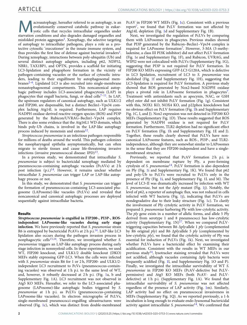
ResultsStreptococcus pneumoniae is engulfed in FIP200-, PI3P-, ROS-independent LAPosome-like vacuoles during early stageinfection. We have previously reported that S. pneumoniae strainR6 is entrapped by bactericidal PcAVs at 2 h p.i.14. LAP-like LC3lipidation also occurs during the pathogen invasion process innonphagocytic cells15,16. Therefore, we investigated whether S.pneumoniae triggers an LAP-like autophagy process during earlystage infection in nonmyeloid cells. In these experiments, we usedWT, FIP200 knockout, and ULK1/2 double knockout (DKO)MEFs stably expressing GFP-LC3. When the cells were infectedwith S. pneumoniae strain R6 for 1 or 2 h, FIP200- and ULK1/2-independent LC3 recruitment to PcVs (pneumococcus-contain-ing vacuoles) was observed at 1 h p.i. to the same level of WT,and, however, it robustly decreased at 2 h p.i. (Fig. 1a, b andSupplementary Fig. 1A). LC3 recruitment was not observed inAtg5 KO MEFs. Hereafter, we refer to the LC3-associated pha-gosome (LAPosome)-like autophagic bodies triggered by S.pneumoniae at 1 h p.i. as PcLVs (pneumococcus-containingLAPosome-like vacuoles). In electron micrographs of PcLVs,single-membraned pneumococci-engulfing ultrastructures wereobserved (Fig. 1c), which was distinct from double-membraned
PcAV in FIP200 WT MEFs (Fig. 1c). Consistent with a previousreport7, we found that PcLV formation was not affected byAtg14L depletion (Fig. 1d and Supplementary Fig. 1B).
Next, we investigated the regulation of PcLVs by comparingthem with LAPosomes in phagocytes. Previous studies showedthat PI3P generated by the Rubicon–Beclin1–Vps34 complex isrequired for LAPosome formation7. However, 3-MA (3-methy-ladenine, a class III PI3K inhibitor) did not affect PcLV formation(Fig. 1e and Supplementary Fig. 1F), and Rubicon, UVRAG, andWIPI2 were not colocalized with PcLVs (Supplementary Fig. 1G),suggesting that PI3P is not required for PcLV formation. InFIP200 KO MEFs expressing GFP-LC3 G120A, which is defectivein LC3 lipidation, recruitment of LC3 to S. pneumoniae wasabolished (Fig. 1f and Supplementary Fig. 1H), suggesting thatLC3-lipidation is required for PcLV formation. A previous studyshowed that ROS generated by Nox2-based NADPH oxidaseplays a pivotal role in LAPosome formation in phagocytes7.Treatment with antioxidants such as apocynin, NAC, or GSHethyl ester did not inhibit PcLV formation (Fig. 1g). Consistentwith this, NOX1 KO, NOX4 KO, and p22phox knockdown hadno obvious effect on PcLV formation (Fig. 1h and SupplementaryFig. 1C, I, and J). Nox2 expression was not detected in FIP200 KOMEFs (Supplementary Fig. 1D). These results suggested that ROSgenerated by NADPH oxidase is not required for PcLVformation. Furthermore, TLR2 depletion had no significant effecton PcLV formation (Fig. 1h and Supplementary Fig. 1E and J).Together, these results clearly showed that PcLVs have non-canonical LAPosome features, such as ROS-, PI3P-, and TLR2-independence, although they are somewhat similar to LAPosomesin the sense that they are FIP200-independent and have a single-membraned structure.
Previously, we reported that PcAV formation 2 h p.i. isdependent on membrane rupture by Ply, a pore-formingcytolysin of S. pneumoniae14. PcLV formation is also dependenton Ply (Fig. 1i and Supplementary Fig. 1K). We found that p62and poly-Ub to PcLVs were recruited to PcLVs only in thepresence of Ply (Fig. 1i, and Supplementary Fig. 1L and M). Thelevel of LC3-II was dramatically increased by infection with WTS. pneumoniae, but not the Δply mutant (Fig. 1j). Notably, thelevel of p62, a reporter of autophagic flux, was not reduced in cellsinfected with WT bacteria (Fig. 1j), indicating that PcLVs arenondegradative due to their leaky structure (Fig. 1c). To clarifythe involvement of Ply cytolytic activity in PcLV formation, weprepared S. pneumoniae harboring Ply with low-cytolytic activity.The ply gene exists in a number of allelic forms, and allele 5 Plyderived from serotype 1 and 8 pneumococci has low-cytolyticactivity (Supplementary Fig. 1N)17. When we compared PcLV-triggering capacities between R6 Δply:allele 1 ply (complementedby R6 original ply) and R6 Δply:allele 5 ply (complemented bylow-cytolytic ply), we found that the cytolytic activity of Ply wasessential for induction of PcLVs (Fig. 1k). Next, we investigatedwhether PcLVs have a bactericidal effect by examining theiracidification. Consistent with the results in WT MEFs in ourprevious study14, lysotracker staining revealed that PcLVs werenot acidified, although vacuoles containing Δply bacteria werefrequently acidified (Fig. 1l, and Supplementary Fig. 1O and P).Finally, we investigated the intracellular survivability of WT S.pneumoniae in FIP200 KO MEFs (PcAV-defective but PcLV-permissive) and Atg5 KO MEFs (both PcAV- and PcLV-defective) at 1 h p.i. (Supplementary Fig. 1A). We found thatintracellular survivability of S. pneumoniae was not affectedregardless of the presence of LAP activity (Fig. 1m). Similarly,LAP activity had no effect on pneumococcal invasiveness inMEFs (Supplementary Fig. 1Q). As we reported previously, a 1-hincubation is long enough to evaluate endo-lysosomal bactericidalactivity against intracellular S. pneumoniae14. We confirmed that
ARTICLE COMMUNICATIONS BIOLOGY | https://doi.org/10.1038/s42003-020-0753-3
2 COMMUNICATIONS BIOLOGY | (2020) 3:25 | https://doi.org/10.1038/s42003-020-0753-3 | www.nature.com/commsbio
10 µg/ml of penicillin G treatment for 15 min had no negativeeffect on intracellular survivability of pneumococci until 3 h, andthat each cell was invaded by at least one bacterium (Supple-mentary Fig. 1R). Putting all these results together, we concludedthat PcLVs do not have bactericidal activity.
Noncanonical and canonical autophagic processes are deployedsequentially against intracellular pneumococci. Multipleautophagic processes, acting simultaneously or sequentially, areinvolved in targeting intracellular bacterial pathogens11,15. Todetermine whether the autophagic processes involving PcLV and
h ig
b
f
j
a
c
d e
k l mN. S.
FIP200KO
(PcLV+)
Atg5KO
(PcLV-)
0
20
40
60
80
100
Sur
viva
bilit
y of
S. p
neum
onia
e in
PcL
V(
% o
f inv
aded
bac
teria
)
WT Δply
*
0
5
10
15
20
25
Aci
difie
d P
cLV
or
PcV
con
tain
ing
cells
(%
)
FIP200 KO MEF
N. S.
si Luc si Atg14L
FIP200 KO MEF
0
5
10
15
20
25
30
LC3-
posi
tive
bact
eria
con
tain
ing
cells
(%
)
R6:
:ply
(alle
le1,
ST
2)R
6::p
ly(a
llele
5, S
T8)
R6Δ
ply
mCherry-LC3Pneumolysin DAPI Merge
S. pneumoniae
LC3-II
LC3-I
p62
Actin
WT ΔplyN.I.
15-
60-
45-
(kDa)
FIP200 KO MEF
FIP200 KO MEF
* *
LC3G120A
LC3WT
*
FIP200 KO MEF
FIP200 KO MEF
0
5
10
15
20
25
30
LC3-
posi
tive
bact
eria
con
tain
ing
cells
(%
)
w/o3-MA
with3-MA
N. S.
FIP200 KO MEF
0
5
10
15
20
25
30
35
LC3-
posi
tive
bact
eria
con
tain
ing
cells
(%
)
N. S.
Non
-tr
eate
d
Apo
cyni
n
GS
H
NA
C
0
20
40
60
80
100
LC3-
posi
tive
bact
eria
con
tain
ing
cells
(%
)
WT
Nox
1 K
O
Nox
4 K
O
siLu
c
sip2
2pho
x
siT
LR2
si Atg14Lto inhibit PcAV
FIP200 KO MEF
N. S.**
*
0
20
40
60
80
100
120LC
3-po
sitiv
e ba
cter
ia c
onta
inin
g ce
lls(r
elat
ive
valu
e)
WT
Δply
poly-Ub p62LC3
*0
5
10
15
20
25
30
Mar
ker-
posi
tive
bact
eria
con
tain
ing
cells
(%
)
FIP200 KO FIP200 WT
FIP200 KOFIP200 WT
1 2Time (h) 1 2 1 2
FIP200KO
1 2
WT ULK1/2DKO
Atg5KO
N. S.
* *
0
5
10
15
20
25
30
35
40
LC3-
posi
tive
bact
eria
con
tain
ing
cells
(%
)
COMMUNICATIONS BIOLOGY | https://doi.org/10.1038/s42003-020-0753-3 ARTICLE
COMMUNICATIONS BIOLOGY | (2020) 3:25 | https://doi.org/10.1038/s42003-020-0753-3 | www.nature.com/commsbio 3
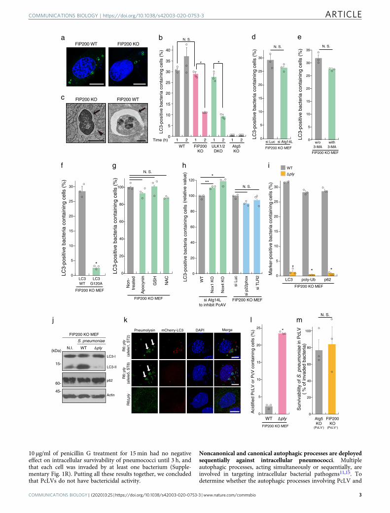
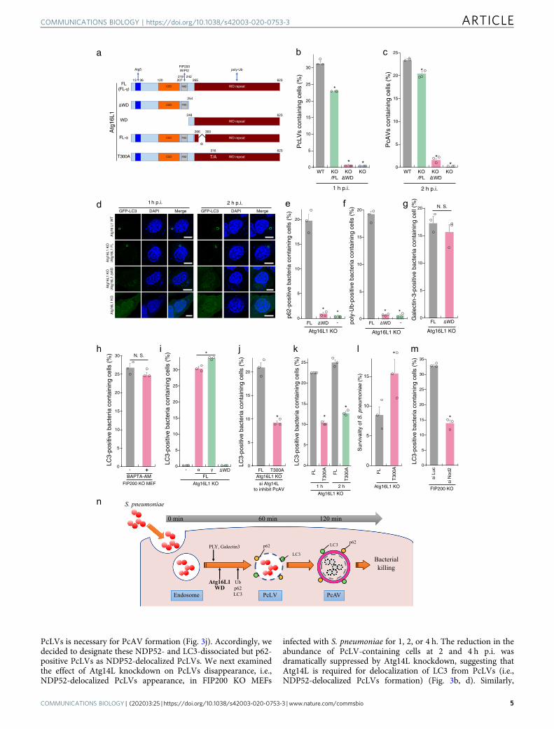
PcAV proceed simultaneously or sequentially, we attempted toconstruct PcLV-deficient MEFs. The C-terminal WD domain(WD; structural domain composed of ~40 amino acids, oftenterminated by a tryptophan–aspartic acid) of Atg16L1 plays acrucial role in the LAPosome-like autophagic bodies induced byentosis and certain bacteria12,15,18. We confirmed that canonicalautophagy and flux were normal in Atg16L1 KO MEFs com-plemented with Atg16 FL (Full Length) or Atg16L1 lacking theWD (ΔWD) (Fig. 2a and Supplementary Fig. 2A–D). Next, weinfected cells stably expressing GFP-LC3 with S. pneumoniae for1 h. PcLV formation was completely abolished in Atg16L1 KOMEFs complemented with Atg16L1 ΔWD (Atg16L1 KO/ΔWDMEFs) (Supplementary Fig. 2B and D, left), suggesting that theAtg16L1 WD also plays a pivotal role also PcLV formation. Wethen investigated the intracellular localization of GFP-Atg16L1ΔWD and WD at 1 h p.i. Only GFP-Atg16L1 FL was recruited toPcLVs (Supplementary Fig. 2E), indicating that both Atg5 bind-ing by the N terminus and poly-Ub binding by the C terminus arerequired for appropriate localization. Next, we investigated PcAVformation using Atg16L1 KO/ΔWD MEFs. At 2 h p.i. in thesePcLV-deficient MEFs, PcAV formation was robustly suppressedin an Atg16L1 WD-dependent manner (Fig. 2c, d, right). In lightof a report that xenophagy is normal in Atg16L1 KO/ΔWDMEFs16, these results clearly suggest that PcLV and PcAV pro-ceed sequentially, rather than simultaneously (Fig. 2n).
Next, we investigated the localization of p62 in Atg16L1 KOMEFs complemented with Atg16L1 FL or ΔWD at 1 h p.i. p62-3Myc recruitment to intracellular bacteria was dramaticallyreduced in Atg16L1 KO/ΔWD MEFs (Fig. 2e and SupplementaryFig. 2F), indicating that the recruitment of p62 and Atg16L1 WDto PcLVs is interdependent. Importantly, poly-Ub deposition onPcVs was also robustly reduced in Atg16L1 KO/ΔWD MEFs(Fig. 2f and Supplementary Fig. 2G), although endosomalmembrane rupture occurred normally in Atg16L1 KO/ΔWDMEFs at 1 h p.i. (Fig. 2g). LC3 deposition via Atg16L1 WD alsoparticipates in membrane repair in a Ca2+-dependent manner19.However, the calcium chelator O,O′-bis(2-aminophenyl)ethyle-neglycol-N,N,N′,N′-tetraacetic acid, tetraacetoxymethyl ester(BAPTA-AM) had no reductive effect on PcLV formation(Fig. 2h). Together, these results reveal the Atg16L1 WD-dependency of p62 and poly-Ub recruitment during PcLVformation.
Next, we investigated domain analysis of Atg16L1 WD domainin PcLV formation. The alpha domain of Atg16L1 (Fig. 2a) playsa role in PI3P-independent noncanonical autophagy induced bymonensin20. Accordingly, we investigated PcLV formation using
Atg16L1 KO MEFs complemented with the α or γ form ofAtg16L1 FL. Intriguingly, PcLV formation in MEFs complemen-ted with the α form was normal (Fig. 2i), suggesting that PcLVsare distinct from monensin-triggered LAPs. A coding poly-morphism of human Atg16L1 (rs2241880; T300A) increases therisk of Crohn’s disease; the T300A mutation (corresponding toT316A in mouse Atg16L1γ, hereafter referred to as T300A,Fig. 2a) in the WD region of Atg16L1 decreases the autophagicclearance of intracellular pathogens such as Shigella21,22, but notSalmonella. Hence, we compared PcLV formation betweenAtg16L1 KO MEFs complemented with Atg16L1 WT or T300Awhere Atg14L had been knocked down to suppress canonicalautophagy. We found that PcLV formation was reduced inT300A-complemented cells (Fig. 2j, and Supplementary Fig. 3Aand B). Notably, subsequent PcAV formation and bactericidalactivity at 2 h p.i. was also reduced in T300A-complemented cells(Fig. 2k, l), providing further confirmation that PcLV and PcAVwere induced sequentially rather than simultaneously. Nod2polymorphism is also a risk factor for Crohn’s disease, and Nod2and Atg16L1 work cooperatively in bactericidal autophagy23,24.Consistent with this, we found that PcLV formation was alsoreduced in Nod2-knockdown cells (Fig. 2m, and SupplementaryFig. 3C and D), implying a correlation between Crohn’s diseaseand the severity of pneumococcal infections such as pneumoniaand IPD25,26. Together, these results support the notion thatPcLV and PcAV are induced sequentially during S. pneumoniaeinfection (Fig. 2n).
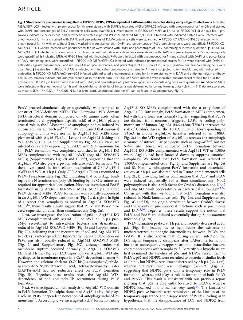
PcLV formation peaked at 1 h p.i. and robustly decreased at 2 hp.i. (Fig. 1b), leading us to hypothesize the existence ofuncharacterized autophagic intermediates between PcLVs andPcAVs. It is also known that, during Listeria infection, theLC3 signal temporarily disappears after LAPosome formation,but then subsequently reappears around intracellular bacteriacontemporaneous with xenophagy11. To verify our hypothesis, wefirst examined the kinetics of p62 and NDP52 recruitment toPcLVs. p62 and NDP52 were recruited to bacteria at similar levelsat 1 h p.i., but NDP52 recruitment decreased by 2 h p.i. (34–14%),whereas p62 recruitment was unchanged (37–36%) (Fig. 3a),suggesting that NDP52 plays only a temporary role in PcLVformation, whereas p62 plays a role in formation of both PcLVsand PcAVs. This result is consistent with our previous reportshowing that p62 is frequently localized to PcAVs, whereasNDP52 localized in this manner very rarely14. The kinetics ofNDP52-positive bacteria were reminiscent of the kinetics of thetemporary appearance and disappearance of PcLVs, leading us tohypothesize that the disappearance of LC3 and NDP52 from
Fig. 1 Streptococcus pneumoniae is engulfed in FIP200-, PI3P-, ROS-independent LAPosome-like vacuoles during early stage of infection. a IndicatedMEFs/GFP-LC3 infected with pneumococci for 1 h were stained with DAPI. b Indicated MEFs/GFP-LC3 infected with pneumococci for 1 or 2 h and stainedwith DAPI, and percentages of PcLV-containing cells were quantified. c Micrographs of FIP200 KO MEFs at 1 h p.i. or FIP200 WT at 2 h p.i.; Bar, 1 µm.Arrows indicate PcLV or PcAV, and arrowhead indicates ruptured PcLV. d Indicated MEFs/GFP-LC3 treated with indicated siRNAs were infected withpneumococci for 1 h and stained with DAPI, and percentages of PcLV-containing cells were quantified. e FIP200 KO MEFs/GFP-LC3 infected withpneumococci for 1 h with or without 3-methyladenine were stained with DAPI, and percentages of PcLV-containing cells were quantified. f FIP200 KOMEFs/GFP-LC3 G120A infected with pneumococci for 1 h were stained with DAPI, and percentages of PcLV-containing cells were quantified. g FIP200 KOMEFs/GFP-LC3 infected with pneumococci for 1 h with or without indicated antioxidants were stained with DAPI, and percentages of PcLV-containing cellswere quantified. h Indicated MEFs/GFP-LC3 treated with indicated siRNA were infected with pneumococci for 1 h and stained with DAPI, and percentagesof PcLV-containing cells were quantified. i FIP200 KO MEFs/GFP-LC3 infected with indicated pneumococcal strains for 1 h were stained with DAPI orantibodies against pneumococci, and anti-poly-Ub or -p62 antibodies, and percentages of LC3-, poly-Ub-, or p62-positive bacteria containing cells werequantified. j Lysates from FIP200 KO MEFs infected with indicated pneumococcal strains for 1 h were subjected to immunoblotting with indicatedantibodies. k FIP200 KO MEFs/mCherry-LC3 infected with indicated pneumococcal strains for 1 h were stained with DAPI and antipneumolysin antibody.Bar, 10 µm. Arrows indicate pneumolysin around or in the bacterium. l FIP200 KO MEFs infected with indicated pneumococcal strains for 1 h in thepresence of 50 nM LysoTracker were stained with DAPI, and percentages of LysoTracker-positive PcV-containing cells were quantified. m Indicated MEFswere infected with pneumococci for 1 h and intracellular survivability of bacteria was determined by colony forming units (cfu); n= 3. Data are expressedas mean ± SEM; *P < 0.01, **P < 0.05, N.S., not significant. Uncropped blots for (j) can be found in Supplementary Fig. 10.
ARTICLE COMMUNICATIONS BIOLOGY | https://doi.org/10.1038/s42003-020-0753-3
4 COMMUNICATIONS BIOLOGY | (2020) 3:25 | https://doi.org/10.1038/s42003-020-0753-3 | www.nature.com/commsbio
PcLVs is necessary for PcAV formation (Fig. 3j). Accordingly, wedecided to designate these NDP52- and LC3-dissociated but p62-positive PcLVs as NDP52-delocalized PcLVs. We next examinedthe effect of Atg14L knockdown on PcLVs disappearance, i.e.,NDP52-delocalized PcLVs appearance, in FIP200 KO MEFs
infected with S. pneumoniae for 1, 2, or 4 h. The reduction in theabundance of PcLV-containing cells at 2 and 4 h p.i. wasdramatically suppressed by Atg14L knockdown, suggesting thatAtg14L is required for delocalization of LC3 from PcLVs (i.e.,NDP52-delocalized PcLVs formation) (Fig. 3b, d). Similarly,
b c
WT KO/FL
KO/ΔWD
KO
1 h p.i.
*
* *
PcL
Vs
cont
aini
ng c
ells
(%
)
WT KO/FL
KO/ΔWD
KO
2 h p.i.
*
**0
5
10
15
20
25
PcA
Vs
cont
aini
ng c
ells
(%
)
a
d
265
WD repeat
Atg5
623
CCD
20712013 36
CCD
248
WD repeat
623
254
poly-UbFIP200WIPI2
FBD
219 242
FBD
Atg
16L1
FL(FL-γ)
WD repeat
623316
CCD T/AFBDT300A
266 300
WD repeatCCD FBDFL-αα
ΔWD
WD
e f g
* *
-ΔWDFL
Atg16L1 KO
0
5
10
15
20
p62-
posi
tive
bact
eria
con
tain
ing
cells
(%
)
-
Atg16L1 KO
ΔWDFL
* *0
5
10
15
20
poly
-Ub-
posi
tive
bact
eria
con
tain
ing
cells
(%
)
Atg16L1 KO
ΔWDFL
N. S.
0
5
10
15
20
Gal
ectin
-3-p
ositi
ve b
acte
ria c
onta
inin
g ce
ll (%
)
j k l m
*
0
5
10
15
20
LC3-
posi
tive
bact
eria
con
tain
ing
cells
(%
)
si Atg14Lto inhibit PcAV
Atg16L1 KOT300AFL
*
*
0
5
10
15
20
25
LC3-
posi
tive
bact
eria
con
tain
ing
cells
(%
)
Atg16L1 KO
1 h 2 h
T30
0AFL
T30
0AFL
Atg16L1 KO
T30
0AFL
*
0
5
10
15
Sur
viva
lity
of S
. pne
umon
iae
(%)
siN
od2
siLu
c
*
0
5
10
15
20
25
30
35
LC3-
posi
tive
bact
eria
con
tain
ing
cells
(%
)
FIP200 KO
h i
FIP200 KO MEF
N. S.
BAPTA-AM+
0
5
10
15
20
25
30
LC3-
posi
tive
bact
eria
con
tain
ing
cells
(%
)
LC3-
posi
tive
bact
eria
con
tain
ing
cells
(%
) *
-- γ ΔWDαFL
Atg16L1 KO
0
5
10
15
20
25
30
GFP-LC3 DAPI Merge
1h p.i.
Atg
16 L
1 W
TA
tg16
L1 K
O/A
tg16
L1 F
LA
tg16
L1 K
O/A
tg16
L1 Δ
WD
Atg
16L1
KO
GFP-LC3 DAPI Merge
2 h p.i.
n S. pneumoniae
Endosome PcLV
LC3
PcAV
LC3p62p62
Ubp62LC3
Atg16L1WD
PLY, Galectin3
Bacterialkilling
120 min60 min0 min
COMMUNICATIONS BIOLOGY | https://doi.org/10.1038/s42003-020-0753-3 ARTICLE
COMMUNICATIONS BIOLOGY | (2020) 3:25 | https://doi.org/10.1038/s42003-020-0753-3 | www.nature.com/commsbio 5
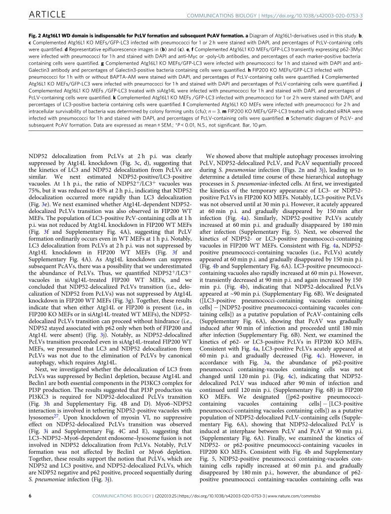
NDP52 delocalization from PcLVs at 2 h p.i. was clearlysuppressed by Atg14L knockdown (Fig. 3c, d), suggesting thatthe kinetics of LC3 and NDP52 delocalization from PcLVs aresimilar. We next estimated NDP52-positive/LC3-positivevacuoles. At 1 h p.i., the ratio of NDP52+/LC3+ vacuoles was75%, but it was reduced to 45% at 2 h p.i., indicating that NDP52delocalization occurred more rapidly than LC3 delocalization(Fig. 3e). We next examined whether Atg14L-dependent NDP52-delocalized PcLVs transition was also observed in FIP200 WTMEFs. The population of LC3-positive PcV-containing cells at 1 hp.i. was not reduced by Atg14L knockdown in FIP200 WT MEFs(Fig. 3f and Supplementary Fig. 4A), suggesting that PcLVformation ordinarily occurs even in WT MEFs at 1 h p.i. Notably,LC3 delocalization from PcLVs at 2 h p.i. was not suppressed byAtg14L knockdown in FIP200 WT MEFs (Fig. 3f andSupplementary Fig. 4A). As Atg14L knockdown can suppresssubsequent PcAVs, there was a possibility that we underestimatedthe abundance of PcLVs. Thus, we quantified NDP52+/LC3+
vacuoles in siAtg14L-treated FIP200 WT MEFs, and weconcluded that NDP52-delocalized PcLVs transition (i.e., delo-calization of NDP52 from PcLVs) was not suppressed by Atg14Lknockdown in FIP200 WT MEFs (Fig. 3g). Together, these resultsindicate that when either Atg14L or FIP200 is present (i.e., inFIP200 KO MEFs or in siAtg14L-treated WT MEFs), the NDP52-delocalized PcLVs transition can proceed without hindrance (i.e.,NDP52 stayed associated with p62 only when both of FIP200 andAtg14L were absent) (Fig. 3j). Notably, as NDP52-delocalizedPcLVs transition proceeded even in siAtg14L-treated FIP200 WTMEFs, we presumed that LC3 and NDP52 delocalization fromPcLVs was not due to the elimination of PcLVs by canonicalautophagy, which requires Atg14L.
Next, we investigated whether the delocalization of LC3 fromPcLVs was suppressed by Beclin1 depletion, because Atg14L andBeclin1 are both essential components in the PI3KC3 complex forPI3P production. The results suggested that PI3P production viaPI3KC3 is required for NDP52-delocalized PcLVs transition(Fig. 3h and Supplementary Fig. 4B and D). Myo6–NDP52interaction is involved in tethering NDP52-positive vacuoles withlysosomes27. Upon knockdown of myosin VI, no suppressiveeffect on NDP52-delocalized PcLVs transition was observed(Fig. 3i and Supplementary Fig. 4C and E), suggesting thatLC3–NDP52–Myo6-dependent endosome–lysosome fusion is notinvolved in NDP52 delocalization from PcLVs. Notably, PcLVformation was not affected by Beclin1 or Myo6 depletion.Together, these results support the notion that PcLVs, which areNDP52 and LC3 positive, and NDP52-delocalized PcLVs, whichare NDP52 negative and p62 positive, proceed sequentially duringS. pneumoniae infection (Fig. 3j).
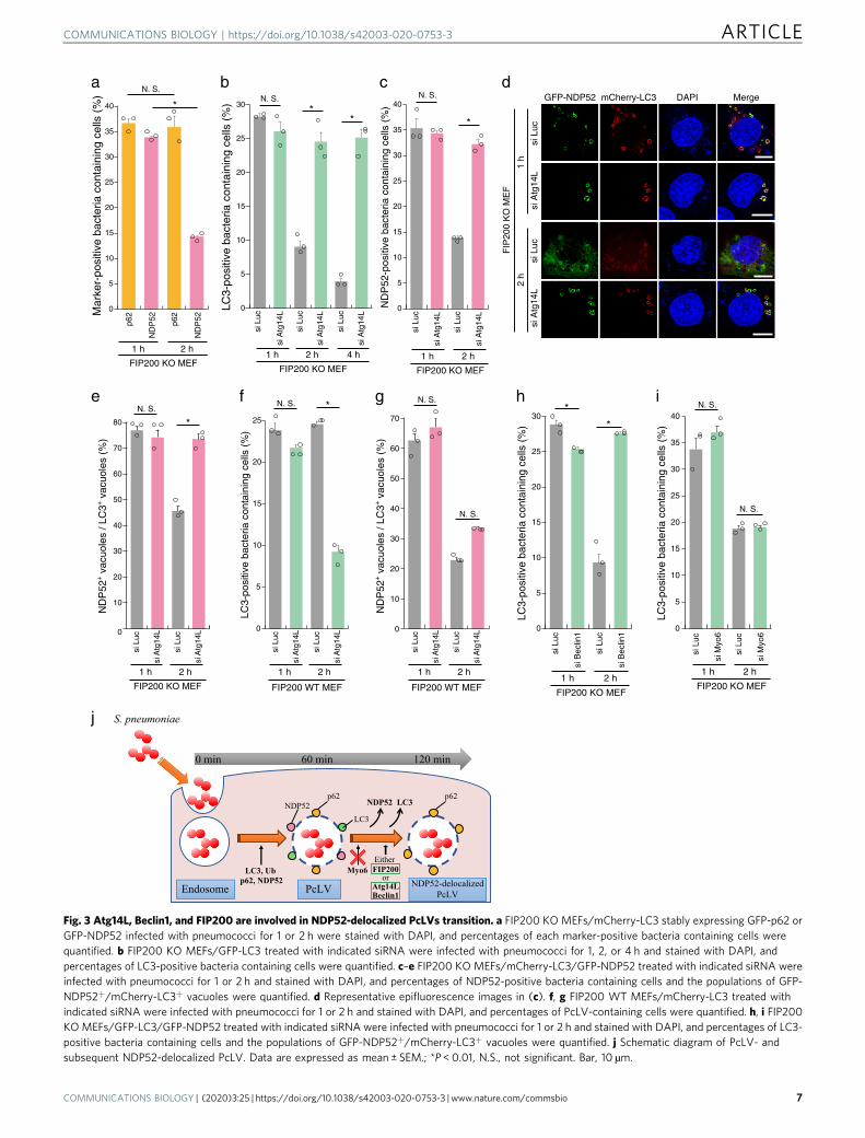
We showed above that multiple autophagy processes involvingPcLV, NDP52-delocalized PcLV, and PcAV sequentially proceedduring S. pneumoniae infection (Figs. 2n and 3j), leading us todetermine a detailed time course of these hierarchical autophagyprocesses in S. pneumoniae-infected cells. At first, we investigatedthe kinetics of the temporary appearance of LC3- or NDP52-positive PcLVs in FIP200 KO MEFs. Notably, LC3-positive PcLVswas not observed until at 30 min p.i. However, it acutely appearedat 60 min p.i. and gradually disappeared by 150min afterinfection (Fig. 4a). Similarly, NDP52-positive PcLVs acutelyincreased at 60 min p.i. and gradually disappeared by 180 minafter infection (Supplementary Fig. 5). Next, we observed thekinetics of NDP52- or LC3-positive pneumococci-containingvacuoles in FIP200 WT MEFs. Consistent with Fig. 4a, NDP52-positive pneumococci-containing vacuoles (i.e., PcLVs) acutelyappeared at 60 min p.i. and gradually disappeared by 150 min p.i.(Fig. 4b and Supplementary Fig. 6A). LC3-positive pneumococci-containing vacuoles also rapidly increased at 60 min p.i. However,it transiently decreased at 90 min p.i. and again increased by 150min p.i. (Fig. 4b), indicating that NDP52-delocalized PcLVsappeared at ~90 min p.i. (Supplementary Fig. 6B). We designated([LC3-positive pneumococci-containing vacuoles containingcells]− [NDP52-positive pneumococci-containing vacuoles con-taining cells]) as a putative population of PcAV-containing cells(Supplementary Fig. 6A), showing that PcAV was graduallyinduced after 90 min of infection and proceeded until 180 minafter infection (Supplementary Fig. 6B). Next, we examined thekinetics of p62- or LC3-positive PcLVs in FIP200 KO MEFs.Consistent with Fig. 4a, LC3-positive PcLVs acutely appeared at60 min p.i. and gradually decreased (Fig. 4c). However, inaccordance with Fig. 3a, the abundance of p62-positivepneumococci containing-vacuoles containing cells was notchanged until 120 min p.i. (Fig. 4c), indicating that NDP52-delocalized PcLV was induced after 90 min of infection andcontinued until 120 min p.i. (Supplementary Fig. 6B) in FIP200KO MEFs. We designated ([p62-positive pneumococci-containing vacuoles containing cells]− [LC3-positivepneumococci-containing vacuoles containing cells]) as a putativepopulation of NDP52-delocalized PcLV-containing cells (Supple-mentary Fig. 6A), showing that NDP52-delocalized PcLV isinduced at interphase between PcLV and PcAV at 90 min p.i.(Supplementary Fig. 6A). Finally, we examined the kinetics ofNDP52- or p62-positive pneumococci-containing vacuoles inFIP200 KO MEFs. Consistent with Fig. 4b and SupplementaryFig. 5, NDP52-positive pneumococci containing-vacuoles con-taining cells rapidly increased at 60 min p.i. and graduallydisappeared by 180 min p.i., however, the abundance of p62-positive pneumococci containing-vacuoles containing cells was
Fig. 2 Atg16L1 WD domain is indispensable for PcLV formation and subsequent PcAV formation. a Diagram of Atg16L1-derivatives used in this study. b,c Complemented Atg16L1 KO MEFs/GFP-LC3 infected with pneumococci for 1 or 2 h were stained with DAPI, and percentages of PcLV-containing cellswere quantified. d Representative epifluorescence images in (b) and (c). e, f Complemented Atg16L1 KO MEFs/GFP-LC3 transiently expressing p62-3Mycwere infected with pneumococci for 1 h and stained with DAPI and anti-Myc or -poly-Ub antibodies, and percentages of each marker-positive bacteriacontaining cells were quantified. g Complemented Atg16L1 KO MEFs/GFP-LC3 were infected with pneumococci for 1 h and stained with DAPI and anti-Galectin3 antibody and percentages of Galectin3-positive bacteria containing cells were quantified. h FIP200 KO MEFs/GFP-LC3 infected withpneumococci for 1 h with or without BAPTA-AM were stained with DAPI, and percentages of PcLV-containing cells were quantified. i ComplementedAtg16L1 KO MEFs/GFP-LC3 were infected with pneumococci for 1 h and stained with DAPI and percentages of PcLV-containing cells were quantified. jComplemented Atg16L1 KO MEFs /GFP-LC3 treated with siAtg14L were infected with pneumococci for 1 h and stained with DAPI, and percentages ofPcLV-containing cells were quantified. k Complemented Atg16L1 KO MEFs /GFP-LC3 infected with pneumococci for 1 or 2 h were stained with DAPI, andpercentages of LC3-positive bacteria containing cells were quantified. l Complemented Atg16L1 KO MEFs were infected with pneumococci for 2 h andintracellular survivability of bacteria was determined by colony forming units (cfu); n= 3. m FIP200 KO MEFs/GFP-LC3 treated with indicated siRNA wereinfected with pneumococci for 1 h and stained with DAPI, and percentages of PcLV-containing cells were quantified. n Schematic diagram of PcLV- andsubsequent PcAV formation. Data are expressed as mean ± SEM.; *P < 0.01, N.S., not significant. Bar, 10 µm.
ARTICLE COMMUNICATIONS BIOLOGY | https://doi.org/10.1038/s42003-020-0753-3
6 COMMUNICATIONS BIOLOGY | (2020) 3:25 | https://doi.org/10.1038/s42003-020-0753-3 | www.nature.com/commsbio
c d
j
ba*
N. S.
p62
ND
P52
p62
ND
P52
1 h 2 h
FIP200 KO MEF
0
5
10
15
20
25
30
35
40 *N. S.
*
siLu
c
siA
t g14
L
siLu
c
siA
t g14
L
siLu
c
siA
tg14
L1 h 2 h 4 h
FIP200 KO MEF
0
5
10
15
20
25
30
LC3-
posi
tive
bact
eria
con
tain
ing
cells
(%
)
Mar
ker-
posi
tive
bact
eria
con
tain
ing
cells
(%
) GFP-NDP52 mCherry-LC3 DAPI Merge
siLu
csi
Atg
14L
1 h
siLu
csi
Atg
14L
2 h
FIP
200
KO
ME
F
*
N. S.
siLu
c
siA
tg14
L
siLu
c
siA
tg14
L
1 h 2 h
FIP200 KO MEF
0
5
10
15
20
25
30
35
40
ND
P52
-pos
itive
bac
teria
con
tain
ing
cells
(%
)
siLu
c
siM
yo6
siLu
c
siM
yo6
1 h 2 h
FIP200 KO MEF
N. S.
0
5
10
15
20
25
30
35
40
LC3-
posi
tive
bact
eria
con
tain
ing
cells
(%
)
siLu
c
Luc
siB
eclin
1
si
siB
eclin
1
1 h 2 h
FIP200 KO MEF
*
0
5
10
15
20
25
30
LC3-
posi
tive
bact
eria
con
tain
ing
cells
(%
)
Endosome PcLV
LC3
LC3, Ub,p62, NDP52
LC3NDP52
0 min 60 min
S. pneumoniae
NDP52-delocalizedPcLV
p62NDP52
p62
120 min
Myo6
e ihgf
10
20
30
40
50
60
70
80
ND
P52
+ v
acuo
les
/ LC
3+va
cuol
es (
%)
ND
P52
+va
cuol
es /
LC3+
vac
uole
s (%
)
0
si L
uc
si A
tg14
L
si L
uc
si A
tg14
L
1 h 2 h
FIP200 KO MEF
0
10
20
30
40
50
60
70
FIP200 WT MEF
0
5
10
15
20
25
LC3-
posi
tive
bact
eria
con
tain
ing
cells
(%
)
*N. S.
*N. S. *
N. S.N. S.
N. S.
si L
uc
si A
tg14
L
si L
uc
si A
tg14
L
1 h 2 h
FIP200 WT MEF
si L
uc
si A
tg14
L
si L
uc
si A
tg14
L
1 h 2 h
Either
orAtg14LBeclin1
FIP200
Fig. 3 Atg14L, Beclin1, and FIP200 are involved in NDP52-delocalized PcLVs transition. a FIP200 KO MEFs/mCherry-LC3 stably expressing GFP-p62 orGFP-NDP52 infected with pneumococci for 1 or 2 h were stained with DAPI, and percentages of each marker-positive bacteria containing cells werequantified. b FIP200 KO MEFs/GFP-LC3 treated with indicated siRNA were infected with pneumococci for 1, 2, or 4 h and stained with DAPI, andpercentages of LC3-positive bacteria containing cells were quantified. c–e FIP200 KO MEFs/mCherry-LC3/GFP-NDP52 treated with indicated siRNA wereinfected with pneumococci for 1 or 2 h and stained with DAPI, and percentages of NDP52-positive bacteria containing cells and the populations of GFP-NDP52+/mCherry-LC3+ vacuoles were quantified. d Representative epifluorescence images in (c). f, g FIP200 WT MEFs/mCherry-LC3 treated withindicated siRNA were infected with pneumococci for 1 or 2 h and stained with DAPI, and percentages of PcLV-containing cells were quantified. h, i FIP200KO MEFs/GFP-LC3/GFP-NDP52 treated with indicated siRNA were infected with pneumococci for 1 or 2 h and stained with DAPI, and percentages of LC3-positive bacteria containing cells and the populations of GFP-NDP52+/mCherry-LC3+ vacuoles were quantified. j Schematic diagram of PcLV- andsubsequent NDP52-delocalized PcLV. Data are expressed as mean ± SEM.; *P < 0.01, N.S., not significant. Bar, 10 µm.
COMMUNICATIONS BIOLOGY | https://doi.org/10.1038/s42003-020-0753-3 ARTICLE
COMMUNICATIONS BIOLOGY | (2020) 3:25 | https://doi.org/10.1038/s42003-020-0753-3 | www.nature.com/commsbio 7
almost unchanged until 180 min p.i. (Fig. 4d). Together, theseresults supported the notion that multiple hierarchical autophagyprocesses, including PcLV, NDP52-delocalized PcLV, andeventually PcAV, sequentially proceed during S. pneumoniaeinfection (Fig. 4i and Supplementary Fig. 6B).
Next, we confirm whether our findings observed in MEF cellscan be replicated in human pulmonary epithelial cells (A549cells). We investigated the kinetics of the temporary induction ofPcLVs in siLuc- or siFIP200-treated A549 cells. Consistent withthe results in MEFs, FIP200-independent PcLV was transiently
i
e f h
Endosome PcLV
LC3
LC3, Ubp62, NDP52
Atg16L1 WD
PLY LC3NDP52
0 min 120 min60 min 90 min
S. pneumoniae
NDP52-delocalizedPcLV PcAV
LC3 LC3p62NDP52
p62
Canonical pathway
Non-canonical pathway
Reprogramming ofpneumococci-containing vesicles
Bacterialkilling
p62
0
5
10
15
20
FLmAtg16L1 ΔWD mock (-)
LC3-
posi
tive
bact
eria
con
tain
ing
cells
(%
)
hAtg16L1 KD
1 h p.i.
A549/GFP-LC3
*
N. S.
g
mAtg16L10
5
10
15
20
hAtg16L1 KD
2 h p.i.
A549/GFP-LC3
FL ΔWD mock (-)LC
3-po
sitiv
e ba
cter
ia c
onta
inin
g ce
lls (
%)
*
N. S.
0
5
10
15
FL ΔWD mock (-)
ND
P52
-pos
itive
bac
teria
con
tain
ing
cells
(%
)
mAtg16L1
hAtg16L1 KD
1 h p.i.
A549/GFP-LC3
*
N. S.
0
5
10
15
FL ΔWD mock (-)
ND
P52
-pos
itive
bac
teria
con
tain
ing
cells
(%
)
mAtg16L1
hAtg16L1 KD
2 h p.i.
A549/GFP-LC3
N. S.
a b c
0
5
10
15
20
25
30
5 15 30 60 90 120150
LC3-
posi
tive
bact
eria
con
tain
ing
cells
(%
)
Time after infection (min)
FIP200 KO MEF
30 60 90 120 150
Time after infection (min)
FIP200 WT MEF
30 60 90 120
Time after infection (min)
FIP200 KO MEF
Time after infection (min)
FIP200 KO MEF
0
5
10
15
20
25
30
35
LC3
NDP52
Mar
ker-
posi
tive
bact
eria
con
tain
ing
cells
(%
)
0
5
10
15
20
25
30
35
Mar
ker-
posi
tive
bact
eria
con
tain
ing
cells
(%
)
LC3dLC3
NDP52p62
30 60 90 120 150 180
NDP52
p62p62
0
5
10
15
20
25
30
Mar
ker-
posi
tive
bact
eria
con
tain
ing
cells
(%
)
Either
orAtg14LBeclin1
FIP200
ARTICLE COMMUNICATIONS BIOLOGY | https://doi.org/10.1038/s42003-020-0753-3
8 COMMUNICATIONS BIOLOGY | (2020) 3:25 | https://doi.org/10.1038/s42003-020-0753-3 | www.nature.com/commsbio

induced in A549 cells (Supplementary Fig. 7A–B and F–G).Notably, at 3 and 4 h p.i., LC3 recruitment remained high insiLuc-treated cells. However, it was decreased in siFIP200-treatedcells, suggesting that subsequent PcAV is induced in siLuc-treatedcells at 3 and 4 h p.i. (Supplementary Fig. 7C). In thisexperimental setting (i.e., siFIP200 knockdown for 3 days inA549 cells), PcLVs appearance was little later than that in MEFs(between 1 and 2 h). Treatment with antioxidants, such asapocynin or NAC, had no effect on PcLV formation in siFIP200-treated A549 cells, suggesting that PcLV in A549 cells is also ROSindependent (Supplementary Fig. 7D and E). Finally, weperformed siRNA rescue experiments using A549 cells stablyexpressing mouse Atg16L1 (Supplementary Fig. 7H). When theseA549 cells were infected with S. pneumoniae under Atg16L1-knockdowned conditions for 1 or 2 h, NDP52-positive PcLVsappeared at 1 h p.i. and disappeared at 2 h p.i. (Fig. 4e, f). Bycontrast, in siRNA-resistant Atg16L1 WD rescued or mock (norescued) A549 cells, the formation of NDP52-positive PcLV wassuppressed (Fig. 4e, f). Next, when LC3 localization wasquantified, LC3-positive PcLVs appeared at 1 h p.i. in siRNA-resistant Atg16L1 FL-rescued A549 cells. However, it was robustlydecreased in siRNA-resistant Atg16L1 WD rescued or mock (norescued) A549 cells (Fig. 4g), which was quite similar to the resultin Atg16L1-complemented and Atg16L1 KO MEFs (Fig. 2b). At2 h p.i., where NDP52 had already disappeared from PcLVs(Fig. 4f), PcAV was apparently observed in siRNA-resistantAtg16L1 FL-rescued A549 cells, but it was robustly suppressed insiRNA-resistant Atg16L1 WD rescued or mock A549 cells(Fig. 4h). These findings were also similar to those in Atg16L1-complemented and Atg16L1 KO MEFs (Fig. 2c). These resultsclearly show that multiple hierarchical autophagy processes alsooccurred in human pulmonary epithelial cells.
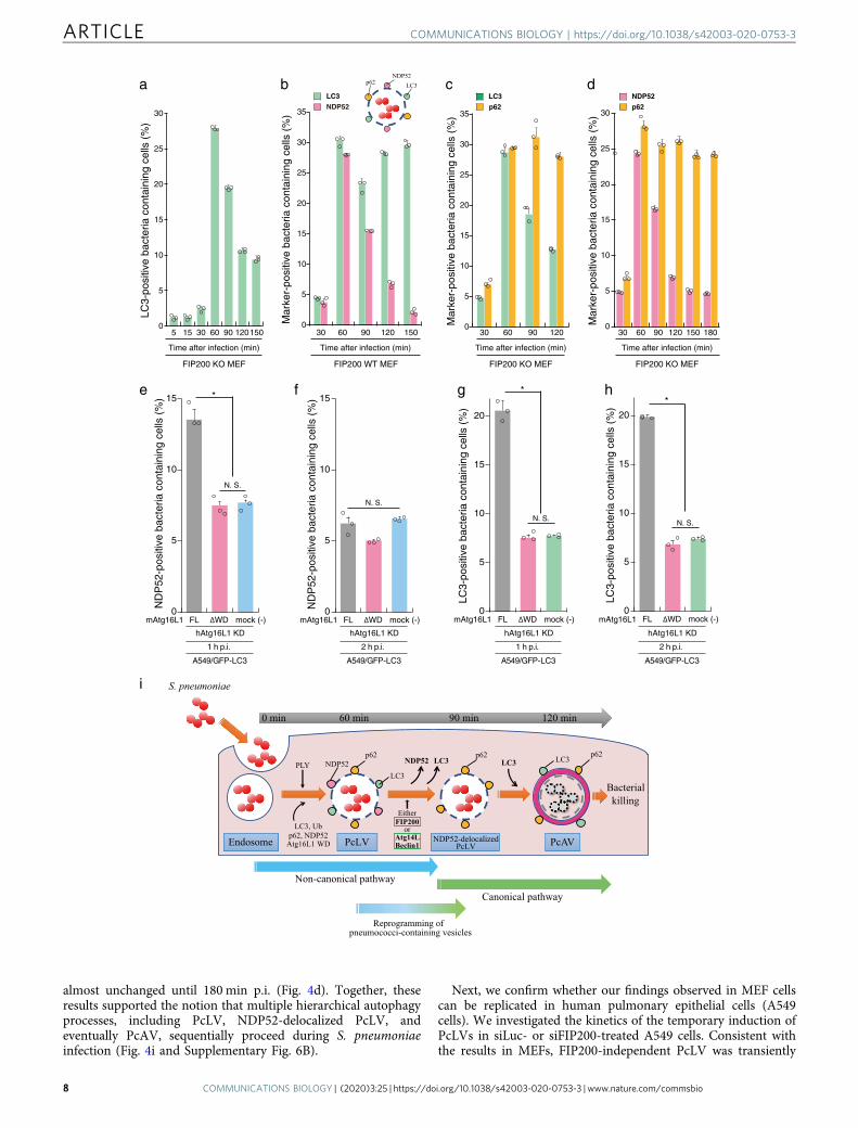
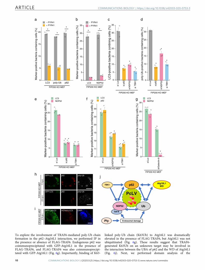
Poly-Ub, p62, NDP52, and Atg16L1 WD are interdependentlyinvolved in PcLV formation. Although we previously reportedthat NDP52 was rarely recruited to PcAVs at 2 h p.i.14, NDP52was frequently recruited to PcLVs at 1 h p.i. at a level comparablewith p62 (Fig. 3a); however, their contribution to PcLV formationremains unclear. Hence, we investigated the interdependence ofpoly-Ub, p62, and NDP52 in PcLV formation. Treatment withPYR-41, a ubiquitin-activating enzyme (E1) inhibitor, led to areduction in the number of LC3-, poly-Ub-, p62-, and NDP52-positive bacteria at 1 h p.i. in FIP200 KO MEFs (Fig. 5a, b). Next,we performed a knockdown experiment to clarify the role of p62and NDP52 in PcLV formation. Upon depletion of p62, NDP52,or TBK1 (activator kinase of these adapters), PcLV formation wasdramatically reduced, suggesting that both p62 and NDP52 arenecessary for PcLV formation (Fig. 5c and SupplementaryFig. 8A–C). Consistent with this, PcLV formation was reduced inAtg14L-knockdown p62 KO MEFs (Supplementary Fig. 8E andF). Intriguingly, poly-Ub deposition on PcVs was also reduced inp62-, NDP52-, or TBK1-knockdown FIP200 KO MEFs, sug-gesting that cargo receptors promote poly-Ub deposition on
PcVs, and vice versa (Fig. 5d). Next, we assessed the inter-dependence of p62 and NDP52 recruitment to intracellular S.pneumoniae in FIP200 KO MEFs. Upon depletion of p62 orTBK1, we observed a robust decrease in NDP52 recruitment tointracellular bacteria (Fig. 5e). Similarly, upon depletion ofNDP52 or TBK1, p62 recruitment to bacteria was also reduced(Fig. 5f), suggesting that the recruitment of p62 and NDP52 tointracellular pneumococci is interdependent. Galectin-8 is a targetof NDP52 in xenophagy5. Upon depletion of Galectin-8, NDP52recruitment was also abolished, but the inhibition of PcLV for-mation was partial (Fig. 5g and Supplementary Fig. 8D). Intra-cellular pneumococci can evade into the cytosol28. We theninvestigated whether LC3, p62, and poly-Ub deposition is tar-geted on the endosomal membrane-engulfing bacteria or on thecytosolic bacterial surface. In PcLV and NDP52-delocalizedPcLV, these signals were clearly decorated on the membrane-engulfing bacteria in a ring shape (Fig. 5h). Similarly, when thephosphoserine-decorated membrane was visualized by GFP-Lact-C2, and most of the intracellular pneumococci were still in themembranous compartment even at the NDP52-delocalized PcLVstage (Fig. 5i). This finding suggests that most of the intracellularpneumococci remained in the damaged vacuoles duringsequential PcLV and NDP52-delocalized PcLV.
Together, these results clearly showed that poly-Ub, p62, NDP52,and Atg16L WD (Fig. 2e, f) are mutually dependent for theirrecruitment to intracellular pneumococci, and that all of theseproteins are required for PcLV formation (Fig. 5j).
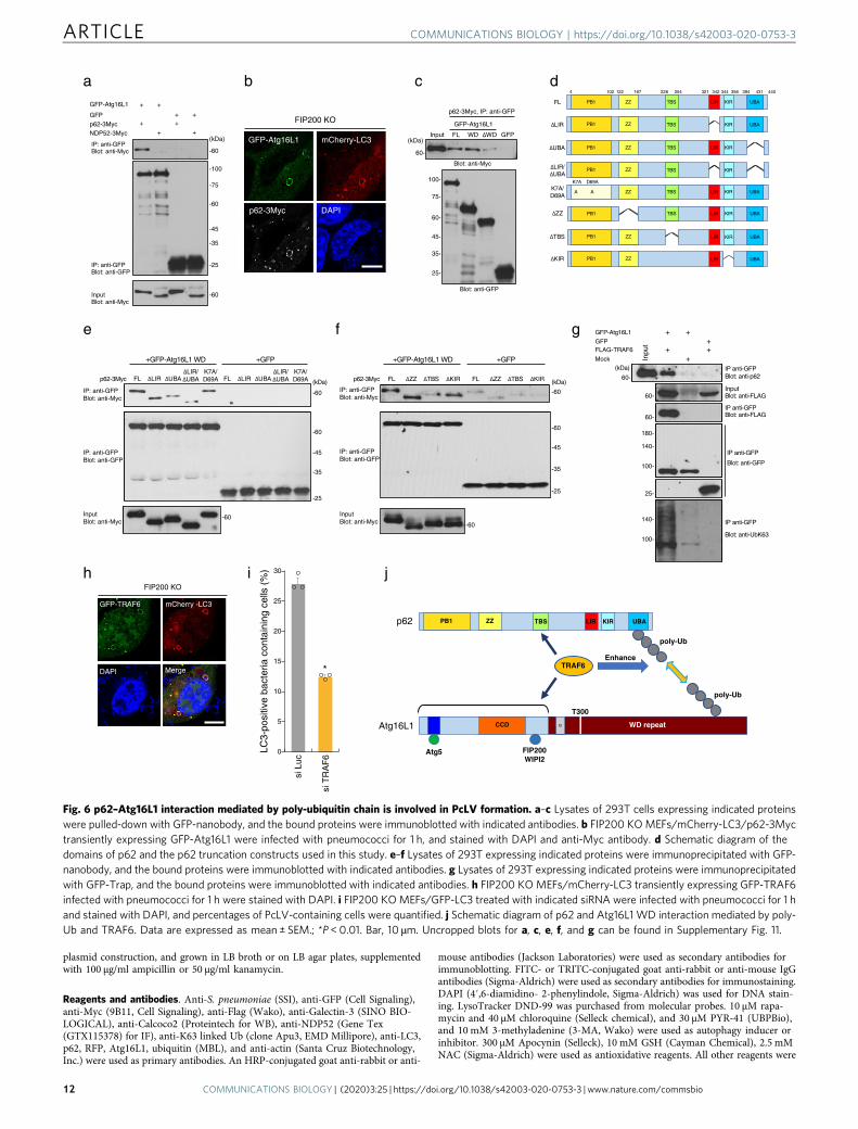
Hence, we sought to assess the interdependence of p62,NDP52, and Atg16L1 in PcLV formation. To this end, weinvestigated whether p62 or NDP52 can interact with Atg16L1through WD. When GFP-Atg16L1 was immunoprecipitated in293T cells transiently expressing GFP-Atg16L1 and p62-3Myc orNDP52-3Myc, p62 was coprecipitated with GFP-Atg16L1, butNDP52 was not (Fig. 6a). p62 and Atg16L were clearly colocalizedwith PcLVs (Fig. 6b). Hence, we performed a domain analysis ofAtg16L1 by immunoprecipitation (IP). The results revealed thatthe C-terminal portion of Atg16L1 (WD domain) is responsiblefor p62 binding (Fig. 6c). Structurally, p62 contains six functionaldomains: the PB1 (Phox and Bem1p), ZZ (ZZ-type zinc finger),TBS (TRAF6-biding sequence), LIR (LC3-interacting region),KIR (Keap1-binding region), and UBA (ubiquitin-associated)domains (Fig. 6d). We prepared a series of truncated versions ofp62-3Myc, including K7A/D69A (dimerization defective), ΔLIR,ΔUBA, or ΔLIR/ΔUBA (Fig. 6d), and investigated their ability tointeract with Atg16L1 WD by IP experiment. We found that thep62–Atg16L1 WD interaction was dramatically reduced in themutant lacking UBA (Fig. 6e). In addition, we prepared otherp62-3Myc variants, including ΔZZ (RIP1-binding defective),ΔTBS, and ΔKIR (Fig. 6d), and tested their ability to interactwith Atg16L1 WD. We also found that the p62–Atg16L1 WDinteraction was dramatically reduced in the ΔTBS mutant (Fig. 6f).These results suggest that the poly-Ub–p62 and TRAF6–p62interactions are important to the p62–Atg16L1 WD interaction.
Fig. 4 Hierarchical autophagy processes during time course of pneumococcal infection. a FIP200 KO MEFs/GFP-LC3 infected with pneumococci forindicated periods were stained with DAPI, and percentages of GFP-LC3 positive bacteria containing cells were quantified. b FIP200 WT MEFs/mCherry-LC3/GFP-NDP52 infected with pneumococci for indicated periods were stained with DAPI, and percentages of each marker-positive bacteria containingcells were quantified. c FIP200 KO MEFs/GFP-LC3 infected with pneumococci for indicated periods were stained with anti-p62 antibody and DAPI, andpercentages of each marker-positive bacteria containing cells were quantified. d FIP200 KO MEFs/GFP-NDP52 infected with pneumococci for indicatedperiods were stained with anti-p62 antibody and DAPI, and percentages of each marker-positive bacteria containing cells were quantified. e–h A549 cells/GFP-LC3/mouse Atg16L1 FL or ΔWD were treated with siRNA for human Atg16L1. After 2 days, the cells were infected with pneumococci for 1 or 2 h, andstained with anti-NDP52 antibody and DAPI, and percentages of each marker-positive bacteria containing cells were quantified. i Schematic diagram ofpneumococci-containing endosomes-remodeling to PcAVs through PcLV and NDP52-delocalized PcLV. Data are expressed as mean ± SEM.; *P < 0.01,N.S., not significant.
COMMUNICATIONS BIOLOGY | https://doi.org/10.1038/s42003-020-0753-3 ARTICLE
COMMUNICATIONS BIOLOGY | (2020) 3:25 | https://doi.org/10.1038/s42003-020-0753-3 | www.nature.com/commsbio 9
To explore the involvement of TRAF6-mediated poly-Ub chainformation in the p62–Atg16L1 interaction, we performed IP inthe presence or absence of FLAG-TRAF6. Endogenous p62 wascoimmunoprecipitated with GFP-Atg16L1 in the presence ofFLAG-TRAF6, and FLAG-TRAF6 was also coimmunoprecipi-tated with GFP-Atg16L1 (Fig. 6g). Importantly, binding of K63-
linked poly-Ub chain (K63Ub) to Atg16L1 was dramaticallyelevated in the presence of FLAG-TRAF6, but Atg16L1 was notubiquitinated (Fig. 6g). These results suggest that TRAF6-generated K63Ub on an unknown target may be involved inthe interaction between the UBA of p62 and the WD of Atg16L1(Fig. 6j). Next, we performed domain analysis of the
a b
f ge
h
i
j
d
LC3 poly-Ub p62
**
*
0
5
10
15
20
– PYR41+ PYR41
Mar
ker-
posi
tive
bact
eria
con
tain
ing
cells
(%
)
Endosomal damagePly
Atg16L1WD
Gal-8
UbNDP52
TBK1 p62
FIP200 KO MEF
siLu
c
sip6
2
siT
BK
1
* ** *
0
5
10
15
20
25
30
Mar
ker-
posi
tive
bact
eria
con
tain
ing
cells
(%
)
FIP200 KO MEF
FIP
200
KO
ME
FF
IP20
0K
O M
EF
1 h
p.i.
2 h
p.i.
2 h
p.i.
* *
siLu
c
siN
DP
52
siT
BK
1
**
0
5
10
15
20
25
30
LC3p62
Mar
ker-
posi
tive
bact
eria
con
tain
ing
cells
(%
)
FIP200 KO MEF siG
alec
tin-8
siLu
c
*
*
0
5
10
15
20
25
30
LC3NDP52
LC3NDP52
Mar
ker-
posi
tive
bact
eria
con
tain
ing
cells
(%
)
FIP200 KO MEF
c
siLu
c
**
*
sip6
2
siN
DP
52
siT
BK
1
*
*
*
0
5
10
15
20
25
30
poly
-Ub-
posi
tive
bact
eria
con
tain
ing
cells
(%
)
siLu
c
sip6
2
siN
DP
52
siT
BK
10
5
10
15
20
25
30
35
LC3-
posi
tive
bact
eria
con
tinin
g ce
lls (
%)
FIP200 KO MEF FIP200 KO MEF
* *
FIP200 KO MEF
0
5
10
15
20
25
30
35- PYR41
+ PYR41
LC3 NDP52
Mar
ker-
posi
tive
bact
eria
con
tain
ing
cells
(%
)
GFP-LC3 p62 poly-Ub
GFP-LC3 p62 poly-Ub
GFP-Lact-C2 mCherry-LC3 DAPI
PcLV
ARTICLE COMMUNICATIONS BIOLOGY | https://doi.org/10.1038/s42003-020-0753-3
10 COMMUNICATIONS BIOLOGY | (2020) 3:25 | https://doi.org/10.1038/s42003-020-0753-3 | www.nature.com/commsbio
Atg16L1–TRAF6 interaction by IP, and we found that the T300Asubstitution did not negatively affect the Atg16L1–TRAF6 andTRAF6-enhanced Atg16L1–p62 interactions (SupplementaryFig. 9A). Notably, TRAF6 can bind to the Atg16L1 N-terminalΔWD domain (Fig. 6j and Supplementary Fig. 9B–C), where it isinvolved in Atg5, FIP200, or WIPI-2 binding. Atg16L1 harbors aconserved TRAF6-interacting motif in the central region(Supplementary Fig. 9B), but the this putative TRAF6-bindingregion of Atg16L1 was not involved in TRAF6 binding(Supplementary Fig. 9C). Next, we investigated whether GFP-TRAF6 was recruited to PcLVs, and found that GFP-TRAF6 wasclearly confined to PcLVs (Fig. 6h). Finally, PcLV formation wasrobustly decreased by TRAF6 knockdown in FIP200 KO MEFs(Fig. 6i and Supplementary Fig. S9D).
DiscussionIn this study, we identified LC3- and NDP52-delocalized LAPo-some-like vacuoles and found evidence that multiple autophagicprocesses involving PcLV, NDP52-delocalized PcLV, and PcAVsequentially target intracellular S. pneumoniae (Fig. 4i). At earlystage of infection, intracellular pneumococci evade the bacter-icidal endocytic pathway by triggering PcLV, and take refuge in atransient niche; however, subsequent PcAVs eventually destroythis survival niche. Furthermore, we demonstrated that severalcomponents of autophagic machinery, including poly-ubiquitin,p62, NDP52, and Atg16L1, are interdependently recruited todamaged PcVs during LAP-like autophagic processes (Fig. 5j),and that TRAF6-mediated poly-Ub formation promotes thep62–Atg16L1 interaction (Fig. 6j) and subsequent PcLV forma-tion. Furthermore, we suggested that Crohn’s disease might becorrelated with pneumococcal diseases (Fig. 2j–m).
Multiple autophagic pathways and distinct autophagic machi-neries are employed during distinct bacterial infections2. Indeed,p62 and NDP52 are interdependently recruited to intracellularShigella during xenophagy, whereas poly-Ub, p62, and NDP52 areindependently recruited to intracellular Listeria during this pro-cess29. Moreover, p62 and NDP52 are independently recruited tointracellular Salmonella during xenophagy30. Thus, the inter-dependence of p62 and NDP52 is distinct in each bacterial species.In S. pneumoniae infection, p62 is required for both PcLV andPcAV formation, but NDP52 is involved only in PcLV formation.In addition, we found that poly-Ub, p62, and NDP52 are mutuallydependent for their recruitment to intracellular pneumococci, andall are required for PcLV formation. Importantly, we obtainedevidence of LC3- and NDP52-delocalized but p62-positive PcLVsduring the transition from PcLVs to PcAVs. However, the roles ofAtg14L, Beclin1, and FIP200 during the NDP52-delocalized PcLVstransition, as well as the events that occur during the disappearanceof LC3 and NDP52, remain to be elucidated.
In LAP and LAP-like autophagy, the involvement of cargoreceptors in each experimental setting is distinct. Poly-Uband p62 are recruited to vacuoles induced by LLOMe, a
lysosome-specific inducer of membrane damage31. In AMDE-1-induced LAP-like autophagy, p62 is recruited, but is not neces-sary32. By contrast, in monensin- or chloroquine-induced LAP-like autophagy, p62 and NDP52 are not recruited33. Similarly,Listeria-induced LAP in phagocytes requires LLO (ListeriolysinO)-induced membrane rupture, but not poly-Ub or p629. InVacA-induced LAP-like autophagy process, p62 is not recrui-ted34. In this study, we revealed that PcLVs are a previouslyunreported type of FIP200-independent LAP-like vesicles towhich Galectin3, poly-Ub, p62, and NDP52 are recruited. Inaddition, PcLVs has unique features among LAP-like vesicles,including ROS-, TLR2-, PI3P-, Myo6-, and Atg16L1 α domainindependence. Importantly, it remains to be elucidated whether acommon molecule or biochemical event is involved in these LAP-like autophagy processes.
Atg16L1 T300A has no negative effect on Salmonella-inducedLAP-like autophagy or xenophagy16. By contrast, Atg16L1 T300Ais associated with reduced xenophagy against Shigella21,22, and wefound that PcLV and PcAV formation was reduced in Atg16L1T300A-complemented cells. LAP-like autophagy and canonicalautophagy proceed concurrently in Salmonella infection15,16.Hence, we speculated that Atg16L1 T300A would have a negativeeffect only on LAP or LAP-like processes, but not on canonicalautophagy (including xenophagy) and that defects in LAP-likeprocesses would be masked by canonical autophagy in Salmonellainfection. It is most exciting to speculate that some pneumococcalvirulence factor can manipulate or delay FIP200-dependentcanonical autophagy during the early stage of infection in orderto acquire a transient survival niche. The Atg16L1 T300A proteinis susceptible to Caspase-3 and -7-mediated digestion in myeloidcells during apoptosis22. However, Atg16L1 T300A is not pre-ferentially cleaved by S. pneumoniae infection (SupplementaryFig. 3E). Notably, pneumococcal infection tends to be moresevere in Crohn’s patients25,26, and it is tempting to speculate thatautophagy exerts a protective effect against severe or IPD.
In summary, several lines of evidence suggest that mammaliancells use several pathways to target bacteria for autophagy: canonicalautophagy, Ub-dependent selective autophagy, and LAP-likeautophagy, including the process involving PcLV. Indeed, S.pneumoniae can trigger autophagy via various pathways in multiplecell types. Our discovery gives insight into the mechanism by whichthe host defense system senses intracellular pulmonary bacterialpathogens. This knowledge may facilitate the development oftherapeutic targets for the control of pneumococcal diseases.
MethodsBacterial strains. S. pneumoniae strain R6 (ATCC BAA-255) and strainATCC6308 (ST8) were purchased from the American Type Culture Collection.Δply S. pneumoniae strain was constructed as described previously14. S. pneumo-niae was grown as standing cultures in THY (Todd–Hewitt Broth (BD) plus 0.5%yeast extract (BD)) broth, or plated THY-agar plates, or Columbia agar plates with5% sheep blood (BD) at 37 °C in a 5% CO2 in air atmosphere. Escherichia colistrains MC1061, DH10B, or C43 (Cosmo Bio) were used for DNA cloning and
Fig. 5 Poly-Ub, p62, and NDP52 regulate PcLV formation interdependently. a, b FIP200 KO MEFs/GFP-LC3 or FIP200 KO MEFs/mCherry-LC3/GFP-NDP52 infected with WT or Δply pneumococci for 1 h with or without PYR-41 were stained with DAPI and antibodies against poly-Ub or p62, andpercentages of each marker-positive bacteria containing cells were quantified. c, d FIP200 KO MEFs/mCherry-LC3 treated with indicated siRNA wereinfected with pneumococci for 1 h and stained with DAPI and anti-poly-Ub antibody, and percentages of LC3- or poly-Ub-positive bacteria containing cellswere quantified. e, f FIP200 KO MEFs/mCherry-LC3 stably expressing GFP-p62 or GFP-NDP52 treated with indicated siRNA were infected withpneumococci for 1 h and stained with DAPI, and percentages of each marker-positive bacteria containing cells were quantified. g FIP200 KO MEFs/mCherry-LC3/GFP-NDP52 treated with indicated siRNA were infected with pneumococci for 1 h and stained with DAPI, and percentages of each marker-positive bacteria containing cells were quantified. h, i FIP200 KO MEFs/GFP-LC3 or FIP200 KO MEFs/mCherry-LC3/GFP-Lact-C2 infected with WTpneumococci for 1 or 2 h were stained with DAPI and antibodies against p62 or poly-Ub. j Autophagy machinery protein dynamics in PcLV formation. Dataare expressed as mean ± SEM.; *P < 0.01.
COMMUNICATIONS BIOLOGY | https://doi.org/10.1038/s42003-020-0753-3 ARTICLE
COMMUNICATIONS BIOLOGY | (2020) 3:25 | https://doi.org/10.1038/s42003-020-0753-3 | www.nature.com/commsbio 11
plasmid construction, and grown in LB broth or on LB agar plates, supplementedwith 100 μg/ml ampicillin or 50 μg/ml kanamycin.
Reagents and antibodies. Anti-S. pneumoniae (SSI), anti-GFP (Cell Signaling),anti-Myc (9B11, Cell Signaling), anti-Flag (Wako), anti-Galectin-3 (SINO BIO-LOGICAL), anti-Calcoco2 (Proteintech for WB), anti-NDP52 (Gene Tex(GTX115378) for IF), anti-K63 linked Ub (clone Apu3, EMD Millipore), anti-LC3,p62, RFP, Atg16L1, ubiquitin (MBL), and anti-actin (Santa Cruz Biotechnology,Inc.) were used as primary antibodies. An HRP-conjugated goat anti-rabbit or anti-
mouse antibodies (Jackson Laboratories) were used as secondary antibodies forimmunoblotting. FITC- or TRITC-conjugated goat anti-rabbit or anti-mouse IgGantibodies (Sigma-Aldrich) were used as secondary antibodies for immunostaining.DAPI (4′,6-diamidino- 2-phenylindole, Sigma-Aldrich) was used for DNA stain-ing. LysoTracker DND-99 was purchased from molecular probes. 10 µM rapa-mycin and 40 µM chloroquine (Selleck chemical), and 30 µM PYR-41 (UBPBio),and 10 mM 3-methyladenine (3-MA, Wako) were used as autophagy inducer orinhibitor. 300 µM Apocynin (Selleck), 10 mM GSH (Cayman Chemical), 2.5 mMNAC (Sigma-Aldrich) were used as antioxidative reagents. All other reagents were
d
e
h i j
f g
PB1 LIRTBS KIR UBA
PB1 LIR KIR UBAZZ
PB1 LIR UBAZZ
ΔZZ
ΔTBS
ΔKIR
PB1 ZZ LIRTBS KIR UBA
4 102 122 167 228 254 321 342 394 431 440344 356
PB1 TBS KIR UBA
PB1 LIR KIRZZ
PB1 ZZ
FL
ΔLIR
ΔUBA
ΔLIR/ΔUBA
ZZ
TBS
LIR UBAZZK7A/D69A
KIR
KIRTBS
K7A D69A
A A
TBS
a
FL ΔZZ ΔTBS ΔKIR FL ΔZZ ΔTBS ΔKIRFLp62-3Myc (kDa)ΔLIR ΔUBAΔLIR/ΔUBA
K7A/D69A FL ΔLIR ΔUBA
ΔLIR/ΔUBA
K7A/D69A
+GFP-Atg16L1 WD +GFP
IP: anti-GFPBlot: anti-GFP
InputBlot: anti-Myc
IP: anti-GFPBlot: anti-Myc
p62-3Myc
IP: anti-GFPBlot: anti-GFP
InputBlot: anti-Myc
IP: anti-GFPBlot: anti-Myc
+GFP-Atg16L1 WD +GFP
siLu
c
siT
RA
F6
*
0
5
10
15
20
25
30
LC3-
posi
tive
bact
eria
con
tain
ing
cells
(%
)
+GFP-Atg16L1
Blot: anti-p62IP anti-GFP
IP anti-GFP
IP anti-GFP
IP anti-GFPBlot: anti-FLAG
InputBlot: anti-FLAG
Blot: anti-GFP
Blot: anti-UbK63
++GFP
Inpu
t
+FLAG-TRAF6
+Mock
+
cbGFP-Atg16L1
-60
-60
-60
-60
-35
-45
-25
(kDa)
-60
(kDa)
60-
60-
60-
100-
180-
140-
100-
140-
25-
-60
-60
-35
-45
-25
-60
(kDa)
GFPp62-3MycNDP52-3Myc
+
+++
+
+
+
+
Blot: anti-MycIP: anti-GFP
Blot: anti-GFPIP: anti-GFP
Blot: anti-MycInput
GFP-Atg16L1
GFPFL WD ΔWDInput
60-
100-
75-
60-
25-
35-
45-
(kDa)
p62-3Myc, IP: anti-GFP
Blot: anti-Myc
Blot: anti-GFP
FIP200 KO
mCherry-LC3
p62-3Myc DAPI
FIP200 KO
GFP-Atg16L1
GFP-TRAF6 mCherry -LC3
DAPI Merge
p62 PB1 ZZ LIRTBS KIR UBA
TRAF6
WD repeatAtg16L1 CCD
Atg5 FIP200WIPI2
T300
poly-Ub
poly-Ub
Enhance
-100
-75
-60
-45
-35
-25
Fig. 6 p62–Atg16L1 interaction mediated by poly-ubiquitin chain is involved in PcLV formation. a–c Lysates of 293T cells expressing indicated proteinswere pulled-down with GFP-nanobody, and the bound proteins were immunoblotted with indicated antibodies. b FIP200 KOMEFs/mCherry-LC3/p62-3Myctransiently expressing GFP-Atg16L1 were infected with pneumococci for 1 h, and stained with DAPI and anti-Myc antibody. d Schematic diagram of thedomains of p62 and the p62 truncation constructs used in this study. e–f Lysates of 293T expressing indicated proteins were immunoprecipitated with GFP-nanobody, and the bound proteins were immunoblotted with indicated antibodies. g Lysates of 293T expressing indicated proteins were immunoprecipitatedwith GFP-Trap, and the bound proteins were immunoblotted with indicated antibodies. h FIP200 KO MEFs/mCherry-LC3 transiently expressing GFP-TRAF6infected with pneumococci for 1 h were stained with DAPI. i FIP200 KOMEFs/GFP-LC3 treated with indicated siRNA were infected with pneumococci for 1 hand stained with DAPI, and percentages of PcLV-containing cells were quantified. j Schematic diagram of p62 and Atg16L1 WD interaction mediated by poly-Ub and TRAF6. Data are expressed as mean ± SEM.; *P < 0.01. Bar, 10 µm. Uncropped blots for a, c, e, f, and g can be found in Supplementary Fig. 11.
ARTICLE COMMUNICATIONS BIOLOGY | https://doi.org/10.1038/s42003-020-0753-3
12 COMMUNICATIONS BIOLOGY | (2020) 3:25 | https://doi.org/10.1038/s42003-020-0753-3 | www.nature.com/commsbio

purchased from Sigma-Aldrich. All antibodies were used at 1:100 for immuno-fluorescence staining and 1:1000 for western blotting.
Plasmids. All PCR was executed by Q5 High-Fidelity DNA Polymerase (NewEngland BioLabs), and all RT-PCR was performed by SuperScript III One-Step RT-PCR System with Platinum Taq (Thermo Fisher Scientific). The vectors for GFP-or mCherry-rat LC3B, or human pIgR stable-expressing MEFs generation wereconstructed as described previously14. Vectors for GFP-Nedd4-1, GFP-Galectin-314, WIPI2-GFP, GFP-Rubicon4, and p62 (FL, ΔLIR, ΔUBA, ΔLIR-ΔUBA, andK7A/D69A)-3Myc6 expression vectors were constructed as described previously.pcDNA-p62-3Myc ΔZZ, ΔTBS, and ΔKIR were constructed by NEB builder (NewEngland BioLabs) using pcDNA-p62-3Myc FL as a template and primers shown inSupplementary Table 1. For stable clone generation, obtained p62-3Myc ΔTBScDNA was subcloned into pMXs-puro vector (Cosmo Bio). GFP-LC3 G120A wasconstructed with QuickChange (Agilent) using pEGFP-rat LC3B as a template andprimers shown in Supplementary Table 1. For pcDNA3.1-NDP52-3Myc con-struction, human NDP52 cDNA amplified by PCR using pEGFP-NDP52 as atemplate and primers shown in Supplementary Table 1 was subcloned intopcDNA3.1-3Myc vector4. For the construction of NDP52-3Myc-stably expressingcells, obtained NDP52-3Myc cDNA was subcloned into pMXs-IRES-blast vector(Cosmo Bio). For construction of pEGFP-Ply variants, allele 1 ply gene of R6, orAllele 5 ply gene of ATCC6308 were subcloned into pEGFP-C1 vector (Clontech).For construction of pEGFP-mouse Atg16L1, mouse Atg16L1 cDNA purchase fromOpenbiosystems (IMAGE:6813377) was subcloned into pEGFP-C3 vector. For theconstruction of Atg16L1-stably expressing cells, nontagged mouse Atg16L1 cDNAwas subcloned into pMXs-puro vector. For the construction of mouse Atg16L1ΔWD or WD, each cDNA was amplified by PCR with primers shown in Sup-plementary Table 1. Human OPTN and UVRAG cDNA were amplified by RT-PCR using primer pairs shown in Supplementary Table 1 and total mRNA fromDetroit 562 or HeLa cell as a template. Obtained DNA fragments were subclonedinto pEGFP-C or -N vectors. The expression vectors for GFP- and FLAG-TRAF635
were a generous gift from Dr Jun-ichiro Inoue (University of Tokyo). Theexpression vectors for GST-GFP-nanobody36 were a generous gift from Dr YoheiKatoh (Kyoto University). The expression vectors for GFP-rat LC3B37 were agenerous gift from Dr Tamotsu Yoshimori (Osaka University).
Cell cultures and transfections. A total of 293T (human embryonic kidneyfibroblasts) cells and variety of MEFs (mouse embryonic fibroblasts) were cultured inDulbecco’s modified Eagle medium (DMEM, Nakarai) supplemented with 10% FCS(Gibco), 100 μg/ml gentamicin (Wako), and 60 μg/ml kanamycin (Wako). platE cellwas maintained in DMEM/10% FCS with 1 μg/ml puromycin (Sigma) and 10 μg/mlblasticidin (Kaken pharmaceutical). Transfections were performed by using PEI MAX(Polysciences) or Lipofectamine LTX (Thermo Fisher Scientific) for 293T and platE,and Fugene 6 (Promega) for MEFs according to the manufacturers’ protocols. MEFs-derived stable clones were cultured in DMEM/10% FCS with 1 μg/ml puromycin and/or 10 μg/ml blasticidin. FIP200 WT or KO was cultured previously38. ULK1/2 WT orDKO MEFs39, Atg5 WT or KO MEFs40, Nox1 WT or KO and Nox4 WT or KOMEFs41, Atg16L1 WT or KO MEFs42, and p62 WT or KO MEFs43 were generousgift from Dr Craig B. Thompson (Memorial Sloan Kettering Cancer Center), DrNoboru Mizushima (The University of Tokyo), Dr Denis Martinvalet (GenèveUniversity), and Drs Tatsuya Saitoh (Tokushima University) and Shizuo Akira(Osaka University), and Toru Yanagawa (University of Tsukuba).
siRNA. siRNAs were synthesized and duplexed by siRNA Co. Ltd. The siRNAsequences for mouse Rabs are shown in Supplementary Table 2. The siRNAs weretransfected into cells by reverse transfection using Lipofectamine RNAi MAX(Thermo Fisher Scientific) according to the manufacturer’s protocol. Knockdownefficiency was checked by RT-PCR using primer pair shown in SupplementaryTable 2.
Construction of S. pneumoniae mutant by allelic exchange mutagenesis.Substitution of the ply gene in S. pneumoniae strain R6 was performed by doublecrossover recombination as described previously14. Briefly, Allele 1 ply gene of R6,or Allele 5 ply gene of ATCC6308, and subsequent erm cassette were joined withlong 5′- and 3′-flanking regions that are homologous to the target locus weregenerated using two-step PCR as described previously44 using the primers listed inSupplementary Table 3. PCR products were introduced into competent cells of theS. pneumoniae strain R6 as described previously14, and transformants were selectedby 1 μg/ml erythromycin. Substitution of the ply gene was confirmed by sequencingof PCR product amplified by primers listed in Supplementary Table 1.
Recombinant retroviruses and infections. The pMXs-puro and pMX-IRES-blastvectors were purchased from Cosmo Bio. Recombinant retroviruses were preparedas previously described14. In short, obtained retroviral plasmid was transfected intoplatE, and after 2 days culture supernatant containing retrovirus was collected andcentrifugated, and clear supernatant was used for retroviral infection. For pre-paration of retrovirus for A549 cells, platE cells were cotransfected with retroviralplasmid and VSV-G plasmid. For retroviral infection, recipient cells were infected
with retroviruses by adding obtained supernatant of platE cells in the presenceof polybrene for 6 h. Stable transformants were selected in DMEM/10% FCS with1 μg/ml puromycin or 10 μg/ml blasticidin.
Infection of cells with S. pneumoniae and sample preparation for fluorescencemicroscopy, western blotting, electron microscopy, and survivability assay.Variety of MEFs/pIgR were infected with S. pneumoniae as previously described14.In short, MEFs seeded at 2 × 10⁵ cells/well on glass coverslips in six-well plates wereinfected with fresh S. pneumoniae grown with THY broth at MOI (multiplicity ofinfection) of 100, and then centrifugated at 1000 rpm for 5 min at room tem-perature. Cells were incubated for 1 h at 37 °C in 5% CO2 to allow bacterial uptakeand invasion, washed with Hank’s balanced salt solution (HBSS) three times, andDMEM/10% FCS with 200 μg/ml gentamycin and 200 U/ml catalase (Bacterialkilling buffer) was added to each well and incubated for 30 min to kill extracellularbacteria. After changing the medium to DMEM/10% FCS with 100 μg/ml genta-mycin and 200 U/ml catalase (incubation buffer), the cells were incubated for theindicated periods at 37 °C in 5% CO2. If not stated otherwise, inhibitors were addedin incubation buffer to avoid the influence on bacterial invasion efficiency. At eachtime point, the cells were fixed with 4% paraformaldehyde/PB (phosphate buffer)(Wako) for 15 min at room temperature. Fixed cells were washed with PBS threetimes and quenched with 50 mM NH4Cl in PBS for 10 min, permeabilized with0.2% Triton X-100 in PBS for 10 min and blocked with 2% bovine serum albuminin TBS (Tris-buffered saline) for 30 min at room temperature. After staining usingthe indicated antibodies, the specimens were analyzed by confocal microscopy(Carl Zeiss LSM700). For western blot analysis, cells were lysed with 100 μl of 2×SDS sample buffer (Nakarai Tesque). Equal volumes of lysates were separated bySDS-PAGE and transferred to a PVDF membrane. For electron microscopyobservation, cells seeded on 35 mm dishes were infected with S. pneumoniae asdescribed above, fixed with 2% glutaraldehyde/4% paraformaldehyde in PBS forovernight at room temperature, post-fixed in 2% OsO4, dehydrated with a gradedethanol series, and embedded in epoxy resin. Ultra-thin sections were stained withboth uranyl acetate and lead citrate. For, intracellular bacterial survivability assay,MEFs seeded on 24-well plates were infected with wild-type of S. pneumoniae asdescribed previously14. Briefly, MEFs were infected with S. pneumoniae strain R6WT to allow bacterial invasion as described above, and were washed with HBSStwice, and were then incubated in bacterial killing buffer containing 200 μg/mlgentamycin for 15 min, and were incubated in bacterial killing buffer containing200 μg/ml gentamycin and 10 μg/ml of penicillin G (Wako) for a further 15 min.After washing cells with HBSS three times, cells were incubated in incubationbuffer for 1 h at 37 °C in 5% CO2, and were then lysed using 1.0% saponin (Sigma-Aldrich), and lysates were serial diluted with 0.1% saponin in PBS and were platedonto THY-agar plates, and numbers of intracellular bacteria were counted andexpressed in colony forming units (CFU).
Quantification of cells with bacteria associated with autophagic markers. Weconfirmed that infection efficiency was at least 100% when MEFs were infectedwith S. pneumoniae at MOI= 100. Thus, we evaluated PcLV formation activity bythe percentage of cells containing PcV (S. pneumoniae-containing vesicles) asso-ciated with autophagic markers, such as GFP-LC3, p62-3Myc, ubiquitin etc., whichwas determined by visual counting with fluorescence microscopy. At least 500PcVs, PcLVs, or S. pneumoniae-containing cells were examined in triplicate foreach experimental condition. Error bars indicate the standard errors of the means.
Purification of GST-GFP-nanobody protein. E. coli BL21 harboring pGST-GFP-nanobody was cultured in L-broth with 50 μg/ml of ampicillin for 2 h at 37 °C, thenisopropyl-1-thio-β-D- galactopyranoside was added to the culture medium at afinal concentration of 0.1 mM. After incubation for 2 h at 37 °C, bacteria wereharvested, and GST fusion proteins were purified according to the manufacturer’sprotocol using glutathione Sepharose 4B (GE Healthcare).
GST pull-down assay using 293T cell lysates. A total of 293T cells transfectedwith indicated plasmids seeded onto six-well plate were suspended in 500 μl ofwash buffer (TBS/5 mM MgCl2, 1 mM EDTA, 0.05% NP-40) including completeprotease inhibitor cocktail (Roche), chilled on ice for 30 min, and centrifuged at16,900 × g at 4 °C for 10 min. Five microliters of GST-GFP-nanobody bound toglutathione Sepharose 4B was mixed with cleared lysates of the 293T cells andincubated with rotation for 2 h at 4 °C. After incubation, the beads were washedwith 1 ml of wash buffer three times, and the bound proteins were analyzed byimmunoblotting using indicated antibodies.
Immunoprecipitation using 293T cell lysates. A total of 293T cells transfectedwith indicated plasmids seeded onto six-well plate were suspended in 500 μl of washbuffer (TBS/5mMMgCl2, 1 mM EDTA, 0.05% NP-40) including complete proteaseinhibitor cocktail (Roche), chilled on ice for 30 min, and centrifuged at 16,900 × g at4 °C for 10min. Five microliters of GFP-Trap beads (Chromotek) was mixed withcleared lysates of the 293T cells and incubated with rotation for 2 h at 4 °C. Afterincubation, the beads were washed with 1 m of wash buffer three times, and thebound proteins were analyzed by immunoblotting using indicated antibodies.
COMMUNICATIONS BIOLOGY | https://doi.org/10.1038/s42003-020-0753-3 ARTICLE
COMMUNICATIONS BIOLOGY | (2020) 3:25 | https://doi.org/10.1038/s42003-020-0753-3 | www.nature.com/commsbio 13
Statistics and reproducibility. At least 500 PcVs, PcLVs, or pneumococcus-containing cells were examined in triplicate, in each experiment. Data are themeans ± SEM. P values were calculated using Student’s t test. If SD is sig-nificantly different (F < 0.05), P values were calculated using Mann–Whitney Utest by using Prism6.
Reporting summary. Further information on research design is available inthe Nature Research Reporting Summary linked to this article.
Data availabilityThe authors declare that all data supporting the findings of this study are available withinthe article and its supplementary information files. All source data underlying the graphspresented in the main figures are made available in Supplementary Data 1.
Received: 7 May 2019; Accepted: 23 December 2019;
References1. Mizushima, N., Yoshimori, T. & Ohsumi, Y. The role of Atg proteins in
autophagosome formation. Annu Rev. Cell Dev. Biol. 27, 107–132 (2011).2. Evans, R. J., Sundaramurthy, V. & Frickel, E. M. The interplay of host
autophagy and eukaryotic pathogens. Front. Cell Dev. Biol. 6, 118 (2018).3. Grumati, P. & Dikic, I. Ubiquitin signaling and autophagy. J. Biol. Chem. 293,
5404–5413 (2017).4. Ogawa, M. et al. A Tecpr1-dependent selective autophagy pathway targets
bacterial pathogens. Cell Host Microbe 9, 376–389 (2011).5. Thurston, T. L., Wandel, M. P., von Muhlinen, N., Foeglein, A. & Randow, F.
Galectin 8 targets damaged vesicles for autophagy to defend cells againstbacterial invasion. Nature 482, 414–418 (2012).
6. Yoshikawa, Y. et al. Listeria monocytogenes ActA-mediated escape fromautophagic recognition. Nat. Cell Biol. 11, 1233–1240 (2009).
7. Heckmann, B. L., Boada-Romero, E., Cunha, L. D., Magne, J. & Green, D. R.LC3-Associated Phagocytosis and Inflammation. J. Mol. Biol. 429, 3561–3576(2017).
8. Huang, J. et al. Activation of antibacterial autophagy by NADPH oxidases.Proc. Natl Acad. Sci. USA 106, 6226–6231 (2009).
9. Lam, G. Y., Cemma, M., Muise, A. M., Higgins, D. E. & Brumell, J. H. Hostand bacterial factors that regulate LC3 recruitment to Listeria monocytogenesduring the early stages of macrophage infection. Autophagy 9, 985–995(2013).
10. Martinez, J. et al. Molecular characterization of LC3-associated phagocytosisreveals distinct roles for Rubicon, NOX2 and autophagy proteins. Nat. CellBiol. 17, 893–906 (2015).
11. Mitchell, G. et al. Listeria monocytogenes triggers noncanonical autophagyupon phagocytosis, but avoids subsequent growth-restricting xenophagy. Proc.Natl Acad. Sci. USA 115, E210–E217 (2018).
12. Fletcher, K. et al. The WD40 domain of ATG16L1 is required for its non-canonical role in lipidation of LC3 at single membranes. EMBO J. 37, e97840(2018).
13. Kadioglu, A., Weiser, J. N., Paton, J. C. & Andrew, P. W. The role ofStreptococcus pneumoniae virulence factors in host respiratory colonizationand disease. Nat. Rev. Microbiol. 6, 288–301 (2008).
14. Ogawa, M. et al. Molecular mechanisms of Streptococcus pneumoniae-targetedautophagy via pneumolysin, Golgi-resident Rab41, and Nedd4-1-mediatedK63-linked ubiquitination. Cell Microbiol. 20, e12846 (2018).
15. Fujita, N. et al. Recruitment of the autophagic machinery to endosomes duringinfection is mediated by ubiquitin. J. Cell Biol. 203, 115–128 (2013).
16. Fujita, N. et al. Differential involvement of Atg16L1 in Crohn disease andcanonical autophagy: analysis of the organization of the Atg16L1 complex infibroblasts. J. Biol. Chem. 284, 32602–32609 (2009).
17. Jefferies, J. M. et al. Presence of nonhemolytic pneumolysin in serotypes ofStreptococcus pneumoniae associated with disease outbreaks. J. Infect. Dis. 196,936–944 (2007).
18. Xiong, Q. et al. The role of ATG16 in autophagy and the ubiquitin proteasomesystem. Cells 8, E2 (2018).
19. Tan, J. M. J. et al. An ATG16L1-dependent pathway promotes plasmamembrane repair and limits Listeria monocytogenes cell-to-cell spread. Nat.Microbiol. 3, 1472–1485 (2018).
20. Lystad, A. H. et al. Distinct functions of ATG16L1 isoforms in membranebinding and LC3B lipidation in autophagy-related processes. Nat. Cell Biol.21, 372–383 (2019).
21. Boada-Romero, E. et al. The T300A Crohn’s disease risk polymorphismimpairs function of the WD40 domain of ATG16L1. Nat. Commun. 7, 11821(2016).
22. Lassen, K. G. et al. Atg16L1 T300A variant decreases selective autophagyresulting in altered cytokine signaling and decreased antibacterial defense.Proc. Natl Acad. Sci. USA 111, 7741–7746 (2014).
23. Homer, C. R., Richmond, A. L., Rebert, N. A., Achkar, J. P. & McDonald, C.ATG16L1 and NOD2 interact in an autophagy-dependent antibacterialpathway implicated in Crohn’s disease pathogenesis. Gastroenterology 139,1630–1641 (2010). 1641 e1631-1632.
24. Travassos, L. H. et al. Nod1 and Nod2 direct autophagy by recruitingATG16L1 to the plasma membrane at the site of bacterial entry. Nat.Immunol. 11, 55–62 (2010).
25. Long, M. D., Martin, C., Sandler, R. S. & Kappelman, M. D. Increased risk ofpneumonia among patients with inflammatory bowel disease. Am. J.Gastroenterol. 108, 240–248 (2013).
26. Casella, G. et al. Pulmonary diseases associated with inflammatory boweldiseases. J. Crohns Colitis 4, 384–389 (2010).
27. Verlhac, P. et al. Autophagy receptor NDP52 regulates pathogen-containingautophagosome maturation. Cell Host Microbe 17, 515–525 (2015).
28. Surve, M. V. et al. Heterogeneity in pneumolysin expression governs the fateof Streptococcus pneumoniae during blood-brain barrier trafficking. PLoSPathog. 14, e1007168 (2018).
29. Mostowy, S. et al. p62 and NDP52 proteins target intracytosolic Shigella andListeria to different autophagy pathways. J. Biol. Chem. 286, 26987–26995(2011).
30. Cemma, M., Kim, P. K. & Brumell, J. H. The ubiquitin-binding adaptorproteins p62/SQSTM1 and NDP52 are recruited independently to bacteria-associated microdomains to target Salmonella to the autophagy pathway.Autophagy 7, 341–345 (2011).
31. Maejima, I. et al. Autophagy sequesters damaged lysosomes to controllysosomal biogenesis and kidney injury. EMBO J. 32, 2336–2347 (2013).
32. Li, M. et al. AMDE-1 is a dual function chemical for autophagy activation andinhibition. PLoS ONE 10, e0122083 (2015).
33. Florey, O., Gammoh, N., Kim, S. E., Jiang, X. & Overholtzer, M. V-ATPaseand osmotic imbalances activate endolysosomal LC3 lipidation. Autophagy 11,88–99 (2015).
34. Yahiro, K. et al. Low-density lipoprotein receptor-related protein-1 (LRP1)mediates autophagy and apoptosis caused by Helicobacter pylori VacA. J. Biol.Chem. 287, 31104–31115 (2012).
35. Kobayashi, N. et al. Segregation of TRAF6-mediated signaling pathwaysclarifies its role in osteoclastogenesis. EMBO J. 20, 1271–1280 (2001).
36. Katoh, Y., Nozaki, S., Hartanto, D., Miyano, R. & Nakayama, K. Architecturesof multisubunit complexes revealed by a visible immunoprecipitation assayusing fluorescent fusion proteins. J. Cell Sci. 128, 2351–2362 (2015).
37. Kabeya, Y. et al. LC3, a mammalian homologue of yeast Apg8p, is localized inautophagosome membranes after processing. EMBO J. 19, 5720–5728 (2000).
38. Hara, T. et al. FIP200, a ULK-interacting protein, is required forautophagosome formation in mammalian cells. J. Cell Biol. 181, 497–510(2008).
39. Cheong, H., Lindsten, T., Wu, J., Lu, C. & Thompson, C. B. Ammonia-induced autophagy is independent of ULK1/ULK2 kinases. Proc. Natl Acad.Sci. USA 108, 11121–11126 (2011).
40. Kuma, A. et al. The role of autophagy during the early neonatal starvationperiod. Nature 432, 1032–1036 (2004).
41. Jacquemin, G. et al. Granzyme B-induced mitochondrial ROS are required forapoptosis. Cell Death Differ. 22, 862–874 (2015).
42. Saitoh, T. et al. Loss of the autophagy protein Atg16L1 enhances endotoxin-induced IL-1beta production. Nature 456, 264–268 (2008).
43. Komatsu, M. et al. Homeostatic levels of p62 control cytoplasmic inclusionbody formation in autophagy-deficient mice. Cell 131, 1149–1163 (2007).
44. Yamamoto, S., Izumiya, H., Morita, M., Arakawa, E. & Watanabe, H.Application of lambda red recombination system to Vibrio cholerae genetics:simple methods for inactivation and modification of chromosomal genes.Gene 438, 57–64 (2009).
AcknowledgementsWe thank Drs Masaaki Komatsu, Craig B. Thompson, Noboru Mizushima, DenisMartinvalet, Tatsuya Saitoh, Shizuo Akira, and Toru Yanagawa for providing reagents.We thank Mr Tomohiro Nakai for technical supports. This work was supported byGrant-in-Aid for Scientific Research (C) (19K07568, 16K08800, 25460555) to Mi.O. fromthe Ministry of Education, Culture, Sports, Science and Technology (MEXT). This workwas supported by grants from the Naito Foundation and the Uehara Foundation.
Author contributionsMi.O. supervised the overall research project. Mi.O., N.T., S.S., and M.T. performed theexperiments. B.C. provided bacterial strains. M.Y., S.K., and I.T. performed EMexperiment. Mi.O., N.T., S.S., M.T., A.R., J.G., H.T. and Ma.O. analyzed the data. Mi.O.wrote the manuscript with input from all co-authors.
ARTICLE COMMUNICATIONS BIOLOGY | https://doi.org/10.1038/s42003-020-0753-3
14 COMMUNICATIONS BIOLOGY | (2020) 3:25 | https://doi.org/10.1038/s42003-020-0753-3 | www.nature.com/commsbio
Competing interestsThe authors declare no competing interests.
Additional informationSupplementary information is available for this paper at https://doi.org/10.1038/s42003-020-0753-3.
Correspondence and requests for materials should be addressed to M.O.
Reprints and permission information is available at http://www.nature.com/reprints
Publisher’s note Springer Nature remains neutral with regard to jurisdictional claims inpublished maps and institutional affiliations.
Open Access This article is licensed under a Creative CommonsAttribution 4.0 International License, which permits use, sharing,
adaptation, distribution and reproduction in any medium or format, as long as you giveappropriate credit to the original author(s) and the source, provide a link to the CreativeCommons license, and indicate if changes were made. The images or other third partymaterial in this article are included in the article’s Creative Commons license, unlessindicated otherwise in a credit line to the material. If material is not included in thearticle’s Creative Commons license and your intended use is not permitted by statutoryregulation or exceeds the permitted use, you will need to obtain permission directly fromthe copyright holder. To view a copy of this license, visit http://creativecommons.org/licenses/by/4.0/.
© The Author(s) 2020
COMMUNICATIONS BIOLOGY | https://doi.org/10.1038/s42003-020-0753-3 ARTICLE
COMMUNICATIONS BIOLOGY | (2020) 3:25 | https://doi.org/10.1038/s42003-020-0753-3 | www.nature.com/commsbio 15