coinfection with mycoplasma pneumoniae and chlamydia pneumoniae in ruptured plaques associated with...
TRANSCRIPT

1 21 21 21 21 2
Higuchi et alMycoplasma and chlamydia in vulnerable plaques associated to AMI
Arq Bras Cardiol2003; 81: 12-22.
Instituto do Coração (InCor) of the Hospital das Clínicas of the FMUSP.Mailing address: Maria de Lourdes Higuchi – Rua Capote Valente, 361/142 –05409-001 – São Paulo, SP, Brazil – E-mail: [email protected] version by Stela Maris C. e Gandour
Arq Bras Cardiol, volume 81 (nº 1), 12-22, 2003
Maria de Lourdes Higuchi, Marcia Martins Reis, Nádia Vieira Sambiase,Suely Aparecida Pinheiro Palomino, Jussara Bianchi Castelli, Paulo Sampaio Gutierrez,
Vera Demarchi Aiello, José Antonio Franchini Ramires
São Paulo, SP - Brazil
Coinfection with Mycoplasma Pneumoniae and ChlamydiaPneumoniae in Ruptured Plaques Associated with Acute
Myocardial Infarction
Original Article
Inflammation of atherosclerotic plaque is an importantfactor of instability. However, the cause of this inflammationstill remains unclear 1,2. Oxidized low-density lipoproteins(LDL) 3,4, tissue factor 5, and other factors have been conside-red important in the development of rupture and thrombosisof the plaque 6,7. Infectious agents have been investigatedand the results have been controversial. When infectiousagents are found, sometimes their presence does not correlatewith the degree of inflammation or stenosis, among otherparameters. Therefore, doubt exists about whether theinfectious agents play an active role in atherogenesis and intriggering of inflammation and plaque rupture or whether theyare passive inhabitants of the plaque 8-10. The low positivityor absence of infectious agents, mainly of Chlamydia pneu-moniae (C. pneumoniae), and the presence of the antigen orheat shock protein of chlamydia has led authors to suggestindirect mechanisms of immunologic activation induced bythose agents 11,12. Most anatomicopathological studies havesought an association between C. pneumoniae and atheros-clerotic plaques in general or the intensity of plaque obstruc-tion 13-15. Thrombosed ruptured plaques associated withmyocardial infarction have not been analyzed, despite theclinical and serological evidence favoring the pathogeneticparticipation of C. pneumoniae in acute myocardial infarc-tion. Studies 16,17 attempting to correlate the levels of antibo-dies against C. pneumoniae or the presence of this bacteriumin the circulating blood found no significant difference bet-ween patients experiencing or not experiencing acute myo-cardial infarction. However, clinical studies comparing pa-tients treated with antibiotics versus those not treated afteran episode of myocardial infarction showed that those treatedwith antibiotics evolved with fewer complications 18,19, butthis result was not maintained in a long-term follow-up 20.
In a previous study 21 using the techniques of immuno-histochemistry, in situ hybridization (ISH), and electron mi-croscopy, we reported a great amount of C. pneumoniae
Objective – To study atheromas, Mycoplasma pneu-moniae (M. pneumoniae), and Chlamydia pneumoniae (C.pneumoniae).
Methods – C. pneumoniae was studied with immuno-histochemistry and M. pneumoniae with in situ hybridi-zation (ISH), in segments of coronary arteries (SCA) asfollows: group A – thrombosed ruptured plaques (TRP) of23 patients who died due to acute myocardial infarction(AMI); group B – 23 nonruptured plaques (NRP) of groupA patients; group C – NRP of 11 coronary patients who didnot die due to AMI; and group D – 11 SCA from patientswith dilated cardiomyopathy or Chagas’ disease withoutatherosclerosis.
Results – The mean number of C. pneumoniae+ cells/400x in groups A, B, C, and D was, respectively, 3.3±3.6;1.0±1.3; 1.2±2.4; and 0.4±0.3; and the percentage of M.pneumoniae area was, respectively, 3.9±3.5; 1.5± 1.6;0.9±0.9; and 0.4±0.2. More M. pneumoniae and C. pneumo-niae were found in of group A than in group B (P<0.01).Good correlation was seen between the area of the vesseland the M. pneumoniae area in the plaque (r = 0.46;P=0.001) and between C. pneumoniae+ cells and CD4+ Tlymphocytes (r = 0.42; P<0.01). The number of C. pneumo-niae+ cells correlated with CD20+ B cells (r=0.48; P<0.01).
Conclusion - M. pneumoniae and C. pneumoniae aremore frequently found in TRP correlate with the intensity ofthe inflammation and diameter of the vessel (positiveremodeling).
Key words: Mycoplasma pneumoniae, Chlamydia pneu-moniae, acute myocardial infarction

Arq Bras Cardiol2003; 81: 12-22.
Higuchi et alMycoplasma and chlamydia in vulnerable plaques associated to AMI
1 31 31 31 31 3
bacteria in the intra- and extracellular space of atherosclero-tic plaques, in the media layer, and mainly in the adventitiaof segments of unstable coronary arteries. However, C.pneumoniae was also identified in segments of coronaryarteries with stable plaques without atherosclerosis, mainlyin the adventitia, suggesting that C. pneumoniae is a com-mon infectious agent of the adventitia of vessels, and thatits increased proliferation leads to inflammation of the vas-cular wall and may contribute to vessel remodeling.
In a sequence of the reported studies 22, we observedthat, in addition to C. pneumoniae, another bacterium isfound in unstable atherosclerotic plaques: Mycoplasmapneumoniae (M. pneumoniae).
Mycoplasmas are the smallest autoreplicating micro-organisms known and they have unique characteristics,such as the absence of an outer wall and the need for cho-lesterol for survival 23. Another important characteristic isthat they induce alterations in the immunologic system ofthe host and, therefore, may favor the proliferation of otherinfectious agents. In the present study, we hypothesizedthat a close association between M. pneumoniae and C.pneumoniae may be related to the development of unstableatherosclerotic plaques and to the inflammation of thevessel as a whole.
The objective of the present study was, therefore, toassess the hypothesis that M. pneumoniae and C. pneumo-niae are present in greater amounts in thrombosed rupturedplaques that cause fatal acute myocardial infarction and areassociated with vessel inflammation.
Methods
The major coronary branches of the hearts obtained atautopsies were dissected, transversely sectioned at 3-mmintervals, and submitted to processing for paraffin embed-ding and histological analysis. The following segments ofthe coronary arteries (SCA) were selected for analysis:
Group A (unstable thrombosed plaque) – 23 SCA of 23patients who died due to acute myocardial infarction (AMI)with the thrombosed segment of the ruptured plaqueresponsible for AMI (10 females and 13 males; mean age of65 + 7 years).
Group B (stable plaques of the same patients in groupA) – 23 SCA containing stable plaques with a degree ofobstruction similar to that of the thrombosed rupturedsegment, but in another coronary artery branch.
Group C (nonruptured plaque with severe atherosclero-sis in patients without AMI) – 11 SCA containing nonruptu-red plaques with obstruction greater than 70%, responsiblefor clinical findings of chronic ischemia, in 11 patients whohad undergone revascularization surgery and died due tocauses other than AMI (11 males; mean age of 57 + 10 years).
Group D (SCA of patients with no atherosclerosis) –11 SCA of 11 patients without atherosclerosis (patientswith Chagas’ disease or with idiopathic dilated cardiomyo-pathy) who died with no ischemic heart disease (3 females;8 males; mean age of 44 + 13 years).
The material of groups A and B corresponded to serialsections of the same segments analyzed in a previous stu-dy in regard to the constitution of the plaques and characte-rization of the cell subtypes of inflammation 24,25, collectedon autopsies performed from 1985 to 1986, when angioplas-ty and thrombolytic agents, which may change the morpho-logy of the plaque and the diameter of the vessel, were notroutinely used.
From each transverse section of the coronary arteryselected, serial sections were taken in the search for Myco-plasma pneumoniae (M. pneumoniae) and Chlamydiapneumoniae (C. pneumoniae). The search for M. pneumo-niae was performed through the detection of DNA by the insitu hybridization technique; the search for C. pneumoniaewas performed through the detection of antigens in itsexternal membrane with the immunoperoxidase technique.To confirm the findings of in situ hybridization and immuno-histochemistry, autopsy material was prospectively collec-ted for electron microscopy analysis from 5 more patients,who had recently died due to acute myocardial infarction.These segments were also studied with immunohistoche-mistry and in situ hybridization for M. pneumoniae and C.pneumoniae, but as the results did not differ, they were notincluded in the groups. Of these cases, 5 thrombosed ruptu-red SCA and 5 stable SCA were analyzed.
Histological 5-µm sections of the paraffin tissueblocks containing the SCA were submitted to antigen retrie-val by heating (microwave) and incubated with the puremonoclonal antibody against C. pneumoniae (RR-402clone, Dako, Carpinteria, CA, USA, antiprotein of theexternal membrane of C. pneumoniae). That antibody doesnot cross-react with adenovirus, respiratory syncytialvirus, influenza virus types A and B, Chlamydia trachoma-tis, and with some serotypes of Chlamydia psittaci. Thespecificity of that antibody was checked in histologicalsections previously known to be positive for cytomegalo-virus and herpes virus. The secondary antibody was anti-immunoglobulin of mouse produced in rabbit conjugatedwith biotin (Dako, Carpinteria, CA, USA) at a dilution of1:200. Streptoavidin was then used conjugated with peroxi-dase (Amersham International, England) at a dilution of1:100. The glass slides were kept in a solution of 3,3' diami-nobenzidine (Sigma Chemical Corporation, St. Louis, MO,USA) and 6% hydrogen peroxide for 10 minutes, being thencounterstained with Harris’ hematoxylin.
Five-µm thick sections of SCA embedded in paraffinwere placed on glass slides covered with silane, were depa-raffinized in xylol, and dehydrated in ethanol.
Cell permeability was provided with a 0.01M solutionof citrate buffer pH 6.0 + 0.1 in a microwave oven by using thesame immunohistochemical protocol for antigen retrievaldescribed above. The endogenous peroxidase was blockedwith 3% H2O2, and the free proteins in the tissue were blo-cked with serum blocker (DAKO Corporation, Carpinteria,CA, USA). Then, 20µl of the following hybridization mixturewere added to the sections: 10µL of the probe in deionizedformamide, 50% of dextran sulfate, 20X SSC, Denhardt
1 41 41 41 41 4
Higuchi et alMycoplasma and chlamydia in vulnerable plaques associated to AMI
Arq Bras Cardiol2003; 81: 12-22.
solution, DNA of salmon sperm, tRNA of yeast, poly A andpoly C, and water with diethylpirocarbonate. The probe ofMycoplasma pneumoniae was prepared from a highlyspecific clone of M. pneumoniae (Diagnósticos de Enzo,Farmingdale, NY, USA). The double strands of the targetDNA and of the probe were denatured at 95 + 5°C for 6minutes. DNA hybridization was performed for 18 hours.The nonspecific hybrids were removed by washings with0.2X SSC. The signal was amplified with the DAKO amplifi-cation system (CSA) (Carpinteria, CA, USA). Developmentwas carried out with 3’3 diaminobenzidine (DAKO Corpora-tion, Carpinteria, CA, USA), and the sections were coun-terstained with Harris’ hematoxylin.
Double marking was performed in serial sections of theSCA of 2 cases in groups A and B, using the immunohisto-chemical technique with alkaline phosphatase (AmershamInternational, England) to mark C. pneumoniae (previouslydescribed protocol) and the ISH technique to mark M.pneumoniae.
Negative controls were obtained omitting the probe orthe primary antibody. As a positive control for all reactions,sections of human tissue known to be positive for Myco-plasma pneumoniae or Chlamydia pneumoniae were used.As a positive control for the in situ hybridization technique,a probe of repeated sequence (alu1/alu2) of the GeneticResearch (AL, USA) was used, and, as a negative control, aplasmid DNA marked with biotin (DAKO Corporation,Carpinteria, CA, USA) was used.
Study of the percentage of the area occupied by the M.pneumoniae DNA was performed with the image analysissystem (Quantimet 500 – Leica) assessing all the fields of theintima layer in a 20x objective. In regard to the number ofpositive cells for C. pneumoniae in the adventitia, the num-ber of cells present in the first 400x field of the adventitiawas counted beginning those in the external elastic mem-brane and surrounding the entire vessel. In the plaque, allintima fields were counted. The result was expressed as amean number of positive cells per 400x field.
The data obtained regarding the percentage of the areaof M. pneumoniae DNA in groups A and B were correlatedwith the amount of C. pneumoniae, with the number of Tlymphocytes (CD4+ and CD8+) and B lymphocytes (CD20+)per 400x field, and with the percentage of the fatty area in theplaque 23. In this study, the number of lymphocytes present inthe width of the adventitia adjacent to the external elasticmembrane surrounding a transverse section of the vessel in a400x field was counted. The mean number of lymphocytes perfield was calculated and divided by 0.36 mm2 (area of themicroscopic field), providing the mean number of lymphocy-tes per mm2. In the plaque, all lymphocytes were counted anddivided by the area of the plaque in mm2.
In comparative analysis between 2 results in the sameindividual, the Wilcoxon test (non-normal distribution) wasused to check the existence of a difference in the amount of M.pneumoniae DNA between groups A and B, and also thenumber of positive cells for C. pneumoniae. In the analysis ofvariance between different individuals, ie, between groups B,
C, and D, the Kruskal-Wallis test was used. Spearman rankcorrelation was used to assess possible correlations betweenthe amount of bacteria and the amount of fat in the plaque, thearea of the vessel, and the number of lymphocytes, becausethey did not have a normal distribution.
In the additional recent 5 cases submitted to electronmicroscopy, segments of coronary artery with rupturedplaque and segments with stable obstructive plaque wereselected for study.
Transverse 0.3cm-thick sections of the selected seg-ments were analyzed with a magnifying glass, and the mate-rial representing the thickest part of the plaque with its cor-responding media layer and adventitia was collected. Frag-ments less than 1-mm thick were fixed in 3% glutaraldehydeand 0.1M sodium phosphate buffer and postfixed in 1%osmium tetroxide. Then the fragments were washed in 0.9%saline solution and contrasted with uranyl acetate. Dehy-dration was performed with ethanol and propylene oxide.The fragments were emblocked in “araldite” resin, andultrathin sections (60 to 90 nm) were obtained and contras-ted with a solution of 5% lead nitrate 26.
Results
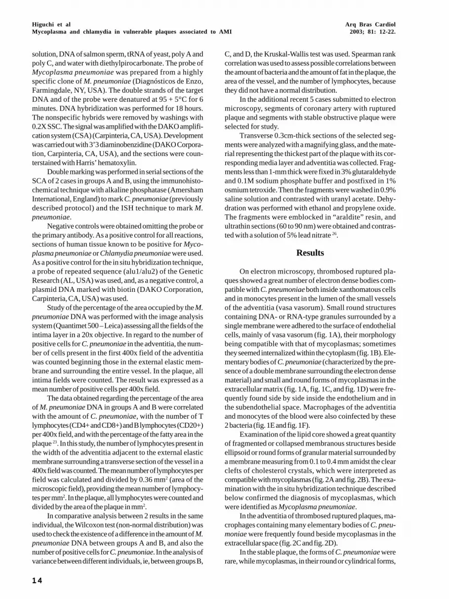
On electron microscopy, thrombosed ruptured pla-ques showed a great number of electron dense bodies com-patible with C. pneumoniae both inside xanthomatous cellsand in monocytes present in the lumen of the small vesselsof the adventitia (vasa vasorum). Small round structurescontaining DNA- or RNA-type granules surrounded by asingle membrane were adhered to the surface of endothelialcells, mainly of vasa vasorum (fig. 1A), their morphologybeing compatible with that of mycoplasmas; sometimesthey seemed internalized within the cytoplasm (fig. 1B). Ele-mentary bodies of C. pneumoniae (characterized by the pre-sence of a double membrane surrounding the electron densematerial) and small and round forms of mycoplasmas in theextracellular matrix (fig. 1A, fig. 1C, and fig. 1D) were fre-quently found side by side inside the endothelium and inthe subendothelial space. Macrophages of the adventitiaand monocytes of the blood were also coinfected by these2 bacteria (fig. 1E and fig. 1F).
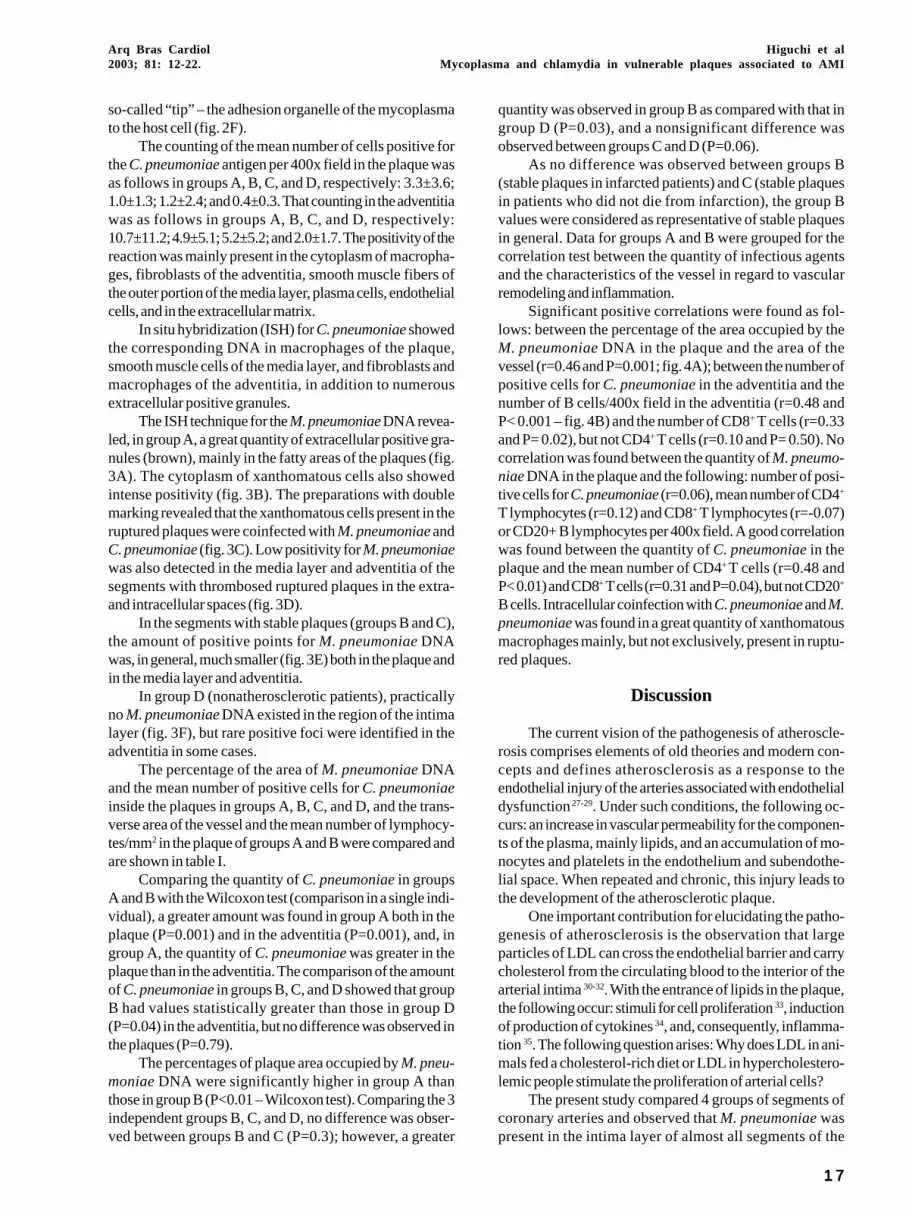
Examination of the lipid core showed a great quantityof fragmented or collapsed membranous structures besideellipsoid or round forms of granular material surrounded bya membrane measuring from 0.1 to 0.4 mm amidst the clearclefts of cholesterol crystals, which were interpreted ascompatible with mycoplasmas (fig. 2A and fig. 2B). The exa-mination with the in situ hybridization technique describedbelow confirmed the diagnosis of mycoplasmas, whichwere identified as Mycoplasma pneumoniae.
In the adventitia of thrombosed ruptured plaques, ma-crophages containing many elementary bodies of C. pneu-moniae were frequently found beside mycoplasmas in theextracellular space (fig. 2C and fig. 2D).
In the stable plaque, the forms of C. pneumoniae wererare, while mycoplasmas, in their round or cylindrical forms,

Arq Bras Cardiol2003; 81: 12-22.
Higuchi et alMycoplasma and chlamydia in vulnerable plaques associated to AMI
1 51 51 51 51 5
Fig. 1 – Ultrastructural characteristics of M. pneumoniae (MP) and its association with C. pneumoniae (CP) in segments of coronary arteries with ruptured and thrombosedplaques: A) panoramic view of vasa vasorum showing mycoplasmas adhered to and being internalized in the endothelial cell. In the subendothelial space, both bacteria M.pneumoniae (MP) and C. pneumoniae (CP) can be seen in the extracellular space (2.600X - originals). The arrows indicate that these regions are magnified in the following figures;B) a great magnification of A, showing in detail the surface of the endothelial cell with M. pneumoniae adhered (arrows), one of which internalized in the endothelial cell (7200X);C) great magnification of A, exhibiting in detail 2 types of extracellular bacteria M. pneumoniae (elliptic form with granules) and C. pneumoniae (electron dense round form) in thesubendothelial space (7200X); D) great magnification of an endothelial cell [identified by the presence of Weibel-Palade bodies (WP)] coinfected with M. pneumoniae and C.pneumoniae (10,000X); E) circulating monocytes with numerous bacteria in their cytoplasm with characteristics of C. pneumoniae and M. pneumoniae, which are better shownin the following magnification (3330X); F) greater magnification of E showing in detail the elementary bodies of C. pneumoniae and the round forms of M. pneumoniae (10,000X).

1 61 61 61 61 6
Higuchi et alMycoplasma and chlamydia in vulnerable plaques associated to AMI
Arq Bras Cardiol2003; 81: 12-22.
were more frequent. In the adventitia of the segments con-taining these stable plaques, forms compatible with largemycoplasmas were detected and characterized by electron
dense granules and filaments (fig. 2E), a surrounding mem-brane, and lack of organelles. These mycoplasmas had intheir extremities a more electron-dense region resembling the
Fig. 2 - Ultrastructural characteristics of the ruptured plaque (A-D) and of the stable plaque (E-F) of a coronary artery: A and B) lipid core of the atheroma showing many membranousremnants, cholesterol crystals (c) in clefts, and small cylindrical forms of M. pneumoniae (MP), 7,200X and 10,000X, respectively; C) segment with ruptured plaque showing, in theadventitia, a macrophage containing many small forms of C. pneumoniae (CP arrow) in the cytoplasm, and, in the extracellular space, round or cylindrical forms of M. pneumoniae (MParrows) 3,300X; D) detail of C, showing M. pneumoniae (MP) with a surrounding membrane and material of the DNA type inside (10,000X); E) a large cylindrical form of M. pneumoniae(MP) in the adventitia of a stable plaque; F) detail of the more electron dense extremity of M. pneumoniae (MP) suggestive of an adhesion organelle (“tip”) (10,000X).
Arq Bras Cardiol2003; 81: 12-22.
Higuchi et alMycoplasma and chlamydia in vulnerable plaques associated to AMI
1 71 71 71 71 7
so-called “tip” – the adhesion organelle of the mycoplasmato the host cell (fig. 2F).
The counting of the mean number of cells positive forthe C. pneumoniae antigen per 400x field in the plaque wasas follows in groups A, B, C, and D, respectively: 3.3±3.6;1.0±1.3; 1.2±2.4; and 0.4±0.3. That counting in the adventitiawas as follows in groups A, B, C, and D, respectively:10.7±11.2; 4.9±5.1; 5.2±5.2; and 2.0±1.7. The positivity of thereaction was mainly present in the cytoplasm of macropha-ges, fibroblasts of the adventitia, smooth muscle fibers ofthe outer portion of the media layer, plasma cells, endothelialcells, and in the extracellular matrix.
In situ hybridization (ISH) for C. pneumoniae showedthe corresponding DNA in macrophages of the plaque,smooth muscle cells of the media layer, and fibroblasts andmacrophages of the adventitia, in addition to numerousextracellular positive granules.
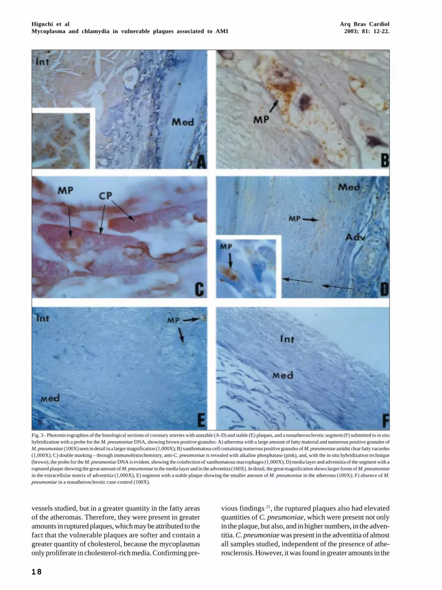
The ISH technique for the M. pneumoniae DNA revea-led, in group A, a great quantity of extracellular positive gra-nules (brown), mainly in the fatty areas of the plaques (fig.3A). The cytoplasm of xanthomatous cells also showedintense positivity (fig. 3B). The preparations with doublemarking revealed that the xanthomatous cells present in theruptured plaques were coinfected with M. pneumoniae andC. pneumoniae (fig. 3C). Low positivity for M. pneumoniaewas also detected in the media layer and adventitia of thesegments with thrombosed ruptured plaques in the extra-and intracellular spaces (fig. 3D).
In the segments with stable plaques (groups B and C),the amount of positive points for M. pneumoniae DNAwas, in general, much smaller (fig. 3E) both in the plaque andin the media layer and adventitia.
In group D (nonatherosclerotic patients), practicallyno M. pneumoniae DNA existed in the region of the intimalayer (fig. 3F), but rare positive foci were identified in theadventitia in some cases.
The percentage of the area of M. pneumoniae DNAand the mean number of positive cells for C. pneumoniaeinside the plaques in groups A, B, C, and D, and the trans-verse area of the vessel and the mean number of lymphocy-tes/mm2 in the plaque of groups A and B were compared andare shown in table I.
Comparing the quantity of C. pneumoniae in groupsA and B with the Wilcoxon test (comparison in a single indi-vidual), a greater amount was found in group A both in theplaque (P=0.001) and in the adventitia (P=0.001), and, ingroup A, the quantity of C. pneumoniae was greater in theplaque than in the adventitia. The comparison of the amountof C. pneumoniae in groups B, C, and D showed that groupB had values statistically greater than those in group D(P=0.04) in the adventitia, but no difference was observed inthe plaques (P=0.79).
The percentages of plaque area occupied by M. pneu-moniae DNA were significantly higher in group A thanthose in group B (P<0.01 – Wilcoxon test). Comparing the 3independent groups B, C, and D, no difference was obser-ved between groups B and C (P=0.3); however, a greater
quantity was observed in group B as compared with that ingroup D (P=0.03), and a nonsignificant difference wasobserved between groups C and D (P=0.06).
As no difference was observed between groups B(stable plaques in infarcted patients) and C (stable plaquesin patients who did not die from infarction), the group Bvalues were considered as representative of stable plaquesin general. Data for groups A and B were grouped for thecorrelation test between the quantity of infectious agentsand the characteristics of the vessel in regard to vascularremodeling and inflammation.
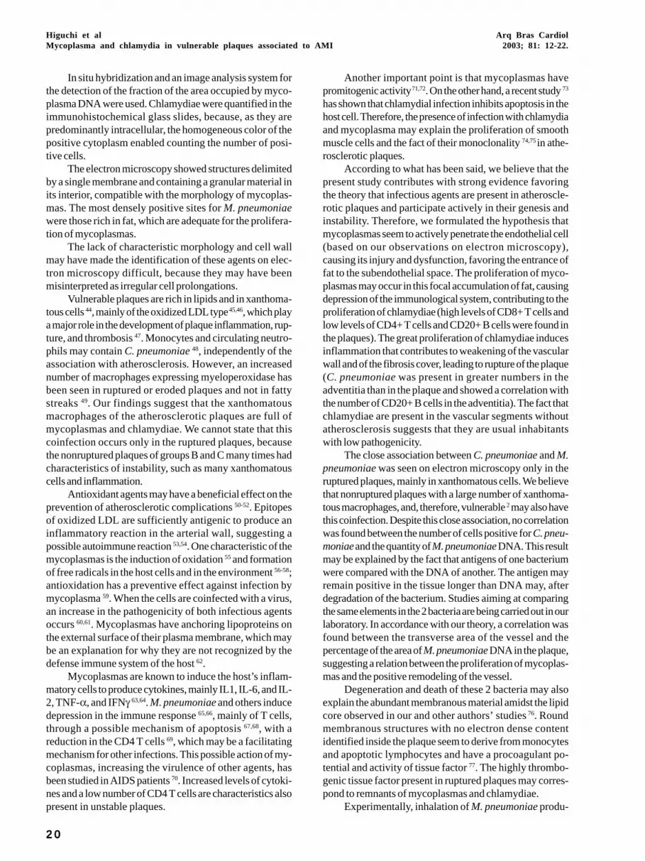
Significant positive correlations were found as fol-lows: between the percentage of the area occupied by theM. pneumoniae DNA in the plaque and the area of thevessel (r=0.46 and P=0.001; fig. 4A); between the number ofpositive cells for C. pneumoniae in the adventitia and thenumber of B cells/400x field in the adventitia (r=0.48 andP< 0.001 – fig. 4B) and the number of CD8+ T cells (r=0.33and P= 0.02), but not CD4+ T cells (r=0.10 and P= 0.50). Nocorrelation was found between the quantity of M. pneumo-niae DNA in the plaque and the following: number of posi-tive cells for C. pneumoniae (r=0.06), mean number of CD4+
T lymphocytes (r=0.12) and CD8+ T lymphocytes (r=-0.07)or CD20+ B lymphocytes per 400x field. A good correlationwas found between the quantity of C. pneumoniae in theplaque and the mean number of CD4+ T cells (r=0.48 andP< 0.01) and CD8+ T cells (r=0.31 and P=0.04), but not CD20+
B cells. Intracellular coinfection with C. pneumoniae and M.pneumoniae was found in a great quantity of xanthomatousmacrophages mainly, but not exclusively, present in ruptu-red plaques.
Discussion
The current vision of the pathogenesis of atheroscle-rosis comprises elements of old theories and modern con-cepts and defines atherosclerosis as a response to theendothelial injury of the arteries associated with endothelialdysfunction 27-29. Under such conditions, the following oc-curs: an increase in vascular permeability for the componen-ts of the plasma, mainly lipids, and an accumulation of mo-nocytes and platelets in the endothelium and subendothe-lial space. When repeated and chronic, this injury leads tothe development of the atherosclerotic plaque.
One important contribution for elucidating the patho-genesis of atherosclerosis is the observation that largeparticles of LDL can cross the endothelial barrier and carrycholesterol from the circulating blood to the interior of thearterial intima 30-32. With the entrance of lipids in the plaque,the following occur: stimuli for cell proliferation 33, inductionof production of cytokines 34, and, consequently, inflamma-tion 35. The following question arises: Why does LDL in ani-mals fed a cholesterol-rich diet or LDL in hypercholestero-lemic people stimulate the proliferation of arterial cells?
The present study compared 4 groups of segments ofcoronary arteries and observed that M. pneumoniae waspresent in the intima layer of almost all segments of the
1 81 81 81 81 8
Higuchi et alMycoplasma and chlamydia in vulnerable plaques associated to AMI
Arq Bras Cardiol2003; 81: 12-22.
vessels studied, but in a greater quantity in the fatty areasof the atheromas. Therefore, they were present in greateramounts in ruptured plaques, which may be attributed to thefact that the vulnerable plaques are softer and contain agreater quantity of cholesterol, because the mycoplasmasonly proliferate in cholesterol-rich media. Confirming pre-
vious findings 21, the ruptured plaques also had elevatedquantities of C. pneumoniae, which were present not onlyin the plaque, but also, and in higher numbers, in the adven-titia. C. pneumoniae was present in the adventitia of almostall samples studied, independent of the presence of athe-rosclerosis. However, it was found in greater amounts in the
Fig. 3 - Photomicrographies of the histological sections of coronary arteries with unstable (A-D) and stable (E) plaques, and a nonatherosclerotic segment (F) submitted to in situhybridization with a probe for the M. pneumoniae DNA, showing brown positive granules: A) atheroma with a large amount of fatty material and numerous positive granules ofM. pneumoniae (100X) seen in detail in a larger magnification (1,000X); B) xanthomatous cell containing numerous positive granules of M. pneumoniae amidst clear fatty vacuoles(1,000X); C) double marking – through immunohistochemistry, anti-C. pneumoniae is revealed with alkaline phosphatase (pink), and, with the in situ hybridization technique(brown), the probe for the M. pneumoniae DNA is evident, showing the coinfection of xanthomatous macrophages (1,000X); D) media layer and adventitia of the segment with aruptured plaque showing the great amount of M. pneumoniae in the media layer and in the adventitia (160X). In detail, the great magnification shows larger forms of M. pneumoniaein the extracellular matrix of adventitia (1,000X); E) segment with a stable plaque showing the smaller amount of M. pneumoniae in the atheroma (100X); F) absence of M.pneumoniae in a nonatherosclerotic case-control (100X).

Arq Bras Cardiol2003; 81: 12-22.
Higuchi et alMycoplasma and chlamydia in vulnerable plaques associated to AMI
19
Table I - Amount of M. pneumoniae and C. pneumoniae, percentage of the fatty area, mean number of B lymphocytes (CD20+) and subtypes of T lymphocytes(CD4+ and CD8+)/mm2 in the plaques of groups A, B, C, and D
Group N. C. pneumoniae /400x % M. pneumoniae area % Fatty area CD20+ CD4+ CD8+
A 3.3 (+3.6) 3.9 (+3.5) 33.7 (+22.3) 5.9 (+19.6) 7.4 (+6.6) 13.8 (+25.4)B 1.0 (+1.3) 1.5 (+1.6) 6.8 (+11.6) 1.1 (+1.6) 3.4 (+4.0) 5.7 (+6.3)C 1.2 (+2.4) 0.9 (+0.9) np np np npD 0.4 (+0.3) 0.3 (+0.2) np np np np
np: not performed; N. C. pneumoniae/400x: mean number of Chlamydia pneumoniae per 400x field;% M. pneumoniae area: percentage of the area occupied byMycoplasma pneumoniae.
Fig. 4 - Positive correlations between the morphologic characteristics of vulnerability of the atherosclerotic plaque and the amount of bacteria. Fig. 4A – Positive correlationbetween the transverse area of the vessel (positive remodeling) and the amount of Mycoplasma pneumoniae (MP) DNA. Fig. 4B – Positive correlation between the number of cellspositive for Chlamydia pneumoniae (CP) and the number of CD20 B lymphocytes in the adventitia.
ruptured segments. Practically no study reporting an asso-ciation between mycoplasmas and atherosclerosis exists inthe literature. Experimental studies about the infection ofrabbits with mycoplasmas could not show a pathogeneticrole played by that bacterium in the development of athe-rosclerosis 36. Our team, studying ultrastructurally and withimmunohistochemistry the lungs of apparently healthylaboratory animals (rats and mice), observed that 100% ofthe animals were infected with Mycoplasma pulmonis andC. pneumoniae 37, leading us to believe that laboratory ani-mals may often be naturally infected with these bacteria.This requires further study, because that infection maypotentially influence the results of experimental research onatherosclerosis.
On electron microscopy, we could document myco-plasmas in the endothelial intercellular space or being inter-nalized by the endothelial cells of ruptured plaques. Theclose association of mycoplasmas and chlamydiae may fa-vor the proliferation of both bacteria. In the present study,these 2 bacteria were present in great quantities in thethrombosed ruptured plaques, and, sometimes in contactwith each other, both in the extracellular space and insideendothelial cells and macrophages. This finding was mainlybased on electron microscopy and was confirmed in the ca-ses in which double marking was performed. The presence
of these bacteria inside ruptured plaques may explain pre-viously known characteristics of these plaques as follows:oxidized LDL, an increase in the production of several cytoki-nes, an increase in TNF, cell proliferation, and inflammation.
Mycoplasmas have been held responsible for humandiseases, such as pneumonia, arthritis, and urethritis 38. Morerecently, they have also been implicated in the progression ofAIDS, chronic fatigue syndrome, acute respiratory distresssyndrome, and others 39. However, these arguments have notsucceeded in sensitizing scientists in regard to a pathogeneticrelation, because most of these arguments are based on casereports whose positivity for mycoplasmas results from sero-logical data, culture, or PCR 40,41. Few studies sought M.pneumoniae in atherosclerotic plaques, and the resultswere negative 42 or slightly positive 43. The difference in theresults here presented may have originated in the methodo-logy used. We did not use PCR but in situ hybridization witha very specific probe and a system of amplification to revealthe reaction (CSA), and electron microscopy.
In the present study, M. pneumoniae and C. pneumo-niae were very frequently shown in atherosclerotic plaques.Quantitative analysis was not performed with all techniquesused, because the qualitative analysis confirmed the pre-sence of these agents with the 3 techniques used in thesame locations and in the same cells.
% a
rea
of D
NA
of M
P
A Transverse area of the vessel mm2 B number of CD 20 B/mm2 lymphocytes
no
. CP
/mm
2 in
the
adve
nti
tio
2 02 02 02 02 0
Higuchi et alMycoplasma and chlamydia in vulnerable plaques associated to AMI
Arq Bras Cardiol2003; 81: 12-22.
In situ hybridization and an image analysis system forthe detection of the fraction of the area occupied by myco-plasma DNA were used. Chlamydiae were quantified in theimmunohistochemical glass slides, because, as they arepredominantly intracellular, the homogeneous color of thepositive cytoplasm enabled counting the number of posi-tive cells.
The electron microscopy showed structures delimitedby a single membrane and containing a granular material inits interior, compatible with the morphology of mycoplas-mas. The most densely positive sites for M. pneumoniaewere those rich in fat, which are adequate for the prolifera-tion of mycoplasmas.
The lack of characteristic morphology and cell wallmay have made the identification of these agents on elec-tron microscopy difficult, because they may have beenmisinterpreted as irregular cell prolongations.
Vulnerable plaques are rich in lipids and in xanthoma-tous cells 44, mainly of the oxidized LDL type 45,46, which playa major role in the development of plaque inflammation, rup-ture, and thrombosis 47. Monocytes and circulating neutro-phils may contain C. pneumoniae 48, independently of theassociation with atherosclerosis. However, an increasednumber of macrophages expressing myeloperoxidase hasbeen seen in ruptured or eroded plaques and not in fattystreaks 49. Our findings suggest that the xanthomatousmacrophages of the atherosclerotic plaques are full ofmycoplasmas and chlamydiae. We cannot state that thiscoinfection occurs only in the ruptured plaques, becausethe nonruptured plaques of groups B and C many times hadcharacteristics of instability, such as many xanthomatouscells and inflammation.
Antioxidant agents may have a beneficial effect on theprevention of atherosclerotic complications 50-52. Epitopesof oxidized LDL are sufficiently antigenic to produce aninflammatory reaction in the arterial wall, suggesting apossible autoimmune reaction 53,54. One characteristic of themycoplasmas is the induction of oxidation 55 and formationof free radicals in the host cells and in the environment 56-58;antioxidation has a preventive effect against infection bymycoplasma 59. When the cells are coinfected with a virus,an increase in the pathogenicity of both infectious agentsoccurs 60,61. Mycoplasmas have anchoring lipoproteins onthe external surface of their plasma membrane, which maybe an explanation for why they are not recognized by thedefense immune system of the host 62.
Mycoplasmas are known to induce the host’s inflam-matory cells to produce cytokines, mainly IL1, IL-6, and IL-2, TNF-α, and IFNγ 63,64. M. pneumoniae and others inducedepression in the immune response 65,66, mainly of T cells,through a possible mechanism of apoptosis 67,68, with areduction in the CD4 T cells 69, which may be a facilitatingmechanism for other infections. This possible action of my-coplasmas, increasing the virulence of other agents, hasbeen studied in AIDS patients 70. Increased levels of cytoki-nes and a low number of CD4 T cells are characteristics alsopresent in unstable plaques.
Another important point is that mycoplasmas havepromitogenic activity 71,72. On the other hand, a recent study 73
has shown that chlamydial infection inhibits apoptosis in thehost cell. Therefore, the presence of infection with chlamydiaand mycoplasma may explain the proliferation of smoothmuscle cells and the fact of their monoclonality 74,75 in athe-rosclerotic plaques.
According to what has been said, we believe that thepresent study contributes with strong evidence favoringthe theory that infectious agents are present in atheroscle-rotic plaques and participate actively in their genesis andinstability. Therefore, we formulated the hypothesis thatmycoplasmas seem to actively penetrate the endothelial cell(based on our observations on electron microscopy),causing its injury and dysfunction, favoring the entrance offat to the subendothelial space. The proliferation of myco-plasmas may occur in this focal accumulation of fat, causingdepression of the immunological system, contributing to theproliferation of chlamydiae (high levels of CD8+ T cells andlow levels of CD4+ T cells and CD20+ B cells were found inthe plaques). The great proliferation of chlamydiae inducesinflammation that contributes to weakening of the vascularwall and of the fibrosis cover, leading to rupture of the plaque(C. pneumoniae was present in greater numbers in theadventitia than in the plaque and showed a correlation withthe number of CD20+ B cells in the adventitia). The fact thatchlamydiae are present in the vascular segments withoutatherosclerosis suggests that they are usual inhabitantswith low pathogenicity.
The close association between C. pneumoniae and M.pneumoniae was seen on electron microscopy only in theruptured plaques, mainly in xanthomatous cells. We believethat nonruptured plaques with a large number of xanthoma-tous macrophages, and, therefore, vulnerable 2 may also havethis coinfection. Despite this close association, no correlationwas found between the number of cells positive for C. pneu-moniae and the quantity of M. pneumoniae DNA. This resultmay be explained by the fact that antigens of one bacteriumwere compared with the DNA of another. The antigen mayremain positive in the tissue longer than DNA may, afterdegradation of the bacterium. Studies aiming at comparingthe same elements in the 2 bacteria are being carried out in ourlaboratory. In accordance with our theory, a correlation wasfound between the transverse area of the vessel and thepercentage of the area of M. pneumoniae DNA in the plaque,suggesting a relation between the proliferation of mycoplas-mas and the positive remodeling of the vessel.
Degeneration and death of these 2 bacteria may alsoexplain the abundant membranous material amidst the lipidcore observed in our and other authors’ studies 76. Roundmembranous structures with no electron dense contentidentified inside the plaque seem to derive from monocytesand apoptotic lymphocytes and have a procoagulant po-tential and activity of tissue factor 77. The highly thrombo-genic tissue factor present in ruptured plaques may corres-pond to remnants of mycoplasmas and chlamydiae.
Experimentally, inhalation of M. pneumoniae produ-
Arq Bras Cardiol2003; 81: 12-22.
Higuchi et alMycoplasma and chlamydia in vulnerable plaques associated to AMI
2 12 12 12 12 1
1. van der Wal AC, Pieck JJ, de Boer OJ, et al. Recent activation of the plaque immuneresponse in coronary lesions underlying acute coronary syndromes. Heart1998;80:14-8.
2. Libby P, Geng YJ, Aikawa M et al. Macrophages and atherosclerotic plaque ins-tability. Curr Opin Lipidol 1996;7:330-5.
3. Yasunobu Y, Hayashi K, Shingu T, Yamagata T, Kajiyama G, Kambe M. Coronaryatherosclerosis and oxidative stress as reflected by autoantibodies against oxidi-zed low-density lipoprotein and oxysterols. Atherosclerosis 2001;155:445-53.
4. Gobel H, van der Wal AC, Teeling P, van der Loos CM, Becker AE. Metallothio-nein in human atherosclerotic lesions: a scavenger mechanism for reactive oxy-gen species in the plaque? Virchows Arch 2000;437:528-33.
5. Wilcox JN, Smith KM, Schwartz S, Gordon D. Localization of tissue factor in thenormal vessel wall and in the atherosclerotic plaque. Proc Natl Acad Sci USA1989;86:2839-43.
6. Marmur JD, Thiruvikraman SV, Fyfe SV, et al. The identification of tissue factor inhuman coronary atheroma. Circulation 1996;94: 226-36.
7. Moreno PR, Bernardi VH, Lopez-Cuellar J, et al. Macrophages, smooth musclecells and tissue factor in unstable angina: implications for cell mediated thrombo-genicity in acute coronary syndromes.Circulation 1996;94:3090-7.
8. Mahony JB, Coombes BK. Chlamydia pneumoniae and atherosclerosis: doesthe evidence support a causal or contributory role? FEMS Microbiol Lett2001;197:1-9.
9. Burian K, Kis Z, Virok D, et al. Independent and joint effects of antibodies tohuman heat-shock protein 60 and Chlamydia pneumoniae in the developmentof coronary atherosclerosis. Circulation 2001;103:1503-8.
10. Vink A, Poppen M, Schoveveld AH, et al. Distribution of Chlamydia pneumo-niae in the human arterial system and its relation to the local amount of atheros-clerosis within the individual. Circulation 2001;103:1613-7.
11. Kol A, Sukhova GK, Lichtman AH, Libby P. Chlamydial heat schock protein 60localizes in human atheroma and regulates macrophage tumor necrosis factor-alpha and matrix metalloproteinase expression. Circulation 1998;98:300-7.
12. Kalayoglu MV, Indrawati, Morrison RP, Morrison SG, Yuan Y, Byrne GI. Chla-mydial virulence determinants in atherogenesis: the role of chlamydial lipopo-lysaccharide and heat shock protein 60 in macrophage-lipoprotein interactions.J Infect Dis 2000; 181(Suppl3): S483-9.
13. Thomas M, Wong Y, Ajaz M, Tsang V, Gallagher PJ, Ward ME. Relation betweendirect detection of Chlamydia pneumoniae DNA in human coronary arteries atpostmortem examination and histological severity (Stary grading) of associatedatherosclerotic plaque. Circulation 1999; 99: 2733-6.
14. Ericson K, Saldeen TG, Lindquist O, Pahlson C, Mehta JL. Relationship ofC.pneumoniae infection to severity of human coronary atherosclerosis. Circula-tion 2000;101:2568-71.
15. Espinola-Klein C, Rupprecht HJ, Blanknenberg S, et al. Impact of infectiousburden on extent and long-term prognosis of atherosclerosis. Circulation2002;105:15-21.
16. Wald NJ, Law MR, Morris JK, Zhou X, Wong Y, Ward ME. Chlamydia pneumo-niae infection and mortality from ischaemic heart disease: large prospectivestudy. BMJ 2000;321:204-7.
17. Maass M, Jahn J, Gieffers J, Dalhoff K, Katus HA, Solbach W. Detection of Chlamy-dia pneumoniae within peripheral blood monocytes of patietns with unstableangina or myocardial infarction. J Infect Dis 2002;181(Suppl 3):S449-51.
18. Gurfinkel E, Bozovich G, Beck E, et al. Treatment with antibiotic roxithromycinin patients with acute non-Q-waved coronary syndromes. The final report of theROXIS Study. Eur Heart J 1999;20:121-7.
References
ced no changes in atherosclerosis in rabbits, favoring ourhypothesis that both bacteria are important for the develop-ment of those complications 78.
In conclusion, our study strongly supports the hypo-thesis that atherosclerosis and plaque rupture are infectiouscomplications of the artery and not simple degenerativeprocesses. M. pneumoniae is present in almost all coronaryatheroma. A large amount of M. pneumoniae and C. pneu-moniae seems fundamental for the development of plaqueinstability, possibly in a symbiotic relation. The increase in
the number of C. pneumoniae was correlated with anincrease in the number of inflammatory cells, which, in turn,was greater in the segments with ruptured plaque. Our mor-phological findings favor the active participation of myco-plasmas in vulnerable plaques, by entering the subendothe-lial space and creating conditions that favor fat accumula-tion, dysfunction of the immunological and endothelialresponse, proliferation of chlamydiae, inflammation, and anincrease in apoptosis, which are fundamental ingredientsfor plaque rupture.
19. Neumann F, Kastrati A, Miethke T, et al. Treatment of Chlamydia pneumoniaeinfection with roxithromycin and effect on neointima proliferation after coronarystent placement (ISAR-3): a randomized, double-blind, placebo-controlled trial.Lancet 2001;357:2085-9.
20. WIZARD study, presented in 51st American College Cardiology ScientificSession, Atlanta 2002.
21. Higuchi ML, Castelli JB, Aiello VD, et al. Great amount of Chlamydia pneumo-niae in ruptured plaque vessel segments at autopsy. A comparative study withstable plaques. Arq Bras Cardiol 2000;74:149-51.
22. Higuchi ML, Sambiase N, Palomino S, et al. Detection of Mycoplasma pneumo-niae and Chlamydia pneumoniae in ruptured atherosclerotic plaques. Braz JMed Biol Res 2000;33:1023-6.
23. Razin S, Yogev D, Naot Y. Molecular Biology and pathogenicity of mycoplas-mas. Microbiol Molec Biol Rev 1998;62:1094-156.
24. Bezerra HG, Higuchi ML, Gutierrez PS, et al. Atheromas that cause fatal throm-bosis are usually larger and frequently accompanied by vessel enlargement. Car-diovasc Pathol 2001;10:189-96.
25. Higuchi ML, Gutierrez PS, Bezerra HG, et al. - Comparison between adventitialand intimal inflammation of ruptured and non-ruptured atherosclerotic plaquesin human coronary arteries. Arq Bras Cardiol, 2002;79:20-24.
26. Reynolds ES. The use of lead citrate at high pH as an electron-opaque stain inelectron microscope. J Cell Biol 1963;17:208-12.
27. Ross R. The pathogenesis of atherosclerosis: a perspective for the 1990s.Nature. 1993;362:801-8.
28. Fuster V, Badimon L, Badimon JJ, Chesebro JH. The pathogenesis of coronary arte-ry disease and the acute coronary syndromes. N Engl J Med 1992;326:242-50.
29. Ross R - Atherosclerosis - an inflammatory disease. N Engl J Med 1999;340:115-26.
30. French JE. Endothelial structure and function. In: Jones RJ, ed. Evolution of theaterosclerotic plaque. Chicago, III: University of Chicago Press; 1963:15-33.
31. Kao V, Wissler RW. A study of the immunohistochemical localization of serumlipoproteins and other plasma proteins in human atherosclerotic lesions. ExpMol Pathol 1965;4:465-79.
32. Zilversmith DB. Cholesterol flux in atherosclerotic plaque. Ann N Y Acad Sci.1968;149:710-24.
33. Ross R. Atherosclerosis: a problem of the biology of arterial wall cells and theirinteractions with blood components. Arteriosclerosis. 1981;1:293-311.
34. Libby P, Friedman GB, Salomon RN. Cytokines as modulators of cell prolifera-tion in fibrotic diseases. Ann Rev Respir Dis. 1989;140:1114-17.
35. Libby P, Hansson GK. Involvement of the immune system in human atherogene-sis: current knowledge and unanswered questions. Lab Invest 1991;64:5-15.
36. Fong IW. Antibiotics effects in a rabbit model of Chlamydia pneumoniae-indu-ced atherosclerosis. J Infect Dis 2000;181(Suppl 3):S514-8.
37. Damy S, Higuchi ML, Timenetsky J, et al. Coinfection of laboratory rats with My-coplasma pulmonis and Chlamydia pneumoniae. Contemp Top Lab Anim Sci2003;42:52-6.
38. Baseman JB, Tully JG. Mycoplasmas: sophisticated reemerging and burdened bytheir notoriety. Emerging Inf Dis 1997;3:21-32.
39. Chambaud I, Wromblewski H, Blanchard A. Interactions between mycoplasmalipoproteins and the host immune syustem. Trends Microbiol 1999;7:493-9.
40. Baseman JB, Tully JB. Mycoplasmas: sophisticated, re-emerging, and burdenedby their notoriety. Emerging Infect Dis. 1997;3:21-32.
41. Maniloff J, McElbaney RN, Finch LR, Baseman JB (ed.). 1992; Mycoplasmas:molecular biology and pathogenesis. American Society for Microbiology,Washington DC

2 22 22 22 22 2
Higuchi et alMycoplasma and chlamydia in vulnerable plaques associated to AMI
Arq Bras Cardiol2003; 81: 12-22.
human T cell lines and normal lymphocytes co-infected with HIV-1 and myco-plasma. Free Radic Res 1995;23:197-212.
61. Blanchard A, Montagnier L. AIDS-associated mycoplasmas. Annu Rev Micro-biol 1994;48:687-712.
62. Chambaud I, Wroblewski H, Blanchard A. Interactions between mycoplasmalipoproteins and the host immune system. Trends Microbiol 1999;7:493-9.
63. Faulkner CB, Simecka JW, Davidson MK, et al. Gene expression and productionof TNFα, IL-1, IL-6 and IFN-γ in acute Mycoplasma pulmonis disease. InfectImmun 1995;63:4084-90.
64. Pietsch K, Ehlers S, Jacobs E. Cytokine gene expression in the lungs of BALB/cmice during primary and secondary intranasal infection with Mycoplasmapneumoniae. Microbiology 1994;140:2043-8.
65. Izutsu KT, Fatherazi S, Belton CM, Oda D, Cartwright FD, Kenny GE. Mycoplas-ma orale infection affects K+ and Cl- currents in the HSG salivary fland cell line. InVitro Cell Dev Biol Anim 1996;32:361-5.
66. Matsui M, Kobayashi M, Mitsup Y. Elevation of endothelin biosynthesis inhuman endothelial cells with mycoplasma infection. In Vitro Cell Dev Biol Anim1995;31:880-5.
67. Rawadi G, Roman-Roman S, Castedo M, et al. Effects of Mycoplasma fermentanson the myelomonocytic lineage. Different molecular entities with cytokine-inducing and cytocidal potential. J Immunol 1996;156:670-8.
68. Shibata K, Watanabe T. Mycoplasma fermentans enhances concavalin A-inducedapoptosis of mouse splenic T cells. FEMS Immunol Med Microbiol 1997; 17:103-9.
69. Sasaki Y, Cunha RA, Gougeon L, Montagnier L, Blanchard A. Mycoplasmapenetrans cytopathic effects: an in vitro study using human peripheral bloodmononuclear cells. IOM Lett. 1996;4:6-7.
70. Grau O, Slizewicz B, Tuppin P, et al. Association of Mycoplasma penetranswith human immunodeficiency virus infection. J Infect Dis 1995;172:672-81.
71. Lapidot Z, Naot Y. Immunohistochemical characterization of Mycoplasmapulmonis membrane mitogens. IOM Lett 1996;4:385.
72. Tsai S, Wear DJ, Shih JW, Lo SC. Mycoplasma and oncogenesis: persistent infec-tion and multistage malignant transformation. Proc Natl Acad Sci 92:10197-201.
73. Fan T, Lu H, Hu H, et al. Inhibition of apoptosis in chlamydia-infected cells: blo-ckade of mitochondrial cytochrome c release and caspase activation. J Exp Med1998; 187(4): 487-96.
74. Murry CE, Gipaya CT, Bartosek T, Benditt EP, Schwartz SM. Monoclonality ofsmooth muscle cells in human atherosclerosis. Am J Pathol 1997;151:697-705.
75. Schwartz SM, Majesky MW, Murry CE. The intima: development and monoclo-nal responses to injury. Atheroslcerosis 1995;118:S125-40.
76. Meijer A, van der Vliet JAQ, Roholl PJ, Gielis-Porper SK, de Vries A, Ossewaar-de JM. Chlamydia pneumoniae in abdominal aortic aneurysms: abundance ofmembrane components in the absence of heat shock protein 60 and DNA. Arte-rioscler Thromb Vasc Biol 1999;19:2680-6.
77. Mallat Z, Hugel B, Ohan J, Leseche G, Freyssinet JM, Tedgui A. Shed membranemicroparticles with procoagulant potential in human atherosclerotic plaques: arole for apoptosis in plaque thrombogenicity. Circulation 1999;99:348-53.
78. Fong IW, Chiu B, Viira E, Jang D, Mahony JB. De Novo induction of atheroscle-rosis by Chlamydia pneumoniae in a rabbit model. Infect Immun 1999;67:6048-55.
42. Balse F, Fagetti L, Allegra L. Chlamydia pneumoniae detection in atheroscle-rotic plaques in Italy. J Infect Dis 2000;181(Suppl 3): S444-6.
43. Maraha B, Berg H, Scheffer GJ, et al. Correlation between detection methods ofChlamydia pneumoniae in atherosclerotic and non-atherosclerotic tissues.Diagn Microbiol Infect Dis 2001;39:139-43.
44. van der Wal AC, Becker AE, van der Loos CM, Tigges AJ, Das PK. Fibrous andlipid-rich atherosclerotic plaques are part of interchangeable morphologies rela-ted to inflammation: a concept. Cor Art Dis 1994;5:463-9.
45. Steinberg D. Antioxidants and atherosclerosis. A current assessment. Circula-tion. 1991;84:1420-5.
46. Fuster V, Gotto AM, Libby P, Loscalzo J, McGill H, Task Force 1. Pathogenesisof coronary disease: The biologic role of risk factors. J Am Coll Cardiol1996;27:964-1047.
47. Fuster V, Badimon JJ, Chesebro JH, Fallon JT. Plaque rupture, thrombosis, andtherapeutic implications. Haemostasis 1996;26(Suppl4):269-84.
48. Berger M, Schroeder B, Daeschlein G, et al. Chlamydia pneumoniae DNA innon-coronary atherosclerotic plaques and circulating leukocytes. J Lab ClinMed 2000;136:194-200.
49. Sugiyama S, Okada Y, Sukhova GK, Virmani R, Heinecke JW, Libby P. Macro-phage myeloproxidase regulation by granulocyte macrophage colony-stimula-ting factor in human atherosclerosis and implications in acute coronary syndro-mes. Am J Pathol 1001;158:879-91.
50. Rikitake Y, Kawashima S, Takeshita S, et al. Anti-oxidative properties of fluvas-tatin, an HMG-CoA reductase inhibitor, contribute to prevention of atheroscle-rosis in cholesterol-fed rabbits. Atherosclerosis 2001;154:87-96.
51. Fuhrman B, Aviram M. Flavonoids protect LDL from oxidation and attenuateatherosclerosis. Curr Opin Lipidol 2001;12:41-8.
52. Durak I, Kamaz M, Cimen MY, Buyukkocak U, Ozturk HS. Blood oxidant/ antio-xidant status of atherosclerotic patients. Int J Cardiol 2001;77:293-7.
53. Palinski W, Yl”a-Herttuala S, Rosenfeld ME, et al. Antisera and monoclonalantibodies for epitopes generated during the oxidative modification of low den-sity lipoprotein. Arterioscler Thromb 1990;10:325-35.
54. Steinbrecher UP, Fisher M, Witztum JL, Curtiss LK. Immunogenicity of homolo-gous low-density lipoprotein after methilation, ethylation, acetylation, orcarbamylation: generation of antibodies specific for derivatized lysine. J LipidRes. 1984;25:1109-16.
55. Almagor M, Kahane I, Yatziv S. Role of superoxide anion in host cell injuryinduced by M.pneumoniae infection. A study in normal and trisomy 21 cells. JClin Invest 1984;73:842-7.
56. Miles RJ, Taylor RR, Varsani H. Oxygen uptake and H2O2 production by fermen-tative Mycoplasma spp. J Med Microbiol 1991;34:219-23.
57. Taylor RR, Mohan K, Miles RJ. Diversity of energy-yelding substrates and meta-bolism in avian mycoplasmas. Vet Microbiol 1996;51:291-304.
58. Ben-Menachem G, Himmelreich R, Herrmann R, Aharonowithz Y, Rottem S. Thethiodoxin reductase system of mycoplasmas. Microbiology 1997;143:1933-40.
59. Almagor M, Kahane I, Gilon C, Yatziv S. Protective effects of glutathione redoxcycle and vitamin E on cultured fibrobasts infected by Mycoplasma penumo-niae. Infect Immun 1986;52:240-4.
60. Chocola J, Strosberg AD, Stanislawski M. Release of hydrogen peroxide from