happy facial expression processing with different social interaction cues: an fmri study of...
TRANSCRIPT
Progress in Neuro-Psychopharmacology & Biological Psychiatry 44 (2013) 108–117
Contents lists available at SciVerse ScienceDirect
Progress in Neuro-Psychopharmacology & BiologicalPsychiatry
j ourna l homepage: www.e lsev ie r .com/ locate /pnp
Happy facial expression processing with different social interaction cues: An fMRIstudy of individuals with schizotypal personality traits
Jia Huang a, Yi Wang a, Zhen Jin b, Xin Di c, Tao Yang a, Ruben C. Gur d, Raquel E. Gur d, David H.K. Shum e,Eric F.C. Cheung f, Raymond C.K. Chan a,g,⁎a Neuropsychology and Applied Cognitive Neuroscience Laboratory, Key Laboratory of Mental Health, Institute of Psychology, Chinese Academy of Sciences, Beijing, Chinab MRI Imaging Center, 306 Hospital, Beijing, Chinac Department of Radiology, University of Medicine and Dentistry of New Jersey, Newark, USAd Department of Psychiatry, Perelman School of Medicine, University of Pennsylvania, Philadelphia, USAe Behavioural Basis of Health Research Program, Griffith Health Institute, Griffith University, Gold Coast, Australiaf Castle Peak Hospital, Hong Kong Special Administrative Region, Chinag Magnetic Resonance Imaging Research Centre, Institute of Psychology, Chinese Academy of Sciences, Beijing, China
Abbreviations: fMRI, functional Magnetic Resonancesonality disorder; SPQ, schizotypal personality questionnTR, repetition time; TE, echo time; FOV, field of view; MP-rapid gradient-echo imaging; SPM, Statistical Parametric Mappearing; D, happiness disappearing; P, praise context; Binterest; ACC, anterior cingulate cortex; PCC, posterior ciCN, caudate nucleus; IFG, inferior frontal gyrus; REST, ReToolkit; STG, superior temporal gyrus; BOLD, blood-Montréal Neurological Institute.⁎ Corresponding author at: Room 526, South Building, I
Academy of Sciences, 16 Lincui Road, Beijing, China. Tel./fE-mail address: [email protected] (R.C.K. Chan).
0278-5846/$ – see front matter © 2013 Elsevier Inc. Allhttp://dx.doi.org/10.1016/j.pnpbp.2013.02.004
a b s t r a c t
a r t i c l e i n f oArticle history:Received 26 November 2012Received in revised form 6 February 2013Accepted 6 February 2013Available online 13 February 2013
Keywords:Functional imagingHappy facial expressionSchizotypal personality traitsSocial interaction cues
In daily life facial expressions change rapidly and the direction of change provides important clues aboutsocial interaction. The aim of conducting this study was to elucidate the dynamic happy facial expression pro-cessing with different social interaction cues in individuals with (n=14) and without (n=14) schizotypalpersonality disorder (SPD) traits. Using functionalmagnetic resonance imaging (fMRI), dynamic happy facial ex-pression processing was examined by presenting video clips depicting happiness appearing and disappearingunder happiness inducing (‘praise’) or reducing (‘blame’) interaction cues. The happiness appearing conditionconsistently elicitedmore brain activations than the happiness disappearing condition in the posterior cingulatebilaterally in all participants. Further analyses showed that the SPD groupwas less deactivated than the non-SPDgroup in the right anterior cingulate cortex in the happiness appearing–disappearing contrast. The SPD groupdeactivated more than the non-SPD group in the left posterior cingulate and right superior temporal gyrus inthe praise–blame contrast. Moreover, the incongruence of cues and facial expression activated the frontal–thalamus–caudate–parietal network, which is involved in emotion recognition and conflict resolution.These results shed light on the neural basis of social interaction deficits in individuals with schizotypal per-sonality traits.
© 2013 Elsevier Inc. All rights reserved.
1. Introduction
Central to successful social interaction is the understanding of andresponding to social stimuli embedded in the environmental context.Impaired understanding and responding to social stimuli was foundin patients with schizophrenia (Maat et al., 2012; Smith et al., 2012),especially those with a first episode of psychosis (Achim et al., 2012).As part of schizophrenia spectrumdisorders, individualswith schizotypalpersonality disorder (SPD) display similar yet attenuated difficulties
Imaging; SPD, schizotypal per-aire; EPI, echoplanar imaging;RAGE, magnetization-preparedapping software; A, happiness, blame context; ROI, region ofngulate cortex; THA, thalamus;sting-State fMRI Data Analysisoxygen-level-dependent; MNI,
nstitute of Psychology, Chineseax: +86 10 64836274.
rights reserved.
(Abbott and Green, 2012; H.J. Li et al., 2012; Roddy et al., 2012; Shiet al., 2012; Zong et al., 2010). In particular, Chan et al. (2012b) showedthat individuals with SPD traits have deficits of hedonic capacity bothphysically and socially as compared to healthy controls. The reducedhedonic capacity is related to problems in the temporal experience ofpleasure (Chan et al., 2012a). One of the social stimuli that providepleasure information is happy facial expression (Wittfoth et al., 2010).Elucidating the neuralmechanismof happy facial expression processingin the context of different social interaction cues in individuals with SPDmay advance understanding of social cognition in schizophrenia spec-trum disorders. In this study, we investigated the neural responses ofindividuals with and without SPD traits when they process dynamichappy facial expressions under different social interaction cues.
Although facial displays of emotion are dynamic in nature, moststudies have primarily examined emotion identification or recogni-tion through a series of static and categorized facial expression photo-graphs (Gur et al., 2007; Kohler et al., 2010; Lee et al., 2010; Lougheadet al., 2008). The use of static emotional expressions may underminethe ecological validity of the emotion-processing tasks in these studies(McDonald et al., 2011; Platt et al., 2010; Sato and Yoshikawa, 2007b).
109J. Huang et al. / Progress in Neuro-Psychopharmacology & Biological Psychiatry 44 (2013) 108–117
Recent studies have shown that dynamic displays of emotion havemore expressivity compared to that of static emotion stimuli, as indicat-ed by electromyographic (Rymarczyk et al., 2011) and behavioral data(Sato and Yoshikawa, 2007a). The underlying mechanism of this facili-tation relates to enhanced activation in visual areas (Recio et al., 2011),and to the emotion properties of dynamic facial expressions (Fujimuraand Suzuki, 2010). Notably, static and dynamic facial expressions maydiffer in their neural substrates (Johnston et al., 2010; Pitcher et al.,2011) and dynamic information exchange may occur in multi-modalemotion communication (Regenbogen et al., 2012).
Among emotions, positive affect such as happiness serves as asource of information for judging meanings (Hicks et al., 2010).Examining the neural correlates of happy facial expression processingmay increase our understanding of the neural system associated withthe pursuit of happiness. In a previous review, Kringelbach andBerridge (2009) suggested that the neural network associated withhappiness pursuit is composed of the orbitofrontal cortex, cingulatecortex, insula, amygdala, ventral pallidum, nucleus accumbens, ventraltegmentum area and hypothalamus. A voxel-based meta-analysis hasprovided convergent evidence for the neural correlates of happiness in-cluding the right superior temporal gyrus, left anterior cingulate cortex,left insula and left thalamus (Vytal and Hamann, 2010). Moreover,recent findings also implicate the frontostriatal neural network inhappiness experience and perception. For example, the experience ofhappiness was associated with activities in the right frontal lobes,nucleus accumbens/ventral striatum and prefrontal cortex (Sharpleyand Bitsika, 2010). Additionally, the offset of happy and onset of angryexpressions show common activations in the orbitofrontal cortex bilat-erally, the left amygdala and the left insula, while the onset of happyand the offset of angry expressions show common activations in theleft dorsal striatum (Muhlberger et al., 2010).
The frontostriatal network associated with happiness provides in-sights into anhedonia, an inability to experience pleasure. Anhedoniacan manifest in depression. Depressed individuals show a negativecorrelation between their depression severity and activation of theright fusiform gyri in response to happy facial expressions, while healthyindividuals demonstrate linear increases in response to expressions ofincreasing happiness in the bilateral fusiform gyri and right putamen(Surguladze et al., 2005). Individualswith anhedonia in social interactionshow less neural activity in facial expression discrimination regions suchas the medial prefrontal cortex, the right superior temporal gyrus, andthe left somatosensory cortex (Germine et al., 2011). Strong relationshipwas found among SPD traits, depressedmood and their poor social func-tion (McCleery et al., 2012). Interpersonal aspects of SPD (particularly so-cial anxiety) have also been associated with reduced accuracy on thefacial expression recognition task (Abbott and Green, 2012). Moreover,individuals with SPD reported less pleasant affect compared with psy-chometrically defined controls and even stable schizophrenia patients(Cohen et al., 2012). Therefore, the current study aimed to investigatethe neural activity of these at-risk individuals with social interactiondifficulties when they process the dynamic happy facial expression.
Real-life scenarios are usuallymore complicated than simplydecodingthe emotion of facial expressions in an artificial laboratory environment.Facial expressions are typically decoded with various social cuesthat involve reciprocity with sensory inputs. Thus, in the currentstudy, we used a dyadic conversation consisting of a question and adynamic facial expression as a response to provide social interactioncues. We attempted to investigate the dynamic happy facial expressionprocessing within social interaction contexts in people with and with-out schizotypal personality traits.
The neural activities underlying the congruence between expressedemotions and the social cues have received increased attention. Whenthe self-expressed happy emotion is congruentwith the observed others'emotion state, the medial orbitofrontal cortex and ventromedial pre-frontal cortex, which have been associated with positive feelings andreward, are activated. In contrast, incongruent emotional states activate
the dorsolateral prefrontal cortex and the posterior superior temporalgyrus (Kuhn et al., 2011). Moreover, incongruence of emotion valencein audiovisual integration activates a frontal–cingulate–parietal net-work (Muller et al., 2011). However, social cues in previous studiesõseldom involved the social interaction component, which is the mostcommon situation in our daily lives. Dyadic conversation is the simplestform of social interaction (Huang et al., 2009a, 2009b). Very little isknown about the neural responses of dynamic happy facial expressionprocessing with social cues in the dyadic conversation context. In thisstudy, we first examined the general neural responses to a dynamichappy facial expression with social cues in a dyadic conversationcontext. Without social cues, happiness appearing on a neutral facewill signify pleasure while happiness disappearing from a happy facewould signify displeasure or disappointment. Based upon previousstudies on the neural basis of happiness (Kringelbach and Berridge,2009; Muhlberger et al., 2010; Sharpley and Bitsika, 2010; Vytal andHamann, 2010), we hypothesized that 1) happiness appearing anddisappearing will have both overlapping and distinct neural net-work, 2) the social interaction cues would influence the activationof regions associatedwith happiness processing, 3) brain areas associat-ed with emotion–cue conflict resolution would be activated when thedynamic happy facial expression is incongruent with the cues in dyadicconversations. We then examined whether individuals with SPD traitsdemonstrated a different neural mechanismwhen compared to indi-viduals without SPD traits. Given the similarity between SPD andschizophrenia, we hypothesized that compared to the psychometricallydefined control (non-SPD) group, the SPD group would show differentneural activities when processing happiness and incongruence withdifferent social interaction cues.
2. Materials and method
2.1. Pilot study
We first conducted a pilot study to ensure the validity of the facialexpressions and social cues in dyadic conversation. After providingwritten informed consent, 20 healthy volunteers (13 female; ageM=22.85, s.d.=3.0) participated in the pilot study. They had to per-form an emotion recognition task by selecting a label from ‘happy’,‘angry’, ‘fearful’, ‘neutral’, and ‘sad’ below each facial expression. Thehappy and neutral facial expressions taken from20psychology graduateswere subjected to emotion recognition. The happy and neutral imageswith the highest recognition accuracy in the pilot study were selectedto be linearly morphed into video clips, which were used in the fMRIstudy. The subjects were also asked to rate the valence of the facialexpressions and the valence of questions with ‘blame’ and ‘praise’ con-tents from 1 (most happy) to 5 (most angry). The questions with themost consistent ratings were selected for the dyadic conversations.
2.2. Subjects for fMRI study
Before the fMRI study, a large scale psychological investigationwas conducted in the university and the community to assess theSPD trait in Chinese adults. This investigation has been reported ear-lier (Chan et al., 2011, 2012b; Shi et al., 2012). In this investigation,the score range and mean score of SPQ in general adult populationwere [Range: 1–64; Mean: 26.46]. For the purpose of this study, weused the Chinese version of the Schizotypy Personality Questionnaire(SPQ) (Chen et al., 1997). According to the scoring criteria suggestedby Raine (1991), subjects whose scores were in the top 10th percen-tile of the score distribution were considered as individuals with SPDtraits and subjects whose scores were in the lowest 50th percentilewere considered as without SPD traits, i.e. psychometrically definednon-SPD group. In total, 14 individuals with SPD traits (7 males; ageM=22.3, s.d.=2.1; SPQ score mean: 45.79±1.84) and 14 individualswithout SPD traits (8 males; age: 20.7±0.46 years; SPQ mean score:
110 J. Huang et al. / Progress in Neuro-Psychopharmacology & Biological Psychiatry 44 (2013) 108–117
11.07±1.25) participated in the fMRI study. We assessed IQ by theChinese version of the Wechsler Adult Intelligence Scale—Revised(Gong, 1992) and found no significant difference between these twogroups [SPD: 124.17±7.6; non-SPD: 123.87±8.74, t(25)=−0.904,n.s.]. However, the SPD group reported higher level of depression thannon-SPD group as measured by the Beck Depression Inventory (Becket al., 1961, Chinese version: Chan and Tsoi, 1984) [SPD: 15.31±9.72;non-SPD: 2.73±1.94, t(26)=−4.914, pb0.001]. None of them reporteda history of psychiatric illness, neurological illness, or drug/alcoholdependence and none of them had a first-degree relative with psy-chiatric illness. All had normal or correct-to-normal vision andwere right-handed (Annett, 1976). Prior to completing the task, thesubjects provided written informed consent to a protocol that wasapproved by the ethics committee of the Institute of Psychology, ChineseAcademyof Sciences. After the experiment, each subject received 50RMBas a reward for their participation.
2.3. Design, stimuli and task
The study employed a 3×2×2 factorial design: dynamic facialexpression (happiness appearing vs. happiness disappearing vs.mosaic),social interaction cues (‘praise’ vs. ‘blame’ questions) and group (SPD vs.non-SPD). The facial emotional images were created from a set of colorphotographs of the Chinese people, which depict happy and neutralexpressions. Using a computer algorithm, the prototype photographswere morphed to create video clips by a set of linear continuum of 50facial images between two endpoints in the same person (100% neutraland 100% happy) (Kee et al., 2006). Linear changes secured the samephysical features for the presentation of the dynamic facial expression.Happiness appearing expressions weremade of facial expression imagesmorphed from 100% neutral to 100% happiness. Happiness disappearingfacial expressions were created in the reverse directions. One facialexpression image was randomly selected and was cut into 100 smallsquares. These small squareswere scrambled to form themosaic images.Two different mosaic images were morphed into a dynamic mosaic clip.In the ‘mosaic’ condition, dynamic mosaic clips were presented asresponders to the questions. The subjects were asked to press either ofthe right or left key when they saw a mosaic image. Social interactionswere presented in the form of dyadic conversations in which A wouldask a question and Bwould provide the above dynamic facial expression.We used the above video clips of happiness appearing and disappearingwith 2 s length and a frame rate of 25 frames per second as dynamicfacial expressions. For the questions in the dyadic conversation, 70 ques-tionswere generated freely in the ‘praise’ content and 70 questionsweregenerated freely in the ‘blame’ content. Examples of ‘praise’ questionsinclude ‘How could you write so quickly?’, ‘How could you dance sowell?’ while examples of ‘blame’ questions include ‘How could you beso rude?’, ‘How could you be so careless?’ All the questions were ratedin the pilot study described above and presented visually in the fMRIexperiment.
The fMRI study was conducted as a block design in two runs(8 min for each run). The questions in the first run were all ‘praise'sand in the second run all the questions were ‘blame's. The three exper-imental conditions of happiness appearing, disappearing and dynamicmosaic were repeated four times per run resulting in 12 experimentalblocks per run. Nine baseline blocks were inserted (fixation cross for12 s) after each experimental block, while the order of experimentalblocks was pseudo-randomized in the way of obtaining the highestsignal-noise ratio. Each block contained five dyadic conversation trialswith one question and one dynamic facial expression or mosaic forresponding. Five trials in the same block were in the same conditionof either baseline, happiness appearing or happiness disappearing.Fig. 1 illustrates the trial procedure in the fMRI experiment. In eachtrial, first a fixation cross was presented for 1000 ms to capture theattention of subjects, then one question would be presented visuallyfor 2000 ms, and then the fixation cross disappeared followed by a
dynamic facial expression for 2500 ms. During this 2500 ms, the firstand last images of dynamic facial expression clips were presented for250 ms, respectively. The stimuli were presented on a black back-ground and the questions were in gray. The subjects were instructedto read the questions carefully and told that theywould be given a ques-tion memory task after the fMRI experiment. After trying to keepthe questions in mind, they were asked to watch the corresponding dy-namic facial expression carefully and judge the gender of the faces. Thistask was designed to secure the attention of the subjects on thedynamic facial expressions.
2.4. Image acquisition
Functional and structural MRIs were performed with a Siemens 3T(SIEMENS 3T-Trio A Tim, Erlangen, Germany) MRI whole body scan-ner using a 32-channel head coil. Functional images were obtainedusing a T2-weighted single-shot gradient echoplanar imaging (EPI)sequence (TR: 2000, TE: 30, 90° flip angle, FOV: 210 mm, matrix:64×64, voxel size: 3.3×3.3×4 mm3). Each EPI volume contained32 axial slices (thickness 4 mm, 0 mm gap), acquired in interleavedorder, covering the whole brain. Each run contained 243 functionalimages. The first three slices of each run were discarded to allowfor T1 equilibration. In addition, a high-resolution T1-weightedmagnetization-prepared rapid gradient-echo imaging (MP-RAGE)3D MRI sequence was obtained from each subject (TR: 2300 ms,TE: 3.01 ms, 9 flip angle, FOV: 240×256, matrix: 256×256, voxelsize: 1×1×1 mm3).
2.5. Imaging preprocessing and analyses
Data were analyzed using the Statistical Parametric Mappingsoftware (SPM8; Wellcome Department of Imaging Neuroscience,London, UK) implemented in Matlab 2009b (Mathworks Inc.,Sherborn, MA, USA). Functional images were realigned by affineregistration to correct for scan head motions. The mean functionalimage was subsequently co-registered to the 3D high resolutionstructural image of each subject. Each subject's structural imagewas normalized to T1 template provided by SPM, and the normalizationparameters were then applied to all the functional images. Imageswerere-sampled at a 2×2×2 mm3 voxel size in the normalization step, andthen spatially smoothed using an 8 mm full width at half maximumGaussian Kernel.
Each experimental condition was modeled using a boxcar refer-ence vector convolved with a canonical hemodynamic response func-tion. The generalized linear model implicitly implemented a high passfilter with 128 s cutoff. Parameter estimates were subsequently cal-culated for each voxel using weighted least squares to provide maxi-mum likelihood estimates based on the non-sphericity assumption ofthe data to get identical and independently distributed error terms.Happiness appearing was abbreviated as ‘A’, happiness disappearingas ‘D’, mosaic as ‘M’, praise cues as ‘P’ and blame cues as ‘B’. ‘AP’washap-piness appearing with praise cues; ‘DP’ was happiness disappearingwith praise cues; ‘AB’ was happiness appearing with blame cues and‘DB’ was happiness disappearing with blame cues; ‘MP’ was mosaicwith praise cues and ‘MB’wasmosaicwith blame cues. For each subject,ten contrasts were computed. Main effects were calculated for happi-ness dynamic facial expression and social interaction cues by contrasts(AP+AB)−(MP+MB) for happiness appearing simple effect, con-trasts (DP+DB)−(MP+MB) for happiness disappearing simple effect,contrasts (AP+DP+MP)−(AB+DB+MB) for ‘praise’ cues simpleeffect and (AB+DB+MB)−(AP+DP+MP) for ‘blame’ cues simpleeffect. The simple effects that examined the difference between happi-ness appearing and happiness disappearing were t-contrasts such as(AP+AB)−(DP+DB) and (DP+DB)−(AP+AB). When the facialexpression was happiness appearing with ‘praise’ social interactioncues and happiness disappearing with ‘blame’ social interaction cues,

(mosaic)
Fig. 1. The illustration of the dynamic happy facial expression paradigm.
111J. Huang et al. / Progress in Neuro-Psychopharmacology & Biological Psychiatry 44 (2013) 108–117
the condition was considered congruent. When the facial expressionwashappiness appearingwith ‘blame’ social interaction cues andhappi-ness disappearingwith ‘praise’ social interaction cues, the conditionwasconsidered incongruent. The interaction between dynamic facialexpression and social interaction contexts was calculated by contrasts(AP+DB)−(AB+DP) for congruent condition vs. incongruent conditionand (AB+DP)−(AP+DB) for incongruent vs. congruent condition.
These first-level individual contrastmapswere fed into a second-levelgroup analysis using an independent sample t-test. Resulting activationpeaks were superimposed on standard high-resolution anatomicalimages. Two independent sample t tests were conducted between theSPD group and the non-SPD group. To further examine the performanceof each group in each condition, activations that were associated withemotion processing and incongruence processing were selected. ROIof the right middle temporal gyrus was defined by the contrasts of(SPD>non-SPD) and (appearing>disappearing). ROI of the right ante-rior cingulate cortex (rACC) was defined by (non-SPD>SPD) and(appearing>disappearing). ROI of the left posterior cingulate cortex(PCC) was defined by (SPD>non-SPD) and (praise>blame). ROI ofthe thalamus (THA), the caudate nucleus (CN) and the left inferiorfrontal gyrus (IFG) were defined by the contrast of (SPD>non-SPD)and (incongruent>congruent). These ROIs were constructed by theMarsbar software (Brett et al., 2002) using a box with a peak activationcenter and 10width. Correction formultiple comparisonswas performedusing Monte Carlo simulation. A corrected threshold of pb0.05(two-tailed) was derived from a combined threshold of pb0.001for each voxel and a cluster size of >78 voxels was determinedusing the AlphaSim program in REST software (Parameters: singlevoxel pb0.001, 1000 iterations, FWHM=8 mm, with gray matter mask(Song et al., 2011)).
3. Results
3.1. Pilot findings
The 70 ‘praise’ questions were ranked in an ascending order from1 to 5 and the first 60 were used in the formal experiment (meanrating±S.D.: 1.72±0.15). Ten questions were selected randomlyfrom the first 60 ‘praise’ questions for the memory test after the for-mal experiment. Similarly, the 70 ‘blame’ questions were rankedin a descending order from 5 to 1 and the first 60 were used in theformal experiment (mean rating±S.D.: 4.24±0.27). Ten questionswere selected randomly from the first 60 for the memory test afterthe formal experiment.
A total of 119 facial expressions were reliably recognized, andhappy and neutral faces of eight persons with the highest recognition
accuracy (mean±S.D., happy: 0.94±0.06, neutral: 0.85±0.04) wereselected for the formal experiment.
3.2. Behavioral results
The performance of gender identification in the fMRI study wasnot significantly different (p>0.05) in the first and second runs: SPD0.94±0.05, non-SPDs 0.95±0.04 (mean±S.D.); SPD 0.96±0.03;non-SPDs 0.98±0.05 (mean±S.D.). The memory test accuracy afterthe fMRI study was also not statistically significant (p>.05): SPD0.75±0.1; non-SPDs: 0.73±0.07 (mean±S.D.).
3.3. fMRI results
3.3.1. Happiness appearing and disappearingCombining the fMRI results of the two groups of subjects, observa-
tion of happiness appearing expressions elicited right cortical activation.Dynamic mosaic was used as a baseline and statistically significant acti-vation (happiness appearing>mosaic) was detected in an activationcluster covering the right fusiform gyrus, the right occipital lobe, theright thalamus and the left cerebellum, posterior and anterior lobes(Table 1, Fig. 2 Panel A). Observation of happiness disappearing expres-sions vs. mosaic elicited activation in the right fusiform gyrus, the rightmiddle frontal gyrus and the left fusiform gyrus (Table 1, Fig. 2 Panel B).The happiness appearing condition activated more brain regions thanthe happiness disappearing condition in the right and left posteriorcingulate as well as the right occipital lobe (Table 1, Fig. 2, Panel C).Moreover, we found a significant interaction between group and facialexpression in the right anterior cingulate cortex (x=8, y=42, z=10,k=81, t=4.7, pb0.05, alphasim corrected; F(1,26)=17.963, pb0.001).Specifically, the SPD group was less deactivated than the non-SPD groupin the happiness disappearing condition (mean±S.E. SPD: −0.02±0.12, non-SPD: −0.33±0.12, pb0.05) and the SPD group tended to bemore deactivated than the non-SPD group in the happiness appearingcondition (mean±S.E. SPD: −0.2±0.12, non-SPD: −0.045±0.13, n.s.)(Fig. 3 Panel A).
3.3.2. Interaction between group and cuesThe ‘praise’ cues activated more regions than the ‘blame’ cues in
the right hippocampus, the right and left insula, the right thalamus,the right superior and inferior parietal lobes, the left lentiform nucleusand the left middle frontal gyrus (Table 1). Significant interactionswere found between group and cues in the left posterior cingulatecortex (x=−8. y=−34, z=22, pb0.05, alphasim corrected, k=78,T=5.83; F(1,26)=7.822, p=0.01) and the right temporal gyrus(x=40, y=−4, z=−20, k=107, T=4.19, pb0.05, alphasim
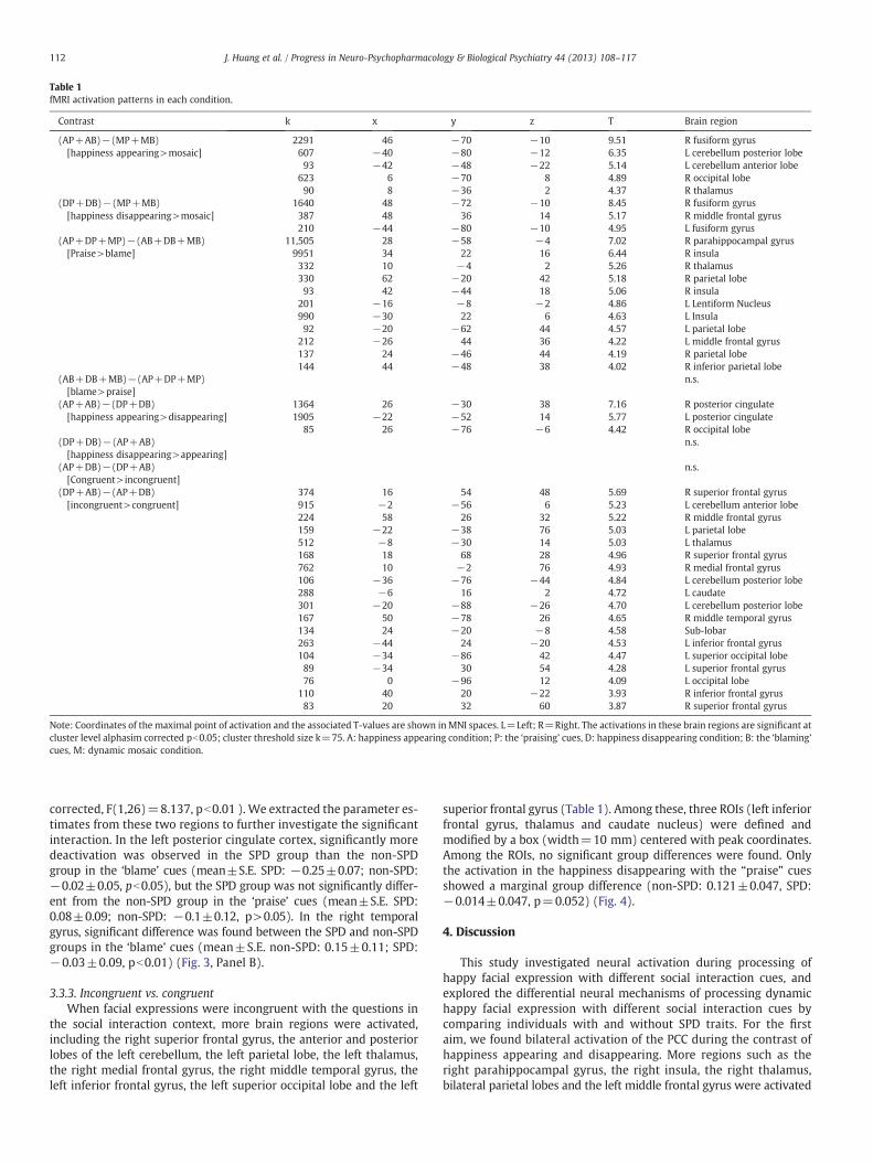
Table 1fMRI activation patterns in each condition.
Contrast k x y z T Brain region
(AP+AB)−(MP+MB)[happiness appearing>mosaic]
2291 46 −70 −10 9.51 R fusiform gyrus607 −40 −80 −12 6.35 L cerebellum posterior lobe93 −42 −48 −22 5.14 L cerebellum anterior lobe
623 6 −70 8 4.89 R occipital lobe90 8 −36 2 4.37 R thalamus
(DP+DB)−(MP+MB)[happiness disappearing>mosaic]
1640 48 −72 −10 8.45 R fusiform gyrus387 48 36 14 5.17 R middle frontal gyrus210 −44 −80 −10 4.95 L fusiform gyrus
(AP+DP+MP)−(AB+DB+MB)[Praise>blame]
11,505 28 −58 −4 7.02 R parahippocampal gyrus9951 34 22 16 6.44 R insula332 10 −4 2 5.26 R thalamus330 62 −20 42 5.18 R parietal lobe93 42 −44 18 5.06 R insula
201 −16 −8 −2 4.86 L Lentiform Nucleus990 −30 22 6 4.63 L Insula92 −20 −62 44 4.57 L parietal lobe
212 −26 44 36 4.22 L middle frontal gyrus137 24 −46 44 4.19 R parietal lobe144 44 −48 38 4.02 R inferior parietal lobe
(AB+DB+MB)−(AP+DP+MP)[blame>praise]
n.s.
(AP+AB)−(DP+DB)[happiness appearing>disappearing]
1364 26 −30 38 7.16 R posterior cingulate1905 −22 −52 14 5.77 L posterior cingulate
85 26 −76 −6 4.42 R occipital lobe(DP+DB)−(AP+AB)[happiness disappearing>appearing]
n.s.
(AP+DB)−(DP+AB)[Congruent>incongruent]
n.s.
(DP+AB)−(AP+DB)[incongruent>congruent]
374 16 54 48 5.69 R superior frontal gyrus915 −2 −56 6 5.23 L cerebellum anterior lobe224 58 26 32 5.22 R middle frontal gyrus159 −22 −38 76 5.03 L parietal lobe512 −8 −30 14 5.03 L thalamus168 18 68 28 4.96 R superior frontal gyrus762 10 −2 76 4.93 R medial frontal gyrus106 −36 −76 −44 4.84 L cerebellum posterior lobe288 −6 16 2 4.72 L caudate301 −20 −88 −26 4.70 L cerebellum posterior lobe167 50 −78 26 4.65 R middle temporal gyrus134 24 −20 −8 4.58 Sub-lobar263 −44 24 −20 4.53 L inferior frontal gyrus104 −34 −86 42 4.47 L superior occipital lobe89 −34 30 54 4.28 L superior frontal gyrus76 0 −96 12 4.09 L occipital lobe
110 40 20 −22 3.93 R inferior frontal gyrus83 20 32 60 3.87 R superior frontal gyrus
Note: Coordinates of the maximal point of activation and the associated T-values are shown inMNI spaces. L=Left; R=Right. The activations in these brain regions are significant atcluster level alphasim corrected pb0.05; cluster threshold size k=75. A: happiness appearing condition; P: the ‘praising’ cues, D: happiness disappearing condition; B: the ‘blaming’cues, M: dynamic mosaic condition.
112 J. Huang et al. / Progress in Neuro-Psychopharmacology & Biological Psychiatry 44 (2013) 108–117
corrected, F(1,26)=8.137, pb0.01 ). We extracted the parameter es-timates from these two regions to further investigate the significantinteraction. In the left posterior cingulate cortex, significantly moredeactivation was observed in the SPD group than the non-SPDgroup in the ‘blame’ cues (mean±S.E. SPD: −0.25±0.07; non-SPD:−0.02±0.05, pb0.05), but the SPD group was not significantly differ-ent from the non-SPD group in the ‘praise’ cues (mean±S.E. SPD:0.08±0.09; non-SPD: −0.1±0.12, p>0.05). In the right temporalgyrus, significant difference was found between the SPD and non-SPDgroups in the ‘blame’ cues (mean±S.E. non-SPD: 0.15±0.11; SPD:−0.03±0.09, pb0.01) (Fig. 3, Panel B).
3.3.3. Incongruent vs. congruentWhen facial expressions were incongruent with the questions in
the social interaction context, more brain regions were activated,including the right superior frontal gyrus, the anterior and posteriorlobes of the left cerebellum, the left parietal lobe, the left thalamus,the right medial frontal gyrus, the right middle temporal gyrus, theleft inferior frontal gyrus, the left superior occipital lobe and the left
superior frontal gyrus (Table 1). Among these, three ROIs (left inferiorfrontal gyrus, thalamus and caudate nucleus) were defined andmodified by a box (width=10 mm) centered with peak coordinates.Among the ROIs, no significant group differences were found. Onlythe activation in the happiness disappearing with the “praise” cuesshowed a marginal group difference (non-SPD: 0.121±0.047, SPD:−0.014±0.047, p=0.052) (Fig. 4).
4. Discussion
This study investigated neural activation during processing ofhappy facial expression with different social interaction cues, andexplored the differential neural mechanisms of processing dynamichappy facial expression with different social interaction cues bycomparing individuals with and without SPD traits. For the firstaim, we found bilateral activation of the PCC during the contrast ofhappiness appearing and disappearing. More regions such as theright parahippocampal gyrus, the right insula, the right thalamus,bilateral parietal lobes and the left middle frontal gyrus were activated
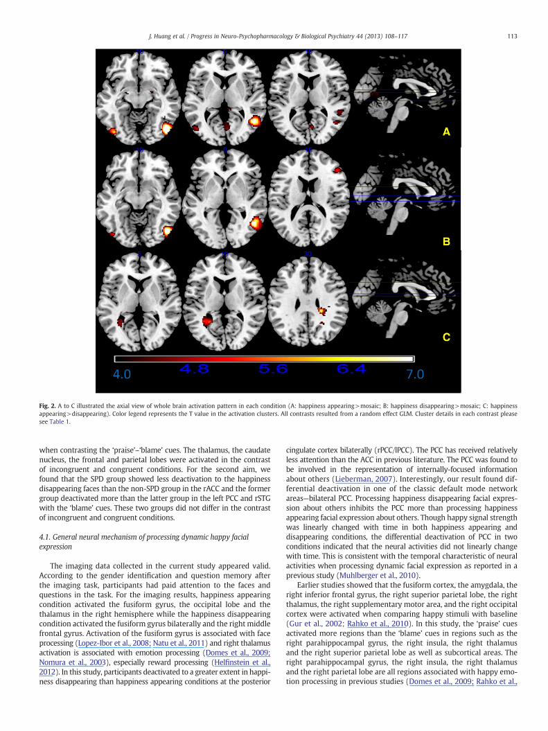
Fig. 2. A to C illustrated the axial view of whole brain activation pattern in each condition (A: happiness appearing>mosaic; B: happiness disappearing>mosaic; C: happinessappearing>disappearing). Color legend represents the T value in the activation clusters. All contrasts resulted from a random effect GLM. Cluster details in each contrast pleasesee Table 1.
113J. Huang et al. / Progress in Neuro-Psychopharmacology & Biological Psychiatry 44 (2013) 108–117
when contrasting the ‘praise’–‘blame’ cues. The thalamus, the caudatenucleus, the frontal and parietal lobes were activated in the contrastof incongruent and congruent conditions. For the second aim, wefound that the SPD group showed less deactivation to the happinessdisappearing faces than the non-SPD group in the rACC and the formergroup deactivated more than the latter group in the left PCC and rSTGwith the ‘blame’ cues. These two groups did not differ in the contrastof incongruent and congruent conditions.
4.1. General neural mechanism of processing dynamic happy facialexpression
The imaging data collected in the current study appeared valid.According to the gender identification and question memory afterthe imaging task, participants had paid attention to the faces andquestions in the task. For the imaging results, happiness appearingcondition activated the fusiform gyrus, the occipital lobe and thethalamus in the right hemisphere while the happiness disappearingcondition activated the fusiform gyrus bilaterally and the right middlefrontal gyrus. Activation of the fusiform gyrus is associated with faceprocessing (Lopez-Ibor et al., 2008; Natu et al., 2011) and right thalamusactivation is associated with emotion processing (Domes et al., 2009;Nomura et al., 2003), especially reward processing (Helfinstein et al.,2012). In this study, participants deactivated to a greater extent in happi-ness disappearing than happiness appearing conditions at the posterior
cingulate cortex bilaterally (rPCC/lPCC). The PCC has received relativelyless attention than the ACC in previous literature. The PCC was found tobe involved in the representation of internally-focused informationabout others (Lieberman, 2007). Interestingly, our result found dif-ferential deactivation in one of the classic default mode networkareas—bilateral PCC. Processing happiness disappearing facial expres-sion about others inhibits the PCC more than processing happinessappearing facial expression about others. Though happy signal strengthwas linearly changed with time in both happiness appearing anddisappearing conditions, the differential deactivation of PCC in twoconditions indicated that the neural activities did not linearly changewith time. This is consistent with the temporal characteristic of neuralactivities when processing dynamic facial expression as reported in aprevious study (Muhlberger et al., 2010).
Earlier studies showed that the fusiform cortex, the amygdala, theright inferior frontal gyrus, the right superior parietal lobe, the rightthalamus, the right supplementary motor area, and the right occipitalcortex were activated when comparing happy stimuli with baseline(Gur et al., 2002; Rahko et al., 2010). In this study, the ‘praise’ cuesactivated more regions than the ‘blame’ cues in regions such as theright parahippocampal gyrus, the right insula, the right thalamusand the right superior parietal lobe as well as subcortical areas. Theright parahippocampal gyrus, the right insula, the right thalamusand the right parietal lobe are all regions associated with happy emo-tion processing in previous studies (Domes et al., 2009; Rahko et al.,
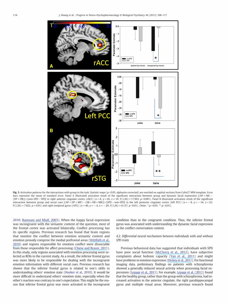
Fig. 3. Activation patterns for the interactions with group in the task. Statistic maps (pb0.05, alphasim corrected) are overlaid on sagittal sections from Colin27 MNI template. Errorbars represent the mean of standard error. Panel A illustrated activation result of the significant interaction between group and dynamic facial expression [(AP+AB)−(DP+DB)]×[non-SPD−SPD] in right anterior cingulate cortex (rACC) (x=8, y=42, z=10; F(1,26)=17.963, pb0.001). Panel B illustrated activation result of the significantinteraction between group and social cues [(AP+DP+MP)−(AB+DB+MB)]×[SPD−non-SPD] in the left posterior cingulate cortex (left PCC) (x=−8, y=−34, z=22;F(1,26)=7.822, p=0.01) and right temporal gyrus (rSTG) (x=40, y=−4, z=−20; F(1,26)=8.137, pb0.01). (Note: *:pb0.05; **:pb0.01).
114 J. Huang et al. / Progress in Neuro-Psychopharmacology & Biological Psychiatry 44 (2013) 108–117
2010; Ramnani and Miall, 2003). When the happy facial expressionwas incongruent with the semantic content of the question, most ofthe frontal cortex was activated bilaterally. Conflict processing hasits specific regions. Previous research has found that brain regionsthat monitor the conflict between emotion semantic content andemotion prosody comprise the medial prefrontal areas (Wittfoth et al.,2010) and regions responsible for emotion conflict were dissociablefrom those responsible for affect processing (Chiew and Braver, 2011).In this study, only regions associated with emotion processing were se-lected as ROIs in the current study. As a result, the inferior frontal gyruswas more likely to be responsible for dealing with the incongruentemotion information with different social cues. Previous research hasshown that the inferior frontal gyrus is related to one's skills inunderstanding others' emotion state (Hooker et al., 2010). It would bemore difficult to understand others' emotion state, especially when theother's reactionwas contrary to one's expectation. Thismight be the rea-son that inferior frontal gyrus was more activated in the incongruent
condition than in the congruent condition. Thus, the inferior frontalgyrus was associated with understanding the dynamic facial expressionin the conflict conversation context.
4.2. Differential neural mechanism between individuals with and withoutSPD traits
Previous behavioral data has suggested that individuals with SPDhave poor social function (McCleery et al., 2012), have subjectivecomplaints about hedonic capacity (Yan et al., 2011) and mighthave problems in emotion expression (Dickey et al., 2011). For functionalimaging data, preliminary findings on patients with schizophreniashowed a generally reduced neural activity when processing facial ex-pressions (Lepage et al., 2011). For example, Lepage et al. (2011) foundthat thehealthy group, rather than the groupwith schizophrenia, had in-creased activation in the anterior cingulate, the right parahippocampalgyrus and multiple visual areas. Moreover, previous research found

Fig. 4. Activation patterns for the contrast between incongruent and congruent conditions. Three ROIs were extracted [left inferior frontal gyrus—IFG(−44 24–20); thalamus—THA(−8 −30 14) and caudate nucleus—CN(−6 16 2)]. AP: happiness appearing with praise cues; DP: happiness disappearing with praise cues; AB: happiness appearing withblame cues; DB: happiness disappearing with blame cues. Statistic maps (pb0.05, alphasim corrected) are overlaid on sagittal sections from Colin27 MNI template. Error barsrepresent the mean of standard error.
115J. Huang et al. / Progress in Neuro-Psychopharmacology & Biological Psychiatry 44 (2013) 108–117
that cingulate volumes were reduced in first episode schizophrenia(Rothlisberger et al., 2012). Our findings showed that individuals withSPD had a differentiated neural mechanism in inhibiting neural activitieswhen processing happiness disappearing and the ‘blame’ cues.We foundthat individuals with SPD deactivated less than individuals without SPDin the right ACC in the happiness disappearing condition. The ACC is aneural region in the hedonic system (Kringelbach and Berridge, 2009).Less deactivation in the ACC in the happiness disappearing conditionmight suggest alteration of neural activities in the hedonic system of in-dividualswith SPD traits. In addition, we also found that individuals withSPD traits deactivatedmore than individualswithout SPD traits in the leftPCC and the rSTG when they processed the ‘blame’ condition. Previousstudies have suggested that the STG and the temporal–occipital lobeare responsible for dynamic human facial expression processing(Pitcher et al., 2011). The rSTGwas also activatedwhen subjects listenedto a semantically and prosodically incongruent sentences and the leftsuperior frontal gyrus and left superior temporal gyrus were activatedin happily intoned sentences expressed in negative content (Wittfothet al., 2010). In this study, ‘blame’ context represented the negative socialcue. The rSTG was associated with the processing of dynamic happyfacial expression with negative social cue. We thus inferred that themore deactivated rSTG in the ‘blame’ condition could provide a piece ofevidence of neural sensitivity for the negative social interaction cues,which induced unhappiness in individuals with SPD traits. Deactivationin rSTG and left PCC is reasonable because the resting state of rSTG(R. Li et al., 2012) and the cingulate cortex (Z. Li et al., 2012) areactivations. Deactivation in the current task could be a potentialneural marker for individuals with social interaction difficulty.Based on the imaging results in the incongruent–congruent contrast,we selected three regions of interest to further clarify whether indi-viduals with and without SPD would be different. The thalamus andthe caudate nucleus are associated with empathy and emotionrecognition (Kemp et al., 2012). Previous studies found that medialprefrontal cortex was associated with happy emotion processing(Keener et al., 2012) and right inferior parietal gyrus activated more
in emotion rating than gender rating (Sarkheil et al., 2012). Such frontaland parietal regions were not activated in the current contrast. Thus,thalamus, caudate and inferior frontal gyruswere considered as regionsof interest (ROIs). In our study, these regions were consistentlydeactivated in conditions without happy emotion such as happinessdisappearing with blame cues. If the facial expression or interactioncues contain happy emotion, the thalamus or caudate nucleus wouldbe activated, otherwise it would be deactivated. Taken together, the dif-ferential neural activity in the rACC, left PCC, rSTG, thalamus and thecaudate nucleus could be the neural mechanisms associated with theaberrant happy emotion processing with social interaction cues in indi-viduals with SPD traits.
5. Limitations
The main limitation of this study is that the block design could notcapture the temporal characteristic of the onset and disappearance ofhappiness emotion, as found in a previous study (Muhlberger et al.,2010), because a block design tends to ignore the temporal changeof BOLD signals. In future studies, a mixed block-event related designshould be considered to avoid such limitation. The second limitationis that the praise and blame cues were not counterbalanced duringthe task. The difference between the two runs might confound adirect comparison between praise and blame cues, though this wasnot the main purpose of the current study. Another limitation isthat only the happiness emotion was examined. This could be prob-lematic when examining congruence and incongruence in the ‘praise’or ‘blame’ interaction cues, because happiness appearing with the‘blame’ cues could be understood as a way to avoid embarrassmentrather than an inappropriate or incongruent facial expression whenbeing blamed. Therefore, angry facial expressions should be furtherexamined with the blame cues. One potentially informative approachfor future research is to determine whether the threat-relateddynamic facial expression change will be influenced by the social cuesin dyadic conversations. We could further investigate whether the left
116 J. Huang et al. / Progress in Neuro-Psychopharmacology & Biological Psychiatry 44 (2013) 108–117
hemisphere could still be dominant when the threat-related conditionwas incongruent with the social cues in dyadic conversations. Finally,it should be noted that SPD trait is associatedwith depression and autis-tic trait. Indeed several studies (e.g. Hurst et al., 2007)demonstratedthat SPQ scores are significantly correlated with the Autism-SpectrumQuotient (Baron-Cohen et al., 2001). In particular “Interpersonal” factorin SPQ is more or less similar to “Social skill” factor in the Autism-Spectrum Quotient. It is likely that individuals with high SPD traits inthe present study would also show autistic trait and have difficultieswith social perception associated with this trait. Given that we did notinclude any scales of autistic trait in the current study, we could nottease out this potential confound on the observed facial perception inourfindings. Future study should evaluate autistic trait and control abil-ity of facial perception correlated with autistic trait, and to analyze andextract neural responses associated with SPD trait only.
6. Conclusions
Notwithstanding the limitations, the current study is the first, toour knowledge, to examine the neural mediation of dynamic happyfacial expression with different social cues in dyadic conversations.Although there were some overlap of activations in some brain regions,happiness appearing and disappearing exhibited differences in activa-tion at the PCC bilaterally. The frontal–thalamus–caudate–parietal net-work was used to recognize happy emotion in the social situation andresolve the conflict between facial expression and social cues. Individualswith SPD traits have different neural inhibition mechanisms whenprocessing the happiness disappearing stimuli with ‘blame’ cues. Thiscould help elucidate the neuralmechanismunderlying the social interac-tion difficulty when facing happiness vanishing stimuli.
Acknowledgments
This work was supported by a grant from the Strategic PriorityResearch Programme (B) of the Chinese Academy of Sciences(XDB02030200) and the National Key Technologies R&D Programme(2012BAI36B01), the National Science Fund China (81088001,91132701), and a grant from the Knowledge Innovation Project ofthe Chinese Academy of Sciences (KSCX2-EW-J-8), to Raymond Chan.Dr. Jia Huang acknowledges supports from the National Science FundChina (31100747), the Young Investigator Scientific Fund of theInstitute of Psychology, Chinese Academy of Sciences (YOCX031S01)and funding from the Key Laboratory of Mental Health, Institute ofPsychology, ChineseAcademyof Sciences. The studywas also supportedby the initiation fund of the CAS/SAFEA International PartnershipProgramme for Creative Research Teams to Raymond Chan, RubenGur, and David Shum (Y2CX131003). These funding agents had nofurther role in the study design; in the collection, analysis and inter-pretation of the data; in the writing of the manuscript; and in thedecision to submit the paper for publication.
References
Abbott GR, Green MJ. Facial affect recognition and schizotypal personality characteristics.Early Interv Psychiatry 2012. http://dx.doi.org/10.1111/j.1751-7893.2012.00346.x.
Achim AM, Ouellet R, Roy MA, Jackson PL. Mentalizing in first-episode psychosis.Psychiatry Res 2012;196:207–13.
Annett M. A coordination of hand preference and skill replicated. Br J Psychol 1976;67:587–92.
Baron-Cohen S, Wheelwright S, Skinner R, Martin J, Clubley E. The autism-spectrumquotient (AQ): evidence from Asperger syndrome/high functioning autism,males and females, scientists and mathematicians. J Autism Dev Disord 2001;31:5-17.
Beck AT, Ward CH, Mendelson M, Mock J, Erbaugh J. An inventory for measuringdepression. Arch Gen Psychiatry 1961;4:561–71.
Brett M, Anton J-L, Valabregue R, Poline JB. Region of interest analysis using an SPMtoolbox. [abstract]Presented at the 8th International Conference on FunctionalMapping of the Human Brain, June 2–6, Sendai, JapanNeuroImage, 16: No 2;2002 [Available on CD-ROM].
Chan CM, Tsoi MM. The BDI and stimulus determinants of cognitive-related depressionamong Chinese college students. Cogn Ther Res 1984;8:501–7.
Chan RCK, Wang Y, Yan C, Song L-l, Wang Y-n, Shi Y-f, et al. Contribution of specificcognitive dysfunction to people with schizotypal personality. Psychiatry Res2011;186:71–5.
Chan RCK, Shi YF, Lai MK, Wang YN, Wang Y, Kring AM. The Temporal Experience ofPleasure Scale (TEPS): exploration and confirmation of factor structure in a healthyChinese sample. PLoS One 2012a;7:e35352.
Chan RCK, Wang Y, Yan C, Zhao Q, McGrath J, Hsi X, et al. A study of trait anhedonia innon-clinical Chinese samples: evidence from the Chapman scales for physical andsocial anhedonia. PLoS One 2012b;7:e34275.
Chen WJ, Hsiao CK, Lin CCH. Schizotypy in community samples: the three-factorstructure and correlation with sustained attention. J Abnorm Psychol 1997;106:649–54.
Chiew KS, Braver TS. Neural circuitry of emotional and cognitive conflict revealedthrough facial expressions. PLoS One 2011;6:e17635.
Cohen AS, Callaway DA, Najolia GM, Larsen JT, Strauss GP. On “risk” and reward:investigating state anhedonia in psychometrically defined schizotypy and schizo-phrenia. J Abnorm Psychol 2012;121:407–15.
Dickey CC, Panych LP, Voglmaier MM, Niznikiewicz MA, Terry DP, Murphy C, et al.Facial emotion recognition and facial affect display in schizotypal personalitydisorder. Schizophr Res 2011;131:242–9.
Domes G, Schulze L, Böttger M, Grossmann A, Hauenstein K, Wirtz PH, et al. The neuralcorrelates of sex differences in emotional reactivity and emotion regulation. HumBrain Mapp 2009;31:758–69.
Fujimura T, Suzuki N. Effects of dynamic information in recognising facial expressionson dimensional and categorical judgments. Perception 2010;39:543–52.
Germine LT, Garrido L, Bruce L, Hooker C. Social anhedonia is associated with neuralabnormalities during face emotion processing. Neuroimage 2011;58:935–45.
GongY.Manual ofWechsler Adult Intelligence Scale—Chinese Version. Changsha: ChineseMap Press; 1992.
Gur RE, McGrath C, Chan RM, Schroeder L, Turner T, Turetsky BI, et al. An fMRI studyof facial emotion processing in patients with schizophrenia. Am J Psychiatry2002;159:1992.
Gur RE, Loughead J, Kohler CG, Elliott MA, Lesko K, Ruparel K, et al. Limbic activationassociated with misidentification of fearful faces and flat affect in schizophrenia.Arch Gen Psychiatry 2007;64:1356–66.
Helfinstein SM, Kirwan ML, Benson BE, Hardin MG, Pine DS, Ernst M, et al. Validation ofa child-friendly version of the monetary incentive delay task. Soc Cogn AffectNeurosci 2012. http://dx.doi.org/10.1093/scan/nss1057.
Hicks JA, Schlegel RJ, King LA. Social threats, happiness, and the dynamics of meaningin life judgments. Pers Soc Psychol Bull 2010;36:1305–17.
Hooker CI, Verosky SC, Germine LT, Knight RT, D'Esposito M. Neural activity duringsocial signal perception correlates with self-reported empathy. Brain Res 2010;1308:100–13.
Huang J, Chan RCK, Lu X, Ma Z, Li Z, Gong Q-y. An exploratory study of the influence ofconversation prosody on emotion and intention identification in schizophrenia.Brain Res 2009a;1281:58–63.
Huang J, Chan RCK, Lu X, Tong Z. Emotion categorization perception in schizophrenia inconversations with different social contexts. Aust N Z J Psychiatry 2009b;43:438–45.
Hurst R, Mitchell J, Kimbrel N, Kwapil T, Nelson-Gray R. Examination of the reliabilityand factor structure of the autism spectrum quotient (AS) in a non-clinical sample.Personal Individ Differ 2007;43:1938–49.
Johnston PJ, Enticott PG, Mayes AK, Hoy KE, Herring SE, Fitzgerald PB. Symptom corre-lates of static and dynamic facial affect processing in schizophrenia: evidence of adouble dissociation? Schizophr Bull 2010;36:680–7.
Kee KS, HoranWP, Wynn JK, Mintz J, Green MF. An analysis of categorical perception offacial emotion in schizophrenia. Schizophr Res 2006;87:228–37.
Keener MT, Fournier JC, Mullin BC, Kronhaus D, Perlman SB, LaBarbara E, et al. Dissocia-ble patterns of medial prefrontal and amygdala activity to face identity versusemotion in bipolar disorder. Psychol Med 2012;42:1913–24.
Kemp J, Berthel MC, Dufour A, Despres O, Henry A, Namer IJ, et al. Caudate nucleus andsocial cognition: neuropsychological and SPECT evidence from a patient with focalcaudate lesion. Cortex 2012. http://dx.doi.org/10.1016/j.cortex.2012.1001.1004.
Kohler CG, Walker JB, Martin EA, Healey KM, Moberg PJ. Facial emotion perception inschizophrenia: a meta-analytic review. Schizophr Bull 2010;36:1009–19.
Kringelbach ML, Berridge KC. Towards a functional neuroanatomy of pleasure andhappiness. Trends Cogn Sci 2009;13:479–87.
Kuhn S, Muller BC, van der Leij A, Dijksterhuis A, Brass M, van Baaren RB. Neural corre-lates of emotional synchrony. Soc Cogn Affect Neurosci 2011;6:368–74.
Lee SJ, Lee HK, Kweon YS, Lee CT, Lee KU. Deficits in facial emotion recognition inschizophrenia: a replication study with Korean subjects. Psychiatry Investig 2010;7:291–7.
Lepage M, Sergerie K, Benoit A, Czechowska Y, Dickie E, Armony JL. Emotional face pro-cessing and flat affect in schizophrenia: functional and structural neural correlates.Psychol Med 2011;41:1833–44.
Li HJ, Chan RCK, Gong QY, Liu Y, Liu SM, Shum D, et al. Facial emotion processing inpatients with schizophrenia and their non-psychotic siblings: a functional magnet-ic resonance imaging study. Schizophr Res 2012a;134:143–50.
Li R, Qin W, Zhang Y, Jiang T, Yu C. The neuronal correlates of digits backward arerevealed by voxel-based morphometry and resting-state functional connectivityanalyses. PLoS One 2012b;7:e31877.
Li Z, Kadivar A, Pluta J, Dunlop J, Wang Z. Test–retest stability analysis of resting brainactivity revealed by blood oxygen level-dependent functional MRI. J Magn ResonImaging 2012c;36:344–54.
117J. Huang et al. / Progress in Neuro-Psychopharmacology & Biological Psychiatry 44 (2013) 108–117
Lieberman MD. Social cognitive neuroscience: a review of core processes. Annu RevPsychol 2007;58:259–89.
Lopez-Ibor JJ, Lopez-Ibor M-I, Mendez M-A, MoronM-D, Ortiz-Teran L, Fernandez A, et al.The perception of emotion-free faces in schizophrenia: a magneto-encephalographystudy. Schizophr Res 2008;98:278–86.
Loughead J, Gur RC, Elliott M, Gur RE. Neural circuitry for accurate identification offacial emotions. Brain Res 2008;1194:37–44.
Maat A, Fett AK, Derks E. Social cognition and quality of life in schizophrenia. SchizophrRes 2012;137:212–8.
McCleery A, Divilbiss M, St-Hilaire A, Aakre JM, Seghers JP, Bell EK, et al. Predictingsocial functioning in schizotypy: an investigation of the relative contributions oftheory of mind and mood. J Nerv Ment Dis 2012;200:147–52.
McDonald S, Li S, De Sousa A, Rushby J, Dimoska A, James C, et al. Impaired mimicryresponse to angry faces following severe traumatic brain injury. J Clin Exp Neuropsychol2011;33:17–29.
Muhlberger A, Wieser MJ, Gerdes AB, Frey MC, Weyers P, Pauli P. Stop looking angryand smile, please: start and stop of the very same facial expression differentiallyactivate threat- and reward-related brain networks. Soc Cogn Affect Neurosci2010:1–9.
Muller VI, Habel U, Derntl B, Schneider F, Zilles K, Turetsky BI, et al. Incongruenceeffects in crossmodal emotional integration. Neuroimage 2011;54:2257–66.
Natu V, Raboy D, O'Toole AJ. Neural correlates of own- and other-race face perception:spatial and temporal response differences. Neuroimage 2011;54:2547–55.
NomuraM, Iidaka T, Kakehi K, Tsukiura T, Hasegawa T,Maeda Y, et al. Frontal lobe networksfor effective processing of ambiguously expressed emotions in humans. Neurosci Lett2003;348:113–6.
Pitcher D, Dilks DD, Saxe RR, Triantafyllou C, Kanwisher N. Differential selectivity fordynamic versus static information in face-selective cortical regions. Neuroimage2011;56:2356–63.
Platt B, Kamboj S, Morgan CJ, Curran HV. Processing dynamic facial affect in frequentcannabis-users: evidence of deficits in the speed of identifying emotional expressions.Drug Alcohol Depend 2010;112:27–32.
Rahko J, Paakki J-J, Starck T, Nikkinen J, Remes J, Hurtig T, et al. Functional mappingof dynamic happy and fearful facial expression processing in adolescents. BrainImaging Behav 2010;4:164–76.
Raine A. The SPQ: a scale for the assessment of schizotypal personality based onDSM-III-R criteria. Schizophr Bull 1991;17:555–64.
Ramnani N, Miall R. Instructed delay activity in the human prefrontal cortex is modu-lated by monetary reward expectation. Cereb Cortex 2003;13:318–27.
RecioG, SommerW, Schacht A. Electrophysiological correlates of perceivingand evaluatingstatic and dynamic facial emotional expressions. Brain Res 2011;1376:66–75.
Regenbogen C, Schneider DA, Gur RE, Schneider F, Habel U, Kellermann T. Multimodalhuman communication — targeting facial expressions, speech content and prosody.Neuroimage 2012;60:2346–56.
Roddy S, Tiedt L, Kelleher I, Clarke MC, Murphy J, Rawdon C, et al. Facial emotionrecognition in adolescents with psychotic-like experiences: a school-based samplefrom the general population. Psychol Med 2012:1-10.
RothlisbergerM, Riecher-Rossler A, Aston J, Fusar-Poli P, Radu EW, Borgwardt S. Cingulatevolume abnormalities in emerging psychosis. Curr Pharm Des 2012;18:495–504.
Rymarczyk K, Biele C, Grabowska A, Majczynski H. EMG activity in response to staticand dynamic facial expressions. Int J Psychophysiol 2011;79:330–3.
Sarkheil P, Goebel R, Schneider F, Mathiak K. Emotion unfolded by motion: a role forparietal lobe in decoding dynamic facial expressions. Soc Cogn Affect Neurosci2012. http://dx.doi.org/10.1093/scan/nss092.
Sato W, Yoshikawa S. Enhanced experience of emotional arousal in response to dynamicfacial expressions. J Nonverbal Behav 2007a;31:119–35.
SatoW, Yoshikawa S. Spontaneous facial mimicry in response to dynamic facial expressions.Cognition 2007b;104:1-18.
Sharpley CF, Bitsika V. The diverse neurogeography of emotional experience: formfollows function. Behav Brain Res 2010;215:1–6.
Shi YF, Wang Y, Cao XY, Wang Y, Wang YN, Zong JG, et al. Experience of pleasure andemotional expression in individuals with schizotypal personality features. PLoSOne 2012;7:e34147.
Smith MJ, Horan WP, Karpouzian TM, Abram SV, Cobia DJ, Csernansky JG. Self-reportedempathy deficits are uniquely associated with poor functioning in schizophrenia.Schizophr Res 2012;137:196–202.
Song X, Dong Z, Long X, Li S, Zuo X, Zhu C, et al. REST: a toolkit for resting-statefunctional magnetic resonance imaging data processing. PLoS One 2011;6:e25031.
Surguladze S, Brammer M, Keedwell P, Giampietro V, Young AW, Travis MJ, et al. Adifferential pattern of neural response toward sad versus happy facial expressionsin major depressive disorder. Biol Psychiatry 2005;57:201–9.
Vytal K, Hamann S. Neuroimaging support for discrete neural correlates of basicemotions: a voxel-based meta-analysis. J Cogn Neurosci 2010;22:2864–85.
Wittfoth M, Schroder C, Schardt DM, Dengler R, Heinze H-J, Kotz SA. On emotion conflict:interference resolution of happy and angry prosody reveals valence-specific effects.Cereb Cortex 2010;20:383–92.
Yan C, Liu WH, Cao Y, Chan RC. Self-reported pleasure experience and motivation inindividuals with schizotypal personality disorders proneness. East Asian ArchPsychiatry 2011;21:115–22.
Zong JG, Chan RCK, Stone WS, Hsi XL, Cao XY, Zhao Q, et al. Coping flexibility in youngadults: comparison between subjects with and without schizotypal personalityfeatures. Schizophr Res 2010;122:185–92.