effects of development and dopamine depletion on striatal nmda receptor-mediated calcium uptake
TRANSCRIPT
ORIGINAL PAPER
Effects of development and dopamine depletionon striatal NMDA receptor-mediated calcium uptake
Thabelo Khoboko & Vivienne A. Russell
Received: 4 January 2007 /Accepted: 4 March 2007 /Published online: 27 June 2007# Springer Science + Business Media, LLC 2007
Abstract Calcium (Ca2+) is the currency of N-methyl-D-aspartate (NMDA) receptormediated signal transduction pathways involved in the modification of synapticefficacy during regulation of excitatory inputs into the striatum. The aim of thepresent study was to investigate the effects of development and dopamine depletionon NMDA receptor function. NMDA receptors were stimulated by incubation ofstriatal sections (350 μm) in buffer containing NMDA (100 μm) for 2 min, the sliceswere washed and uptake of radioactively labelled calcium (45Ca2+) was measured.Dopamine depletion has been reported to result in alterations of glutamate receptorexpression and upregulation of NMDA receptor activity. However, the results of thepresent study show that dopamine depletion does not alter NMDA-stimulated Ca2+
uptake into rat striatal slices in vitro. Unilateral striatal dopamine depletion wasachieved by infusion of 6-hydroxydopamine (6-OHDA, 13.5 μg/4.5 μl) into themedial forebrain bundle (MFB) of the left hemisphere of ten rats. NMDA-stimulated45Ca2+ uptake into striata following dopamine depletion was not significantlydifferent from NMDA-stimulated 45Ca2+ uptake into striata obtained from sham-operated rats. Other factors that induce changes in NMDA receptor function includedevelopment and aging. In young rats aged 7 weeks old (n=7) and 16 weeks old (n=6) a significant 2–3 fold decrease in striatal NMDA receptor function was observedwith increasing age over the 9 week period of development. To our knowledge theseare the first results to show developmental decreases of NMDA receptor function inthe striatum of juvenile rats.
Metab Brain Dis (2008) 23:9–30DOI 10.1007/s11011-007-9050-9
T. Khoboko : V. A. Russell (*)Department of Human Biology, Faculty of Health Sciences, University of Cape Town,Anzio Road, Observatory 7925, Cape Town, South Africae-mail: [email protected]
T. Khobokoe-mail: [email protected]
Present address:T. KhobokoDivision of Neural Systems, Memory and Aging, Arizona Research Laboratories,University of Arizona, Tucson, AZ, USA
Keywords NMDA receptors . Calcium . Striatum . Development . Dopamine
Introduction
N-methyl-D-aspartate (NMDA) receptors play a critical role in synaptic strengthen-ing and weakening through calcium (Ca2+) ion fluxes across the cell membrane(Nakanishi 1992; Mori and Mishina 1995; Blackstone and Sheng 1999). NMDAreceptors are comprised of NR1 and NR2 subunits which bond to form NR1/NR2heteromers. NR3 subunits have also been described but are not widespread withinthe brain. NR1 subunits are expressed indiscriminately throughout the developedbrain, but the expression of the NR2 subtypes, NR2A-NR2D, exhibits more regionalspecialization (Nakanishi 1992; Mori and Mishina 1995; Erreger et al. 2005). In thestriatum, spiny projection neurons predominantly express NR1/NR2A and NR1/NR2B NMDA receptor complexes (Chen and Reiner 1996; Götz et al. 1997; Loftisand Janowsky 2003). Altering NMDA receptor subtype composition confersdifferent kinetics and glutamate affinity to the receptor channels and henceinfluences ionic Ca2+ conductances. NR1/NR2A NMDA channels have the fastestgating constant ≈ 120 ms and the gating constant for NR1/NR2B NMDA channels is≈ 400 ms (Mori and Mishina 1995). The kinetics of the different NMDA receptorchannels regulates different signaling mechanisms mediated by Ca2+. Rapidincreases activate mechanisms that are not activated by slower ionic increaseshence subunit composition may confer temporal activation of signaling mechanisms(Cull-Candy et al. 2001; Erreger et al. 2005). Ca2+ activates signaling cascades thatinfluence synapse formation, dendritic spine morphology, gene transcription,glutamate receptor expression, ion channel regulation, neuronal excitability andconsequently the firing response of striatal spiny neurons to glutamate stimulation(Bourne and Nicoll 1993; Mori and Mishina 1995; Kennedy 1997; Wickens andWilson 1998; Blackstone and Sheng 1999; van Rossum and Hanisch 1999; Carrolland Zukin 2002; Cohen-Cory 2002; Reynolds and Wickens 2002; Saimi and Kung2002; Sheng and Kim 2002; Kasai et al. 2003).
Dopamine has differential influences on excitability of medium spiny neurons atdifferent membrane potentials (Wickens and Wilson 1998; Reynolds and Wickens2002). Dopamine reduces cell excitability during neuronal hyperpolarized ‘down’resting state (Lin et al. 2003) but potentiates firing at depolarized ‘up’ potentials(Hernandez-Lopez et al. 1997). The electroresponsive properties of striatal neuronsare mediated by different types of potassium (K+) currents (Llinás 1988; Calabresi etal. 1990; Nisenbaum and Wilson 1995). Inwardly rectifying K+ currents (IKir) areresponsible for high input resistance of the spiny neurons at resting membranepotentials (Nisenbaum and Wilson 1995; Charpier et al. 1999; Venance andGlowinski 2003). Dopamine through activation of D1 receptors fortifies theresistance to uncoordinated excitability by increasing the activity of IKir (Lin et al.2003; Venance and Glowinski 2003) and reduces neuronal firing output athyperpolarized potentials (−82 mV; Hernandez-Lopez et al. 1997). Partially due toIKir, D1 receptor-mediated potentiation of NMDA currents is minimal athyperpolarized potentials (Cepeda et al. 1998). NMDA receptors are also underMg2+ regulating block during membrane hyperpolarisation and this contributes
10 Metab Brain Dis (2008) 23:9–30
strongly to minimal NMDA mediated currents during the ‘down state.’ Theseinfluences are a result of dopamine mediated intracellular signaling cascadesactivating kinases and phosphatases that change the phosphorylation state ofvoltage gated ion channels and ligand gated ion channels such as NMDAreceptors. Changes in receptor phosphorylation state cause alterations in NMDAreceptor channel function (Yu et al. 1997). The efficiency of NMDA receptors ishighly regulated by phosphorylation of their receptor subunits.
There are multiple effects of dopamine depletion on medium spiny neuronexcitability. The membrane potential of striatal neurons is increased (alleviating theMg2+ block of NMDA receptor channels) and firing activity of striatal neurons isincreased (Kish et al. 1999; Tseng et al. 2001; Wichmann and DeLong 2003; Tsenget al. 2004). There is an alteration of dendritic spine morphology and synapticNMDA receptor expression (Lange et al. 1997; Arbuthnott et al. 2000; Reynoldsand Wickens 2002; Yasumoto et al. 2002; Dunah and Standaert 2003). Changes inNMDA phosphorylation occur following dopamine depletion in a parkinsonian ratmodel (Oh et al. 1998; Dunah et al. 2000). The phosphorylation state of NR1 serineresidues and NR2 tyrosine residues increases (Oh et al. 1998; Dunah et al. 2000) andit has been suggested that NMDA receptor function is upregulated (Chase et al.1998). In the clinical treatment of Parkinson’s disease, attenuation of NMDAreceptor activity is a strategy developed using NMDA antagonists as adjuncts todopamine replacement therapy, L-DOPA (Montastruc et al. 1997). In animal modelsof Parkinson’s disease, NMDA antagonists are reported to have similar anti-parkinsonian effects to dopamine receptor agonists (Nash and Brotchie 2002).During aging striatal medium spiny neurons exhibit lower spontaneous firing ratesand have higher firing thresholds (Cepeda et al. 1989). The age effects of NMDA-stimulated Ca2+ uptake in vitro have not been described in young adult male rats. Inthe current study we tested the hypothesis that within the striatum, stimulation ofNMDA receptors would result in a reduction of Ca2+ influx in older animals whencompared with Ca2+ influx in younger animals. The second purpose of the currentstudy was to examine NMDA-stimulated Ca2+ uptake in a parkinsonian rat model.We hypothesized that 6-hydroxydopamine (6-OHDA) degeneration of thenigrostriatal pathway will result in increased NMDA-stimulated Ca2+ uptake invitro as a consequence of NMDA receptor upregulation.
Materials and methods
Animals
The design of the current study and the use of animals were approved by theUniversity of Cape Town Research Ethics Committee. Long Evans rats wereobtained from the Faculty of Health Sciences Animal Unit where they were bred.The rats were housed in individual cages in the Department of Human Biologyanimal house. The rats were maintained on a 12-h light/dark cycle and wereprovided with food pellets and water ad libitum. The effects of age on NMDAreceptor function was investigated in male Long Evans rats aged 6–7 weeks, n=7(106–210 g) and 16 weeks, n=6 (482–556 g); these rats did not receive pharma-
Metab Brain Dis (2008) 23:9–30 11
cological or surgical treatment prior to uptake assays being conducted. Male LongEvans rats weighing 330–398 g (aged 18–19 weeks at the time of 45Ca2+ uptakeassays) were used to investigate the effects of 6-OHDA lesion of the nigrostriatalpathway on NMDA receptor function. To identify the site of lesion in the medialforebrain bundle (MFB) using the method of cresyl violet staining, ten rats wereused. The nigrostriatal pathway was lesioned with 6-OHDA in 15 rats and 10 ratswere sham lesioned using sterile saline. Of the rats lesioned with 6-OHDA, threewere used to visually verify the extent of degeneration using tyrosine hydroxylase(TH) staining following behavioural assessment. From the sham-lesioned group, onerat, was used for the TH staining, in addition to one unlesioned rat. NMDA-stimulated uptake assays were conducted on ten of the 6-OHDA-lesioned rats and onnine of the sham-lesioned rats.
NMDA-stimulated 45Ca2+ uptake
Two studies to investigate the effects of dopamine and age on NMDA receptors wereconducted using the method of NMDA-stimulated calcium uptake assays aspreviously described (Feldman et al. 1990; Lehohla et al. 2000; Lehohla et al.2001). Each rat was deeply anaesthetized with halothane and decapitated. The brainwas rapidly removed and cooled for 5 min in continuously oxygenated, ice-coldincubation HEPES buffer (120 mM NaCl, 1 mM MgCl2, 3.36 mM KCl, 10 mM D-Glucose, 20 mM HEPES buffer pH 7.4, 1.2 mM CaCl2). After 5 min incubation, theposterior part of the brain, ≈ 2 mm anterior to the cerebellum, was cut. The posteriorbrain surface of the block was glued onto the vibratome cutting stage using Loctite406 glue. Prior to use, the vibratome bath was filled with continuously oxygenatedice-cold HEPES buffer slush made from frozen buffer.
Dissection of striata
Serial coronal sections of 350 μm thickness containing the anterior, intermediate andposterior dorsal striata were cut. The serial sections were cut from slicescorresponding approximately to antero–posterior co-ordinates 10.20 to 8.74 mmwith reference to the interaural line (Paxinos and Watson 1986). A nick was made inthe left cerebrum to enable differentiation of the left hemisphere from the righthemisphere. Striata were carefully dissected from each hemisphere using a curvedblade. The striatal sections were transferred to individual Teflon vials with nylonmesh stretched across the base and held firmly to the bottom with a rubber o-ringfitted to a groove near the base of the tube. The vials were marked to indicate theslice number and hemisphere (right or left) of the striatum. The vials were heldfirmly in a rack that was used to facilitate the simultaneous transfer of all the striatalsections between sets of polyethylene wells containing different buffers insubsequent phases of the assay.
Incubation phases
In the first phase, dissected striata were incubated in 10 ml continuously oxygenatedHEPES buffer contained in individual wells and equilibrated at room temperature for
12 Metab Brain Dis (2008) 23:9–30
45 min. The sections were then transferred to 35°C continuously oxygenated HEPESbuffer and incubated for 5 min. NMDA-stimulated uptake of radioactive 45Ca2+
(Perkin Elmer Life and Analytical Sciences, USA) was determined by incubatingdissected striatal sections in HEPES buffer containing 100 μM NMDA (SigmaChemical Company, USA) and comparing the 45Ca2+ accumulation with striatalsections incubated in control buffer to which NMDAwas not added. The incubationperiod lasted 2 min at 35°C. Dissected striatal sections from the anterior (1L and 1R)and posterior (3L and 3R) slices were used as the test sections incubated instimulation buffer and the striatal sections dissected from the intermediate (2L and2R) slice served as the controls. ‘L’ and ‘R’ represent striatal sections dissected fromthe left and right hemispheres respectively. The experiment was concluded with aseries of lanthanum washes at 4°C to remove excess 45Ca2+ from the striatalsections. The buffered lanthanum chloride (LaCl3) solution had similar compositionto the normal HEPES buffer solution except that CaCl2 was replaced by 10 mMLaCl3. The first wash was a rapid wash for 10 s followed by a 15 min wash. Thesewashes were conducted in individual wells containing buffered LaCl3. The final15 min wash was in flat petri dishes containing buffered LaCl3 solution.
The striatal sections were sonicated in 1 ml ice-cold distilled water to rupture thecells and release accumulated 45Ca2+. A 600 μl aliquot of the sonicate was mixedthoroughly with 3.4 ml of scintillation fluid (Zinsser Analytic, UK) and the countsper minute (CPMs) of the sample were measured in a liquid scintillation counter(Packard Tri-Carb 2100TR). A standard quench curve was constructed for eachexperiment using the NMDA-containing and non-NMDA-containing incubationbuffers. The purpose of the quench curve was to determine the amount ofradioactivity in the samples by comparison with a set of quenched standards.Varying volumes of distilled water 0 ml, 50 μl, 250 μl, 600 μl, 1.0 ml, 1.5 ml and2.0 ml were added to 5 ml Teflon vials. Using a Finn pipette, 20 μl of the testincubation buffer containing 45Ca2+ was added into each of the vials. A separatestandard curve was constructed for the control incubation buffer. Scintillation fluid(3.4 ml) was added to the vials and mixed thoroughly. The vials were placed intolarger 20 ml polyethylene vials and taken to the liquid scintillation counter. From thecounts per minute (CPMs) recorded for the quenched standards, a curve ofpercentage efficiency (this had to be calculated) was plotted against tSIE (amountof quench determined by the counter).
Protein determination
The remaining sonicate was used to determine the protein content using the Miller(1959) method of protein determination. Protein dilutions using 1 mg/ml bovineserum albumin (BSA) were used to construct a standard curve. Duplicate volumes ofBSA (0, 10, 20, 40 and 80 μl) were made up to 1 ml with distilled water. Duplicatesamples of the tissue sonicate (80 μl) were made up to 1 ml with distilled water. Oneml of freshly prepared copper tartarate solution was added to all the test tubes usinga Finn pipette and mixed thoroughly on the vortex. The samples were allowed tostand at room temperature for 10 min. Folin phenol reagent (3 ml) was forcefullyadded to each test tube using a Finn pipette and mixed thoroughly on the vortex. Thetest tubes were incubated in a 50°C water bath for 10 min. After 10 min the samples
Metab Brain Dis (2008) 23:9–30 13
were cooled to room temperature, before being transferred to cuvettes formeasurement of absorbance at 650 nm in the spectrophotometer (UV-9100,Laboratory & Scientific Equipment Company). The absorbance of the BSA solutionswas used to construct a standard curve from which the dilutions of the sonicate wereread to obtain the amount of protein in 80 and 160 μl of sonicate. The amount of45Ca2+ accumulated in the tissue samples was expressed as nanomoles of 45Ca2+
accumulation per milligram of protein (nmol 45Ca2+/mg protein) in 2 min. Toinvestigate the effects of dopamine depletion on NMDA stimulated 45Ca2+ uptake,calcium uptake assays were conducted as described above following the surgicaltreatment and behavioral studies of the rats described below.
Calculations and statistical analysis
The NMDA-stimulated 45Ca2+ uptake data were analysed with Student t tests(dependent samples) for within factor comparison with Bonferroni correction formultiple comparisons (while pi≤α/n reject Hi, otherwise accept Hi, where n is equalto the number of null hypotheses that are simultaneously tested; García 2004) ofuptake into left (lesioned) vs. right (unlesioned) hemispheres. One-way ANOVAwasused for comparison between 6-OHDA-lesioned and sham-lesioned rats and forcomparison between rats aged 6–7 weeks and 16 weeks. In all the statisticalanalyses, statistical significance was taken as p<0.05. The Statistica 7 softwarepackage was used for all analyses. Results are given as the mean±standard error ofthe mean (SEM) and uptake was calculated as nmol 45Ca2+/mg protein/2 min.
Surgical procedure
Surgery was conducted to lesion the nigrostriatal pathway and to obtain ahistological representation of the site of lesion. All surgical lesioning procedureswere conducted under semi-sterile conditions on rats weighing 330–398 g (aged 13–14 weeks) at time of surgery. The rats were maintained under deep anaesthesia with3% halothane/oxygen anaesthesia (Safeline Pharmaceuticals, South Africa) admin-istered using a Blease vaporizer via a specifically designed and constructed nosecone. The co-ordinates of the MFB were determined using the stereotaxic rat atlas ofPaxinos and Watson (1986). Co-ordinates were: anterior–posterior with reference tothe interaural line—4.7 mm and lateral relative to the midline—1.6 mm. A midlineincision of the skin overlying the skull was made and a burr hole drilled to exposethe dura mater above the MFB target site in the left hemisphere. The dura wascarefully lifted using the bent tip of a 20Gx1 1/2 needle and a 0.3 mm stainless steelinfusion needle was lowered 8.4 mm from dura to the MFB. The needle was left inplace for 5 min before being withdrawn slowly. The rats used to obtain histologicalrepresentations of the site of lesion were immediately killed by the method oftranscardial perfusion.
This surgical technique was used to infuse 6-OHDA (Sigma Chemical Company,USA) into the MFB of 15 rats and to sham-lesion ten rats with infusion of sterilesaline. Each rat was injected intraperitoneally (i.p.) with desipramine hydrochloride(25 mg/kg; Sigma Chemical Company, USA) at least 25 min prior 6-OHDA orsaline infusion into the MFB (Magill et al. 2001).
14 Metab Brain Dis (2008) 23:9–30
A syringe pump connected to the infusion needle regulated the rate of infusion of13.5 μg/4.5 μl dose of 6-OHDA into the left hemisphere (Magill et al. 2001). The 6-OHDA solution was prepared with sterile saline and 0.02% L-ascorbic acid (Merck,Germany) just before use. At the end of the infusion period, 5 min was allowed forthe toxin to diffuse before the needle was withdrawn slowly from the brain. The burrhole was sealed with bone wax and the skin incision was sutured with silk suture andsterilized with 70% alcohol and iodine. Sham-operated control rats underwent thesame surgical procedure receiving only 4.5 μl of the vehicle (sterile saline with0.02% ascorbic acid).
Behavioral assessment of lesion
Fourteen days after surgery, the 6-OHDA-lesioned and sham-lesioned ratsunderwent behavioural assessment to determine the extent of degeneration ofdopamine neurons innervating the striatum. An established technique of apomorphine-induced rotations was used (Ungerstedt and Arbuthnott 1970; Tillerson et al. 2001).The rats were placed in rotameter drums and the rotameter recorded the number ofquarter rotations made by the rat over a period of 90 min using the Automatic RodentMonitor programme. The first 30 min of the recording session was used to acclimatizethe rats to the new environment and to record the baseline rotational movement. After30 min, the rats were removed from the drums and were subcutaneously injected with0.5 mg/kg apomorphine hydrochloride hemihydrate (Sigma Chemical Company,USA). The rats were returned to the drums immediately following the injection andthe rotational response to apomorphine was recorded. 6-OHDA-lesioned rats (n=3)that exhibited less than 400 contraversive quarter rotations i.e. 100 complete rotationswere removed from further analysis involving NMDA-stimulated 45Ca2+ assays. Morethan 100 complete contralateral rotations has been established to represent a loss of>90% of dopamine in the striatum (Ungerstedt 1971). On the basis of this criterion, 126-OHDA-lesioned rats (completing >100 contralateral rotations) were selected forfurther study. Sham-lesioned rats did not exhibit contralateral turning in response toapomorphine, but rather conducted predominantly ipsilateral rotations. At the end of therecording session, the rats were returned to their individual cages and transported to theanimal house.
Histology
Transcardial perfusion
To conduct the histology on brain tissue to obtain a representation of the site oflesion using cresyl violet staining and to obtain a visual record of the extent of lesionusing tyrosine hydroxylase staining, rats were transcardially perfused. The rats thathad undergone lesioning operations, were perfused five weeks after 6-OHDAinjection i.e. 21 days after behavioural assessment. The method of transcardialperfusion was employed as it was the only method that allowed the efficient clearingof blood from the brain tissue and the complete perfusion of the brain with fixative.
The operation was conducted as humanely as possible with each rat under deepanaesthesia. The rat was perfused with 150 ml 0.15 M phosphate buffer saline (PBS)
Metab Brain Dis (2008) 23:9–30 15

solution pH 7.4 through a 20Gx1 1/2 perfusion needle inserted into the left atrium.Perfusion with saline was followed by fixation with 300 ml of 4% paraformaldehyde(PFA) in 0.15 M PBS pH 7.4 solution. Following the perfusion procedure, the brainwas removed from the cranium and was placed in a glass vial containing 20%sucrose PBS pH 7.4 solution for cryoprotection overnight. The brain was removedfrom the sucrose solution and was embedded in tissue freezing medium within tinfoil moulds, frozen over liquid nitrogen vapour and stored at −80°C wrapped inparafilm until removed for sectioning. Using a cryostat, 20 μm sections of braincontaining the striatum and the midbrain (substantia nigra and ventral tegmentalarea) were cut at −20°C and placed on gelatinized glass slides in preparation forcresyl violet and tyrosine hydroxylase staining.
Cresyl violet staining
The brain sections were incubated in cresyl violet solution (0.2% cresyl violet in0.2 M acetate buffer containing 1% thionin, pH 3.5) for 20 min. Following cresylviolet incubation the sections were cleared in distilled water for 1 min and subjectedto a series of dehydration steps in 90% alcohol (1 min), 95% alcohol (1 min) and100% alcohol (3x 1 min) followed by clearing in xylene. The slides were mountedwith Entellan.
Tyrosine hydroxylase staining
Sections of the brain tissue containing the midbrain (substantia nigra pars compacta—SNc and ventral tegmental area—VTA) and the striatum were stained for thedopamine synthesis regulating enzyme, tyrosine hydroxylase, to confirm whetherdegeneration had occurred. Tyrosine hydroxylase staining was conducted on the brainsof two 6-OHDA-lesioned rats completing >100 complete rotations and one ratcompleting<100 rotations. The brains obtained from one sham-lesioned rat and oneunlesioned animal were also stained for tyrosine hydroxylase immunoreactivity.
Prior to processing for immunohistochemistry, the sections had to reach roomtemperature. The brain sections were prepared for staining by initially quenchingtheir endogenous peroxidase activity with 0.3% hydrogen peroxide solution toreduce background staining. The sections were incubated with the hydrogenperoxide solution in a humidifying chamber for 30 min at room temperaturefollowed by a series of three washes of 10 min each in 0.15 M PBS pH 7.4.Blocking serum (normal horse serum, 150 μl) was diluted in 10 ml 0.15 M PBScontaining 0.2% Triton X100 and immediately applied to each slice; the sectionswere incubated for 30 min at room temperature in the humidifying incubationchamber. After 30 min, the blocking solution was blotted from the sections. Primarymouse monoclonal anti-tyrosine hydroxylase antibody solution (Sigma ChemicalCompany) was diluted to 1:8,000 in blocking solution just before use. The primaryantibody solution was applied onto each slice and the sections were incubatedovernight in the humidifying chamber kept in a fridge at 4°C.
The overnight incubation was terminated with a series of 5x 10 min washes in0.15 M PBS pH 7.4. The sections were then incubated with biotinylated secondaryantibody (Vectastain anti-mouse IgG biotinylated antibody, 1:400 dilution in
16 Metab Brain Dis (2008) 23:9–30
blocking serum without Triton X100) in the humidifying chamber for 1 h at roomtemperature. The biotinylated secondary antibody solution was prepared just beforeuse. The sections were washed four times with 0.15 M PBS pH 7.4 with each washlasting 10 min. Avidin–Biotin–Peroxidase Complex (ABC) reagent (Elite kit, VectorLaboratories, USA) was used to visualise the staining. ABC reagent was prepared asper kit instructions. The sections were incubated with ABC reagent for 30 min atroom temperature in a humidifying chamber. This was followed by 2x 5 min washesin 0.15 M PBS pH 7.4. 3,3’-Diaminobenzidine (DAB) peroxidase (VectorLaboratories, USA) was used to obtain a dark blue stain. DAB solution was usedand prepared as per kit instructions. The sections were incubated at roomtemperature until a dark blue stain had developed. The sections were washed for15 min in running distilled water. Sections were then dehydrated in alcohol, clearedin xylene and mounted with Entellan.
Results
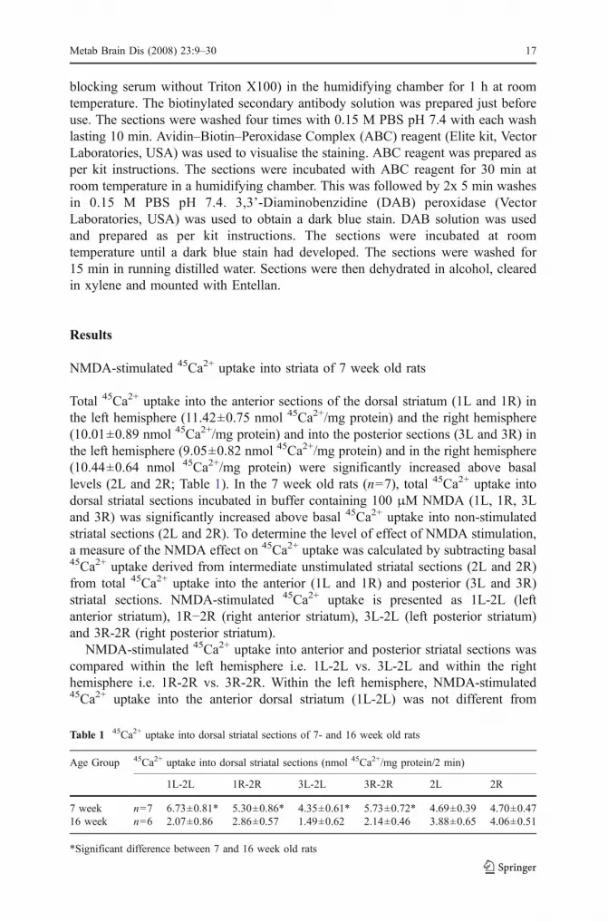
NMDA-stimulated 45Ca2+ uptake into striata of 7 week old rats
Total 45Ca2+ uptake into the anterior sections of the dorsal striatum (1L and 1R) inthe left hemisphere (11.42±0.75 nmol 45Ca2+/mg protein) and the right hemisphere(10.01±0.89 nmol 45Ca2+/mg protein) and into the posterior sections (3L and 3R) inthe left hemisphere (9.05±0.82 nmol 45Ca2+/mg protein) and in the right hemisphere(10.44±0.64 nmol 45Ca2+/mg protein) were significantly increased above basallevels (2L and 2R; Table 1). In the 7 week old rats (n=7), total 45Ca2+ uptake intodorsal striatal sections incubated in buffer containing 100 μM NMDA (1L, 1R, 3Land 3R) was significantly increased above basal 45Ca2+ uptake into non-stimulatedstriatal sections (2L and 2R). To determine the level of effect of NMDA stimulation,a measure of the NMDA effect on 45Ca2+ uptake was calculated by subtracting basal45Ca2+ uptake derived from intermediate unstimulated striatal sections (2L and 2R)from total 45Ca2+ uptake into the anterior (1L and 1R) and posterior (3L and 3R)striatal sections. NMDA-stimulated 45Ca2+ uptake is presented as 1L-2L (leftanterior striatum), 1R−2R (right anterior striatum), 3L-2L (left posterior striatum)and 3R-2R (right posterior striatum).
NMDA-stimulated 45Ca2+ uptake into anterior and posterior striatal sections wascompared within the left hemisphere i.e. 1L-2L vs. 3L-2L and within the righthemisphere i.e. 1R-2R vs. 3R-2R. Within the left hemisphere, NMDA-stimulated45Ca2+ uptake into the anterior dorsal striatum (1L-2L) was not different from
Table 1 45Ca2+ uptake into dorsal striatal sections of 7- and 16 week old rats
Age Group 45Ca2+ uptake into dorsal striatal sections (nmol 45Ca2+/mg protein/2 min)
1L-2L 1R-2R 3L-2L 3R-2R 2L 2R
7 week n=7 6.73±0.81* 5.30±0.86* 4.35±0.61* 5.73±0.72* 4.69±0.39 4.70±0.4716 week n=6 2.07±0.86 2.86±0.57 1.49±0.62 2.14±0.46 3.88±0.65 4.06±0.51
*Significant difference between 7 and 16 week old rats
Metab Brain Dis (2008) 23:9–30 17

stimulated uptake into the posterior dorsal striatum (3L-2L; Table 1). Within theright hemisphere, NMDA-stimulated 45Ca2+ uptake in the anterior dorsal striatalsection (1R-2R) was also not different from the posterior dorsal striata (3R-2R;Table 1). There were therefore no significant differences in NMDA-stimulated 45Ca2+
uptake between anterior and posterior dorsal striata within either the left or the righthemisphere (Table 1). The response to NMDA stimulation was compared across thehemispheres using Bonferroni correction for multiple comparisons. There were nodifferences between NMDA-stimulated 45Ca2+ uptake into the anterior dorsal striatumof the left hemisphere and the anterior dorsal striatum of the right hemisphere i.e. 1L-2L vs. 1R-2R (Table 1). There were also no differences between 45Ca2+ uptake intothe posterior dorsal striata of the left and right hemispheres i.e. 3L-2L vs. 3R-2R(Table 1).
NMDA-stimulated 45Ca2+ uptake into striata of 16 week old rats
Comparison of total 45Ca2+ uptake into dorsal striatal sections (1L, 1R, 3L and 3R)incubated in buffer containing NMDA (100 μM) with non-stimulated striatalsections (2L and 2R) showed that NMDA evoked a significant increase in 45Ca2+
uptake above basal levels into striatal sections obtained from rats aged 16 weeks(Table 1). Measurement of NMDA-stimulated 45Ca2+ uptake (calculated as describedabove) showed that within the left hemisphere, 45Ca2+ uptake into the anteriorstriatal sections (1L-2L) was not different from 45Ca2+ uptake into the posteriorstriatal sections (3L-2L; Table 1). In the right hemisphere there was no differencebetween 45Ca2+ uptake into the anterior striatal section (1R-2R) and 45Ca2+ uptakeinto the posterior striatum (3R-2R; Table 1). Comparison of 45Ca2+ uptake intostriatal sections in the left hemisphere with uptake into striata in the right hemisphereshowed there were no differences in 45Ca2+ uptake between the anterior striata (1L-2L vs. 1R-2R) and posterior striata (3L-2L vs. 3R-2R; Table 1).
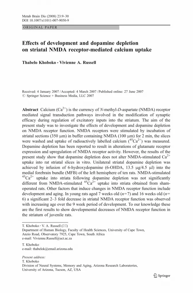
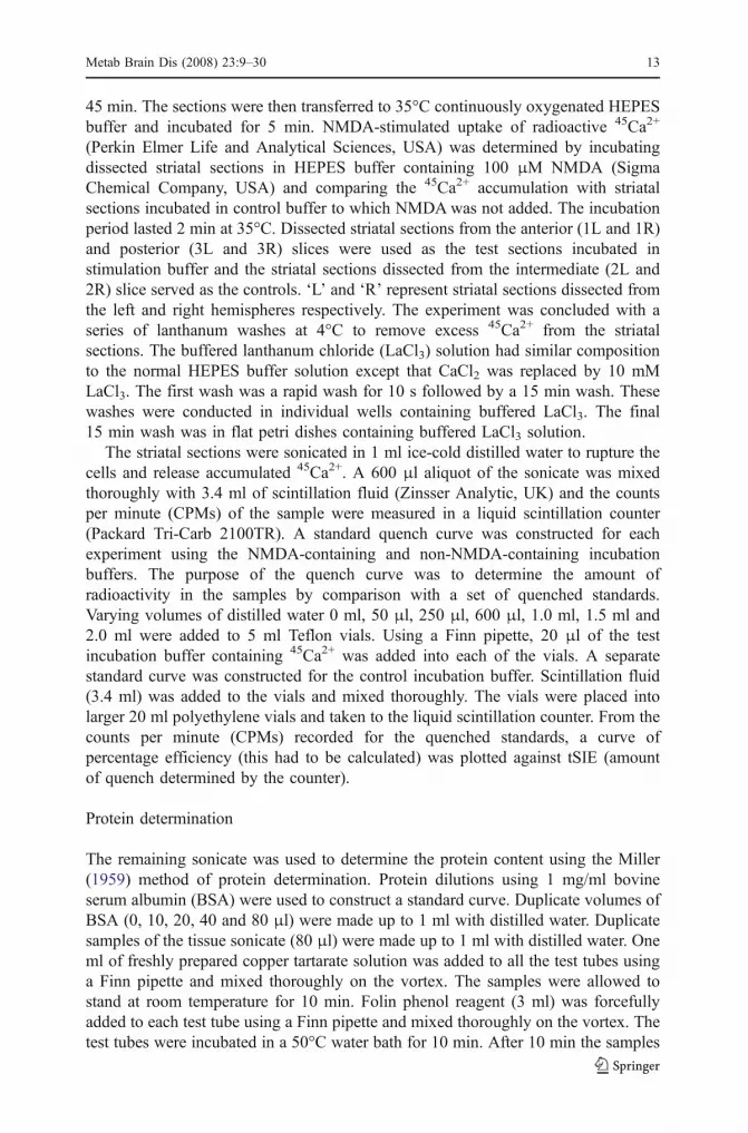
Developmental effects on NMDA-stimulated 45Ca2+ uptake: comparisonof 7 week- vs. 16 week old rats
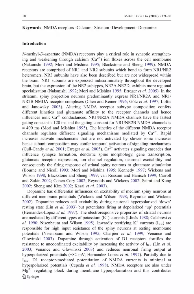
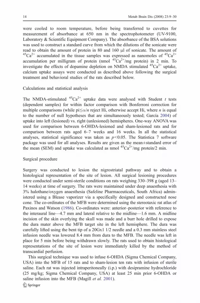
To determine whether there were any age-dependent changes in NMDA-stimulated45Ca2+ uptake into striata, uptake into striata obtained from rats aged 7 weeks (n=7)was compared with uptake into striata obtained from rats aged 16 weeks (n=6). Inboth the left and right hemispheres, stimulated 45Ca2+ uptake into the anterior dorsalstriata (1L-2L and 1R-2R) was significantly greater in 7 week old rats than in16 week old rats (Fig. 1). In the posterior striata (3L-2L and 3R-2R), levels ofstimulated 45Ca2+ accumulation were also significantly higher in 7 week old ratsthan in 16 week old rats (Fig. 1). The results demonstrate that NMDA evokedsignificantly greater 45Ca2+ uptake into dorsal striata of 7 week old rats than 16 weekold rats. In addition to comparing stimulated uptake between the two age groups,basal unstimulated uptake was compared. In the left hemisphere mean uptake of7 week old rats was 4.69±0.39 nmol 45Ca2+/mg protein and 16 week old rats was3.88±0.65 nmol 45Ca2+/mg protein (Fig. 1). In the right hemisphere the uptake forthe 7 week and 16 week old rats was 4.7±0.47 nmol 45Ca2+/mg protein and 4.06±0.51 nmol 45Ca2+/mg protein, respectively (Fig. 1). Comparison of basal uptake into
18 Metab Brain Dis (2008) 23:9–30

the dorsal striatum showed there was no significant difference between the two ages(Fig. 2).
Extent of degeneration of dopamine neurons
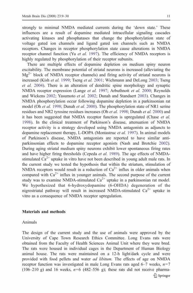
Immunocytochemistry conducted on a sagittal section of the brain stained for thedopamine synthesis-regulating enzyme, tyrosine hydroxylase, highlights the dopa-mine concentrated areas of the basal ganglia. Figure 3 shows sections obtained fromrats that had undergone apomorphine-induced rotations. The sham-lesioned ratconducted 55 ipsilateral rotations and stained positive for tyrosine hydroxylase in themidbrain (SNc/VTA) and in the striatum in both hemispheres (Figs. 3 and 4). 6-OHDA induced degeneration of dopamine producing neurons of the midbrain in theleft lesioned hemisphere, resulted in an absence of tyrosine hydroxylase (Figs. 3 and4). The fully lesioned 6-OHDA-infused rat shown in the figure conducted 381complete contralateral rotations. Tyrosine hydroxylase staining revealed that the 6-OHDA-lesioned rat that had conducted 101 ipsilateral rotations had partialdegeneration of the left midbrain.
The absence of tyrosine hydroxylase staining of the striatum ipsilateral to the siteof injection of 6-OHDA, 5 weeks post surgery confirmed degeneration to have been
1L-2L 1R-2R 3L-2L 3R-2R0
1
2
3
4
5
6
7
8
16 WEEK OLD RATS
7 WEEK OLD RATS
NMDA
stim
ulat
ed45
Ca2+
upt
ake
(nm
ol/m
g pr
otei
n)
**
**
Fig. 1 Effect of age on NMDA-stimulated 45Ca2+ uptake intodorsal striata of 7 (n=7) and16 week old rats (n=6).*Significant difference between7 and 16 week old rats(ANOVA, p<0.05). Results aremean±SEM. Data are reportedin Table 1
2L 2R
1
2
3
4
5
6
16 WEEKS
7 WEEKS
Basa
l45Ca
2+ u
ptak
e (n
mol
/mg
prot
ein)
Fig. 2 Basal 45Ca2+ uptake intothe dorsal striata obtained fromthe left hemisphere (2L) and theright hemisphere (2R) of 16 and7 week old rats. Results aremean±SEM. Data are reportedin Table 1
Metab Brain Dis (2008) 23:9–30 19

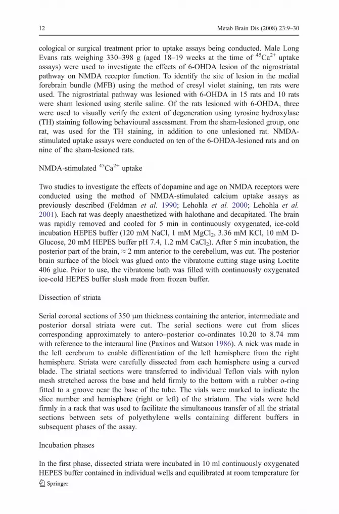
complete within the striatum at the time that the 45Ca2+ uptake assays wereconducted. Figure 4 shows stained coronal sections representative of the anterior (1Land 1R), intermediate (2L and 2R) and posterior (3L and 3R) striata of sham and 6-OHDA-lesioned rats used for the NMDA-stimulated 45Ca2+ uptake assay. In thesham-lesioned rats, tyrosine hydroxylase was intact in both the sham-lesioned leftand the right hemispheres. Following 6-OHDA injection, tyrosine hydroxylase losswas uniform throughout the dorsal striata from anterior to posterior regions in ratsthat had completed >100 contralateral rotations (Fig. 4). Degeneration of neurons inthe midbrain SNc and the VTA (Fig. 3) resulting from infusion of 6-OHDA into theMFB demonstrates retrograde degeneration. Lack of tyrosine hydroxylase staining inthe striatum (Fig. 4) demonstrates anterograde degeneration.
NMDA-stimulated 45Ca2+ uptake into dorsal striatum of sham-lesioned rats
In the sham-lesioned rats, the anterior (1L and 1R) and posterior (3L and 3R) striatalsections incubated in buffer containing NMDA (100 μM) had significantly greater45Ca2+ accumulation above basal 45Ca2+ accumulation in non-stimulated striatalsections (2L and 2R; Table 2). Basal 45Ca2+ uptake into the anterior striatum of theleft hemisphere (2L) was not different from basal 45Ca2+ uptake into the striatum ofthe right hemisphere (2R; Table 2). NMDA (100 μM) significantly increased 45Ca2+
uptake into dorsal striatal sections in sham-lesioned rats. NMDA-stimulated 45Ca2+
uptake was calculated as previously described by subtracting basal uptake from the
Fig. 3 Tyrosine hydroxylase staining in the substantia nigra pars compacta (SNc) and ventral tegmentalarea (VTA) of individual rats. a In the sham-lesioned rat, tyrosine hydroxylase staining is complete in bothhemispheres. The sham-lesioned rat conducted 55 complete ipsilateral rotations in response toapomorphine. b Tyrosine hydroxylase staining in the fully lesioned 6-OHDA-infused rat shows virtuallyno tyrosine hydroxylase in the lesioned left hemisphere (appearing on the right of the image) indicatingnear complete degeneration of the SNc and the VTA. This fully lesioned rat conducted 381 completecontralateral rotations. c An incomplete lesion following 6-OHDA infusion into the left MFB resulted insignificant TH staining in both left and the right hemispheres, although a reduction in TH staining wasobserved in the left hemisphere. This rat conducted 101 complete ipsilateral rotations
20 Metab Brain Dis (2008) 23:9–30

total uptake. Stimulated 45Ca2+ uptake into the anterior striata (1L-2L) and uptakeinto the posterior striata (3L-2L) of the left hemisphere were not significantlydifferent (Table 2). NMDA-stimulated 45Ca2+ uptake into the anterior and posteriorstriatum of the right hemisphere were similar; there was no difference betweenuptake into the anterior (1R-2R) and in the posterior (3R-2R) striatum (Table 2).Analysis of NMDA-stimulated 45Ca2+ uptake into left and right hemispheres showedthat stimulated 45Ca2+ uptake into the anterior striata of the left hemisphere (1L-2L)was not different from uptake into the anterior striata of the right hemisphere (1R-2R; Table 2). Similarly, stimulated 45Ca2+ uptake into the posterior striata (3L-2Land 3R-2R) was not different (Table 2).
NMDA stimulation increases 45Ca2+ uptake into dorsal striatumof 6-OHDA-lesioned rats
Measurement of total 45Ca2+ uptake into dorsal striatal sections of 6-OHDA-lesionedrats (n=10), showed that the total 45Ca2+ accumulation of the anterior (1L and 1R) and
Fig. 4 Representative serial coronal sections of rat brain showing dopamine terminals stained for tyrosinehydroxylase within striata. The serial sections were from sham-lesioned (on the left) and 6-OHDA-lesioned rats (on the right). Lack of tyrosine hydroxylase staining in the left hemisphere of the 6-OHDA-lesioned rats (1L, 2L, 3L) confirms near complete dopamine loss following 6-OHDA injection into theMFB. These sections show that in the 6-OHDA-lesioned rats dopamine loss in the left hemisphere wasuniform from the anterior to the posterior striatum. These sections are representative of the sections of theanterior, intermediate and posterior striata from which the dorsal striata were dissected for NMDA-stimulated 45Ca2+ uptake studies
Metab Brain Dis (2008) 23:9–30 21

posterior (3L and 3R) sections was significantly different (p<0.05) from the basaluptake of the control sections (2L and 2R; Table 2). NMDA (100 μM) stimulated asignificant increase of 45Ca2+ uptake into dorsal striatal sections of 6-OHDA-lesionedrats. Measurements of basal 45Ca2+ uptake into the striata of the left lesionedhemisphere (2L) and the right unlesioned (2R) hemisphere were not significantlydifferent (Table 2). Measurement of stimulated 45Ca2+ uptake was used to determinewhether there were any differences in uptake into anterior and posterior striata withineach hemisphere, and to determine whether there were any differences in uptakebetween the left hemisphere and the right hemisphere. There was no difference instimulated 45Ca2+ uptake into the anterior striata of the left lesioned hemisphere (1L-2L) and the posterior striata (3L-2L) neither was there any difference in stimulated45Ca2+ uptake into the anterior striata of the right hemisphere (1R-2R) and theposterior right striata (3R-2R; Table 2). There were no significant differences betweenNMDA-stimulated 45Ca2+ uptake into the anterior or posterior striata of the left andright hemispheres (Table 2). There was no significant difference between NMDA-stimulated 45Ca2+ uptake into the left 6-OHDA-lesioned hemisphere from stimulated45Ca2+ uptake into the right unlesioned hemisphere.
NMDA-stimulated 45Ca2+ uptake: comparison of 6-OHDA-lesionedvs. sham-lesioned rats
NMDA (100 μM) stimulated 45Ca2+ uptake into dopamine depleted striata of 6-OHDA-lesioned rats was not significantly different from that of sham-lesioned rats.45Ca2+ uptake into anterior (1L-2L and 1R-2R) striata of 6-OHDA-lesioned and sham-lesioned rats was not significantly different, nor was uptake into the posterior (3L-2Land 3R-2R) striata (ANOVA, p>0.05; Fig. 5). Data suggest that dopamine depletioninduced by 6-OHDA lesions did not cause significant changes in 45Ca2+ influx in
Table 2 45Ca2+ uptake into dorsal striatal sections of sham-lesioned and 6-OHDA-lesioned rats
Lesion Group 45Ca2+ uptake into dorsal striatal sections (nmol 45Ca2+/mg protein/2 min)
1L-2L 1R-2R 3L-2L 3R-2R 2L 2R
Sham n=9 3.54±0.66 3.49±0.57 3.12±0.57 4.20±0.93 6.56±0.61 5.89±0.636-OHDA n=10 3.83±0.64 3.90±0.80 4.58±0.69 4.51±0.89 4.92±0.65 4.98±0.55
1L-2L 1R-2R 3L-2L 3R-2R0
1
2
3
4
5
6
6-OHDA LESIONED RATS
SHAM LESIONED RATS
NMDA
stim
ulat
ed45
Ca2+
upt
ake
(nm
ol/m
g pr
otei
n)
Fig. 5 NMDA-stimulated 45Ca2+ uptake into the dorsal striata of6-OHDA-lesioned (n=10) andsham-lesioned rats (n=9).Results are mean±SEM. Dataare reported in Table 2
22 Metab Brain Dis (2008) 23:9–30
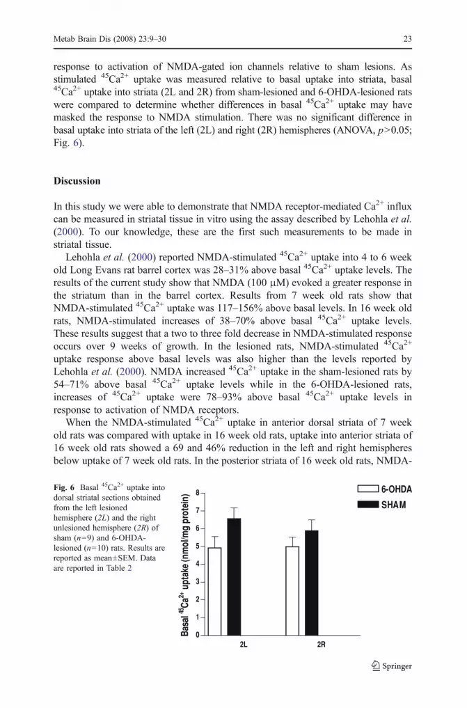
response to activation of NMDA-gated ion channels relative to sham lesions. Asstimulated 45Ca2+ uptake was measured relative to basal uptake into striata, basal45Ca2+ uptake into striata (2L and 2R) from sham-lesioned and 6-OHDA-lesioned ratswere compared to determine whether differences in basal 45Ca2+ uptake may havemasked the response to NMDA stimulation. There was no significant difference inbasal uptake into striata of the left (2L) and right (2R) hemispheres (ANOVA, p>0.05;Fig. 6).
Discussion
In this study we were able to demonstrate that NMDA receptor-mediated Ca2+ influxcan be measured in striatal tissue in vitro using the assay described by Lehohla et al.(2000). To our knowledge, these are the first such measurements to be made instriatal tissue.
Lehohla et al. (2000) reported NMDA-stimulated 45Ca2+ uptake into 4 to 6 weekold Long Evans rat barrel cortex was 28–31% above basal 45Ca2+ uptake levels. Theresults of the current study show that NMDA (100 μM) evoked a greater response inthe striatum than in the barrel cortex. Results from 7 week old rats show thatNMDA-stimulated 45Ca2+ uptake was 117–156% above basal levels. In 16 week oldrats, NMDA-stimulated increases of 38–70% above basal 45Ca2+ uptake levels.These results suggest that a two to three fold decrease in NMDA-stimulated responseoccurs over 9 weeks of growth. In the lesioned rats, NMDA-stimulated 45Ca2+
uptake response above basal levels was also higher than the levels reported byLehohla et al. (2000). NMDA increased 45Ca2+ uptake in the sham-lesioned rats by54–71% above basal 45Ca2+ uptake levels while in the 6-OHDA-lesioned rats,increases of 45Ca2+ uptake were 78–93% above basal 45Ca2+ uptake levels inresponse to activation of NMDA receptors.
When the NMDA-stimulated 45Ca2+ uptake in anterior dorsal striata of 7 weekold rats was compared with uptake in 16 week old rats, uptake into anterior striata of16 week old rats showed a 69 and 46% reduction in the left and right hemispheresbelow uptake of 7 week old rats. In the posterior striata of 16 week old rats, NMDA-
2L 2R0
1
2
3
4
5
6
7
8 6-OHDA
SHAM
Basa
l45Ca
2+ u
ptak
e (n
mol
/mg
prot
ein)
Fig. 6 Basal 45Ca2+ uptake intodorsal striatal sections obtainedfrom the left lesionedhemisphere (2L) and the rightunlesioned hemisphere (2R) ofsham (n=9) and 6-OHDA-lesioned (n=10) rats. Results arereported as mean±SEM. Dataare reported in Table 2
Metab Brain Dis (2008) 23:9–30 23

stimulated 45Ca2+ uptake was decreased 66 and 63% in the left and right striatabelow NMDA-stimulated 45Ca2+ uptake of 7 week old rats. These results show asignificant decrease in stimulated uptake of 16 week old rats compared to 7 week oldrats and suggests that during 9 weeks of growth from juvenile to young adultdevelopment, there is an average decrease of ≈ 60% in dorsal striatal NMDAmediated Ca2+ influx.
The above results showing significant reduction in NMDA receptor mediated45Ca2+ influx with increasing age from 7 to 16 weeks concur with reported agedependent attenuation of NMDA receptor activity attributed to reduction of NMDAreceptor density occurring in rats aged 3–31 months and in Rhesus monkeys(Mitchell and Anderson 1998; Wenk and Barnes 2000; Bai et al. 2004). There aremajor changes of dendritic spines that occur, including shrinkage and loss and theseundoubtedly contribute to the reduction of NMDA receptor density (Levine et al.1988; Dunia et al. 1996; Segovia et al. 2001).
Basal uptake was compared to determine whether differences in NMDA-stimulated 45Ca2+ uptake between 7 and 16 week old rats could be a factorinfluencing the levels of 45Ca2+ influx. There was no significant difference in basal45Ca2+ uptake into dorsal striata between adolescent 7 week old rats and young adult16 week old rats (Fig. 1). These results are consistent with findings reported byGriffith et al. (2000) that intracellular Ca2+ concentrations are unchanged and thatCa2+ homeostasis is not compromised with age.
In the current study the 6-OHDA model of Parkinson’s disease was established byconducting histological mappings of the needle tip positions around the MFB and byconducting behavioural assessments of the lesion followed by immunocytochemis-try. In 10 sham-lesioned rats and 15 6-OHDA-lesioned rats, apomorphine inducedrotations were used to assess the extent of dopamine depletion. Sham-lesioned rats inthe current study conducted 19 8 rotations ipsilateral to the lesion. The mean numberof rotations conducted by 6-OHDA-lesioned rats was 233±43 complete contralateralrotations. This is consistent with Hudson et al. (1993) who showed thatapomorphine injection did not produce a significant number of rotations in non-lesioned rats. Moore et al. (2001) found that a contralateral rotational bias inresponse to apomorphine only occurred when >70–80% degeneration of midbraindopamine neurons and striatal dopamine fibres had occurred in rats infused with 6-OHDA into the MFB. This suggests that in the current study, >70% degenerationwas achieved in 6-OHDA-lesioned rats selected for NMDA-stimulated 45Ca2+
uptake assays.Tyrosine hydroxylase staining of the midbrain and the striatum confirmed that
degeneration, shown by TH staining, was minimal in sham-lesioned rats; there wascomplete staining in the striatum and midbrain of both hemispheres. In a 6-OHDA-lesioned rat that had conducted 381 contralateral rotations, there was near completeloss of TH ipsilateral to the site of 6-OHDA injection. Immunocytochemistry wasconducted on a 6-OHDA-lesioned rat that had exhibited 101 ipsilateral rotations inresponse to apomorphine. There was a reduced TH in the midbrain and in the medialdorsal and medial ventral striata but the reduction was not as extensive as thedegeneration observed in 6-OHDA rats conducting >100 contralateral rotations.These results confirm the findings by Carman et al. (1991) that the number of THimmunoreactive neurons that remain unlesioned is correlated to the number of
24 Metab Brain Dis (2008) 23:9–30

contralateral rotations conducted in response to apomorphine; the fewer the numberof degenerated neurons the fewer the number of contralateral rotations. Apomor-phine induced rotations are a useful predictor of the extent of midbrain degenerationof dopamine neurons and dopamine terminals in the striatum (Carman et al. 1991).The results of TH immunocytochemistry obtained from the serial sections confirmedthat 5 weeks post surgery, there was near complete depletion of dopamine neurons inthe entire striatum of the lesioned left hemisphere in 6-OHDA-lesioned rats selectedfor NMDA-stimulated 45Ca2+ uptake assay and that in selected sham-lesioned rats,dopamine innervation remained intact within both hemispheres.
Having confirmed through TH immunoreactivity that there was near completedepletion of dopamine in the left striatum, ipsilateral to the 6-OHDA lesion, it wasexpected that there would be increases in NMDA receptor mediated 45Ca2+ influx inthe left striatum relative to NMDA-stimulated 45Ca2+ uptake in the striatum of theright hemisphere that had intact TH immunoreactivity. NMDA-stimulated 45Ca2+
uptake into the anterior striatum of the left hemisphere was 2% less than NMDA-stimulated 45Ca2+ uptake into the anterior striatum of the right hemisphere whereasin the posterior striata NMDA-stimulated 45Ca2+ uptake into the left lesionedhemisphere was 2% more than uptake into the right hemisphere. These differences inNMDA-stimulated 45Ca2+ uptake between striata of the 6-OHDA-lesioned lefthemisphere and the unlesioned right hemisphere were marginal and not significant.These results from 6-OHDA-lesioned rats were similar to the results of NMDA-stimulated 45Ca2+ uptake measured in sham-lesioned rats. In the posterior striataNMDA-stimulated 45Ca2+ uptake levels in the right hemisphere were 17% higherthan NMDA-stimulated 45Ca2+ uptake levels in posterior striatum of the lefthemisphere. There was no significant difference in NMDA-stimulated 45Ca2+ uptakebetween the sham operated left hemisphere and the unlesioned right hemisphere.These results suggest that mechanical stress induced by the surgical procedure didnot disturb NMDA mediated 45Ca2+ influx.
NMDA-stimulated 45Ca2+ uptake into the anterior dorsal striata of 6-OHDA-lesioned rats showed increases of 8% and 11% in the left and right hemispheresrespectively above NMDA-stimulated 45Ca2+ uptake in sham-lesioned rats. In theposterior striata of 6-OHDA-lesioned rats, NMDA-stimulated 45Ca2+ uptake in theleft and right hemispheres showed 32 and 7% increases respectively above NMDA-stimulated 45Ca2+ uptake levels of sham-lesioned rats. There was no significantdifference in NMDA-stimulated 45Ca2+ uptake between 6-OHDA-lesioned andsham-lesioned rats. It is of interest that while the measurement of the left posteriorstriatum was the highest measurement in the 6-OHDA-lesioned rats, themeasurement from the same striatal section was the lowest measurement in sham-lesioned rats. While these findings could suggest that NMDA receptor function inthe posterior striata of the lesioned hemisphere was attenuated in response to shamoperations and enhanced in 6-OHDA-lesioned rats, this does not seem likely as therewere no significant differences between the left lesioned and the right unlesionedhemispheres in either sham-lesioned or 6-OHDA-lesioned rats.
To determine whether changes in basal uptake may have masked upregulation ofNMDA receptor mediated 45Ca2+ influx in dopamine depleted animals, basal uptakeinto striata of sham-lesioned and 6-OHDA-lesioned rats was analyzed. Althoughthere was a tendency for basal 45Ca2+ uptake to be lower in striatal sections obtained
Metab Brain Dis (2008) 23:9–30 25

from 6-OHDA-lesioned rats compared to sham-lesioned rats, there was nosignificant difference between the two groups. Sham operated rats had 25%greater uptake in the left hemisphere and 15% greater basal uptake in the righthemisphere than 6-OHDA-lesioned rats. Our investigations examining thedifferences in NMDA receptor function in the dopamine depleted striatum do notsupport the original hypothesis that reported upregulation of NMDA receptors wouldtranslate into increased ionic flux of 45Ca2+ in response to NMDA stimulation.
A factor that could have masked any upregulation of NMDA receptor functionmay have been the morphological changes that occur i.e. dendritic spine atrophyfollowing dopamine depletion (Ingham et al. 1998; Meshul et al. 1999; Arbuthnottet al. 2000). This would influence the number of functional NMDA receptorsstimulated in the assay. The morphological changes could be attributed to increasedCa2+ influx which causes a reduction in the binding affinity of NMDA receptorsubunits for crosslinking structural proteins resulting in depolymerisation ofcytoskeletal proteins (Krupp et al. 1999; van Rossum and Hanisch 1999). As aresult, despite their necessary role in synaptic function and modification, Ca2+
influxes are tightly regulated in neurons as they can also induce neuropathologicaldisturbances that cause cell death (Hardingham and Bading 2003). One couldspeculate that to counter possible cytoskeletal collapse following dopaminedepletion, homeostatic mechanisms in surviving spines would be increased inresponse to increased NMDA receptor mediated Ca2+ influx. Ca2+ influx issubstantially influenced by homeostatic regulation of intracellular Ca2+ levels thatoccurs to counter changes in neuronal excitability (Yeung et al. 2004). Increases ofthe membrane potential of the medium spiny neurons that follow dopaminedepletion would also increase NMDA receptor mediated influx of Ca2+ ions (asthe Mg2+ block is removed); in addition the opening of voltage gated Ca2+ channelswould increase Ca2+ intracellular levels (Tseng et al. 2001). It would be necessary tostudy whether the activity of Ca2+-ATPase is enhanced following dopaminedepletion in the striatum to understand some of the mechanisms involved inregulating levels of intracellular Ca2+.
The issue of sensitivity of the NMDA-stimulated 45Ca2+ uptake assay wasconsidered particularly as NMDA receptor mediated currents are maximal atdepolarised potentials and with glycine co-stimulation; two conditions not includedin the assay. To induce membrane depolarization in the study would have skewedour measurements of the NMDA mediated 45Ca2+ uptake response as a consequenceof the invariable activation of other channels including L-type voltage activated Ca2+
channels. Though as mentioned above, previous studies have found that themembrane potential of spiny neurons does become less negative in response todopamine loss and this was not a variable we could control easily in our preparation.Co-stimulation of NMDA receptors with glycine increases the open probability ofreceptor channels hence the number of open NMDA receptors and this influences theamplitude of Ca2+ currents (Jahr 1992). Lehohla et al. (2000) in developing the assayfound that addition of 1 mM glycine to the assay did not result in a significantdifference in NMDA-stimulated 45Ca2+ uptake into the barrel cortex and it wasdeemed unnecessary to add glycine to the assays used in the current study. A furtherfactor that may have influenced activation of the NMDA receptors is the use ofhalothane anaesthetic prior to killing the rats for the assay. Halothane reduces the
26 Metab Brain Dis (2008) 23:9–30
amplitude of NMDA receptor mediated excitatory postsynaptic potentials (Nishikawaand Maclver 2000). Finally, the conclusive results of the effects of age on NMDA-stimulated 45Ca2+ uptake serve as a strong indication that the assay was indeedsufficiently sensitive to measure NMDA mediated Ca2+ influx into the striatum.
Conclusions
In the current study we set out to determine whether aging would result in decreasedNMDA-stimulated 45Ca2+ uptake into the striatum and whether dopamine depletioninduced by degeneration of the nigrostriatal pathway increases NMDA-stimulated45Ca2+ uptake. We were able to demonstrate that 9 weeks of growth fromadolescence to young adulthood in male Long Evans rats, results in reduction ofNMDA receptor mediated 45Ca2+ influx of up to 69%. These results support thefindings that striatal glutamate innervation and NMDA receptor responsivenesshence neuronal excitability are reduced with age (Cepeda et al. 1989; Cepeda et al.1996; Segovia et al. 2001). To our knowledge, this is the first study to demonstratesuch a significant reduction of NMDA receptor function in the dorsal striatum usingthe in vitro NMDA-stimulated 45Ca2+ uptake assay.
The second hypothesis we tested was that degeneration of the nigrostriatalpathway would result in increased NMDA-stimulated 45Ca2+ uptake. The resultsfrom the study did not concur with this stated hypothesis. Instead we found that therewere no differences in NMDA-stimulated 45Ca2+ uptake between 6-OHDA-lesionedand sham-lesioned rats and neither were there any differences in NMDA-stimulated45Ca2+ uptake within the 6-OHDA-lesioned rats comparing the striatal 45Ca2+ uptakefrom the lesioned and the unlesioned hemispheres. These results rather suggest thatreported modifications of striatal expression and phosphorylation state of NMDAreceptor subunits in parkinsonian rats do not necessarily lead to increased NMDAreceptor-mediated Ca2+ influx into striatal slices in vitro.
Acknowledgements This work was supported by the Medical Research Council of South Africa and theUniversity of Cape Town. Thabelo Khoboko was supported by the Levi-Montalcini Fellowship inNeuroscience for African Women awarded by the International Brain Research Organization (IBRO).
References
Arbuthnott GW, Ingham CA, Wickens JR (2000) Dopamine and synaptic plasticity in the neostriatum.J Anat 196(4):587–596
Bai L, Hof PR, Standaert DG, Xing Y, Nelson SE, Young AB, Magnusson KR (2004) Changes in theexpression of the NR2B subunit during aging in macaque monkeys. Neurobiol Aging 25(2):201–208
Blackstone C, Sheng M (1999) Protein targeting and calcium signalling microdomains in neuronal cells.Cell Calcium 26(5):181–192
Bourne HR, Nicoll R (1993) Molecular machines integrate coincident synaptic signals. Neuron 10(Suppl):65–75
Calabresi P, Mercuri NB, Bernardi G (1990) Synaptic and intrinsic control of membrane excitability ofneostriatal neurons. II. An in vitro analysis. J Neurophysiol 63(4):663–675
Carman LS, Gage FH, Shults CW (1991) Partial lesion of the substantia nigra: relation between extent oflesion and rotational behavior. Brain Res 553(2):275–283
Carroll RC, Zukin RS (2002) NMDA receptor trafficking and targeting: implications for synaptictransmission and plasticity. Trends Neurosci 25(11):571–577
Metab Brain Dis (2008) 23:9–30 27

Cepeda C, Walsh JP, Hull CD, Buchwald NA, Levine MS (1989) Intracellular neurophysiological analysisreveals alterations in excitation in striatal neurons in aged rats. Brain Res 494(4):215–226
Cepeda C, Li Z, Levine MS (1996) Aging reduces neostriatal responsiveness to N-methyl-D-aspartate anddopamine: an in vitro electrophysiological study. Neuroscience 73(3):733–750
Cepeda C, Colwell CS, Itri JN, Chandler SH, Levine MS (1998) Dopaminergic modulation of NMDA-induced whole cell currents in neostriatal neurons in slices: contribution of calcium conductances.J Neurophysiol 79:82–84
Charpier S, Mahon, S, Deniau JM (1999) In vivo induction of striatal long-term potentiation by low-frequency stimulation of the cerebral cortex. Neuroscience 91(4):1209–1222
Chase TN, Oh JD, Blanchet PJ (1998) Neostriatal mechanisms in Parkinson’s disease. Neurology 51(Suppl 2):S30–S35
Chen Q, Reiner A (1996) Cellular distribution of the NMDA receptor NR2A/2B subunits in the ratstriatum. Brain Res 743(1–2):346–352
Cohen-Cory S (2002) The developing synapse: construction and modulation of synaptic structures andcircuits. Science 298:770–776
Cull-Candy S, Brickley S, Farrant M (2001) NMDA receptor subunits: diversity, development anddisease. Curr Opin Neurobiol 11(3):327–335
Dunah AW, Standaert DG (2003) Subcellular segregation of distinct heteromeric NMDA glutamatereceptors in the striatum. J Neurochem 85(4):935–943
Dunah AW, Wang Y, Yasuda RP, Kameyama K, Huganir RL, Wolfe BB, Standaert DG (2000) Alterationsin subunit expression, composition, and phosphorylation of striatal N-methyl-D-aspartate glutamatereceptors in a rat 6-hydroxydopamine model of Parkinson’s disease. Mol Pharmacol 57(2):342–352
Dunia R, Buckwalter G, Defazio T, Villar FD, McNeill TH, Walsh JP (1996) Decreased duration of Ca2+-mediated plateau potentials in striatal neurons from aged rats. J Neurophysiol 76(4):2353–2363
Erreger K, Dravid SM, Banke TG, Wyllie DJ, Traynelis SF (2005) Subunit-specific gating controls ratNR1/NR2A and NR1/NR2B NMDA channel kinetics and synaptic signalling profiles. J Physiol 563(2):345–358
Feldman D, Sherin JE, Press WA, Bear MF (1990) N-methyl-D-aspartate-evoked calcium uptake by kittenvisual cortex maintained in vitro. Exp Brain Res 80:252–259
Garcia LV (2004) Escaping the Bonferroni iron claw in ecological studies. OIKOS 105(3):657–663Götz T, Kraushaar U, Geiger J, Lubke J, Berger T, Jonas P (1997) Functional properties of AMPA and
NMDA receptors expressed in identified types of basal ganglia neurons. J Neurosci 17(1):204–215Griffith WH, Jasek MC, Bain SH, Murchison D (2000) Modification of ion channels and calcium
homeostasis of basal forebrain neuron during aging. Behav Brain Res 115:219–233Hardingham GE, Bading H (2003) The Yin and Yang of NMDA receptor signalling. Trends Neurosci 26
(2):81–89Hernández-López S, Bargas J, Surmeier DJ, Reyes A, Galarraga E (1997) D1 receptor activation enhances
evoked discharge in neostriatal medium spiny neurons by modulating an L-type Ca2+ conductance.J Neurosci 17(9):3334–3342
Hudson JL, van Horne CG, Stromberg I, Brock S, Clayton J, Masserano J, Hoffer BJ, Gerhardt GA (1993)Correlation of apomorphine- and amphetamine-induced turning with nigrostriatal dopamine content inunilateral 6-hydroxydopamine lesioned rats. Brain Res 626(1–2):167–174
Ingham CA, Hood SH, Taggart P, Arbuthnott GW (1998) Plasticity of synapses in the rat neostriatum afterunilateral lesion of the nigrostriatal dopaminergic pathway. J Neurosci 18(12):4732–4743
Jahr CE (1992) High probability opening of NMDA receptor channels by L-glutamate. Science 255:470–472
Kasai H, Matsuzaki M, Noguchi J, Yasumatsu N, Nakahara H (2003) Structure–stability–functionrelationships of dendritic spines. Trends Neurosci 26(7):360–368
Kennedy MB (1997) The postsynaptic density at glutamatergic synapses. Trends Neurosci 20(6):264–268Kish LJ, Palmer MR, Gerhardt GA (1999) Multiple single-unit recordings in the striatum of freely moving
animals: effects of apomorphine and D-amphetamine in normal and unilateral 6-hydroxydopamine-lesioned rats. Brain Res 833(1):58–70
Krupp JJ, Vissel B, Thomas CG, Heinemann SF, Westbrook GL (1999) Interactions of calmodulin andalpha-actinin with the NR1 subunit modulate Ca2+−dependent inactivation of NMDA receptors.J Neurosci 19(4):1165–1178
Lange KW, Kornhuber J, Riederer P (1997) Dopamine/glutamate interactions in Parkinson’s disease.Neurosci Biobehav Rev 21(4):393–400
Lehohla M, Russell V, Kellaway L, Govender A (2000) Development of a method to evaluate glutamatereceptor function in rat barrel cortex slices. Metab Brain Dis 15(4):305–314
28 Metab Brain Dis (2008) 23:9–30

Lehohla M, Russell V, Kellaway L (2001) NMDA-stimulated Ca2+ uptake into barrel cortex slices ofspontaneously hypertensive rats. Metab Brain Dis 16(3/4):135–143
Levine MS, Adinolfi AM, Fisher RS, Hull CD, Guthrie D, Buchwald NA (1988) Ultrastructural alterationsin caudate nucleus in aged cats. Brain Res 440(2):267–279
Lin JY, Dubey R, Funk GD, Lipski J (2003) Receptor subtype-specific modulation by dopamine ofglutamatergic responses in striatal medium spiny neurons. Brain Res 959:251–262
Llinás RR (1988) The intrinsic electrophysiological properties of mammalian neurons: insights into centralnervous system function. Science 242:1654–1655
Loftis JM, Janowsky A (2003) The N-methyl-D-aspartate receptor subunit NR2B: localization, functionalproperties, regulation, and clinical implications. Pharmacol Ther 97(1):55–85
Magill PJ, Bolam JP, Bevan MD (2001) Dopamine regulates the impact of cerebral cortex on thesubthalamic nucleus-globus pallidus network. Neuroscience 106(2):313–330
Meshul CK, Emre N, Nakamura CM, Allen C, Donohue MK, Buckman JF (1999) Time-dependentchanges in striatal glutamate synapses following a 6-hydroxydopamine lesion. Neuroscience 88(1):1–16
Miller GL (1959) Protein determination for large number of samples. Anal Chem 31:964Mitchell JJ, Anderson KJ (1998) Age-related changes in [3H]MK-801 binding in the Fischer 344 rat brain.
Neurobiol Aging 19(3):259–265Montastruc JL, Rascol O, Senard JM (1997) Glutamate antagonists and Parkinson’s disease: a review of
clinical data. Neurosci Biobehav Rev 21(4):477–480Moore AE, Cicchetti F, Hennen J, Isacson O (2001) Parkinsonian motor deficits are reflected by
proportional A9/A10 dopamine neuron degeneration in the rat. Exp Neurol 172(2):363–376Mori H, Mishina M (1995) Structure and function of the NMDA receptor channel. Neuropharmacology 34
(10):1219–1237Nakanishi S (1992) Molecular diversity of glutamate receptors and implications for brain function.
Science 258:597–603Nash JE, Brotchie JM (2002) Characterisation of striatal NMDA receptors involved in the generation of
parkinsonian symptoms: intrastriatal microinjection studies in the 6-OHDA-lesioned rat. Mov Disord17(3):455–466
Nisenbaum ES, Wilson CJ (1995) Potassium currents responsible for inward and outward rectification inrat neostriatal spiny projection neurons. J Neurosci 15(6):4449–4463
Nishikawa K, Maclver MB (2000) Excitatory synaptic transmission mediated by NMDA receptors is moresensitive to isoflurane than are non-NMDA receptor mediated responses. Anethesiology 92(1):228–236
Oh JD, Russell DS, Vaughan CL, Chase TN (1998) Enhanced tyrosine phosphorylation of striatal NMDAreceptor subunits: effect of dopaminergic denervation and L-DOPA administration. Brain Res 813(1):150–159
Paxinos G, Watson C (1986) The rat brain in stereotaxic coordinates, 2nd edn. Academic, San Diego, CAReynolds JN, Wickens JR (2002) Dopamine-dependent plasticity of corticostriatal synapses. Neural Netw
15(4–6):507–521Saimi Y, Kung C (2002) Calmodulin as an ion channel subunit. Annu Rev Physiol 64:289–311Segovia G, Porras A, Arco AD, Mora F (2001) Glutamatergic neurotransmission in aging: a critical
perspective. Mech Ageing Dev 122:1–29Sheng M, Kim MJ (2002) Postsynaptic signaling and plasticity mechanisms. Science 298:776–780Tillerson JL, Cohen AD, Philhower J, Miller GW, Zigmod MJ, Schallert T (2001) Forced limb-use effects
on the behavioral and neurochemical effects of 6-hydroxydopamine. J Neurosci 21(12):4427–4435Tseng KY, Kasanetz F, Kargieman L, Riquelme LA, Murer MG (2001) Cortical slow oscillatory activity is
reflected in the membrane potential and spike trains of striatal neurons in rats with chronicnigrostriatal lesions. J Neurosci 21(16):6430–6439
Tseng KY, Riquelme LA, Murer MG (2004) Impact of D1-class dopamine receptor on striatal processingof cortical input in experimental parkinsonism in vivo. Neuroscience 123(2):238–293
Ungerstedt U (1971) Postsynaptic supersensitivity after 6-hydroxy-dopamine induced degeneration of thenigro-striatal dopamine system. Acta Physiol Scand Suppl 367:69–93
Ungerstedt U, Arbuthnott GW (1970) Quantitaive recording of rotational behavior in rats after 6-hydroxy-dopamine lesions of the nigrostriatal dopamine system. Brain Res 24(3):485–493
van Rossum D, Hanisch U-K (1999) Cytoskeletal dynamics in dendritic spines: direct modulation byglutamate receptors? Trends Neurosci 22(7):290–295
Venance L, Glowinski J (2003) Heterogeneity of spike frequency adaptation among medium spinyneurons from the rat striatum. Neuroscience 122:77–92
Metab Brain Dis (2008) 23:9–30 29

Wenk GL, Barnes CA (2000) Regional changes in the hippocampal density of AMPA and NMDAreceptors across the lifespan of the rat. Brain Res 885:1–5
Wichmann T, DeLong MR (2003) Pathophysiology of Parkinson’s disease: the MPTP primate model ofthe human disorder. Ann NY Acad Sci 991:199–213
Wickens JR, Wilson CJ (1998) Regulation of action potential firing in spiny neurons of the rat neostriatumin vivo. J Neurophysiol 79:2358–2364
Yasumoto S, Tanaka E, Hattori G, Maeda H, Higashi H (2002) Direct and indirect actions of dopamine onthe membrane potential in medium spiny neurons of the mouse neostriatum. J Neurophysiology87:1234–1243
Yeung LC, Shouval HZ, Blais BS, Cooper LN (2004) Synaptic homeostasis and input selectivity followfrom a calcium-dependent plasticity model. Proceedings of the National Academy of Sciences USA101(41):14943–14948
Yu X, Askalan R, Keil GJ, Salter MW (1997) NMDA channel regulation by channels-associated proteintyrosine kinase Src. Science 275:674–678
30 Metab Brain Dis (2008) 23:9–30