characteristics and mechanisms of norepinephrine depletion
TRANSCRIPT

Loyola University Chicago Loyola University Chicago
Loyola eCommons Loyola eCommons
Dissertations Theses and Dissertations
1982
Characteristics and Mechanisms of Norepinephrine Depletion Characteristics and Mechanisms of Norepinephrine Depletion
from the Myocardium and Spleen of Endotoxic Rats from the Myocardium and Spleen of Endotoxic Rats
Benet J. Pardini Loyola University Chicago
Follow this and additional works at: https://ecommons.luc.edu/luc_diss
Part of the Physiology Commons
Recommended Citation Recommended Citation Pardini, Benet J., "Characteristics and Mechanisms of Norepinephrine Depletion from the Myocardium and Spleen of Endotoxic Rats" (1982). Dissertations. 2166. https://ecommons.luc.edu/luc_diss/2166
This Dissertation is brought to you for free and open access by the Theses and Dissertations at Loyola eCommons. It has been accepted for inclusion in Dissertations by an authorized administrator of Loyola eCommons. For more information, please contact [email protected].
This work is licensed under a Creative Commons Attribution-Noncommercial-No Derivative Works 3.0 License. Copyright © 1982 Benet J. Pardini

CHARACTERISTICS AND MECHANISMS OF NOREPINEPHRINE DEPLETION
FROM THE MYOCARDIUM AND SPLEEN
OF ENDOTOXIC RATS
by
Benet J. Pardini
A Dissertation Submitted to the Faculty of the Graduate School
of Loyola University of Chicago in Partial Fulfillment
of the Requirements for the Degree of
Doctor of Philosophy
May
1982
LIBRARY LOYOLA UNIVER3iTY MEDICAL CENI1ER

ACKNOWLEDGMENTS
A debt of gratitude is extended to the members of the committee
for their willingness to participate i.n this venture and for their
suggestions and criticisms of this work. Many thanks to Dr. John x.
Thomas, Jr. for his
gratitude is due my
sharing his guidance,
assistance and the use of his computer. Special
advisor and good friend, Dr. Stephen Jones, for
patience, and half of his office with me
throughout this endeavor.
ii

VITA
Benet J. Pardini is the son of Benedict and Mary Pardini. He
was born on August 26, 1954, in Evanston, Illinois.
He attended elementary and secondary schools in suburban
Northbrook and was graduated from Glenbrook North High School in 1972.
Undergraduate studies were carried out at Northwestern University in
Evanston, Illinois. Benet earned a Bachelor of Arts degree in Biology
and was graduated from Northwestern University in June of 1976.
After a year of graduate studies in biology at Northwestern
University, the author began doctoral training in the Department of
Physiology at Loyola University Medical Center. For the past four years
he worked under the direction of Dr. Stephen B. Jones. He was the
recipient of a University Dissertation Fellowship from Loyola University
in 1980, and is presently a student member of the American Physiological
Society. Benet received the First Place Award in the Young
Investigator's Competition of the Shock Society in 1981, for a portion
of his doctoral research.
iii

PUBLICATIONS
1. PARDINI, B. J., S. B. Jones, and J. P. Filkins. Myocardial norepinephrine depletion in endotoxin shock: role of hypoglycemia and neural mediation. The Physiologist 22(4):98, 1979.
2. PARDINI, B. J., S. B. Jones, and J. norepinephrine (NE) depletion in Physiologist 23(4):36, 1980.
P. Filkins. Mechanisms of endotoxin shock. The
3. Jones, s. B. and B. J. PARDINI. Cardiovascular tolerance to severe hypoxia in hibernators vs. non-hibernators. The Physiologist 23(4):153, 1980.
4. PARDINI, B. J., S. B. Jones, and J. P. Filkins. Role of depressed reuptake in splenic and myocardial norepinephrine depletion during endotoxin shock. Circ. Shock 8(2):202, 1981.
5. PARDINI, B. J., S. B. Jones, and turnover in heart and spleen Physiologist 24(4):116, 1981.
J. P. Filkins. Norepinephrine during endotoxicosis. The
6. PARDINI, B. depressed heart and in press.
J., S. B. Jones, and J. P. Filkins. Contribution of reuptake to the depletion of norepinephrine from rat spleen during endotoxin shock. Circ. Shock 9(2), 1982,
7. Sayeed, M. M., D. M. Klein, and B. J. PARDINI. Effects of diltiazem on endotoxicosis in the rat. Fed. Proc. 41(5):1607, 1982.
8. PARDINI, B. J., S. B. Jones, and J. P. Filkins. Cardiac and splenic norepinephrine turnovers in the endotoxic rat. submitted, Am. ~· Physiol., 1982.
iv

TABLE OF CONTENTS
Page
ACKNOWLEDGMENTS . . . . . . . . . . . ii
VITA • • • • • • • • • • iii
TABLE OF CONTENTS
LIST OF TABLES . . . . . . . . . . . . . . . . . . .
. . . . . . . . v
vii
LIST OF ILLUSTRATIONS . . . . . . . • viii
Chapter
I. INTRODUCTION . . . . . . . . . . . . II. REVIEW OF RELATED LITERATURE . . . . . . . . 4
A. Evidence for Sympathetic Activation in Endotoxin Shock • • • • • • • • • • • • • • • • • • 4 1. Elevation of plasma catecholamines • • • 4 2. Depletion of tissue catecholamines • • • 10 3. Radioisotopic studies • • • • • • • • 15 4. Electrophysiological studies • • • • 19
B. Evidence for the "Pathogenetic" Properties of the Sympathetic Nervous System in Endotoxin Shock 21
c. Tissue Norepinephrine Levels -Physiological and Pathological Processes 28 1. Maintenance of tissue norepinephrine levels
under normal conditions • • • • • • • 30 2. Norepinephrine depletion during hemorrhagic
shock • • • • • • • • • • • • • • • 36 a. demonstration of the phenomenon 36 b. mechanisms of hemorrhage-induced
norepinephrine depletion • • • • • • • • 41 c. absence of norepinephrine
depletion during hemorrhage • • • • • • 43 3. Mechanisms of norepinephrine depletion
in endotoxin shock • • • • • • 45
III. RATIONALE AND SPECIFIC AIMS . . . . 53
59 IV. METHODS
A. B. c.
Animals and Care Shock Model Plasma Glucose Measurement
v
. . . . . . . . . . . . .
. . . . . . . . . . . . . . . . . . . . . . . . . . . . . . . . . . . .
59 60 61

D.
E. F.
G. H.
Tissue Catecholamine Measurement • • • • • • • • • • 1. Tissue sampling and storage • • • ••• 2. Catecholamine purification ••••••••• 3. Catecholamine analysis ••••••••••••• 4. Calculation of norepinephrine and
epinephrine • • • • • • • • • • . . . 5. Recovery of catecholamines from column
chromatography • • • • • • • • • • • • • • 3H and 3H-Norepinephrine Analysis • • • • Norepinephrine Reuptake Measurement 1. In vivo reuptake measurement •••••••• 2. In vitro reuptake measurement Norepinephrine Turnover Measurement Statistical Analysis ••••••
. . . . . .
63 63 64 65
69
82 84 86 86 90 91 94
v. RESULTS . . . . . . . . . . . . . . . . . 96
A.
B.
c.
D.
Demonstration of Norepinephrine Depletion during Endotoxin Shock • • • • • • • Characterization of the Norepinephrine Depletion Phenomenon • • • • • • • 1. The singular role of hypoglycemia on
norepinephrine content • • • • • • • • • • • 2. The role of neural mediation in the
norepinephrine depletion • • • • • • • • • 3. Effects of adrenergic manipulation on
lethality and norepinephrine depletion Norepinephrine Reuptake in Endotoxic Shock • • • • 1. In vivo 3H-norepinephrine uptake •••••• 2. In V'i'tro 3H-norepinephrine uptake ••••• 3. Intracellular degradation of 3H-norepinephrine
after uptake • • • • • • • • • • • • • • • • Measurement of Sympathetic Activity in Hearts and Spleens of Control and Endotoxic Rats 1. Tissue norepinephrine levels in control
and endotoxic rats • • • • • • • • • • • • • • • 2. Norepinephrine turnover in endotoxcosis 3. Effect of desmethylimipramine on
norepinephrine turnover • • • • •
96
103
103
119
122 125 125 129
143
143
143 146
152
VI. DISCUSSION • • • • • • • • • • • 162 A. Demonstration of Norepinephrine Depletion
during Endotoxin Shock • • • • • • • • • • • • • • 162 B. Characterization of Norepinephrine Depletion 166 C. Norepinephrine Reuptake in Endotoxic Shock • • • • 173 D. Norepinephrine Turnovers during Endotoxicosis • • • 181 E. Implications and Conclusions • • • • • • • • • 186
SUMMARY . . . . . 189
REFERENCES . . . 192
vi

LIST OF TABLES
Table
1 • Actual and Calculated Concentrations of Norepinephrine and Epinephrine from Data of Figures 4 and 5 • • • • • • • . . . . . . . . . . . Recovery of Norepinephrine (NE) from Column Chromatography • • • • • • • • • • • • • • . . . . . Plasma Glucose and Tissue Catecholamine Content of Endotoxic and Control Rats ••••••••••• . . . . . . Comparison between Endotoxin- and Insulin-Induced Hypoglycemia on Heart and Spleen Norepinephrine Content • • • • • • • • • • • • • . . . . . .
5. Effects of Propranolol or Phentolamine Pretreatment on Endotoxin Shock in the Rat • • • • • • • • • •
6. Effect of Sympathectomy with 6-Hydroxydopamine (6-0HDA) on Endotoxin-Induced Lethality ••••••••••••
s.
9.
10.
11 •
12.
13.
In Vivo 3H-Norepinephrine (3H-NE) Uptake in Rat Myocardium . . . . . . . . In Vivo 3H-Norepinephrine (3H-NE) Uptake in Rat Spleen . . . . . . Myocardium and Spleen Weights in Control and Endotoxic Rats • • • • • • • • • • •
. .
. .
Tissue Levels of Norepinephrine in Hearts and Spleens of Control and Endotoxic Rats
. .
. .
Norepinephrine (NE) Turnover in Hearts and Spleens of Control and Endotoxic Rats . . . . . . . . . Tissue Levels of Norepinephrine (NE) in Hearts and Spleens of Desmethylimipramine-Treated Rats
. .
. .
. .
. Norepinephrine (NE) Turnover in Hearts and Spleens of Control and Desmethylimipramine-Treated Rats • • • •
vii
.
.
.
.
. . .
. . .
. . .
. . .
. . .
Page
83
85
97
111
123
126
127
128
140
147
153
155
161

Figure
1 •
LIST OF ILLUSTRATIONS
Major Processes that Influence Norepinephrine Content in Nerve Terminals • • • • • • • • • . . . . . . .
2. Schematic Diagram of Autoanalyzer Tubing Arrangement . . .
4.
5.
6.
7.
8.
9.
10.
11 •
12.
FOCAL Program for Calculation of Tissue Catecholamine Content • • • • • • • • •
Fluorometer Output from Analysis of Catecholamines in pH 6.5 Buffer . . . . . . . . . .. . . . . . . Fluorometer Output from Analysis of Catecholamines in pH 3.5 Buffer . . . . . . . . . . . . . . Relationship of Fluorescence to Catecholamine Concentration when Analyzed in pH 6.5 Buffer . . Relationship of Fluorescence to Catecholamine Concentration when Analyzed in pH 3.5 Buffer
FOCAL Program for Quench Correction and Calculation of 3H in Tissue Samples . . . . . . . . . . . .
. . . . .
. . . . .
. . . . .
. . . . . . . . . . . . .
Cardiac Norepinephrine and Plasma Glucose Levels in Endotoxic Rats • • • •••••••••• . . . . . . Splenic Norepinephrine and Plasma Glucose Levels in Endotoxic Rats • • • • • • • • • • •
Relationship of Cardiac Norepinephrine to Plasma Glucose in Endotoxic Rats •••••••••••
Relationship of Splenic Norepinephrine to Plasma
. . . . . . Glucose in Endotoxic Rats • • • • • • • • • • . . . . .
Page
35
68
72
74
76
79
81
88
100
102
105
107
13. Relationship of Plasma Glucose to Dose of Injected Insulin • 109
14. Relationship of Cardiac Norepinephrine to Plasma Glucose in Insulin Hypoglycemic Rats • • • • • • • • • • • 114
15. Relationship of Splenic Norepinephrine to Plasma Glucose in Insulin Hypoglycemic Rats • • • • • • • • • • • 116
viii

16.
17·
18.
19.
20.
21.
22.
23.
24.
25.
Relationship of Cardiac Epinephrine to Plasma Glucose in Insulin Hypoglycemic Rats • • • • • • • • • • • • . . . Effect of Ganglionic Blockade on Cardiac Norepinephrine and Plasma Glucose in Endotoxic Rats . . . . 3H Retention in Myocardial Slices of Control and Shocked Rats Incubated with 'H-Norepinephrine •••
Total Norepinephrine Retention in Myocardial Slices of Control and Shocked Rats Incubated with 3H-Norepinephrine ••••••••••••••
. . . .
3H Retention in Spleen Slices of Control and Shocked Rats Incubated with 3H-Norepinephrine . . . . . . . Total Norepinephrine Retention in Splenic Slices of Control and Shocked Rats Incubated with 3H-Norepinephrine ••••••••••••
Total Norepinephrine Retention in Splenic Slices of Control and Shocked Rats Corrected for Weight Change Differences during Shock
Recovery of 3H as 3H-Norepinephrine in Spleen Slices
. . . . of Control and Shocked Rats • • • • • • ••••
Decay of Myocardial Specific Activity Versus Time After 3H-Norepinephrine Injection during Endotoxicosis
Decay of Splenic Specific Activity Versus Time After 3H-Norepinephrine Injection during Endotoxicosis
118
121
131
133
135
137
142
145
149
151
26. Decay of Myocardial Specific Activity Versus Time After 3H-Norepinephrine Injection in Desmethylimipramine-Treated Rats • • • • • • • • • • • • • 157
27. Decay of Splenic Specific Activity Versus Time After 3H-Norepinephrine Injection in Desmethylimipramine-Treated Rats • • • • • • • • • • • • • 159
ix

Sympathetic
manifestation of
CHAPTER I
INTRODUCTION
nervous system activation is accepted as a general
circulatory shock. While early increases in
sympathetic drive support the cardiovascular and metabolic adaptations
to these stressful stimuli, excessive periods of increased sympathetic
activity are considered detrimental, and contribute to the development
of irreversibility. Indeed, blunting the actions of the sympathetic
nervous system has beneficial effects in the outcome of circulatory
shock. More specifically, protection with adrenergic blockade has been
demonstrated in sepsis, both clinically and in various experimental
models including endotoxin
·hyperacti vation is, therefore,
pathogenesis of endotoxin shock.
One critical issue
shock.
considered
Inappropriate sympathetic
a major factor in the
regarding sympathetic nervous system
activation in endotoxin shock is the methodology employed to quantitate
nerve activity. Most demon~trations of increased sympathetic activity
have been indirect in nature. These include increased circulating
concentrations of norepinephrine and epinephrine as well as decreased
tissue content of both bioamines. Interpretations of indirect
neurochemical indices are limited. Neither marker of the sympathetic
nervous system, i.e. circulating or tissue content of norepin~phrine,

2
can adequately quantitate sympathetic activity. Furthermore, increased
circulating norepinephrine may be due to reduced neuronal reuptake or
decreased peripheral catabolism of the bioamine rather than increased
sympathetic nerve activity. Depletion of peripheral tissue
norepinephrine may be caused by inhibition of biosynthesis, decreased
neuronal
augmented
activity
reuptake, or increased intraneuronal catabolism rather than
sympathetic nerve activation. Electronic recording of nerve
provides a direct and quantitative assessment of sympathetic
function. However, nerve recording requires invasive procedures on
anesthetized animals and has provided conflicting results when applied
to endotoxic models. Thus, although extensive work has implicated the
sympathetic nervous system in the pathogenesis of endotoxin shock, few
experiments have directly and quantitatively assessed sympathetic
activity.
Two key, related aspects of sympathetic activation during
endotoxin shock raise a second important issue. One is that
norepinephrine depletion from tissues occurs during shock; a second is
that blocking sympathetic activity is beneficial during endotoxic shock.
Therefore, the depletion of norepinephrine transfers excessive amounts
of the bioamine to areas accessible to adrenergic receptors, enhancing
the effective sympathetic stimulation of the whole animal. Description
of the characteristcs of the depletion and elucidation of the underlying
mechanisms causing it are key issues to address if sympathetic
dysfunction in endotoxin shock is to be understood and reversed." It may
then be possible to apply the findings from experimental studies of

endotoxic
3
shock to its clinical counterpart, and facilitate the
management of sepsis.
Therefore, this dissertation: 1) describes the scientific
evidence implicating the sympathetic nervous system in the pathogenesis
of endotoxin shock, 2) reviews the normal mechanisms involved in
maintenance of tissue catecholamine stores and the information known
about the pathologic features of norepinephrine depletion in circulatory
shock, 3) develops a logical approach to investigate sympathetic
dysfunction in endotoxic shock, 4) presents findings pertinent to the
questions raised, and 5) relates the present results to the existing
literature.

CHAPTER II
REVIEW OF RELATED LITERATURE
A. Evidence for Sympathetic Activation in Endotoxin Shock
As outlined in the introduction, the sympathetic nervous system
has been implicated in the pathogenesis of endotoxin shock. Although
the demonstrations of sympathetic activity have been convincing, the
methodologies employed have been qualitative and indirect in nature.
Essentially four lines of investigation have provided evidence for
sympathetic activation during endotoxin shock: 1) demonstration of
increased plasma catecholamine concentrations, 2) depletion of tissue
catecholamine stores, 3) radioisotopic experiments that traced the fate
of injected radiolabeled catecholamines, and 4) direct
electrophysiologic measurement of nerve activity. Each of these four
areas will be reviewed in the following sections.
1. ELEVATION OF PLASMA CATECHOLAMINES
Plasma catecholamines have been measured in a variety of
endotoxic models. Various results have been reported. In some models a
singular rise in plasma epinephrine has been demonstrated. Spink!..! al.
(142) measured femoral arterial catecholamine concentrations in five
groups of endotoxic dogs. Increasing doses of endotoxin (Group I)
4

5
resulted in progressive increases in plasma epinephrine, but only a
minimal increase of norepinephrine at the highest dose (2 mg/kg). The
rate of endotoxin infusion (Group II) had the effect of raising
epinephrine levels minimally. Rapid infusion of an endotoxin dose
produced greater plasma epinephrine levels than slow infusion at only
one time point (one hour) after infusion even though rapid infusion
resulted in a 50% per cent higher lethality than slow infuson. Dogs
tolerant to endotoxin (Group III; surviving an initial dose 3 weeks
previously) demonstrated lower lethality than non-tolerant dogs, but
both groups showed the same plasma epinephrine levels. Adrenalectomy
(Group IV) or complete cervical spinal cord section (Group V) sensitized
the dogs to endotoxin. The procedures also totally eliminated the
elevated plasma epinephrine associated with endotoxin infusion. Thus,
the experiments showed that endotoxin caused a selective increase in
plasma epinephrine that was dose-dependent and neurally-mediated. The
catecholamine response was subsequently shown by the same group of
investigators to be more pronounced in puppies (124). Nykiel and
Glaviano (112) in studies on anesthetized endotoxic dogs reported
similar results. The experimental model was different to some extent.
Blood was
function.
sampled
The
from the
results
adrenal vein to assess adrenal medullary
indicated that epinephrine, but not
norepinephrine was increased in adrenal venous blood of endotoxic
animals.
decreased
response
The increased epinephrine occurred only in the presence of
arterial blood pressure. Additionally, the
was eliminated by section of the splanchnic nerves.
epinephrine
Thus, the

6
increase in plasma catecholamines was again shown to be dependent on
nerve activation.
Further investigation of adrenal medullary contribution to
circulating catecholamines has been performed. Egdahl (35,36) studied
the differential effects of endotoxin on adrenal medullary and cortical
function in the dog. Adrenal venous blood was collected from indwelling
catheters in unanesthetized dogs and analyzed for total catecholamines
and 17-hydroxycorticosteroids to assess medullary and cortical function,
respectively. Low doses of endotoxin (0.01 mg/dog) stimulated adrenal
cortical secretion and fever but infrequently led to increased
catecholamine levels. A higher dose of endotoxin (0.2 mg/10 to 18 kg
dog; 10% lethality) resulted in adrenal medullary secretion as well as
stimulation of the adrenal cortex and a febrile response. Spinal
transection at C-7 abolished the catecholamine release but left the
adrenal cortical and febrile response intact. Thus, differential
effects on the adrenal medulla and cortex were observed. Endotoxin
elicited a sympathoadrenal response that was presumably dependent on
descending spinal input. The results of this study were generally
confirmed by a subsequent study by Hokfelt (70). Prager et al. (122)
investigated the role of adrenal corticosteroids in the adrenal
medullary response to endotoxin. Adrenal vein output of catecholamines
and systemic arterial catecholamine concentrations were measured in dogs
treated
followed
Endo toxin
with either methylprednisolone sodium succinate (30 mg/kg)
by endotoxin (0.5 mg/kg; 60% lethality) or endotoxin alone.
elicited increases in arterial and adrenal venous

7
norepinephrine and epinephrine. Dogs pretreated with the corticosteroid
demonstrated lower norepinephrine and epinephrine output from the
adrenal gland than dogs not given corticosteroids; arterial
norepinephrine was the same and arterial epinephrine was lower in dogs
treated with the corticosteroid. Thus, the study demonstrated that the
sympathetic nerves are activated during shock and that the adrenal
component of the activation (epinephrine output) could be suppressed by
corticosteroid administration - an intervention shown to be beneficial
for survival in shock.
Plasma catecholamine increases during endotoxic shock have also
been shown to be blunted by other interventions. Feuerstein et al. (43)
described increased plasma epinephrine and norepinephrine concentrations
during two hours of endotoxemia in anesthetized cats. Prior injection
with indomethacin, a prostaglandin synthesis inhibitor, attenuated the
blood pressure decrease associated with endotoxin administration. The
better hemodynamic status obtained with indomethacin also attenuated the
norepinephrine and epinephrine increases. Devereux et al. (33)
investigated the effect of heparin pretreatment on dogs in endotoxin
shock. In control dogs injected with only endotoxin, plasma
norepinephrine (superior vena caval cannula) but not epinephrine was
increased. Heparin pretreatment effectively suppressed the
norepinephrine increases. This report of no alterations in plasma
epinephrine in endotoxic dogs with an intact splanchnic sympathetic
supply is rare. Most studies have demonstrated that adrenal medullary
secretion of epinephrine was extremely sensitive to the effects of

8
endotoxin. No speculation was made for the singular increase in plasma
norepinephrine or the blunted norepinephrine response after heparin.
Other studies using similar dog models have demonstrated
increased plasma concentrations of norepinephrine and epinephrine
(66,126,127). Rosenberg et al. (126,127) found that both norepinephrine
and epinephrine increased markedly after endotoxin injection (dose was
only reported as a "lethal dose") in morphine-sedated dogs. Plasma
samples were taken from systemic venous blood. The increased
epinephrine levels soon returned to control level, but norepinephrine
remained slightly elevated throughout the ten hour procedure. When
animals approached the throes of death and blood pressure fell
precipitously, both plasma norepinephrine and epinephrine rose
dramatically. The inverse correlation between blood pressure and plasma
catecholamines is similar to the report of Nykiel and Glaviano (112)
cited earlier. However, not all studies that measured hemodynamic
parameters as well as circulating catecholamine levels showed the same
relationship. Heiffer et al. (66) have documented increased circulating
norepinephrine and epinephrine (blood samples taken from the marginal
ear vein) during the post-hypotensive phase of endotoxic shock in the
rabbit. It was noted that during the initial hypotensive phase, the
plasma catecholamines were not changed from control.
It is evident that many different results were obtained in the
' outlined studies. Some of the catecholamine responses were dependent on
blood pressure responses; some were not. Some models responded to

9
endotoxin with increases in epinephrine or norepinephrine or both
catecholamines. The different findings can be attributed to different
animal species, different doses of endotoxin, different sensitivities to
endotoxin within a given animal species, and also different locations
for blood withdrawal. With respect to species differences, Hall and
Hodge (62) performed a comparative study on the effects of endotoxin on
plasma catecholamine and angiotensin levels in dogs and cats. The
vasoactive hormones were measured by bioassay (contractile response)
using a continuous superfusion of rat colon and stomach strips for
measurement of angiotensin and catecholamines (total norepinephrine and
epinephrine), respectively. In light of other methodologies available
at the time of this study (1971) to measure catecholamines, one might
question the validity of the methods. However, the control and
calibration experiments seemed adequate and the study deserves mention
because of its comparative nature. In dogs, an immediate rise in
angiotensin was observed, followed by a later variable rise in
catecholamines. In cats, an early rise in plasma catecholamines
occurred followed by a later rise in angiotensin. The results of this
study demonstrated the variability of circulating neurohormonal levels
in different species in response to endotoxicosis. The endotoxin dosage
was 2 mg/kg infused over a 5 to 15 minute period. No indication was
made concerning differential sensitivity of the different species to
endotoxin except the note that all dogs and 11 of 12 cats survived the
90 minutes of endotoxicosis observed.

10
Quantitatively, different results vere observed in the above
reports. However, the important aspect of the studies is that, viewed
as a whole, the vork constitutes a body of literature that indicates
heightened sympathe~ic activity during endotoxic shock. Naturally this
evidence is largely · indirect in nature since increased plasma
catecholamines may be brought about by changes in not only release, but
also reuptake, and degradation of the bioamines. However, no matter
what the mechanism may be for increased circulating catecholamines, they
vill activate postsynaptic receptors as well as, albeit less
discriminately than, locally released catecholamines. The end result is
a functional, if not actual, increase in sympathetic activity. However,
the demonstration of increased plasma catecholamines does not answer the
fundamental question: To what extent does activation of sympathetic
nerves occur during endotoxin shock?
2. DEPLETION OF TISSUE CATECHOLAMINES
Terminals of neurons generally contain constant amounts of
neurotransmitter greatly in excess of that necessary to respond to
normal bursts of electrical activity. This is true in the case of
sympathetic post-ganglionic neurons. Even under high stimulation rates
the level of norepinephrine in sympathetic nerve endings (and hence in
sympathetically-innervated tissues) normally remains constant.
Depletion of the norepinephrine reserve can occur in a number of
circumstances, as will be discussed in a later section, and generally
indicates sympathetic dysfunction or intense hyperactivation of the

nerves.
11
Unless synthesis of norepinephrine is blocked, norepinephrine
depletion is usually interpreted as increased release or transfer of the
neurotransmitter out of the neuron and into the interstitium. In
endotoxin shock the occurrence of depressed tissue levels of
catecholamines concomitant with increased circulating levels of the same
has been interpreted as strong evidence for increased sympathetic drive.
The first mention of depressed tissue catecholamine levels in
endotoxin shock was by Dennis in 1939 (32). In a two page article,
Dennis described the general reaction of laboratory animals (rabbits,
guinea pigs, mice, and rats) to trichloroacetic acid extracts of
defatted typhoid bacilli. Gross observations were reported during both
the shock period and during necropsy. Within the discussion of the
microscopic findings a single statement was made concerning
catecholamine depletion in the adrenal glands: "The adrenals showed
rapid loss of chromaffin material 3 hours after injection." The first
report that focused on decreased tissue content of norepinephrine in
endotoxic shock was by Heiffer, Mundy, and Mehlman in 1959 (25) and 1960
(113). The brief summary described the effect of one or six hours of E.
coli endotoxin administration (5 mg/kg) on the catecholamine content of
the adrenal glands of rabbits. One hour after endotoxin the epinephrine
content was significantly reduced; the norepinephrine content remained
unchanged. After six hours, however, adrenal epinephrine content was
reduced 68% and norepinephrine content was reduced 44% from
time-matched, saline-injected, control rabbits.

12
Although the first reports were made years earlier, Fine and his
associates were the first sroup or investigators to study this
phenomenon
subjected
(164)·
lethality.
in detail (113,114,120,164,165). One study used dogs
to either intravenous or cerebral intraventricular endotoxin
Both routes of endotoxin administration resulted in 100%
Catecholamines were analyzed by the trihydroxyindole
fluorometric method. Four to seven hours after intravenous endotoxin
injection, hearts, livers, and spleens were depleted of norepinephrine
by 49%, 44%, and 22%, respectively. Intraventricular endotoxin (death
occurred within ten to fourteen hours in all animals) caused decreased
cardiac, hepatic, and splenic norepinephrine content of 31%, 17%, and
22%, respectively. Additional work by the same researchers demonstrated
that denervation of the abdominal viscera protected against endotoxic
shock (114,165) as well as hemorrhagic shock (114,164,165). The celiac
and superior mesenteric ganglia were surgically denervated in both dogs
and rabbits (114). Animals were allowed three to eight weeks to recover
before undergoing endotoxin administration. Both species were treated
with endotoxin intravenously; endotoxin injection into cerebral
ventricles was performed in another group of dogs, and additional
rabbits were injected subdurally with endotoxin. Denervation of the
abdominal viscera significantly increased survival rate (surviving
animals were sacrificed four to twelve days after the shock procedure)
in all
endotoxin.
rabbits.
models except those rabbits injected intravenously with
The protection in dogs was far more dramatic than in
Hemodynamic data were presented that demonstrated better
perfusion to the splanchnic area in denervated animals. The- authors

attributed the
reticuloendothelial
protection
function.
to the sparing of liver
13
and
Their conclusion was that chronic
surgical denervation of the splanchnic nerves decreased both the release
of norepinephrine and the subsequent vasoconstriction and flow
deprivation to the liver and intestine which resulted in better function
of these organs.
Fine and associates also demonstrated that acute denervation of
the abdominal viscera protected against the norepinephrine depletion
during endotoxic shock (165). One half of the nervous supply to spleens
of dogs was interrupted acutely prior to endotoxin administration.
During shock, the denervated hemi-spleens were not depleted of
norepinephrine, continued
norepinephrine, and showed
innervated half-spleens.
conclusion drawn by the
to contract in response to exogenous
no histopathologic changes compared to the
Throughout these studies, the fundamental
investigators was that the depletion of
norepinephrine was caused by massive release of the catecholamine (in a
rate exceeding the ability of the neuron to synthesize it). The
importance of the depletion was that the action of the liberated
norepinephrine damaged the splanchnic organs and the myocardium and was
thus one of the major mechanisms responsible for the pathogenesis of
shock.
Pohorecky, Wurtman, Taam, and Fine demonstrated norepinephrine
depletion in the endotoxic rat for the first time in 1972 (120). Five
hours after various doses of endotoxin (1 to 5 mg endotoxin per 200-250

14
gm rat, intraperitoneally) norepinephrine content was measured in heart,
spleen, and adrenal glands. A positive dose-response relationship was
found i.e. the greater the endotoxin challenge, the greater the
norepinephrine depletion. Additional experiments showed that for a
given dose of endotoxin (4 mg per rat) the norepinephrine depletion in
spleen and adrenals was proportional to the length of time of
endotoxicosis.
Recently, Bolton and Atuk, in a study of the effects of the
sympathetic nervous system on endotoxin-induced lethality in rabbits,
demonstrated norepinephrine depletion in hearts, kidneys, and lungs (9).
Endotoxin was administered to rabbits (1.5-2.0 kg) at two times: an
original slow intravenous injection of 0.4 mg followed 18 hr later by a
0.2 mg intravenous injection. Approximately 30% of the animals died
within 18 hours of the initial injection; 30% died within 24 hours of
the second endotoxin injection. The remaining rabbits were killed 24
hours after the second injection. When animals dying at all times were
analyzed together, norepinephrine depletion was evident in heart,
kidney, and lung. The long term effect of endotoxin on norepinephrine
depletion in surviving animals could potentially have been studied if
the authors had grouped the animals according to time of death.
Thus, norepinephrine depletion has been demonstrated in a
variety of endotoxic animal models. The work of Fine and associates has
shown that the depletion was avoided if the nervous supply to the organ
in question was sectioned or blocked with an anesthetic. These results

suggested that the norepinephrine depletion
15
was dependent on
nerve-stimulated release and that endotoxin did not act directly on the
nerve terminals to elicit transmitter release. Thus, the mechanism of
depletion must be subsequent to nerve stimulation. However, the
magnitude of sympathetic activation in shock is still in question. Some
of the above and other studies investigated the mechanism of
norepinephrine depletion and will be reviewed in a latter section.
3. RADIOISOTOPIC STUDIES
Radioisotopic labeling of neurotransmitters and their
incorporation into existing transmitter pools enable design of
experiments that reveal much more information than measurement of
endogenous transmitter levels in tissue or plasma. Several studies have
been performed in endotoxic models and have suggested that sympathetic
activity was increased. However, while a potentially powerful tool was
utilized, the experimental design limited the interpretation of the
results.
The study of Pohorecky .!_!al. (120) that described a time- and
dose- response relationship between endotoxin and norepinephrine
depletion also investigated catecholamine metabolism using radiolabeled
norepinephrine. One hour before endotoxin injection (intraperitoneally)
rats were injected intravenously with 3H-norepinephrine to allow the
labeled transmitter to be taken up by sympathetic nerve endings and
equilibrate with the endogenous pool. Different doses of endotoxin were

16
injected and five hours later hearts, spleens, and adrenal glands were
analyzed for 3H-norepinephrine content. 'l'he experiments demonstrated
that endotoxin (in a dose-dependent manner) reduced the amount of
'H-norepinephrine that remained in the tissues in question. Also,
similar experiments
endotoxin (4 mg/rat)
endotoxin) on the
were performed to test the effect of one dose of
at hourly intervals (up to five hours after
'H-norepinephrine content of tissues. A
time-dependent decrease in labeled norepinephrine was observed. The
results were interpreted as indicative of increased norepinephrine
turnover and hence increased sympathetic activity during endotoxicosis.
Kinetic analysis of 3H-norepinephrine decay in tissues to measure
norepinephrine turnovers requires a different experimental design than
used in the study. In the dose-dependent experiments endogenous
norepinephrine as well as labeled norepinephrine decreased. It is
difficult to interpret the results of various endotoxin dosages at one
time after endotoxin injection. One might interpret the results as a
dose-dependent effect of endotoxin to inhibit synthesis and increase
degradation with no effect on norepinephrine release. Inhibition of
norepinephrine synthesis would result in parallel decreases in
endogenous and labeled norepinephrine; increased degradation due to
endotoxin could also, in a dose-dependent manner, cause a parallel
decrease in both labeled and unlabeled norepinephrine in a tissue. In
the other set of experiments the time-dependent decrease in both
endogenous and labeled norepinephrine (using one dose of endotoxin)
demonstrates very little. No time-matched control series were performed
to determine a normal decay of labeled norepinephrine in the tissues.

17
Additionally, because the endogenous content of norepinephrine also
decreased over the five hours of the experiment, one might make the
interpretation that endotoxin simply inhibited norepinephrine synthesis.
Thus, both endogenous and labeled norepinephrine fell in parallel and no
changes in specific activity were noted.
A study by Rao et al. (123) investigated norepinephrine
metabolism in the endotoxic dog using 3H-norepinephrine. Endotoxin (2
mg/kg) was administered to dogs 30 minutes after injection of
3H-norepinephrine. Femoral artery blood samples were drawn 3, 5, and 30
minutes after endotoxin administration. Plasma 3H-norepinephrine
concentrations
respectively.
were 63%, 90%, and 75% of pre-injected controls,
Analysis of 3H-normetanephrine at the same times
disclosed labeled metabolite increases of 48%, 92%, and 102% at the
indicated times. Labeled norepinephrine and normetanephrine were also
measured in individual organs. 3H-norepinephrine in atria, left
ventricles, and kidneys was unchanged 30 minutes after endotoxin
injection; the levels in spleens and adrenal glands were increased. In
all organs, labeled normetanephrine increased. Vague conclusions and
interpretations were made: The authors suggested that increased release
and metabolism of catecholamines had occurred.
information was gained by this study.
Little conclusive
Cavanagh!_! al. (.16) measured numerous hemodynamic, hematologic,
and neurohumoral parameters in a primate model of endotoxicosis. Plasma
norepinephrine was assessed by quantitating the am~unt of

18
'H-norepinephrine present in blood after an endotoxic challenge (7
mg/kg). Prior to endotoxin, the labeled amine was injected into the
baboons, allowed to be taken up in sympathetic nerve endings, and
equilibrate with the endogenous pool. The radioactivity in the plasma
associated with norepinephrine increased 50% within 3 minutes of
endotoxin injection. The 3H-norepinephrine level in plasma then
decreased with time and was no different than control by two hours after
endotoxin. This study did not quantitate the actual norepinephrine
concentration in plasma, and also failed to quantitate the amount of 3H
that was associated with the products of norepinephrine metabolism. The
level of 3H-norepinephrine in the plasma is dependent on release and
degradation of
labeled amine
the bioamine; it is also dependent on the catabolism of
within the nerve terminal prior to its release.
Interpretation of the data is difficult. The conclusions of increased
sympathetic activity may be entirely correct, but the experimental
design does not allow more than a vague, qualitative conclusion to be
made.
Conclusions from the above studies have indicated increased
sympathetic drive during shock. Indeed, the study of Pohorecky (120)
has been widely quoted in the shock literature as demonstrative of
increased norepinephrine turnovers during endotoxic shock in the rat.
However, closer inspection of the design of the studies raises questions
regarding the strength if not the validity of the conclusions.

19
4. ELECTROPHYSIOLOGICAL STUDIES
The most direct method to assess sympathetic nerve function is
to measure it directly by electrophysiologic methods. Several studies
have employed this approach to assess sympathetic function in the
endotoxic cat and dog, but the results of the different studies are
conflicting.
Halinen (61), studied early effects of endotoxin (up to 15
minutes after administration) on cardiac efferent nerve activity in
chloralose-anesthetized acid-base-controlled dogs. He reported increased
cardiac postganglionic sympathetic activity for the duration of the
experiment compared to control. At the time, aortic pressure had
dropped markedly. Concomitantly, aortic arch baroreceptor activity was
decreased. Unexpectedly, cervical vagal efferent activity increased
after endotoxin administration. It was concluded that sympathetic
pathways were functional early after endotoxin administration and that
the increased discharges were primarily reflex in nature to maintain
blood pressure. Endotoxin was speculated to modify baroreceptors,
mechanoreceptors, or chemoreceptors. It was proposed that vagal tone
increased after endotoxin because of chemoreceptor activation. A group
of dogs was additionally given a small dose of pentobarbital to examine
the possible modifying effects of the anesthetic on the results. The
essential results were that the pentobarbital accentuated both the
increased cardiac sympathetic efferent and cervical vagal efferent

activity.
20
The modification vas performed because many previous
hemodynamic studies had used pentobarbital as an anesthetic.
In contrast to the work of Halinen (61), two reports in abstract
form by Koyama et al. (86) and Santiesteben et al. (130) in the same
laboratories, indicated that preganglionic splanchnic nerve activity was
decreased after endotoxin injection in the chloralose-anesthetized cat
even though blood pressure was significantly reduced. At no time during
the 60 minute experiment vas nerve activity increased over normal.
Preganglionic splanchnic nerve activity was increased, however, in
response to hypotension induced by hemorrhage in the same animal model
(86). The authors concluded that arterial baroreceptors did not buffer
endotoxic hypotension as effectively as hypovolemic hypotension.
Autonomic afferent function has also been assessed before and
after administration of endotoxin (149). Baroreceptor discharge
frequency was measured as a function of intrasinus pressure using an
isolated sinus model in the dog. The relationship of discharge
frequency versus pressure was shifted to the left indicating that the
baroreceptors were firing
stimulus. The conclusion
maintain blood pressure at
at
was
a
a higher rate for a given pressure
that the baroreceptors were reset to
hypotensive level. It would then be
consistent with these results that baroreceptor-stimulated sympathetic
efferent activity was depressed in endotoxicosis.

21
Nerve recording would be expected to yield the most direct and
clearest results. However, investigations during endotoxin shock have
not been conclusive. Differences in the studies may be attributed to
differences in species or anesthesia. Additionally, the experiments of
Koyama and Santiesteban measured sympathetic preganglionic activity from
the splanchnic region.
cardiac nerve activity.
Halinen measured sympathetic postganglionic
It should also be noted that in all cases an
anesthetized preparation was used which can easily modify autonomic
nerve activity. In summary, no clear conclusions can be drawn from the
existing literature on direct sympathetic nerve recording during
endotoxin shock.
B. Evidence Supporting the "Pathogenetic" Properties of the Sympathetic
Ne~ System in Endotoxin Shock
The role of sympathetic nervous system activation in the
pathogenesis of endotoxic shock has been extensively investigated and
tested. Probably the first proposal for an autonomic mechanism in the
action of endotoxin was by Reilly and associates, a group of French
investigators, in 1935 (125). According to Thomas in a 1954 review on
the physiological disturbances produced by endotoxins (147), the "Reilly
phenomenon" received relatively little attention in American scientific
literature. The theory held that there was a "selective affinity of
endotoxin for sympathetic nervous system tissues." Evidence that
supported this concept included the effect of small amounts of endotoxin
injected in the vicinity of the splanchnic nerve that caused hemorrhage

22
and necrosis in the splanchnic region, shock, and death. Even larger
amounts of endotoxin injected intramyocardially or by other routes had
no effect.
The production of synthetic analogues of norepinephrine and
epinephrine in the 1950's spurred much research in this area. Through
the 1950's, therapeutic augmentation of sympathetic activity during all
types of circulatory shock was an accepted clinical procedure (97).
However, not all evidence supported this concept. Although adrenergic
agonists were capable of maintaining blood pressure during the early
phases of shock (154), overall survival was not improved (96,136).
Zweifach et al. (167) demonstrated that endotoxin sensitized blood
vessels to the effects of catecholamines. This finding was not
confirmed in a similar preparation, however (104). Injection of
epinephrine intradermally was shown to cause hemorrhagic necrosis when
either preceded or followed by endotoxin injection (148). Indeed,
infusion of catecholamines alone were shown to cause a shock-like state
(49,103,158,161). Thus, by the 1960's, the foundations for a new
working hypothesis regarding the protective effects of sympathetic
blockade during shock were formed. Interestingly, one of the first
reports of adrenergic blockade during endotoxicosis was as early as 1950
by Boquet and Izard (10). Dibenzyline, an alpha adrenergic antagonist,
reversed the vascular constriction of rabbit ears to typhoid endotoxin.
However, the administration of dibenzyline after endotoxin did not
affect the lethality of the syndrome. The authors concluded that
endotoxin liberated epinephrine or epinephrine-like substances which

23
caused intense vasoconstriction that could be blocked by sympathoadrenal
antagonists. Major contributions by Nickerson and Lillehei documented
the beneficial effects of blunting the actions of the sympathetic
nervous system. It is at this juncture that pertinent literature of
this area will be reviewed.
Gourzis, Hollenberg, and Nickerson (58) confirmed in rabbits the
findings of Zweifach et al. (167) that endotoxin increased the pressor
response to epinephrine and norepinephrine. Additional studies
demonstrated that isotonic contractions of aortic strips incubated in
vitro with endotoxin and whole blood were increased in response to
endotoxin. No such increase was noted if blood or its constituents were
replaced with a buffer solution. The Nickerson study also tested the
lethality of endotoxin in rabbits and mice with and without two agents
that blunt adrenergic responses. Results indicated that pretreatment
with reserpine (causing depletion of endogenous norepinephrine stores)
or phenoxybenzamine (blocking alpha adrenergic receptors) significantly
decreased endotoxic lethality in both species. Pretreatment of mice
with phenoxybenzamine doubled the dose of endotoxin necessary to induce
a 50% lethality.
In a landmark publication Lillehei and MacLean (96) demonstrated
the benefit of adrenergic blocking agents in canine endotoxin shock.
Adrenergic · agonists were shown to sensitize dogs to lethality in their
endotoxic model (84% lethality with endotoxin alone; 100% lethality with
norepinephrine or metaraminol given concomitantly with endotoxin).

24
Metaraminol also potentiated the effect of a sublethal dose of endotoxin
(measured as an increase in lethality). Pretreatment with either
chlorpromazine or phenoxybenzamine was used to block the vasoconstrictor
effects
dogs of
of catecholamines released during endotoxicosis. All of the
both these groups survived (Seventy-two hours after endotoxin
dogs were considered permanent survivors.). On autopsy, animals treated
with adrenergic antagonists showed normal bowel morphology and only
moderate liver congestion and renal tubular damage compared to the
endotoxic controls. Subsequently, a hemodynamic mechanism to explain
the beneficial effects of adrenergic blockade in a similar model was
investigated (73). Results demonstrated that alpha adrenergic blockade
augmented venous return in dogs treated with endotoxin by decreasing the
pooling of blood in the liver and other areas. The initial blood
pressure drop after endotoxin was eliminated by phenoxybenzamine, but
pressures were comparable in both groups by thirty minutes after
endotoxin administration.
blood pH, or hematocrit.
Alpha blockade had no effect on heart rate,
More recently, the combination of a prostaglandin synthesis
inhibitor, indomethacin, and phenoxybenzamine has been shown to increase
survival of endotoxic rats (57). This effect was attributed to the
superior hemodynamic status of the treated rats. The increased survival
of endotoxic rats pretreated with alpha adrenergic blocking agents has
also been shown to be associated with improved metabolic homeostasis.
Filkins (46) demonstrated that alpha adrenergic blockade prevented the
depression of hepatic gluconeogenesis associated with endotoxic shock

25
and protected against endotoxic hypoglycemia. However, the same study
reported that beta adrenergic blockade sensitized rats to endotoxic
lethality, depression of hepatic gluconeogenesis, and endotoxic
hypoglycemia. A subsequent study by Kaelin and Rink (82) measured
hepatic oxygen supply in the same model. They ascribed the improved
metabolic status of the alpha blocked endotoxic rat to the ability of
phentolamine to increase hepatic oxygen supply through its vasodilating
properties.
Attempts have been made to blunt adrenergic vasoconstriction in
endotoxic shock by making animals tolerant to the effects of
catecholamines (34,71). Tolerance can be accomplished by injecting
animals regularly with sublethal doses of epinephrine; subsequent
injection with normally lethal doses of epinephrine does not result in
death (37,152). In their study, Dietzman et al. (99) demonstrated that
while 10% of control dogs survived an endotoxin challenge, 100% of
epinephrine tolerant dogs survived the same dose of endotoxin. Data
were presented to demonstrate the improved hemodynamic status of
epinephrine
adrenergic
tolerant dogs.
responsiveness in
Hruza et al. (71) produced blunted
rats by injecting epinephrine or
norepinephrine suspended in oil 30 minutes prior to endotoxin injection.
The procedure significantly protected rats against endotoxic lethality.
If rats were previously eviscerated, the pretreatment did not affect
lethality, implicating visceral vasoconstriction in endotoxic shock. It
was concluded that the slowly diffusing catecholamines induced a
decreased responsiveness to adrenergic stimuli or "autoregulatory

escape " before
protection. The
stressed by Fine
26
endotoxin was administered and thereby afforded
importance of splanchnic vasoconstriction has been
previously (47), and the beneficial effects of
splanchnic denervation have already been discussed in a previous section
{114,164,165).
Finally, sympathectomy has been shown to have variable effects
prior to endotoxin shock {2,9,58,99,150). Chemical sympathectomy in
newborn rats (within the first few hours of birth) by either
guanethidine, bretylium tosylate, or 6-hydroxydopamine did not affect
survival after endotoxin injection compared to non-sympathectomized
control rats (2). The lack of protection in newborn rats may have
occurred because the sympathetic nerves were not fully developed at that
time. Therefore, even the non-sympathectomized control rats may have
had negligible
pretreatment did
adrenergic responses to endotoxin. 6-Hydroxydopamine
afford protection (9) against endotoxin lethality in
adult rabbits (64% control rabbits died; 25% of the sympathectomized
rabbits died). Sympathectomy with reserpine in rabbits and mice also
decreased lethality associated with endotoxin administration (58).
Spinal block by epidural anesthesia sensitized dogs to hemodynamic
demise and lethality in endotoxin shock (99). Because blood pressure
fell precipitously after endotoxin in the spinal group, the authors
concluded that total lack of sympathetic vasoconstriction was
detrimental in endotoxicosis. However, sympathectomy by complete spinal
blockade lacks a great deal of specificity and compromises hemodynamic
st~tus much more than sympathetic chain extirpation. - Chemical

27
sympathectomy in dogs with 6-hydroxydopamine in the presence of adrenal
demedullation also did not affect cardiopulmonary responses compared to
dogs treated with endotoxin alone (150). However, 6-hydroxydopamine
8 ympathectomy was not investigated in the presence of intact adrenals.
In addition to the literature cited showing no beneficial
effects of sympathectomy during endotoxic shock, many reports describe
the lack of protective affects afforded by adrenergic blockade in shock
(63,69,87,102). Hinshaw and associates are of the strong influence that
catecholamines and sympathetic activation are not a primary cause of
pathologic hemodynamic changes during endotoxicosis, but that they may
be secondary contributors to the splanchnic pooling and pre- and
post-capillary resistance changes (69,87,102). In this regard,
splanchnic denervation and adrenalectomy (69), alpha adrenergic blockade
(102), and extirpation of the sympathetic chain and vagi (87) in dogs
subsequently treated with endotoxin afforded no protection. Combined
alpha and beta adrenergic blockade in the dog also did not afford any
protective effects on hemodynamics during endotoxemia (63).
However, in the majority of cases, blunting of the sympathetic
nervous system has been shown to protect against the pathogenesis of
endotoxic shock. Evidence indicates that alpha adrenergic activation in
shock compromises both hemodynamic and metabolic homeostasis.
Conversely, beta adrenergic stimulation has not specifically been shown
to be detrimental and through its vasodilating properties may actually
be hypothesized to protect against the pathogenesis of endotoxin shock.

28
c. Tissue Norepinephrine Levels Physiological and Pathological
processes
Depletion of norepinephrine from tissues has been reported in a
variety of circumstances. Pharmacologic manipulation of catecholamine
metabolic processes that inhibit synthesis (140) or vesicularization
(106) of norepinephrine depletes the transmitter from nerve terminals.
Also, chemical sympathectomy with 6-hydroxydopamine (107,121) or
guanethidine (44) displaces norepinephrine from nerve terminals thereby
depleting them of transmitter. Pathophysiological situations have also
been shown to deplete norepinephrine. Clinically, norepinephrine is
reduced in cardiac muscle from patients in congestive heart failure
(18,21). In experimental models of cardiovascular disease,
norepinephrine depletion has been documented in: 1) hearts of dogs (20)
and rodents (131) in surgically induced chronic heart failure, 2) hearts
and spleens of hamsters with genetic cardiomyopathies (138,139), and 3)
canine hearts after left anterior descending artery occlusion (166).
Acute induction into hypothermia (81) or chronic cold exposure in
rodents (80) lowers norepinephrine levels in peripheral tissues.
Immobilization stress also activates the sympathetic nervous system and
causes cardiac norepinephrine depletion (139). Evidence has already
been presented that documents norepinephrine depletion from both central
nervous and peripheral tissues during endotoxin shock. In hemorrhagic
shock the same phenomenon has also been reported (23,30,40,52,55,68).

29
Basic knowledge of the physiologic processes that regulate
norepinephrine levels in tisses is a prerequisite for: 1) effective
evaluation of literature addressing the pathologic changes underlying
norepinephrine depletion during endotoxicosis and 2) design of
experiments that further investigate the phenomenon. A brief review of
the factors that determine norepinephrine content in peripheral tissues
under normal conditions will first be presented. A survey of
circulatory shock literature reveals that norepinephrine depletion in
tissues is not confined to endotoxic shock. As mentioned, hemorrhagic
models of shock have been shown to induce similar situations of
norepinephrine depletion. Indeed, some reports have simultaneously
investigated the depletion in both endotoxic and hemorrhagic models.
Because knowledge of norepinephrine depletion during hemorrhage can shed
light on possible mechanisms of endotoxin-induced norepinephrine
depletion, and because many of the vascular lesions are similar in both
of these types of shock, the literature addressing norepinephrine
depletion during hemorrhagic shock will also be reviewed. No literature
review of norepinephrine depletion in other types of circulatory shock
will appear
investigated.
mechanisms of
reviewed.
because the phenomenon has not been extensively
Finally, the existing literature that addresses possible
catecholamine depletion in endotoxin shock will be

30
1. MAINTENANCE OF TISSUE NOREPINEPHRINE LEVELS UNDER NORMAL CONDITIONS
The adrenergic neuron stores norepinephrine within vesicles that
exhibit characteristic dense cores when examined under the electron
microscope after
norepinephrine
biosynthesis of
in
appropriate fixation (60,101). Genesis of a pool of
nerve terminals is ultimately dependent upon
the neurotransmitter. The normal precursor of
norepinephrine is tyrosine, usually derived from dietary sources. Three
enzymatic reactions within the neuron convert tyrosine to the final
product, norepinephrine. The first reaction is the hydroxylation of
tyrosine to DOPA (L-3,4-dihydroxyphenylalanine) by tyrosine hydroxylase,
which is also the rate limiting step in the biosynthetic pathway (95).
The enzyme requires oxygen and a pteridine cofactor (11 ,74,108). The
second enzyme in norepinephrine biosynthesis, aromatic amino acid
decarboxylase, is a relatively nonspecific enzyme that catalyzes the
decarboxylation of DOPA to dopamine (98). The final reaction,
conversion of dopamine to norepinephrine, is catalyzed by dopamine beta
hydroxylase, and takes place within or on the storage granules
(8,84,93). The rate of norepinephrine synthesis is closely related to
the activity of the sympathetic neuron. Thus, both in vivo (31 ,56) and
in vitro (128,155) sympathetic nerve stimulation are accompanied by
increased norepinephrine synthesis from tyrosine, via increased activity
of tyrosine hydroxylase. Early studies concluded that this occurred
because tyrosine hydroxylase activity was modulated by end-product
(norepinephrine) feedback inhibition (74,141 ,151). Recent evidence

31
suggests that tyrosine hydroxylase exists in high and low activity
forms, with high and low affinity for the pterin cofactor, respectively.
Stimulation of adrenergic nerves results in conversion of the enzyme to
the high affinity form (155,156). It has been postulated that the
conversion to the high affinity form by nerve stimulation may be
dependent
tyrosine
Regardless
on a cyclic AMP-dependent protein phosphorylation of either
hydroxylase or one of its activators or inhibitors (156).
of the mechanism, increased norepinephrine synthesis in
response to nerve stimulation is an important mechanism to replenish the
stores of norepinephrine released, and maintain constant levels of
norepinephrine in the nerve terminal (6).
Catabolism of norepinephrine is initiated through two major
metabolic pathways: oxidative deamination by monoamine oxidase (MAO) and
methylation by catechol .Q-methyltransferase (COMT) (134). Monoamine
oxidase is primarily localized in mitochondria (28,132), is present in
adrenergic neurons (137), and is responsible for catabolism of
intraneuronal,
the monoamine
norepinephrine
cytoplasmic norepinephrine. Treatment of animals with
oxidase inhibitor, pargyline, leads to increased
levels in adrenergically innervated tissues (141). This
evidence implicates intraneuronal degradation in modulation of tissue
norepinephrine levels. Catechol .Q-methyltransferase is probably not
important for catabolism of intraneuronal norepinephrine. It is more
important for catabolism of circulating norepinephrine, and is found in
highest concentrations in the liver and kidney (4).

An active
norepinephrine from
32
neuronal uptake process (Uptake 1) recovers
the interstitium against a concentration gradient
(76). This process is carrier-mediated, requires energy, is
temperature-dependent, and can be inhibited by prolonged exposure to
8 noxia or metabolic poisons such as dinitrophenol or cyanide (76). The
presence of extracellular sodium ions is necessary (75,129) as are low
concentrations of potassium ions (51,116). Neuronal reuptake can be
selectively blocked by a variety of agents, cocaine and
desmethylimipramine being two of the most potent antagonists (77).
Because Uptake is rapid and efficient (75% to 80% of norepinephrine
released from nerve terminals may be recovered) it has been implicated
as the process that terminates the effects of sympathetic nerve
stimulation by removing the neurotransmitter from the synaptic area
(76,77). Another role of Uptake 1 is to conserve norepinephrine within
the nerve terminal and contribute to maintenance of constant levels,
especially during conditions of high sympathetic nerve activity. Thus,
when rats were pretreated with specific Uptake 1 blockers and exposed to
cold (to increase sympathetic activity), cardiac catecholamine levels
dropped markedly, but were unaltered if Uptake 1 was not inhibited (6).
Although norepinephrine is synthesized within the granule,
norepinephrine is also present in the cytoplasm due to its uptake from
the extracellular space. The storage of norepinephrine within vesicles
(repackaging) is dependent on an active uptake process from the
cytoplasm that concentrates norepinephrine against a concentration
gradient (78). The process is temperature dependent, requires ATP and

33
Mg++, and can be selectively inhibited by reserpine (38,106,143).
Inhibition of vesicular reuptake results in the degradation of
norepinephrine recovered by Uptake 1 in the cytoplasm by the action of
monoamine oxidase and lowers norepinephrine content in the terminal
(106). Thus, constant norepinephrine levels are dependent on vesicular
reuptake as well as Uptake 1.
The amount of norepinephrine released per nerve stimulus can be
modulated by numerous factors (92,157). In this regard, many
investigations have been directed toward the role of presynaptic
receptors. Stimulation of presynaptic alpha adrenergic receptors
decreases the amount of norepinephrine overflow measured from various
tissues in response to nerve stimulation. Similarly, adenosine,
dopamine, and muscarine receptors inhibit nerve-stimulated release of
norepinephrine (157). In contrast, stimulation of presynaptic beta
adrenergic receptors augments nerve stimulated norepinephrine release
(1). Prostaglandins have been reported to have both stimulatory and
inhibitory effects related to their series (13). Although manipulation
of these processes have not been demonstrated to affect norepinephrine
levels in tissues, modulation of norepinephrine release by this
mechanism may have effects on norepinephrine pool size under conditions
of high sympathetic activity.
Figure summarizes the various processes that determine the
norepinephrine content in nerve terminals. Two major processes augment
the tissue content of norepinephrine. Biosynthesis of norepinephrine

Figure 1.
Diagram illustrates the processes that determine the
norepinephrine content in peripheral tissues. Arrows and + or -
indicate the relative contribution of the process on the norepinephrine
content (+: increase or-: decrease). See text for details.

Reuptake
Biosynthesis
PERIPHERAL ORGAN NOREPINEPHRINE
~ CONTENT
+ +H Modulation of Nerve-Stimulated
Release
-
Postganglionic Sympathetic Nerve
Activity
Intraneuronal Degradation

36
from its precursor, tyrosine, is the initial process in creating a pool
of norepinephrine in the nerve terminal and contributes to maintaining
the pool by increases in tyrosine bydroxylase activity and hence
synthesis in the face of increased sympathetic drive. Uptake 1 is
responsible for conservation of released norepinephrine. The reuptake
process is important not only in augmenting the norepinephrine pool, but
also is the primary mechanism for terminating the action of
norepinephrine at the neuroeffector site. Two major processes decrease
the content of norepinephrine in nerve endings: intraneuronal
degradation by the action of monoamine oxidase, and release of
norepinephrine from nerve t~rminals by postganglionic sympathetic nerve
activity. Finally, modulation of nerve-stimulated release of
norepinephrine can either increase or decrease the amount of
neurotransmitter released per nerve stimulus. Normally, a steady-state
exists between these five processes to maintain a constant pool of
norepinephrine in the tissue.
2. NOREPINEPHRINE DEPLETION DURING HEMORRHAGIC SHOCK
a.) Demonstration of the Phenomenon
The earliest quantitation of norepinephrine depletion from
peripheral tissues during hemorrhagic shock was in 1961 by Glaviano and
Coleman (52). Twelve unanesthetized rabbits were subjected to
hemorrhagic
Compared
hypotension (arterial pressure of 50 mmHg) for three hours.
to the twelve normotensive animals, left ventricular
norepinephrine content of hypotensive animals was reduced 85% from a

control
37
of 1.05 + 0.13 »g/gm to 0.16 + 0.05 vg/gm (p<0.001).
conversely, left ventricular epinephrine in the same animals was
increased 72% in the hypotensive group from a control of 0.14 ~ 0.06
vg/gm to 0.24 ~ 0.06 ~g/gm (p<0.01). Injections of graded doses (25 to
500 ug/kg) of norepinephrine in hypotensive rabbits caused elevation of
myocardial norepinephrine in a dose-dependent fashion. The increased
myocardial epinephrine content was interpreted as uptake of circulating
epinephrine rather than de novo synthesis of the bioamine in the
myocardium. This is highly likely because: 1) circulating epinephrine
is increased during hemorrhagic shock and 2) the enzyme responsible for
the conversion of norepinephrine to epinephrine,
phenylethanolamine-N-methyltransferase (PNMT), is present primarily in
the adrenal medulla (8). In light of catecholamine-induced augmentation
of myocardial contraction and metabolism, the authors speculated that
the genesis of myocardial failure in hemorrhagic shock may be due to
norepinephrine depletion and concomitant lack of norepinephrine release. I
In the same model, Coleman and Glaviano (23) subsequently demonstrated
that three hours of hemorrhagic hypotension caused norepinephrine
depletion from brain, liver, and the previously described depletion from
the heart and spleen, but not from skeletal muscle. Epinephrine content
was significantly increased in heart, spleen, brain, and liver, and not
changed in skeletal muscle.
Subsequently, norepinephrine depletion was reported to occur in
other hemorrhagic models. Hift and Campos demonstrated that
irreversible hemorrhagic shock depleted cardiac norepinephrine in a

38
canine model (68). Fasted, pentobarbital-anesthetized mongrel dogs were
subjected to a modified Wiggers hemorrhagic procedure (67): Dogs were
bled rapidly to 40 mmHg arterial pressure, were maintained at this level
by withdrawing or infusing small amounts of blood on demand until the
"agonal state", and were sacrificed at that time by excision of the
heart. Paired time-matched control dogs were subjected to similar
pentobarbital anesthesia and blood pressure measurement. Ventricles and
atria of shocked dogs contained on the average 50% less norepinephrine
than their time-matched controls. In a regional study of cardiac
norepinephrine depletion, Gomez-Povina and Canepa (55) reported that the
norepinephrine depletion in pentobarbital-anesthetized hemorrhagic dogs
was more marked in the left ventricle than in the right ventricle. Dogs
were bled to 30 mmHg arterial pressure and maintained for 135 or 180
minutes and then either sacrificed or reinfused with their own blood and
maintained for another 300 minutes. The more severe depletion of left
ventricular norepinephrine was evident in both the 135 and 180 minute
hemorrhage group. The depletion was partially reversed by retransfusion
of blood into the dogs. As in the study of Coleman and Glaviano (52),
myocardial epinephrine was increased during the hemorrhagic insult.
Farnebo and Hamberger (40) utilized fluorescent microscopy and
biochemical techniques to demonstrate depletion of adrenergic nerves in
pentobarbital-anesthetized hemorrhagic rats. Rats were bled through
femoral arterial catheters to a blood pressure of 35 mmHg, maintained
for four hours, and sacrificed. Histochemical determination of
norepinephrine demonstrated a decrease of the bioamine in the iris,

and blood vessel
39
innervation of submandibular and su~lingual spleen,
gland.
sublingual
The heart and parenchymal innervation of the submandibular and
gland were unaffected by hemorrhage. Biochemical
determination of norepinephrine demonstrated decreased norepinephrine in
brain tissue, marginally decreased norepinephrine in salivary gland, and
increased norepinephrine in cardiac tissue. The discrepancy betwe~n
histofluorescent and biochemical determinations of norepinephrine in the
heart were attributed to the interference of quenching phenomenon in the
histofluorescent technique. The conclusions were that discrete
activation of sympathetic nerves occurred during hemorrhagic shock in
the rat, resulting in depletion of norepinephrine from discrete
sympathetically innervated organs as well as discrete areas within
organs. Recent work by the same group (41) described a more pronounced
sympathetic
anesthetized
by higher
hemorrhagic
drive in the unanesthetized hemorrhagic rat compared to the
counterpart. More active sympathetic drive was evidenced
plasma catecholamine levels. In the unanesthetized
rat skeletal muscle and adrenal tissue was depleted of
catecholamines and cardiac norepinephrine stores were decreased
non-significantly.
Neurochemical techniques have localized the norepinephrine
depletion to the sympathetic nerve terminal (30,68). In the
aforementioned article by Hift and Campos (68) tissues were centrifuged
at various speeds to localize the norepinephrine content to specific
subcellular fractions. The depletion of norepinephrine was greatest in
the particulate fraction that sedimented on high speed centrifugation

40
(63% in atria; 68% in ventricles) and less in the soluble and coarse
fractions. Presumably the particulate fraction was indicative of
norepinephrine bound in vesicles at the terminal sites of sympathetic
nerves. The report by Hift and Campos (68) was confirmed independently
by Dahlstrom and Zetterstrom (30) using a different technique to measure
catecholamines. Experiments employed another modified Wiggers
procedure: arterial pressure reduced to 35 mmHg, maintained until 40%
of the bled volume was reinfused, and followed until death ensued 2 to 4
hours later. Norepinephrine was estimated in the dog spleen by the
histochemical fluorescent technique of Falck and Hillarp (39). The
fluorescent method allowed visualization of sympathetic postganglionic
fibers and their terminal endings as a green fluorescent light. The
specificity of the technique was good, but allowed only qualitative
assessment of norepinephrine content. Sympathetic nerve terminals of
shocked dog spleens were shown to be depleted of norepinephrine. The
fluorescence was shown to be associated with what was generally accepted
to be adrenergic nerve terminals. Acute denervation of the spleen
protected against the depletion of fluorescence during shock. Chronic
denervation (4 weeks prior to the experiment) eliminated all
norepinephrine fluorescence in the spleen and also protected against
gross anatomic abnormalities associated with hemorrhagic shock. Thus,
the norepinephrine depletion that occurred during hemorrhagic shock has
been shown using different types of measurement to involve depletion of
norepinephrine from sympathetic nerve terminals rather than from some
other source (i.e. interstitium, cardiac muscle tissue, etc.).

41
Thus, norepinephrine depletion from various sympathetically
innervated organs has been demonstrated in hemorrhagic models of shock.
The depletion has been localized to the sympathetic nerve terminals.
The difference in the degree of depletion and the number of organs that
it encompasses is most readily explained by the different animal species
and hemorrhagic protocols that have been studied. Most of the
biochemical techniques for catecholamine measurement were variations of
the trihydroxyindole fluorescent technique. Because each study used
subjected to the same tissues from
biochemical
non-hemorrhaged
technique, the
control animals
catecholamine analysis is probably not a
source of difference for results obtained in the above studies.
b.) Mechanisms of Hemorrhage-induced Norepinephrine Depletion
Other experiments addressed the consequences and mechanisms of
norepinephrine depletion during hemorrhagic shock. Extensive
investigation of the depletion phenomenon was performed in the
laboratories of Fine. Zetterstrom, Palmerio, and Fine (164) subjected
adult white rabbits and mongrel dogs to both reversible and irreversible
shock. Experiments with rabbits addressd the two types of hemorrhagic
shock. Results showed that hearts, livers, and spleens of reversibly
shocked rabbits had significantly reduced levels of norepinephrine (42%,
45%, and 60% respectively, compared to controls). The reduction was
proportional to the severity of shock. Therefore, irreversible shock
caused a greater reduction in cardiac, liver, and spleen norepinephrine
content (34%, 36%, and 51% of respectively, compared to controls).
Experiments using dogs had similar outcomes. Both hemorrhagic -protocols

42
resulted in decreased norepinephrine content of hearts, livers, and
spleens; the depletion vas again proportional to the severity of the
hemorrhagic insult.
norepinephrine to
Reversible shock reduced cardiac, liver, and spleen
39%, 84%, and 76% of control, respectively;
irreversible shock in dogs further reduced norepinephrine content in the
same tissues to 36%, 32%, and 32% of control, respectively. A local
anesthetic was applied to the splanchnic nerves and celiac ganglia of an
additional group of dogs prior to six hours of hemorrhagic shock. The
hearts of .these animals (innervated) contained reduced levels of
norepinephrine compared to controls; livers and spleens (effectively
denervated by the anesthetic) showed no reduction. These data
indicated: 1) peripheral tissue norepinephrine content of dogs and
rabbits in hemorrhagic shock decreased and 2) blockade of the nervous
supply to the liver and spleen of dogs in hemorrhagic shock resulted in
normal content of norepinephrine. The authors attributed the
norepinephrine-sparing effect of denervation to both a decrease in the
stimulus for release and an increase in blood flow to the denervated
organs leading to better oxygenation and thus better metabolic function
of peripheral nerve endings.
neural mediation and did
stimulation. Previously,
Therefore, the depletion was dependent on
not occur in the absence of sympathetic
the same group of investigators (114,165)
showed that acute denervation (surgical) or blockade (application of
dibucaine in oil to the celiac ganglia) of the sympathetic nerves to the
splanchnic organs protected against tissue injury and reduced lethality
in hemorrhagic shock as well as preventing norepinephrine depletion.
They concluded that the release of norepinephrine from the storage sites

43
was the critical factor in development of the-shock syndrome rather than
the absolute content of norepinephrine in the nerve terminals of
peripheral tissues. The conclusions are similar to their findings
during endotoxin shock in the dog (114,164,165).
The report of Farnebo and Hamberger (40) attempted to assess the
amount of reuptake and stimulated release of norepinephrine that
occurred in tissues isolated from shocked rats. Either a decreased
norepinephrine reuptake or an increased norepinephrine release per
stimulus is consistent with the depletion of norepinephrine from tissues
of animals undergoing high sympathetic drive. Isolated irises (where
norepinephrine depletion did not occur) and isolated brain slices (where
norepinephrine depletion did occur) were tested. Results indicated that
3H-norepinephrine uptake into irises incubated in vitro from shocked
rats was no different from controls. However, brain slices from shocked
rats took up significantly less (14%) 3H-norepinephrine than their
control counterparts. Release of 3H-norepinephrine from both groups of
tissues demonstrated no hemorrhage-induced changes.
c.) Absence of Norepinephrine Depletion during Hemorrhage
It must be noted that norepinephrine depletion has not been
shown to occur in all models of hemorrhagic shock. Glaviano and Klouda
(53) demonstrated no differences in left ventricular norepinephrine
content between alpha chloralose-anesthetized dogs bled to 40 mmHg for
four hours and sham-operated time-matched controls. The authors
attributed the results to the open-chested preparation and the-extensive

44
surgical procedures performed on both shock and control dogs. They also
mentioned the species difference between this and their earlier studies
in the rabbit. Harrison, Chidsey, and Braunwald (64) demonstrated no
depletion of atrial norepinephrine in chloralose-anesthetized dogs bled
to an arterial pressure of 50 mmHg for four hours. However, they
demonstrated that the effect of a tyramine-induced pressor response was
reduced greater during shock than a norepinephrine-induced pressor
response. Radiolabeled norepinephrine release experiments demonstrated
that the amount of norepinephrine released by tyramine was reduced
during shock even though the norepinephrine content was unchanged. They
speculated that the lack of norepinephrine depletion may have been due
to a decrease in a small portion of the norepinephrine store releasable
by tyramine, with no change in overall tissue content.
Thus, the evidence for depletion of norepinephrine from tissues
of animals in hemorrhagic shock is extensive, although in some models,
most notably the anesthetized dog, the depletion is equivocal. The
results of Farnebo et al. (41) indicated that differences occurred in
sympathetic drive between the anesthetized and unanesthetized rat
hemorrhagic preparation. Additionally, it should be realized that the
two preparations in which no norepinephrine depletion was observed
(53,64) were the only reports using chloralose anesthesia. Therefore,
not only the presence of anesthesia but also the type of anesthesia may
be responsible for the differences in norepinephrine depletion that have
been observed during hemorrhage. It is also important to realize that

45
the underlying mechanism responsible for the depletion during hemorrhage
has not been fully elucidated and warrants further study.
3. MECHANISMS OF NOREPINEPHRINE DEPLETION IN ENDOTOXIN SHOCK
Considering the numerous reports of sympathetic hyperactivation
and norepinephrine depletion during endotoxic shock, there exists a
paucity of information concerning the mechanism of depletion.
Investigations by Fine and coworkers that have already been reviewed
indicated that the depletion of norepinephrine from splanchnic tissues
was neurally mediated (114,164,165). Thus, surgical section or
anesthetic infiltration of the splanchnic nerves protected against
catecholamine depletion. If endotoxin had a direct effect to release
catecholamines from neurons without neural input, denervation would not
have had such an effect. The following studies have further
investigated the depletion phenomenon and are pertinent to the design of
the proposed experiments.
Bhagat et al. (7) tested atrial strips from endotoxic guinea
pigs (2 to 3 mg/kg) for responsiveness to sympathetic stimulation.
Generated isometric tension was recorded from isolated,
Tyrode-superfused,
(concentration of
left atrial strips. Dose-response curves
drug versus increase in force generated, reported as
per cent of maximal response) were generated for norepinephrine and
tyramine. A 50% of maximal effective dose (ED50) was calculated for
each experiment. The ratio of ED50 for endotoxic preparation~ to the

46
ED50 for control preparations was defined as the sensitivity ratio.
Thus, a sensitivity ratio of greater than 1.00 indicated decreased
sensitivity of endotoxic preparations compared to controls; a ratio less
than 1.00 indicated increased sensitivity of endotoxic preparations
compared to controls. Endotoxic pigs were killed from one to forty-two
hours after endotoxin. Atrial tissue from endotoxic guinea pigs was
less sensitive to norepinephrine than tissue from control animals. The
sensitivity ratio had increased by one hour after endotoxin (1.91);
lowest sensitivity to norepinephrine was measured at 18 hours after
endotoxin (sensitivity ratio of 4.00). By 42 hours after endotoxin
challenge, the sensitivity ratio had returned to control levels (1.05).
Results were shown for response to tyramine in preparations from control
and one hour post-endotoxin guinea pigs. The sensitivity to tyramine
was also decreased in the endotoxic tissue (sensitivity ratio: 1.8).
The authors hypothesized that increased neuronal reuptake may have been
responsible for the results in the endotoxic tissue. Increased reuptake
would inactivate the effect of exogenous catecholamines (decreased
sensitivity to norepinephrine) as well as locally released
norepinephrine (decreased sensitivity to tyramine). This hypothesis was
tested by incubating atria from control and endotoxic guinea pigs with
3H-norepinephrine for 30 minutes. Tissues were then rinsed and analyzed
for 3H-norepinephrine. Results showed that labeled norepinephrine was
actually reduced in atria from endotoxic guinea pigs (760 ~ 25 dpm/mg;
mean + S.E.M.) compared to controls (1150 ~ 43 dpm/mg). Dose-response
curves for norepinephrine were also generated after cocaine treatment.
No differences between groups of tissues were reported: Cocaine

47
increased the sensitivity to norepinephrine by 18-fold in control and
15-fold in endotoxic preparations. Situations in which decreased
reuptake of norepinephrine occur should result in decreased sensitivity
to cocaine. No statistical analysis was made (factoral analysis of
variance would have been appropriate) to test the differences between
18- and 15-fold sensitization to norepinephrine after cocaine.
Additional studies were conducted that demonstrated similar
l~ngth-tension relationships between control and endotoxic preparations.
Thus, decreased sensitivity to norepinephrine and tyramine were not
ascribed to differences in physical characteristics between the two
groups. The conclusions were that endotoxin pretreatment: 1) decreased
atrial sensitivity to norepinephrine, 2) did not affect release (or at
least the tyramine-releasable fraction) of norepinephrine because the
sensitivity ratio was similar for norepinephrine and tyramine, and 3)
did not affect uptake of norepinephrine into nerve terminals because of
similar sensitivity to cocaine in both groups. Because of reduced
3H-norepinephrine accumulation in endotoxic tissues and similar effects
of cocaine in both groups, it was speculated that a reserpine-like
effect may have occurred in endotoxic preparations: norepinephrine was
transported as usual into nerve terminals, but was then metabolized
rather than stored intact as norepinephrine. This hypothesis is
possible, but the method used for norepinephrine isolation (elution from
acid-washed alumina columns) did not exclude the monoamine oxidase
end-product DOPEG (3,4-dihydroxyphenylglycol) from the material that was
counted for radioactivity. Therefore, even if norepinephrine was
metabolized within the nerve terminal by monoamine oxidase, the DOPEG as

48
well as the norepinephrine would have been measured, resulting in
similar amounts of tritium recovered in both control and endotoxic
preparations. Although no measurements of atrial norepinephrine content
were made, the study investigated norepinephrine reuptake, depression of
which is a possible mechanism for norepinephrine depletion. In fact,
Bhagat and Friedman concluded in an earlier paper (6) that under normal
conditions, reuptake may not play a significant role in maintenance of
cardiac catecholamine levels; constant levels were dependent on
synthesis. However, und~r conditions of high sympathetic nerve
activity, reuptake was important in supplementing synthesis to maintain
constant norepinephrine levels.
Additional studies have investigated the process of
norepinephrine reuptake during endotoxicosis in various animal models
(120,123). Pohorecky et al. (120) in the study that documented
norepinephrine depletion, assessed norepinephrine uptake and metabolism
in endotoxic rats. Animals were injected intravenously w1th
3H-norepinephrine one hour after intraperitoneal endotoxin. Rats were
killed ten minutes, one, and four hours later. Spleens, hearts, and
adrenals were analyzed for 3H-norepinephrine. The method for analysis
was similar to that of Bhagat et al. (7), and did not exclude deaminated
metabolites of norepinephrine. Results indicated in all three tissues
examined that labeled catechols were present in higher concentrations
in tissues from endotoxic rats compared to controls. By four hours
after 3H-norepinephrine injection the groups were reversed and tissues
from control rats contained greater labeled catechols compared to

49
tissues from the endotoxic group. The results were interpreted to
indicate that endotoxin increased the uptake and accelerated the
turnover of the catecholamine. Increased norepinephrine reuptake is not
consistent with norepinephrine depletion from tissues. However, the
measurement was.made early after endotoxin (one hour), prior to the time
when norepinephrine depletion was observed in the model. It has been
proposed that norepinephrine reuptake increases during periods of high
sympathetic activity (17,50) to conserve the greater amounts of
norepinephrine that are liberated. It is evident from an earlier
discussion that plasma catecholamine levels are increased after
endotoxin administration, although they were not measured in the present
study. Increased concentrations of circulating catecholamines may be a
partial explanation for the increased reuptake measured.
Rao et al. (123) studied the effects of endotoxin on
hemodynamics and norepinephrine metabolism in the dog.
3H-norepinephrine was administered to dogs 30 minutes before endotoxin
(2 mg/kg) injection. Blood samples drawn from the femoral artery were
collected 5 minutes before and 3, 5, and 30 minutes after endotoxin
administration and analyzed for 3H-norepinephrine. Two hours after
endotoxin, spleens, ventricles, left atria, right atria, adrenals, and
kidneys were analyzed for 3H-norepinephrine and 3H-normetanephrine.
3H-norepinephrine was measured by a similar technique to Bhagat et al.
(7) and did not exclude deaminated metabolites. However, aliquots of
the sample were analyzed for the catechol-o-methyl transferase (COMT)
end-product, normetanephrine, by a further purification step. Two hours

50
after endotoxin 3H-norepinephrine was increased in spleens and adrenals,
but remained unchanged in the other organs sampled. The data were
interpreted to indicate that norepinephrine reuptake was increased in
the endotoxic dog. However, neuronal reuptake of tracer doses of
norepinephrine occurs rapidly (within 10 minutes). In the experiment,
the time lapse between 3H-norepinephr~ne injection and tissue sampling
was extremely long (150 minutes). Furthermore, endotoxin was injected
long after norepinephrine reuptake was complete. The results may be
more indicative of endotoxin modulation of catecholamine metabolism
after reuptake. In all tissues 3H-normetanephrine was increased,
suggesting that metabolism of norepinephrine was accelerated during
endotoxicosis. However, normetanephrine is formed by an enzyme present
only outside the sympathetic nerve terminal. Thus, the increased
catabolism is indicative of extraneuronal metabolism. Finally,
3H-norepinephrine was decreased compared to control in plasma of
endotoxic dogs at all times sampled; 3H-normetanephrine was increased
above control in the endotoxic dog at all times tested. These results
were also interpeted to indicate that an increased release and
metabolism of norepinephrine took place in the endotoxic dog.
Norepinephrine depletion may occur more readily if release is
increased. A modulatory effect on the presynaptic terminal to release
more norepinephrine per nerve impulse may be partially responsible for
depletion of norepinephrine from nerve terminals. Studies (115,117,118)
using electrophysiological techniques investigated in vitro effects of
endotoxin to alter presynaptic and postsynaptic events. Experiments

51
performed on neuromuscular preparations of crayfish indicated that in
vitro addition of endotoxin increased the frequenc7 of miniature -------excitatory postsynaptic potentials, decreased facilitation, and
increased the evoked excitatory postsynaptic potential without changing
membrane resistance (115). The results were consistent with the
hypothesis that endotoxin acted on the presynaptic nerve terminal to
increase the amount of transmitter substance released in response to an
applied stimulus. Electrophysiological experiments that tested the
effect of either protein-contaminated endotoxin (crude extract) (117) or
RNA-free lipopolysaccharide (purified extract) (118) on frog
neuromuscular junctions have been performed. Addition of the crude
extract to the in vitro preparation first induced an increase in
spontaneous miniature endplate potential frequency followed by a
decreased frequency with no change in miniature endplate potential
amplitude. The effect was calcium sensitive and suggested that
endotoxin altered calcium conductance channels in the presynaptic
terminal membrane. Use of the purified extract depressed spontaneous
and evoked transmitter release in the same model. These studies
demonstrated that endotoxin had direct neurotoxic effects. A similar
modulation of spontaneous or nerve-stimulated release may have occurred
during endotoxicosis in the rat to accelerate norepinephrine turnovers
and increase sympathetic stimulation to neuroeffector sites. Thus,
local effects of endotoxin or its mediators on peripheral neurons may
affect spontaneous or evoked norepinephrine release. These hypotheses
are consistent with findings from Fine and associates (114,164,165) that
demonstrated the protective effect of denervation on peripheral tissue

52
norepinephrine depletion during endotoxicosis. Although endotoxin may
have an effect to increase nerve-stimulated release of norepinephrine,
the effect would not be functionally evident if the nervous supply was
cut and no nerve activity was present.
As can be surmised, relatively little information is present in
the literature that addresses mechanisms for norepinephrine depletion
during endotoxicosis. This is surprising in light of the protective
effect that blunting the sympathetic response has to pathologic changes
and lethality associated with endotoxic shock.

CHAPTER III
RATIONALE AND SPECIFIC AIMS
Two general themes regarding sympathetic nervous system function
during endotoxic shock are evident after review of the related
literature. One is that the sympathetic nervous system is activated,
indeed, over-activated during endotoxicosis. A second is that the
massive, generalized, sympathetic tone is detrimental to the organism in
endotoxic shock. Thus, extensive work has implicated the sympathetic
nervous system in the pathogenesis of endotoxic shock. The present
experiments focus on one manifestation of sympathetic activity during
endotoxicosis: norepinephrine depletion from peripheral nerve
Experiments of Fine and colleagues (114,164,165) terminals.
demonstrated that norepinephrine depletion was avoided if the
sympathetic nerve supply to an organ was interrupted before either
endotoxin or hemorrhage. Furthermore, splanchnic denervation was
associated with reduced histopathologic changes in the splanchnic organs
during shock as well as a reduced overall lethality. These types of
experiments demonstrated that the depletion was important because it
represented the transfer of excessive amounts of norepinephrine from
storage sites in the nerve terminal to active sites accessible to
receptors in the periphery. Thus, characterization of norepinephrine
depletion and elucidation of the mechanisms causing it are essential
53

54
steps toward reversal or prevention of the functional hyperactivity of
the sympathetic nervous system associated with endotoxic shock.
From the discussion of physiological maintenance of tissue
norepinephrine levels (as summarized in Figure 1) several points become
apparent. First, for a massive decrease in norepinephrine levels to
occur the normal relationship between the five listed processes must
change or an additional "depleting" process (such as a direct effect of
endotoxin to release norepinephrine) must occur. Second, if a change in
an existing process occurred, the net effect would be an increase in one
or more processes that decrease content (sympathetic drive,
intraneuronal degradation) and/or a decrease in one or more processes
that increase content (biosynthesis, neuronal reuptake). Third, from
the conclusions of denervation experiments the depletion should be
dependent on mechanisms involved subsequent to nerve stimulation,
because denervation eliminated the depletion. An additional conclusion
is that endotoxin does not act directly on sympathetic nerve endings to
increase their permeability and cause release of norepinephrine in the
absence of nerve stimulation. This would be an example of an additional
"depleting" process mentioned above.
What mechanisms that may cause depletion of norepinephrine are
consistent with the findings of the existing literature? Increased
release of norepinephrine is a possible consequence of endotoxicosis and
may occur because of two effects: increased sympathetic nerve activity
and/or increased release of transmitter per nerve stimulus. Increased

55
release is consistent with the elevation of plasma catecholamine levels
and depression of tissue catecholamines during endotoxic shock.
Increased sympathetic activity and release of catecholamines is also
consistent with baroreceptor activation that would be expected during
the hypotensive periods after endotoxin administration.
Another hypothesis for norepinephrine depletion which is
consistent with the sympathetic neural manifestations of endotoxicosis
is depression of neuronal reuptake of norepinephrine. Decreased
recovery of released norepinephrine would lead to increased circulating
levels of the catecholamine. Because reuptake conserves much of the
released norepinephrine, depression of the process sensitizes the nerve
terminals to depletion of the neurotransmitter. Because the depletion
associated with decreased reuptake is dependent on release of the
transmitter, blockade of the nervous supply to the tissue would also
protect against the depletion.
Increased intraneuronal degradation is also a possibility
consistent with norepinephrine depletion. This could occur by either an
increased activity or amount of monoamine oxidase or by a reduction of
the intraneuronal vesicular reuptake process. However, the singular
effect of increased degradation is not consistent with some of the
neural manifestations of endotoxicosis. No increased circulating
catecholamine levels would be expected. In fact, a decrease may occur
because of an overall decrease in release subsequent to depressed tissue
levels. The mechanism of singular depression of catecholamine

56
biosynthesis is also consistent with tissue depletion of norepinephrine
and inconsistent with the elevated circulating catecholamines of
endotoxicosis. Again, depressed synthesis would be expected to decrease
release subsequent to depressed tissue levels.
Thus, the major candidates for norepinephrine depletion during
endotoxicosis are increased release (by increased sympathetic activity
or modulation of nerve-stimulated release) and decreased neuronal
reuptake of norepinephrine. Minor or contributory candidates that may
synergize with the major proposed mechanisms are depressed biosynthesis
and increased intraneuronal degradation.
In light of the existing literature on the sympathetic nervous
system during endotoxic shock and subsequent interpretations of the
studies, the following specific aims are presented for design of the
present experiments:
1. Demonstrate norepinephrine depletion in an endotoxic rat model and
further characterize the conditions under which it occurs. The
spleen and heart will be used for the following reasons: Both organs
contain dense sympathetic innervation and relatively high
norepinephrine content and are therefore appropriate for depletion
studies. Both organs have been used previously in similar
investigations and their further study would facilitate
interpretations in light of the existing literature. The heart and
spleen are both circulatory organs - the heart is representative of

57
pumping processes vith most innervation being directly to myocardial
cells. The spleen is representative of the vascular or resistance
compartment vith most of its innervation on vascular tissue rather
than parenchymal or structural cells.
2. Assess sympathetic activity in the endotoxic model by a more
reliable and direct method than previously used. The method of
choice is norepinephrine turnover measurement in hearts and spleens
by the 3H-norepinephrine decay technique. This procedure reliably
reflects sympathetic activity to the organ in question (12,26,27).
Additionally, it quantitates sympathetic activity by a non-invasive
method in an unanesthetized animal, thus eliminating two of the
problems of earlier studies. Analysis of norepinephrine turnovers
also reveals semi-quantitative data concerning norepinephrine
synthesis during the period of turnover measurement.
3. Measure neuronal reuptake of norepinephrine in hearts and spleens of
endotoxic animals that display norepinephrine depletion. Use of
radiolabeled norepinephrine either injected into the circulation or
incubated vith tissues in vitro vill allow tracing the amount of
norepinephrine taken up by tissues. It is important to perform both
in vitro as vell as in vivo studies because reuptake after
norepinephrine injection is dependent on circulatory distribution of
the bioamine. Distribution of blood flow has been shown to change
dramatically in the endotoxic state (42,83,159,160). From the in
vitro studies some additional qualitative information may be

58
obtained regarding intraneuronal degradation of norepinephrine by
assessing the amount of radiolabeled norepinephrine retained in its
unmetabolized state.
The aims of the present research are founded on a logical
extension of the existing literature and are designed to answer
important questions regarding sympathetic function during endotoxic
shock. Execution of the experiments should enhance and expand the
understanding of the mechanisms of norepinephrine depletion as well as
promote the development of interventions to reduce sympathetic
hyperactivity in endotoxic shock and its clinical counterparts, sepsis
and septic shock.

CHAPTER IV
METHODS
A. Animals and Care
Male Holtzman rats of 300-350 gm body weight were used
throughout these studies. Prior to experimentation all rats were
maintained in the animal research facility for a minimum of 7 days after
receipt from the supplier (Holtzman Company, Madison, Wisconsin) to: 1)
adjust to a 12 hr light-dark cycle (light from 0700 hr to 1900 hr,
central daylight time), and 2) recover from the stress of shipping.
Rats were housed four per cage (cage dimensions: 16" length, 9.5" width,
and 7" depth) except where noted to be housed individually (cage
dimensions: 7" length, 9.5" width, and 7" depth). Ambient temperature
was approximately 72° F and relative humidity was 30% to 50%. Animals
were maintained on Purina rat chow and tap water ad libitum. Fasting
was induced by removing food but not water at 4 to 6 pm on the day
preceding the experiment. Cages were of a hanging variety with wire
mesh floors so that rats did not have access to their own feces during
the evening of the fast. Thus, rats were post-absorptive by the start
of experiments on the following morning.
59

60
B. Shock Model
Endotoxins derived from the outer cell walls of gram-negative
bacteria have been widely used experimentally to model septic shock.
The present experiments employed a simple, reproducible model of
endotoxic shock in the rat. The biological activity of an endotoxin
isolated from the cell walls of gram-negative bacteria depends upon the
cell wall constituents associated with it. The preparation of endotoxin
used in these experiments was Salmonella enteritidis endotoxin prepared
by the Boivin method (extraction by trichloroacetic acid). The toxin
thus obtained was primarily lipopolysaccharide in complex with protein
from the cell wall. Endotoxin was purchased from Difeo Laboratories,
Detroit, Michigan. The endotoxin was prepared daily in 0.9% saline.
Fasted rats under light ether anesthesia were injected with endotoxin (5
mg/rat or about 14 to 17 mg/kg, except where noted) intravenously via
the dorsal vein of the penis. All injections were performed between
0700 hr and 1000 hr to minimize the effects of diurnal sensitivity to
endotoxin. This regimen induced a 60% to 80% lethality at 24 hours
(seasonal variation in lethality occurred). Each batch of endotoxin was
checked for potency prior to experiments. Control rats under ether
anesthesia were injected intravenously with 0.9% saline. The rats were
returned to their cages (allowed access to water but not food) and
followed until just prior to death (evidenced by loss of the righting
reflex) if it occurred by 5 hours post-endotoxin; rats still alive 5
hours post-injection were sacrificed at that time.

61
The shock state progresses at different rates in individual
animals. Use of the present protocol allowed study or both mildly and
severely shocked rats. Because glucoregulatory dysfunction of endotoxic
shock results in a progressive decline of plasma glucose after an
initial hyperglycemia (45), the plasma glucose concentration was used as
an indicator of the severity of the shock state. Mild or early phase
shock is indicated by a plasma glucose greater than 40 mg/dl; severe or
late phase shock is indicated by a plasma glucose less than 40 mg/dl.
All endotoxic rats were, therefore, divided into mild or severe shock on
the basis of their plasma glucose levels at the time of sacrifice.
C. Plasma Glucose Measurement
Rats were sacrificed by decapitation with a guillotine (Stoetler
Apparatus Company). Mixed arterial and venous blood samples were
collected from the neck wound in 250 ul heparin-treated microfuge tubes.
The blood was stored on ice for short periods (5 to 30 minutes) before
centrifugation in a Beckman Model 152 microfuge. Samples were then
frozen until glucose analysis was performed (usually within one week).
Plasma glucose was measured on a Yellow Springs Instrument Model
23A Glucose Analyzer. The instrument utilizes an oxidase enzyme
hydrogen peroxide probe developed by Dr. Leland Clark. The principles
on which the analyzer operates are as follows: D-Glucose and oxygen are
converted to gluconic acid and hydrogen peroxide via the enzyme glucose

62
oxidase (reaction 1). The hydrogen peroxide is then oxidized by a
platinum electrode (the anode) to liberate hydrogen ions, oxygen, and
electrons (reaction 2). A silver cathode then reduces the oxygen and
tiydrogen ions to water (reaction 3). The oxidation or hydrogen peroxide
creates a current that is proportional to the amount of glucose in the
sample.
In practice, the glucose probe is the platinum anode covered by
a two-layered membrane. The outer membrane is composed or a
polycarbonate material that will allow diffusion of small molecules but
will exclude enzymes. The inner membrane is composed of a cellulose
acetate material that will pass hydrogen peroxide, oxygen, water, and
salts, but will exclude glucose, ascorbic acid and other substances that
may interfere with the analysis. The glucose oxidase is immobilized
between the membranes in a thin layer of resinous material. Thus,
glucose in the sample diffuses through the outer membrane, is oxidized
to hydrogen peroxide by the glucose oxidase, and then diffuses through
the inner membrane for the reaction at the platinum electrode. The
buffer in which the glucose is diluted contains a catalase that destroys
any hydrogen peroxide that diffuses back into the sample chamber. The
electrode system acts as a polarograph. The platinum electrode is
polarized at 0.7 volts with respect to the cathode to maximize the
specificity of reaction 3 for oxidation of hydrogen peroxide. The
polarization voltage and the inner membrane effectively filter out all
interfering substances. The current necessary to maintain a constant
voltage (0.7 volts) across the electrodes is then proportional to the

63
amount of hydrogen peroxide oxidized, and hence the amount of glucose
present in the sample. Therefore, the glucose measurement is dependent
on the amount of glucose in the sample, and not on its concentration.
For accurate measurements it is critical to maintain constant sample
size. This method has been shown to give comparable results as other
methods (72). The instrument was routinely calibrated against known
standards and checked for linearity before sample analysis. All samples
were analyzed in duplicate, and an average of the results was calculated
for the final glucose concentration.
D. Tissue Catecholamine Measurement
Tissue levels of catecholamines (norepinephrine and epinephrine)
were estimated by the trihydroxyindole fluorometric assay according to
Crout (29). According to this method, the catecholamines are purified
by the acid-washed alumina method first used by Shaw (135) and later
modified by Lund (100). The fluorometric analysis has been adapted to
the Autoanalyzer by Jellinek et al. (79). The following section
includes a description of these procedures as well as an illustration of
typical fluorometric data.
1. TISSUE SAMPLING AND STORAGE
After decapitation, hearts and spleens were rapidly removed and
blotted to remove excess blood. Tissues were frozen rapidly by

64
compression between a pair of Wollenberger tongs precooled in liquid
nitrogen and immediately immersed in liquid nitrogen to accelerate the
freezing process. Spleens were frozen intact, without removal of the
capsule. Hearts were frozen with only the ventricles compressed between
the faces of the tongs. The atria were cut away before immersion in
liquid nitrogen. Thus, all cardiac catecholamine levels reflect average
ventricular (left and right) content of the bioamines. The total time
to remove and freeze the samples was about two minutes from the time of
decapitation. Frozen tissues were subsequently stored in liquid
nitrogen until analyzed (typically one to two weeks). A timed storage
study in this laboratory demonstrated that degradation of catecholamines
during storage time was negligible.
2. CATECHOLAMINE PURIFICATION
Tissues were weighed frozen on a Roller-Smith balance and placed
in 10 ml of 10% trichloroacetic acid. As tissues thawed, they were
homogenized with a Brinkman Kinematica Polytron for 20 seconds at full
speed. The homogenate was filtered on fluted Sharkskin filter paper;
the precipitated protein was discarded.
Five ml of the filtrate was transferred to a 50 ml Erlenmeyer
flask containing the following: 1) 10.0 ml of cold 0.2 M sodium acetate
(NaOAc), 2) 0.5 ml of cold 0.2 M ethylenediaminetetraacetate (EDTA), and
3) drop of 1% phenolphthalein. The solution was titrated with cold
0.5 N NaOH to pH s.4, indicated by a pale pink color. The solution was

r ! i
65
poured onto a glass column containing 0.5 gms of acid-washed alumina.
columns were custom-made and were composed of a 50 ml reservoir at the
top, below which was a 15 cm column (I.D. 0.5 cm). At the lower end of
the column a constriction allowed placement of glass wool that acted as
a trap for the alumina. Flow rate was typically one to two ml/min. At
pH 8.4 good specificity was obtained for adsorption of the negatively
charged catecholamines onto the positively charged alumina. Ten ml of
cold distilled water were added to the column as a rinse. Finally, the
catecholamines were eluted from the column by addition of 5 ml of 0.2 M
acetic acid. The eluates were sealed with parafilm and stored at 5° C
until analyzed (within one to two weeks). The catecholamines have been
shown in this laboratory to be stable over this period. Recovery of
norepinephrine from the columns was routinely 80%. Values were reported
as uncorrected for the per cent recovery.
3. CATECHOLAMINE ANALYSIS
After mixing with slightly acidic sodium acetate buffer (NaOAc +
EDTA), the eluate was analyzed for catecholamines by oxidation of the
norepinephrine and epinephrine to their respective 2-iodo aminochrome
derivatives with iodine. Subsequent treatment with strong NaOH (30%)
caused tautomerization of the aminochromes to fluorescent derivatives of
3,5,6-trihydroxyindole. Because the indoles were unstable in the
presence of oxygen, ascorbic acid was added to remove dissolved oxygen
from the system. The flow diagram for adaptation of this procedure to
an automated process on the Technicon Autoanalyzer is illustrated in

66
Figure 2. Concentrations of the reagents were as follows: 0.2 N acetic
acid, 30.0% NaOH, 1.0 M NaOAc (either at pH 6.5 or 3.5), 0.1 N I 2 , 0.05
N thiosulfate, and 7.5 mM ascorbate. The fluorometer was outfitted with
a flow-through cell and interfaced with a Beckman strip chart recorder.
The primary fluorometer filter (47B) was of a narrow pass variety at 436
nm. The secondary filter (2A-12) was a sharp cutoff filter above 510
nm.
Differential estimation of norepinephrine and epinephrine was
accomplished by carrying out the oxidation to the aminochrome with
buffer at pH 3.5 and pH 6.5. At the less acidic pH both bioamines are
oxidized to the aminochrome stage; at the more acidic pH only
epinephrine is significantly oxidized. In practice, the differentiation
of catecholamines was performed by analyzing the sample four times in
the following modified conditions: 1) NaOAc at pH 6.5, 2) NaOAc at pH
3.5, 3) NaOAc at pH 6.5 containing 50 ng/ml norepinephrine, and 4) NaOAc
at pH 6.5 with the ascorbate and distilled water reagent tubes reversed.
Under condition 1, both norepinephrine and epinephrine are oxidized,
while under condition 2 only epinephrine is oxidized. The fluorometric
peak height in condition 2 is proportional to the epinephrine
concentration in the sample; the difference between the peak heights in
condition and 2 is proportional to the norepinephrine concentration.
Condition 3 is an internal standard that raises the background level of
norepinephrine and allows for estimation of fluorescent quench that
occurs. Condition 4 introduces the ascorbate into the sample mixture
after oxygen has destroyed the 3,5,6-trihydroxyindole derivatiVe of the

67
Figure 2.
Schematic diagram illustrates the tubing organization for
catecholamine analysis on the Autoanalyzer. Numbers along right margin
indicate the inner diameter (inches) of tubes that draw the indicated
reagents. Because the roller pump rotates at a constant velocity, the
tubing diameter regulates the flow.

To Sampler 11 Reservoir
H3
Double Coils
Acetate
NaOH Ascorbate
Air NaOAc
Sample
12
Na2S203
H20
Waste Waste
F luorometer Recorder Primary Filter 4 78
Secondary Filter 2A-12
10--.073
.030
.040
.045 .045 .045 .040 .020 .040
.073
O'I CD

69
catecholamines. This measurement is a blank that yields background
fluorescence
concentration
epinephrine
acid sample
in the sample. In addition to unknown samples, one known
(repeated three additional times) of norepinephrine and
(referred to as daily standards) as well as a 0.2 M acetic
(repeated three additional times), the vehicle for the
catecholamines, was run under the above conditions as standards and
blank, respectively. Because the fluorometer output describes a linear
relationship to catecholamine concentration, only one blank and one
known concentration of norepinephrine and epinephrine are necessary to
calculate catecholamine concentrations of the unknown samples.
4. CALCULATION OF NOREPINEPHRINE AND EPINEPHRINE
Norepinephrine (NE) and epinephrine (E) were calculated for each
sample by solving the following simultaneous equations:
(a/b) x !NE! + (c/d) x !E! = peak height of sample at pH 6.5
(e/b) x !NE! + (f/d) x !E! = peak height of sample at pH 3.5
where:
a = peak height of norepinephrine standard at pH 6.5;
b s concentration of norepinephrine standard;
c = peak height of epinephrine standard at pH 6.5;
d = concentration of epinephrine standard;
e = reading of norepinephrine standard at pH 3.5;
f s reading of epinephrine standard at pH 3.5;
!NE! s concentration of norepinephrine in sample;

70
!E! = concentration of epinephrine in sample;
Note: All peak heights are previously corrected for quench using the
internal standard.
The calculations were performed on a PDP-12 computer (Digital
Equipment Co.). The computer program (FOCAL) is shown in Figure 3. The
peak heights from the strip chart recorder were entered into the
computer using the following code letters:
Standards -
p = NE reading at pH 6.5 NaOAc buffer;
Q .. NE reading at pH 3.5 NaOAc buffer;
R = E reading at pH 6.5 NaOAc buffer;
s = E reading at pH 3.5 NaOAc buffer;
Samples -
LABEL • Name of sample;
BLK = Blank reading;
RED -= Reading at pH 6.5 NaOAc buffer;
EPI • Reading at pH 3.5 NaOAc buffer;
STD = Reading using internal standard;
WT = Weight of tissue;
An example of the recorder output from the fluorometer for known
samples is shown in Figures 4 and 5. Nine different concentrations of
norepinephrine and/or epinephrine were made from stock solutions of 10
pg/ml and diluted into 0.2 M acetic acid. No mixture was made from
serial dilutions of another. Nine pairs of samples were run. Figure 4

71
Figure 3.
FOCAL program used to calculate tissue norepinephrine and
epinephrine content.
weight.
Output is in ~g of bioamine per gm of tissue wet

C-f·Sl'f. f OC:fil, 1~73
f11. 10 C TtlH· f'F'(1(,F:f:~1 (:Pol C:lll f;H ~. H:E C:(lt.i(:Er<l F:f;1 JCtf .. C•f Cf;U U!Ctl FltiJr!E 8j. '1 C f•ff.: l-!£1 a.!f J G1:1 (If 1 H·!·llf SF:l·if'L E. llSU:\' J!. F:(.(:[lJ;:t•WC. 1C• (llj.12 C CIO:Ctlll, F1l•H•~ifilEr• f:'i' .JELLil~H:, flt.
73
Figure 4.
Output from Beckman chart recorder illustrates fluorometric
peaks from samples run in buffer at pH 6.5. Concentrations of
norepinephrine (NE) and epinephrine (E) are indicated above the peaks.
Samples 7-12 contain a mixture of norepinephrine and epinephrine that is
a total of 60 ng of catecholamine per ml.

E ' L&J 0 c 0 U>
-E
' I.LI OI c: 0 f()
E I I ' I.LI
OI c: I()
18 17 16 15 14 13
-'E e .E E .E E' ~~ ~+~ ~ ... ~ o•o c c g' g' 0 OI c c: IC'> I() 00 I() I() v- f() f() -V
I
I I I I
/ I I I
12 II 10 9 8 7
SAMPLE N
74
~ ~ 0
lcmJ c
~
E
' ~ 0 c 0 f()
.e I.LI z 0 c I()
6 5 4 3 2 I

75
Figure 5.
Output from Beckman chart recorder illustrates fluorometric
peaks from samples run in buffer at pH 3.5. Concentrations of
norepinephrine (NE) and epinephrine (E) are indicated above the peaks.
Samples 7-12 contain a mixture of norepinephrine and epinephrine that is
a total of 60 ng of catecholamine per ml.

~ w Cl c: I{)
18 17 16 15 14 13
I i I
12 II 10 9 8 7
SAMPLE N
76
lcmI
E E e ........ ........ ........ w w w z z z Cl 2' Cl c: c: 0 0 I{) U) f()
6 5 4 3 2 I

depicts peak heights
depicts peak heights
77
for samples run with pH 6.5 buffer; Figure 5
for samples run with pH 3.5 buffer. Bote that
samples 5 through 14 contained a total catecholamine concentration of 60
ng/ml (norepinephrine and/or epinephrine). At pH 6.5 both
catecholamines were oxidized to approximately the same degree.
Therefore, peak heights 5 through 14 were of similar magnitude.
However, at pH 3.5 only the epinephrine was significantly oxidized, and
thus the peak heights were primarily dependent on the concentration of
epinephrine present in the sample.
The linearity of the system is shown graphically in Figures 6
and 7. The peak heights (cm) for the pure epinephrine and
norepinephrine knowns (15, 30, and 60 ng/ml) were plotted versus their
concentrations (ng/ml). Additional points were the blank (0 ng/ml) and
the normal daily standard (50 ng/ml). These two concentrations are the
two standards used for calibration of a typical analysis. Normal
unknown data values fall within the extremes of the graphs. Figure 6
depicts the results for the samples run in pH 6.5 buffer (data from
Figure 4). Note that norepinephrine oxidation, and hence its
fluorescence, is marginally greater than for epineprine at this pH.
Figure 7 represents the data recorded at pH 3.5 (data from Figure 5).
At this pH epinephrine oxidation and fluorescence is approximately
twenty times higher than norepinephrine oxidation. Regression lines
vere calculated from all five data points for each line. The
correlation coefficient, r, for each of the four lines was greater than
0.99. The equations for the lines in the form y = m(x) + b are:

78
Figure 6.
Graph displays the linear response of the fluorometer to
epinephrine and norepinephrine analyzed in pH 6.5 buffer. The peak
height (cm) is plotted versus the catecholamine concentration (ng/ml)
for the raw data of Figure 4. Details of linear regression are in text.

24
22
20
18
~ 16 u
1-:r: c:> -w :r: ~ <[ w a..
14
4
2
0
79
o EPINEPHRINE
X NOREPINEPHRINE
0 10 20 30 40 50 60
CATECHOLAMINE NG/ML

80
Figure 7.
Graph displays the linear response of the fluorometer to
epinephrine and norepinephrine analyzed in pH 3.5 buffer. The peak
height (cm) is plotted versus the catecholamine concentration (ng/ml)
for the raw data of Figure 5. Details of linear regression are in text.

81
24 o EPINEPHRINE
22 X NOREPINEPHRINE
20
18
~ 16 u
._ 14 J: (.!) 12 w J: ~
10
(5 8 a..
6
4
2
0
0 10 20 30 40 60
CATECHOLAMINE NG/ML

Norepinephrine at pH 6.5:
Epinephrine at pH 6.5:
Norepinephrine at pH 3.5:
Epinephrine at pH 3.5:
Table I
analysis system.
y • o.197x + 0.023,
y • o.111x + o.oa9,
y • o.149x + 0.053, and
y • 0.381x + 0.223.
82
for epinephrine
illustrated.
and 5, and
illustrated).
FOCAL program
depicts the repeatability and the accuracy of the
The actual concentrations of norepinephrine and
each known sample as well as the calculated values are
Calculated values were derived from the data of Figures 4
the respective internal standard and blank runs (not
Results
with a
were calculated using the previously described
slight modification to account for absence of
tissue weights and to calculate the output as ng/ml. The calculated
values are repeatable and extremely close to the actual concentrations.
5. RECOVERY OF CATECHOLAMINES FROM COLUMN CHROMATOGRAPHY
All of the catecholamines from the filtrate are not recovered in
the eluate from the alumina chromatography isolation. The amount of
recovery is closely related to the preparation of acid-washed alumina
and the technique by which the catecholamines are isolated. Typically,
the recovery of catecholamines from the column is 80%, and is highly
reproducible. The per cent recovery was measured numerous times while
the technique was initially learned, and was subsequently performed
every time a new batch of acid-washed alumina was prepared. The
procedure for checking the per cent recovery is as follows:

83
TABLE I: ACTUAL AND CALCULATED CONCENTRATIONS
OF NOREPINEPHRINE AND EPINEPHRINE FROM DATA OF FIGURES 4 AND 5
NOREPINEPHRINE EPINEPHRINE (ng/ml) (ng/ml)
Sam;Ele No. Actual Calculated Actual Calculated
1 15 15 0 0 2 15 15 0 0
3 30 30 0 0 4 30 29 0 0
5 60 60 0 0 6 60 61 0 0
7 45 43 15 16 8 45 43 15 16
9 30 29 30 31 10 30 31 30 31
11 15 14 45 45 12 15 17 45 44
13 0 -1 60 60 14 0 -1 60 61
15 0 0 30 31 16 0 0 30 31
17 0 -1 15 16 18 0 -1 15 16
Nine different concentration mixtures (norepinephrine and/or epinephrine) were made in 0.2 M acetic acid. Two samples (1 and 2, 3 and 4, etc.) were taken.from each mixture, treated as column eluates, and analyzed as described in the text. Therefore, these data did not rely on serial dilutions of samples. Data from Figures 4 and 5 as well as additional data using an internal standard and blank were used to calculate the values with the FOCAL program.

Several tissues vere ground and pooled to yield about 50 ml of
filtrate; eight 5 ml samples vere removed. Samples 1-4 were processed
as described above for catecholamine purification. Samples 5-8 were
purified after addition of 250 ng of norepinephrine. The result was an
additional 50 ng/ml in the final 5 ml of eluate. The results of a
typical per cent recovery are shown on Table II.
E. 3H and 3H-Norepinephrine Analysis
For experiments in which 3H-norepinephrine was used, total 3H or
3H-norepinephrine in tissues was measured as follows: Aliquots (0.5 ml)
of the protein-free filtrate (in the case of total 3H measurement) or
the alumina eluates (for 3H-norepinephrine measurement) were dissolved
in 14.5 ml of PCS scintillation fluor (Amersham). Samples were counted
for 3H in an Isocap/300 scintillation counter (Nuclear Chicago).
Quench correction was performed by the standard channels ratio
method (153). Use of this technique relies on the effect of quench to
decrease the average pulse height of the 3H energy spectrum; the pulse
height spectrum will shift toward a lower energy level in a count rate
versus pulse height graph. A known standard with no quench and with
various amounts of added quench is counted under two conditions: 1) with
the window discriminators open only to a 3H channel (A) and 2) with the
window discriminators open to a full 14c window (B). The ratio of
channel A to channel B counts is dependent on the amount of pulse height

85
TABLE II. RECOVERY OF NOREPINEPHRINE (NE) FROM COLUMN CHROMATOGRAPHY
(Explanation of protocol is in text.)
Substance
50 ng/ml NE Standard
50 ng/ml NE Standard
50 ng/ml NE Standard
50 ng/ml NE Standard
Sample
Sample 2
Sample 3
Sample 4
Sample 5 + NE
Sample 6 + NE
Sample 7 + NE
Sample 8 + NE
where: standard = A (known)
sample s B (unknown)
Peak Height
6.5
6.5
6.6
6.5
2.9
2.9
2.s
2.9
8.2
8.5
8.2
8.0
sample + NE = C (known + unknown)
then: 100 x (C - B)/A = per cent recovery
or: 100 x (8.23 - 2.88)/6.53 = 81.9% recovery.
Avg Peak Height
6.53
2.88
8.23

86
shift and hence the amount of quench. From the data obtained on known
activity standards, a standard curve is constructed plotting per cent
counting efficiency versus ratio of channel A to channel B counts per
minute. For each experimental sample, the channel count ratio is
determined and the per cent counting efficiency is determined from the
standard curve. In practice, a FOCAL program has been written to
construct a standard curve for each daily run from the commercially
purchased quenched standards and to correct each sample for its counting
efficiency and convert it from cpm to dpm. The program is illustrated
in Figure 8. Minor alterations of the program were made to tailor the
output to the particular experiment. The specific program depicted was
used for the in vitro norepinephrine uptake experiments. Efficiency for
counting 3H in this system was typically 40-45%. All tritium data were
expressed as dpm per unit of tissue wet weight.
F. Norepinephrine Reuptake Measurement
1. IN VIVO REUPTAKE MEASUREMENT
Norepinephrine reuptake was assessed in vivo by measuring the
amount of intravenously injected 3H-norepinephrine retained by hearts
and spleens. Four groups of rats were used: saline-injected controls,
mild shock rats, severe shock rats, and rats pretreated with the
specific neuronal reuptake blocker, desmethylimipramine (20 mg/kg) sixty
minutes prior to the experiment. All rats were injected intravenously
in the dorsal vein of the penis with small amounts of high specific

Figure 8.
FOCAL program used to perform standard channels ratio quench
correction and calculate 3H, 3H-norepinephrine, and net norepinephrine
uptake.

c-PS/e FOCAL. 1971
. 11. 1e T ! ! ! ·srnDATA·' ! ; s S1•EI; s S2•fl; s S!•EI; s !-4•£1; s S5•fJ; 81. 30 ft ·How MRIW C·A~·s SINCE CtEC. )1, S.979? •T; T ! ! e1. 32 s ACT=5Ct0C•C•CVC FEXP«. l01*(T+&-e::.2>/447S. 63>•FLOGC18))] M. 33 T "f.. 02, •"ET FtCTJ\.'lT'r' C•F STFINC-flF:t• JS: •fctT, ! ! e1. 35 fl ·"L OH (.(ILlfMN? ·oN; A • "L OFF CC•LUMN? •oFF; fc • Gl::INDJ NC:i VC•L? ·Q 81. 36 T ! ! ; A •[:ftCt:uli:OLll~Co Cf'f'I<• •f:(:I(; T ! ! 81. 38 T •£1H£R DATA FROM 0.ll£NCH SET STf!Nt•Akt-S•, ! 81. 40 F I•1,7;D 2 81. 58 G 4.18
82. 10 ft •tHftrmEL 8 CP11? •cHE:; T !; fl •sTANt·R~:C· CHANtJEL RATIO? •sC:li:; T 112. 28 S EFFaH•C••<CHE:-EICK)/CftCT-f:CIOs S Sti::•SCF.•. fJ1 ez. 32 T "~ 02.·PER CENT EFFJCIENC~ Js:•.EFF. !!! 82. 50 S S1s51+SCR;S S2•S2+SCR-2 82. 60 S S3•S3+£FF;S S4•S4+SCR•EFF;S SS•SS+EFF-2
84. 10 e4. 2(1
e4. J(I 84. 40 84. 5(1 84. 55 84. (.(!!
84. 70 84. 60
s s s s s s s T T
A1•< S3•,S2-S4*S1>/(7•52-51 -2 > fl2•<7•S4-S1•Sl>r<7•S2-s1-2>;S fl•ft2-2;S 8•A1•A2 t-•< ... s2-s1-2 S•ft2'·2•S2+2•A2"'A1•S1-2"'A2•S4 S•<S-2,..A1•S3+SS>/7+A1-2 E11•FS~T<7•S/D);S EE•FSRT<S•S2/~) R•(711S4-S1•SJ>/FS~TC<7•S2-S1-2>•<7•Ss-sJ-2))
! !. "f.. 02. ·THE SLC•F'E 1s•f12. •+r-•£M, ! ·THE INTEl':C:EF'T 1s•fl1. •+/-•££: !~6 02. ·THE CORF'ELATlOH CDEFFlC:JEHT 1s·R
0~. 11?1 fl ! ! •H(l14 MFttl\' E>:F·EF:Il'IElffAL SAMF·LES? •t e~ .. 1'5 A !•Sf"HJFJC FtCTH'In" <Cl/MOLE UE> C•F INC'lfE:RTJCIN l'IEMA? •JNC;T es. 20 T !!.ENTEF' Co~TA FROM EXF'E~IMENTAL SAMPLES"! 95. 30 F J•1,C;Co 6 es. JS A !•ANY 110RE?•QU;T ! es. Jt. I <QU-0V>9. 10.s. 1ei
86. 15 A •sftMPLE NAME: •JUUK;T 85. 21?1 A• CHANNEL 8 CPM? •st-AT;T e6. 30 ft • STANNdi:t• CHANt~ELS ~ATJC•? •st-RT; T e6. 36 ft. TJSSllE WT. JN GRAMS? •,x,T ! 86. 41?1 S PsA2*SCoAT•. 01+A1 86. 42 S ECoftT•8~AT-EICK 96. 45 S EM•E:DAT/CP/100> 86. 50 S BNET=CE:DAT/CP/100)J/CX*10> 8£. 54 S CAF•INC•2.2 86. 55 S NOR•BNET•OFF•CQ+X)/(. 4•0tl•CAR> e.:;. 61?1 T !. ?.€-. e:::.. THE PER (:EIH EFFIC:JEHc~· JS•f·, ! e.:;. t.S T ::s 02.. THE NET t-F·M FOi': THJS SAMF·LE 1s·. :SS.E:M. I
8f.. 70 T ?.S. 02, • THE NET t-f'l1/10C1 l'IG FC•fi: THIS SAMF·LE JS •, : SS. E:NET, ' It;. eei T ?.El. e2.. THE l~ET llF'TAt::E (IF NE <Pl(;(IMOLES/1(1(1 MG> IS". :SS. N(IF:, ! ! !
ISi. 10 T ! •THIS IS THE EHCo OF THE LlC!lllt• SCINTILLATJC11J C•FtTA P~:OGJO:ffM•
88

89
activity (11 Ci/mmol) L-(7,8-3H)-norepinephrine (20 ~Ci/kg). Therefore,
rats were killed
3H-norepinephrine.
by decapitation ten minutes after injection with
Hearts and spleens were removed, blotted, and
and 'H-norepinephrine content as described assayed for norepinephrine
above. Data were expressed as dpm/gm tissue vet weight.
Preliminary experiments demonstrated that maximum uptake of
labeled norepinephrine occurred within ten minutes of its injection.
Saline control and five hour endotoxic rats were injected with 20 ~Ci/kg
of 3H-norepinephrine. Animals were selected at random either 5 or 10
minutes later and decapitated. Hearts and spleens were assayed for
3H-norepinephrine. In the saline series, there was no difference in
3H-norepinephrine content of hearts between the 5 minute (12675 ~ 816
dpm/gm; n•5) and 10 minute (11518 ~ 1142 dpm/gm; n•5) circulation time
groups. In saline control spleens, the 5 minute group (2168 ~ 151
dpm/gm; n•4) was no different from the 10 minute group (2315 ~ 309
dpm/gm; n•5). Similar results were found in tissues from endotoxic
rats.
dpm/gm
(n•10)
Cardiac 3H-norepinephrine in endotoxic rats was 13871 ~ 929
(n•10) for 5 minutes of circulation time and 14077 ~ 1254 dpm/gm
in the 10 minute group. Spleen 3H-norepinephrine in the
endotoxic group was 1642 ~ 270 dpm/gm (n•10) in the 5 minute group and
1382 + 359 dpm/gm (n•8) in the 10 minute group. Statistical analysis
was performed between each set of 5 and 10 minute groups (e.g. spleens
of saline treated rats sacrificed 5 versus 10 minutes after
3H-norepinephrine) by an independent Student's t test. No significant
differences in 3H-norepinephrine content existed between the 5 and 10

minute circulation time groups for either control or endotoxic rats.
Thus, for in vivo uptake experiments, tissue sampling at 10 minutes
after 3H-norepinephrine injection was considered to measure total uptake
of the labeled amine in both control and endotoxic series of rats.
2. IN VITRO REUPTAKE MEASUREMENT
Norepinephrine reuptake was assessed by measuring the amount of
3H-norepinephrine incorporated in tissue slices incubated in vitro in
two groups of rats: saline-injected controls and severely shocked rats.
After decapitation, hearts and spleens were removed from both groups of
rats. Tissues were sliced at 4° C with a Stadie-Riggs tissue slicer to
a thickness of 0.5 mm. Approximately 100 mg samples were placed in
flasks containing 4.9 ml of Krebs Ringer Bicarbonate buffer. The
samples were preincubated for 10 minutes at 37° C in a Dubnoff metabolic
shaker while being gassed with 95% oxygen and 5% carbon dioxide.
Norepinephrine and 3H-norepinephrine were added in a total volume of 0.1
ml to bring the final concentration of norepinephrine to 10-7 Mand 0.1
~Ci/ml. Tissues were then incubated for an additional O, 10, 20, or 30
minutes. At the end of the incubation period tissues were removed from
the incubation media, rinsed twice in cold (4° C) Krebs Ringer
Bicarbonate buffer, frozen rapidly between tongs precooled in liquid
nitrogen, and subsequently stored in liquid nitrogen. Samples were
analyzed for total 3H-norepinephrine content as described above. Total
norepinephrine retention (the amount of norepinephrine that was taken up
and retained as unmetabolized norepinephrine) was expressed in pmol/100

91
mg tissue wet weight. The value was calculated as the quotient of the
tissue 3H-norepinephrine content (dpm/mg tissue) and the specific
activity of the incubation media (dpm/pmol norepinephrine). In some
experiments an additional sample of tissues was incubated for 30 minutes
at 37° C in the presence of 10-3 M desmethylimipramine, a potent Uptake
inhibitor, to characterize the measured reuptake as neuronal in
nature. A number of dose response curves were generated that
demonstrated over 90% of the 3H retention was eliminated in both control
and endotoxic rats by incubation of the tissues with 10-3 M
desmethylimipramine.
G. NorepineEhrine Turnover Measurement
Norepinephrine turnovers were measured in hearts and spleens by
the 3H-norepinephrine decay method (26). This technique relies on the
neuronal uptake of norepinephrine and its rapid equilibration with
endogenous stores. Once the tissue pool of norepinephrine has been
labeled, 3H-norepinephrine is released along with the unlabeled
norepinephrine. Replenishment of the pool by biosynthesis of unlabeled
norepinephrine decreases the specific activity of norepinephrine in the
tissue. Thus, the greater the nerve activity, the more rapidly
3H-norepinephrine disappears from the tissue. If the total pool of
norepinephrine remains constant, the reduction of norepinephrine
specific activity obeys first order kinetics and rate constants of decay
are calculated. During endotoxin shock the depletion of norepinephrine
from peripheral organs is dose and time dependent (120). Therefore, in

92
the present experiments low dose endotoxin treatment (6.7 mg/kg or
approximately 2 mg/300 gm rat; preliminary testing indicated a lethality
of 5% at 24 hours) was used to minimize the tissue depletion of
norepinephrine.
For turnover studies, rats were housed individually as described
above for three days prior to and at all times during the 12 hour
experiment. Two experimental groups were used: 1) endotoxic rats (6.7
mg/kg) and 2) saline-injected controls. Rats (321 ~ 3 gm) were
injected intravenously with high specific activity (11 Ci/mmol)
l-(7,8-3H)-norepinephrine (20 vCi/kg) one minute prior to injection with
either saline or endotoxin. The rats were killed at preselected times
by decapitation. Each time point of the turnover study represents four
to seven animals from each experimental group. Hearts and spleens were
rapidly removed, blotted, and frozen between brass tongs precooled in
liquid nitrogen. The samples were subsequently stored in liquid
nitrogen until analyzed for norepinephrine content and the specific
activity of norepinephrine as described above. The specific activity of
norepinephrine (dpm/ug norepinephrine) was calculated as the quotient of
3H-norepinephrine present in the sample (dpm/gm tissue) and tissue
norepinephrine content (»g norepinephrine/gm tissue).
The specific activity of tissue norepinephrine was plotted
versus time on a semi-logarithmic scale after injection of a tracer dose
of 3H-norepinephrine. The decay of specific activity versus time obeys
first-order kinetics (and thus describes a straight line on a

93
semi-logarithmic plot) when low doses of high specific activity
3H-norepinephrine are injected (25). This was the case in the present
experiments. A regression line was calculated by the method of least
squares. Kinetic data were determined as follows (12,27,105):
where: NE = endogenous concentration of norepinephrine in the tissue
(ug/gm tissue vet weight);
NE* • concentration of 3H-norepinephrine divided by NE at any
time, t (i.e. the specific activity of norepinephrine in
the tissue in dpm/ug norepinephrine);
NE*. NE* at time zero (i.e. the time of tracer injection); 0
k • the rate constant • of NE decay in the tissue, i.e. the
fraction of the norepinephrine pool replaced per unit
time (hr-1);
Tt = turnover time, i.e. the time required to synthesize the
amount of norepinephrine stored in the tissue (hr); Tt is
the reciprocal of the rate constant, k;
Tr = turnover rate, i.e. the rate at which the norepinephrine
pool is renewed per unit time (ug norepinephrine/gm
tissue wet weight)/hr);
* T1; 2 • time required for NE0
to decline by one half;
Then, according to first order kinetics:
* • dNE /dt • -k(NE );
integration yields:
and,
log NE*• log NE: - 0.434(k)(t).

94
The equation is now of the form: yamx+b, so
o.434(k) • the slope,
and
1 NE* -og 0 - the y intercept
* of the graph log NE versus time.
Then by substituting T1; 2 for t and (1/2)NE* for NE: in the integrated
form of the equation above,
T1/2 • (0.693)/k,
Tt • 1/k • (1.44)T1; 2 ,
and
Tr • (NE)k.
Although the graphs depict the specific activity plotted versus
time on semi-logarithmic coordinates, the values of the slope and rate
constant (k) were calculated on the basis of the logarithm of the
specific activity to conform to the usual convention.
H. Statistical Analysis
Unless otherwise stated, all data were expressed as mean.:!:_ SEM.
Statistical significance was tested by two methods except where noted.
For comparison of two groups an independent Student's t test was used.
The comparison of more than two groups was accomplished with an
independent analysis of variance. Significant F tests were followed by
the mean separation technique of a least significant difference (LSD).

95
A p value of less than 0.05 was accepted as achieving a significant
difference.
For norepinephrine turnover experiments, differences between the
slopes of the regression lines were tested with a Student's t test,
using the standard error
norepinephrine content were
variance.
of sample
tested by
regression. Differences
an independent analysis
in
of

CHAPTER V
RESULTS
A. Demonstration of Norepinephrine Depletio~ durint Endotoxin Shock
Rats were injected with saline or endotoxin (5 mg/300 gm rat)
and sacrificed at the earlier of two times: five hours after injection
or near death. Plasma glucose was measured from a mixed arterial/venous
blood sample; hearts and spleens were analyzed for norepinephrine and
epinephrine content. Table III depicts the results of these experiments
for all endotoxic rats versus saline controls. The plasma glucose
concentration was significantly reduced in both groups of endotoxic rats
(p<0.01). Cardiac norepinephrine was significantly reduced (p<0.01) in
the endotoxic group from a control value of 0.995 ~ 0.05 pg/gm (mean~
SEM) to 0.600 ~ 0.06 pg/gm. Similarly, splenic norepinephrine content
was depressed in the endotoxic group: 1.214 + 0.07 ~g/gm for controls
and 0.553 ~ 0.07 ~g/gm for endotoxic rats (p<0.01).
Epinephrine content is reported only for cardiac samples. The
trihydroxyindole technique limits the quantitation of epinephrine in a
mixed norepinephrine/epinephrine sample to those epinephrine values that
are greater than 5% of the norepinephrine content for that sample (29).
Thus, in samples that contain relatively high amounts of norepinephrine
(as in spleens from control rats) the epinephrine values are usually too
96

97
TABLE III. PLASMA GLUCOSE AND TISSUE CATECHOLAMINE CONTENT
OF ENDOTOXIC AND CONTROL RATS
CARDIAC NOREPINEPHRINE (llg/ gm)
CARDIAC
Plasma Glucose (mg/dl)
N
EPINEPHRINE (llg/ gm)
Plasma Glucose (mg/dl)
N
SPLENIC NOREPINEPHRINE (µg/gm)
Plasma Glucose (mg/dl)
N
Mean + SEM;
Saline Control
0.995 ~ 0.05
90 ~ 3
15
0.067 + 0.02
92 ~ 5
5
1.214 + 0.07
90 ~ 3
11
Endotoxic
0.600 + 0.06 *
53 ~ 7.
21
0.167 ~ 0.03 *
53 ~ 7 *
21
0.553 ~ 0.01 *
47 + 7 *
14
* p<o-:-01 endotoxic group compared to saline control by independent Student's t test;

98
low to allow quantitative significance. In the myocardium, however, the
mean epinephrine content rose from a control value of 0.087 .! 0.02 ug/gm
to 0.187 .! 0.03 ug/gm (p<0.01). Note that due to the limitations of the
epinephrine measurement, only 5 of 15 control rats were included in the
calculation of epinephrine values.
To assess the severity of shock on the catecholamine content of
hearts and spleens, endotoxic rats were divided into mild and severe
shock on the basis of their plasma glucose levels. Mild shock is
denoted by a plasma glucose greater than 40 mg/dl; severe shock is
denoted by a plasma glucose less than 40 mg/dl. The results of this
differentiation are shown in Figures 9 and 10 for cardiac and splenic
norepinephrine, respectively. In hearts (Figure 9), norepinephrine
levels were significantly reduced 17% in mild shock rats (p<0.01).
Severe shock rats demonstrated a more profound depletion in
norepinephrine content (control: 0.985 + 0.05 ug/gm; severe shock: 0.312
+ 0.04 ug/gm; p<0.01). This constituted a 68% reduction compared to
control. Similar results (Figure 10) occurred in spleens from a
different group of rats. Rats in mild shock displayed a moderate
depletion of 36% compared to control (control: 1.21 .! 0.06 ug/gm; mild
shock: 0.77 + 0.08 ug/gm; p<0.01). The depletion was much more
pronounced in severely shocked rats than in the mild shock group;
norepinephrine content was reduced 77%, from the control level of 1.21
+ 0.06 ug/gm to 0.28 .::_ 0.03 ug/gm (p<0.01). The relationship between
plasma glucose and norepinephrine content for all endotoxic rats is
illustrated in Figures 11 and 12 for hearts and spleens, respectively.

99
Figure 9.
Mean + SEM of cardiac norepinephrine content and plasma glucose
concentration for control and endotoxic rats. Endotoxic rats were
divided into mild and severe groups on the basis of their plasma glucose
concentration (see text for details). Numbers of animals are indicated
in parentheses. * p<0.01, ** p<0.05 compared to control by independent
analysis of variance.

CARDIAC
NOREPIN
EPHRINE
µg/gm
wet wt.
. PLASMA
LOO
.90
.80
.70
.60
.50
AO
.30
. 20
.I 0
100
90
80
70
GLUCOSE 60
50
40
30
20
10
..
~
~
•
•
~
..
•
•
•
~
•
~
SALINE . MILD CONTROL SHOCK
I I •
I I
.985 .e 16 .t .05 i .03
(15) (12)
I • I •
90 75 t 3 !7 (13) (12)
SEVERE SHOCK
• I I
.312 ;t.04
(9)
• I I
24 ;t 2
(9)
100

101
Figure 1 O.
Mean + SEM of splenic norepinephrine content and plasma glucose
concentration for control and endotoxic rats. Endotoxic rats were
divided into mild and severe groups on the basis of their plasma glucose
concentration (see text for details). Numbers of animals are indicated
in parentheses. * p<0.01 compared to control by independent analysis of
variance.
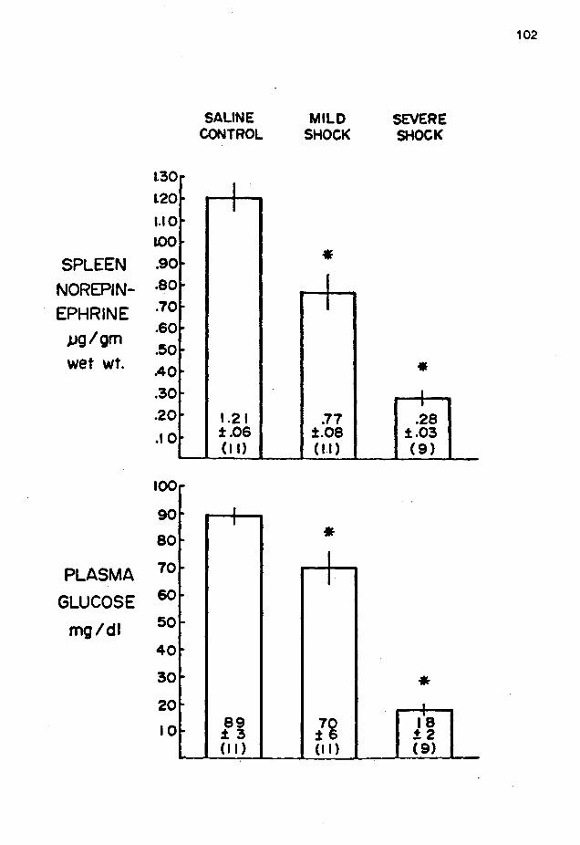
L30
l20
1.10
IOO
SPLEEN .90
NOREPIN- .so EPHRINE
µg/gm
wet wt.
PLASMA
GLUCOSE
mg/di
.70
.60
.50
.40
.30
.20
.I 0
100
90
so 70
60
50
40
30
20
10
.. ~
~
~
..
..
..
..
..
"
..
..
..
..
..
..
..
..
SALINE CONTROL
J I
1.21 ±.06 (II)
I I
S9 13 (II)
MILD SHOCK
•
.77 :t.08 (ll)
• I I
70 :J: 6 (II)
SEVERE SHOCK
• I I
.28 :t.03 (9)
• I
i's t2 (9)
102

103
Linear regression analysis was performed to determine any correlation
between the two parameters. For hearts (Figure 11), the equation of the
regression line (of the form y • 111% + b) was y • o.0063x + 0.267. The
correlation coefficient, r, was 0.73, and indicated a significant
correlation (p<0.01) between plasma glucose and cardiac norepinephrine.
Similar results were obtained in the spleen (Figure 12). The equation
of the regression line was y • 0.0082x + 0.167, and demonstrated a
significant correlation (r•0.78, p<0.01) between plasma glucose and
splenic norepinephrine content during endotoxin shock.
B. Characterization of the Norepinephrine Depletion Phenomenon
1. THE SINGULAR ROLE OF HYPOGLYCEMIA ON NOREPINEPHRINE CONTENT IN HEAR?
AND SPLEEN
To assess the role of low plasma glucose on the norepinephrine
depletion of endotoxicosis, one group of rats was treated with insulin
to lower plasma glucose concentration to varying degrees. Under light
ether anesthesia rats were injected intravenously with various amounts
(0 to 3.5 U/kg) of regular insulin (Eli Lilly and Co.) and sacrificed 90
minutes later. Plasma glucose was measured, and hearts and spleens were
analyzed for catecholamine content. Figure 13 is a graph of injected
insulin dose (U/kg) versus plasma glucose concentration (mg/dl) 90
minutes after insulin injection. The figure illustrates the range and
distribution of insulin doses as well as the classic relationship
between insulin and plasma glucose levels.

104
Figure 11.
Cardiac norepinephrine content is plotted against plasma glucose
concentration for all endotoxic rats of Figure 9. Linear regression
analysis indicates a significant correlation between cardiac
norepinephrine and plasma glucose concentration (r .. 0.73, p<0.01).
Equation (of the form y = mx + b) for the line is y .. 0.0063x + 0.267.

105
1.25
1.00 * *
r-..
* L: C)
' * C)
:::J 0 .. 75 * * - * * * * w z u o .. so * a ........ 0 0: a u * o .. 2s
** * *
o .. oo
0 25 50 75 100 125
PL~SM~ GLUCOSE CMG%)

106
Figure 12.
Splenic norepinephrine content is plotted against plasma glucose
concentration for all endotoxic rats of Figure 10. Linear regression
analysis indicates a significant correlation between splenic
norepinephrine and plasma glucose concentration (r=0.78, p<0.01).
Equation (of the form y = mx + b) for the line is y = 0.0082x + 0.167.

107
1.25 *
1 .. 00 ,...... L: C)
' C)
:::l 0 .. 75 ............
w z u o .. so ...__. z w _J CL CJ)
Q .. 25
* *
o.oo
0 25 50 75 100 125
PL~SM~ GLUCOSE (MG/.)

108
Figure 13.
Graph depicts the relationship between the injected dose of
insulin (U/kg) and the plasma glucose concentration (mg/dl) 90 minutes
after intravenous insulin injection.

109
100
* 90
* * * ao
,_ * I ~ (..!) 70 * I L: -... \ w 60 * I * (J) * ** * D * u so * ::J _J
* l.:) * * * 40 * * a I * L: * * • (J) 30 * * a
_J * * CL * 20
10
0
o.o o.s 1.0 1.s 2.0 2.s 3.0 3.s
INSULIN DOSE CU/KG"l

110
The results of the experiments are shown in Table IV. For each
organ, the data are first shown for the entire shock or insulin
hypoglycemia group. The groups are then shovn separated into mild and
severe groups (insulin-treated rats were separated into mild and severe
groups by the same criteria as endotoxic rats). The plasma glucose
concentrations were comparable between shock and insulin groups.
However during the mild phase, insulin hypoglycemia had a tendency
toward lower plasma glucose than respective endotoxic samples (not
statistically significant), while during the severe phase, the shock
group demonstrated lower plasma glucose levels (p<0.01). Comparison
between norepinephrine content for all animals in the shock and insulin
groups demonstrated that for both hearts and spleens the norepinephrine
content of the insulin hypoglycemic group always contained greater
amounts of norepinephrine than the shock group (p<0.01 in both cases).
Similar results were found when the total groups were separated into
mild and severe groups. Cardiac norepinephrine was significantly
reduced 12% in the mild shock group (p<0.05) and 64% in the severe shock
group (p<0.01) compared to the respective insulin hypoglycemic groups.
Similarly, splenic norepinephrine was significantly reduced 38% in the
mild shock group and 75% in the severe shock group compared to the
respective insulin hypoglycemic groups (p<0.01 in both cases).
Closer inspection of organ norepinephrine content in the insulin
hypoglycemic group revealed that no significant reduction from saline
controls occurred. In the spleen, norepinephrine content in saline

111
TABLE IV. COMPARISON BETWEEN ENDOTOXIN- AND INSULIN-INDUCED HYPOGLYCEMIA
ON HEART AND SPLEEN NOREPINEPHRINE CONTENT
HEART
All Shock
All Insulin Hypoglycemia
Mild Shock
Mild Insulin Hypoglycemia
Severe Shock
Severe Insulin Hypoglycemia
SPLEEN
All Shock
All Insulin Hypoglycemia
Mild Shock
Mild Insulin Hypoglycemia
Severe Shock
Severe Insulin Hypoglycemia
N
21
41
12
27
9
14
20
33
11
24
9
9
Tissue Plasma Norepinephrine Glucose
(11~/gm) (ms/dl)
0.600 + 0.06 53 .:!:. 7
• 0.905 .:!:. 0.03 52 .:!:. 4
0.816 .:!:. 0.03 75 .:!:. 7 .. 0.924 .:!:. 0.03 63 .:!:. 3
0.312 .:!:. 0.04 24 ~ 2
* * 0.868 .:!:. 0.05 33 .:!:. 1
0.553 .:!:. 0.07 47 ~ 7
• 1 .225 ~ 0.07 59 ~ 4
0.772 + o.os 70 + 6
• 1.254 ~ 0.09 69 ~ 3
0.284 + 0.04 18 + 2
* * 1.149~0.14 31 + 2
• p<0.01, ** p<0.05 for shock versus insulin hypoglycemia group by independent Student's t test;

112
controls was 1.214 ~ 0.07 ug/gm. This was not significantly different
from either the mild insulin hypoglycemic group (1.254 ~ 0.09 ug/gm) or
the severe insulin hypoglycemic group (1.149 ~ 0.14 ug/gm). In the
heart, norepinephrine content of saline controls was 0.985 ~ 0.05 ug/gm.
There was no statistical reduction in either the mild (0.924 ~ 0.03
ug/gm) or severe insulin hypoglycemic group (0.868 ~ 0.05 ug/gm).
Figures 14 and 15 illustrate the relationship between plasma glucose and
norepinephrine content for hearts and spleens of all insulin
hypoglycemic rats. Linear regression analysis was performed by the
method of least squares. The equation of the regression line (of the
form y "' mx + b) for cardiac norepinephrine versus plasma glucose
(Figure 14) was y • o.0014x + o.s33. The correlation coefficient, r
(0.15), was not significantly different from 0 (p>0.05); no correlation
between cardiac norepinephrine content and plasma glucose occurred.
Similar results were obtained in the spleen (Figure 15). The equation
of the regression line was y • -0.0002x + 1.23s. No correlation between
splenic norepinephrine and plasma glucose was evident (rs-0.01, p>o.05).
Cardiac epinephrine was also quantitated in the insulin
hypoglycemic rats. Epinephrine content of saline-injected controls was
0.087 + 0.02 ug/gm (n=5). Mild insulin hypoglycemia significantly
raised cardiac epinephrine levels 10% (0.096 ~ 0.01 ug/gm; n=24); severe
insulin hypoglycemia resulted in a marked 115% increase in cardiac
epinephrine levels (0.189 + 0.04 µg/gm). Figure 16 depicts the
relationship between plasma glucose and cardiac epinephrine for all
insulin hypoglycemic rats. The equation of the regression line was y =

113
Figure 14.
Graph depicts cardiac norepinephrine content versus plasma
glucose concentration for all rats treated with insulin. Linear
regression analysis indicates no correlation between norepinephrine
content and plasma glucose concentration (r= 0.15, p>0.05). Equation of
the line (of the form y = mx + b) is y = o.0014x + o.s33.

114
1-25
* * * * ** * *
1.00 * * * ** *
** ,--...
I: C) * ' ~ * * C)
* * * :J 0.75 * * - * 4
* * w * * z u o.so * a t---4
D CY: a u
0.25
o.oo
0 25 50 75 100 125
PL~SM~ GLUCOSE CMG/.)

115
Figure 15.
Graph depicts splenic norepinephrine content versus plasma
glucose concentration for all rats treated with insulin. Linear
regression analysis indicates no correlation between norepinephrine
content and plasma glucose concentration (r= -0.01, p>0.05). Equation
of the line (of the form y = mx + b) is y s -0.0002x + 1.23s.

116
o .. oo
0 25 50 75 100 125
Plr:ISM~ GLUCOSE CMG/.·)

117
Figure 16.
Graph depicts cardiac epinephrine content versus plasma glucose
concentration for all rats treated with insulin. Linear regression
analysis indicates a significant negative correlation between
norepinephrine content and plasma glucose concentration (r= -0.66,
p<0.01). Equation of the line (of the form y = mx + b) is y = -0.0019x
+ 0.213.

118
o.so
Q.40 ;o-"\.
L: t-'
' t-' ::J Q.30 -f--1
* o._ * w *
u 0.20 CI * f--1
* D CY *. CI u
0 .10 * I * * * f
* * * * * *
o.oo
0 20 60 BO 100
PL~SM~ GLUCOSE CMG/.)

119
-0.0019x + 0.213. A significant negative correlation (r•-0.656; p<0.01)
existed between cardiac epinephrine and plasma glucose for insulin
hypoglycemic rats.
2. THE ROLE OF NEURAL MEDIATION IN THE NOREPINEPHRINE DEPLETION OF
ENDOTOXIC SHOCK
To test the hypothesis that the norepinephrine depletion was
dependent on neural activity, rats were injected intravenously with the
ganglionic blocker, chlorisondamine (5 mg/kg), one hour after endotoxin
injection (5 mg/300 gm rat). Preliminary experiments showed that
pretreatment or cotreatment of chlorisondamine sensitized rats to
endotoxin lethality (rats usually died while still under ether
anesthesia or within five to ten minutes). Therefore, ganglionic
blockade was administered one hour after endotoxin (post-hypotensive
phase). This regimen was tolerated better by rats, and the time to
death of the ganglionic blockade group (176 + 21 min) was not
significantly different
blockade (169 ~ 35 min).
than the endotoxic rats without ganglionic
cardiac norepinephrine for
Figure 17 illustrates the plasma glucose and
these experiments. An additional group of
endotoxic rats was sacrificed at one hour after endotoxin to act as a
control for rats with ganglionic blockade. Ganglionic blockade
effectively eliminated any norepinephrine depletion after one hour
post-endotoxin (0.832 + 0.05 ~g/gm for one hour of endotoxin; 0.748 +
0.04 ~g/gm for endotoxin plus ganglionic blockade). Plasma glucose

120
Figure 17.
Illustration of cardiac norepinephrine content and plasma
glucose concentration in control, endotoxic, and endotoxic rats
pretreated with chlorisondamine (5 mg/kg). Within the bars the mean +
SEM are shown. Number of rats is illustrated in parentheses. ** p<0.01
versus adjoining bar by independent analysis of variance.

LOO
. 90
. so
NOREPIN- ·70
EPHRINE
pg/om wet wt.
.60
.50
.40
.30
.20
.I 0
110
100
90
80
PLASMA 70
GLUCOSE 60
mg 0k 50
40
30
20
10
.
.
•
~
.
. •
•
•
SALINE ONE HR SHOCK+ CONTROL SHOCK GANGLIA
BLOCK I I NS
NS I I I
I
.985 .832 .748 t .05 !.05 t.06
(15) (6) (12)
NS
I I
• •
I I
90 106 30 !3 117 16
(13) (6) (12)
• •
LATE SHOCK
I I
.312 !.04
(9)
NS
I I
24 12 (9)
121

122
levels in the_ganglionic blockade group (30 ..:!:. 6 mg/dl) reached levels as
low as the severe shock group (24 ..:!:. 2 mg/dl).
3. EFFECT OF ADRENERGIC MANIPULATION ON ENDOTOXIN-INDUCED LETHALITY AND
NOREPINEPHRINE DEPLETION
Adrenergic blockade and sympathectomy have been shown to have
variable effects on the pathogenesis of endotoxin shock. To further
investigate this area, rats were treated with either postsynaptic
adrenergic blocking agents or chemically sympathectomized to prevent
presynaptic release of norepinephrine. In the first set of experiments
rats were treated with either an alpha adrenergic blocking agent
(phentolamine) or beta adrenergic blocking agent (propranolol) and then
subjected to the present shock model. In awake rats, phentolamine (3
mg/kg) or propranolol (4 mg/kg) was injected intraperitoneally 15
minutes before endotoxin was injected intravenously (5 mg/300 gm rat)
under light ether anesthesia. A control group of rats was injected with
only endotoxin. Rats were killed 5 hours later or near death. Results
of these experiments are shown in Table v. All phentolamine plus
endotoxin rats lived for the maximum five hours (300 ..:!:. 0 min) of the
experiment. This was significantly longer than either the endotoxin
alone group (277 ..:!:. 12 min) or the propranolol plus endotoxin group (18
..:!:. 1 min). Propranolol significantly decreased time to death compared to
the endotoxin alone group. Although plasma glucose was no different
between endotoxin and propranolol plus endotoxin in this series,
phentolamine plus endotoxin rats had significantly higher glucose levels

123
TABLE V. EFFECTS OF PROPRANOLOL OR PHENTOLAMINE PRETREATMENT
ON ENDOTOXIN SHOCK IN THE RAT
Plasma Glucose (mg/dl)
Time to Death (minutes)
Cardiac Norepinephrine
(µg/gm)
Splenic Norepinephrine
(v.g/ gm)
Endo toxin Alone
51 + 14 (8)
277 + 12 (8)
0.617 + 0.10 (8)
0.520 + 0.11 Cf)
Propranolol Plus Endotoxin
43 + 5 Cf)
• 18 + 1
(12)
0.761 + 0.05 (7)
0.812 + 0.07 Cf)
Phentolamine Plus Endo toxin
• II 78 + 5
(8)
• II 300 + 0
( 1T)
• 0.853 + 0.07
(8)
* 0.887 + 0.13
(7)
Values are mean + SEM. The number of samples is in parentheses below each value. * p<0.05 versus endotoxin alone, II p<0.05 versus propranolol plus endotoxin,
by independent analysis of variance.

124
than either of the other eroups. Borepinephrine content in both the
heart and spleen was statistically ereater in the phentolamine plus
endotoxin eroup than in the endotoxin alone group. However,
norepinephrine content of hearts and spleens in propranolol-treated rats
was intermediate and no different than in either of the other two
groups.
The effects of blunted release of norepinephrine was next
investigated by chemically sympathectomizing rats before endotoxin
challenge. Animals sympathectomized with 6-hydroxydopamine contain
minimal norepinephrine in peripheral sympathetic nerve endings, and
higher than normal catecholamine levels in adrenal medullary tissue
(norepinephrine and epinephrine). Because norepinephrine has relatively
more alpha adrenergic effect and epinephrine has more beta adrenergic
effect, chemical sympathectomy would accentuate beta adrenergic activity
and minimize alpha adrenergic activity. In light of the phentolamine
and propranolol
decrease alpha
experiments, 6-hydroxydopamine is hypothesized to
adrenergic stimulation of sympathetically innervated
tissues, leave adrenal medullary secretion of catecholamines intact, and
significantly protect against endotoxic lethality. Under light ether
anesthesia, rats
me/kg) and then
6-hydroxydopamine
were injected intravenously with phentolamine (10
either saline or 6-hydroxydopamine (80 mg/kg).
was weighed out as the free base of the amine
complexed with hydrobromide and dissolved in saline containing 1%
ascorbate. Phentolamine was administered to minimize the blood pressure
increase that occurred after 6-hydroxydopamine injection and subsequent

125
norepinephrine liberation. Preliminary experiments indicated that this
regimen of treatment with 6-hydroxydopamine reduced cardiac
norepinephrine levels by 88% and splenic norepinephrine levels by 93%.
Endotoxin was administered 48 hours later. The lethality data is
illustrated in Table VI. Chemical sympathectomy significantly reduced
the lethality associated with endotoxin injection. The protective
effect of sympathectomy was first demonstrated at 6 hours after
endotoxin and was present for the duration of the study up to 24 hours
post endotoxin.
c. Norepinephrine Reuptake
1. IN VIVO 3H-NOREPINEPHRINE UPTAKE IN ENDOTOXIC SHOCK
The results of in vivo assessment of the reuptake process in
endotoxicosis are presented in Tables VII and VIII for hearts and
spleens, respectively. Four groups of rats were evaluated: 1)
saline-injected controls, 2) mild, and 3) severe shock rats, and 4) a
separate group of rats pretreated with the neuronal reuptake blocker of
norepinephrine, desmethylimipramine (DMI). The last group was employed
to evaluate the singular effect of depressed reuptake in this
experimental paradigm.
In the myocardium (Table VII) DMI had no effect on the level of
endogenous norepinephrine, but DMI reduced the amount of
3H-norepinephrine accumulated in the tissue by 95% ten minutes after

TABLE VI. EFFECT OF SYMPATHECTOMY WITH 6-HYDROXYDOPAMINE (6-0HDA) ON ENDOTOXIN-INDUCED LETHALITY
Hours After % Lethality % Lethality Endo toxin Control 6-0HDA
0 0 ( 0/27)# 0 ( 0/27)
0 ( 0/27) 0 ( 0/27)
2 7 ( 2/27) 0 ( 0/27)
3 7 ( 2/27) 0 ( 0/27)
4 19 ( 5/27) 7 ( 2/27)
5 22 ( 6/27) 7 ( 2/27)
6 26 ( 7/27) * 7 ( 2/27)
7 26 ( 7/27) * 7 ( 2/27)
8 30 ( 8/27) * 11 ( 3/27)
9 52 (14/27) ** 15 ( 4/27)
10 56 (15/27) ** 15 ( 4/27)
11 56 (15/27) ** 19 ( 5/27)
12 56 (15/27) ** 19 ( 5/27)
24 56 (15/27) ** 26 ( 7/27)
* p<0.05, ** p<0.01 compared to control by Chi Square; #numbers in parentheses indicate number of deaths I total initial
number of animals;
126
Rats were treated with 6-0HDA 48 hours before endotoxin challenge. There were a total number of 27 rats in each group ~t time 0 hours.

127
TABLE VII. IN VIVO 3H-NOREPINEPHRINE (3H-NE) UPTAKE IN RAT MYOCARDIUM
Plasma Glucose Norepinephrine 3H-NE
Group N (mg/dl) (11g/gm) (dpm/gm)
91 o.eo 11838 Control 7
,:3 ,:0.05 ,:845
Desmethyl- 87 0.72 ** 650 7
imipramine +7 +0.04 +110
Mild 95 ** o.66 13414 15
Shock ,:5 +0.01 ,:740
Severe ** 23 ** 0.48 17783 7
Shock ,:3 +0.06 ,:2904
Mean + SEM
** p<0.05 compared to control by independent analysis of variance;

128
TABLE VIII. IN VIVO 3H-NOREPINEPHRINE (3H-NE) UPTAKE IN RAT SPLEEN
Plasma Glucose Norepinephrine 3H-NE
Group N (mg/dl) ()lg/gm) (dpm/gm)
91 0.86 2309 Control 7
_:3 +0.02 _:224
Desmethyl- 87 0.94 .. 366 7
imipramine +7 _:0.09 +48
Mild 95 ** 0.50 ** 1522 15
Shock _:238
Severe .. 23 .. 0.27 .. 270 7
Shock +0.06
Mean + SEM
** p<0.05 compared to control by independent analysis of variance;

intravenous injection of the labeled bioamine.
129
In both mild and
severely shocked endotoxic rats, 3H-norepinephrine uptake was not
changed from control. However, note that the relative depletion of
endogenous norepinephrine is similar to the group of rats in which the
depletion was demonstrated (Figure 9).
In the spleen (Table VIII), DMI pretreatment again had no effect
on endogenous norepinephrine content, but it depressed the accumulation
of injected 3H-norepinephrine by 84%. Mild and severe shock produced
depression of the endogenous norepinephrine content of the spleen.
However, unlike the myocardium, during both mild and severe
endotoxicosis, spleen uptake of circulating 3H-norepinephrine was
depressed. In mild shock, 3H-norepinephrine content in spleens was
reduced from a control value of 2309 ~ 224 dpm/gm to 1522 ~ 238 dpm/gm
(34% reduction). Severe shock rats demonstrated an 88% decrease in
3H-norepinephrine accumulation to 270 ~ 69 dpm/gm.
2. IN VITRO 3H-NOREPINEPHRINE UPTAKE
Because in vivo experiments suggested that reuptake depression
would be most prominent in severe shock rats, in vitro analysis of
norepinephrine uptake was performed in control and severe shock groups.
The results of these experiments for heart and spleen slices are shown
in Figures 18 through 21.
or 30 minutes at 37° C
norepinephrine. Total
Tissue slices were incubated for 0, 10, 20,
in the presence of labeled and unlabeled
3H and 3H-norepinephrine were measured.

Figure 18.
Total 3H retention in myocardial slices of control and severely
shocked rats incubated for the indicated time with 3H-norepinephrine.
Each bar represents the mean + SEM for 6 preparations, each from a
different rat. NS represents no significant difference for severe shock
group versus control.
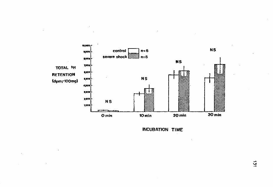
TOTAL 'H
RETENTION
(dpm/100mg)
t0,000
•.ooo
..... 1,000
..... 1,000
4,HO
..... t.OOD
control c=J n=6
severe shock~ n=6
NS
NS
Omin 10mln
NS
20mln
INCUBATION TIME
NS
30mtn

132
Figure 19.
Total norepinephrine retention for myocardial slice preparations
of Figure 18 calculated from the 3H-norepinephrine content of the tissue
and the specific activity of the incubation media. Each bar represents
the mean + SEM for 6 preparations, each from a different rat. NS
represents no significant difference for severe shock group versus
control.

NOREPIN
EPHRINE
RETENTION
(pmol/100mg)
11
10
9
8
7
6
5
4
3
control c:J n=6 severe shock ~ n=6
NS
NS
2 NS
1
Omin 10min 20mln
INCUBATION Tl ME
NS
30min

134
Figure 20.
Total 3H retention in splenic slices of control and severely
shocked rats incubated for the indicated time with 3H-norepinephrine.
Each bar represents the mean + SEM for 5 preparations, each from a
different rat. NS represents no significant difference for severe shock
group versus control.

TOTAL 'H
RETENTION
(dpm /100 mg)
tl,000
t4,000
tl,000
to,000
1,000
1,000
4,000
t,ODD
control severe shock
p<.01
0 min
[=:J n=S ~n::S
•
•
10min
•
20mln
INCUBATION TIME
•
30mln 30mln cold.

Figure 21.
Total norepinephrine retention for splenic slice preparations of
Figure 20 calculated from the 3H-norepinephrine content of the tissue
and the specific activity of the incubation media. Each bar represents
the mean_:!:. SEM for 5 preparations, each from a different rat.

100
90 80
NOREPIN- 70
EPHRINE 60
RETENTION 50
( pmol/IOOmg) 4 0 30
20
10
control [=:J n= 5 severe shock ~ n=5
p<.01 •
p<.05 ••
•
•
Omin 10 min
•
20min
INCUBATION TIME
•
30min
••
30 min cold

138
Norepinephrine retention (pmol/100 mg) was calculated as the quotient of
the tissue 'H-norepinephrine content (dpm/mg tissue) and the specific
activity of the incubation media (dpm/pmol norepinephrine). In
myocardial slices (Figure 18) at all times of incubation at 37° C there
were no differences in total 3H retention between control and severe
shock
both
21),
rats. Similarly, total norepinephrine retention was the same in
groups of heart slices (Figure 19). In the spleen (Figures 20 and
total 3H and total norepinephrine retention were severely reduced
in tissue slices of severely shocked rats at all times of incubation at
37° C. The reduction was on the order of 50% at all times tested. The
results of in vitro analyses are thus consistent with in vivo
experiments, i.e. depressed reuptake of norepinephrine occurs in
spleens, but not in hearts of endotoxic rats.
Because neuronal reuptake is temperature sensitive, incubations
were also carried out for 30 minutes at 4° C. Cold incubation (Figures
20 and 21) effectively abolished all norepinephrine retention.
Preliminary experiments with DMI in the incubation media indicated
reuptake depression equal to cold incubation. Thus, the measured
norepinephrine retention is dependent on neuronal reuptake and not
non-neuronal reuptake or non-specific binding.
One profound consequence of shock is splanchnic congestion and
accumulation of cellular debris in the spleen. Increase in tissue wet
weight during shock would thus lower the measured norepinephrine
accumulation values. To evaluate this possibility, both spleens and

139
hearts were remoyed from control and severely shocked animals, weighed,
and then dried to constant weight. The results are shown in Table IX.
There were no differences in wet weight, dry weight, or dry to wet
weight ratios in hearts from control and shock animals. In the spleen,
wet weight increased from 0.851 .:!: 0.038 gm to 1.013 .:!: 0.082 gm and dry
weight increased from 0.195 + 0.009 gm to 0.239 .:!: 0.023 gm in the
endotoxic state. Both of these increases approached but did not reach
significance (p•0.10 for both vet and dry weight differences). The
splenic dry to wet weight ratio also did not change in the shock state
(control: 0.229 .:!: 0.001; severe shock: 0.234 .:!: 0.004; p•0.23).
Although the weight changes in the spleen were not significant,
the difference represents a 19% increase in wet weight and a 22%
increase in dry weight in the severe shock rats. The parallel tendency
for both spleen wet and dry weight to increase with no change in the dry
to wet weight ratio suggests an increase in total cellular mass (most
likely a congestion with red blood cells or cellular debris) of the
spleen rather than a pure edema. The number of reuptake sites per unit
of tissue wet weight will decrease if the spleen is infiltrated by
cellular debris. Therefore, the measured amount of norepinephrine
retention per 100 mg of tissue would be expected to decrease (in this
case by 19%) simply due to an increase in cellular mass of the spleen.
Therefore, the norepinephrine data for the spleen was corrected for
changes in spleen vet weight of the endotoxic group (Figure 22). Even
using this conservative modification, spleen slices from endotoxic rats
retained significantly less norepinephrine than those from controls.

140
TABLE IX: MYOCARDIUM AND SPLEEN WEIGHTS IN CONTROL AND ENDOTOXIC RATS
CONTROL MILD SHOCK SEVERE SHOCK
N 8 8 7
Plasma Glucose 110 • 82 • 35 (mg/dl) _:4 _:5 _:5
MYOCARDIUM
Wet Weight 1 .491 1.485 1.429 (gm) _:0.035 _:0.036 +0.088
Dry Weight 0.351 0.347 0.331 (gm) +0.010 +0.008 +0.020
Dry/Wet 0.236 0.234 0.234 Weight Ratio _:0.003 +0.002 _:0.003
SPLEEN
Wet Weight 0.951 ** 1.154 1 .013 (gm) ;!;_0.038 _:0.091 +0.082
Dry Weight 0.195 ** 0.261 0.239 (gm) ;!;_0.009 ;!;_0.023 ;!;_0.023
Dry/Wet 0.229 0.234 0.234 Weight Ratio +0.001 ;!;_0.003 +0.004
Mean .:!:._ SEM;
* p<0.01 vs control, ** p<0.05 vs control by independent analysis of variance;

141
Figure 22.
Total norepinephrine retention for spleen slice data of Figure
21 corrected for a 19% wet weight increase in spleens from severely
shocked rats. Each bar represents the mean .:. SEM for 5 preparations,
each from a different rat. NS represents no significant difference for
severe shock group versus control.

IOO
90 BO
NOREPIN- 70
EPHRINE 60
RETENTION 50
( pmol/IOOmg) 40
30
control CJ n=5
severe shock - n = 5
p<.01 • •
20 •
10
0 min IOmin 20min
INCUBATION TIME
•
30min 30min cold
.... ~ N

143
3· INRACELLULAR DEGRADATION OF 3H-NOREPINEPHRINE AFTER UPTAKE
Because the neuronal reuptake process (Uptake 1) is energy
dependent and is compromised in the spleen, it is reasonable to suspect
other energy sensitive processes to be affected in the spleen. After
reuptake of norepinephrine, repackaging of the catecholamine into
vesicles requires energy. One consequence of decreased rate of
repackaging is an increase in intraneuronal degradation by the soluble
enzyme, monoamine oxidase. The ratio of 3H-norepinephrine to 3H
recovered by a tissue is indicative of the amount of norepinephrine
taken up, repackaged, and conserved in its unmetabolized form. Figure
23 depicts the results of these calculations in spleen slices. At all
times of incubation, tissues from shocked rats demonstrated
significantly less 3H retained in the form of 3H-norepinephrine.
Therefore, these data are consistent with the hypothesis that in
endotoxic rats, norepinephrine degradation is accelerated in the nerve
terminals of peripheral tissues.
D. Measurement of Sympathetic Activity in Hearts and Spleens of Control
and Endotoxic Rats
1. TISSUE NOREPINEPHRINE LEVELS IN CONTROL AND ENDOTOXIC RATS
Kinetic
3H-norepinephrine
analysis of norepinephrine
decay method is dependent on
turnover by the
steady state tissue

144
Per cent
unmetabolized form
severely shocked
Figure 23.
of radioactive label (3H) recovered in its
(3H-norepinephrine) in spleen slices of control and
rats coincubated with 3H-norepinephrine for the
indicated time. Each bar represents the mean + SEM for 5 preparations,
each from a different rat.

PER CENT
IOO
90
80
70
OF 3H 60
RECOVERED 50 AS 5H-NE 4o
30
20
10
control CJ (n=5)
•
0 min
•
severe shock~ (n=5)
10 min 20min
INCUBATION Tl ME
p<.01 •
•
30min
•
30min cold
-.,. \J1

146
levels of the bioamine throughout the 12 hour experiment (12,27). Table
X illustrates the norepinephrine content in hearts and spleens of
saline-injected control and endotoxic rats. Groups of rats were
sacrificed at each of the indicated times after 3H-norepinephrine
injection. An independent analysis of variance was performed on each of
the four groups of values. No statistical differences (p>0.10) in
norepinephrine content exist between sampling times for any single organ
experimental group. Thus, the reduced dose of endotoxin (2 mg/rat;
5-7-6.7 mg/kg) used in this series of experiments was sufficient to
prevent the norepinephrine depletion of endotoxicosis. However, it is
important to note that the stress of endotoxicosis in these particular
experiments (LD 5%) is much less than in the general shock model (60 -
70%) used in the rest of the experiments.
2. NOREPINEPHRINE TURNOVER IN ENDOTOXICOSIS
Figure 24 illustrates the decay of myocardial 3H-norepinephrine
specific activity in control and endotoxic rats. Norepinephrine
specific activity (dpm/ug norepinephrine) is plotted on a logarithmic
scale.
hearts
The slope of the specific activity versus time decay line for
from endotoxic rats (-0.047) was significantly increased
(p<0.001) compared to the slope calculated for saline-injected control
rats (-0.006). Figure 25 depicts specific activity decay lines for
spleens of control and endotoxic rats. The slope of the line for the
endotoxic group (-0.049) was markedly increased (p<0.001) over the
control slope (-0.015). The constant level of endogenous norepinephrine

147
TABLE X. TISSUE LEVELS OF NOREPINEPHRINE IN HEARTS
AND SPLEENS OF CONTROL AND ENDOTOXIC RATS
CONTROL ENDOTOXIN
NE HOURS AFTER NE n µg/g 3H-NE INJECTION n Jlg/g
HEART
5 0.70.: 0.04 0.17 7 o.66 .: 0.03
6 0.72 + 0.06 3.00 7 0.69 .: 0.04
6 0.69.: 0.04 6.00 6 0.68 + 0.06
6 0.67 + 0.04 9.00 6 0.72.: 0.05
6 o.66 .: 0.03 12.00 4 0.69 .: 0.04
SPLEEN
5 0.96 .: 0.05 1.00 4 0.97 .: 0.10
7 1 .01 + 0.11 3.00 6 0.76 + 0.06
7 0.90.: 0.06 6.00 5 o. 96 .: 0.07
6 0.93 .: 0.08 9.00 6 0.89 .: 0.09
3 0.76 + o.os 12.00 4 o. 78 .: 0.01
Mean + SEM; There are no statistical differences in norepinephrine content at different times after injection within any single group (independent analysis of variance).

'148
Figure 24.
Decay of myocardial 3H-norepinephrine (3H-NE) specific activity
in control and endotoxic rats. Rats were injected with 3H-NE one minute
before injection of either saline or endotoxin. Each point represents 4
to 7 rats. Slope of the decay line for endotoxic rats is significantly
steeper than control (p<0.001).

149
...... ~··· ao.•••
NE SPECIFIC H.000 s1opo • -.ooe
ACTIVITY (dpmlpg NE)
...... •.o•• ..... ••••• yoo , •. 001
, ... ••••• .....
• SALIN£ CONTROL ••••• • ENDOTOXIC
..... ._ ....... ~~--------___..__...._ ___ __...__..._.....___.~.._...._ ........ ~---0 a ' • • '° u " t• I
TIME AFT ER INJECTION OF 'tf·NE (hours)

150
Figure 25.
Decay of splenic 3H-norepinephrine (3H-NE) specific activity in
control and endotoxic rats. Rats were injected with 3H-NE one minute
before injection of either saline or endotoxin. Each point represents 4
to 7 rats. Slope of the decay line for endotoxic rats is significantly
steeper than control (p<0.001).

NE SPECIFIC ACTIVITY (dpm~g NE)
....
.,, .. \000 ... 000 ,.. ... ... -.. ..
Slo519•~•t
• • .001
e SALINE CONTROL
• ENDOTOJI IC
Slop••-:011
.. .__.__.__.~..__._~__..__..__.__..___..L..-.._......___.___..__...__
0 I • • • ,. TIME AFTER INJECTION OF IH·NE (houra)
151

152
and increased rate or 3H-norepinephrine disappearance from tissues of
endotoxic rats indicate increased turnover or norepinephrine in the
endotoxic state.
The kinetic turnover data for hearts and spleens is summarized
in Table XI. In hearts the mean norepinephrine content for all samples
comprising a single decay curve was unchanged in the endotoxic group
compared to control. Rate constants were calculated from the slopes and
application or first order kinetics. The rate of cardiac norepinephrine
turnover (the product of the rate constant and the endogenous
norepinephrine content) increased more than 700% in the endotoxic group
(0.075 µgxgm- 1xhr-1) compared to controls (0.009 µgxgm- 1xhr-1). Similar
results occurred in the spleen. Endogenous norepinephrine content of
all endotoxic spleens (0.87 + 0.04 pg/gm) was not significantly
different (p=0.24) than control spleens (0.93 _: 0.04 µg/gm). Endotoxin
treatment more than tripled the rate constant of decay from 0.035 hr-1
in the control state to 0.114 hr-1• Thus, the turnover rate of
norepinephrine in the spleen increased 200% in the endotoxic state
(control: 0.033 µgxgm- 1xhr-1; endotoxin: 0.099 ~gxgm- 1 xhr- 1 ).
3. EFFECT OF DESMETHYLIMIPRAMINE ON NOREPINEPHRINE TURNOVER
Increased norepinephrine turnover rates in endotoxicosis
indicate increased sympathetic tone. However, the experiments just
reported demonstrated that spleens but not hearts from endotoxic rats
displayed depressed norepinephrine reuptake when measured in both in

153
TABLE XI. NOREPINEPHRINE (NE) TURNOVER IN HEARTS AND SPLEENS
OF CONTROL AND ENDOTOXIC RATS
NE RATE CONSTANT TURNOVER RATE TREATMENT n µg/g SLOPE hr-1 ug/(gxhr)
HEART
Control 29 0.69 -0.006 0.013 0.009 +0.02 +0.004
Endo toxin 30 0.69 -0.047* 0.108 0.075 +0.02 +0.006
SPLEEN
Control 28 0.93 -0.015 0.035 0.033 +0.04 +0.008
Endotoxin 25 0.87 -0.049* 0.114 0.099 +0.04 +0.007
Mean + SEM; * p<o-:-001 compared to control using the standard error of the sample
regression.

154
vivo and in vitro situations. It is possible that the depressed eplenic
norepinephrine reuptake of endotoxic rats may contribute to increased
rate of transmitter loss from the terminal (both labeled and unlabeled)
and be measured as increased turnover rate. Therefore, this hypothesis
was tested by measuring norepinephrine turnovers in control rats
pretreated with the neuronal norepinephrine reuptake inhibitor, DMI.
Turnovers in both hearts and spleens were examined to determine if
Uptake 1 had similar effects on sympathetic activity in both organs.
Table XII illustrates the norepinephrine content in hearts and
spleens of DMI-treated rats. The critical organ for turnover
measurement is the spleen, since reuptake has been shown to be depressed
in endotoxic spleens. At each time point splenic norepinephrine content
was found to not be statistically different than any other time point.
The same holds true for myocardial norepinephrine content. The control
group of rats is the same as in the endotoxic turnover experiments. The
cardiac and splenic norepinephrine content for the control rats is
depicted in Table X.
The specific activity decay lines for hearts and spleens of
control and DMI-treated rats are depicted in Figures 26 and 27,
respectively. Although the initial specific activity is not the same,
the slopes for specific activity decay are not statistically different
from one another in hearts of control (-0.006) and DMI-treated (-0.004)
rats (p>0.015; Figure 26). Figure 27 illustrates that the slop~s of the
splenic specific activity decay lines were the same (0.015 hr-1) for

HEART
SPLEEN
TABLE XII. TISSUE LEVELS OF NOREPINEPHRINE (NE) IN HEARTS
AND SPLEENS OF DESMETHYLIMIPRAMINE-TREATED RATS
NE JJ.g/gm
0.83 ~ .05
0.73 ~ .06
0.77 ~ .05
0.72 + .05
0.70 ~ .08
0.85 ~ .10
0.78 + .04
0.75 ~ .09
0.72 + .05
0.81 + .08
HOURS AFTER 3H-NE INJECTION
1.0
6.0
9.0
12.0
1.0
3.0
6.0
9.0
12.0
n
6
6
6
6
5
4
5
6
6
5
155
~ean ~ SEM; Rats were injected with desmethylimipramine 10 minutes after H-NE injection. There are no statistical differences in norepinephrine
content at different times after injection within any single group (independent analysis of variance).

156
Figure 26.
Decay of myocardial 3H-norepinephrine (3H-NE) specific activity
in control and desmethylimipramine-treated rats. Rats were injected
with 3H-NE ten minutes before injection of desmethylimipramine. Each
point represents 4 to 7 rats. Slope of the decay line for
desmethylimipramine-treated rats is no different from control (p>0.015).

" ... ...... .....
NE SPECIFIC --.••• ACTIVITY
(dpm~g NE)
...... ..... ... 00
\000 ..... .....
• ... 015
1,000 o SALINE CONTROL
A DESMETHYLIMIPRAllllNE
.....
Slopo • -:001
..... .._ __ _.___...__~_.....__..~.._---_..~~~---~.._-----__......__. 0 2 • • • 10 16
TIME AFTER INJECTION OF 3H-NE (hours)
157

158
Figure 27.
Decay of splenic 3H-norepinephrine (3H-NE) specific activity in
control and desmethylimipramine-treated rats. Rats were injected with
3H-NE ten minutes before injection of desmethylimipramine. Each point
represents 4 to 7 rats. Slope of the decay line for
desmethylimipramine-treated rats is no different from control (p>0.50).

4,DDD
NE SPECIFIC _,DD
ACTIVITY Cdpm~g NE)
11,DDD eDD IDD
JOD
eDD
MID
•DD
IDD
IDD
NS ••• 50
Slope • -.015
O SALINE CONTROL
& DESMETHYLIMIPRAMINE
teD~~~~~~~~~~__..~~-----~~---~~~~--
0 2 • • • " TIME AFTER INJECTION OF 3H·NE (hours)
159

160
both control and DMI-treated rats. Although the specific activity at O
hours after 'H-norepinephrine was greater in the control sroup, the
fractional loss of tracer from both groups was exactly the same.
Table XIII summarizes the turnover data for control versus
DMI-treated rats. In hearts, DMI pretreatment increased the endogenous
level of norepinephrine. However, the rate constant of decay in hearts
of DMI-treated rats was reduced compared to control. The turnover rates
in both groups of rats were similar (0.009 µg/(gxhr) in controls, 0.006
~g/(gxhr) for DMI treatment) and extremely lower than the turnover rate
for endotoxic rats (0.075 µg/(gxhr). In spleens, DMI treatment reduced
endogenous levels of norepinephrine slightly, but significantly. The
fraction of norepinephrine turned over per hour was the same in both
groups. Therefore, the
(0.028
splenic turnover rate of DMI-treated rats was
~gxgm- 1 xhr-1) than in controls (0.033 slightly lower
pgxgm-1xhr-1). Again, as in the heart, note that turnovers in spleens
of control and DMI-treated rats were dramatically lower than in spleens
of endotoxic rats (0.099 pg/(gxhr)).

161
TABLE XIII. NOREPINEPHRINE (NE) TURNOVER IN HEARTS AND SPLEENS
OF CONTROL AND DESMETHYLIMIPRAMINE-TREATED RATS
NE RATE CONSTANT TURNOVER RATE TREATMENT n ~ SLOPE hr-1 ;ig/(gxhr)
HEART
Control 29 0.69 -0.006 0.013 0.009 +0.02 +0.004
Desmethyl- 29 0.75* -0.004* 0.008 0.006 imipramine ~0.03 ~0.003
SPLEEN
Control 28 0.93 -0.015 0.035 0.033 +0.04 +0.008
Desmethyl- 26 0.78* -0.015 0.036 0.028 imipramine ~0.03 ~0.029
Mean + SEM; * p<o":-001 compared to control using the standard error of the sample
regression.

The
sympathetic
CHAPTER VI
DISCUSSION
present experiments
dysfunction during
have focused on
endotoxin shock:
one aspect
depletion
of
of
norepinephrine from sympathetic nerve terminals. These studies can be
categorized into four areas: 1) demonstration of the phenomenon, 2)
characterization of the depletion, 3) assessment of norepinephrine
reuptake, and 4) measurement of sympathetic activity during endotoxin
shock. Each of these areas will be discussed separately.
A. Demonstration of Norepinephrine DeEletion during Endotoxin Shock
Fundamental to any series of experiments is an appropriate,
reproducible model. In the present studies an endotoxic rat model was
employed: intravenous injection of Salmonella enteritidis endotoxin at a
dose (14 to 17 mg/kg) to generate a 60%-80% lethality. The albino rat
has been widely used in endotoxic studies, primarily for metabolic,
immune, and in vitro preparation experiments. The inbred genetic line
of the laboratory rat minimizes animal to animal variations, and the
size and cost per animal facilitates experimental repetition. Larger
animal models, such as the dog or primate, are better suited for
hemodynamic and cardiovascular experimentation - the so-called "pump and
pipe" investigations. The area of present interest, norepinephrine
162

163
depletion from peripheral tissues, is particularly well-suited to a
small animal model such as the rat. The rat is large enough to obtain
adequately-sized tissues for analysis, and small enough to be
economically feasible to generate an adequate number of samples. The
terminal nature of the experiments and the sampling technique enable the
studies to be performed in the absence of anesthesia. Previous
endotoxic research using rats has generated a solid data base of
information with respect to many metabolic, immunological, and even
hemodynamic responses to endotoxin administration. Finally, although
endotoxin-induced norepinephrine
demonstrated in the rat (120),
depletion has been previously
the characteristics and mechanisms
involved have not been adequately defined. Thus, the rat is an
appropriate model for study of norepinephrine depletion and sympathetic
dysfunction during endotoxicosis.
Initial experiments demonstrated norepinephrine depletion from
hearts and spleens of endotoxic rats (Table III). The depletion was
shown to be correlated with the terminal plasma glucose of the rats
(Figures 9-12). This finding was consistent throughout the course of
all the experiments. Previous work has shown that initially plasma
glucose rises and then falls to hypoglycemic levels during endotoxicosis
(45). Using this criteria, the norepinephrine depletion was shown to be
associated with the severity of hypoglycemia, and hence the severity of
the shock state. These results are consistent with the study of
Pohorecky et al. (120) who observed that norepinephrine depletion from
peripheral tissues in the endotoxic rat was a dose- and time-dependent

164
phenomenon. According to the protocol, rats were sacrificed one to five
hours after endotoxin administration. Because animals progress through
the shock state at different rates, the present model of sacrificing
animals at 5 hours after endotoxin or near death sampled animals at all
stages of shock. A qualitative observation was that animals surviving
to the 5 hour limit were found to be of the mild shock group (plasma
glucose greater than 40 mg/dl). Rats that neared death before the 5
hour period (and were sacrificed at that time) were usually of the
severe shock group (plasma glucose less than 40 mg/dl). On this basis,
the norepinephrine depletion could be considered to be inversely related
to the time of endotoxicosis, and hence contrary to the results of the
Pohorecky study.
sampling
protocol
procedure
allowed
However, the discrepancy is resolved because the
was different in the two studies. The present
sacrifice of animals.before five hours only if they
were near death; the Pohorecky study selected animals only on the basis
of time after endotoxin injection. If "time- and dose- dependence" is
interpreted as the severity of shock, the results of both studies are in
agreement.
Tissue epinephrine content was also measured in the present
experiments. In many cases epinephrine data was not reported because of
the limitations of the measurement. According to Crout (29),
quantitative significance should only be ascribed to epinephrine values
when they exceed 5% of the calculated norepinephrine content.
Phenylethanolamine-N-methyl transferase (PNMT), the enzyme responsible
for conversion of norepinephrine to epinephrine, has been found

165
primarily in the adrenal medulla (8) and to a small extent in the
central nervous system (5,22,119). Low levels of epinephrine-forming
activity have even been demonstrated in hearts of rabbits although the
physiologic significance of it remains questionable (5). Therefore,
epinephrine is
tissues, although
not readily synthesized
they generally contain
in non-adrenal peripheral
small amounts of it. The
origin of peripheral tissue epinephrine is most likely from circulating
epinephrine that is recovered by the neuronal uptake process, Uptake 1
(76). Thus, the amount of epinephrine in a peripheral tissue is
dependent upon the synthesis of epinephrine in the tissue, the
circulating concentration of epinephrine in the blood, the blood flow to
the tissue, the rate of neuronal uptake, and the extent of degradation
of the amine inside the nerve terminal. Therefore, interpretation of
peripheral tissue epinephrine content must be made with the above
considerations in mind.
Cardiac epinephrine content increased during endotoxicosis
(Table III). However, it is important to note that only 5 of 15
epinephrine values of control samples were used in the analysis while 21
of 21 endotoxic samples were included. The necessity to delete
epinephrine
norepinephrine
selected for
values that were less than 5% of the corresponding
values may have introduced a sampling error by having
data with abnormally high epinephrine to norepinephrine
ratios. Therefore, saline control epinephrine content may be skewed to
the high end of normal. This effect would minimize observed increases
in epinephrine content during endotoxicosis. A significant increase was

166
noted, however, during shock and is considered a real change, although
the quantitative significance or the control data must be qualified.
Similarly, increased epinephrine content in peripheral non-adrenal
tissue has been reported previously in hemorrhagic models (23,52,55).
During endotoxin shock in the dog, a tendency toward increased
epinephrine content
demonstrated, but the
contols (164). In
catecholamines during
in hearts,
results were
light of the
endotoxicosis,
livers, and spleens has been
not statistically different from
evidence for
it is likely
elevated plasma
that circulating
epinephrine was taken up by cardiac sympathetic nerve endings and
subsequently stored there. The present experiments that demonstrated
uncompromised neuronal reuptake in the myocardium also support this
hypothesis.
B. Characterization of Norepinephrine Depletion
One consistent finding in the initial experimental series was
the relationship between norepinephrine content and plasma glucose
during endotoxicosis (see Figures 11 and 12). Classic views support the
thesis that activation of the sympathoadrenal system during fasting or
hypoglycemic states plays a role in maintaining metabolic substrate
homeostasis (15,48,94). Another view has been championed recently by
Landsberg
in various
the same
and Young who demonstrated decreased norepinephrine turnovers
peripheral organs during fasting and increased turnovers in
organs during feeding and overfeeding (3,89,163). Adrenal
medullary function (assessed by plasma epinephrine concentrations),

however, was shown to more
traditional theories (90,162).
167
closely approximate results expected by
Despite the conflicting theories, it was
reasonable to question whether the norepinephrine depletion associated
with endotoxic shock was solely a function of decreased plasma glucose
concentration. The singular role of hypoglycemia on heart and spleen
norepinephrine content was assessed in a separate model of insulin
hypoglycemia. Tissue samples were collected 90 minutes after
intravenous injection of various doses of regular insulin. This
particular regimen was
glucose of endotoxic
hypoglycemic endotoxic
developed to mimic the variable fall in plasma
rats. Additionally, because most of the
rats were sacrificed from 3 to 5 hours
post-injection, and because rats first experience a hyperglycemic phase
before hypoglycemia, a 90 minute falling phase in plasma glucose (in the
insulin group) is a reasonable parallel to the endotoxic state. The
data indicated that insulin hypoglycemic rats did not display the
norepinephrine depletion characteristic of endotoxicosis (Table IV). In
similar fashion, regression analysis of tissue norepinephrine versus
plasma glucose for insulin hypoglycemic rats failed to reveal a
significant correlation (Figures 14 and 15). Lastly, neither the mild
nor severe insulin hypoglycemic group displayed cardiac or splenic
norepinephrine content significantly lower than saline controls. The
results obtained do not preclude norepinephrine depletion in other, more
severe (either lower plasma glucose or longer maintenance of
hypoglycemia) models of insulin hypoglycemia. Thus, the conclusion of
the experiments is that, in the model of insulin hypoglycemia employed,
norepinephrine metabolism in peripheral tissues was not adversely

168
affected. Revealing experiments to be pursued in this area are: 1)
tissue norepinephrine measurement during prolonged hypoglycemia to test
the effect of the severity of the stress, and 2) measurement of
norepinephrine turnovers in a prolonged model of insulin hypoglycemia
(perhaps using longer acting lente and ultra-lente insulins). The
latter experiments would reveal whether conditions for norepinephrine
depletion were present (increased sympathetic activity) and would also
provide a different situation (high insulin and low plasma glucose) for
investigation of the Landsberg and Young hypothesis (when feeding or
fasting induced physiologic parallel changes in plasma glucose and
insulin).
Cardiac epinephrine content
hypoglycemia. During hypoglycemia
ratio was generally high enough
was also measured during insulin
the epinephrine to norepinephrine
so that 37 of 41 samples met the
criteria for epinephrine quantitation. Mean epinephrine content of the
myocardium was increased during insulin hypoglycemia and was found to be
inversely correlated with the plasma glucose concentration. These
results are consistent with the interpretation that adrenal medullary
secretion of epinephrine into the circulation was increased during the
hypoglycemia, augmenting circulating levels of catecholamines, and
thereby increasing uptake of amines into peripheral sympathetic nerve
endings.
Fine and associates demonstrated in the dog that abdominal
denervation protected against the lethality and splanchnic

169
histopathologic changes associated with endotoxic shock (114,165). The
denervation also prevented the norepinephine depletion of the abdominal
organs (165). Their conclusion that norepinephrine depletion was
dependent upon neural drive was confirmed in the present experiments:
Treatment of rats with the ganglionic blocker, chlorisondamine,
prevented norepinephrine depletion in the heart during endotoxicosis
(Figure 17). Because nerve section or blockade, as well as ganglionic
blockade eliminated norepinephrine depletion, endotoxin probably did not
directly cause release of norepinephrine. If endotoxin or its mediators
directly elicited release of norepinephrine, the depletion would have
occurred in the absence of neural activity.
Experiments other than the denervation studies of Fine have
implicated the sympathetic nervous system in the pathogenesis of
endotoxic shock. Abundant literature has been cited that documents the
beneficial effects of alpha adrenergic blockade during endotoxicosis
(10,57,58,96). One report also demonstrated that beta adrenergic
blockade sensitized rats to endotoxic hypoglycemia and lethality. Based
on the literature two questions become apparent: 1) Do interventions
that protect against endotoxic shock (e.g. alpha adrenergic blockade)
also protect against norepinephrine depletion, and vice versa (i.e. the
effects of beta antagonist sensitization on tissue norepinephrine
content)? 2) Does prior depletion of norepinephrine stores and hence
decreased adrenergic stimulation by peripheral sympathectomy protect
against endotoxic shock? Both questions were investigated.

170
Filkins has reported (46) that intraperitoneal pretreatment of
rats with propranolol, a nonselective beta adrenergic antagonist,
sensitized rats to lethality associated with endotoxic shock and that
the animals also displayed compromised glucose homeostatic capability
(depressed hepatic gluconeogenesis and hypoglycemia). Opposite results
were found after alpha adrenergic blockade pretreatment with
phentolamine. A similar model was employed in the present studies,
although the dose of endotoxin administered to beta-blocked rats was
higher (5 mg/300 gm rat) than in the previous study (3 mg/300 gm rat).
Results confirmed the change in lethality with adrenergic blockade: all
alpha adrenergic blockade rats survived the 5 hour protocol and all beta
adrenergic blocked rats died within 30 minutes of endotoxin injection
(mean time to death was 18 + 1 minutes). Phentolamine pretreated rats
had higher plasma glucose levels than rats administered endotoxin alone.
However, no statistical decreases in plasma glucose of
propranolol-treated rats was evident. This may be explained by the
increased endotoxin dose employed in the propranolol rats. Death may
have been caused by acutely fulminating cardiovascular or pulmonary
changes (early systemic hypotension and pulmonary hypertension are
common complications of endotoxicosis) that were not compensated for by
a myocardium unresponsive to beta adrenergic stimulation. Thus, the
late metabolic changes were not yet evident at the time of death
(evidenced by fairly normal plasma glucose levels). In similar fashion,
no depletion of tissue norepinephrine was seen in propranolol-treated
rats. Again, the time to death may have been too short to observe any

171
changes. However, in phentolamine rats, cardiac and splenic
norepinephrine content was greater at 5 hours than rats treated with
endotoxin alone. It can be concluded that phentolamine protection
during endotoxicosis is also associated with a tissue norpinephrine
It is impossible, however, to determine whether, in the sparing effect.
alpha-blocked rat, the improved glucose homeostasis, improved perfusion
organs, or some direct effect of phentolamine to decrease of various
sympathetic drive was responsible for the improved tissue norepinephrine
levels.
As reviewed earlier, various forms of total peripheral
sympathectomy have been investigated during endotoxin shock (2,9,58,99).
Chemical sympathectomy with either guanethidine, bretylium tosylate, or
6-hydroxydopamine, was not shown to have a protective effect in newborn
rats (2). However, sympathectomy with 6-hydroxydopamine did protect
against lethality and fibrin deposition in rabbits (9). Reserpine
pretreatment also significantly depleted tissue norepinephrine content
and protected against endotoxin lethality in rabbits and mice (58).
Finally, epidural spinal block sensitized dogs to endotoxic lethality
(99). Despite the contradictory results, experiments in
sympathectomized rats were performed for the following reasons: 1) The
studies on newborn rats were conducted so early after birth that normal
sympathetic reflexes in the control, non-sympathectomized rats may not
yet have been developed enough to affect the shock state (54). 2)
Studies that utilized spinal epidural anesthesia were simply not
specific for ablation of only the sympathetic nervous system. All

172
descending spinal pathways were interrupted, causing stress and trauma
independent of the endotoxic insult. 3) The phentolamine protection and
propranolol sensitization experiments of this study suggested an
intriguing concept. Chemical sympathectomy with 6-hydroxydopamine
eliminates peripheral norepinephrine stores but leaves the adrenal
medulla intact (containing an epinephrine/norepinephrine mixture) (146).
In a very nonspecific way, sympathectomy eliminates much of the alpha
adrenergic dominant norepinephrine and leaves much of the beta
adrenergic epinephrine intact. Thus, sympathectomy, while drastically
reducing sympathetic tone, also to some extent is weighted toward
minimizing "alpha" tone and not affecting "beta" tone a functional
alpha blockade with minimal beta blockade. Chemical sympathectomy in
the present model with 6-hydroxydopamine significantly decreased
endotoxic lethality from 6 hours after endotoxin through the end of the
24 hour protocol. Post-mortem examination revealed qualitatively that
mesenteric lesions were less severe in the sympathectomized rats. The
lethality data is consistent with similar experiments of Bolton and Atuk
(9) in the rabbit, and of Gourzis, Hollenberg, and Nickerson in the
rabbit and mouse (58). These data indicated that this form of blunted
sympathetic activity was also protective in the endotoxic rat. Further
evaluation of metabolic parameters in the sympathectomized rat model may
be important to elucidate any protective effects on glucose or fat
metabolism.

173
C. Norepinephrine Reuptake in Endotoxic Shock
The experiments described thus far, have shown norepinephrine
depletion to be associated with the severity of shock as assessed by
plasma glucose concentrations, and to be dependent on neural drive. As
Figure illustrates, a constant tissue content of norepinephrine is
dependent on an equilibrium between processes that increase the pool
size (biosynthesis and neuronal reuptake) and that decrease it
(sympathetic activity and intraneuronal degradation). The depletion of
norepinephrine from tissues during endotoxic shock must result from the
disturbance of one or more of the above described processes. Because
the depletion is dependent on release of norepinephrine, and because
reuptake
synaptic
depletion
is responsible for conservation of transmitter present in the
cleft, decreased reuptake may contribute to norepinephrine
from nerve terminals. Indeed, Bhagat and Friedman (6) have
stressed the importance of neuronal reuptake in maintaining pool size
during conditions of high sympathetic tone. Additional previous
observations during endotoxic shock are consistent vith this hypothesis.
Depressed reuptake would result in more release of catecholamines into
the general circulation, a finding well-documented by increased
concentrations of circulating catecholamines in endotoxicosis. Because
reuptake also is the main mechanism for termination of transmitter
action, a decrease in this process would intensify sympathetic-mediated
responses. Increased vascular sensitivity to epinephrine has been shown
after endotoxin administration (58,167), although conflicting results

174
have been reported (104). However, the demonstration that vasodilators
protected against endotoxic shock (10,57,58,96) indicated that
adrenergic vasoconstriction of the peripheral circulation was high
during shock. Thus, adequate evidence has implicated depressed neuronal
reuptake of norepinephrine in the norepinephrine depletion of endotoxin
shock.
The present experiments were designed to evaluate whether
depression of neuronal reuptake of norepinephrine occurred during
endotoxic shock. Initial experiments assessed norepinephrine uptake by
measuring the amount of injected 3H-norepinephrine taken up and stored
by hearts and spleens. Previous studies have been conducted that
utilized similar protocols in the rat (120) and dog (123). These
experiments have already been reviewed in detail, but the differences in
methodology from the present work need to be stressed. Pohorecky et al.
(120) described increased norepinephrine reuptake measured in vivo early
after endotoxin administration. The methods were similar to those used
in the present study. However, the measurement was made only one hour
after endotoxin injection shorter than in the present experiments.
.Also, in the Pohorecky study the endotoxin was administered
intraperitoneally and not intravenously. The demonstration of increased
reuptake may be due to the model employed or to elevated plasma
catecholamine levels that are associated with endotoxicosis.
Observations have been made that indicate increased norepinephrine
reuptake during periods
concentrations are high
of high
( 17 '50).
sympathetic activity when substrate
The Rao et al. investigafion (123)
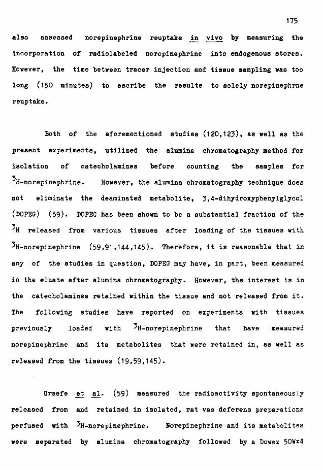
175
also assessed norepinephrine reuptake in vivo by measuring the
incorporation of radiolabeled norepinephrine into endogenous stores.
However, the time between tracer injection and tissue sampling was too
long (150 minutes) to ascribe the results to solely norepinephrne
reuptake.
Both of the aforementioned studies (120,123), as well as the
present experiments, utilized the alumina chromatography method for
isolation of catecholamines before counting the samples for
3H-norepinephrine. However, the alumina chromatography technique does
not eliminate the deaminated metabolite, 3,4-dihydroxyphenylglycol
(DOPEG) (59). DOPEG has been shown to be a substantial fraction of the
3H released from various tissues after loading of the tissues with
3H-norepinephrine (59,91 ,144,145). Therefore, it is reasonable that in
any of the studies in question, DOPEG may have, in part, been measured
in the eluate after alumina chromatography. However, the interest is in
the catecholamines retained within the tissue and not released from it.
The following studies have
previously loaded with
reported on experiments
3H-norepinephrine that
with tissues
have measured
norepinephrine and its metabolites that were retained in, as well as
released from the tissues (19,59,145).
Graefe et al. (59) measured the radioactivity spontaneously
released from and retained in isolated, rat vas deferens preparations
perfused with 3H-norepinephrine. Norepinephrine and its metabolites
were separated by alumina chromatography followed by a Dowex 50Wx4

176
column (to separate norepinephrine from DOPEG). The spontaneous outflow
of 'H-catechols from the preparation was measured two hours after the
end of incubation with 3H-norepinephrine. Results indicated that 70% of
3H released spontaneously from the isolated, perfused organ was in the
However, at the same time after incubation,
3H-norepinephrine accounted for over 95% of the total radioactivity
retained within the tissue. In similar fashion, Tarlov and Langer (145)
investigated the effect of field stimulation on the release of labeled
norepinephrine from rat and guinea pig atria. Norepinephrine and its
metabolites were separated by paper chromatography. Results indicated
that 90% of the radioactivity released spontaneously from atria was
associated with norepinephrine metabolites primarily deaminated
byproducts (including DOPEG). Field stimulation increased the release
of radioactivity and also the fraction of 3H recovered as
3H-norepinephrine (60% norepinephrine and 40% deaminated metabolites).
Analysis of the radioactivity retained within atria 150 minutes after
the end of incubation with 3H-norepinephrine demonstrated that more than
90% of the radioactivity corresponded to the norepinephrine
chromatographic peak. Similar results have been demonstrated in
isolated canine hearts (19).
Therefore, the present and cited studies which assessed
norepinephrine reuptake during endotoxicosis with 3H-norepinephrine and
alumina column chromatography have assurance from related studies that
the measured radioactivity was associated with norepinephrine rather
than its metabolites. It should also be noted that even if the

177
recovered 'H-norepinephrine was metabolized prior to measurement, the
total radioactivity would still be an accurate measure or uptake since
the metabolism occurred after reuptake. Had metabolism of
norepinephrine occurred before reuptake, the byproducts would have been
o-methylated extraneuronally by COMT. Uptake 1 will not transport
amines with methoxyl groups present on the aromatic ring (76). The
results of the present in vivo experiments (Tables VII and VIII)
indicated that norepinephrine reuptake was depressed in spleens, but not
in hearts of endotoxic rats. The depression of reuptake in the spleen
was directly related to the severity of the shock state. Experiments in
this study have already demonstrated that norepinephrine depletion from
the heart and spleen was also related to the severity of the shock
state. Although the present experiments indicated that the spleen
functionally takes up less norepinephrine in endotoxicosis, the results
may be reasonably interpreted in two ways: 1) reuptake was depressed in
nerve endings of the spleen, and 2) during endoxicosis, blood flow was
shunted away from the spleen, and hence less delivery of labeled
norepinephrine to the spleen occurred.
Support for the latter hypothesis is present in existing
literature. Measurements of blood flow distribution in various
endotoxic models indicated that while the per cent of cardiac output to
the myocardium remained unaltered or elevated, the proportion to
splanchnic organs was severely limited (42,83,159,160). In the
unanesthetized monkey, Wyler et al. (159) used radiolabeled microspheres
to measure cardiac output distribution both acutely (at the end of a 40

178
minute infusion period) and 24 hours after E. ~ endotoxin infusion.
Acutely, all vascular beds demonstrated some degree of vasodilatation
except the spleen, in which vascular resistance tripled, and blood flow
decreased from 134 to 24 ml/(min x 100 gm tissue). At the same time,
coronary resistance fell, and total coronary flow actually increased to
142 ml/(min x 100 gm) compared to saline-injected, time-matched,
controls (98 ml/(min x 100 gm)). Twenty-four hours after endotoxin
infusion, results from heart and spleen were essentially the same:
directional changes in vascular resistance compared to time-matched
controls were the same, but the magnitude of the responses were reduced.
The same group of investigators (160) studied the distribution of
cardiac output in unanesthetized endotoxic rabbits using similar
techniques. Measurements were made O, 60, and 150 minutes after
endotoxin injection. Vascular resistance increased most dramatically in
the spleen, kidneys, and skin of endotoxic rabbits compared to
time-matched controls. Vasoconstriction in the coronary circulation was
minimal during endotoxicosis and often was no different from
time-matched control values.
Blood flow measurement using radiolabeled microspheres has also
been applied to rodent models. Ferguson et al. (42) measured regional
blood flow in the unanesthetized endotoxic guinea pig. The percentage
of cardiac output to the spleen was drastically reduced at both one and
three hours after endtoxin injection. The coronary circulation received
a higher proportion of the cardiac output during endotoxicosis.
However, since cardiac output fell, both organs had lover than control

179
flow after endotoxin, but the flow deprivation to the spleen was much
greater than to the myocardium. In the rat, Keeler et al. {83) used two
slow infusions of endotoxin {10 mg/kg over 4 hours; 2.5 to 3 mg/kg
infused over a period of four days) to more closely simulate a septic
state. The acute (4 hour) infusion was lethal to 64% of the rats;
chronic infusion (4 days) resulted in no lethality. Under anesthesia,
cardiac output and its distribuion were measured using radioactive
microspheres. Cardiac output was reduced in both acute and chronic
models of endotoxicosis, although the reduction was greatest in the
acute situation. The per cent of cardiac output to the myocardium
remained unchanged in both acute and chronic endotoxicosis compared to
control. Percentage distribution of cardiac output to liver,
gastrointestinal tract, and skin fell in both models of endotoxemia.
Although spleen flows were not measured, hepatic and gastrointestinal
data may be examined as representative of abdominal organ responses.
Thus, distribution of cardiac output during endotoxicosis has
been shown to consistently decrease in the spleen, but may not change or
actually increase in the heart. Labeled norepinephrine uptake from the
circulation is especially sensitive to blood flow distribution to
various organs. In the present experiments norepinephrine reuptake
assessment was dependent on circulatory distribution; thus the decreased
amount of labeled norepinephrine found in the spleen may have been the
result of decreased distribution of flow to that region. Because flow
distribution changes little in the myocardium, results in that organ may

180
be more representative of the actual uptake process rather than a
function of delivery.
Therefore, experiments were undertaken to study norepinephrine
reuptake in a model where delivery of the labeled bioamine was not
flow-limited. Incubation of tissue slices in vitro effectively
controlled substrate availability to the reuptake sites. It also
ensured that both control and endotoxic preparations were oxygenated to
the same extent. Results of in vitro experiments indicated similar
findings as the in vivo studies: norepinephrine reuptake and retention
was depressed in spleen slices but not myocardial slices from endotoxic
rats (Figures 19 and 21). Because spleen vet weight increased during
endotoxicosis and results were based on a wet weight basis, the
norepinephrine retention values were corrected to eliminate the possible
error. After correction, spleen slices from endotoxic rats still
retained far less norepinephrine than spleen slices from controls. The
differential depression of norepinephrine uptake between organs raises a
question regarding the mechanism responsible for it. If endotoxin or
its mediators were responsible for reuptake inhibition, the effect
should have been evident in both hearts and spleens. However, because
neuronal reuptake of norepinephrine is energy dependent (76), and
because blood flow is severely reduced to the spleen (but not the heart)
during endotoxicosis (42,83,159,160), reuptake depression in the spleen
may have been caused by flow and hence energy deprivation secondary to
endotoxin administration. The ischemia and acidosis in the spleen was
probably more severe in the latter stages of shock, which is.also the

181
time when norepinephrine uptake was most severely limited in the in vivo
experiments.
Another energy dependent process associated with monoamine
metabolism is the uptake of norepinephrine from neuronal cytoplasm into
vesicles. Because neuronal reuptake has been compromised, it is
reasonable to assume that the repackaging process bas also been
affected. Neuronal reuptake without concomitant revesicularization
results in degradation of the norepinephrine by monoamine oxidase.
Thus, if repackaging was depressed, much of the norepinephrine taken up
by the reuptake process would be reduced to metabolites. The data in
Figure 23 illustrate that relatively more of the norepinephrine taken up
by spleen slices of control rats remained urunetabolized than in the
spleens of severely shocked rats. Thus, these results suggest that
increased intraneuronal degradation occurred in spleens of endotoxic
rats, which may contribute to the norepinephrine depletion that occurs
in that organ. Experiments on endotoxic guinea pigs (7) also provided
evidence consistent with this hypothesis.
D. Norepinephrine Turnovers during Endotoxicosis
Norepinephrine turnovers accurately reflect the level of
sympathetic nerve activity (12,26,27) in various organs. Assessment may
be made by any of the following methods: 1) Measurement of radiolabeled
norepinephrine formed after infusion (111,133) or rapid inject~on (133)
of radiolabeled tyrosine yields an accurate estimate of norepinephrine

182
synthesis and can be used to calculate turnover rates. 2) When
norepinephrine synthesis is inhibited with alpha-methyltyrosine,
endogenous norepinephrine levels decline at a rate proportional to the
concentration of remaining amine. A graph of endogenous norepinephrine
content versus time mathematically describes a first order relationship,
and is used to calculate a rate constant of decay and a turnover rate
(12,109). 3) Injection of labeled norepinephrine is taken up by
sympathetic neurons and released along with endogenous norepinephrine.
Under conditions of constant endogenous levels of norepinephrine the
relationship of specific activity of tissue norepinephrine versus time
describes a first order decay (105,26).
Each technique has its positive aspects as well as its
drawbacks. The labeled tyrosine methodology yields synthesis rates from
single determinations while the other techniques necessitate use of
groups of animals. However, the tyrosine method also relies on either
an indwelling cannula or an acutely anesthetized animal model to
facilitate infusion of tyrosine. The unanesthetized model would be of
choice, but the possibility of stress and infection from prior surgery
may complicate the endotoxin model. Additionally, methodology for
analysis of tyrosine would be required. The alpha-methyltyrosine
technique is reliable and can be performed in groups of unanesthetized
animals, but the technique measures the decline in norepinephrine
content over time. Because endotoxin administration alone can deplete
norepinephrine from tissues, the first order decay of norepinephrine
content induced by alpha-methyltyrosine injection may not occur during

endotoxicosis.
183
The 3H-norepinephrine technique also depends on groups
of animals to calculate a turnover rate. The method relies on constant
levels of endogenous norepinephrine vhich may be a drawback during
endotoxin shock. This methodology, however, does not by itself disturb
the constancy of the endogenous norepinephrine content (which is not
possible after alpha-methyltyrosine injection). However, the
'H-norepinephrine technique was the procedure of choice to estimate
sympathetic activity during endotoxicosis for the following reasons: 1)
Low doses of endotoxin eliminated norepinephrine depletion as well as
lethality. Furthermore, the constancy of the endogenous norepinephrine
content was measured to ensure proper kinetic analysis. 2) Animals were
studied in a non-invasive, unanesthetized model. 3) Sympathetic
activity was assessed over a long period of time, rather than acutely
during any short phase during endtoxicosis.
Because the depletion of norepinephrine from peripheral organs
was shown to be ·time- and dose-dependent (120), low dose endotoxin
treatment (lethality was 5% at 24 hours) was used to minimize the
depletion of norepinephrine. Preliminary experiments demonstrated that
this regimen did not deplete norepinephrine from hearts or spleens. On
occasion, however, tissue content of norepinephrine was drastically
depressed in samples obtained 12 hours after endotoxin. These rats were
probably more sensitive to endotoxin than others and were excluded from
the study. Basal cardiac turnover rates in control animals are similar
to those found in the literature, but tend to be on the slower end of
those published (3,89,24). However, the experimental paradigm used in

the present experiments may be responsible for this. The rats were
housed singly for three days before the start of the turnover studies.
Isolation may have an effect to lover sympathetic activity.
Additionally, the rats were fasted for eighteen hours before and also
during the turnover measurement. Fasting has been shown to decrease
norepinephrine turnovers to various peripheral organs (89,90,163).
Comparison of the present turnover rates with those of other studies
that have also measured them in a fasting state show good similarity
(3,89).
Endotoxin treatment accelerated the turnover of norepinephrine
in hearts and spleens of rats compared to controls (Figs. 24 and 25,
Table XI). Thus, the average sympathetic activity to hearts and spleens
of endotoxic rats was increased over the 12 hours after endotoxin
injection. This conclusion is consistent with previous studies that
used radiolabeled norepinephrine injections, although none of the
investigations were designed to assess turnover rates quantitatively
(16,120,123). While the results of the present experiments are also
consistent with some electrophysiological results during endotoxicosis
(61), they do not agree with the work of others (86,130). Halinen
demonstrated increased cardiac sympathetic efferent nerve activity early
after endotoxin injection in dogs (61). Koyama et al. (86) and
Santiesteban et al. (130) demontrated decreased preganglionic splanchnic
nerve activity in the endotoxic cat. Major differences in models and
experimental design exist between the electrophysiological and turnover
studies. The use of an invasive, anesthetized cat or dog model is

185
difficult to compare to the unanesthetized rat. Furthermore, the
turnovers estimated nerve activity over a twelve hour period after
endotoxin; nerve recording was performed for brief periods of time early
after endotoxin. Additionally, the differences in species and endotoxin
doses could easily have induced differential changes in sympathetic
activity.
Experiments of the present study demonstrated that
norepinephrine reuptake was reduced in spleens of endotoxic rats in
severe shock. Even though the low dose endotoxin injection for turnover
measurement induced a modest lethality (5%) compared to the fundamental
shock model (70% lethality), similar reuptake depression may have
occurred. Depressed reuptake leads to a reduction of released
transmitter that is recovered by the nerve terminal. It is possible
that during turnover measurement in the spleen, reduced recovery of
norepinephrine and 3H-norepinephrine caused an accelerated decay rate of
norepinephrine specific activity, thus falsely indicating increased
sympathetic nerve activity to that organ. This possibility was tested
by treating control rats with a potent neuronal reuptake inhibitor,
desmethylimipramine, before norepinephrine turnover measurement. The
results indicated that the singular effect of decreased norepinephrine
reuptake
(Figures
did not alter the fraction of norepinephrine released per hour
26 and 27, Table XIII). Therefore, it is concluded that even
if splenic norepinephrine reuptake was depressed in the present
endotoxic model, it did not contribute to the observed increased rate of
norepinephrine turnover.
186
The increased norepinephrine turnovers in hearts and spleens may
have been the result of increased central drive of sympathetic nerves.
However, electrophysiological experiments on isolated nerve-muscle
preparations suggested that endotoxin affected information processing at
the presynaptic site (115,117,118). One hypothesis was that endotoxin
increased the amount of transmitter released per nerve impulse. If this
indeed occurred in the in situ situation, norepinephrine turnovers may
have increased without a concomitent increase in nerve activity.
Alternatively, increased release per stimulus coupled with even a modest
increase in sympathetic activity could result in greatly increased
norepinephrine turnovers. The effect of increased norepinephrine
release per
norepinephrine
stimulus would effectively accelerate the loss of
from the existing pool (both labeled and unlabeled) and
increase the replacement with newly formed unlabeled norepinephrine.
Regardless
hearts and
magnified
of the mechanism for the increased turnovers measured in
spleens of endotoxic rats, the turnover rates may have been
under conditions of higher endotoxin administration and
contribute to the norepinephrine depletion that occurs in lethal
endotoxic shock.
E. Implications and Conclusions
The role of the sympathetic nervous system in endotoxic shock is
multi-faceted, and many of its aspects remain to be elucidated.
Demonstrations of increased circulating concentrations and depleted

187
tissue content of catecholamines suggested that sympathetic activity was
high during endotoxic shock. While a classic view of the sympathetic
nervous system is that it aids in the maintenance of homeostasis during
stressful or "fight or flight" situations, the sympathetic system may be
a double edged sword in shock. Early during shock the sympathetic
system may maintain blood presure and enhance metabolic substrate
availability. However, the utilization of these compensatory mechanisms
may exhaust both circulatory and metabolic reserve leading to the
irreversible phase of shock. Presumably the vasoactive properties of
catecholamines cause prolonged flow deficits to multiple organ systems,
most notably the splanchnic region, to maintain perfusion of more flow
sensitive areas such as heart and brain. Additionally, the metabolic
effects of catecholamines may alter substrate availability as well as
metabolism. Although the sympathetic nervous system may tide an animal
through the early phases of shock, it may ultimately exacerbate the
situation later in the form of ischemic organs and metabolic
derangements. Many past reports as well as data presented here
demonstrated the beneficial effects of reduced sympathetic stimulation
during endotoxicosis.
Norepinephrine depletion from peripheral tissues is one
manifestation of sympathetic dysfunction during endotoxicosis that
warrants further study. In light of increased plasma concentrations of
catecholamines during shock, the depletion most likely represents a
massive transfer of catecholamines from nerve terminals to synaptic
spaces and finally to the general circulation. Thus, prevention of the

188
depletion would also prevent hyperactivation of adrenergic receptors.
As discussed earlier, the major candidates for the mechanism of
norepinephrine depletion were considered to be increased sympathetic
activity and depressed neuronal reuptake. These hypotheses are
consistent with the existing literature and also fit within the
conceptual framework of sympathetic exacerbation of the shock syndrome.
Increased sympathetic drive and decreased neuronal reuptake both result
in greater release of catecholamines from nerve terminals. This is in
contrast to hypotheses of singular inhibition of norepinephrine
synthesis or increased intraneuronal degradation which would result in
depletion, but not increased liberation of transmitter.
Elucidation of the mechanism and subsequent prevention of the
increased sympathetic activity as well as interventions to maintain
splenic reuptake of norepinephrine may well be future goals to formulate
protective interventions in not only endotoxic shock, but also its
clinical counterpart, sepsis.

SUMMARY
Activation of the sympathetic nervous system during endotoxic
shock has been demonstrated by a variety of methods: elevation of plasma
catecholamines, depletion of tissue norepinephrine, altered disposition
of administered radiolabeled catecholamines, and increased nerve
activity measured directly by electrophysiological techniques. However,
these studies have been largely indirect and/or contradictory in nature.
Furthermore, a large volume of literature has implicated the sympathetic
nervous system
pathogenesis
investigate
of
the
either in a primary or secondary role - in the
endotoxic shock. Experiments were designed to
following aspects of sympathetic nervous system
dysfunction during endotoxicosis in the white laboratory rat: 1)
demonstrate and further characterize norepinephrine depletion from heart
and spleen, 2) assess norepinephrine reuptake both in vivo and in vitro
in hearts and spleens by measuring the amount of 3H-norepinephrine
incorporated into those organs, and 3) quantitatively estimate the
degree of sympathetic activity in both hearts and spleens of endotoxic
rats by measurement of norepinephrine turnovers by the 3H-norepinephrine
decay technique.
Initial experiments demonstrated that the depletion from hearts
and spleens was related to the severity of the shock state as evidenced
by the significant negative correlation of norepinephrine content to the
189

190
plasma glucose concentration. A separate group of rats treated with
insulin demonstrated no depletion and suggested that the depletion with
endotoxin was not caused by the singular effect of low plasma glucose.
Ganglionic blockade with chlorisondamine prevented the depletion from
the heart (spleen was not tested) which provided evidence that the
depletion was neurally-mediated. Alpha adrenergic blockade
(phentolamine) before endotoxin injection significantly increased
survival time, maintained normal plasma glucose levels, and resulted in
higher cardiac and splenic norepinephrine content than in non-treated
endotoxic rats. Beta adrenergic blockade (propranolol) before endotoxin
injection significantly decreased survival time and exacerbated the
hypoglycemia of endotoxicosis, but did not result in significant changes
in cardiac or splenic norepinephrine content compared to non-treated
endotoxic rats. Depletion of peripheral tissue norepinephrine content
before endotoxin administration (sympathectomy with 6-hydroxydopamine)
significantly decreased the lethality associated with endotoxin
administration.
Uptake of 3H-norepinephrine from the circulation was depressed
in spleens, but not hearts of endotoxic rats (uptake depression was
greater in spleens of severely shocked rats). Because blood flow
distribution has been shown to change during endotoxicosis, in vitro
experiments were repeated on hearts and spleens of control and severely
shocked rats. Results of in vitro incubation of tissue slices with
3H-norepinephrine were similar to in vivo results: uptake of labeled
norepinephrine was depressed in spleens, but not hearts of endotoxic

rats compared to control.
191
Additionally, less of the labeled
norepinephrine was retained in its unmetabolized form in endotoxic
spleens. The results indicated that reuptake depression may be a factor
in the depletion of norepinephrine from spleens, but not hearts of
endotoxic rats. Furthermore, uptake and degradation may also play a
role in splenic norepinephrine depletion.
Norepinephrine turnovers were increased in both hearts and
spleens of rats administered a low dose of endotoxin (LD 5%). Turnovers
were also measured in a group of rats injected with the norepinephrine
reuptake blocker, desmethylimipramine to assess the singular role of
reuptake inhibition on turnover rate. Comparison of turnover rates in
control and desmethylimipramine-treated rats demonstrated no
differences. Results indicated quantitatively that sympathetic activity
was increased to hearts and spleen of endotoxic rats.
It is concluded that norepinephrine depletion from hearts and
spleens may be mediated by increased sympathetic activity to those
organs. Additionally, decreased neuronal reuptake of norepinephrine may
play a role in the norepinephrine depletion in the spleen, but not in
the heart. Both mechanisms would result in increased stimulation of
adrenergic receptors and contribute to the hypersympatheti~ state
associated with the pathogenesis of endotoxic shock.

REFERENCES
1. Adler-Graschinsky, E. and s.z. Langer. Possible role of a beta adrenergic adrenoceptor in the regulation of noradrenaline release by nerve stimulation through a positive feedback mechanism. Br. J. Pharmacol. 53:43-50, 1975.
2. Agarwal, M.K. Influence of chemical sympathectomy on endotoxin lethality in young rats. Path. Microbiol. 40:268-274, 1974.
3. Avakian, E.V. and S.M. Horvath. Starvation suppresses sympathoadrenal medullary response to cold exposure in rats. Am. J. Physiol. 241 :E316-E320, 1981.
4. Axelrod, J. Metabolism of epinephrine and other sympathomimetic amines. Physiol. Rev. 39:751-776, 1959.
5. Axelrod, J. Purification and phenylethanolamine-N-methyl transferase. 237:1657-1666, 1962.
properties J. Biol.
of Chem.
6. Bhagat, B. and E. Friedman. Factors involved in maintenance of cardiac catecholamine content: relative importance of synthesis and re-uptake. Br. J. Pharmac. 37:24-33, 1969.
7. Bhagat, B., D. Cavanagh, B.N. Merrild, M.W. Rana, and P.S. Rao. Noradrenaline and tyramine action on isolated atrial muscle of endotoxin-treated guinea-pigs. Br. J. Pharmac. 39:688-695, 1970.
8. Blaschko, H. Catecholamine biosynthesis. Br. Med. Bull. 29:105-109, 1973.
9. Bolton, W.K. and endotoxin-induced 13:263-270, 1978.
N.O. Atuk. Study of chemical sympathectomy in lethality and fibrin deposition. Kidney Int.
10. Bouquet, P. and Y. Izard. Effect of dibenamine on vascular response of rabbits to typhoid endotoxin. Proc. Soc. Exp. Biol. Med. 75:254, 1950.
11. Brenneman, A.R. and S. Kaufman. The role of tetrahydropteridines in the enzymatic conversion of tyrosine to 3,4-dihydroxyphenylalanine. Biochem. Biophys. Res. Commun. 17:177-183, 1964.
12. Brodie, B.B., E. Costa, A. Dlabac, N.H. Neff, and H.H. Smookler. Application of steady state kinetics to the estimation of synthesis rate and turnover time of tissue catecholamines. J. Pharmacol. Exp. Ther. 154:493-498, 1966.
192

193
13. Brody, M.J. and P.J. Kadowitz. Prostaglandins as modulators of the autonomic nervous system. Fed. Proc. 33:48-60, 1974.
14. Canepa, J.F. and O.A. Gomez-Povina. Electrolyte changes in hemorrhagic ventricular myocardium following experimental
hypotension in the dog. J. Surg. Res. 5:335-340, 1965.
15. Cannon, W.B., conditions of 69:46-66, 1924.
M.A. Mciver, and s.w. Bliss. activity in endocrine glands.
Studies Am. J.
on the Physiol.
16. Cavanagh, D., P.s. Rao, D.M.c. Sutton, B.D. Bhagat, and F. Bachmann. Pathophysiology of endotoxin shock in the primate. Am. J. Obstet. Gynecol. 108:705-722, 1970.
17. Chang, C.C. and C.C. Chiueh. Increased uptake of noradrenaline in rat submaxillary gand during sympathetic nerve stimulation. J. Pharm. Pharmacol. 20:158-159, 1968.
18. Chidsey, C.A., E. Braunwald, A.G. Morrow, Myocardial norepinephrine concentration in reserpine and of congestive heart failure. 269:653-658, 1963.
and D.T. Mason. man. Effects of
New Engl. J. Med.
19. Chidsey, C.A., R.L. Kahler, L.L. Kelminson, and E. Braunwald. Uptake and metabolism of tritiated norepinephrine in the isolated canine heart. Circ. Res. 12:220-227, 1963.
20. Chidsey, C.A., G.A. Kaiser, E.H. Sonnenblick, J.F. Spann, Jr., and E. Braunwald. Cardiac norepinephrine stores in experimental heart failure in dogs. J. Clin. Invest. 43:2389-2393, 1964.
21. Chidsey, C.A., E. Braunwald, and A.G. Morrow. Catecholamine excretion and cardiac stores of norepinephrine in congestive heart failure. Am. J. Med. 39:442-451, 1965.
22. Ciaranello, R.D., R.E. Barchas, G.S. Byers, D.W. Stemmle, and J.D. Barchas. Enzymatic synthesis of adrenaline in mammalian brain. Nature 221 :368-369, 1969.
23. Coleman, B., and v.v. Glaviano. Tissue levels of norepinephrine and epinephrine in hemorrhagic shock. Science 139:54, 1963.
24. Costa, E. and N.H. Neff. Isotopic and non-isotopic measurements of the rate of catecholamine biosynthesis. in Biochemistry and Pharmacology of the Basal Ganglia, E. Costa, .J. Cote, and M.D. Yahr, eds., Raven:New York, pp. 141-155, 1965.
25. Costa, E., D.J. Interactions of 18:577' 1966.
Boullin, w. Hammer, w. Vogel, and B.B. Brodie. drugs with adrenergic neurons. Pharmacol. Rev.,

194
26. Costa, E., and N.H. Neff. Importance of turnover rate measurements to elucidate the function of neuronal monoamines. In: Topics in Medicinal Chemistry, Vol. 2, edited by J.L. Rabinowitz, and R.M. Myerson. New York:Interscience Publishers, 1968, 65-95·
27. Costa, E., and N.H. Neff. Estimation of turnover rates to study the metabolic regulation of the steady-state level of neuronal monoamines. In: Handbook of Neurochemistry, Vol. 4, Control Mechanisms in the C.N.S., edited by A. Lajtha, New York:Plenum Press, 1968, 45-90.
28. Cotzias, G. and localization of 78:157-160, 1951.
V. Dole. Metabolism of amines. II: Mitochondrial monoamine oxidase. Proc. Soc. Exp. Biol. Med.
29. Crout, J.R. Catechol Standard Methods of 62-80, 1961.
amines Clinical
in urine. Chemistry.
in D. Seligson, ed., New York:Academic, pp.
30. Dahlstrom, A.B. and B.E.M. Zetterstrom. Noradrenaline stores in nerve terminals of the spleen: changes during hemorrhagic shock. Science 147:1583-1585, 1965.
31. Dairman, W., R. Gordon, s. Spector, A. Sjoerdsma, and s. Udenfriend. Increased synthesis of catecholamines in the intact rat following administration of alpha adrenergic blocking agents. Molec. Pharmacol. 4:457-464, 1968.
32. Dennis, E.W. Toxicity of acid-soluble typhoid toxin for laboratory animals. Proc. Soc. Exp. Biol. Med. 42: 553-554, 1939.
33. Devereux, D.F., C.A. Michas, and S. Rice. Heparin pretreatment suppresses norepinephrine concentrations in dogs in endotoxic shock. Clin. Chem. 23:1346-1347, 1977.
34. Dietzman, R.H., C.B. Beckman, L.H. Romero, L.S. Schultz, and R.C. Lillehei. Effects of blunted sympathetic nervous system response on regional tissue perfusion in experimental endotoxic shock. J. Surg. Res. 14:412-419, 1973.
35. Egdahl, R.H. The effect of bacterial endotoxin on adrenal medullary function. Clin. Res. 7:158,1959.
36. Egdahl, medulla 1959.
R.H. The differential response of the adrenal cortex and to bacterial endotoxin. J. Clin. Invest. 38:1120-1125,
37. Essex, H.E. Further doses of epinephrine. Am. J. Physiol. 171 :78-86, 1952.

195
38. von Euler, U.S. and F. Lishajko. Catecholamine release and uptake in isolated adrenergic nerve granules. Acta Physiol. Scand. 57:468-480, 1963.
39. Falck, B. Observations on the possibilities of the cellular localization of monoamines by a fluorescence method. Acta Physiol. Scand. 56(Suppl. 197):1-25, 1962.
40. Farnebo, L.O. and B. Hamberger. Noradrenergic mechanisms in hemorrhagic shock of the rat. Acta Chir. Scand. 143:1-8, 1977.
41. Farnebo, L.O., H. Hallman, B. Hamberger, and G. Jonsson. Catecholamines and hemorrhagic shock in awake and anesthetized rats. Circ. Shock 6:109-118, 1979.
42. Ferguson, J.L., J.J. Spitzer, and H.I. Miller. Effects of endotoxin on regional blood flow in the unanesthetized guinea pig. J. Surg. Res. 25:236-243, 1978.
43. Feuerstein, indomethacin responses to 1981.
J.A. Dimicco, A. Ramu, and I.J. Kopin. Effect of on the blood pressure and plasma catecholamine acute endotoxaemia. J. Pharm. Pharmacol. 33:576-579,
44. Fielden, R. and A.L. Green. A comparative study of the noradrenaline-depleting and sympathetic-blocking actions of guanethidine and (-)-beta-hydroxyphenethylguanidine. Br. J. Pharmacol. Chemother. 30:155-165, 1967.
45. Filkins, J.P. Phases of glucose dyshomeostasis in endotoxicosis. Circ. Shock 5:347-355, 1978.
46. Filkins, J.P. Adrenergic blockade and glucoregulation in endotoxin shock. Circ. Shock 6:99-107, 1979.
47. Fine, J. The intestinal circulation in shock. Gastroenterology 52:454-460, 1967.
48. Garber, A., P. Cryer, J. Santiago, M. Haymond, A. Pagliara, and D. Kipnis. The role of adrenergic mechanisms and hormonal responses to insulin-induced hypoglycemia in man. J. Clin. Invest. 58:7-14, 1976.
49. Gilbert, R.P. and R. Hobf. Hemodynamic basis of norepinephrine shock. Proc. Soc. Exp. Biol. Med. 116:43-46, 1964.
50. Gillis, C.N. Increased retention of exogenous norepinephrine by cat atria after electrical stimulation of the cardio-accelerator nerves. Biochem. Pharmac. 12:593-595, 1963.

51. Gillis, C.N. and D.M. Paton. Cation transmitter retention by slices of Pharmacol. 29:309-318, 1967.
196
dependence of sympathetic rat ventricles. Br. J.
52. Glaviano, v.v., and B. Coleman. Myocardial depletion of norepinephrine in hemorrhagic hypotension. Proc. Soc. Exp. Biol. Med. 107:761-763, 1961.
53. Glaviano, v.v. and M.A. Klouda. Myocardial catecholamines and stimulation of the stellate ganglion in hemorrhagic shock. Am. J. Physiol. 209:751-756, 1965.
54. Glowinski, J., J. Axelrod, I. Kopin, and R. Wurtman. Physiological disposition of H3-norepinephrine in the developing rat. J. Pharmacol. Exp. Ther. 146:48-53, 1964.
55. Gomez-Povina, O.A. and J.F. Canepa. Catecholamine content of the ventricular myocardium in dogs following hemorrhagic hypotension. J. Surg. Res. 5:341-345, 1965.
56. Gordon, R. J.v.o. Reid, A. Sjoerdsma, and S. Udenfriend. Increased synthesis of norepinephrine in the cat heart on electrical stimulation of the stellate ganglion. Molec. Pharmacol. 2:606-613, 1966.
57. Goto, F., T. Fujita, E. Otani, and M. Yamamuro. The effect of indomethacin and adrenergic receptor blocking agents on rats and canine responses to endotoxin. Circ. Shock 7:413-424, 1980.
58. Gourzis, J.T., M.W. Hollenberg, and M. Nickerson. Involvement of adrenergic factors in the effects of bacterial endotoxin. J. Exp. Med. 114:593-604, 1961.
59. Graefe, K.H., F.J.E. Stefano, and s.z. Langer. Preferential metabolism of (-)-3H-norepinephrine through the deaminated glycol in the rat vas deferens. Biochem. Pharmacol. 22:1147-1160, 1973.
60. Grillo, M.A. Electron microscopy of sympathetic tissues. Pharmacol. Rev. 18:387-399, 1966.
Initial effects of endotoxin on cardiovascular reflex circulation in dogs. Acta Physiol. Scand. (Suppl.)
61. Halinen, M. functions and 439:1-61, 1976.
62. Hall, R.C. and R.L. Hodge. Vasoactive hormones in endotoxin shock: a comparative study in cats and dogs. J. Physiol. 213:69-84, 1971.
63. Halmagyi, D.F.J., M. Kennedy, A.H. Goodman, and D. Varga. Simple and combined adrenergic receptor blockade in canine endotoxemia. Eur. Surg. Res. 3:326-339, 1971.

197
64. Harrison, D.C., C.A. Chidsey, and E. Braunwald. Effect of hemorrhagic shock on release of norepinephrine by tyramine. Am. J. Physiol. 206:1262-1266, 1964.
65. Heiffer, M.H., concentrations 18: 66, 1959.
R.L. Mundy, and B. Mehlman. Adrenal catechol amine following endotoxin administration. Fed. Proc.
66. Heiffer, M.H., R.L. Mundy, and B. Mehlman. Effect of lethal doses of bacterial endotoxin (E. coli) on sympathetic neurohormones in the rabbit. Am. J. Physiol-.-198:1307-1311, 1960.
67. Hift, H. and J.G. Strawitz. Irreversible hemorrhagic shock in dogs: problem of onset or irreversibility. Am. J. Physiol. 200:269-273, 1961.
68. Hift, H., and H.A. Campos. Changes in the subcellular distribution of cardiac catecholamines in dogs dying in irreversible haemorrhagic shock. Nature 196:678-679, 1962.
69. Hinshaw, L.B., C.M. Brake, T.E. Emerson, Jr., M.M. Jordan, and F.D.
70.
Masucci. Participation of sympathoadrenal system in endotoxin shock. Am. J. Physiol. 207:925-930, 1964.
Hokfelt, B., Bygdeman, S., and Sekkenes, J. The participation of the adrenal glands in endotoxin shock. in K.D. Bock (ed.), Shock -Pathogenesis and Therapy, pp. 151-161. Springer-Verlag:Berlin, 1962.
71. Hruza, z. Protective effect of depot catecholamines in shock. Circ. Shock 2:65-72, 1975.
72. Huntington, J.L., and J.M. Johnson. Proof-of-claims test report comparing YSI model 23A vs. hexokinase method for blood plasma, blood serum, and whole blood. Yellow Springs, Ohio: YSI Corporation. unpublished.
73. Iampietro, P.F., LL.B. Hinshaw, and C.M. Brake. Effect of an adrenergic blocking agent on vascular alterations associated with endotoxin shock. Am. J. Physiol. 204:611-614, 1963.
74. Ikeda, M. L.A. Fabien, and S. Udenfriend. A kinetic study of bovine adrenal tyrosine hydroxylase. J. Biol. Chem. 241 :4452-4456, 1966.
75. Iversen, L.L. and E.A. Kravitz. Sodium dependence of transmitter uptake at adrenergic nerve terminals. Molec. Pharmacol. 2:360-362, 1966.
76. Iversen, L.L. Catecholamine uptake processes. Br. Med. Bull. 29:130-135, 1973.

198
77. Iversen, L.L. Neuronal and extraneuronal catecholamine uptake mechanisms. in Frontiers in Catecholamine Research, E. Usdin and S. Snyder, eds., Pergamon:London, pp. 403-408, 1973.
78. Iversen, L.L. Uptake processes for biogenic amines. in Handbook of Psychopharmacology, vol. l· Biochemistry of Biogenic Amines. L."'E:" Iverson, S.D. Iverson, and S.H. Snyder, eds., Plenum:New York, pp. 381-442, 1975.
79. Jellinek, M., N. Sperelakis, L.M. Napolitano, and T.J. Cooper. 3,4-Dihydroxyphenylalanine in cultured ventricular cells from chick embryo heart. J. Neurochem. 15:959-963, 1968.
80. Jones, S.B. and X.J. Musacchia. Tissue catecholamine levels of the golden hamster (Mesocricetus Auratus) acclimated to 7, 22, and 34° C. Comp. Biochem. Physiol. 52:91-94, 1975.
81. Jones, S.B. Myocardial norepinephrine turnover during induced hypothermia and rewarming. J. Appl. Physiol. 50:962-966, 1981.
82. Kaelin, C.R. and R.D. Rink. Effects of phentolamine on hepatic p02 in endotoxemia. Can. J. Physiol. Pharmacol. 58:281-286, 1980.
83. Keeler, R. A. Barrientos, and K. Lee. Circulatory effects of acute or chronic endotoxemia in rats. Can. J. Physiol. Pharmacol. 59:204-208, 1981.
84. Kirshner, N. Pathway of noradrenaline formation from DOPA. J. Biol. Chem. 226:821-825, 1957.
85. Kobold, E.E., R. Lovell, W. Katz, and A.P. Thal. Chemical mediators released by endotoxin. Surg. Gynecol. Obstet. 118:807-813, 1964.
86. Koyama, S., H.L. Santiesteban, w.s. Ammons, and J.W. Manning. Effects of sinoaortic denervation on hypotension induced by E. coli endotoxin and by hypovolemia in cats (Abstract). Physiologist 23:36, 1980.
87. Kux, M., D.D. Holmes, L.B. Hinshaw, and W.H. Massion. Effects of injection of live Escherichia coli organisms on dogs after denervation of the abdominal viscera. Surgery 70:392-398, 1971.
88. Landsberg, L. and J. Axelrod. Influence of pituitary, thyroid, and adrenal hormones on norepinephrine turnover and metabolism in the rat heart. Circ. Res. 22:559-571, 1968.
89. Landsberg, L. and J.B. Young. Fasting, feeding and regulation of the sympathetic nervous system. New Engl. J. Med. 298:1295-1301, 1978.

90. Landsberg, L., L. Greff, S. Gunn, and J.B. Young. Adrenergic mechanisms in the metabolic adaptation to fasting and feeding: effects of phlorizin on diet-induced changes in sympathoadrenal activity in the rat. Metabolism 29:1128-1137, 1980.
91. Langer, S.Z. The metabolism of 3H-noradrenaline released by electrical stimulation from the isolated nictitating membrane of the cat and from the vas deferens of the rat. J. Physiol. (London) 208:515-546, 1970.
92. Langer, s.z. The regulation of transmitter release elicited by nerve stimulation through a presynaptic feedback mechanism. in: Frontiers in Catecholamine Research, E. Usdin and S. Snyder, eds., Pergamon:London, PP• 543-549, 1973.
93. Levin, E.Y. and s. Kaufman. Studies on the enzyme catalyzing the conversion of 3,4-dihydroxyphenylethylamine to norepinephrine. J. Biol. Chem. 236:2043-2049, 1961.
94. Levine, R.J. and L. Landsberg. Catecholamines and the adrenal medulla. in: Diseases of Metabolism, Chapter 19, P.K. Bondy and L.E. Rosenberg, eds., Saunders:Philadelphia, pp. 1181-1224, 1974.
95. Levitt, M., s. Spector, A. Sjoerdsma, and Elucidation of the rate-limiting step in biosynthesis in the perfused guinea pig heart. J. Ther. 148:1-8, 1965.
s. Udenfriend. norepinephrine
Pharmacol. Exp.
96. Lillehei, R.C. and L.D. MacLean. Physiological approach to successful treatment of endotoxin shock in the experimental animal. Arch. Surg. 78:464-471, 1959.
97. Lillehei, R.C. History of vasodilation in treating shock and low flow states. Adv. Shock Res. 1 :1-17, 1979.
98. Lovenberg, W., H. Weissbach, ands. Udenfriend. Aromatic 1-amino acid decarboxylase. J. Biol. Chem. 237:89-93, 1962.
99. Lowry, P., T. Blanco, E.A. Santiago-Delpin. Histamine and sympathetic blockade in septic shock. Am. Surg. 43:12-19, 1977.
100. Lund, A. Fluorimetric determination of adrenaline in blood. III. A new sensitive and specific method. Acta Pharmacol. Toxicol. 5:231-247, 1949.
101. Machado, A.B.M. Straight OS04 versus glutaraldehyde-OSO~ in sequence as fixatives for the granular vesicles in sympathetic axons of the rat pineal body. Stain Technol. 42:293-300, 1967.

200
102. Masucci, F.D., and L.B. Hinshaw. Evaluation of protection with phenoxybenzamine against lethal endotoxin shock. Proc. Soc. Exp. Biol. Med. 116:1057-1060, 1964.
103. McKay, D.G., A.N. Whitaker, and v. Cruse. Studies of catecholamine shock: II. An experimental model of microangiopathic hemolysis. Am. J. Pathol. 56:177-192, 1969.
104. Meyer, M.W. and H.M. Ballin. Failure to observe alteration of epinephrine activity after endotoxin. Proc. Soc. Exp. Biol. Med. 100:288-290, 1959.
105. Montanari, R., E. Costa, M.A. Beaven, and B.B. Brodie. Turnover rates of norepinephrine in hearts of intact mice, rats and guinea pigs using tritiated norepinephrine. Life Sci. 4:232-240, 1963.
106. Muscholl, E. and M. Vogt. The action of reserpine on the peripheral sympathetic system. J. Physiol. (Lond) 141:132-155, 1958.
107. Nadeau, R.A., J. de Champlain, and G.M. Tremblay. Supersensitivity of the isolated rat heart after chemical sympathectomy with 6-hydroxydopamine. Can. J. Physiol. Pharmacol. 49:36-44, 1971.
108. Nagatsu, T., M. Levitt, ands. Udenfriend. Tyrosine hydroxylase. The initial step in norepinephrine biosynthesis. J. Biol. Chem. 239:2910-2917, 1964.
109. Neff, N.H. and the study of inhibition or 40-47, 1967.
E. Costa. Application of steady-state kinetics to catecholamine turnover after monoamine oxidase
reserpine administration. J. Pharmacol. Exp. Ther.
110. Neff, N.H., T.N. Tozer, W. Hammer, E. Costa, and B.B. Brodie. A~plication of steady-state kinetics to the uptake and decline of H -NE in the rat heart. J. Pharmacol. Exp. Ther. 160:48-52, 1968.
111. Neff, N.H., S.H. Ngai, C.T. Wang, and E. Costa. Calculation of the rate of catecholamine synthesis from the rate of conversion of tyrosine-14c to catecholamines - effect of adrenal demedullation on synthesis rates. Mol. Pharmacol. 5:90-99, 1969.
112. Nykiel, F. and v.v. Glaviano. Adrenal catecholamies in E. coli endotoxin shock. J. Appl. Physiol. 16:348-350, 1961.
113. Palmerio, c., s.c. Ming, E.D. Frank, and J. Fine. Cardiac tissue response to endotoxin. Proc. Soc. Exp. Biol. Med. 109:773-776, 1962.
114. Palmerio, c., B. Zetterstrom, J. Shammash, E. Euchbaum, E. Frank, and J. Fine. Denervation of the abdominal viscera for the treatment of traumatic shock. Nev Engl. J. Med. 269:709-716, 1963.

201
115. Parnes, I., R. Reinhold, J. Fine. Synaptic transmission in the crayfish: increased release of transmitter substance by bacterial endotoxin. Science 171:1153-1155, 1971.
116. Paton, D.M. metaraminol 1968.
Cation by rat
and metabolic uterine horn.
requirements for retention of Br. J. Pharmacol. 33:277-286,
117. Person, R.J. Endotoxin alters spontaneous transmitter release at the frog neuromuscular junction. J. Neurosci. Res. 3:63-72, 1977.
118. Person, R.J. Bacterial lipopolysaccharide depresses spontaneous, evoked, and ionophore-induced transmitter release at the neuromuscular junction. J. Neurosci. Res. 4:105-114, 1979.
119. Pohorecky, L.A., M. Zigmond, H. Karten, and R.J. Wurtman. Enyzmatic conversion of norepinephrine to epinephrine by the brain. J. Pharmacol. Exp. Ther. 165:190-195, 1969.
120. Pohorecky, L.A., R.J. Wurtman, D. Taam, and J. Fine. Effects of endotoxin on monoamine metabolism in the rat. Proc. Soc. Exp. Biol. Med. 140:739-746, 1972.
121. Porter, C.C., 6-hydroxydopamine norepinephrine in 140:308-316, 1963.
J.A. Totaro, and C.A. Stone. Effect of and some other compounds on the concentration of
the hearts of mice. J. Pharmacol. Exp. Ther.
122. Prager, R.L., E.L. Dunn, and J.F. Seaton. Increased adrenal secretion of norepinephrine and epinephrine after endotoxin and its reversal with corticosteroids. J. Surg. Res. 18:371-375, 1975.
123. Rao, P.S., B.D. Bhagat, and D. Cavanagh. Effect of endotoxin on hemodynamics and norepinephrine metabolism in the dog. Proc. Soc. Exp. Biol. Med. 141:412-418, 1972.
124. Reddin, J.L., B. Starzecki, W.W. Spink. Comparative hemodynamic and humoral responses of puppies and adult dogs to endotoxin. Am. J. Physiol. 210:540-544, 1966.
125. Reilly, J., E. Rivlier, A. Compagnon, R. Laplane, and H. duBuit. Sur la pathogenie de la dothienenterie; le role du systeme neuro-vegetatif dens la genese des lesions intestinales. Ann .• de Med. 37:321-358, 1935·
126. Rosenberg, J.C., R.C. Lilllehei, W.H. Moran, and B. Zimmermann. Effect of endotoxin on plasma catechol amines and serum serotonin. Proc. Soc. Exp. Biol. Med. 102:335-337, 1959.

202
127. Rosenberg, J.C., R.C. Lillehei, J. Longerbeam, B. Zimmerman. Studies on hemorrhagic and endotoxin shock in relation to vasomotor changes and endogenous circulating epinephrine, norepinephrine and serotonin. Ann. Surg. 154:611-627, 1961.
128. Roth, R.H., L. Stjarne, and U.S. von Euler. Factors influencing the rate of norepinephrine biosynthesis in nerve tissue. J. Pharmacol. Exp. Ther. 158:373-377, 1967.
129. Sanchez-Armass, S. and F. Orrego. Noradrenaline transport by rat heart sympathetic nerves: A re-examination of the role of sodium ions. Naunyn-Schmiedeberg's Arch. Pharmacol. 302:255-261, 1978.
130. Santiesteban, H.L., s. Koyama, W.S. Ammons, and J.W. Manning. The effects of naloxone on the peripheral sympathetics in endotoxin shock (Abstract). Fed. Proc. 40:573, 1981.
131. Schmid, P.G., D.D. Lund, and R. Roskoske, Jr. Efferent autonomic dysfunction in heart failure. in: Disturbances in Neurogenic Control of the Circulation. F.M. Abboud, H.A. Fozzard, J.P. Gilmore, and D.J. Reis, eds., William and Wilkins:Baltimore, pp. 33-49, 1981.
132. Schnaitman, C., V.G. Erwin, and J.W. Greenawalt. The submitochondrial localization of monoamine oxidase. J. Cell Biol. 32:719-735, 1967.
133. Sedvall, G.C., V.K. Weise, and I.J. Kopin. The rate of norepinephrine synthesis measured in vivo during short intervals; influence of adrenergic nerve impulse activity. J. Pharmacol. Exp. Ther. 159:274-282, 1968.
134. Sharman, D.F. The catabolism of catecholamines - recent studies. Br. Med. Bull. 29:110-115, 1973.
135. Shaw, F.H. The estimation of adrenaline. Biochem. J. 32:19-25, 1938.
136. Smith, L.L., W. Muller, and L.B. Hinshaw. The management of experimental endotoxin shock. Arch. Surg. 89:630-636, 1964.
137. Snyder, S.H., J. Fischer, and J. Axelrod. Evidence for the presence of monoamine oxidase in sympathetic nerve endings. Biochem. Pharmacol. 14:363-365, 1965.
138. Sole, M.J., c. Lo, c.w. Laird, E.H. Sonnenblick, and R.J. Wurtman. lorepinephrine turnover in the heart and spleen of the cardiomyopathic hamster. Circ. Res. 37:855-862, 1975.

203
139. Sole, M.J., A.B. Kamble, and M.J. Hussain. A possible chance in the rate-limiting step for cardiac norepinephrine synthesis in the cardiomyopathic Syrian hamster. Circ. Res. 41:814-817, 1977.
140. Spector, s., A. Sjoerdsma, and endogenous norepinephrine synthesis inhibitor of tyrosine hydroxylase. 147 :86-95' 1965.
s. Udenfriend. Blockade of by alpha methyl tyrosine, an J. Pharmacol. Exp. Ther.
141. Spector, s., R. Gordon, A. Sjoerdsma, and s. Udenfriend. End-product inhibition of tyrosine hydroxylase as a possible mechanism for regulation of norepinephrine synthesis. Molec. Pharmacol. 3:549-555, 1967.
142. Spink, W.W., J. Reddin, S.J. Zak, M. Peterson, B. Starzecki, and E. Seljeskog. Correlation of plasma catecholamine levels with hemodynamic changes in canine endotoxin shock. J. Clin. Invest. 45 :78-85' 1966.
143. Stjarne, L. Studies of catecholamine uptake, storage, and release mechanisms. Acta Physiol. Scand. 62 (Suppl. 228):1-97, 1964.
144. Su, C. and I.A. strips studied stimulation. J.
Bevan. The release of 3H-norepinephrine in arterial by the technique of superfusion and transmural
Pharmacol. Exp. Ther. 172:62-68, ·1970.
145. T~rlov, S.R. and S.Z. Langer. The fate of 3H-norepinephrine released from isolated atria and vas deferens: effect of field stimulation. J. Pharmacol. Exp. Ther. 179:186-197, 1971.
146. Thoenen, H. and J.P. Tranzer. The pharmacology of 6-hydroxydopamine. Annu. Rev. Pharmacol. 13:169-180, 1973.
147. Thomas, L. The physiologial disurbances produced by endotoxins. Ann. Rev. Physiol. 16:467-490, 1954.
148. Thomas, L. The role of epinephrine in the reactions produced by the endotoxins of gram-negative bacteria: I. Hemorrhagic necrosis produced by epinephrine in the skin of endotoxin treated rabbits. J. Exp. Med. 104:865-880, 1956.
149. Trank, J.w. and M.B. modifications associated 202:971-977, 1962.
Visscher. Carotid sinus baroreceptor with endotoxin shock. Am. J. Physiol.
150. Trippodo, J.C. and D.L. Traber. Endotoxic shock in dogs after 6-hydroxydopamine and adrenal denervation and after 48/80. Circ. Shock 3:115-121, 1976.

204
151. Udenfriend, S., P. Zaltzman-Nirenberg, and T. Nagatsu. Inhibitors of purified beef adrenal tyrosine hydroxylase. Biochem. Pharmacol. 14:837-845, 1964.
152. Vigran, I.M. and H.E. Essex. Studies on physiologic effects of larger doses or epinephrine. Am. J. Physiol. 162:230-242, 1950.
153. Wang, C.H., D.L. Willis, and W.D. Loveland. Radiotracer Methodology in the Biological, Environmental, and Physical Sciences, McElroy, W.D. and C.P. Swanson, editors. Prentice-Hall:New Jersey, 181-232, 1975.
154. Weil, M.H., L.B. Hinshaw, M.B. Visscher, W.W. Spink, and L.D. MacLean. Hemodynamic effects of vasopressor agent (metaraminol) on hypotension in dogs produced by endotoxin. Proc. Soc. Exp. Biol. Med. 92:610-612, 1956.
155. Weiner, N. synthesis guinea-pig Pharmacol.
and M. Rabadjija. The effect of nerve stimulation on the and metabolism of norepinephrine in the isolated hypogastric nerve vas deferens preparation. J.
Exp. Ther. 160:61-71, 1968.
156. Weiner, N., F. Lee, E. Dreyer, and E. Barnes. The activation of tyrosine hydroxylase in noradrenergic neurons during acute nerve stimulation. Life Sci. 22:1197-1216, 1978.
157. Westfall, T.C. Local regulation of adrenergic neurotransmission. Physiol. Rev. 57: 659-728, 1977.
158. Whitaker, A.N., D.G. McKay, and I. Csavossy. Studies of catecholamine shock: I. Disseminated intravascular coagulation. Am. J. Pathol. 56:153-169, 1969.
159. Wyler, F., R.P Forsyth, A.S. Nies, J.M. Neutze, and K.L. Melmon. Endotoxin-induced regional circulatory changes in the unanesthetized monkey. Circ. Res. 24:777-786, 1969.
160. Wyler, F., J.M. Neutze, and A.M. Rudolph. Effects of endotoxin on distribution of cardiac output in unanesthetized rabbits. Am. J. Physiol. 219:246-251, 1970.
161. Yard, A.C. and M. Nickerson. Shock produced in dogs by infusions of norepinephrine. Fed. Proc. 15:502, 1956.
162. Young, J.B. and L. Landsberg. Sympathoadrenal activity in fasting pregnant rats. J. Clin. Invest. 64:109-116, 1979.
163. Young, J.B. and L. Landsberg. Effect of diet and cold exposure on norepinephrine turnover in pancreas and liver. Am. J. Physiol. 236:E524-E533, 1981.

205
164. Zetterstrom, B.E., C. Palmerio, and J. line. Changes in tissue content of catechol amines in traumatic shock. Acta Chir. Scand. 128:13-19, 1964.
165. Zetterstrom, B.E., c. Palmerio, and J. Pine. Protection of functional and vascular integrity of the spleen in traumatic shock by denervation. Proc. Soc. Exp. Biol. Med. 117:373-376, 1964.
166. Ziegelhoffer, A., M. Fedelesova, and R. Kvetnansky. Myocardial catecholamines and related metabolic changes during acute coronary occlusion. in Catecholamines and Stress, Usdin, E., R. Kvetnansky, and I.J. Kopin, eds. Pergamon:New York, pp. 483-490, 1975.
167. Zweifach, B.W., A.L. Nagler, and L. Thomas. The role of epinephrine in the reactions produced by the endotoxins of gram-negative bacteria. J. Exp. Med. 104:881-896, 1956.

206
APPROVAL SHEET
The dissertation submitted by Benet John Pardini has been read and approved by the following committee:
Stephen B. Jones, Ph.D., Chairman of Committee Associate Professor, Physiology
Loyola, Stritch School of Medicine
James P. Filkins, Ph.D. Professor and Chairman, Physiology Loyola, Stritch School of Medicine
Robert D. Wurster, Ph.D. Professor, Physiology
Loyola, Stritch School of Medicine
Michael A. Collins, Ph.D. Associate Professor, Biochemistry and Biophysics
Loyola, Stritch School of Medicine
Phillip G. Schmid, M.D. Professor, Internal Medicine
University of Iowa College of Medicine, Iowa City, Iowa
The final copies have been examined by the director of the dissertation and the signature which appears below verifies the fact that any necessary changes have been incorporated and that the dissertation is now given final approval by the Committee with reference to content and form.
The dissertation is therefore accepted in partial fulfillment of the requirements for the degree of Doctor of Philosophy.