neurokinin 1 receptor antagonism requires norepinephrine to increase serotonin function
TRANSCRIPT

www.e l sev i e r . com/ loca te /eu roneu ro
Neurokinin 1 receptor antagonism requiresnorepinephrine to increase serotonin function
Gabriella Gobbi a,c,*,1, Tommaso Cassano b, Fatiha Radja a,c,Maria Grazia Morgese b, Vincenzo Cuomo d, Luca Santarelli e,Rene Hen e, Pierre Blier f
a Department of Psychiatry, McGill University, 1033 Pine Avenue, West Montreal, QC, Canada H3A 1A1b Department of Biochemical Sciences, University of Foggia, Foggia, Italyc Centre de Recherche Fernand Seguin, Universite de Montreal, Montreal, QC, Canadad Department of Human Physiology and Pharmacology, University bLa SapienzaQ, Rome, Italye Center for Neurobiology and Behavior, Columbia University, New York, NY, USAf Mood Disorders Research, Institute of Mental Health Research, University of Ottawa, Ottawa, Ontario Canada K1Z 7K4
Received 23 January 2006; received in revised form 29 May 2006; accepted 18 July 2006
0924-977X/$ - see front matter D 200doi:10.1016/j.euroneuro.2006.07.004
* Corresponding author. Tel.: +1 514E-mail address: Gabriella.Gobbi@M
1 Financial support: G.G. received aQuebec (FRSQ). P.B. holds the Canada
KEYWORDSNK1 receptor;Serotonin;5-HT1A receptor;Substance P;Antidepressant;Anxiolytic
Abstract The present studies examined the role of norepinephrine (NE) system in mediatingthe enhancement of 5-HT function produced by neurokinin (NK)1 receptor antagonism. Dorsalraphe 5-HT and locus coeruleus NE neurons were recorded in vivo in mice lacking NK1 receptorsin wildtype mice pretreated with the NK1 antagonist RP67580 and its inactive enantiomer RP68651. RP67580 and RP68651 were also tested on 5-HT neurons of mice lacking the 5-HT1Areceptor. RP67580 increased the firing rate of 5-HT neurons in wildtype mice and in 5-HT1A nullmutant mice to the same degree, thus indicating that the mechanism by which NK1 antagonistsenhances 5-HT firing is independent of 5-HT1A receptors. NE neuronal burst activity wasincreased in NK1 null mutant and wildtype mice given RP67580, but not with RP68651. After NEdepletion, RP67580 was ineffective in increasing 5-HT neuronal firing activity in NK1 wildtypemice, and the enhancement of 5-HT neuronal firing observed in NK1 null mutant mice wasabolished. In conclusion, NE neurons are essential for the action of NK1 antagonists on 5-HTneurons. In addition, the desensitization of 5-HT1A autoreceptors produced by NK1 receptorantagonism is not critical for enhancing 5-HT neuronal firing.D 2006 Elsevier B.V. and ECNP. All rights reserved.
European Neuropsychopharmacology (2007) 17, 328—338
6 Elsevier B.V. and ECNP. All rights reserved.
398 4650; fax: +1 514 398.cGill.ca (Gabriella Gobbi).salary award (n.1991) and a young investigator grant (n.2782) from Fonds de la Recherche en Sante duResearch Chair in Psychopharmacology.

Neurokinin 1 receptor antagonism requires norepinephrine to increase serotonin function 329
1. Introduction
Tricyclic antidepressant drugs (TCAs), selective serotonin (5-hydroxytryptamine; 5-HT) reuptake inhibitors (SSRIs) and/ornorepinephrine (NE) reuptake inhibitors (SNRIs) and mono-amine oxidase inhibitors (MAOIs), used in the treatment ofboth mood and anxiety disorders, modulate the function ofthese two monoamine systems by different mechanisms.Their net long-term effect is an enhancement of 5-HTneurotransmission (Blier and de Montigny, 1999) and amodification of locus coeruleus firing activity (Szabo andBlier, 2001). Nevertheless, the use of these drugs is limited bytheir side-effect profiles, their delayed onset of action andtheir partial effectiveness. Indeed, approximately 50% ofdepressed patients do not show an acceptable response to aninitial antidepressant trial (Thase and Rush, 1997). SSRIsrequire prolonged administration before a significant clinicalimprovement occurs because the rapid increase of synaptic 5-HT availability caused by reuptake inhibition in the raphenuclei stimulates cell body 5-HT1A autoreceptors, which inturn exerts a negative feedback on 5-HT neuronal firing andrelease in projection areas. Only when 5-HT1A autoreceptorsare desensitized can SSRIs, SNRIs and MAOIs markedlyenhance 5-HT neurotransmission (Blier and de Montigny,1999). In addition, the firing activity of locus coeruleus NEneurons is reduced after long-term administration of SSRIs,that is, when the anxiolytic action of SSRIs also becomesevident (Szabo et al., 2000; Seager et al., 2004).
The observation that antagonists of Substance P (SP;neurokinin 1; NK1) receptors can be effective in thetreatment of depression in patients with significant anxietysymptoms has generated research endeavors in a potentialtherapeutic strategy for the treatment of depression andanxiety (Kramer et al., 1998, 2004; Ranga and Krishnan,2002). Subsequently, several studies did not replicate suchearlier findings (Keller et al., 2006), although clinical trialsare still ongoing with the possibility that even if NK1antagonists are not eventually used as a monotherapy, theycould still be prescribed as an augmentation strategy (seeBlier et al., 2004). The mechanism of action of NK1antagonists is presently not fully understood. Nevertheless,the presence of NK1 receptors and SP on some 5-HT neuronsof dorsal raphe, on NE neurons of locus coeruleus, and onmesolimbic dopamine neurons lead to the hypothesis thatNK1 receptor antagonists may exert, at least in part, theirclinical actions by modulating monoaminergic transmissions(for a review see Gobbi and Blier, 2005).
Genetic deletion, as well as the pharmacological block-ade of NK1 receptors, decreases anxiety-related behaviors,possibly by increasing the spontaneous firing rate of 5-HTneurons and desensitizing 5-HT1A autoreceptors (Santarelliet al., 2001; Haddjeri and Blier, 2001a). As for other types ofantidepressant treatments, long-term but not subacuteadministration of a non-peptidic NK1 antagonist enhancesthe degree of tonic activation of postsynaptic 5-HT1Areceptors in the hippocampus (Haddjeri et al., 1998;Haddjeri and Blier, 2001a). This was attributed to a greaterincrease in 5-HT neuronal firing achieved after 14 than after2 days of administration, possibly resulting from a morepronounced desensitization of the 5-HT1A autoreceptors(Haddjeri and Blier, 2001a). Prior investigations havereported that NK1 receptor antagonism enhances the
activity of NE neurons (Millan et al., 2001), promotes theirburst firing pattern (Maubach et al., 2002), and attenuatesthe responsiveness of the A2-adrenergic autoreceptor on thecell body of locus coeruleus NE neurons (Haddjeri and Blier,2000, 2001b).
In the light of these results, the goals of the present studywere to investigate: 1) whether, as for SSRIs, 5-HT1Aautoreceptor desensitization represents an essential condi-tion for the pharmacological action of the NK1 antagonistson 5-HT firing activity; 2) whether spontaneous NE neuronalfiring activity is modified by the pharmacological andgenetic blockage of NK1 receptors; 3) and whether thepharmacological activity of NK1 receptor antagonism on5-HT neurotransmission is mediated by the NE system.Electrophysiological experiments were thus performed usingwildtype mice and mice lacking NK1 receptors (NK1 �/�)and mice lacking 5-HT1A receptors (5-HT1A�/�). The NK1antagonist RP67580, as well as its inactive enantiomerRP68651, was used to ascertain the pharmacological NK1receptor selectivity of the active compound (Garret et al.,1991).
2. Experimental procedures
2.1. Animals
NK1 and 5-HT1A receptor knockouts were generated as previouslydescribed (Santarelli et al., 2001; Ramboz et al., 1998). Knockoutand wildtype 129/SvEv age-matched adult male mice (12—20 weeks)derived from heterozygote crossings (Phillips et al., 1999) wereused in electrophysiological experiments. Male Swiss and 129/SvEvmice were used for neurochemical analyses. Mice were housed 4—5per cage in a colony room at 22 8C with food and water ad libitum.The animals were maintained on a 12:12 h cycle (lights on 06:00—18:00). All procedures were approved by local institutional care anduse committees and followed guidelines released by the CanadianInstitutes of Health Research, National Institutes of Health and theItalian Ministry of Health (D.L. 116/92).
2.2. Drugs
RP67580 (Aventis, Bridgewater, NJ, USA) and its inactive enantiomerdevoid of activity at NK1 receptor, RP68651 (Garret et al., 1991),were dissolved in 0.1% HCl diluted in saline (0.9% NaCl); 8-hydroxy-2-(di-n-propylamino) tetralin (8-OH-DPAT) was dissolved in saline.N-(2-chloroethyl)-N-ethyl-2-bromobenzylamine (DSP-4) was dilutedin saline.
2.3. Preparation of the electrophysiologicalexperiments
Mice were anaesthetised with chloral hydrate (400mg kg�1, i.p., usinga 2% solution) and placed in a stereotaxic frame (using the Kopf mouseadaptor) with the skull positioned horizontally. In order to maintain afull anaesthetic state in which there was no reaction to a tail or pawpinch, chloral hydrate supplements of 100 mg kg�1 were given asneeded. The extracellular recordings were carried out using single ordouble-barreled glassmicropipettes (R and D Scientific Glass, Spencer-ville, MD, USA). Single and double-barreled glass micropipettes werepreloaded with fiberglass strands in order to promote capillary fillingwith, respectively, a 1 M and 1.5 M NaCl solution. The singlemicropipettes were used for recording both dorsal raphe 5-HT andlocus coeruleus NE neurons and their tips were of 1—3 Am in diameter.Double-barreled micropipettes were used to assess the responsivenessof 5-HT neurons to the 50-secmicroiontophoretic applications of 8-OH-
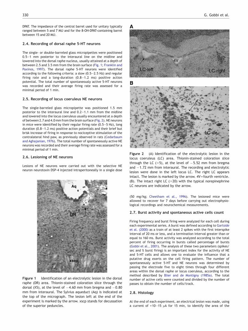
Figure 2 (A) Identification of the electrolytic lesion in thelocus coeruleus (LC) area. Thionin-stained coloration slicethrough the LC (�5), at the level of �5.52 mm from bregmaand �1.72 mm from interaural. The recording and electrolyticlesion were done in the left locus LC. The right LC appears
G. Gobbi et al.330
DPAT. The impedance of the central barrel used for unitary typicallyranged between 5 and 7 MX and for the 8-OH-DPAT-containing barrelbetween 15 and 20 MX.
2.4. Recording of dorsal raphe 5-HT neurons
The single- or double-barreled glass micropipettes were positioned0.5—1 mm posterior to the interaural line on the midline andlowered into the dorsal raphe nucleus, usually attained at a depth ofbetween 2.5 and 3.5 mm from the brain surface (Fig. 1; Franklin andPaxinos, 1997). The dorsal raphe 5-HT neurons were identifiedaccording to the following criteria: a slow (0.5—2.5 Hz) and regularfiring rate and a long-duration (0.8—1.2 ms) positive actionpotential. The total number of spontaneously active 5-HT neuronswas recorded and their average firing rate was assessed for aminimal period of 1 min.
2.5. Recording of locus coeruleus NE neurons
The single-barreled glass micropipette was positioned 1.5 mmposterior to the interaural line and 0.2—1.1 mm from the midlineand lowered into the locus coeruleus usually encountered at a depthof between 2.7 and 4.0mm from the brain surface (Fig. 2). NE neuronsin mice were identified by their regular firing rate (0.5—5 Hz), longduration (0.8—1.2 ms) positive action potentials and their brief butbrisk increase of firing in response to nociceptive stimulation of thecontralateral hind paw, as previously observed in rats (Cedarbaumand Aghajanian, 1976). The total number of spontaneously active NEneurons was recorded and their average firing rate was assessed for aminimal period of 1 min.
2.6. Lesioning of NE neurons
Lesions of NE neurons were carried out with the selective NEneuron neurotoxin DSP-4 injected intraperitoneally in a single dose
Figure 1 Identification of an electrolytic lesion in the dorsalraphe (DR) area. Thionin-stained coloration slice through thedorsal (X5), at the level of �4.60 mm from bregma and �0.80mm from interaural. The Sylvius aqueduct (Aq) is indicated atthe top of the micrograph. The lesion left at the end of theexperiment is marked by the arrow. xscp stands for decussationof the superior peduncles.
intact. The lesion is marked by the arrow. 4V=fourth ventricle.(B). The intact right LC (�20) with the typical norepinephrineLC neurons are indicated by the arrow.
(50 mg/kg; Cheetham et al., 1996). The lesioned mice wereallowed to recover for 7 days before carrying out electrophysio-logical recordings and neurochemical measurements.
2.7. Burst activity and spontaneous active cells count
Firing frequency and burst firing were analyzed for each cell duringeach experimental series. A burst was defined according to Gartsideet al. (2000) as a train of at least 2 spikes with the first interspikeinterval of 20 ms or less, and a termination interval greater than orequal to 160 ms. Burst activity was analyzed according to the totalpercent of firing occurring in bursts called percentage of bursts(Gobbi et al., 2001). The analysis of these two parameters (spikes/sec and % burst firing) is an important index for the activity of NEand 5-HT cells and allows one to evaluate the influence that aputative drug exerts on the cell firing pattern. The number ofspontaneously active 5-HT and NE neurons was determined bypassing the electrode five to eight times through four differentareas within the dorsal raphe or locus coeruleus, according to themethod described by Blier and de Montigny (1985a). The totalnumber of active cells were counted and divided by the number ofpasses to obtain the number of cells/track.
2.8. Histology
At the end of each experiment, an electrical lesion was made, usinga current of +10—15 AA for 15 min, to identify the area of the
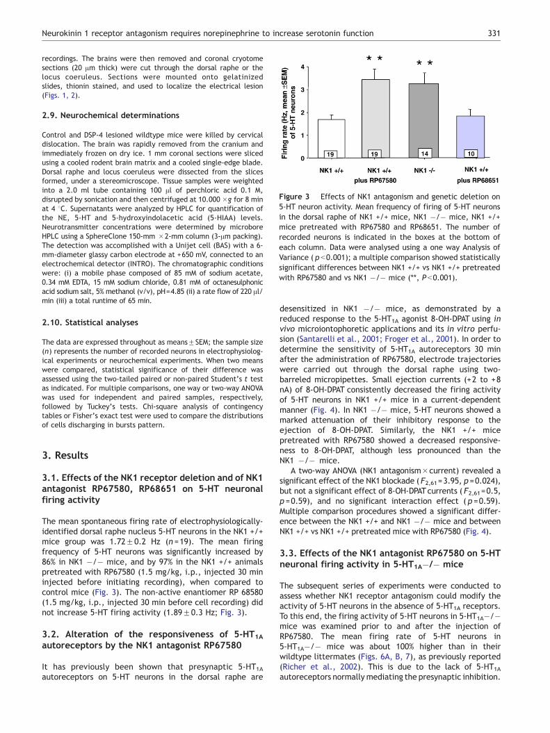
Figure 3 Effects of NK1 antagonism and genetic deletion on5-HT neuron activity. Mean frequency of firing of 5-HT neuronsin the dorsal raphe of NK1 +/+ mice, NK1 �/� mice, NK1 +/+mice pretreated with RP67580 and RP68651. The number ofrecorded neurons is indicated in the boxes at the bottom ofeach column. Data were analysed using a one way Analysis ofVariance ( p b0.001); a multiple comparison showed statisticallysignificant differences between NK1 +/+ vs NK1 +/+ pretreatedwith RP67580 and vs NK1 �/� mice (**, P b0.001).
Neurokinin 1 receptor antagonism requires norepinephrine to increase serotonin function 331
recordings. The brains were then removed and coronal cryotomesections (20 Am thick) were cut through the dorsal raphe or thelocus coeruleus. Sections were mounted onto gelatinizedslides, thionin stained, and used to localize the electrical lesion(Figs. 1, 2).
2.9. Neurochemical determinations
Control and DSP-4 lesioned wildtype mice were killed by cervicaldislocation. The brain was rapidly removed from the cranium andimmediately frozen on dry ice. 1 mm coronal sections were slicedusing a cooled rodent brain matrix and a cooled single-edge blade.Dorsal raphe and locus coeruleus were dissected from the slicesformed, under a stereomicroscope. Tissue samples were weightedinto a 2.0 ml tube containing 100 Al of perchloric acid 0.1 M,disrupted by sonication and then centrifuged at 10.000 �g for 8 minat 4 8C. Supernatants were analyzed by HPLC for quantification ofthe NE, 5-HT and 5-hydroxyindolacetic acid (5-HIAA) levels.Neurotransmitter concentrations were determined by microboreHPLC using a SphereClone 150-mm �2-mm column (3-Am packing).The detection was accomplished with a Unijet cell (BAS) with a 6-mm-diameter glassy carbon electrode at +650 mV, connected to anelectrochemical detector (INTRO). The chromatographic conditionswere: (i) a mobile phase composed of 85 mM of sodium acetate,0.34 mM EDTA, 15 mM sodium chloride, 0.81 mM of octanesulphonicacid sodium salt, 5% methanol (v/v), pH=4.85 (ii) a rate flow of 220 Al/min (iii) a total runtime of 65 min.
2.10. Statistical analyses
The data are expressed throughout as meansFSEM; the sample size(n) represents the number of recorded neurons in electrophysiolog-ical experiments or neurochemical experiments. When two meanswere compared, statistical significance of their difference wasassessed using the two-tailed paired or non-paired Student’s t testas indicated. For multiple comparisons, one way or two-way ANOVAwas used for independent and paired samples, respectively,followed by Tuckey’s tests. Chi-square analysis of contingencytables or Fisher’s exact test were used to compare the distributionsof cells discharging in bursts pattern.
3. Results
3.1. Effects of the NK1 receptor deletion and of NK1antagonist RP67580, RP68651 on 5-HT neuronalfiring activity
The mean spontaneous firing rate of electrophysiologically-identified dorsal raphe nucleus 5-HT neurons in the NK1 +/+mice group was 1.72F0.2 Hz (n =19). The mean firingfrequency of 5-HT neurons was significantly increased by86% in NK1 �/� mice, and by 97% in the NK1 +/+ animalspretreated with RP67580 (1.5 mg/kg, i.p., injected 30 mininjected before initiating recording), when compared tocontrol mice (Fig. 3). The non-active enantiomer RP 68580(1.5 mg/kg, i.p., injected 30 min before cell recording) didnot increase 5-HT firing activity (1.89F0.3 Hz; Fig. 3).
3.2. Alteration of the responsiveness of 5-HT1A
autoreceptors by the NK1 antagonist RP67580
It has previously been shown that presynaptic 5-HT1Aautoreceptors on 5-HT neurons in the dorsal raphe are
desensitized in NK1 �/� mice, as demonstrated by areduced response to the 5-HT1A agonist 8-OH-DPAT using invivo microiontophoretic applications and its in vitro perfu-sion (Santarelli et al., 2001; Froger et al., 2001). In order todetermine the sensitivity of 5-HT1A autoreceptors 30 minafter the administration of RP67580, electrode trajectorieswere carried out through the dorsal raphe using two-barreled micropipettes. Small ejection currents (+2 to +8nA) of 8-OH-DPAT consistently decreased the firing activityof 5-HT neurons in NK1 +/+ mice in a current-dependentmanner (Fig. 4). In NK1 �/� mice, 5-HT neurons showed amarked attenuation of their inhibitory response to theejection of 8-OH-DPAT. Similarly, the NK1 +/+ micepretreated with RP67580 showed a decreased responsive-ness to 8-OH-DPAT, although less pronounced than theNK1 �/� mice.
A two-way ANOVA (NK1 antagonism�current) revealed asignificant effect of the NK1 blockade (F2,61=3.95, p =0.024),but not a significant effect of 8-OH-DPATcurrents ( F2,61=0.5,p =0.59), and no significant interaction effect ( p =0.59).Multiple comparison procedures showed a significant differ-ence between the NK1 +/+ and NK1 �/� mice and betweenNK1 +/+ vs NK1 +/+ pretreated mice with RP67580 (Fig. 4).
3.3. Effects of the NK1 antagonist RP67580 on 5-HTneuronal firing activity in 5-HT1A�/� mice
The subsequent series of experiments were conducted toassess whether NK1 receptor antagonism could modify theactivity of 5-HT neurons in the absence of 5-HT1A receptors.To this end, the firing activity of 5-HT neurons in 5-HT1A�/�mice was examined prior to and after the injection ofRP67580. The mean firing rate of 5-HT neurons in5-HT1A�/� mice was about 100% higher than in theirwildtype littermates (Figs. 6A, B, 7), as previously reported(Richer et al., 2002). This is due to the lack of 5-HT1Aautoreceptors normally mediating the presynaptic inhibition.

Figure 4 Reduced responsiveness of 5-HT1A autoreceptors to the 5-HT1A agonist 8-OH-DPAT after NK1 receptor blockade.Responsiveness, assessed in percentage inhibition (meanFSEM), of 5-HT neurons to the microiontophoretic application of the 5-HT1Aagonist 8-OH-DPAT (2, 4 and 8 nA). The number of neurons tested is given at the bottom of each column. (**) p b0.01, (*) p b0.05 usinga two-way Analysis of Variance, and a multiple comparison procedure (Tukey’s test).
G. Gobbi et al.332
Remarkably, the systemic injection of RP67580 caused afurther increase of the mean firing rate of 5-HT neurons in5-HT1A�/� animals, indicating that 5-HT neuronal firing can
Figure 5 NK1 receptor antagonism increases spontaneous firing achistograms of 5-HT neurons illustrating the activity of all 5-HT neurespectively, of a 5-HT1A +/+ mouse (A), a 5-HT1A �/� mouse (B)RP67580 (C) and RP68651 (D). The dots at the bottom of the tracestime base applies to all traces.
be enhanced by NK1 receptor antagonism despite the absenceof 5-HT1A autoreceptors (Figs. 5C, 6C, 7). Importantly, thenumber of neurons firing in doublets and/or displaying burst
tivity of 5-HT neurons in 5-HT1A �/�mice. Integrated firing raterons recorded in single trajectories through the dorsal raphe,, and a 5-HT1A +/+ mouse pretreated with the NK1 antagonist(in C) represent interruptions of the physiograph recording. The
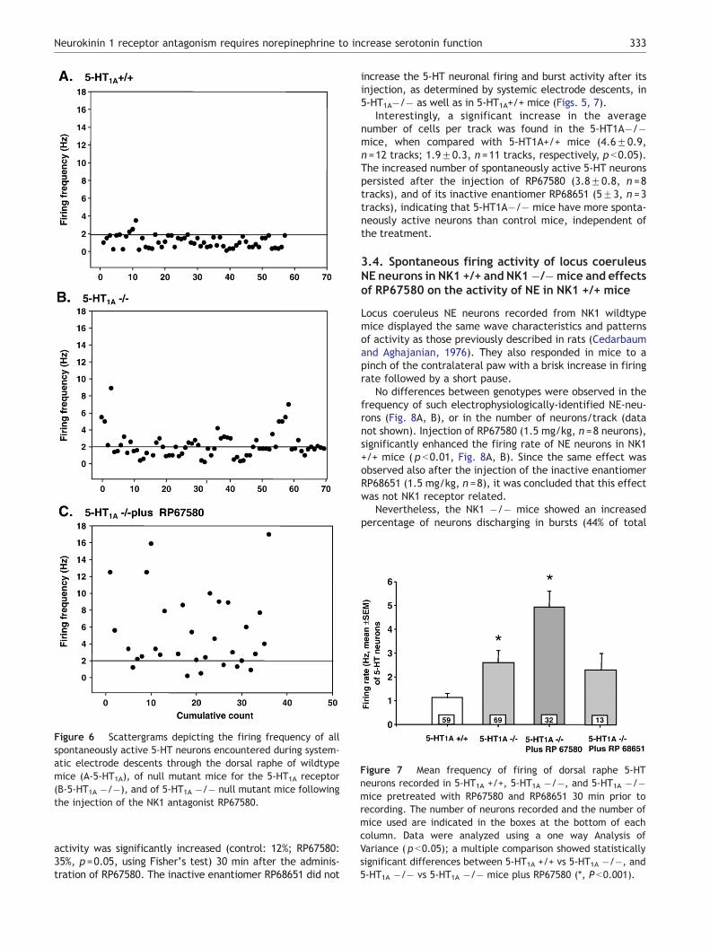
Figure 6 Scattergrams depicting the firing frequency of allspontaneously active 5-HT neurons encountered during system-atic electrode descents through the dorsal raphe of wildtypemice (A-5-HT1A), of null mutant mice for the 5-HT1A receptor(B-5-HT1A �/�), and of 5-HT1A �/� null mutant mice followingthe injection of the NK1 antagonist RP67580.
Figure 7 Mean frequency of firing of dorsal raphe 5-HTneurons recorded in 5-HT1A +/+, 5-HT1A �/�, and 5-HT1A �/�mice pretreated with RP67580 and RP68651 30 min prior torecording. The number of neurons recorded and the number ofmice used are indicated in the boxes at the bottom of eachcolumn. Data were analyzed using a one way Analysis ofVariance ( p b0.05); a multiple comparison showed statisticallysignificant differences between 5-HT1A +/+ vs 5-HT1A �/�, and5-HT1A �/� vs 5-HT1A �/� mice plus RP67580 (*, P b0.001).
Neurokinin 1 receptor antagonism requires norepinephrine to increase serotonin function 333
activity was significantly increased (control: 12%; RP67580:35%, p =0.05, using Fisher’s test) 30 min after the adminis-tration of RP67580. The inactive enantiomer RP68651 did not
increase the 5-HT neuronal firing and burst activity after itsinjection, as determined by systemic electrode descents, in5-HT1A�/� as well as in 5-HT1A+/+ mice (Figs. 5, 7).
Interestingly, a significant increase in the averagenumber of cells per track was found in the 5-HT1A�/�mice, when compared with 5-HT1A+/+ mice (4.6F0.9,n =12 tracks; 1.9F0.3, n =11 tracks, respectively, p b0.05).The increased number of spontaneously active 5-HT neuronspersisted after the injection of RP67580 (3.8F0.8, n =8tracks), and of its inactive enantiomer RP68651 (5F3, n =3tracks), indicating that 5-HT1A�/� mice have more sponta-neously active neurons than control mice, independent ofthe treatment.
3.4. Spontaneous firing activity of locus coeruleusNE neurons in NK1 +/+ and NK1�/�mice and effectsof RP67580 on the activity of NE in NK1 +/+ mice
Locus coeruleus NE neurons recorded from NK1 wildtypemice displayed the same wave characteristics and patternsof activity as those previously described in rats (Cedarbaumand Aghajanian, 1976). They also responded in mice to apinch of the contralateral paw with a brisk increase in firingrate followed by a short pause.
No differences between genotypes were observed in thefrequency of such electrophysiologically-identified NE-neu-rons (Fig. 8A, B), or in the number of neurons/track (datanot shown). Injection of RP67580 (1.5 mg/kg, n =8 neurons),significantly enhanced the firing rate of NE neurons in NK1+/+ mice ( p b0.01, Fig. 8A, B). Since the same effect wasobserved also after the injection of the inactive enantiomerRP68651 (1.5 mg/kg, n =8), it was concluded that this effectwas not NK1 receptor related.
Nevertheless, the NK1 �/� mice showed an increasedpercentage of neurons discharging in bursts (44% of total

Figure 8 (A) Acute effects of the NK1 antagonist RP67580 on NE firing activity. Integrated firing rate histograms of NE neuronsillustrating the activity of all NE neurons recorded in the locus coeruleus, respectively, of a NK1 +/+ mouse (a), a NK1 �/�mouse (b),and a NK1 +/+ mouse pretreated with the NK1 antagonist RP67580 (c) and with RP68651 (d). The time base applies to all traces.Events/sec indicates the burst activity, that is when at least two action potentials occurred with an interval b20 ms. (B). On the leftand bars: mean frequency of firing of NE neurons in the locus coeruleus of NK1 +/+, NK1 �/�mice, NK1 +/+ pretreated with RP67580and RP68651. The number of recorded neurons is indicated in the boxes at the bottom of each column. Data were analyzed using oneway Analysis of Variance ( p b0.001); a multiple comparison showed statistically significant differences between NK1 +/+ treated withRP67580 or RP68651 vs NK1 +/+ (y, P b0.01). On the right and lines: percentage of NE recorded cells showing a burst pattern activity.Chi-square test showed a significant difference between the NK1 �/� mice vs NK1 +/+ mice, and between NK1 +/+ mice pretreatedwith RP67580 vs NK1 +/+ mice (*, P b0.05).
G. Gobbi et al.334
recorded cells) compared to NK1 +/+ mice (14%, (v2(1)=3.7,p =0.05, Fig. 6B). RP67580 (1.5—3 mg/kg), but not RP68651(1.5 mg—3 mg/kg, i.p.), robustly enhanced the burst firingpattern (68% of recorded cells showing burst activity withRP67580 vs 25% with RP68651 (v2(1)=2.8, p =0.05, Fig. 8B,right).
3.5. Effects of DSP-4 on norepinephrine andserotonin levels
To assess whether NE neurons contribute to the alteration of5-HT neuronal firing after NK1 receptor antagonist, theconcentrations of NE were determined in various brain

Table 1 Effect of DSP-4 (50 mg/kg i.p.) on NE, 5-HT and 5-HIAA levels in dorsal raphe and locus coeruleus
Brain areas NE 5-HT 5-HIAA
Control DSP-4 Control DSP-4 Control DSP-4
Dorsal raphe 9.6F1.3 2.7F0.5*** 7.3F2.0 6.6F1.6 2.2F0.3 2.0F0.5Locus coeruleus 13.2F2.7 6.0F1.2* 2.5F0.3 4.7F0.8* 1.5F0.2 2.8F0.6*
Values are expressed as meansFS.E.M in pmol/mg of tissue. Differences between groups were analysed by unpaired t test.*p b0.05; ***p b0.001 in comparison with the control groups.
Neurokinin 1 receptor antagonism requires norepinephrine to increase serotonin function 335
structures following the lesion of NE neurons. Noradrenergicneurons were lesioned using the selective toxin DSP-4. Sevendays after DSP-4 administration (50 mg/kg i.p.), NE levelswere significantly altered in all target areas. In particular, NElevels were decreased by 72% in dorsal raphe (t17=4.717,p b0.001, n =9—10) and by 55% in locus coeruleus (t17=2.372,p b0.05, n =10, Table 1).
Noradrenergic lesion altered 5-HT levels in the locuscoeruleus but not in the dorsal raphe (t17=0.2783, n.s.,n =9—10). Serotonin concentrations were significantly in-creased (91%) in locus coeruleus following noradrenergiclesion (t17=2.751, p b0.05, n =9—10). 5-HIAA levels paral-leled 5-HT alterations observed in lesioned mice, remainingunchanged in dorsal raphe (t17=0.3561, n.s., n =9—10) andsignificantly increased (+89%) in locus coeruleus (t17=2.263,p b0.05, n =10, Table 1).
3.6. Effects of the noradrenergic lesion on 5-HTneuronal firing in NK1 �/� and NK1 +/+ micepretreated with the NK1 antagonist RP67580
Seven days after the DSP-4 lesion, in vivo electrophysiolog-ical experiments were conducted to assess the effects of the
Figure 9 After NE depletion, NK1 receptor antagonism anddeletion fail to increase 5-HT activity. Mean frequency of firingof dorsal raphe 5-HT neurons recorded in intact NK1 +/+ mice,NK1 +/+ pretreated with RP67580 and NK1 �/� mice (white)and in NE-lesioned mice using DSP-4 (gray). The number ofneurons recorded is indicated in the boxes at the bottom ofeach column. The asterisk (*p b0.001) indicates the differencesobtained between intact mice and NE-lesioned mice; the cross(y), indicates the p value (p b0.001) of non-lesioned groups vscontrol group using the One Way Analysis of Variance.
NE lesion and of NK1 receptor antagonism on the spontane-ous firing rate of 5-HT neurons (Fig. 9). In NE-lesionedwildtype mice, the mean spontaneous firing rate of 5-HTneurons was similar to that of the controls. In NK1 +/+ mice,RP67580 (1.5 mg/kg, i.p.) significantly increased the meanfiring rate of 5-HT neurons by 100% when NE neurons wereintact, but not in NE-lesioned mice (+13%). The firingfrequency of 5-HT neurons was approximately enhanced tothe same degree in NK1 +/+ mice (110%) having receivedRP67580 and in NK1 �/� mice, when compared to controls.However, in intact NK1 �/� mice, the NE-lesion abolishedthe increase in spontaneous firing rate of 5-HT neuronsnormally observed in such mutant mice.
4. Discussion
The major finding of the present study was that the increasein the firing rate of 5-HT neurons induced by NK1 receptorantagonism and genetic deletion is dependent on theintegrity of NE neurons, but independent of 5-HT1A auto-receptors.
4.1. NK1 receptor antagonism and the 5-HT system
In this study, it was confirmed that both the pharmacologicalblockage of NK1 receptor and its genetic deletion increase5-HT neuronal firing activity. It was reported that thisenhancement was associated with a desensitization of5-HT1A autoreceptors resembling that induced by SSRItreatments (Froger et al., 2001). It is proposed herein that5-HT1A autoreceptor desensitization is not the only mecha-nism responsible for the increased 5-HT activity by NK1receptor antagonism. Although NK1 receptor antagonismattenuates the response of 5-HT neurons to a 5-HT1A agonist(Fig. 4), it may still increase 5-HT firing activity in theabsence of 5-HT1A autoreceptors, as shown in 5-HT1A nullmutant mice (Fig. 5A, B).
Interestingly, 5-HT1A null mutant mice show anxiety-related behaviors compared to wildtype mice in theelevated plus-maze (Ramboz et al., 1998; Zhuang et al.,1999; Gross et al., 2002), that are decreased by RP67580(Gobbi et al., 2002). Moreover, these mutant mice werestill responsive to both NE reuptake inhibitors imipramineand desipramine in the novelty-suppressed feeding para-digm, but not to the SSRIs fluoxetine (Santarelli et al.,2003). These results indicate that some antidepressants,such as SSRIs, require 5-HT1A receptors for their behavioralactivity, but not other classes as TCAs or NK1 antagonists,suggesting that the psychotropic action of NK1 antagonistsis due in part to a neuronal system other than the 5-HTsystem.

G. Gobbi et al.336
It is important to emphasize that an increase in 5-HTneuronal firing may not necessarily lead to an increase in5-HT transmission. Indeed, a two-day administration of theNK1 antagonist CP-96,365 enhanced 5-HT neuronal firing by50% but did not alter the tonic activation of 5-HT1A receptorsin the rat hippocampus (Haddjeri et al., 2001a). After 14days of treatment with the same drug, the increase in firingwas of 90% which intensified 5-HT1A transmission in thehippocampus. Consequently, it would appear that limitedincreases in firing of 5-HT neurons can be offset by 5-HTreuptake and/or the negative feedback action of terminal5-HT autoreceptors. Consistent with this possibility is theobservation that a NK1 antagonist, while having no effect onits own on extracellular 5-HT levels measured with micro-dialysis in the frontal cortex, potentiated the enhancingeffect of a SSRI (Millan et al., 2001; Lejeune et al., 2002;Zocchi et al., 2003; Guiard et al., 2004). Another possibilityfor the lack of effect of NK1 antagonists on 5-HT levels inpostsynaptic structures using microdialysis is that intra-synaptic 5-HT concentrations are increased; but that thespillover into the extracellular compartment may not besufficient to be detected by a microdialysis probe of0.25 mm in diameter. Consistent with this is the increasein dialysate 5-HT in the rat dorsal raphe after long-termadministration of a NK1 antagonist, even after a washoutperiod (Guiard et al., 2005). This significant increasedetected in that particular brain region possibly resultedfrom the fact that in the raphe nuclei 5-HT cell bodies anddendrites are densely intertwined where 5-HT in thedialysate may more adequately reflect synaptic concentra-tions of 5-HT.
4.2. Neurochemical effects of DSP-4 administration
Although different strains of mice were used for theelectrophysiological recordings and the neurochemicaldeterminations, this is not likely to have influenced theeffects of DSP-4. Indeed, Fornai et al. (1996) did not observea differential effect of DSP-4 in depleting NE in differentbrain areas of inbred C57 black mice and Swiss Webster micewhich are not inbred mice. The greater decrease of NElevels in the dorsal raphe, as a projection area of locuscoeruleus NE neurons (Anderson et al., 1977), than in thecell body region may appear unusual. These NE depletionratios are, however, fully consistent with those obtained in aprior study examining locus coeruleus and hippocampususing DSP-4 (Hutter et al., 1996). This pattern of depletion,whereby terminals are more affected than cell bodies, isusually obtained using various monoaminergic neurotoxins(Blier and de Montigny, 1985b). Finally, the increase in 5-HTconcentrations in the locus coeruleus is likely the result ofdecreased NE activating a2-adrenoceptors on 5-HT terminalswhich normally exert an inhibitory role on 5-HT release(Mongeau et al., 1997).
4.3. NK1 antagonism and the NE system
In the present experiments, it was observed that the acuteadministration of the NK1 antagonist RP67580 and thegenetic deletion of NK1 receptors increased noradrenergicburst activity. Conversely, the transient increase in overallNE firing activity generated by the i.v. injection of RP67580
does not seem to be specific for two reasons. First, it ismimicked by its inactive enantiomer and second, other NK1antagonists (CP-96,345 and WIN 51,708) do not modify thefiring activity of NE neurons, but desensitize the a2-adrenergic autoreceptors (Haddjeri and Blier, 2000). Asustained burst firing activity of NE neurons recorded invitro was also reported after long-term administration ofthe potent NK1 antagonist L-760735 and imipramine (Mau-bach et al., 2002). It is known that the burst pattern of firingis linked to an augmentation of monoamine release interminal areas (Gonon, 1988; Gartside et al., 2000; Florin-Lechner et al., 1996). In support of this notion, an increasein NE release in frontal cortex and dorsal hippocampus wasobserved in microdialysis experiments following acuteadministration of the NK1 antagonist GR205171 (Millan etal., 2001). It is unlikely that RP68580 increased NE neuronalfiring and burst activity by an interference with a2-adrenoceptors because the a2-adrenergic antagonist ida-zoxan enhances the firing rate of NE neurons withoutaltering burst activity (Dremencov et al., in press). Sinceit has been observed that NK3 receptor agonism facilitatesburst activity in neonatal rat spinal cord (Marchetti andNistri, 2001), it is also possible that a preferential action ofSP at NK3 receptors occurring in the presence of selectiveNK1 receptor blockage could account for this shift in firingpattern, since a permissive action of NK3 on NK1 receptormodulation in the locus coeruleus has been reported by Bertet al. (2002).
In DSP-4 lesioned mice, both wildtype animals pretreatedwith the NK1 antagonist RP67580 andNK1�/�mice no longerdisplayed an enhanced 5-HT firing activity. These results areanalogous to those obtained with the a2-adrenergic antago-nist mirtazapine (Haddjeri et al., 1996) and the NE releaserbupropion (Dong and Blier, 2001) that no longer increase 5-HTneuronal firing activity in the absence of NE neurons,indicating that the integrity of the NE neurons is also requiredfor the effect of these two antidepressant drugs on 5-HTneuronal activity. Cryan et al. (2004) reported on theimportance of the NE system for the antidepressant-likeactivity of various classes of antidepressants. They observedthat NE-deficient mice failed to exhibit behavioral effects ofvarious antidepressants, including SNRIs, MAOIs, bupropionand SSRIs; the ability of fluoxetine to increase 5-HT levels inhippocampus was also blocked.
It has been postulated that NK1 antagonists activatedorsal raphe 5-HT neurons via the lateral habenula, acerebral area involved in the mechanism of stress responsesthat has a high density of NK1 receptors (Conley et al.,2002). The amygdala, which feedbacks to the raphe (Boskeret al., 1997), also appears to be a leading structure for theanxiolytic effects of NK1 antagonists (Ebner et al., 2004).While the habenula and the amygdala may play a significantrole in the effects of NK1 antagonists on 5-HT neurons, theresults presented herein indicate that the impact of NK1receptor antagonism on 5-HT neurons is also influenced to amajor extent by NE neurons.
In conclusion, substance P receptor antagonists, becauseof their modulatory effects on monoamine systems thatresemble the actions of antidepressant treatments, maywell become a new class of therapeutic agents for thetreatment of mood and anxiety disorders used alone or topotentiate antidepressant therapies.

Neurokinin 1 receptor antagonism requires norepinephrine to increase serotonin function 337
Acknowledgement
The authors would like to thank Dr. Amir Shimon (Con-cordia University, Montreal) for his help in the preparationof the photomicrographs, and Dr. Silvana Gaetani (Univer-sity bLa SapienzaQ, Rome) for her help in neurochemicalexperiments.
References
Anderson, C.D., Pasquier, D.A., Forbes, W.B., Morgane, P.J., 1977.Locus coeruleus-to-dorsal raphe input examined by electro-physiological and morphological methods. Brain Res. Bull. 2,209–221.
Bert, L., Rodier, D., Bougault, I., Allouard, N., Le-Fur, G., Soubrie,P., Steinberg, R., 2002. Permissive role of neurokinin NK(3)receptors in NK(1) receptor-mediated activation of the locuscoeruleus revealed by SR 142801. Synapse 43, 62–69.
Blier, P., de Montigny, C., 1985a. Serotoninergic but not noradren-ergic neurons in rat central nervous system adapt to long-termtreatment with monoamine oxidase inhibitors. Neuroscience 16,949–955.
Blier, P., de Montigny, C., 1985b. Short-term lithium administrationenhances serotonergic neurotransmission: electrophysiologicalevidence in the rat CNS. Eur. J. Pharmacol. 113, 69–77.
Blier, P., de Montigny, C., 1999. Serotonin and drug-inducedtherapeutic responses in major depression, obsessive-com-pulsive and panic disorders. Neuropsychopharmacology 21,91S–98S.
Blier, P., Gobbi, G., Haddjeri, N., Santarelli, L., Mathew, G., Hen,R., 2004. Impact of substance P receptor antagonism on theserotonin and norepinephrine systems: relevance to theantidepressant/anxiolytic response. J. Psychiatry Neurosci. 29,208–218.
Bosker, F.J., Klompmakers, A., Westenberg, H.G., 1997. Postsynap-tic 5-HT1A receptors mediate 5-hydroxytryptamine release inthe amygdala through a feedback to the caudal linear raphe. Eur.J. Pharmacol. 333, 147–157.
Cheetham, S.C., Viggers, J.A., Butler, S.A., Prow, M.R., Heal, D.J.,1996. [3H]nisoxetine-a radioligand for noradrenaline reuptakesites: correlation with inhibition of [3H]noradrenaline uptakeand effect of DSP-4 lesioning and antidepressant treatments.Neuropharmacology 35, 63–70.
Cedarbaum, J.M., Aghajanian, G.K., 1976. Noradrenergic neurons ofthe locus coeruleus: inhibition by epinephrine and activation bythe alpha-antagonist piperoxane. Brain Res. 112, 413–419.
Conley, R.K., Cumberbatch, M.J., Mason, G.S., Williamson, D.J.,Harrison, T., Locker, K., Swain, C., Maubach, K., O’Donnell, R.,Rigby, M., Hewson, L., Smith, D., Rupniak, N.M., 2002.Substance P (neurokinin 1) receptor antagonists enhance dorsalraphe neuronal activity. J. Neurosci. 22, 7730–7736.
Cryan, J.F., O’Leary, O.F., Jin, S.H., Friedland, J.C., Ouyang, M.,Hirsch, B.R., Page, M.E., Dalvi, A., Thomas, S.A., Lucki, I., 2004.Norepinephrine-deficient mice lack responses to antidepressantdrugs, including selective serotonin reuptake inhibitors. Proc.Natl. Acad. Sci. U. S. A. 101, 8186–8191.
Dong, J., Blier, P., 2001. Modification of norepinephrine andserotonin, but not dopamine, neuron firing by sustainedbupropion treatment. Psychopharmacology 155, 52–57.
Dremencov, E., El Mansari, M., Blier, P., in press. Noradrenergicaugmentation of escitalopram response by risperidone: Electro-physiological studies in the rat brain. Biological Psychiatry.
Ebner, K., Rupniak, N.M., Saria, A., Singewald, N., 2004. SubstanceP in the medial amygdala: emotional stress-sensitive release andmodulation of anxiety-related behavior in rats. Proc. Natl. Acad.Sci. U. S. A. 101, 4280–4285.
Florin-Lechner, S.M., Druhan, J.P., Aston-Jones, G., Valentino, R.J.,1996. Enhanced norepinephrine release in prefrontal cortex withburst stimulation of the locus coeruleus. Brain Res. 742, 89–97.
Fornai, F., Bassi, L., Torracca, M.T., Alessandri, M.G., Scalori, V.,Corsini, G.U., 1996. Region- and neurotransmitter-dependentspecies and strain differences in DSP-4-induced monoaminedepletion in rodents. Neurodegeneration 5, 241–249.
Franklin, K.B.J., Paxinos, G., 1997. The Mouse Brain in StereotaxicCoordinates. Academic Press Inc., San Diego, CA, USA.
Froger, N., Gardier, A.M., Moratalla, R., Alberti, I., Lena, I., Boni,C., De Felipe, C., Rupniak, N.M., Hunt, S.P., Jacquot, C., Hamon,M., Lanfumey, L., 2001. 5-hydroxytryptamine (5-HT)1A auto-receptor adaptive changes in substance P (neurokinin 1)receptor knock-out mice mimic antidepressant-induced desen-sitization. J. Neurosci. 21, 8188–8197.
Garret, C., Carruette, A., Fardin, V., Moussaoui, S., Peyronel, J.F.,Blanchard, J.C., Laduron, P.M., 1991. Pharmacological proper-ties of a potent and selective nonpeptide substance P antago-nist. Proc. Natl. Acad. Sci. U. S. A. 88, 10208–10212.
Gartside, S.E., Hajos-Korcsok, E., Bagdy, E., Harsing, L.G. Jr.,Sharp, T., Hajos, M., 2000. Neurochemical and electrophysio-logical studies on the functional significance of burst firing inserotonergic neurons. Neuroscience 98, 295–300.
Gobbi, G., Blier, P., 2005. Effect of neurokinin-1 receptor antago-nists on serotoninergic, noradrenergic and hippocampal neurons:comparison with antidepressant drugs. Peptides 26, 1383–1393.
Gobbi, G., Muntoni, A., Diana, M., Gessa, G.L., 2001. Clonidine failsto modify dopaminergic neuronal activity in morphine withdraw-al. Psychopharmacology 158, 1–6.
Gobbi, G., Santarelli, L., Hen, R., Debonnel, G., Blier, P., 2002.Understanding the mechanism of action of NK1 receptorantagonist: in vivo electrophysiological studies in mutant mice.Int. J. Neuropsychopharmacol. 5 (suppl.1), S97.
Gonon, F.G., 1988. Nonlinear relationship between impulse flow anddopamine released by rat midbrain dopaminergic neurons asstudied by in vivo electrochemistry. Neuroscience 24, 19–28.
Gross, C., Zhuang, X., Stark, K., Ramboz, S., Oosting, R., Kirby, L.,Santarelli, L., Beck, S., Hen, R., 2002. Serotonin1A receptor actsduring development to establish normal anxiety-like behaviourin the adult. Nature 416 (6879), 396–400.
Guiard, B.P., Przybylski, C., Guilloux, J.P., Seif, I., Froger, N., DeFelipe, C., Hunt, S.P., Lanfumey, L., Gardier, A.M., 2004.Blockade of substance P (neurokinin 1) receptors enhancesextracellular serotonin when combined with a selective sero-tonin reuptake inhibitor: an in vivo microdialysis study in mice.J. Neurochem. 89, 54–63.
Guiard, B.P., Froger, N., Hamon, M., Gardier, A.M., Lanfumey, L.,2005. Sustained pharmacological blockade of NK1 substance Preceptors causes functional desensitization of dorsal raphe 5-HT1A autoreceptors in mice. J. Neurochem. 95, 1713–1723.
Haddjeri, N., Blier, P., 2000. Effect of neurokinin-1 receptorantagonists on the function of 5-HT and noradrenaline neurons.Neuroreport 11, 1323–1327.
Haddjeri, N., Blier, P., 2001a. Sustained blockade of neurokinin-1receptors enhances serotonin neurotransmission. Biol. Psychia-try 50, 191–199.
Haddjeri, N., Blier, P., 2001b. Effects of neurokinin 1 antagonists onthe firing of locus coeruleus norepinephrine neurons. Soc.Neurosci. Abstr. 27 (479.19).
Haddjeri, N., Blier, P., de Montigny, C., 1996. Effect of the alpha-2 adrenoceptor antagonist mirtazapine on the 5-hydroxytryp-tamine system in the rat brain. J. Pharmacol. Exp. Ther. 277,861–871.
Haddjeri, N., Blier, P., de Montigny, C., 1998. Long-term antide-pressant treatments result in a tonic activation of forebrain 5-HT1A receptors. J. Neurosci. 18, 10150–10156.
Hutter, P., Johansson, M., Saria, A., Humpel, C., 1996. Acute andchronic noradrenergic regulation of neurotrophin messenger RNA

G. Gobbi et al.338
expression in rat hippocampus: evidence from lesions andorganotypic cultures. Neuroscience 70, 15–29.
Keller, M., Montgomery, S., Ball, W., Morrison, M., Snavely, D., Liu,G., Harreaves, R., Hietala, M., Lines, C., Beebe, K., Reines, S.,2006. Lack of efficacy of the substance p (neurokin-1 receptor)antagonist aprepitant in the treatment of major depressivedisorder. Biol. Psychiatry 59 (3), 216–223.
Kramer, M.S., Cutler, N., Feighner, J., Shrivastava, R., Carman, J.,Sramek, J.J., Reines, S.A., Liu, G., Snavely, D., Wyatt-Knowles,E., Hale, J.J., Mills, S.G., MacCoss, M., Swain, C.J., Harrison, T.,Hill, R.G., Hefti, F., Scolnick, E.M., Cascieri, M.A., Chicchi, G.G.,Sadowski, S., Williams, A.R., Hewson, L., Smith, D., Carlson,E.J., Hargreaves, R.J., Rupniak, N.M., 1998. Distinct mechanismfor antidepressant activity by blockade of central substance Preceptors. Science 281, 1640–1645.
Kramer, M.S., Winokur, A., Kelsey, J., Preskorn, S.H., Rothschild,A.J., Snavely, D., Ghosh, K., Ball, W.A., Reines, S.A., Munjack,D., Apter, J.T., Cunningham, L., Kling, M., Bari, M., Getson, A.,Lee, Y., 2004. Demonstration of the efficacy and safety of anovel substance P (NK1) receptor antagonist in major depres-sion. Neuropsychopharmacology 29, 385–392.
Lejeune, F., Gobert, A., Millan, M.J., 2002. The selective neurokinin(NK)(1) antagonist, GR205,171, stereospecifically enhancesmesocortical dopaminergic transmission in the rat: a combineddialysis and electrophysiological study. Brain Res. 935, 134–139.
Marchetti, C., Nistri, A., 2001. Neuronal bursting induced by NK3receptor activation in the neonatal rat spinal cord in vitro. J.Neurophysiol. 86, 2939–2950.
Maubach, K.A., Martin, K., Chicchi, G., Harrison, T., Wheeldon, A.,Swain, C.J., Cumberbatch, M.J., Rupniak, N.M., Seabrook, G.R.,2002. Chronic substance P (NK1) receptor antagonist andconventional antidepressant treatment increases burst firing ofmonoamine neurones in the locus coeruleus. Neuroscience 109,609–617.
Millan, M.J., Lejeune, F., De Nanteuil, G., Gobert, A., 2001.Selective blockade of Neurokinin (NK)1 receptors facilitatesthe activity of adrenergic pathways projecting to frontal cortexand dorsal hippocampus rats. J. Neurochem. 76, 1949–1954.
Mongeau, R., Blier, P., de Montigny, C., 1997. The serotonergic andnoradrenergic systems of the hippocampus: their interactionsand the effects of antidepressant treatments. Brain Res. BrainRes. Rev. 23, 145–195.
Phillips, T.J., Hen, R., Crabbe, J.C., 1999. Complications associatedwith genetic background effects in research using knockoutmice. Psychopharmacology 147, 5 –7.
Ramboz, S., Oosting, R., Amara, D.A., Kung, H.F., Blier, P.,Mendelsohn, M., Mann, J.J., Brunner, D., Hen, R., 1998. Serotoninreceptor 1A knockout: an animal model of anxiety-relateddisorder. Proc. Natl. Acad. Sci. U. S. A. 95, 14476–14481.
Ranga, K., Krishnan, R., 2002. Clinical experience with Substance Preceptor (NK1) antagonist in depression. J. Clin. Psychiatry 63(suppl. 11), 25–29.
Richer, M., Hen, R., Blier, P., 2002. Modification of serotonin neuronproperties in mice lacking 5-HT1A receptors. Eur. J. Pharmacol.435, 195–203.
Santarelli, L., Gobbi, G., Debs, P.C., Sibille, E., Blier, P., Hen, R.,Heath, M., 2001. Genetic and pharmacological disruption of NK1receptor function decreases anxiety-related behaviors andincreases serotonergic function. Proc. Natl. Acad. Sci. U. S. A.98, 1912–1917.
Santarelli, L., Saxe, M., Gross, C., Surget, A., Battaglia, F., Dulawa,S., Weisstaub, N., Lee, J., Duman, R., Arancio, O., Belzung, C.,Hen, R., 2003. Requirement of hippocampal neurogenesis forthe behavioral effects of antidepressants. Science 301 (5634),805–809.
Seager, M.A., Huff, K.D., Barth, V.N., Phebus, L.A., Rasmussen, K.,2004. Fluoxetine administration potentiates the effect ofolanzapine on locus coeruleus neuronal activity. Biol. Psychiatry55, 1103–1109.
Szabo, S.T., Blier, P., 2001. Response of the norepinephrine systemto antidepressant drugs. CNS Spectr. 6, 679–684.
Szabo, S.T., de Montigny, C., Blier, P., 2000. Progressive attenuationof the firing activity of locus coeruleus noradrenergic neurons bysustained administration of selective serotonin reuptake inhibi-tors. Int. J. Neuropsychopharmacol. 3, 1 –11.
Thase, M.E., Rush, A.J., 1997. When at first you don’t succeed:sequential strategies for antidepressant nonresponders. J. Clin.Psychiatry 58 (Suppl.13), 23–29.
Zhuang, X., Gross, C., Santarelli, L., Compan, V., Trillat, A.C., Hen,R., 1999. Altered emotional states in knockout mice lacking 5-HT1A or 5-HT1B receptors. Neuropsychopharmacology 21 (Suppl.2), 52S–60S.
Zocchi, A., Varnier, G., Arban, R., Griffante, C., Zanetti, L.,Bettelini, L., Marchi, M., Gerrard, P.A., Corsi, M., 2003. Effectsof antidepressant drugs and GR 205171, an neurokinin-1 (NK1)receptor antagonist, on the response in the forced swim test andon monoamine extracellular levels in the frontal cortex of themouse. Neurosci. Lett. 345, 73–76.