distinct modulation of voltage-gated and ligand-gated ca 2+ currents by ppar-γ agonists in cultured...
TRANSCRIPT
Department of Molecular and Biomedical Pharmacology, University of Kentucky Medical Center, 800 Rose Street, MS 310, Lexington,
Kentucky 40536-0298, USA
Several studies have reported that similar neuropathologicalmechanisms are present in aging and/or Alzheimer’s disease(AD) and in diabetes mellitus. An increased risk of dementia(Alzheimer’s and vascular type) is seen in people withdiabetes (Ott et al. 1999; Schnaider Beeri et al. 2004;Biessels et al. 2006), and aging is a recognized risk factorfor both AD and diabetes (Hoyer 2004; Yaffe et al. 2004;Launer 2005; McNay 2005; Reagan 2007). Furthermore,insulin resistance associated with type 2 diabetes mellitus(T2DM) may be an important pathogenic factor in AD(Watson and Craft 2003; Steen et al. 2005; Li and Holscher2007), and thought to be present in 80% of the ADpopulation (Janson et al. 2004). It has been suggested thatcognitive dysfunction in older adults and AD patients mightreflect a CNS state of diabetes characterized by reducedinsulin signaling and/or resistance, and termed ‘Type 3diabetes’ (Lannert and Hoyer 1998; Gasparini et al. 2002;Steen et al. 2005; de la Monte et al. 2006). In older T2DMadults, cognitive dysfunction is associated with poor controlof diabetes (Munshi et al. 2006), and improving glycemic
control leads to improved cognition and memory (Ryan et al.2006). Therefore, it appears that common underlying mech-anisms coexist in T2DM, aging and/or AD.
Peroxisome proliferator-activated receptor (PPAR)-c is anuclear receptor that is a target of thiazolidinediones (TZDs)and regulates the expression of genes controlling inflamma-tion, metabolism, cell growth, and differentiation (Berger andMoller 2002; Hauner 2002). PPAR subtypes (a, b, and c) arepresent in almost all cells, including adipocytes, fibroblasts,
Received March 25, 2009; accepted April 11, 2009.Address correspondence and reprint requests to Olivier Thibault,
Department of Molecular and Biomedical Pharmacology, University ofKentucky Medical Center, 800 Rose street, MS310, Lexington, KY40536-0298, USA. E-mail:[email protected] used: AD, Alzheimer’s disease; DIV, days in vitro;
DMSO, dimethylsulfoxide; NMDAR, NMDA receptor; PIO, pioglitaz-one; PPAR, peroxisomal proliferator-activated receptor; PPRE, peroxi-some proliferator response element; ROSI, rosiglitazone; T2DM, type 2diabetes mellitus; TEA, tetraethylammonium; TIO, T0070907; TZD,thiazolidinedione; VGCC, voltage-gated Ca2+ channel.
Abstract
Type 2 diabetes mellitus is a metabolic disorder characterized
by hyperglycemia and is especially prevalent in the elderly.
Because aging is a risk factor for type 2 diabetes mellitus, and
insulin resistance may contribute to the pathogenesis of Alz-
heimer’s disease (AD), anti-diabetic agents (thiazolidinedi-
ones-TZDs) are being studied for the treatment of cognitive
decline associated with AD. These agents normalize insulin
sensitivity in the periphery and can improve cognition and
verbal memory in AD patients. Based on evidence that Ca2+
dysregulation is a pathogenic factor of brain aging/AD, we
tested the hypothesis that TZDs could impact Ca2+ signaling/
homeostasis in neurons. We assessed the effects of pioglit-
azone and rosiglitazone (TZDs) on two major sources of Ca2+
influx in primary hippocampal cultured neurons, voltage-gated
Ca2+ channel (VGCC) and the NMDA receptor (NMDAR).
VGCC- and NMDAR-mediated Ca2+ currents were recorded
using patch-clamp techniques, and Ca2+ intracellular levels
were monitored with Ca2+ imaging techniques. Rosiglitazone,
but not pioglitazone reduced VGCC currents. In contrast,
NMDAR-mediated currents were significantly reduced by
pioglitazone but not rosiglitazone. These results show that
TZDs modulate Ca2+-dependent pathways in the brain and
have different inhibitory profiles on two major Ca2+ sources,
potentially conferring neuroprotection to an area of the brain
that is particularly vulnerable to the effects of aging and/or AD.
Keywords: aging, Alzheimer’s disease, Ca2+ signaling, dia-
betes, electrophysiology, neuroprotection.
J. Neurochem. (2009) 109, 1800–1811.
JOURNAL OF NEUROCHEMISTRY | 2009 | 109 | 1800–1811 doi: 10.1111/j.1471-4159.2009.06107.x
1800 Journal Compilation � 2009 International Society for Neurochemistry, J. Neurochem. (2009) 109, 1800–1811� 2009 The Authors
macrophages, microglia, and neurons (Berger and Moller2002; Hauner 2002; Cimini et al. 2005; Inestrosa et al.2005). TZDs’ mechanisms of action and biological effects arecell-type specific (Berger et al. 2005) and are also dependenton the structural makeup of the ligand (Nolte et al. 1998;Rangwala and Lazar 2002; Zhang et al. 2007; Gani and Sylte2008). In adipocytes, TZDs promote lipid accumulation byincreasing gene transcription for several fatty acid bindingproteins (Schoonjans et al. 1996; Martin et al. 1997; Bergerand Moller 2002). TZDs also appear capable of reestablishinginsulin sensitivity in fat, muscle, and liver tissues, bydecreasing plasma free fatty acids, lipid levels (Berger et al.2005), and inflammation (Hofmann et al. 1994).
In a preliminary study, TZD treatment with rosiglitazoneduring early stages of AD has been shown to improvecognition (Watson et al. 2005). Testing the effects of rosiglit-azone using the AD assessment scale (ADAS-Cog) in a largerpopulation ofADpatients has revealed that patients lacking theAPOE4 allele appear selectively protected by the TZD (Risneret al. 2006). Several studies have focused on the mechanismsof action and functional role of TZDs in the brain (Feinstein2003; Sundararajan et al. 2006; Jiang et al. 2008; Lichtoret al. 2008). In models of AD and neurodegeneration,mechanisms identified include decreased inflammation(Combs et al. 2000; Heneka et al. 2000, 2005; Daynes andJones 2002; Garcia-Bueno et al. 2005; Hunter et al. 2007;Xing et al. 2008), decreased oxidative stress (Aoun et al.2003; Garcia-Bueno et al. 2005; Kumar et al. 2009),decreased Ab load (Combs et al. 2000; Yan et al. 2003;Camacho et al. 2004; Heneka et al. 2005; Jiang et al. 2008),improved mitochondrial function/number (Dello Russo et al.2003; Feinstein et al. 2005; Fuenzalida et al. 2007; Strumet al. 2007; Hunter et al. 2008), and increased glial glutamateuptake (Romera et al. 2007).
Despite evidence that TZDs are neuroprotective, fewstudies have considered the impact of PPAR-c agonists onneuronal Ca2+ homeostasis. This is surprising given thedegree of neuronal Ca2+ dysregulation present in aging/AD,and the evidence that in animal models of diabetes, alterationsin Ca2+ signaling are seen. In the streptozotocin model ofdiabetes, increased cytosolic and mitochondrial Ca2+ (Huanget al. 2002, 2005), larger Ca2+ action potentials and Ca2+-dependent afterhyperpolarization, and reduced intracellularCa2+ release (Huang et al. 2002; Kamal et al. 2003; Krug-likov et al. 2004) are seen. Further, in this model, alterationsin the NMDA receptor (NMDAR) complex are thought tomediate deficits in learning and memory (Biessels et al. 1998;Li and Wei 2001; Gardoni et al. 2002). Recent evidence alsoshows the PPAR-c agonist pioglitazone attenuates memoryimpairment in the intracerebral streptozotocin model (Pathanet al. 2006). Here, therefore, we tested the hypothesis thatpioglitazone and rosiglitazone could reduce signalingthrough two aging-sensitive neuronal Ca2+ targets, thevoltage-gated Ca2+ channel (VGCC) and NMDAR. Because
Ca2+ dysregulation is a hallmark of brain aging that is linkedto cognitive decline (Disterhoft et al. 1996; Thibault andLandfield 1996; Moyer et al. 2000; Tombaugh et al. 2005;Murphy et al. 2006), re-establishment of Ca2+ homeostasiswith PPAR-c agonists might represent a mechanism by whichthese compounds improve cognition with aging/AD.
Materials and methods
Cell culturesMixed (neuron/glia) hippocampal cultures were established from
pregnant Sprague-Dawley rats as previously described (Porter et al.1997). Briefly, hippocampi from E18 fetuses were removed and
treated with trypsin 0.25% in Hank’s balanced salt solution for
10 min. The cell suspension was triturated, diluted with minimum
essential medium at a final concentration of 5 · 105 cells/mL, and
added to 35 mm culture dishes. For electrophysiological experiments,
the 35 mm culture dish contained three plastic coverslips (Corning
Inc., Corning, NY,USA). For Ca2+ imaging experiments 35 mmglass
bottom culture dishes were used (Mattek Corp., Ashland MA, USA).
All dishes were coated with poly-L-lysine, and were maintained in an
incubator until used at 13–16 days in vitro (DIV). For PPAR-c bindingandwestern blot assays, six-well plates (35 mm/well; Corning) coated
with poly-L-lysine were used. Approximately 1 · 106 cells were
plated in each well and used at 13–16 DIV. Solutions and media were
obtained from Invitrogen Corp. (Carlsbad, CA, USA).
PPAR-c binding assayQuantitative measurement of PPAR-c activation was achieved using
an ELISA-based assay (TransAM kit; Active Motif, Carlsbad, CA,
USA). The assay was performed according to the manufacturer’s
protocol. Signals were normalized to vehicle-treated cells [dimeth-
ylsulfoxide (DMSO)] and were background corrected. In brief, cells
from at least three 35 mm dishes were harvested for each treatment
group and processed to obtain a purified nuclear extract. The
TransAM kit was supplied with a 96 well plate containing the
peroxisome proliferator response element (PPRE) sequence bound at
the bottom of each well, the primary antibody against the activated
form of PPAR-c and the secondary antibody (horseradish peroxidase
conjugated). The nuclear extract was added to each well to let the
activated PPAR-c bind specifically to the bound oligonucleotide.
After washing, the sample was incubated with the primary antibody
and then with the secondary antibody. Quantification of the PPRE-
bound PPAR-c colorimetric reaction was obtained using a
HTS7000+ plate reader (PerkinElmer, Waltham, MA, USA).
Western blotsThe nuclear fraction from at least six 35 mm dishes per condition
was isolated as above for the PPAR-c binding assay. Total protein
content was evaluated using a Bradford assay and results were used
to load the same amount of protein per lane on a 10% Tris-HCl gel
(Bio-Rad, Hercules, CA, USA). After electrophoresis, proteins were
transferred to a nitrocellulose membrane and then incubated
overnight at 4�C with the primary antibody against the N-terminus
fragment of PPAR-c 1 : 1000 (Santa Cruz Biotechnologies, Santa
Cruz, CA, USA). The membrane was then incubated for 1 h with
the secondary antibody, horseradish peroxidase-conjugated 1 : 5000
� 2009 The AuthorsJournal Compilation � 2009 International Society for Neurochemistry, J. Neurochem. (2009) 109, 1800–1811
TZD’s selective calcium current inhibition | 1801
(Santa Cruz), and developed after treatment with ECL plus. The
membrane was exposed to a radiographic film (ISCBioExpress,
Kaysville, UT, USA), and imaged/quantified on a Gel Logic 2200
Imaging System (Kodak Inc., Rochester, NY, USA). The same
procedure was used for b-actin immunostaining. Signal intensities
for each lane was normalized to b-actin signal for that lane.
Electrophysiology and Ca2+ imaging
VGCC recording solutionsThe extracellular solution was as follows (in mM): 111 NaCl, 5
BaCl2, 5 CsCl, 2 MgCl2, 10 Glucose, 10 HEPES, 20 tetraethyl-
ammonium (TEA)-Cl, pH 7.35 with NaOH. Tetrodotoxin (500 nM)
was added before recording. Intracellular pipette solution was (in
mM): 145 methanesulfonic acid, 10 HEPES, 3 MgCl2, 11 EGTA, 1
CaCl2, 13 TEA-Cl, 14 phosphocreatine Tris-salt, 4 Tris-ATP, 0.3
Tris-GTP, pH 7.3 with CsOH.
NMDAR recording solutionsThe extracellular solution was as follows (in mM): 145 NaCl, 2.5
KCl, 10 HEPES, 10 Glucose, 2 CaCl2, 10 TEA-Cl, pH 7.35 with
NaOH. Glycine (10 lM), tetrodotoxin (500 nM), and 6-cyano-7-
nitroquinoxaline-2,3-dione (10 lM) were added immediately prior
to recording. Intracellular pipette solution was (in mM): 145
methanesulfonic acid, 10 HEPES, 11 EGTA, 1 CaCl2, 14 phospho-
creatine Tris-salt, 4 Tris-ATP, 0.3 Tris-GTP, pH 7.3 with CsOH.
Ca2+ imaging solutionsBath solution contained (in mM): 145 NaCl, 2.5 KCl, 10 HEPES, 10
D-glucose, 2 CaCl2, 1 MgCl2, 0.01 glycine, pH 7.3 with NaOH.
Whole-cell recordingElectrodes were made from glass capillary tubes (Drummond
Scientific, Broomall, PA, USA) using a P-87 micropipette puller
(Sutter Instruments, Novato, CA, USA), coated with polystyrene Q-
dope (GC Electronics, Rockford, IL, USA) and fire polished before
recording. A coverslip was taken from the incubator, rinsed twice
with recording solution and placed in the recording chamber
containing 2 mL of the extracellular solution. The recording
chamber was then fixed on the stage of an E600FN microscope
(Nikon Inc. Melville, NY, USA). An Axopatch-1D amplifier
(Molecular Devices, Sunnyvale, CA, USA) and a Digidata 1200
(Molecular Devices) were used in combination with pClamp7
(Molecular Devices) to control membrane voltage and to acquire
current records (5–10 KHz). Tip resistance was 3.2 ± 0.7 MW.
Junction potential was nulled prior to each experiment and pipette
capacitance was compensated. Once whole cell configuration was
achieved, a 5–10 min run-up period was allowed prior to recording
of currents. Data were lowpass filtered at 2–5 kHz. For VGCC
recording, an I/V relationship was conducted (from )60 to +30 mV,
150 ms step) to identify the voltage step corresponding to the largest
current. Each cell was then held at )70 mVor )40 mVand currents
were recorded in response to a step depolarization (150 or 350 ms,
respectively) eliciting the largest current (as determined from the
I/V). For each cell, leak subtraction was accomplished online using a
fractional method (5–8 scaled hyperpolarizing sub-pulses). Series
resistance compensation was not routinely performed since we have
found that at these current amplitudes, compensation (�80%) has
minimal impact (Porter et al. 1997). For NMDA-mediated current
recording, cells were held at )70 mV and the current elicited by
exposure to NMDA was monitored (see drug delivery below). All
experiments were performed at room temperature (22–24�C).Because cell size contributes to current amplitude, all currents were
normalized to cell capacitance (pF) and are reported as current
density measures (pA/pF). Membrane capacitance was derived from
the integral of the capacitative transient evoked by a 150 ms/10 mV
hyperpolarizing pulse from )70 mV (Porter et al. 1997). For eachcell, input resistance was determined from the steady current
necessary to hyperpolarize the cell by 10 mV.
Ca2+ imagingGlass pieces from broken 35 mm glass bottom dishes containing
hippocampal cells were incubated for 30 min in the dark, in the Ca2+
imaging solution supplemented with 2 lM final Fura-2 AM,
0.085% final DMSO, and 0.015% final Pluronic F127 (Molecular
Probes, Invitrogen). Following a 20 min deesterification period in
indicator-free imaging solution, glass fragments were transferred to
an imaging chamber (glass-bottom 35 mm dish) on the stage of an
E600FN microscope (Nikon Inc.). Excitation of the fluorophore
(340 and 380 nm excitation) was achieved using a high speed filter
changer (Lambda DG4, Sutter instruments), and an Andor iXon
EMCCD camera (Andor Technology, Belfast, Ireland) was used to
capture emitted light (510 nm). Data acquisition and analysis were
performed using Imaging Workbench 5.0 (Indec BioSystems, Santa
Clara, CA, USA). Signal intensity from the somatic area of each cell
was background subtracted by removing the average signal from an
area adjacent the cell imaged but devoid of cellular components.
Ratios were then converted to absolute Ca2+ levels using the
following equation [Ca2+] = KDb (R)Rmin)/(Rmax)R) where R is
the 340/380 emission ratio, and Rmin and Rmax represent ratios of the
lowest and the highest standard Ca2+ concentration imaged
(respectively). Calibration of the ratios was accomplished using a
series of increasing free Ca2+ concentrations from 0 to 39 lMcontaining 1 mM Mg2+(Invitrogen). Rmin, Rmax, and KDb were
determined by fitting ratios from the calibration curve with a 4-terms
sigmoid function (SigmaPlot, Systat, Chicago, IL, USA), and were
0.29, 5.17, and 2.14 lM, respectively.
Drugs and drug deliveryThiazolidinedione concentrations and exposure durations were
chosen to match conditions where these drugs are protective in
culture (Uryu et al. 2002; Dello Russo et al. 2003; Camacho et al.2004), take into consideration the higher affinity of rosiglitazone
(ROSI) for PPAR-c compared to pioglitazone (PIO) (Willson et al.1996; Awais et al. 2007), and are clinically relevant (Asano et al.1999; Brunton et al. 2005; Feinstein et al. 2005). ROSI and
T0070907 (TIO) were purchased from Cayman Chemical (Ann
Arbor, MI, USA). TIO, a selective/irreversible PPAR-c antagonist
promotes the recruitment of co-repressors and blocks agonist-
induced recruitment of co-activators (Lee et al. 2002). PIO was
donated by MW Kilgore. These drugs were dissolved in DMSO and
stored at )20�C until used as a 1 : 1000 dilution into cell culture
medium. Cells were washed twice following exposure (2, 24, 72 h)
and TZDs were absent during Ca2+ imaging or electrophysiological
experiments. Vehicle-treated cultures were exposed to 0.1% DMSO
for similar times. NMDA (Sigma-Aldrich, St. Louis, MO, USA) was
Journal Compilation � 2009 International Society for Neurochemistry, J. Neurochem. (2009) 109, 1800–1811� 2009 The Authors
1802 | T. Pancani et al.
dissolved in water and stored at )20�C. All other drugs and salts
were purchased from Sigma-Aldrich and were dissolved in HPLC-
grade water. NMDAR-mediated Ca2+ currents and transients were
recorded in response to NMDA exposure delivered with a SF77A
rapid solution exchange system (Warner Instruments Corp., Ham-
den, CT, USA). This system is composed of a computer-controlled
stepper motor which rapidly switches the position of separate glass
barrels (700 micron-wide) carrying a constant stream of recording
solutions across the cells (�300 lL/min). Barrels are placed � 75
microns above the cells and control the environment surrounding the
cells. For NMDAR-mediated currents, a 500 ms, 300 lM NMDA
exposure was used, and for Ca2+ imaging, a 5 s application of the
same concentration was used. This NMDA concentration was
chosen because it induces maximal currents in hippocampal cultures
recorded at this DIV (Brewer et al. 2007).
StatisticsCells with membrane resistance < 300 MW, holding current
> 200 pA (from )70 mV holding potential) or resting Ca2+ levels
> 200 nM were removed from the analysis. Effects of treatments on
electrophysiologic and imaging variables were assessed with one-
way ANOVA (with repeated measure as noted) using Prism (GraphPad
software, La Jolla, CA, USA). Tukey’s post-hoc test was used for
pairwise comparisons. A one-way ANOVA by rank analysis (Kruskal-
Wallis) was used for statistical comparisons on DNA binding fold
change analyses, and a t-test was used for analysis of western blots.
All data presented in graph form represent means ± SEM.
Results
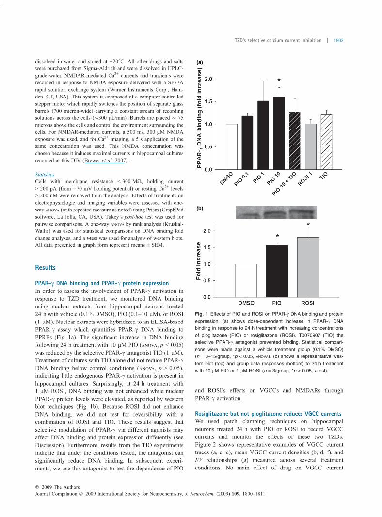
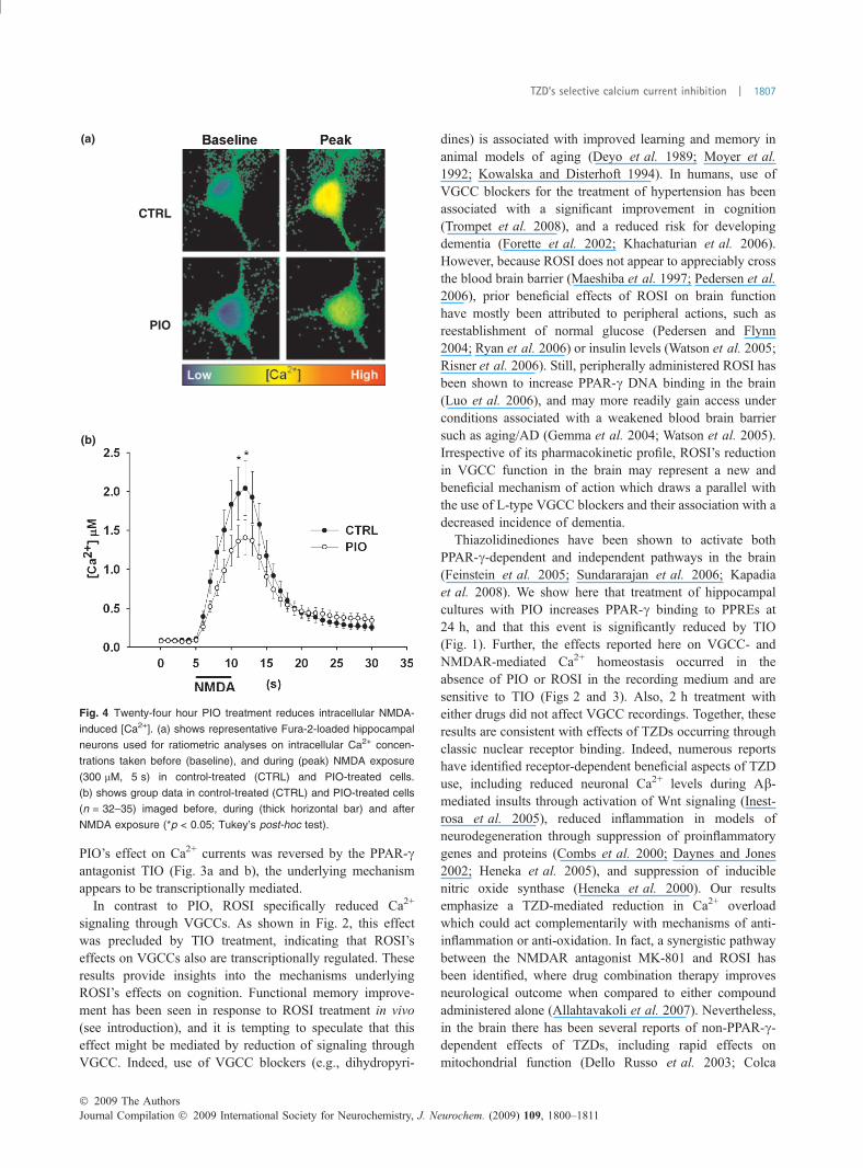
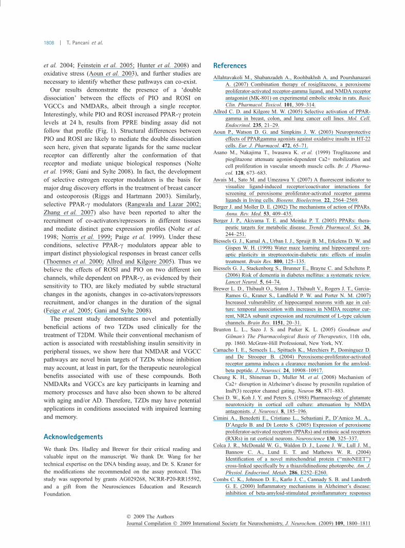
PPAR-c DNA binding and PPAR-c protein expressionIn order to assess the involvement of PPAR-c activation inresponse to TZD treatment, we monitored DNA bindingusing nuclear extracts from hippocampal neurons treated24 h with vehicle (0.1% DMSO), PIO (0.1–10 lM), or ROSI(1 lM). Nuclear extracts were hybridized to an ELISA-basedPPAR-c assay which quantifies PPAR-c DNA binding toPPREs (Fig. 1a). The significant increase in DNA bindingfollowing 24 h treatment with 10 lM PIO (ANOVA, p < 0.05)was reduced by the selective PPAR-c antagonist TIO (1 lM).Treatment of cultures with TIO alone did not reduce PPAR-cDNA binding below control conditions (ANOVA, p > 0.05),indicating little endogenous PPAR-c activation is present inhippocampal cultures. Surprisingly, at 24 h treatment with1 lM ROSI, DNA binding was not enhanced while nuclearPPAR-c protein levels were elevated, as reported by westernblot techniques (Fig. 1b). Because ROSI did not enhanceDNA binding, we did not test for reversibility with acombination of ROSI and TIO. These results suggest thatselective modulation of PPAR-c via different agonists mayaffect DNA binding and protein expression differently (seeDiscussion). Furthermore, results from the TIO experimentsindicate that under the conditions tested, the antagonist cansignificantly reduce DNA binding. In subsequent experi-ments, we use this antagonist to test the dependence of PIO
and ROSI’s effects on VGCCs and NMDARs throughPPAR-c activation.
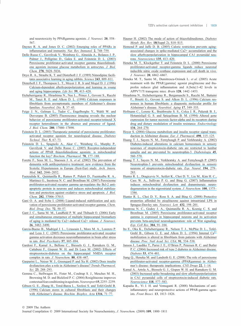
Rosiglitazone but not pioglitazone reduces VGCC currentsWe used patch clamping techniques on hippocampalneurons treated 24 h with PIO or ROSI to record VGCCcurrents and monitor the effects of these two TZDs.Figure 2 shows representative examples of VGCC currenttraces (a, c, e), mean VGCC current densities (b, d, f), andI/V relationships (g) measured across several treatmentconditions. No main effect of drug on VGCC current
(a)
(b)
Fig. 1 Effects of PIO and ROSI on PPAR-c DNA binding and protein
expression. (a) shows dose-dependent increase in PPAR-c DNA
binding in response to 24 h treatment with increasing concentrations
of pioglitazone (PIO) or rosiglitazone (ROSI). T0070907 (TIO) the
selective PPAR-c antagonist prevented binding. Statistical compari-
sons were made against a vehicle treatment group (0.1% DMSO)
(n = 3–15/group, *p < 0.05, ANOVA). (b) shows a representative wes-
tern blot (top) and group data responses (bottom) to 24 h treatment
with 10 lM PIO or 1 lM ROSI (n = 3/group, *p < 0.05, t-test).
� 2009 The AuthorsJournal Compilation � 2009 International Society for Neurochemistry, J. Neurochem. (2009) 109, 1800–1811
TZD’s selective calcium current inhibition | 1803
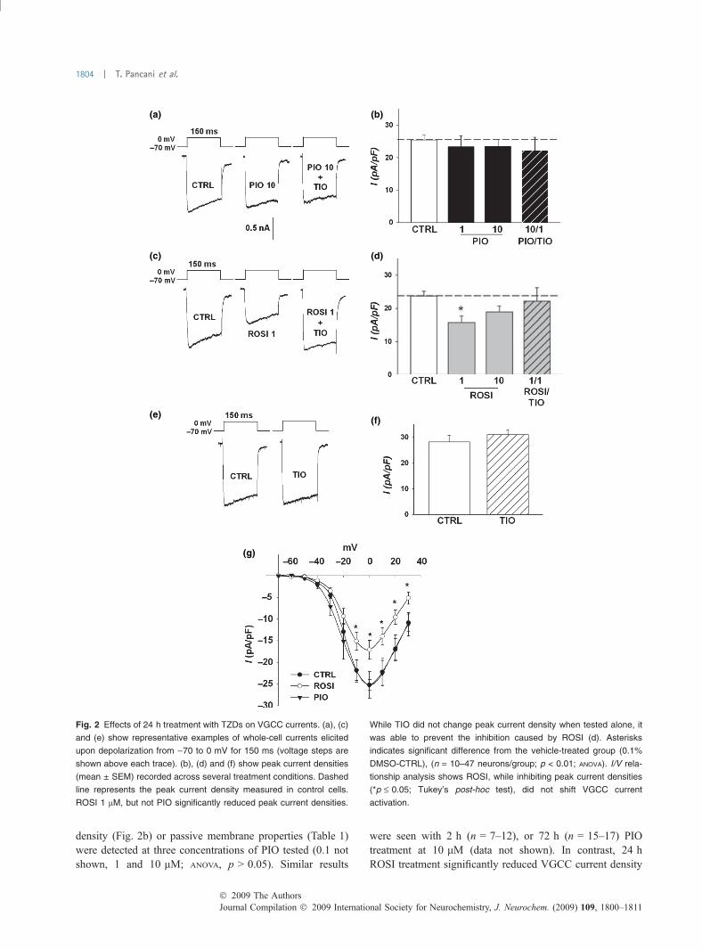
density (Fig. 2b) or passive membrane properties (Table 1)were detected at three concentrations of PIO tested (0.1 notshown, 1 and 10 lM; ANOVA, p > 0.05). Similar results
were seen with 2 h (n = 7–12), or 72 h (n = 15–17) PIOtreatment at 10 lM (data not shown). In contrast, 24 hROSI treatment significantly reduced VGCC current density
(a) (b)
(c) (d)
(e)
(g)
(f)
Fig. 2 Effects of 24 h treatment with TZDs on VGCC currents. (a), (c)
and (e) show representative examples of whole-cell currents elicited
upon depolarization from )70 to 0 mV for 150 ms (voltage steps are
shown above each trace). (b), (d) and (f) show peak current densities
(mean ± SEM) recorded across several treatment conditions. Dashed
line represents the peak current density measured in control cells.
ROSI 1 lM, but not PIO significantly reduced peak current densities.
While TIO did not change peak current density when tested alone, it
was able to prevent the inhibition caused by ROSI (d). Asterisks
indicates significant difference from the vehicle-treated group (0.1%
DMSO-CTRL), (n = 10–47 neurons/group; p < 0.01; ANOVA). I/V rela-
tionship analysis shows ROSI, while inhibiting peak current densities
(*p £ 0.05; Tukey’s post-hoc test), did not shift VGCC current
activation.
Journal Compilation � 2009 International Society for Neurochemistry, J. Neurochem. (2009) 109, 1800–1811� 2009 The Authors
1804 | T. Pancani et al.
measured from )70 mV (Fig. 2d; ANOVA; p < 0.05) or)40 mV (data not shown). Interestingly, greater ROSI-mediated VGCC inhibition was noted on currents elicitedfrom )40 mV compared to )70 mV (data not shown,p < 0.0005), suggesting high voltage-activated L-typecurrents might be more sensitive than low voltage-activatedCa2+ currents to the inhibitory effects of ROSI. In vascularmyocytes, rapid PPAR-c-independent inhibitory effects ofROSI on L-VGCC currents have been observed (Knocket al. 1999). A direct effect of ROSI on L-VGCC is notlikely the mechanism of inhibition reported here, however,because co-treatment of cultures with ROSI (1 lM) and thePPAR-c antagonist TIO (1 lM) for 24 h, was able tocompletely reverse ROSI’s effects on VGCCs (Fig. 2d).Further, ROSI was not present during these experiments.TIO alone for 24 h (n = 18), or ROSI for 2 h (n = 6, datanot shown) had no detectable effect on VGCC currentdensities (Fig. 2e and f). As seen in the I/V relationshipanalysis (Fig. 2g), ROSI inhibited peak currents density butdid not shift the activation curve and voltage-sensitivity ofthe currents. Taken together, these data demonstrate thatROSI’s effects on VGCC are dependent on PPAR-cactivation, and also suggest little basal PPAR-c modulationof these Ca2+ currents.
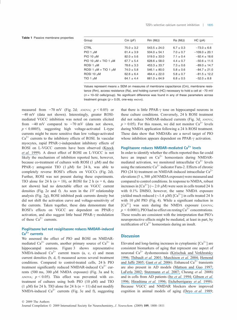
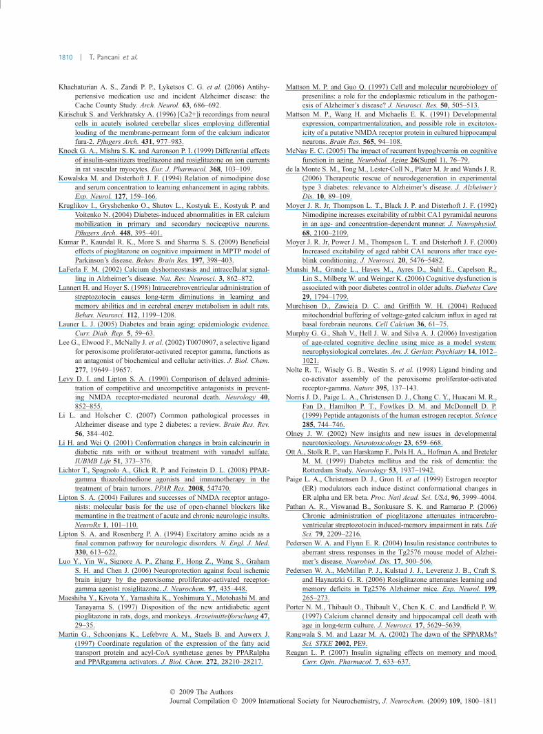
Pioglitazone but not rosiglitazone reduces NMDAR-inducedCa2+ currentsWe assessed the effect of PIO and ROSI on NMDAR-mediated Ca2+ currents, another primary source of Ca2+ inhippocampal neurons. Figure 3 shows representativeNMDA-induced Ca2+ current traces (a, c, e) and meancurrent densities (b, d, f) measured across several treatmentconditions. Compared to control-treated cells, 24 h PIOtreatment significantly reduced NMDAR-induced Ca2+ cur-rents (500 ms, 300 lM NMDA exposure) (Fig. 3a and b;ANOVA; p < 0.05). This effect was prevented with co-treatment of cultures using both PIO (10 lM) and TIO(1 lM) for 24 h. TIO alone for 24 h (n = 11) did not modifyNMDA-induced Ca2+ currents (Fig. 3e and f), suggesting
that there is little PPAR-c tone on hippocampal neurons inthese culture conditions. Conversely, 24 h ROSI treatmentdid not reduce NMDAR-induced currents (Fig. 3d; ANOVA;p > 0.05). For this reason, we did not monitor Ca2+ levelsduring NMDA application following a 24 h ROSI treatment.These data show that NMDARs are a novel target of PIOwhose inhibition appears dependent on PPAR-c activation.
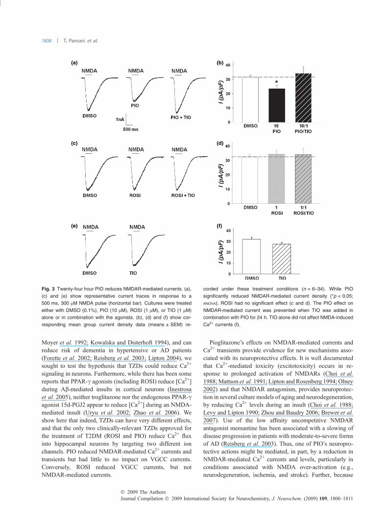
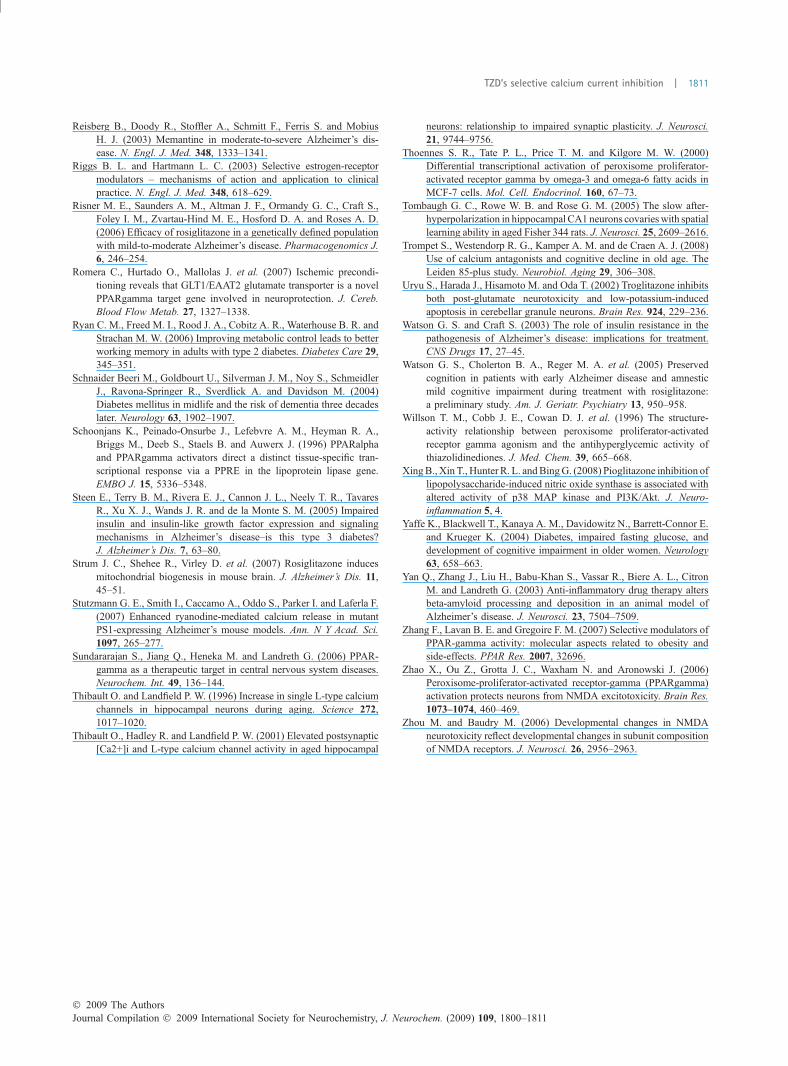
Pioglitazone reduces NMDAR-mediated Ca2+ levelsIn order to identify whether the effects reported thus far couldhave an impact on Ca2+ homeostasis during NMDAR-mediated activation, we monitored intracellular Ca2+ levelsusing the ratiometric Ca2+ indicator Fura-2. Effects of chronicPIO (24 h) treatment on NMDAR-induced intracellular Ca2+
elevations (5 s, 300 lMNMDAexposure) weremeasured andcompared to control conditions. In response to NMDA, robustincreases in [Ca2+] (� 2.0 lM) were seen in cells treated 24 hwith 0.1% DMSO, however, the same NMDA exposureyielded much reduced (�1.4 lM) [Ca2+] in cells treated 24 hwith 10 lM PIO (Fig. 4). While a significant reduction in[Ca2+] was seen during the NMDA exposure (ANOVA;p < 0.0001), PIO had no effect on resting Ca2+ concentrations.These results are consistent with the interpretation that PIO’sneuroprotective effects might be mediated, at least in part, byrectification of Ca2+ homeostasis during an insult.
Discussion
Elevated and long-lasting increases in cytoplasmic [Ca2+] areconsistent biomarkers of aging that represent one aspect ofneuronal Ca2+ dyshomeostasis (Kirischuk and Verkhratsky1996; Thibault et al. 2001; Murchison et al. 2004; Hemondand Jaffe 2005; Gant et al. 2006). Enhanced Ca2+ transientsare also present in AD models (Mattson and Guo 1997;LaFerla 2002; Stutzmann et al. 2007; Cheung et al. 2008)and in cells from AD patients (Ito et al. 1994; Gibson et al.1996; Hirashima et al. 1996; Etcheberrigaray et al. 1998).Because VGCC and NMDAR blockers show improvedcognition in animal models of aging (Deyo et al. 1989;
Table 1 Passive membrane propertiesGroup Cm (pF) Rm (MW) Ra (MW) HC (pA)
CTRL 75.0 ± 3.2 543.5 ± 24.0 6.7 ± 0.3 )73.0 ± 6.6
PIO 1 lM 61.4 ± 3.9 504.0 ± 54.1 7.0 ± 0.7 )109.0 ± 20.1
PIO 10 lM 82.0 ± 5.6 519.0 ± 33.0 7.1 ± 0.4 )92.4 ± 18.6
PIO 10 lM + TIO 1 lM 67.7 ± 5.4 628.6 ± 58.0 4.4 ± 0.7 )50.6 ± 11.5
ROSI 1 lM 78.6 ± 3.3 453.3 ± 33.7 7.3 ± 0.6 )89.0 ± 14.7
ROSI 1 lM + TIO 1 lM 74.0 ± 5.8 546.1 ± 80.0 5.8 ± 0.6 )84.7 ± 21.0
ROSI 10 lM 62.6 ± 6.4 464.4 ± 22.0 5.8 ± 0.7 )81.5 ± 12.2
TIO 1 lM 64.1 ± 4.4 661.0 ± 64.9 6.8 ± 0.5 )52.0 ± 8.8
Values represent means ± SEM on measures of membrane capacitance (Cm), membrane resis-
tance (Rm), access resistance (Ra), and holding current (HC) necessary to hold a cell at )70 mV
(n = 10–52 cells/group). No significant difference was found in any of these parameters across
treatment groups (p > 0.05, one-way ANOVA).
� 2009 The AuthorsJournal Compilation � 2009 International Society for Neurochemistry, J. Neurochem. (2009) 109, 1800–1811
TZD’s selective calcium current inhibition | 1805
Moyer et al. 1992; Kowalska and Disterhoft 1994), and canreduce risk of dementia in hypertensive or AD patients(Forette et al. 2002; Reisberg et al. 2003; Lipton 2004), wesought to test the hypothesis that TZDs could reduce Ca2+
signaling in neurons. Furthermore, while there has been somereports that PPAR-c agonists (including ROSI) reduce [Ca2+]during Ab-mediated insults in cortical neurons (Inestrosaet al. 2005), neither troglitazone nor the endogenous PPAR-cagonist 15d-PGJ2 appear to reduce [Ca2+] during an NMDA-mediated insult (Uryu et al. 2002; Zhao et al. 2006). Weshow here that indeed, TZDs can have very different effects,and that the only two clinically-relevant TZDs approved forthe treatment of T2DM (ROSI and PIO) reduce Ca2+ fluxinto hippocampal neurons by targeting two different ionchannels. PIO reduced NMDAR-mediated Ca2+ currents andtransients but had little to no impact on VGCC currents.Conversely, ROSI reduced VGCC currents, but notNMDAR-mediated currents.
Pioglitazone’s effects on NMDAR-mediated currents andCa2+ transients provide evidence for new mechanisms asso-ciated with its neuroprotective effects. It is well documentedthat Ca2+-mediated toxicity (excitotoxicity) occurs in re-sponse to prolonged activation of NMDARs (Choi et al.1988;Mattson et al. 1991; Lipton and Rosenberg 1994; Olney2002) and that NMDAR antagonism, provides neuroprotec-tion in several culture models of aging and neurodegeneration,by reducing Ca2+ levels during an insult (Choi et al. 1988;Levy and Lipton 1990; Zhou and Baudry 2006; Brewer et al.2007). Use of the low affinity uncompetitive NMDARantagonist memantine has been associated with a slowing ofdisease progression in patients with moderate-to-severe formsof AD (Reisberg et al. 2003). Thus, one of PIO’s neuropro-tective actions might be mediated, in part, by a reduction inNMDAR-mediated Ca2+ currents and levels, particularly inconditions associated with NMDA over-activation (e.g.,neurodegeneration, ischemia, and stroke). Further, because
(a) (b)
(c)
(e)
(d)
(f)
Fig. 3 Twenty-four hour PIO reduces NMDAR-mediated currents. (a),
(c) and (e) show representative current traces in response to a
500 ms, 300 lM NMDA pulse (horizontal bar). Cultures were treated
either with DMSO (0.1%), PIO (10 lM), ROSI (1 lM), or TIO (1 lM)
alone or in combination with the agonists. (b), (d) and (f) show cor-
responding mean group current density data (means ± SEM) re-
corded under these treatment conditions (n = 6–34). While PIO
significantly reduced NMDAR-mediated current density (*p < 0.05;
ANOVA), ROSI had no significant effect (c and d). The PIO effect on
NMDAR-mediated current was prevented when TIO was added in
combination with PIO for 24 h. TIO alone did not affect NMDA-induced
Ca2+ currents (f).
Journal Compilation � 2009 International Society for Neurochemistry, J. Neurochem. (2009) 109, 1800–1811� 2009 The Authors
1806 | T. Pancani et al.
PIO’s effect on Ca2+ currents was reversed by the PPAR-cantagonist TIO (Fig. 3a and b), the underlying mechanismappears to be transcriptionally mediated.
In contrast to PIO, ROSI specifically reduced Ca2+
signaling through VGCCs. As shown in Fig. 2, this effectwas precluded by TIO treatment, indicating that ROSI’seffects on VGCCs also are transcriptionally regulated. Theseresults provide insights into the mechanisms underlyingROSI’s effects on cognition. Functional memory improve-ment has been seen in response to ROSI treatment in vivo(see introduction), and it is tempting to speculate that thiseffect might be mediated by reduction of signaling throughVGCC. Indeed, use of VGCC blockers (e.g., dihydropyri-
dines) is associated with improved learning and memory inanimal models of aging (Deyo et al. 1989; Moyer et al.1992; Kowalska and Disterhoft 1994). In humans, use ofVGCC blockers for the treatment of hypertension has beenassociated with a significant improvement in cognition(Trompet et al. 2008), and a reduced risk for developingdementia (Forette et al. 2002; Khachaturian et al. 2006).However, because ROSI does not appear to appreciably crossthe blood brain barrier (Maeshiba et al. 1997; Pedersen et al.2006), prior beneficial effects of ROSI on brain functionhave mostly been attributed to peripheral actions, such asreestablishment of normal glucose (Pedersen and Flynn2004; Ryan et al. 2006) or insulin levels (Watson et al. 2005;Risner et al. 2006). Still, peripherally administered ROSI hasbeen shown to increase PPAR-c DNA binding in the brain(Luo et al. 2006), and may more readily gain access underconditions associated with a weakened blood brain barriersuch as aging/AD (Gemma et al. 2004; Watson et al. 2005).Irrespective of its pharmacokinetic profile, ROSI’s reductionin VGCC function in the brain may represent a new andbeneficial mechanism of action which draws a parallel withthe use of L-type VGCC blockers and their association with adecreased incidence of dementia.
Thiazolidinediones have been shown to activate bothPPAR-c-dependent and independent pathways in the brain(Feinstein et al. 2005; Sundararajan et al. 2006; Kapadiaet al. 2008). We show here that treatment of hippocampalcultures with PIO increases PPAR-c binding to PPREs at24 h, and that this event is significantly reduced by TIO(Fig. 1). Further, the effects reported here on VGCC- andNMDAR-mediated Ca2+ homeostasis occurred in theabsence of PIO or ROSI in the recording medium and aresensitive to TIO (Figs 2 and 3). Also, 2 h treatment witheither drugs did not affect VGCC recordings. Together, theseresults are consistent with effects of TZDs occurring throughclassic nuclear receptor binding. Indeed, numerous reportshave identified receptor-dependent beneficial aspects of TZDuse, including reduced neuronal Ca2+ levels during Ab-mediated insults through activation of Wnt signaling (Inest-rosa et al. 2005), reduced inflammation in models ofneurodegeneration through suppression of proinflammatorygenes and proteins (Combs et al. 2000; Daynes and Jones2002; Heneka et al. 2005), and suppression of induciblenitric oxide synthase (Heneka et al. 2000). Our resultsemphasize a TZD-mediated reduction in Ca2+ overloadwhich could act complementarily with mechanisms of anti-inflammation or anti-oxidation. In fact, a synergistic pathwaybetween the NMDAR antagonist MK-801 and ROSI hasbeen identified, where drug combination therapy improvesneurological outcome when compared to either compoundadministered alone (Allahtavakoli et al. 2007). Nevertheless,in the brain there has been several reports of non-PPAR-c-dependent effects of TZDs, including rapid effects onmitochondrial function (Dello Russo et al. 2003; Colca
(a)
(b)
CTRL
PIO
Fig. 4 Twenty-four hour PIO treatment reduces intracellular NMDA-
induced [Ca2+]. (a) shows representative Fura-2-loaded hippocampal
neurons used for ratiometric analyses on intracellular Ca2+ concen-
trations taken before (baseline), and during (peak) NMDA exposure
(300 lM, 5 s) in control-treated (CTRL) and PIO-treated cells.
(b) shows group data in control-treated (CTRL) and PIO-treated cells
(n = 32–35) imaged before, during (thick horizontal bar) and after
NMDA exposure (*p < 0.05; Tukey’s post-hoc test).
� 2009 The AuthorsJournal Compilation � 2009 International Society for Neurochemistry, J. Neurochem. (2009) 109, 1800–1811
TZD’s selective calcium current inhibition | 1807
et al. 2004; Feinstein et al. 2005; Hunter et al. 2008) andoxidative stress (Aoun et al. 2003), and further studies arenecessary to identify whether these pathways can co-exist.
Our results demonstrate the presence of a ‘doubledissociation’ between the effects of PIO and ROSI onVGCCs and NMDARs, albeit through a single receptor.Interestingly, while PIO and ROSI increased PPAR-c proteinlevels at 24 h, results from PPRE binding assay did notfollow that profile (Fig. 1). Structural differences betweenPIO and ROSI are likely to mediate the double dissociationseen here, given that separate ligands for the same nuclearreceptor can differently alter the conformation of thatreceptor and mediate unique biological responses (Nolteet al. 1998; Gani and Sylte 2008). In fact, the developmentof selective estrogen receptor modulators is the basis formajor drug discovery efforts in the treatment of breast cancerand osteoporosis (Riggs and Hartmann 2003). Similarly,selective PPAR-c modulators (Rangwala and Lazar 2002;Zhang et al. 2007) also have been reported to alter therecruitment of co-activators/repressors in different tissuesand mediate distinct gene expression profiles (Nolte et al.1998; Norris et al. 1999; Paige et al. 1999). Under theseconditions, selective PPAR-c modulators appear able toimpart distinct physiological responses in breast cancer cells(Thoennes et al. 2000; Allred and Kilgore 2005). Thus webelieve the effects of ROSI and PIO on two different ionchannels, while dependent on PPAR-c, as evidenced by theirsensitivity to TIO, are likely mediated by subtle structuralchanges in the agonists, changes in co-activators/repressorsrecruitment, and/or changes in the duration of the signal(Feige et al. 2005; Gani and Sylte 2008).
The present study demonstrates novel and potentiallybeneficial actions of two TZDs used clinically for thetreatment of T2DM. While their conventional mechanism ofaction is associated with reestablishing insulin sensitivity inperipheral tissues, we show here that NMDAR and VGCCpathways are novel brain targets of TZDs whose inhibitionmay account, at least in part, for the therapeutic neurologicalbenefits associated with use of these compounds. BothNMDARs and VGCCs are key participants in learning andmemory processes and have also been shown to be alteredwith aging and/or AD. Therefore, TZDs may have potentialapplications in conditions associated with impaired learningand memory.
Acknowledgements
We thank Drs. Hadley and Brewer for their critical reading and
valuable input on the manuscript. We thank Dr. Wang for her
technical expertise on the DNA binding assay, and Dr. S. Kraner for
the modifications she recommended on the assay protocol. This
study was supported by grants AG029268, NCRR-P20-RR15592,
and a gift from the Neurosciences Education and Research
Foundation.
References
Allahtavakoli M., Shabanzadeh A., Roohbakhsh A. and PourshanazariA. (2007) Combination therapy of rosiglitazone, a peroxisomeproliferator-activated receptor-gamma ligand, and NMDA receptorantagonist (MK-801) on experimental embolic stroke in rats. BasicClin. Pharmacol. Toxicol. 101, 309–314.
Allred C. D. and Kilgore M. W. (2005) Selective activation of PPAR-gamma in breast, colon, and lung cancer cell lines. Mol. Cell.Endocrinol. 235, 21–29.
Aoun P., Watson D. G. and Simpkins J. W. (2003) Neuroprotectiveeffects of PPARgamma agonists against oxidative insults in HT-22cells. Eur. J. Pharmacol. 472, 65–71.
Asano M., Nakajima T., Iwasawa K. et al. (1999) Troglitazone andpioglitazone attenuate agonist-dependent Ca2+ mobilization andcell proliferation in vascular smooth muscle cells. Br. J. Pharma-col. 128, 673–683.
Awais M., Sato M. and Umezawa Y. (2007) A fluorescent indicator tovisualize ligand-induced receptor/coactivator interactions forscreening of peroxisome proliferator-activated receptor gammaligands in living cells. Biosens. Bioelectron. 22, 2564–2569.
Berger J. and Moller D. E. (2002) The mechanisms of action of PPARs.Annu. Rev. Med. 53, 409–435.
Berger J. P., Akiyama T. E. and Meinke P. T. (2005) PPARs: thera-peutic targets for metabolic disease. Trends Pharmacol. Sci. 26,244–251.
Biessels G. J., Kamal A., Urban I. J., Spruijt B. M., Erkelens D. W. andGispen W. H. (1998) Water maze learning and hippocampal syn-aptic plasticity in streptozotocin-diabetic rats: effects of insulintreatment. Brain Res. 800, 125–135.
Biessels G. J., Staekenborg S., Brunner E., Brayne C. and Scheltens P.(2006) Risk of dementia in diabetes mellitus: a systematic review.Lancet Neurol. 5, 64–74.
Brewer L. D., Thibault O., Staton J., Thibault V., Rogers J. T., Garcia-Ramos G., Kraner S., Landfield P. W. and Porter N. M. (2007)Increased vulnerability of hippocampal neurons with age in cul-ture: temporal association with increases in NMDA receptor cur-rent, NR2A subunit expression and recruitment of L-type calciumchannels. Brain Res. 1151, 20–31.
Brunton L. L., Sazo J. S. and Parker K. L. (2005) Goodman andGilman’s The Pharmacological Basis of Therapeutics, 11th edn,pp. 1860. McGraw-Hill Professional, New York, NY.
Camacho I. E., Serneels L., Spittaels K., Merchiers P., Dominguez D.and De Strooper B. (2004) Peroxisome-proliferator-activatedreceptor gamma induces a clearance mechanism for the amyloid-beta peptide. J. Neurosci. 24, 10908–10917.
Cheung K. H., Shineman D., Muller M. et al. (2008) Mechanism ofCa2+ disruption in Alzheimer’s disease by presenilin regulation ofInsP(3) receptor channel gating. Neuron 58, 871–883.
Choi D. W., Koh J. Y. and Peters S. (1988) Pharmacology of glutamateneurotoxicity in cortical cell culture: attenuation by NMDAantagonists. J. Neurosci. 8, 185–196.
Cimini A., Benedetti E., Cristiano L., Sebastiani P., D’Amico M. A.,D’Angelo B. and Di Loreto S. (2005) Expression of peroxisomeproliferator-activated receptors (PPARs) and retinoic acid receptors(RXRs) in rat cortical neurons. Neuroscience 130, 325–337.
Colca J. R., McDonald W. G., Waldon D. J., Leone J. W., Lull J. M.,Bannow C. A., Lund E. T. and Mathews W. R. (2004)Identification of a novel mitochondrial protein (‘‘mitoNEET’’)cross-linked specifically by a thiazolidinedione photoprobe. Am. J.Physiol. Endocrinol. Metab. 286, E252–E260.
Combs C. K., Johnson D. E., Karlo J. C., Cannady S. B. and LandrethG. E. (2000) Inflammatory mechanisms in Alzheimer’s disease:inhibition of beta-amyloid-stimulated proinflammatory responses
Journal Compilation � 2009 International Society for Neurochemistry, J. Neurochem. (2009) 109, 1800–1811� 2009 The Authors
1808 | T. Pancani et al.
and neurotoxicity by PPARgamma agonists. J. Neurosci. 20, 558–567.
Daynes R. A. and Jones D. C. (2002) Emerging roles of PPARs ininflammation and immunity. Nat. Rev. Immunol. 2, 748–759.
Dello Russo C., Gavrilyuk V., Weinberg G., Almeida A., Bolanos J. P.,Palmer J., Pelligrino D., Galea E. and Feinstein D. L. (2003)Peroxisome proliferator-activated receptor gamma thiazolidinedi-one agonists increase glucose metabolism in astrocytes. J. Biol.Chem. 278, 5828–5836.
Deyo R. A., Straube K. T. and Disterhoft J. F. (1989) Nimodipine facili-tates associative learning in aging rabbits. Science 243, 809–811.
Disterhoft J. F., Thompson L. T., Moyer J. R. Jr and Mogul D. J. (1996)Calcium-dependent afterhyperpolarization and learning in youngand aging hippocampus. Life Sci. 59, 413–420.
Etcheberrigaray R., Hirashima N., Nee L., Prince J., Govoni S., RacchiM., Tanzi R. E. and Alkon D. L. (1998) Calcium responses infibroblasts from asymptomatic members of Alzheimer’s diseasefamilies. Neurobiol. Dis. 5, 37–45.
Feige J. N., Gelman L., Tudor C., Engelborghs Y., Wahli W. andDesvergne B. (2005) Fluorescence imaging reveals the nuclearbehavior of peroxisome proliferator-activated receptor/retinoid Xreceptor heterodimers in the absence and presence of ligand.J. Biol. Chem. 280, 17880–17890.
Feinstein D. L. (2003) Therapeutic potential of peroxisome proliferator-activated receptor agonists for neurological disease. DiabetesTechnol. Ther. 5, 67–73.
Feinstein D. L., Spagnolo A., Akar C., Weinberg G., Murphy P.,Gavrilyuk V. and Dello Russo C. (2005) Receptor-independentactions of PPAR thiazolidinedione agonists: is mitochondrialfunction the key? Biochem. Pharmacol. 70, 177–188.
Forette F., Seux M. L., Staessen J. A. et al. (2002) The prevention ofdementia with antihypertensive treatment: new evidence from theSystolic Hypertension in Europe (Syst-Eur) study. Arch. Intern.Med. 162, 2046–2052.
Fuenzalida K., Quintanilla R., Ramos P., Piderit D., Fuentealba R. A.,Martinez G., Inestrosa N. C. and Bronfman M. (2007) Peroxisomeproliferator-activated receptor gamma up-regulates the Bcl-2 anti-apoptotic protein in neurons and induces mitochondrial stabiliza-tion and protection against oxidative stress and apoptosis. J. Biol.Chem. 282, 37006–37015.
Gani O. A. and Sylte I. (2008) Ligand-induced stabilization and acti-vation of peroxisome proliferator-activated receptor gamma. Chem.Biol. Drug. Des. 72, 50–57.
Gant J. C., Sama M. M., Landfield P. W. and Thibault O. (2006) Earlyand simultaneous emergence of multiple hippocampal biomarkersof aging is mediated by Ca2+-induced Ca2+ release. J. Neurosci.26, 3482–3490.
Garcia-Bueno B., Madrigal J. L., Lizasoain I., Moro M. A., Lorenzo P.and Leza J. C. (2005) Peroxisome proliferator-activated receptorgamma activation decreases neuroinflammation in brain after stressin rats. Biol. Psychiatry 57, 885–894.
Gardoni F., Kamal A., Bellone C., Biessels G. J., Ramakers G. M.,Cattabeni F., Gispent W. H. and Di Luca M. (2002) Effects ofstreptozotocin-diabetes on the hippocampal NMDA receptorcomplex in rats. J. Neurochem. 80, 438–447.
Gasparini L., Netzer W. J., Greengard P. and Xu H. (2002) Does insulindysfunction play a role in Alzheimer’s disease? Trends Pharmacol.Sci. 23, 288–293.
Gemma C., Stellwagen H., Fister M., Coultrap S. J., Mesches M. H.,Browning M. D. and Bickford P. C. (2004) Rosiglitazone improvescontextualfearconditioninginagedrats.Neuroreport15,2255–2259.
Gibson G. E., Zhang H., Toral-Barza L., Szolosi S. and Tofel-Grehl B.(1996) Calcium stores in cultured fibroblasts and their changeswith Alzheimer’s disease. Biochim. Biophys. Acta 1316, 71–77.
Hauner H. (2002) The mode of action of thiazolidinediones. DiabetesMetab. Res. Rev. 18(Suppl 2), S10–S15.
Hemond P. and Jaffe D. B. (2005) Caloric restriction prevents aging-associated changes in spike-mediated Ca2+ accumulation and theslow afterhyperpolarization in hippocampal CA1 pyramidal neu-rons. Neuroscience 135, 413–420.
Heneka M. T., Klockgether T. and Feinstein D. L. (2000) Peroxisomeproliferator-activated receptor-gamma ligands reduce neuronalinducible nitric oxide synthase expression and cell death in vivo.J. Neurosci. 20, 6862–6867.
Heneka M. T., Sastre M., Dumitrescu-Ozimek L. et al. (2005) Acutetreatment with the PPAR{gamma} agonist pioglitazone and ibu-profen reduces glial inflammation and A{beta}1-42 levels inAPPV717I transgenic mice. Brain 128, 1442–1453.
Hirashima N., Etcheberrigaray R., Bergamaschi S., Racchi M., BattainiF., Binetti G., Govoni S. and Alkon D. L. (1996) Calcium res-ponses in human fibroblasts: a diagnostic molecular profile forAlzheimer’s disease. Neurobiol. Aging 17, 549–555.
Hofmann C., Lorenz K., Braithwaite S. S., Colca J. R., Palazuk B. J.,Hotamisligil G. S. and Spiegelman B. M. (1994) Altered geneexpression for tumor necrosis factor-alpha and its receptors duringdrug and dietary modulation of insulin resistance. Endocrinology134, 264–270.
Hoyer S. (2004) Glucose metabolism and insulin receptor signal trans-duction in Alzheimer disease. Eur. J. Pharmacol. 490, 115–125.
Huang T. J., Sayers N. M., Fernyhough P. and Verkhratsky A. (2002)Diabetes-induced alterations in calcium homeostasis in sensoryneurones of streptozotocin-diabetic rats are restricted to lumbarganglia and are prevented by neurotrophin-3. Diabetologia 45,560–570.
Huang T. J., Sayers N. M., Verkhratsky A. and Fernyhough P. (2005)Neurotrophin-3 prevents mitochondrial dysfunction in sensoryneurons of streptozotocin-diabetic rats. Exp. Neurol. 194, 279–283.
Hunter R. L., Dragicevic N., Seifert K., Choi D. Y., Liu M., Kim H. C.,Cass W. A., Sullivan P. G. and Bing G. (2007) Inflammationinduces mitochondrial dysfunction and dopaminergic neuro-degeneration in the nigrostriatal system. J. Neurochem. 100, 1375–1386.
Hunter R. L., Choi D. Y., Ross S. A. and Bing G. (2008) Protectiveproperties afforded by pioglitazone against intrastriatal LPS inSprague-Dawley rats. Neurosci. Lett. 432, 198–201.
Inestrosa N. C., Godoy J. A., Quintanilla R. A., Koenig C. S. andBronfman M. (2005) Peroxisome proliferator-activated receptorgamma is expressed in hippocampal neurons and its activationprevents beta-amyloid neurodegeneration: role of Wnt signaling.Exp. Cell Res. 304, 91–104.
Ito E., Oka K., Etcheberrigaray R., Nelson T. J., McPhie D. L., Tofel-Grehl B., Gibson G. E. and Alkon D. L. (1994) Internal Ca2+
mobilization is altered in fibroblasts from patients with Alzheimerdisease. Proc. Natl Acad. Sci. USA, 91, 534–538.
Janson J., Laedtke T., Parisi J. E., O’Brien P., Petersen R. C. and ButlerP. C. (2004) Increased risk of type 2 diabetes in Alzheimer disease.Diabetes 53, 474–481.
Jiang Q., Heneka M. and Landreth G. E. (2008) The role of peroxisomeproliferator-activated receptor-gamma (PPARgamma) in Alzhei-mer’s disease: therapeutic implications. CNS Drugs 22, 1–14.
Kamal A., Artola A., Biessels G. J., Gispen W. H. and Ramakers G. M.(2003) Increased spike broadening and slow afterhyperpolarizationin CA1 pyramidal cells of streptozotocin-induced diabetic rats.Neuroscience 118, 577–583.
Kapadia R., Yi J. H. and Vemuganti R. (2008) Mechanisms of anti-inflammatory and neuroprotective actions of PPAR-gamma agon-ists. Front Biosci. 13, 1813–1826.
� 2009 The AuthorsJournal Compilation � 2009 International Society for Neurochemistry, J. Neurochem. (2009) 109, 1800–1811
TZD’s selective calcium current inhibition | 1809
Khachaturian A. S., Zandi P. P., Lyketsos C. G. et al. (2006) Antihy-pertensive medication use and incident Alzheimer disease: theCache County Study. Arch. Neurol. 63, 686–692.
Kirischuk S. and Verkhratsky A. (1996) [Ca2+]i recordings from neuralcells in acutely isolated cerebellar slices employing differentialloading of the membrane-permeant form of the calcium indicatorfura-2. Pflugers Arch. 431, 977–983.
Knock G. A., Mishra S. K. and Aaronson P. I. (1999) Differential effectsof insulin-sensitizers troglitazone and rosiglitazone on ion currentsin rat vascular myocytes. Eur. J. Pharmacol. 368, 103–109.
Kowalska M. and Disterhoft J. F. (1994) Relation of nimodipine doseand serum concentration to learning enhancement in aging rabbits.Exp. Neurol. 127, 159–166.
Kruglikov I., Gryshchenko O., Shutov L., Kostyuk E., Kostyuk P. andVoitenko N. (2004) Diabetes-induced abnormalities in ER calciummobilization in primary and secondary nociceptive neurons.Pflugers Arch. 448, 395–401.
Kumar P., Kaundal R. K., More S. and Sharma S. S. (2009) Beneficialeffects of pioglitazone on cognitive impairment in MPTP model ofParkinson’s disease. Behav. Brain Res. 197, 398–403.
LaFerla F. M. (2002) Calcium dyshomeostasis and intracellular signal-ling in Alzheimer’s disease. Nat. Rev. Neurosci. 3, 862–872.
Lannert H. and Hoyer S. (1998) Intracerebroventricular administration ofstreptozotocin causes long-term diminutions in learning andmemory abilities and in cerebral energy metabolism in adult rats.Behav. Neurosci. 112, 1199–1208.
Launer L. J. (2005) Diabetes and brain aging: epidemiologic evidence.Curr. Diab. Rep. 5, 59–63.
Lee G., Elwood F., McNally J. et al. (2002) T0070907, a selective ligandfor peroxisome proliferator-activated receptor gamma, functions asan antagonist of biochemical and cellular activities. J. Biol. Chem.277, 19649–19657.
Levy D. I. and Lipton S. A. (1990) Comparison of delayed adminis-tration of competitive and uncompetitive antagonists in prevent-ing NMDA receptor-mediated neuronal death. Neurology 40,852–855.
Li L. and Holscher C. (2007) Common pathological processes inAlzheimer disease and type 2 diabetes: a review. Brain Res. Rev.56, 384–402.
Li H. and Wei Q. (2001) Conformation changes in brain calcineurin indiabetic rats with or without treatment with vanadyl sulfate.IUBMB Life 51, 373–376.
Lichtor T., Spagnolo A., Glick R. P. and Feinstein D. L. (2008) PPAR-gamma thiazolidinedione agonists and immunotherapy in thetreatment of brain tumors. PPAR Res. 2008, 547470.
Lipton S. A. (2004) Failures and successes of NMDA receptor antago-nists: molecular basis for the use of open-channel blockers likememantine in the treatment of acute and chronic neurologic insults.NeuroRx 1, 101–110.
Lipton S. A. and Rosenberg P. A. (1994) Excitatory amino acids as afinal common pathway for neurologic disorders. N. Engl. J. Med.330, 613–622.
Luo Y., Yin W., Signore A. P., Zhang F., Hong Z., Wang S., GrahamS. H. and Chen J. (2006) Neuroprotection against focal ischemicbrain injury by the peroxisome proliferator-activated receptor-gamma agonist rosiglitazone. J. Neurochem. 97, 435–448.
Maeshiba Y., Kiyota Y., Yamashita K., Yoshimura Y., Motohashi M. andTanayama S. (1997) Disposition of the new antidiabetic agentpioglitazone in rats, dogs, and monkeys. Arzneimittelforschung 47,29–35.
Martin G., Schoonjans K., Lefebvre A. M., Staels B. and Auwerx J.(1997) Coordinate regulation of the expression of the fatty acidtransport protein and acyl-CoA synthetase genes by PPARalphaand PPARgamma activators. J. Biol. Chem. 272, 28210–28217.
Mattson M. P. and Guo Q. (1997) Cell and molecular neurobiology ofpresenilins: a role for the endoplasmic reticulum in the pathogen-esis of Alzheimer’s disease? J. Neurosci. Res. 50, 505–513.
Mattson M. P., Wang H. and Michaelis E. K. (1991) Developmentalexpression, compartmentalization, and possible role in excitotox-icity of a putative NMDA receptor protein in cultured hippocampalneurons. Brain Res. 565, 94–108.
McNay E. C. (2005) The impact of recurrent hypoglycemia on cognitivefunction in aging. Neurobiol. Aging 26(Suppl 1), 76–79.
de la Monte S. M., Tong M., Lester-Coll N., Plater M. Jr and Wands J. R.(2006) Therapeutic rescue of neurodegeneration in experimentaltype 3 diabetes: relevance to Alzheimer’s disease. J. Alzheimer’sDis. 10, 89–109.
Moyer J. R. Jr, Thompson L. T., Black J. P. and Disterhoft J. F. (1992)Nimodipine increases excitability of rabbit CA1 pyramidal neuronsin an age- and concentration-dependent manner. J. Neurophysiol.68, 2100–2109.
Moyer J. R. Jr, Power J. M., Thompson L. T. and Disterhoft J. F. (2000)Increased excitability of aged rabbit CA1 neurons after trace eye-blink conditioning. J. Neurosci. 20, 5476–5482.
Munshi M., Grande L., Hayes M., Ayres D., Suhl E., Capelson R.,Lin S., Milberg W. and Weinger K. (2006) Cognitive dysfunction isassociated with poor diabetes control in older adults. Diabetes Care29, 1794–1799.
Murchison D., Zawieja D. C. and Griffith W. H. (2004) Reducedmitochondrial buffering of voltage-gated calcium influx in aged ratbasal forebrain neurons. Cell Calcium 36, 61–75.
Murphy G. G., Shah V., Hell J. W. and Silva A. J. (2006) Investigationof age-related cognitive decline using mice as a model system:neurophysiological correlates. Am. J. Geriatr. Psychiatry 14, 1012–1021.
Nolte R. T., Wisely G. B., Westin S. et al. (1998) Ligand binding andco-activator assembly of the peroxisome proliferator-activatedreceptor-gamma. Nature 395, 137–143.
Norris J. D., Paige L. A., Christensen D. J., Chang C. Y., Huacani M. R.,Fan D., Hamilton P. T., Fowlkes D. M. and McDonnell D. P.(1999) Peptide antagonists of the human estrogen receptor. Science285, 744–746.
Olney J. W. (2002) New insights and new issues in developmentalneurotoxicology. Neurotoxicology 23, 659–668.
Ott A., Stolk R. P., van Harskamp F., Pols H. A., Hofman A. and BretelerM. M. (1999) Diabetes mellitus and the risk of dementia: theRotterdam Study. Neurology 53, 1937–1942.
Paige L. A., Christensen D. J., Gron H. et al. (1999) Estrogen receptor(ER) modulators each induce distinct conformational changes inER alpha and ER beta. Proc. Natl Acad. Sci. USA, 96, 3999–4004.
Pathan A. R., Viswanad B., Sonkusare S. K. and Ramarao P. (2006)Chronic administration of pioglitazone attenuates intracerebro-ventricular streptozotocin induced-memory impairment in rats. LifeSci. 79, 2209–2216.
Pedersen W. A. and Flynn E. R. (2004) Insulin resistance contributes toaberrant stress responses in the Tg2576 mouse model of Alzhei-mer’s disease. Neurobiol. Dis. 17, 500–506.
Pedersen W. A., McMillan P. J., Kulstad J. J., Leverenz J. B., Craft S.and Haynatzki G. R. (2006) Rosiglitazone attenuates learning andmemory deficits in Tg2576 Alzheimer mice. Exp. Neurol. 199,265–273.
Porter N. M., Thibault O., Thibault V., Chen K. C. and Landfield P. W.(1997) Calcium channel density and hippocampal cell death withage in long-term culture. J. Neurosci. 17, 5629–5639.
Rangwala S. M. and Lazar M. A. (2002) The dawn of the SPPARMs?Sci. STKE 2002, PE9.
Reagan L. P. (2007) Insulin signaling effects on memory and mood.Curr. Opin. Pharmacol. 7, 633–637.
Journal Compilation � 2009 International Society for Neurochemistry, J. Neurochem. (2009) 109, 1800–1811� 2009 The Authors
1810 | T. Pancani et al.
Reisberg B., Doody R., Stoffler A., Schmitt F., Ferris S. and MobiusH. J. (2003) Memantine in moderate-to-severe Alzheimer’s dis-ease. N. Engl. J. Med. 348, 1333–1341.
Riggs B. L. and Hartmann L. C. (2003) Selective estrogen-receptormodulators – mechanisms of action and application to clinicalpractice. N. Engl. J. Med. 348, 618–629.
Risner M. E., Saunders A. M., Altman J. F., Ormandy G. C., Craft S.,Foley I. M., Zvartau-Hind M. E., Hosford D. A. and Roses A. D.(2006) Efficacy of rosiglitazone in a genetically defined populationwith mild-to-moderate Alzheimer’s disease. Pharmacogenomics J.6, 246–254.
Romera C., Hurtado O., Mallolas J. et al. (2007) Ischemic precondi-tioning reveals that GLT1/EAAT2 glutamate transporter is a novelPPARgamma target gene involved in neuroprotection. J. Cereb.Blood Flow Metab. 27, 1327–1338.
Ryan C. M., Freed M. I., Rood J. A., Cobitz A. R., Waterhouse B. R. andStrachan M. W. (2006) Improving metabolic control leads to betterworking memory in adults with type 2 diabetes. Diabetes Care 29,345–351.
Schnaider Beeri M., Goldbourt U., Silverman J. M., Noy S., SchmeidlerJ., Ravona-Springer R., Sverdlick A. and Davidson M. (2004)Diabetes mellitus in midlife and the risk of dementia three decadeslater. Neurology 63, 1902–1907.
Schoonjans K., Peinado-Onsurbe J., Lefebvre A. M., Heyman R. A.,Briggs M., Deeb S., Staels B. and Auwerx J. (1996) PPARalphaand PPARgamma activators direct a distinct tissue-specific tran-scriptional response via a PPRE in the lipoprotein lipase gene.EMBO J. 15, 5336–5348.
Steen E., Terry B. M., Rivera E. J., Cannon J. L., Neely T. R., TavaresR., Xu X. J., Wands J. R. and de la Monte S. M. (2005) Impairedinsulin and insulin-like growth factor expression and signalingmechanisms in Alzheimer’s disease–is this type 3 diabetes?J. Alzheimer’s Dis. 7, 63–80.
Strum J. C., Shehee R., Virley D. et al. (2007) Rosiglitazone inducesmitochondrial biogenesis in mouse brain. J. Alzheimer’s Dis. 11,45–51.
Stutzmann G. E., Smith I., Caccamo A., Oddo S., Parker I. and Laferla F.(2007) Enhanced ryanodine-mediated calcium release in mutantPS1-expressing Alzheimer’s mouse models. Ann. N Y Acad. Sci.1097, 265–277.
Sundararajan S., Jiang Q., Heneka M. and Landreth G. (2006) PPAR-gamma as a therapeutic target in central nervous system diseases.Neurochem. Int. 49, 136–144.
Thibault O. and Landfield P. W. (1996) Increase in single L-type calciumchannels in hippocampal neurons during aging. Science 272,1017–1020.
Thibault O., Hadley R. and Landfield P. W. (2001) Elevated postsynaptic[Ca2+]i and L-type calcium channel activity in aged hippocampal
neurons: relationship to impaired synaptic plasticity. J. Neurosci.21, 9744–9756.
Thoennes S. R., Tate P. L., Price T. M. and Kilgore M. W. (2000)Differential transcriptional activation of peroxisome proliferator-activated receptor gamma by omega-3 and omega-6 fatty acids inMCF-7 cells. Mol. Cell. Endocrinol. 160, 67–73.
Tombaugh G. C., Rowe W. B. and Rose G. M. (2005) The slow after-hyperpolarization in hippocampalCA1 neurons covarieswith spatiallearning ability in aged Fisher 344 rats. J. Neurosci. 25, 2609–2616.
Trompet S., Westendorp R. G., Kamper A. M. and de Craen A. J. (2008)Use of calcium antagonists and cognitive decline in old age. TheLeiden 85-plus study. Neurobiol. Aging 29, 306–308.
Uryu S., Harada J., Hisamoto M. and Oda T. (2002) Troglitazone inhibitsboth post-glutamate neurotoxicity and low-potassium-inducedapoptosis in cerebellar granule neurons. Brain Res. 924, 229–236.
Watson G. S. and Craft S. (2003) The role of insulin resistance in thepathogenesis of Alzheimer’s disease: implications for treatment.CNS Drugs 17, 27–45.
Watson G. S., Cholerton B. A., Reger M. A. et al. (2005) Preservedcognition in patients with early Alzheimer disease and amnesticmild cognitive impairment during treatment with rosiglitazone:a preliminary study. Am. J. Geriatr. Psychiatry 13, 950–958.
Willson T. M., Cobb J. E., Cowan D. J. et al. (1996) The structure-activity relationship between peroxisome proliferator-activatedreceptor gamma agonism and the antihyperglycemic activity ofthiazolidinediones. J. Med. Chem. 39, 665–668.
XingB.,Xin T., Hunter R. L. andBingG. (2008) Pioglitazone inhibition oflipopolysaccharide-induced nitric oxide synthase is associated withaltered activity of p38 MAP kinase and PI3K/Akt. J. Neuro-inflammation 5, 4.
Yaffe K., Blackwell T., Kanaya A. M., Davidowitz N., Barrett-Connor E.and Krueger K. (2004) Diabetes, impaired fasting glucose, anddevelopment of cognitive impairment in older women. Neurology63, 658–663.
Yan Q., Zhang J., Liu H., Babu-Khan S., Vassar R., Biere A. L., CitronM. and Landreth G. (2003) Anti-inflammatory drug therapy altersbeta-amyloid processing and deposition in an animal model ofAlzheimer’s disease. J. Neurosci. 23, 7504–7509.
Zhang F., Lavan B. E. and Gregoire F. M. (2007) Selective modulators ofPPAR-gamma activity: molecular aspects related to obesity andside-effects. PPAR Res. 2007, 32696.
Zhao X., Ou Z., Grotta J. C., Waxham N. and Aronowski J. (2006)Peroxisome-proliferator-activated receptor-gamma (PPARgamma)activation protects neurons from NMDA excitotoxicity. Brain Res.1073–1074, 460–469.
Zhou M. and Baudry M. (2006) Developmental changes in NMDAneurotoxicity reflect developmental changes in subunit compositionof NMDA receptors. J. Neurosci. 26, 2956–2963.
� 2009 The AuthorsJournal Compilation � 2009 International Society for Neurochemistry, J. Neurochem. (2009) 109, 1800–1811
TZD’s selective calcium current inhibition | 1811