ppar? agonists improve renal preservation in kidneys subjected to chronic in vitro perfusion:...
TRANSCRIPT

ORIGINAL ARTICLE
PPARa agonists improve renal preservation in kidneyssubjected to chronic in vitro perfusion: interaction withmannitolTravis C. Jackson,1 Zaichuan Mi,1 Sheldon I. Bastacky,2 Teresa McHale,2 Mona F. Melhem,2
Prajakta A. Sonalker,3 Stevan P. Tofovic1 and Edwin K. Jackson3
1 Department of Medicine, University of Pittsburgh School of Medicine, Pittsburgh, PA, USA
2 Department of Pathology, University of Pittsburgh School of Medicine, Pittsburgh, PA, USA
3 Department of Pharmacology, Center for Clinical Pharmacology, University of Pittsburgh School of Medicine, Pittsburgh, PA, USA
Introduction
Identification of drugs that preserve organs would have
important implications. Effective drugs added to organ
preservation solutions, such as University of Wisconsin
solution, Celsior, St. Thomas Hospital 2 solution,
Ringer-lactate, Euro-Collins solution or Bretschneider
HTK solution, could extend the time for extracorporeal
survival of grafts prior to transplantation, decrease the
incidence of primary graft dysfunction and augment
the pool of available donors [1]. Moreover, such drugs
may find utility for the prevention and treatment of
acute organ failure, for example acute renal failure for
which no effective pharmacological agent is currently
known [2]. Finally, preservation drugs, if sufficiently
efficacious, could allow for the prolonged in situ perfu-
sion of organ systems with solutions to restore organ
function.
Keywords
kidney, mannitol, organ preservation, PPARa,
renal preservation, transplantation.
Correspondence
Edwin K. Jackson, PhD, Department of
Pharmacology, Center for Clinical
Pharmacology, University of Pittsburgh School
of Medicine, 100 Technology Drive, Suite
450, Pittsburgh, PA 15219-3130, USA.
Tel.: +412 648 1505; fax: +412 648 1837;
e-mail: [email protected]
Received: 26 July 2006
Revision requested: 5 September 2006
Accepted: 30 October 2006
doi:10.1111/j.1432-2277.2006.00431.x
Summary
We developed methods for prolonged (12 h), sterile, normothermic perfusion
of rat kidneys and screened compounds for renal preservation including: mit-
ochondrial transition pore inhibitor (decylubiquinone); caspase inhibitor
(Z-VAD); peroxisome proliferator-activated receptor-alpha (PPARa) agonists
(gemfibrozil, WY-14643); antioxidants (trolox, luteolin, quercetin); growth
factors (HGF, PDGF, EGF, IGF-1, VEGF, transferrin); calpain inhibitor (Z-Val-
Phe-CHO); calmodulin inhibitor (W7); KATP opener (minoxidil, minoxidil
sulfate); PARP inhibitor (3-aminobenzamide); calcium channel blocker (verap-
amil); V2 agonist (DDAVP); diuretics (acetazolamide, hydrochlorothiazide,
furosemide, mannitol); peroxisome proliferator-activated receptor-beta agonist
(L-165041); dopamine agonist (dopamine); essential fatty acid (linolenic acid);
b-NAD; urea; uric acid; and aldosterone. In pilot studies, only PPARa agonists
and mannitol provided promising results. Accordingly, these agents were inves-
tigated further. Fifteen rat kidneys were perfused for 12 h with L-15 media at
37 �C in the absence or presence of mannitol, gemfibrozil, gemfibrozil +
mannitol or WY-14643. Chronic perfusion in untreated kidneys caused
destruction of glomerular and tubular architecture (light and electron micros-
copy), disappearance of Na+-K+-ATPase-a1 (Western blotting), and apoptosis
(Apoptag staining). Gemfibrozil and WY-14643 marginally improved some bio-
markers of renal preservation. However, the combination of gemfibrozil with
mannitol markedly improved all parameters of renal preservation. We conclude
that PPARa agonists, particularly when combined with mannitol, protect
organs from normothermic, perfusion-induced damage.
Transplant International ISSN 0934-0874
ª 2006 The Authors
Journal compilation ª 2006 European Society for Organ Transplantation 20 (2007) 277–290 277

The purpose of the present investigation was to explore
several available drug classes for possible organ preserva-
tion. In this regard, we developed a method for perfusing
rat kidneys in a rigorously sterile environment such that
the kidney can be maintained at 37 �C for prolonged
periods of time without infection. Using this model sys-
tem, we can reproducibly induce by 12-h perfusion at
37 �C severe acute tubular necrosis as well as deterior-
ation of the glomerular and renal interstitial elements.
Using this relatively inexpensive and convenient model
system, we began systematically examining a number of
drug classes for possible efficacy. Here we describe our
model system and provide a summary of our screening
efforts.
In our screening, the osmotic diuretic mannitol and
the peroxisome proliferator-activated receptor-alpha
(PPARa) agonists gemfibrozil and WY-14643 demonstra-
ted promise. Previous studies have recognized mannitol
as an effective drug for improving organ preservation
[3–11]. However, to our knowledge the identification of
PPARa agonists as organ-preserving agents is a novel
finding. Accordingly, we engaged a more detailed exam-
ination of PPARa agonists. Also, because mannitol is
already employed in some organ preservation solutions
[1], we investigated the interaction between mannitol and
PPARa agonists on kidney preservation.
Methods
Perfusion system
A glass perfusion system (Hugo Sachs Elektronik-Harvard
Apparatus GmbH, March-Hugstetten, Germany) was dis-
assembled, autoclaved (35 min at 135 �C), reassembled in
a laminar flow hood (Baker Company, model SG-400,
Sanford, ME, USA), and irradiated with ultraviolet light.
The system, shown schematically in top panel of Fig. 1,
included the following components: a jacketed glass per-
fusate reservoir for warming and initial oxygenation of
perfusate; a jacketed glass-oxygenator for additional oxy-
genation; a Windkessel chamber for dampening of pulsa-
tions; a jacketed heat exchanger for warming the
perfusate and trapping bubbles; and a jacketed organ
chamber to keep the kidney’s surrounding environment
at 37 �C. The Hugo Sachs system was modified in five
ways. First, the plexiglass framework that came with the
system was eliminated and replaced with a custom-built
aluminum framework that could withstand ultraviolet
irradiation. Second, the plastic inline filter holder that
came with the system was eliminated and replaced with
two high-pressure stainless steel filter holders (filter diam-
eter 47 mm, filtration area, 11.2 cm2, catalog number
c-06644–50, Cole-Palmer, Vernon Hills, IL, USA; glass
fiber filters, size 0.7 lm, catalog number GF7547MM,
Advantec MFS, Inc., Pleasanton, CA, USA) that could be
autoclaved and that provided a greater surface area for fil-
tration (to prevent back-pressure) and that were config-
ured in parallel (also to reduce back-pressure) between
the oxygenator and Windkessel. Third, the tubing that
came with the system that connected the glass compo-
nents was replaced with Tygon� Norprene� tubing (cata-
log number 72–0948; Harvard Apparatus, Holliston, MA,
USA) that could withstand repeated autoclaving. Fourth,
a plastic platform was inserted into the organ chamber
upon which the kidney rested to prevent kinking of the
renal artery and ureter. Fifth the various sections of tub-
ing were either connected with plastic male Luer adapters
(Harvard Apparatus) or metal stopcocks (Harvard Appar-
atus) or disposable plastic stopcocks (Cole-Palmer). The
perfusate was pumped with a roller pump (model
ISM834A, Ismatec, Glattbrug-Zurich, Switerland), and the
jacketed devices were maintained at 37�C with a thermo-
static circulator (model 1136, VWR Scientific, Niles, IL,
USA). Tygon roller pump tubing (Harvard Apparatus)
was used to pump perfusate from the reservoir toward
the oxygenator (catalog number 731839), from the over-
flow of the oxygenator back toward the reservoir (catalog
number 730155), from the main outflow of the oxygen-
ator toward the Windkessel (catalog number 731836),
and from the organ chamber back toward the reservoir
(catalog number 730155).
Drug preparation
Leibovitz L-15 cell culture medium (Sigma, St. Louis,
MO, USA) was mixed according to manufactures specifi-
cations to make 2 l, and 120 mg of penicillin was added.
We used L-15 as the perfusate for four reasons. First,
L-15 maintains vascular integrity and function in small
skeletal arteries for 48 h [12]. Second, preglomerular
microvessels are better maintained in L-15 compared with
Tyrode’s solution [13]. Third, L-15 maintains human
renal tissue in tissue culture for up to 9 days [14].
Fourth, the pH of L-15 can be maintained at 7.4 without
gassing with carbon dioxide; therefore, 100% oxygen,
rather than 95% oxygen, can be used to gas the perfusate.
The pH of 1 l of L-15 was adjusted to 7.4, and this solu-
tion was used during the kidney isolation procedure. The
other liter was further prepared with the addition of var-
ious pharmacological agents, and then the pH adjusted to
7.4. Both solutions were filtered (0.22 lm) into 1 l con-
tainers. The solution with the experimental drug was per-
fused through the kidney perfusion system at a rate of
2.6 ml/min. The other solution was placed underneath a
second laminar flow hood (catalog number 3740002; Lab-
conco, Kansas City, MO, USA) where the surgery was
performed.
PPARa agonist improve renal preservation Jackson et al.
ª 2006 The Authors
278 Journal compilation ª 2006 European Society for Organ Transplantation 20 (2007) 277–290

Kidney isolation
Kidneys were isolated from adult male Sprague-Dawley
rats under a laminar flow hood (Labconco). Animals were
anesthetized with Inactin (90 mg/kg, ip), the abdomen
was shaved, washed with 75% alcohol, and covered with
autoclaved towels. A sterile catheter was assembled, placed
in a roller pump and filled with L-15. The catheter
Water-JacketedGlass
PerfusateReservoir
Water-JacketedGlass
Oxygenator
Water-JacketedGlass
Heat Exchanger& Bubble Trap
Water-JacketedGlassOrgan
Chamber
Overflow
RollerPump
Thermo-Circulator(37EC)
Filter
OxygenTank
PressureRecording
System
0.5 Hour Perfusion 12 Hour Perfusion
RollerPump
RollerPump
RollerPump
GlomerularAmorphous
Deposits
Tubular Casts & Disintegration
Pyknotic Nucleus
Interstitial Edema
Windkessel
Filter
Figure 1 The top panel is a schematic diagram of the kidney perfusion system. Perfusate was pumped from a water-jacketed glass reservoir to a
water-jacketed glass oxygenator. Oxygen (100%) was delivered to both the reservoir and oxygenator via an oxygen tank. Some perfusate was
pumped through parallel filters, whereas excess perfusate was diverted back to the reservoir with a pump. After inline filtration, the perfusate entered
a Windkessel and then a heat exchanger/bubble trap. From the heat exchanger, the perfusate entered the kidney which resided in a water-jacketed
glass organ chamber. Venous effluent from the kidney was collected in a funnel and pumped back to the reservoir. Urine was collected separately
and was not returned to the system. For clarity, the diagram shows four separate pumps; however, the system utilized only one pump with four chan-
nels for pumping four different lines simultaneously. All the aforementioned components resided in a sterile laminar flow hood and were sterilized by
autoclaving before each experiment. A single thermo-circulator (placed under the laminar flow hood) was used to circulate warm (37 �C) water to the
jackets of all jacketed glass components. Also, a pressure monitoring system was connected to the kidney inflow line to measure continuously renal
perfusion pressure. The bottom panel shows hematoxylin and eosin stain of kidney sections obtained from rat kidneys perfused with the system des-
cribed above. The left section shows the histology of a renal section from a kidney that was not chronically perfused, but only flushed for 30 min with
perfusate. This section provides an image of ‘baseline’ histology of a kidney not damaged by 12 h of perfusion at 37 �C. Histology is normal except
for tubular dilatation due to the short-term flush. The right section shows the histology of a renal section from kidney that was chronically perfused
(12 h at 37 �C), but in the absence of mannitol or gemfibrozil. The section shows nearly complete destruction of glomerular architecture, tubular dis-
integration, tubular casts, pyknotic nuclei, diminished number of nuclei and interstitial edema (see notational arrows on right side of section).
Jackson et al. PPARa agonist improve renal preservation
ª 2006 The Authors
Journal compilation ª 2006 European Society for Organ Transplantation 20 (2007) 277–290 279

consisted of three sequentially smaller sections, the middle
of which was the pump tubing (Harvard Apparatus). The
smallest of the sections (consisting of PE-50 tubing) was
for insertion into the renal artery. The largest size tubing
was placed into the 1 l of room temperature L-15. The
animal’s left kidney was exposed, and the ureter was
cleaned and cannulated with a short section of sterile PE-
10 tubing connected to a longer section of sterile PE-50
tubing. All side branches of the renal artery and veins
were ligated and cut. The aorta, vena cava, renal artery
and renal vein were dissected free, and ligatures were
loosely placed around these vessels. The infra-renal aorta
was ligated, a small clamp was placed on the aorta just
above the ligation but below the renal artery, a cut was
made between the clamp and ligation, and the PE-50 tub-
ing was inserted, secured with a ligature and advanced
into the left renal artery. The roller pump was activated
to perfuse the kidney with room temperature L-15 at
5 ml/min. The supra-renal aorta and renal vein were
immediately ligated and severed. The kidney, now com-
pletely isolated from the cardiovascular system of the ani-
mal, was freed from surrounding tissue, placed in sterile
saline solution in a sterilized Petri dish, and transferred
to the perfusion apparatus within seconds of interrupting
flow. The ureter catheter was directed to a urine collec-
tion system. The kidney was then perfused for 12 h at
37 �C with L-15 gassed with 100% oxygen. Throughout
the 12-h experiment, perfusion pressure was time-aver-
aged every 5 min using a Digi-Med (Louisville, KY, USA)
model BPA digital pressure measuring system, and pres-
sure data were captured with a computer running the
Digi-Med software package.
Treatments
To identify possible pharmacological agents for organ
preservation, we screened (n ¼ 1–2) a number of phar-
macological agents including: decylubiquinone (16 mg/l;
mitochondrial transition pore inhibitor); Z-VAD (2.5 mg/l;
caspase inhibitor); gemfibrozil and WY-14643 (50 and
16 mg/l, respectively; PPARa agonists); trolox, luteolin
and quercetin (2.5 g/l, 3 mg/l and 3 mg/l, respectively;
antioxidants); recombinant hepatocyte growth factor,
recombinant platelet-derived growth factor-BB, recombin-
ant epidermal growth factor, recombinant fibroblast
growth factor-basic, recombinant vascular endothelial
growth factor, recombinant insulin-like growth factor-1
and transferrin (5 lg/l, 5 lg/l, 10 lg/l, 25 lg/l, 10 lg/l,
10 lg/l and 5 mg/l, respectively; growth factors); Z-Val-
Phe-CHO (4 mg/l; calpain inhibitor); W7 (50 mg/l; cal-
modulin inhibitor); minoxidil and its active metabolite,
minoxidil sulfate (20 mg/l and 25 mg/l, respectively; KATP
openers); 3-aminobenzamide [680 mg/l; inhibitor of poly
(ADP-ribose) polymerases]; verapamil (50 mg/l; calcium
channel blocker); DDAVP (1 lg/l; selective V2 agonist);
acetazolamide, hydrochlorothiazide, furosemide and
mannitol (22 mg/l, 30 mg/l, 33 mg/l and 20 g/l, respect-
ively; diuretics); L-165041 (241 lg/l; peroxisome prolifer-
ator-activated receptor-beta agonist); dopamine (10 mg/l;
dopamine receptor agonist); linolenic acid (14 mg/l;
essential fatty acid); b-NAD (340 mg/l; involved in energy
metabolism); urea and uric acid (300 mg/l and 40 mg/l,
respectively; normally exist in high concentrations in
renal interstitium); and aldosterone (1 lg/l, respectively;
hormone). Both PPARa agonists and mannitol appeared
to provide moderate histological preservation as assessed
by light microscopy, and aldosterone, urea, uric acid,
3-aminobenzamide and minoxidil sulfate demonstrated
mild protection. The remaining agents either had no
effect or worsened the histological outcome.
Because PPARa agonist showed the most promising
results and because mannitol is known to enhance organ
preservation, we investigated in a formal protocol the
protecting effects of PPARa agonists in comparison to
mannitol as the ‘gold-standard’ for preservation. Also,
because mannitol is used in some renal preservation solu-
tions to which could be added a PPARa agonist, we also
investigated the efficacy of mannitol combined with a
PPARa agonist. Accordingly, we randomized 12 kidneys
for treatment with L-15 alone (damaged control; Group A)
or L-15 containing mannitol (20 g/l; Sigma; Group B),
gemfibrozil (50 mg/l; Sigma; Group C) or gemfibrozil +
mannitol (50 mg/l and 20 g/l, respectively; Group D).
These kidneys were perfused as described above and proc-
essed for light microscopy, electron microscopy, Apoptag
staining, and Western blotting. An additional three
kidneys were treated with WY-14643 (16 mg/l; Biomol,
Plymouth Meeting, PA, USA) and examined by light
microscopy only.
Collection of kidneys
A timed urine collection was performed at the beginning
and end of each perfusion experiment, and urine was also
collected throughout the 12 h of perfusion. After 12 h,
the kidney was removed and rapidly sectioned into three
pieces. One piece was placed in 10% buffered formalin
for light microscopy and Apoptag staining, another slice
was placed in Karnovsky’s fixative for electron micro-
scopy, and the third section was rapidly frozen and stored
at )80 �C for Western blot analysis.
Light microscopy
The portion of kidney stored in 10% buffered formalin
was processed and embedded in paraffin. Two-lm thick
PPARa agonist improve renal preservation Jackson et al.
ª 2006 The Authors
280 Journal compilation ª 2006 European Society for Organ Transplantation 20 (2007) 277–290

sections were cut and stained with hematoxylin–eosin.
Specimens were examined on a Nikon 50i compound
microscope, and images were captured using a color digi-
tal camera (RT-KE Slider F Mount Camera) that was
controlled by a computerized imaging system (SPOT RT-
RE Camera Controller and Board) (all microscope and
accessories from Fryer Company, Huntley, IL, USA).
Electron microscopy
Separate 1 mm3 pieces of fresh rat kidney tissue were
placed in Karnovsky’s fixative, rinsed in buffer, post-fixed
in osmium tetroxide, processed and embedded in Epon.
Thin sections were cut on copper grids using an ultra-
microtome, stained with uranyl acetate and lead citrate
and examined on a FEI/Philips CM12 electron micro-
scope (Eindhoven, The Netherlands). Digital images were
obtained using an AMT digital camera (Danvers, MA,
USA).
Apoptag staining
Five-micron sections were cut from the paraffin-embed-
ded tissue blocks, and these sections were heated in an
oven (55–58 �C for 45 min), deparaffinized and hydrated.
A commercial ApopTag Peroxidase In situ Apoptosis
Detection Kit (Chemicon International, Temecula, CA,
USA) was used to detect apoptotic cells. Detection of
apoptosis is based on the TUNEL method and the use of
an anti-digoxigenin antibody that is conjugated to a per-
oxidase reporter molecule. Apoptotic cells showed brown
nuclear staining on microscopic examination.
Western blotting
Tissues were homogenized in lysis buffer (Tris–HCl, 2%
SDS, glycerol, PMSF and protease inhibitors). Protein
concentrations were measured using the BCA protein
assay. The proteins in whole homogenates were solubi-
lized at 60 �C for 15 min in Laemmli sample buffer. SDS-
PAGE was performed on gradient polyacrylamide gels
(4–12%) loaded with 20 lg protein per lane. For immu-
noblotting, proteins were transferred electrophoretically
to PVDF membranes. Membranes were blocked in 5%
milk for 2 h, probed overnight at 4 �C with primary anti-
body (anti-Na+-K+-ATPase-a1, 1:5000, Chemicon) in PBS
containing 1% milk. Membranes were probed with anti-
b-actin (1:10 000, Sigma) for 1 h to determine loading
efficiency. Subsequently, membranes were exposed to a
secondary HRP-conjugated donkey anti-rabbit polyclonal
antibody (1:5000, Pierce Biotechnology Inc., Rockford, IL,
USA) in PBS containing 1% milk for 1 h at room
temperature. Bound antibodies were visualized using a
luminol-based enhanced chemiluminescence substrate
(SupersignalWest Dura Extended Duration Substrate,
Pierce) before exposure to X-ray film (Kodak 165–1579;
Eastman Kodak Co., Rochester, NY, USA). Densitometric
analysis was performed using ImageQuant TL (Amersham
Biosciences, Piscataway, NJ, USA) and band densities
were normalized to b-actin.
Statistics
Data were normally distributed and were analyzed with a
1-factor or 2-factor analysis of variance using NCSS 2004
statistical package (Kaysville, UT, USA). P < 0.05 was the
criterion of significance.
Results
Some kidneys were not perfused chronically but were
only briefly perfused (30 min) with L-15 and then proc-
essed for light microscopy. Figure 1 shows the typical his-
tology of such a kidney and provides a standard for the
very best achievable histological picture for a 12-h per-
fused kidney. The histological image of a merely ‘flushed’
kidney is normal with the exception of tubular dilatation
caused by the flushing.
Figure 1 also shows the typical histology of kidneys
perfused for 12 h at 37 �C with L-15 gassed with 100%
oxygen. Twelve-hour perfusion resulted in nearly com-
plete destruction of the renal architecture. Bowman’s
space and the glomerular capillary loops were collapsed
and/or filled with an amorphous substance, the tubules
were fragmented and occluded with cellular debri/casts,
tubular epithelial cells were detached from basement
membrane, nuclei were pyknotic and the interstitial space
was edematous.
Figure 2 shows representative light microscopy results
from all 12 kidneys randomized to L-15 alone (Group A),
mannitol (Group B), gemfibrozil (Group C) and gemfi-
brozil + mannitol (Group D). As is clearly evident from
the images from Group A, 12 h of perfusion obliterated
the glomerular, tubular and interstitial renal architecture
in all three kidneys perfused with L-15 alone. At the light
microscopic level, histology was modestly improved by
mannitol (Group B) or gemfibrozil (Group C) alone, but
was markedly improved by the combination of gemfibro-
zil + mannitol (Group D). In this regard, in the combina-
tion group Bowman’s space and capillary lumens were
observable, tubules were mostly intact, few epithelial cells
were detached from the underlying basement membrane,
cells appeared less pyknotic and the interstitium con-
tained much less edema. However, the histological picture
did not achieve that in the 30-min flushed kidney (Fig. 1)
and damage was still evident.
Jackson et al. PPARa agonist improve renal preservation
ª 2006 The Authors
Journal compilation ª 2006 European Society for Organ Transplantation 20 (2007) 277–290 281

In an attempt to semi-quantify the effects of mannitol,
gemfibrozil and gemfibrozil + mannitol, a histology scor-
ing system was devised in which 0 represented histological
damage that was so severe that the micro-structure was
unrecognizable and 10 represented the histology evident
in a 30-min flushed kidney. A score was then assigned by
a blinded observer that considered the extent and severity
of all of the aforementioned histopathology. As shown in
Fig. 3, mannitol’s effect per se did not achieve statistical
significance. However, gemfibrozil statistically significantly
improved the histological score in both non-mannitol-
treated and mannitol-treated kidneys. However, the
effects of gemfibrozil were statistically significantly greater
in mannitol-treated kidneys than in non-mannitol-treated
kidneys.
Four kidneys, one from each group, were randomly
selected for electron microscopic studies. Figure 4 shows
glomerular ultrastructure. At the electron microscopic
level, in the L-15 alone kidney (Group A), Bowman’s
space was filled with cellular detritus, capillary loops were
obliterated, and visceral epithelial cells were absent.
Glomerular ultrastructure appeared slightly improved in
both the mannitol-treated (Group B) and gemfibrozil-
treated (Group C) kidneys and was markedly improved
in the mannitol + gemfibrozil-treated kidney (Group D).
The combination treatment resulted in many glomeruli
with open capillaries, a Bowman’s space with little cellular
detritus, a covering of visceral epithelial cells, unfragment-
ed podocytes with foot processes extending to the glom-
erular basement membrane and endothelial cells with
large non-condensed nuclei.
As shown in Fig. 5, the proximal tubular epithelial
structures were destroyed by 12 h of perfusion with L-15
alone (Group A). In this regard, no brush border was
12 h perfusion – no treatments
12 h perfusion – mannitol
12 h perfusion – gemfibrozil
12 h perfusion – gemfibrozil+ mannitol
(a)
(d)
(c)
(b)
Kidney #1 Kidney #2 Kidney #3
Kidney #1 Kidney #2 Kidney #3
Kidney #1 Kidney #2 Kidney #3
Kidney #1 Kidney #2 Kidney #3
Figure 2 Hematoxylin and eosin stain
of kidney sections obtained from isola-
ted, perfused rat kidneys chronically per-
fused (12 h at 37 �C) in the absence (a)
and presence of mannitol (b), gemfibro-
zil (c) or both (d). In all three untreated
kidneys, there was nearly complete dest-
ruction of glomerular architecture, tubu-
lar disintegration, tubular casts, pyknotic
nuclei, diminished number of nuclei and
interstitial edema. Histology was slightly
better preserved in the mannitol-treated
and gemfibrozil-treated kidneys, and
was much better preserved in the kid-
neys treated with both mannitol and
gemfibrozil.
PPARa agonist improve renal preservation Jackson et al.
ª 2006 The Authors
282 Journal compilation ª 2006 European Society for Organ Transplantation 20 (2007) 277–290

evident, tubular epithelial cells were unrecognizable and
the tubular lumen was filled with amorphous casts. This
severe histopathology was evident throughout the
observed section, with no areas showing preservation.
Tubular ultrastructure was only marginally better in
mannitol-treated (Group B) and gemfibrozil-treated
(Group C) kidneys. Notably, in the gemfibrozil + manni-
tol-treated kidney (Group D), ultrastructure was highly
preserved in many, but not all, tubules. As shown in
Group D, proximal tubular epithelial cells with normal
brush border and tight junctions, large and numerous
mitochondria and a normal basement membrane were
evident.
The light and electron microscopic images provided
supporting evidence for preservation by gemfibrozil, par-
ticularly when combined with mannitol. To test the
hypothesis of gemfibrozil-induced preservation using an
objective criterion, we examined by Western blotting
and densitometry the expression of Na+-K+-ATPase-a1.
Na+-K+-ATPase provides the driving force for tubular
reabsorption in renal epithelial cells and therefore is
richly expressed in the basolateral membrane of all renal
epithelial cells. Damage of epithelial cells would be
expected to be associated with a reduction in the expres-
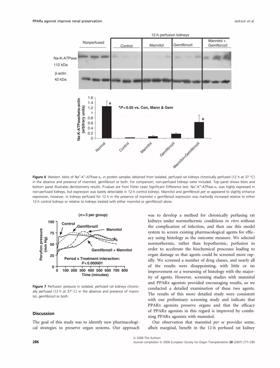
sion of this protein. As shown in Fig. 6, Na+-K+-AT-
Pase-a1 was strongly expressed in normal (freshly
isolated) kidneys. However, Na+-K+-ATPase-a1 expres-
sion was nearly undetectable in kidneys perfused with L-
15 alone and in mannitol-treated kidneys, but was
slightly higher in gemfibrozil-treated kidneys. Import-
antly, Na+-K+-ATPase-a1 was statistically, significantly
greater in gemfibrozil + mannitol-treated kidneys com-
pared with kidneys treated with L-15 alone, mannitol
alone or gemfibrozil alone. Although gemfibrozil +
mannitol markedly increased Na+-K+-ATPase-a1 expres-
sion, the combination treatment did not normalize the
expression of Na+-K+-ATPase-a1.
In our model system, we used a renal perfusion flow
rate that was low but within the normal limits for an
adult rat. This lower flow rate allowed the addition of
mannitol to the L-15 without unacceptable back-pressure
behind the 0.7 lm inline filters. Because of the low flow
rate and low viscosity of L-15 relative to blood, the initial
perfusion pressure in L-15 alone kidneys was approxi-
mately 50 mmHg, increased rapidly and within 30 min to
approximately 65 mmHg, and thereafter changed little
(Fig. 7). In kidneys receiving mannitol, either alone or
with gemfibrozil, initial renal perfusion pressure was
lower (approximately 25 mmHg). The rapid increase in
perfusion pressure observed in L-15 alone kidneys was
delayed by gemfibrozil, more so by mannitol and even
more by the combination of gemfibrozil with mannitol
(Fig. 7).
Figure 8 illustrates the effects of mannitol, gemfibrozil
and gemfibrozil + mannitol on initial urine flow rate,
terminal urine flow rate and total urine volume. Both
gemfibrozil and mannitol increased total urine volume;
however, the diuretic effects of mannitol were greater
than those observed with gemfibrozil, and the diuretic
actions of gemfibrozil + mannitol were less than those
observed for mannitol per se. Thus there was a statisti-
cally significant interaction between gemfibrozil and
mannitol on total urine volume. Similar trends were
observed with respect to the initial and terminal urine
flow rates; however the interaction did not achieve sta-
tistical significance with the terminal urine flow rates
and none of the treatments significantly altered terminal
flow rates.
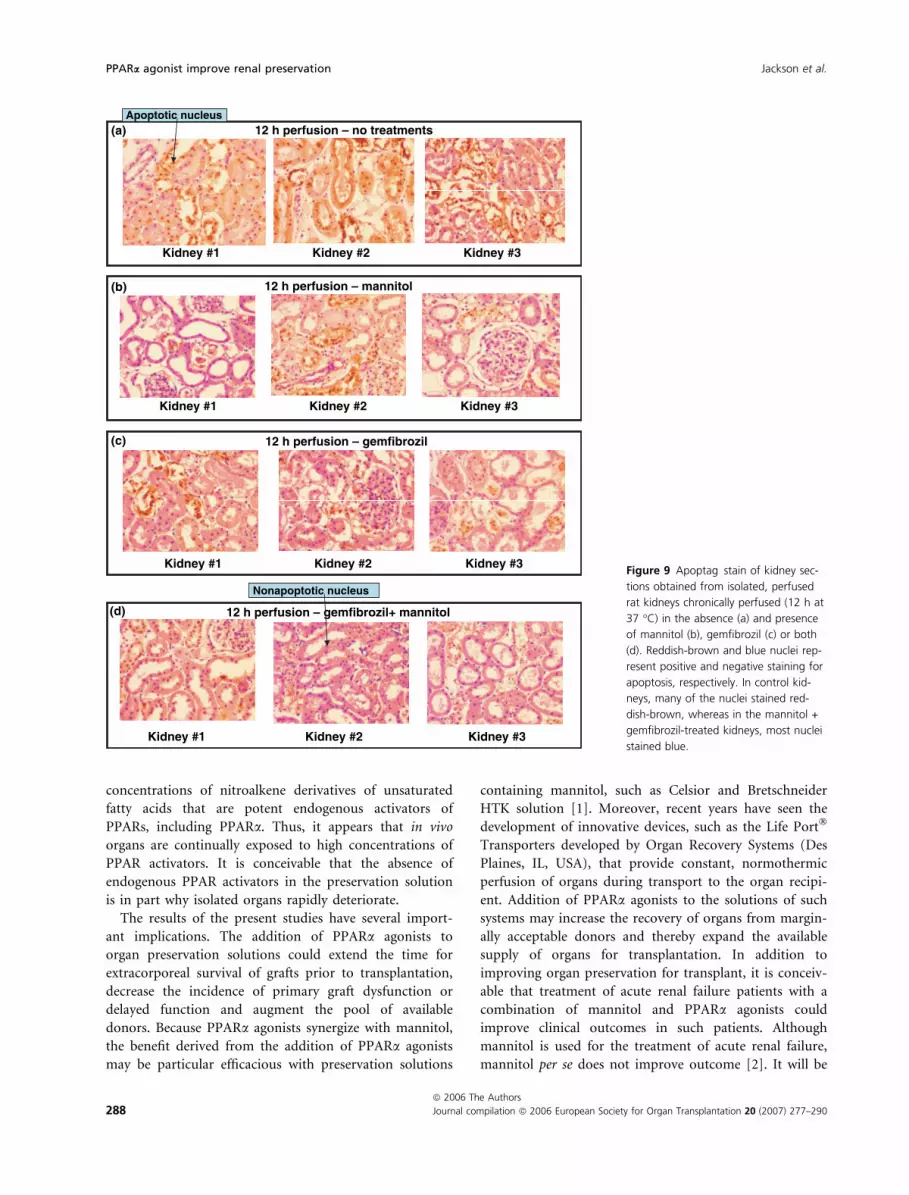
As shown in Fig. 9, staining for apoptosis with Apoptag
revealed diffuse areas of apoptosis in L-15 alone kidneys
(Group A). Mannitol (Group B) and gemfibrozil (Group
C) reduced the degree of apoptosis, and the combination
(Group D) appeared to be even more effective in this
regard.
An additional three kidneys were perfused with the
potent PPARa agonist WY-14643 and compared with the
L-15 alone group (Fig. 10). At the light microscopic level,
histological preservation was improved by WY-14643
compared with L-15 alone. These results extended the
findings with gemfibrozil to a structurally different
PPARa agonist, thus supporting the conclusion that the
preserving effects of gemfibrozil are shared by the phar-
macological class of PPARa agonists.
Figure 3 Histology scores in kidney sections obtained from isolated,
perfused rat kidneys chronically perfused (12 h at 37 �C) in the
absence and presence of mannitol, gemfibrozil or both. Scoring was
calibrated with 0 being complete destruction of glomerular and tubu-
lar architecture and 10 being a histological pattern similar to kidneys
flushed only for 30 min. Arrows connect statistically different groups
(P < 0.05, Fishers’ Least Significant Difference test).
Jackson et al. PPARa agonist improve renal preservation
ª 2006 The Authors
Journal compilation ª 2006 European Society for Organ Transplantation 20 (2007) 277–290 283

Control(a) (b)
(c) (d)
Mannitol
Gemfibrozil
2 µm 2 µm
Gemfibrozil + Mannitol
2 µm 2 µm
Figure 4 Electron micrographs of glomeruli obtained from isolated, perfused rat kidneys chronically perfused (12 h at 37 �C) in the absence (a)
and presence of mannitol (b), gemfibrozil (c) or both (d). Note the nearly complete destruction of glomerular architecture in the untreated glom-
erulus (a) and the near normal glomerular structure in the mannitol + gemfibrozil-treated glomerulus (d) (open Bowman’s space with parietal and
visceral epithelial cells intact, open capillary loops with intact endothelial cells and well-preserved foot processes). Ultrastructure in the mannitol-
treated (b) and gemfibrozil-treated (c) glomeruli was improved, but not as much as the combination treatment.
PPARa agonist improve renal preservation Jackson et al.
ª 2006 The Authors
284 Journal compilation ª 2006 European Society for Organ Transplantation 20 (2007) 277–290

Control(a) (b)
(c) (d)
Mannitol
Gemfibrozil
2 µm 2 µm
Gemfibrozil + Mannitol
2 µm 2 µm
Figure 5 Electron micrographs of proximal tubular epithelial cells obtained from isolated, perfused rat kidneys chronically perfused (12 h at
37 �C) in the absence (a) and presence of mannitol (b), gemfibrozil (c) or both (d). Note the nearly complete destruction of tubular epithelial ultra-
structure in the untreated (a), mannitol-treated (b) and gemfibrozil-treated (c) tubular epithelial cells, and the near normal epithelial ultrastructure
in the mannitol + gemfibrozil-treated epithelial cells (d) (open lumen and intact microvilli (brush border), tight junctions, mitochondria, nuclei, vac-
uoles and basement membrane).
Jackson et al. PPARa agonist improve renal preservation
ª 2006 The Authors
Journal compilation ª 2006 European Society for Organ Transplantation 20 (2007) 277–290 285
Discussion
The goal of this study was to identify new pharmacologi-
cal strategies to preserve organ systems. Our approach
was to develop a method for chronically perfusing rat
kidneys under normothermic conditions in vitro without
the complication of infection, and then use this model
system to screen existing pharmacological agents for effic-
acy using histology as the outcome measure. We selected
normothermic, rather than hypothermic, perfusion in
order to accelerate the biochemical processes leading to
organ damage so that agents could be screened more rap-
idly. We screened a number of drug classes, and nearly all
of the results were disappointing, with little or no
improvement or a worsening of histology with the major-
ity of agents. However, screening studies with mannitol
and PPARa agonists provided encouraging results, so we
conducted a detailed examination of these two agents.
The results of this more detailed study were consistent
with our preliminary screening study and indicate that
PPARa agonists preserve organs and that the efficacy
of PPARa agonists in this regard is improved by combi-
ning PPARa agonists with mannitol.
Our observation that mannitol per se provides some,
albeit marginal, benefit in the 12-h perfused rat kidney
Figure 6 Western blots of Na+-K+-ATPase-a1 in protein samples obtained from isolated, perfused rat kidneys chronically perfused (12 h at 37 �C)
in the absence and presence of mannitol, gemfibrozil or both. For comparison, non-perfused kidneys were included. Top panel shows blots and
bottom panel illustrates densitometry results. P-values are from Fisher Least Significant Difference text. Na+-K+-ATPase-a1 was highly expressed in
non-perfused kidneys, but expression was barely detectable in 12-h control kidneys. Mannitol and gemfibrozil per se appeared to slightly enhance
expression; however, in kidneys perfused for 12 h in the presence of mannitol + gemfibrozil expression was markedly increased relative to either
12-h control kidneys or relative to kidneys treated with either mannitol or gemfibrozil alone.
Figure 7 Perfusion pressure in isolated, perfused rat kidneys chronic-
ally perfused (12 h at 37� C) in the absence and presence of manni-
tol, gemfibrozil or both.
PPARa agonist improve renal preservation Jackson et al.
ª 2006 The Authors
286 Journal compilation ª 2006 European Society for Organ Transplantation 20 (2007) 277–290

model is not novel, and this finding is entirely consistent
with an extensive literature regarding mannitol as a cell-
impermeable solute in organ preservation [3–9]. For
example, administration of mannitol to human donors
prior to interruption of flow for harvesting the organ
reduces postoperative acute tubular necrosis [5]. Simi-
larly, flushing and storing rat kidneys in a mannitol-con-
taining solution increases functional outcomes in
transplantation studies [7], and perfusing human donor
kidneys with a mannitol solution decreases the incidence
of acute renal failure [8]. Flushing with a mannitol solu-
tion improves histology in rat kidneys stored under cold
ischemia [9], and cold storage in a mannitol-containing
solution improves cortical hemoglobin oxygenation in a
rabbit renal transplant model [10]. Indeed, a flushing-
storage solution containing mannitol as the cell-imperme-
able solute is patented [11], and mannitol-containing
organ preservation solutions are widely employed [1].
Although our findings with mannitol are not novel, the
consistency between the present findings and previous
reports of efficacy with mannitol (and cell-impermeable
solutes in general) supports the predictability of our
model system and the validity of extrapolating findings in
our model system to organ preservation in other model
systems and humans.
The present study strongly suggests that PPARa agon-
ists provide organ preservation and that there is a striking
synergy between PPARa agonists and mannitol in this
regard. To our knowledge, both these observations are
novel findings. An exhaustive Medline search and patent
database search did not uncover any prior publications
regarding the organ preserving efficacy of PPARa agon-
ists, either alone or in combination with mannitol. There-
fore, the present findings represent a potentially
important advance in the development of solutions for
organ preservation.
When given chronically to patients with dyslipidemias,
PPARa agonists, such as gemfibrozil, lower plasma tri-
glycerides and increase high-density lipoprotein choles-
terol levels. Consequently, PPARa agonists are widely
prescribed for post-transplant hyperlipidemia and appear
entirely safe in such a setting [15–18]. In fact, therapy
with PPARa agonists may provide renoprotection from
the damaging effects of chronic dyslipidemia [19]. There-
fore, it is highly likely that the addition of gemfiborzil or
other PPARa agonists to organ preservation solutions
would carry little, if any, risk of adverse effects to the
transplanted organ or its recipient.
Peroxisome proliferator-activated receptor-alpha is a
ligand-activated transcriptional factor that is a member of
the nuclear receptor family [20]. PPARa modulates the
expression of multiple genes that participate in fatty acid
metabolism including fatty acid transport protein, acyl-
coA synthetase, fatty acid binding protein, medium chain
acyl-coenzyme A dehydrogenase, acyl-coA oxidase, cyto-
chrome P450 fatty acid x-hydroxylase and carnitine
palmitoyl transferase I [20]. It is conceivable that the
organ-preserving effects of PPARa agonists are related to
enhancing the utilization of fatty acids as an alternative
energy source in organs with a reduced energy supply.
Further studies are required to address this issue. Also,
PPARa agonists increase the expression of nephrin by
glomerular podocytes [21]. Importantly, very recent stud-
ies by Baker et al. [22] have identified in blood high
Figure 8 Urine flow rates at the beginning and end of perfusion and
over the entire 12-h period in isolated, perfused rat kidneys chronic-
ally perfused (12 h at 37 �C) in the absence and presence of manni-
tol, gemfibrozil or both. Arrows connect statistically different groups
(P < 0.05, Fishers’ Least Significant Difference test).
Jackson et al. PPARa agonist improve renal preservation
ª 2006 The Authors
Journal compilation ª 2006 European Society for Organ Transplantation 20 (2007) 277–290 287
concentrations of nitroalkene derivatives of unsaturated
fatty acids that are potent endogenous activators of
PPARs, including PPARa. Thus, it appears that in vivo
organs are continually exposed to high concentrations of
PPAR activators. It is conceivable that the absence of
endogenous PPAR activators in the preservation solution
is in part why isolated organs rapidly deteriorate.
The results of the present studies have several import-
ant implications. The addition of PPARa agonists to
organ preservation solutions could extend the time for
extracorporeal survival of grafts prior to transplantation,
decrease the incidence of primary graft dysfunction or
delayed function and augment the pool of available
donors. Because PPARa agonists synergize with mannitol,
the benefit derived from the addition of PPARa agonists
may be particular efficacious with preservation solutions
containing mannitol, such as Celsior and Bretschneider
HTK solution [1]. Moreover, recent years have seen the
development of innovative devices, such as the Life Port�
Transporters developed by Organ Recovery Systems (Des
Plaines, IL, USA), that provide constant, normothermic
perfusion of organs during transport to the organ recipi-
ent. Addition of PPARa agonists to the solutions of such
systems may increase the recovery of organs from margin-
ally acceptable donors and thereby expand the available
supply of organs for transplantation. In addition to
improving organ preservation for transplant, it is conceiv-
able that treatment of acute renal failure patients with a
combination of mannitol and PPARa agonists could
improve clinical outcomes in such patients. Although
mannitol is used for the treatment of acute renal failure,
mannitol per se does not improve outcome [2]. It will be
12 h perfusion – no treatments
12 h perfusion – mannitol
12 h perfusion – gemfibrozil
12 h perfusion – gemfibrozil+ mannitol
(a)
(b)
(c)
(d)
Apoptotic nucleus
Nonapoptotic nucleus
Kidney #1 Kidney #2 Kidney #3
Kidney #1 Kidney #2 Kidney #3
Kidney #1 Kidney #2 Kidney #3
Kidney #1 Kidney #2 Kidney #3
Figure 9 Apoptag stain of kidney sec-
tions obtained from isolated, perfused
rat kidneys chronically perfused (12 h at
37 �C) in the absence (a) and presence
of mannitol (b), gemfibrozil (c) or both
(d). Reddish-brown and blue nuclei rep-
resent positive and negative staining for
apoptosis, respectively. In control kid-
neys, many of the nuclei stained red-
dish-brown, whereas in the mannitol +
gemfibrozil-treated kidneys, most nuclei
stained blue.
PPARa agonist improve renal preservation Jackson et al.
ª 2006 The Authors
288 Journal compilation ª 2006 European Society for Organ Transplantation 20 (2007) 277–290

important to test this concept in vivo. Finally, protecting
drugs, if sufficiently efficacious, could allow for the pro-
longed in situ perfusion of organ systems with solutions
to restore organ function or to deliver selectively other
drugs or molecular biological constructs (plasmids, viral
vectors, siRNAs, anti-sense oligonucleotides) directly to
the target organ.
As noted above, we used normothermic perfusion to
hasten organ injury and facilitate more rapid screening of
potentially protective compounds. Nonetheless, it is poss-
ible that the results are applicable to hypothermic organ
preservation as well because the biochemical mechanisms
of injury in normothermic and hypothermic conditions
likely overlap. Also, pretreatment of hypothermically
maintained organs with PPARa agonists would provide
high levels of PPARa activation during the critical re-
warming period following transplantation. However,
additional experiments are required to clarify whether
PPARa agonists would protect organs preserved by cold
storage and subsequently subjected to ischemic/reper-
fusion injury.
References
1. Nydegger UE, Carrel T, Maumonier T, Mohacsi LP. New
concepts in organ preservation. Transpl Immunol 2002; 9:
215.
2. Dishart MK, Kellum JA. An evaluation of pharmacological
strategies for the prevention and treatment of acute renal
failure. Drugs 2000; 59: 79.
3. Cerilli J, Lauridsen P, Saurbrey J, Husfeldt E, Wandall
HH. Effect of mannitol, saline, and urea on the function
of a recently autotransplanted kidney. Arch Surg 1966; 92:
178.
4. Weimar W, Geerlings W, Bijnen AB, et al. A controlled
study on the effect of mannitol on immediate renal func-
tion after cadaver donor kidney transplantation. Trans-
plantation 1983; 35: 99.
5. Tiggeler RG, Berden JH, Hoitsma AJ, Koene RA. Preven-
tion of acute tubular necrosis in cadaveric kidney trans-
plantation by the combined use of mannitol and moderate
hydration. Ann Surg 1985; 201: 246.
6. Grino JM, Miravitlles R, Castelao AM, et al. Flush solution
with mannitol in the prevention of post-transplant renal
failure. Transplant Proc 1987; 19: 4140.
7. Kline R, Churchill M, Churchill P, Bidani A, Schwartz M.
High osmolality-low pH flush solutions improve renal
transplant function in rats. Urol Res 1991; 19: 81.
8. Lopez-Costea MA, Grino JM, Seron D, et al. Mannitol
solution (M-400) versus mannitol solution combined with
allopurinol in the prevention of post-transfusion acute
renal failure. Actas Urol Esp 1992; 16: 446.
9. Herrero I, Torras J, Carrera M, et al. Evaluation of a pre-
servation solution containing fructose-1,6-diphosphate and
mannitol using the isolated perfused rat kidney. Compar-
ison with Euro-Collins and University of Wisconsin solu-
tions. Nephrol Dial Transplant 1995; 10: 519.
10. Lane NJ, Thorniley MS, Manek S, Fuller BJ, Green CJ.
Effect of mannitol and polyethylene glycol on the action of
frusemide during renal storage and transplantation. Trans-
plantation 1996; 62: 575.
11. Churchill PC, Churchill MC. Flush-storage solution for
donor organs. United States Patent 5,834,178, 1998.
12. Bolz SS, Pieperhoff S, De Wit C, Pohl U. Intact endothelial
and smooth muscle function in small resistance arteries
after 48 h in vessel culture. Am J Physiol 2000; 279:
H1434.
13. Jackson TC, Mi Z, Jackson EK. Modulation of cyclic AMP
production by signal transduction pathways in preglomer-
12 h perfusion – no treatments
12 h perfusion – WY-14643
Kidney #1 Kidney #2 Kidney #3
Kidney #1 Kidney #2 Kidney #3
Figure 10 Hematoxylin and eosin stain
of kidney sections obtained from isola-
ted, perfused rat kidneys chronically per-
fused (12 h at 37 �C) in the absence
and presence of WY- 14643 showing
protective effects of WY-14643 (glomer-
ular and tubular architectures were pre-
served).
Jackson et al. PPARa agonist improve renal preservation
ª 2006 The Authors
Journal compilation ª 2006 European Society for Organ Transplantation 20 (2007) 277–290 289

ular microvessels and microvascular smooth muscle cells.
J Pharmacol Exp Ther 2004; 310: 349.
14. Briere N. Human foetal kidney explants in serum-free
organ culture. Anat Embryol 1987; 176: 105.
15. Bastani B, Robinson S, Heisler T, et al. Post-transplant
hyperlipidemia: risk factors and response to dietary modi-
fication and gemfibrozil therapy. Clin Transplant 1995; 9:
340.
16. Vergoulas G, Miserlis G, Solonaki F, et al. Combined treat-
ment of hypercholesterolemia of renal transplant allograft
recipients with fluvastatin and gemfibrozil. Transpl Int
2000; 13(Suppl 1): S64.
17. Kasiske BL, Heim-Duthoy KL, Singer GG, Watschinger B,
Germain MJ, Bastani B. The effects of lipid-lowering
agents on acute renal allograft rejection. Transplantation
2001; 72: 223.
18. Chan TM, Cheng IK, Tam SC. Hyperlipidemia after renal
transplantation: treatment with gemfibrozil. Nephron 1994;
67: 317.
19. Izzedine H, Launay-Vacher V, Baumelou A, Deray G.
Renal effects of PPARa-agonists. Minerva Urol Nefrol 2004;
56: 339.
20. van Raalte DH, Li M, Pritchard PH, Wasan KM. Peroxisome
proliferator-activated receptor (PPAR)-a: a pharmacological
target with a promising future. Pharm Res 2004; 21: 1531.
21. Ren S, Xin C, Beck KF, et al. PPARa activation upregulates
nephrin expression in human embryonic kidney epithelial
cells and podocytes by a dual mechanism. Biochem Biophys
Res Commun 2005; 338: 1818.
22. Baker PRS, Lin YM, Schopfer FJ, et al. Fatty acid transduc-
tion of nitric oxide signaling: Multiple nitrated unsaturated
fatty acid derivatives exist in human blood and urine and
serve as endogenous peroxisome proliferator-activated
receptor ligands. J Biol Chem 2005; 280: 42464.
PPARa agonist improve renal preservation Jackson et al.
ª 2006 The Authors
290 Journal compilation ª 2006 European Society for Organ Transplantation 20 (2007) 277–290