rosiglitazone and 15-deoxy-Δ 12,14 -prostaglandin j 2 , ppar γ agonists, differentially regulate...
TRANSCRIPT

Rosiglitazone and 15-deoxy-D12,14-prostaglandin J2
cause potent neuroprotection after experimentalstroke through noncompletely overlappingmechanisms
Marta P Pereira1, Olivia Hurtado1, Antonio Cardenas2, Lisardo Bosca3, Jose Castillo4,Antoni Davalos5, Jose Vivancos6, Joaquın Serena7, Pedro Lorenzo1, Ignacio Lizasoain1,8
and Marıa A Moro1,8
1Departamento de Farmacologıa, Facultad de Medicina, Universidad Complutense de Madrid (UCM),Madrid, Spain; 2Instituto de Farmacologıa y Toxicologıa, CSIC, Madrid, Spain; 3Instituto de Bioquımica yBiologıa Molecular, CSIC, Madrid, Spain; 4Hospital Clınico Universitario, Santiago de Compostela, Spain;5Hospital Germans Trias i Pujol, Badalona, Spain; 6Hospital Universitario La Princesa, Madrid, Spain;7Hospital Josep Trueta, Girona, Spain
Stroke triggers an inflammatory cascade which contributes to a delayed cerebral damage, thusimplying that antiinflammmatory strategies might be useful in the treatment of acute ischaemicstroke. Since two unrelated peroxisome proliferator-activated receptor-c (PPARc) agonists, thethiazolidinedione rosiglitazone (RSG) and the cyclopentenone prostaglandin 15-deoxy-D12,14-prostaglandin J2 (15d-PGJ2), have been shown to possess antiinflammatory properties, we havetested their neuroprotective effects in experimental stroke. Rosiglitazone or 15d-PGJ2 wereadministered to rats 10 mins or 2 h after permanent middle cerebral artery occlusion (MCAO).Stroke outcome was evaluated by determination of infarct volume and assesment of neurologicalscores. Brains were collected for protein expression, gene array analyses and gene shift assays.Our results show that both compounds decrease MCAO-induced infarct size and improveneurological scores. At late times, the two compounds converge in the inhibition of MCAO-inducedbrain expression of inducible NO synthase and the matrix metalloproteinase 9. Interestingly, at earlytimes, complementary DNA microarrays and gene shift assays show that different mechanisms arerecruited. Analysis of early nuclear p65 and late cytosolic IjBa protein levels shows that bothcompounds inhibit nuclear factor-jB signalling, although at different levels. All these resultssuggest both PPARc-dependent and independent pathways, and might be useful to design boththerapeutic strategies and prognostic markers for stroke.Journal of Cerebral Blood Flow & Metabolism (2006) 26, 218–229. doi:10.1038/sj.jcbfm.9600182; published online20 July 2005
Keywords: inflammation; middle cerebral artery occlusion; MMP-9; NF-kB; nitric oxide synthase
Introduction
Stroke is one of the leading causes of death anddisability worldwide. In spite of this, its acute thera-
peutic management is almost limited to thrombo-lysis. As the inflammatory cascade triggered by theischaemic injury in both occluded blood vessels andbrain parenchyma is an important feature of thepathophysiological response to the ischaemic injury,antiinflammatory strategies might be a usefultherapy for acute stroke treatment.
The compounds rosiglitazone (RSG) and 15-deoxy-D12,14-prostaglandin J2 (15d-PGJ2) are knownto exert antiinflammatory actions (rev. in Straus andGlass, 2001; Martens et al, 2002; Stumvoll, 2003).They are agonists of the nuclear receptor peroxisomeproliferator-activated receptor-g (PPARg; Willson etal, 2001; Berger and Moller, 2002), a ligand-dependent nuclear transcription factor belonging
Received 18 February 2005; revised 17 May 2005; accepted 13June 2005; published online 20 July 2005
Correspondence: Dr MA Moro, Departamento de Farmacologıa,Facultad de Medicina, UCM, 28040 Madrid, Spain.E-mail: [email protected].
This work was supported by grants from the Spanish Ministries of
Health FIS-PI030304 (MAM) and Science and Technology
SAF2002-04487-C02-01 (IL), BIO2000-0405-P4-05 (PL), and Local
Madrid Government GR/SAL/0056/2004 (PL). MPP and OH are
recipients of fellowships funded by FIS and CAM, respectively.
8These senior authors contributed equally to this work.
Journal of Cerebral Blood Flow & Metabolism (2006) 26, 218–229& 2006 ISCBFM All rights reserved 0271-678X/06 $30.00
www.jcbfm.com

to the nuclear hormone receptor superfamilyof nuclear receptors, that has been proposedto participate in a broad range of cellularfunctions that include adipocyte differentiation,glucose homeostasis, apoptosis and antiinflam-matory actions (rev. in Willson et al, 2001;Berger and Moller, 2002). Rosiglitazone is amember of the thiazolidinedione (TZD) class ofcompounds used for its antidiabetic properties,which has shown a high selectivity for PPARgwith a Kd of 40 nmol/L, whereas 15d-PGJ2, anaturally occurring derivative of prostaglandin D2,is a high-affinity ligand for PPARg and has beenproposed as one of the natural agonists of thisreceptor.
Peroxisome proliferator-activated receptor-gagonists have been shown to inhibit proinflam-matory and inflammatory mediators (Jiang et al,1998; Marx et al, 1998; Ricote et al, 1998) andto exert antiinflammatory effects in several settings,including the central nervous system (CNS) (rev.in Landreth and Heneka, 2001; Feinstein, 2003;Kielian and Drew, 2003). Indeed, both RSG and15d-PGJ2 antagonise the expression of inflammatorymediators; however, these effects seem to be accom-plished through both PPARg receptor-dependentand -independent mechanisms (Castrillo et al,2000; Li et al, 2000; Rossi et al, 2000; Straus et al,2000; Chawla et al, 2001; Moore et al, 2001; Takataet al, 2002; Park et al, 2003; Perez-Sala et al, 2003;Welch et al, 2003).
Regardless of the mechanism involved, the anti-inflammatory actions of these compounds could bebeneficial in stroke. We have therefore investigatedthe effects of RSG and 15d-PGJ2 on stroke outcomein a rodent model of cerebral ischaemia by perma-nent occlusion of the middle cerebral artery(MCAO).
Materials and methods
Materials
Rosiglitazone was from Calbiochem (San Diego, CA, USA)and 15d-PGJ2 was from Cayman Chemical (Ann Arbor, MI,USA). The rest of the reagents were from Sigma (Madrid,Spain), or as indicated in the text.
Animals
Adult male Fischer rats weighing 250 to 275 g were used.All experimental protocols adhered to the guidelines ofthe Animal Welfare Committee of the UniversidadComplutense (following EU directives 86/609/CEE and2003/65/CE). Rats were housed individually under stan-dard conditions of temperature and humidity, and a 12 hlight/dark cycle (lights on at 08:00), with free access tofood and water.
Middle Cerebral Artery Occlusion (MCAO)
Animals were anaesthetised with 1% to 1.5% halothane ina mixture of 70% nitrogen/30% oxygen, and bodytemperature was maintained at 371C using a heating padthroughout the surgery procedure and during postsurgeryrecovery. Permanent focal cerebral ischaemia was inducedby occlusion of the ipsilateral middle cerebral artery(MCA) as described (De Cristobal et al, 2001; Hurtado etal, 2003). Briefly, for the CCA ligature, a midline ventralcervical incision was made, and the CCA was isolated andpermanently occluded with a silk ligature. For the MCAocclusion, a 1 cm incision perpendicular to the lineconnecting the lateral canthus of the left eye and theexternal auditory canal was made to expose and retract thetemporalis muscle. A 2-mm burr hole was drilled and theMCA was exposed by cutting and retracting the dura. TheMCA was elevated and cauterised. Rats in which the MCAwas exposed but not occluded served as sham-operatedcontrols (SHAM). After surgery, subjects were returned totheir cages and allowed free access to water and food.
Experimental Groups
Several groups were used for determinations of infarctsize, neurological assessment, and determination of bio-chemical and molecular parameters: MCAO 10 mins beforean intraperitoneal injection of saline (MCAO; n¼ 23) ordimethyl sulfoxide (DMSO) (vehicle; 10% in saline;n¼ 21), and MCAO 10 mins before an intraperitonealinjection of either RSG 1 to 3 mg/kg (MCAOþRSG1/3;n¼ 21) or 15d-PGJ2 1 mg/kg (MCAOþ 15d-PGJ2; n¼ 21).For infarct size and neurological assesment, additionalgroups of animals were administered either 3 mg/kg RSGor 1 mg/kg 15d-PGJ2 2 h after the occlusion, or a combina-tion of both 1 mg/kg RSG and 1 mg/kg 15d-PGJ2 10 minsafter the occlusion (n¼ 6). An additional control groupconsisted of SHAM-operated animals 10 mins before anintraperitoneal injection of saline (SHAM; n¼ 18). Injec-tion volume was r0.5 mL/250 g body weight.
Infarct Size
Infarct outcome was assessed at 48 h, a time at which wehave observed that infarct lesions have reached theirmaximal degree (unpublished results). Animals werekilled, brains were removed, and a series of 2 mm ofcoronal slices were obtained and stained in 1% 2,3,5-triphenyl-tetrazolium chloride (TTC) in 0.1 mol/L phos-phate buffer, and infarct size was determined as described(Mackensen et al, 2001).
Neurological Characterisation After Middle CerebralArtery Occlusion
Before the killing, a neurological test was made by twounaware independent observers, as described previously(Hunter et al, 2000). According to the test, animals werescored as: 0 points – no deficit; 1 point – failure to extendright forepaw fully; 2 points – decreased grip of right
Neuroprotection by rosiglitazone and 15d-PGJ2
MP Pereira et al
219
Journal of Cerebral Blood Flow & Metabolism (2006) 26, 218–229

forelimb while tail pulled; 3 points – spontaneous circlingor walking to the contralateral side; 4 points – walks onlywhen stimulated with depressed level of consciousness; 5points – unresponsive to stimulation.
Protein Expression in Brain Homogenates
Brain cortical and striatal tissue was collected from theinfarcted and surrounding areas. For determination ofinducible NO synthase (iNOS), cyclooxygenase (COX)-2and matrix metalloproteinase 9 (MMP-9) protein expres-sion levels, rats were killed 18 h after MCAO. Brain areascorresponding to the infarct and surrounding areas werecollected. Brain samples were homogenised by sonicationfor 10 secs at 41C in 4 volumes of homogenisation buffercontaining 320 mmol/L sucrose, 1 mmol/L DL-dithiothrei-tol, 10 mg/mL leupeptin, 10 mg/mL soybean trypsin inhi-bitor, 2mg/mL aprotinin and 50 mmol/L Tris, brought to pH7.0 at 201C with HCl. The homogenate was centrifuged at41C at 12,000 g for 20 mins and the pellet was discarded.
Protein Expression in Cytosolic and Nuclear Extracts
Cytosolic and nuclear extracts were prepared as described(Cardenas et al, 2000). Determination of p65 wasperformed in nuclei obtained from brains of rats killed1 h after MCAO, whereas, for PPARg, rats were killed 2 and18 h after MCAO. For IkBa, cytosolic extracts wereobtained from brains of rats killed 5 h after MCAO.
Gene Microarray Analysis
Rats were decapitated 5 h after MCAO and ipsilateralcortices were used immediately for total RNA extractionwith the TRIzols Reagent (Invitrogen, Carlsbad, CA, USA).The RNA was then cleaned (Rneasy Mini Kit, Qiagen,Valencia, CA, USA) and analysed in a 2100 Bioanalyzerfollowing the instructions of the manufacturer of the array(Affymetrix). Fragmentation of biotin-labelled comple-mentary RNA (cRNA) was performed at 941C using40 mmol/L Tris-acetate, pH 8.1, 100 mmol/L KOAc and30 mmol/L MgOAc. Samples were hybridised to Affyme-trix Genechip Test3 Array and then to AffymetrixGeneChip Rat expression probe array 230A. The datawere analysed with the GeneChip Analysis Suite software.
Quantitative Real-Time Reverse Transcriptase-Polymerase Chain Reaction (QRT-PCR)
The mRNA levels of selected genes that were found to bemodified in the array (see Supplemental material) werequantified by real-time QRT-PCR amplification. Total RNAwas extracted from isolated brain slices of four animals,using Trizol solution (Gibco BRL, NY, USA). RNA quantitywas determined spectrophotometrically and the puritywas confirmed by the relative absorbance at 260 nm versus280 nm. RNA samples were stored in diethyl pyrocarbo-nate-treated water at �801C. Complementary DNA (cDNA)synthesis was performed with 4mg of total RNA and
M-MLU reverse transcriptase (Invitrogen) in accordancewith the manufacturer’s instruction, using oligo-dT asprimer. Complementary DNA was stored at �201C.
Specific primer sequences can be found in Supplemen-tal Table 1. Primers and probes against the selected genesof interest were designed from rat sequences available inGenBank using Primer Express Software (PE AppliedBiosystems). Polymerase chain reaction amplification andquantification were performed using SyBr Green PCR kit(PE Applied Biosystem) in a model 7700 Sequencedetector (PE Applied Biosystems), and the results wereanalysed using sodium dodecyl sulphate (SDS) 1.9 Soft-ware (PE Applied Biosystems). The absence of genomiccontamination in the RNA samples and of DNA contam-ination in RNA mimics was confirmed with reversetranscriptase-negative controls for each experiment.
Western Blot Analysis
Samples containing 20 mg of protein were loaded and theproteins size-separated in 7% to 10% SDS-polyacrylamidegel electrophoresis (110 mA). Proteins were blotted onto apolyvinylidene fluoride (PVDF) membrane (Hybondt-P,Amersham Biosciences Europe GmbH, Friburg, Germany)and incubated with specific primary antibodies againstPPARg (Santa Cruz Biotechnology, Santa Cruz, CA, USA;1:1000), iNOS (Santa Cruz; 1:500), COX-2 (Santa Cruz,1:1000), MMP-9 (Chemicon, Temecula, CA, USA; 1:2000),p65 (Santa Cruz; 1:1000) and IkBa (Santa Cruz; 1:1000).Proteins recognised by the antibody were revealed byECLt-kit following the manufacturers’ instructions(Amersham Biosciences Europe GmbH, Friburg, Ger-many). b-Actin and Sp1 levels were used as loadingcontrols for total-cytosolic and nuclear protein expression,respectively.
Gel Mobility Shift Assays
A double-stranded oligonucleotide corresponding to PPARresponse element (PPRE), composed of GGGGACCAGGA-CAAAGGTCA, which is located in the 50 upstream of theacyl-CoA oxidase gene (Tugwood et al, 1992), was used asprobe. Labelled probe (5� 105 cpm) was incubated withnuclear extracts (5mg) in a 20-mL reaction consisting of 2 mgpoly dI:dC, 10 mmol/L Hepes, pH 7.9, 50 mmol/L KCl, 5%glycerol, 2% Ficoll, 0.05% NP-40, and 1 mL rabbit serum.Reactions were incubated for 10 mins on ice, radiolabelledprobe was added, and reaction was continued for another10 mins on ice. Reactions were run on a 5% nondenatur-ing polyacrylamide gel in 0.5� TBE buffer.
NOx� (NO2
� and NO3�) Assay
NO release was estimated from the amounts of nitrite(NO2
�) and nitrate (NO3�) in brain homogenates. NO3
� wascalculated by first reducing NO3
� into NO2� in the presence
of Cd, and NO2� was determined by a colorimetric assay
based on the Griess reaction as described (Cortas andWakid, 1990).
Neuroprotection by rosiglitazone and 15d-PGJ2
MP Pereira et al
220
Journal of Cerebral Blood Flow & Metabolism (2006) 26, 218–229

Statistical Analysis
Results are expressed as mean7s.e.m. of the indicatednumber of experiments; statistical analysis involved one-way analysis of variance (ANOVA, or the Kruskal–Wallistest when the data were not normally distributed),followed by individual comparisons of means (Student–Newman–Keuls or Dunns method, when the data were notnormally distributed). Po0.05 was considered statisticallysignificant.
Results
Effect of Rosiglitazone and 15-Deoxy-D12,14-Prostaglandin J2 on Infarct Outcome AfterPermanent Middle Cerebral Artery Occlusion
The administration of RSG or 15d-PGJ2 10 mins afterthe occlusion caused a decrease in MCAO-inducedinfarct size determined 48 h after the ischaemicinjury (Figure 1). In addition, the animals treatedwith these compounds also showed better scores in aneurological assessment scale after MCAO (Table 1).When these drugs were administered 2 h after theocclusion, the decrease in infarct size persisted (94.970.8 or 101.872.9 mm3 after 3 mg/kg RSG or 1 mg/kg15d-PGJ2, respectively, n¼ 6). When RSG (1 mg/kg)and 15d-PGJ2 (1 mg/kg) were coadministered 10 minsafter the occlusion, infarct size was further decreased(77.474.8 mm3, n¼ 6, Po0.05 versus MCAOþRSG1or MCAOþ 15d-PGJ2).
Expression of Inducible NO Synthase andcyclooxygenase-2, and Nitrite/Nitrate (NOx
�) LevelsAfter Middle Cerebral Artery Occlusion. Effect ofRosiglitazone and 15-Deoxy-D12,14-Prostaglandin J2
Expression of the enzymes iNOS and COX-2 wasstudied, as both are described to mediate inflammatorydamage after stroke (del Zoppo et al, 2000). Occlusionof the MCA caused the expression of the inflammatoryenzymes iNOS and COX-2 in rat brain, as shown bythe levels of these proteins found 18 h after theischaemic insult (Figures 2A and 2B). The adminis-tration of either RSG or 15d-PGJ2 inhibited MCAO-induced expression of iNOS, but did not affect COX-2levels at the time examined (Figures 2A and 2B).
Levels of NOx� 18 h after MCAO were determined
as an indicator of NO synthesis. Permanent MCAOcaused an increase in brain NOx
�, which wasdecreased by the two compounds tested (Figure 2C).
Expression of Matrix Metalloproteinase 9 AfterMiddle Cerebral Artery Occlusion. Effect ofRosiglitazone and 15-Deoxy-D12,14-Prostaglandin J2
Occlusion of the MCA caused an increase in thelevels of MMP-9, a matrix metalloproteinase whichis induced and mediates damage caused by proin-flammatory stimuli, including cerebral ischaemia(Mun-Bryce and Rosenberg, 1998). Both RSG and15d-PGJ2 decreased the levels of this MMP afterexperimental stroke (Figure 2D).
Nuclear Levels of Nuclear Factor-jB (NF-jB) afterMiddle Cerebral Artery Occlusion. Effect ofRosiglitazone and 15-Deoxy-D12,14-Prostaglandin J2
Nuclear factor-kB is a transcription factor that playsa key role in the expression of a variety of genesinvolved in inflammatory responses (Gosh et al,1998). As a sign of its activation, the nuclear levelsof its subunit p65 were determined 1 h after MCAO.Experimental ischaemia caused activation of NF-kB,as revealed by the nuclear translocation of the NF-kB subunit p65. The cyclopentenone prostaglandin15d-PGJ2 decreased MCAO-induced p65 transloca-tion. Conversely, RSG did not modify p65 nuclearlevels after MCAO (Figure 3A).
Cytosolic Levels of IjBa after Middle Cerebral ArteryOcclusion. Effect of Rosiglitazone and 15-Deoxy-D12,14-Prostaglandin J2
As IkB itself is an NF-kB-dependent gene, late IkBlevels were determined as an indicator of NF-kBtranscriptional activity induced after its nucleartranslocation. Cytosolic IkBa levels increased 5 hafter the ischaemic insult when compared withcontrol brains (Figure 3B). Both RSG and thecyclopentenone prostaglandin 15d-PGJ2 abolishedthe increase in IkBa levels induced 5 h after MCAO.
200
180
160
140
120
100
80
Infa
rct
volu
me
(mm
3 )
MCAO
MCAO+vehicle
MCAO+RSG 1
MCAO+RSG 3
MCAO+15dPGJ2
*
*
*
Figure 1 Neuroprotective effect of RSG and 15d-PGJ2 afterexperimental stroke. The TZD RSG (1 to 3 mg/kg) and thecyclopentenone prostaglandin (15d-PGJ2, 1 mg/kg) reduceinfarct volume after permanent MCAO. Data are mean7s.e.m., n¼6 to 8, *Po0.05 versus MCAO (see Materialsand methods for details). Photographs of brain slices fromrepresentative experiments.
Neuroprotection by rosiglitazone and 15d-PGJ2
MP Pereira et al
221
Journal of Cerebral Blood Flow & Metabolism (2006) 26, 218–229
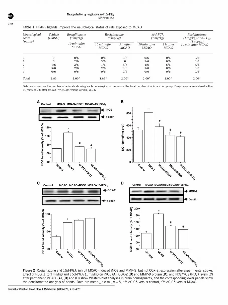
Table 1 PPARg ligands improve the neurological status of rats exposed to MCAO
Neurologicalscore(points)
Vehicle(DMSO)
Rosiglitazone(1 mg/kg)
Rosiglitazone(3 mg/kg)
15d-PGJ2
(1 mg/kg)Rosiglitazone
(1 mg/kg)+15d-PGJ2
(1 mg/kg)10 min after
MCAO10 min after
MCAO2 h afterMCAO
10 min afterMCAO
2 h afterMCAO
10 min after MCAO
0 0 0/6 0/6 0/6 0/6 0/6 0/61 0 2/6 3/6 0 1/6 0/6 0/62 1/6 2/6 1/6 6/6 4/6 6/6 6/63 5/6 2/6 2/6 0/6 1/6 0/6 0/64 0/6 0/6 0/6 0/6 0/6 0/6 0/6
Total 2.83 2.00* 1.83* 2.00* 2.00* 2.00* 2.00*
Data are shown as the number of animals showing each neurological score versus the total number of animals per group. Drugs were administered either10 mins or 2 h after MCAO. *Po0.05 versus vehicle, n¼6.
Control MCAO MCAO+RSG1 MCAO+15dPGJ2
Control MCAO MCAO+RSG3 MCAO+15dPGJ2Control MCAO MCAO+RSG3 MCAO+15dPGJ2
MCAO
MCAO+RSG1
MCAO+15dPGJ2
iNOS
β-actin
COX-2
β-actin
MMP-9
β-actin
∗
∗
##
#
#
∗
##
#
120
100
80
60
40
iNO
S b
and
Inte
nsi
ty (
% o
f M
CA
O)
Control
MCAO
MCAO+RSG3
MCAO+15dPGJ2
Control
MCAO
MCAO+RSG3
MCAO+15dPGJ2
Control
MCAO
MCAO+RSG1
MCAO+RSG3
MCAO+15dPGJ2
Control
800
600
400
200
0
NO
x-(
pm
ol/m
g p
rot)
180
120
60
CO
X-2
ban
d in
ten
sity
(%
of
MC
AO
)
MM
P-9
ban
d in
ten
sity
(%
of
MC
AO
)
∗
80
100
120
140
160
180
200
A B
C D
Figure 2 Rosiglitazone and 15d-PGJ2 inhibit MCAO-induced iNOS and MMP-9, but not COX-2, expression after experimental stroke.Effect of RSG (1 to 3 mg/kg) and 15d-PGJ2 (1 mg/kg) on iNOS (A), COX-2 (B) and MMP-9 protein (D), and NO2
�/NO3� (NOx
�) levels (C)after permanent MCAO. (A), (B) and (D) show Western blot analyses in brain homogenates, and the corresponding lower panels showthe densitometric analysis of bands. Data are mean7s.e.m., n¼5, *Po0.05 versus control, #Po0.05 versus MCAO.
Neuroprotection by rosiglitazone and 15d-PGJ2
MP Pereira et al
222
Journal of Cerebral Blood Flow & Metabolism (2006) 26, 218–229

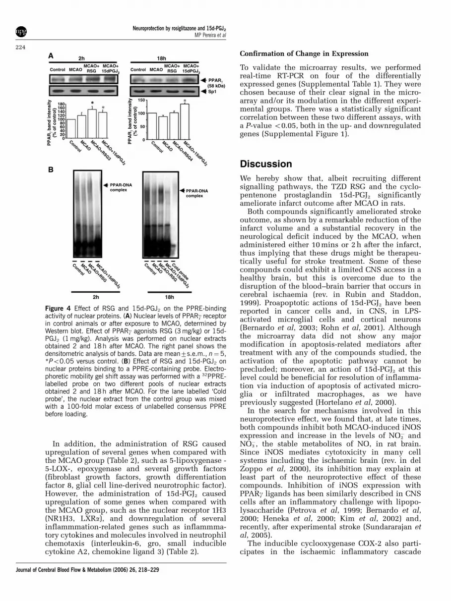
Expression of Peroxisome Proliferator-ActivatedReceptor-c in Nuclear Extracts After MiddleCerebral Artery Occlusion. Effect of Rosiglitazoneand 15-Deoxy-D12,14-Prostaglandin J2
Western blot analysis showed the presence of PPARgin nuclear extracts obtained from control animals(Figure 4A). Middle cerebral artery occlusion didnot significantly modify the expression of thisreceptor when measured 2 and 18 h after theischaemic insult (Figure 4A). In the presence ofeither RSG (3 mg/kg) or 15d-PGJ2 (1 mg/kg), PPARglevels were significantly increased 2 h after MCAO,but this increase was maintained at 18 h only in thepresence of 15d-PGJ2 (Figure 4A).
Effect of Rosiglitazone and 15-Deoxy-D12,14-Prostaglandin J2 on the Binding of Nuclear Proteinsto the Peroxisome Proliferator-Activated ReceptorResponse Element
We studied by gel shift analysis whether RSG or15d-PGJ2 increase the binding of PPAR isoforms tothe PPRE. When studied 2 h after the MCAO,binding of nuclear elements to the probe containingthe PPRE was essentially unaltered in most condi-tions tested, excluding nuclei from MCAOþ 15d-PGJ2 animals, in which binding was inhibited(Figure 4B). At 18 h after the ischaemic insult, RSG(3 mg/kg) increased the amount of nuclear proteinbound to the PPRE probe (Figure 4B), whereas 15d-PGJ2 (1 mg/kg) virtually abrogated the gel retardationband associated with PPRE/nuclear proteins (Figure4B). The binding was specific, as addition of excessamount of cold oligonucleotide blocked the binding(Figure 4B).
DNA Microarray Assesment of Global GeneExpression After Middle Cerebral Artery Occlusion.Effects of Rosiglitazone and 15-Deoxy-D12,14-Prostaglandin J2
At 5 h after MCAO, RNA was extracted from thebrain of animals treated with RSG (3 mg/kg), 15d-PGJ2 (1 mg/kg) or vehicle, and DNA microarrayanalyses were performed. This short time frame oftreatment was chosen to identify predominantlydirect transcriptional responses.
Regarding the transcripts corresponding to theinflammatory proteins studied in this work, atshort times, MCAO did not affect iNOS or MMP-9genes, the expression of which has been described tooccur at later times after permanent focal ischaemia(Iadecola et al, 1995; Romanic et al, 1998) (Table 2).Permanent MCAO did cause the upregulation ofIkBa, an effect that was inhibited by both RSG and15d-PGJ2 (Table 2). Similarly, COX-2 mRNA wasincreased after MCAO, although it was differentiallyaffected by the two drugs tested (Table 2).
Control MCAO MCAO+RSG3MCAO+15dPGJ2
Control MCAOMCAO+RSG3
MCAO+ 15dPGJ2
Control
MCAO
MCAO+RSG3
MCAO+15dPGJ2
Control
MCAO
MCAO+RSG3
MCAO+15dPGJ2
p65
Sp1
180
160
140
120
100
80
60
40
20
0
100
80
60
40
20
0
p65
ban
d in
ten
sity
(%
of
MC
AO
)lκ
Bα
ban
d in
ten
sity
(%
of
MC
AO
)
1h after MCAO
5h after MCAO
#
∗∗
##
∗
lκBα
β-actin
A
B
Figure 3 Effect of RSG and 15d-PGJ2 on NF-kB activation andtranscriptional activity. Effect of RSG (3 mg/kg) and 15d-PGJ2
(1 mg/kg) on nuclear p65 levels at 1 h (A) and cytosolic IkBaprotein levels at 5 h (B) after exposure to permanent MCAO,determined by Western blot analysis in brain homogenates. Thelower panel shows the densitometric analysis of bands. Data aremean7s.e.m., n¼5, *Po0.05 versus control, #Po0.05versus MCAO.
Neuroprotection by rosiglitazone and 15d-PGJ2
MP Pereira et al
223
Journal of Cerebral Blood Flow & Metabolism (2006) 26, 218–229
In addition, the administration of RSG causedupregulation of several genes when compared withthe MCAO group (Table 2), such as 5-lipoxygenase -5-LOX-, epoxygenase and several growth factors(fibroblast growth factors, growth differentiationfactor 8, glial cell line-derived neurotrophic factor).However, the administration of 15d-PGJ2 causedupregulation of some genes when compared withthe MCAO group, such as the nuclear receptor 1H3(NR1H3, LXRa), and downregulation of severalinflammmation-related genes such as inflammma-tory cytokines and molecules involved in neutrophilchemotaxis (interleukin-6, gro, small induciblecytokine A2, chemokine ligand 3) (Table 2).
Confirmation of Change in Expression
To validate the microarray results, we performedreal-time RT-PCR on four of the differentiallyexpressed genes (Supplemental Table 1). They werechosen because of their clear signal in the micro-array and/or its modulation in the different experi-mental groups. There was a statistically significantcorrelation between these two different assays, witha P-value o0.05, both in the up- and downregulatedgenes (Supplemental Figure 1).
Discussion
We hereby show that, albeit recruiting differentsignalling pathways, the TZD RSG and the cyclo-pentenone prostaglandin 15d-PGJ2 significantlyameliorate infarct outcome after MCAO in rats.
Both compounds significantly ameliorated strokeoutcome, as shown by a remarkable reduction of theinfarct volume and a substantial recovery in theneurological deficit induced by the MCAO, whenadministered either 10 mins or 2 h after the infarct,thus implying that these drugs might be therapeu-tically useful for stroke treatment. Some of thesecompounds could exhibit a limited CNS access in ahealthy brain, but this is overcome due to thedisruption of the blood–brain barrier that occurs incerebral ischaemia (rev. in Rubin and Staddon,1999). Proapoptotic actions of 15d-PGJ2 have beenreported in cancer cells and, in CNS, in LPS-activated microglial cells and cortical neurons(Bernardo et al, 2003; Rohn et al, 2001). Althoughthe microarray data did not show any majormodification in apoptosis-related mediators aftertreatment with any of the compounds studied, theactivation of the apoptotic pathway cannot beprecluded; moreover, an action of 15d-PGJ2 at thislevel could be beneficial for resolution of inflamma-tion via induction of apoptosis of activated micro-glia or infiltrated macrophages, as we havepreviously suggested (Hortelano et al, 2000).
In the search for mechanisms involved in thisneuroprotective effect, we found that, at late times,both compounds inhibit both MCAO-induced iNOSexpression and increase in the levels of NO2
� andNO3
�, the stable metabolites of NO, in rat brain.Since iNOS mediates cytotoxicity in many cellsystems including the ischaemic brain (rev. in delZoppo et al, 2000), its inhibition may explain atleast part of the neuroprotective effect of thesecompounds. Inhibition of iNOS expression withPPARg ligands has been similarly described in CNScells after an inflammatory challenge with lipopo-lysaccharide (Petrova et al, 1999; Bernardo et al,2000; Heneka et al, 2000; Kim et al, 2002) and,recently, after experimental stroke (Sundararajan etal, 2005).
The inducible cyclooxygenase COX-2 also parti-cipates in the ischaemic inflammatory cascade
2h 18h
2h 18h
Control MCAOMCAO+
RSGMCAO+ 15dPGJ2
Control MCAOMCAO+
RSGMCAO+ 15dPGJ2
180160140120100806040200
100
50
150
0
PP
AR
γ ban
d in
ten
sity
(% o
f co
ntr
ol)
PP
AR
γ ban
d in
ten
sity
(% o
f co
ntr
ol)
Control
MCAO
MCAO+RSG3
MCAO+15dPGJ2
Control
MCAO
MCAO+RSG4
MCAO+15dPGJ2
Control
MCAO
MCAO+RSG
MCAO+15dPGJ2
Control
MCAO
MCAO+RSG
MCAO+15dPGJ2
Cold probe
∗∗∗
∗
PPARγ(58 kDa)Sp1
PPAR-DNAcomplex PPAR-DNA
complex
A
B
Figure 4 Effect of RSG and 15d-PGJ2 on the PPRE-bindingactivity of nuclear proteins. (A) Nuclear levels of PPARg receptorin control animals or after exposure to MCAO, determined byWestern blot. Effect of PPARg agonists RSG (3 mg/kg) or 15d-PGJ2 (1 mg/kg). Analysis was performed on nuclear extractsobtained 2 and 18 h after MCAO. The right panel shows thedensitometric analysis of bands. Data are mean7s.e.m., n¼5,*Po0.05 versus control. (B) Effect of RSG and 15d-PGJ2 onnuclear proteins binding to a PPRE-containing probe. Electro-phoretic mobility gel shift assay was performed with a 32PPRE-labelled probe on two different pools of nuclear extractsobtained 2 and 18 h after MCAO. For the lane labelled ‘Coldprobe’, the nuclear extract from the control group was mixedwith a 100-fold molar excess of unlabelled consensus PPREbefore loading.
Neuroprotection by rosiglitazone and 15d-PGJ2
MP Pereira et al
224
Journal of Cerebral Blood Flow & Metabolism (2006) 26, 218–229

(Planas et al, 1995; Collaco-Moraes et al, 1996;Ohtsuki et al, 1996). Our results confirm that COX-2expression is induced in rat brain, but neither RSGnor 15d-PGJ2 affected its levels at late times.However, the microarray results indicate that, atearlier times, MCAO-induced increase in COX-2transcript is partly increased or decreased, respec-tively, by RSG or 15d-PGJ2, effects that are likely tobe vanished at the time that protein expressionlevels are determined, 18 h after MCAO, and reflectthe controversy in the literature regarding theactions of these molecules. Indeed, whereas inhibi-tion of COX-2 expression by PPARg ligands has beenshown (Inoue et al, 2000; Subbaramaiah et al, 2001),other reports also show that PPARg activation doesnot affect (Staels et al, 1998), or even increases, theexpression of COX-2 (Pontsler et al, 2002; Pang et al,2003).
The MMP-9 (gelatinase B) is another inflammatorymediator that contributes to ischaemic cerebraldamage (Mun-Bryce and Rosenberg, 1998), becauseit participates in extracellular matrix degradation(Asahi et al, 2001); we and others have shown thatMMP-9 participates in the haemorrhagic transforma-tion in acute ischaemic stroke in humans (Montaneret al, 2001; Castellanos et al, 2003). Our data confirmthat experimental stroke increases the expression ofthis metalloproteinase. More importantly, both RSGand 15d-PGJ2 inhibit MCAO-induced MMP-9 expres-sion. It has been reported that increased NO produc-tion is necessary for MMP-9 activation (Gu et al,
2002; Marcet-Palacios et al, 2003; Mayers et al, 2003),that might constitute a potential extracellular pro-teolysis pathway to neuronal cell death in cerebralischaemia. Given the relevance of both interrelatedsignalling for cell damage, the dual inhibition ofiNOS and MMP-9 expression after MCAO is likely tobe one of the main mechanisms responsible for theneuroprotective effect of these compounds. However,we cannot discard that the reduced damage causedby these compounds on other targets may indirectlyaffect the levels of these mediators. The microarraystudies did not show any change in iNOS or MMP-9mRNA levels 5 h after MCAO, in agreement withprevious findings showing that, whereas COX-2mRNA is detectable 6 h after a permanent MCAO(Nogawa et al, 1997), iNOS mRNA and MMP-9protein have not been detected before 12 h afterpermanent occlusions (Iadecola et al, 1995; Romanicet al, 1998). In contrast with our results, a report hasdescribed the proinflammatory actions of TZDs onepithelial cells (Desmet et al, 2005).
As commented above, both drugs have beenshown to act as PPARg agonists. In agreement withearly work (Kainu et al, 1994; Cullingford et al,1998), PPARg receptor is present in rat brain in thedifferent experimental conditions of our work.Interestingly, 15d-PGJ2 causes a sustained increasein the levels of this receptor after MCAO, whichdeserves further studies.
There is a great deal of controversy on the role ofPPARg in explaining the antiinflammatory actions of
Table 2 Effect of rosiglitazone or 15d-PGJ2 on several transcripts 5 h after MCAO
Gene title Genesymbol
MCAO fold changeversus control
(change P-value)
MCAO+rosiglitazonefold change versus
MCAO (change P-value)
MCAO+15d-PGJ2 foldchange versus MCAO
(change P-value)
IkBa Nfkbia 4.32 (0.000020) �0.69 (0.999308) �0.65 (0.999973)iNOS Nos2 n.c. n.c. n.c.COX-2 Ptgs2 9.32 (0.000020) 1.35 (0.005409) �0.75 (0.996959)MMP-9 Mmp9 n.c. n.c. n.c.Heat shock protein 70 kDa protein 1A Hspa1a 6.06 (0.000020) n.c. n.c.AMP-activated kinase Prkaa2 �2.62 (0.999911) 1.08 (0.005933) 3.07 (0.004925)Arachidonic acid epoxygenase Cyp2c23 n.c. 2.15 (0.002490) n.c.Arachidonate 5-lipoxygenase Alox5 n.c. 2.27 (0.000167) n.c.Growth differentiation factor 8 Gdf8 n.c. 1.52 (0.003699) n.c.Glial cell line-derived neurotrophic factor Gdnf n.c. 1.70 (0.003041) n.c.Fibroblast growth factor 16 Fgf16 n.c. 1.43 (0.000865) n.c.Fibroblast growth factor 8 Fgf8 n.c. 1.95 (0.004925) n.c.Fibroblast growth factor 14 Fgf14 n.c. 1.43 (0.000865) n.c.Enoyl-coA, hydratase/3-hydroxyacyl CoAdehydrogenase
Ehhadh n.c. n.c. 3.10 (0.002753)
Nuclear receptor subfamily 1, group H, member 3(LXRa)
Nr1h3 n.c. n.c. 2.31 (0.004481)
Small inducible cytokine A2 Scya2 6.22 (0.000020) �0.37 (0.999886) �1.63 (0.999980)Lymphocyte antigen 68 Ly68 0.39 (0.004481) n.c. �1.61 (0.999786)Interleukin 6 Il6 4.18 (0.000147) n.c. �3.71 (0.999954)Chemokine (C–C motif) ligand 3 Ccl3 2.66 (0.000020) n.c. �1.38 (0.999980)gro Gro1 4.64 (0.000273) n.c. �1.98 (0.999448)
Positive and negative fold-change values indicate increase or decrease in expression, respectively. Change P-values close to 0.0 indicate the likelihood for anincrease in transcript expression level; values near 0.5 indicate a weak likelihood for change in any direction, whereas values close to 1.0 indicate the likelihoodfor a decrease in transcript expression level (n.c.: no change).
Neuroprotection by rosiglitazone and 15d-PGJ2
MP Pereira et al
225
Journal of Cerebral Blood Flow & Metabolism (2006) 26, 218–229

RSG and 15d-PGJ2. It has been proposed thattransrepressional inhibition of the transcriptionalactivity of NF-kB, a transcription factor that plays akey role in the induction of inflammatory responsegenes such as iNOS and COX-2 (Gosh et al, 1998),mediates the antiinflammatory actions of PPARgagonists (Jiang et al, 1998; Ricote et al, 1998; Li et al,2000). Alternatively, it has been shown that theantiinflammatory actions of PPARg agonistsoccur via PPARg-independent mechanisms (rev. inBerger and Moller, 2002). In this context, we andothers have shown that 15d-PGJ2 is able to inhibitNF-kB activation in a PPARg-independent manner(Castrillo et al, 2000; Rossi et al, 2000; Straus et al,2000) by obstructing its nuclear translocation. Thisis confirmed by our results showing that MCAO-induced NF-kB activation, shown by the transloca-tion of p65 to the nucleus at early times, 1 h, isreduced by 15d-PGJ2. On the contrary, RSG did notalter p65 translocation, indicating that its neuropro-tective actions are not exerted by a direct inhibitionof NF-kB nuclear translocation, although this ex-periment does not discard whether it affects NF-kBtranscriptional activity. To ascertain this point, westudied the levels of the inhibitory protein IkBa,which is rapidly induced after NF-kB activation (LeBail et al, 1993; Sun et al, 1993), thus constituting anindicator of NF-kB transcriptional activity. Ourresults show that IkBa is remarkably increased inthe brain of MCAO-exposed animals 5 h after theocclusion, indicating prior NF-kB activation, andthat this effect is completely blocked by themolecules tested, showing that both inhibit NF-kBsignalling, although at different steps. Microarraydata obtained at 5 h exhibit a similar profile for IkBa,although the effect on the transcript is less robust, asituation which will likely correspond to IkBaprotein levels recovery at later times. Indeed, IkBais expected to return to normal levels to maintainnormal functioning of NF-kB, and also because IkBaitself plays an important role in ischaemic penum-bra (Aronowski et al, 2000).
To determine whether RSG or 15d-PGJ2 activatesPPARg in our setting, gel shift analyses wereperformed. Rosiglitazone increased the amount ofprotein bound to a PPRE mainly at the latest timestudied, strongly suggesting that PPAR is activatedand may mediate RSG-induced effects. A novelfinding is that 15d-PGJ2, at the concentration tested,remarkably inhibits the binding of nuclear protein tothe PPRE, indicating that, at the times studied,PPARg activation might not be involved in itsneuroprotective actions in this model. This inhibi-tion could be because this prostaglandin is able tomodify covalently critical cysteine residues in theDNA-binding domains of some transcription factors(Straus et al, 2000; Perez-Sala et al, 2003). In anycase, the differences in binding are not due todifferent levels of receptor in each group, as shownby our results, indicating that PPARg is present atthe times studied.
A global gene microarray survey was performed toexplore gene expression responses after RSG and15d-PGJ2. The study was performed 5 h after theMCAO, in an attempt to identify predominantlydirect transcriptional responses elicited by thesecompounds. To our knowledge, most studies ongene expression induced by PPARg agonists havebeen performed at later times, usually longer than24 h. Interestingly, although the inhibitory effects ofRSG and 15d-PGJ2 on NF-kB transcriptional activityare confirmed as indicated by reduced IkBa levels,other results regarding gene expression after MCAOwere unrelated, thus confirming that the initialpathways recruited by each compound are not fullyoverlapping. Of note, a novel finding of our work isthe potent induction of 5-LOX and epoxygenaseafter RSG. Since lipoxygenase (LOX) and epoxygen-ase products are polyunsaturated fatty acids whichare able to activate PPARg and a receptors (VandenHeuvel, 1999), our results suggest that RSG couldfunction by inducing the synthesis of PPAR ago-nists, and poses an interesting hypothesis suggest-ing that RSG is an indirect PPAR agonist. Anothernovel finding is the potent induction of severalgrowth factors caused by RSG, that may participatein repair processes of the damaged cerebral tissue.
However, treatment of MCAO animals with 15d-PGJ2 caused a very significant decrease in severalinflammation-related genes at early times, such ascytokines (IL-6), and molecules involved in neutro-phil chemotaxis, a result which may explain at leastpart of the antiinflammatory actions of this prosta-glandin by inhibiting local inflammation, as well asthat derived from leukocyte infiltration. Interest-ingly, 15d-PGJ2 also caused a potent upregulation ofthe mRNA for the nuclear receptor LXRa (Nr1h3),the activation of which has been shown to antag-onise NF-kB signalling and to have antiinflamma-tory activity in vivo (Joseph et al, 2003).
All these results strongly suggest that, at shorttimes, the mechanisms of RSG and 15d-PGJ2 do notoverlap; this is supported by the fact that, whencoadministered in combination, there is a potentia-tion of the effects achieved separately. However, atlater times, both drugs converge in the inhibition ofnoxious inflammatory proteins such as iNOS andMMP-9. All these findings open new lines ofinvestigation on the mechanisms to explain theantiinflammatory actions of these compounds.
Recent data have reported protective effects ofTZDs such as pioglitazone and troglitazone ontransient focal ischaemia (Shimazu et al, 2005;Sundararajan et al, 2005). Our present report is thefirst evidence of the differential actions of twounrelated PPARg agonists on cerebral ischaemia,and constitutes a thorough study of the mechanismsimplicated.
Given that these compounds were administeredafter the onset of the ischaemic damage, our findingsmay possess therapeutic repercussions in the man-agement of acute ischaemic stroke. Furthermore, our
Neuroprotection by rosiglitazone and 15d-PGJ2
MP Pereira et al
226
Journal of Cerebral Blood Flow & Metabolism (2006) 26, 218–229

results imply that the endogenous levels of 15d-PGJ2
could serve as a helpful prognostic marker in strokepatients, as we have recently shown (Blanco et al,2005). In summary, our results imply that, regardlessof the signalling involved, these compounds exertpotent neuroprotective actions and might be usefulas therapies or markers in acute-stroke patients.
Acknowledgements
The authors are greatly indebted to ProfessorFederico Gago (Universidad de Alcala, Spain) forhis continuous support, to Dr Paloma Martın-Sanzand Dr Magali Silberman for help with gene shiftassays, and to Mr Rafael Mayoral and Mr AlfonsoMoyano for assistance in the QPCR assays.
References
Aronowski J, Strong R, Kang HS, Grotta JC (2000) Selectiveup-regulation of IkappaB-alpha in ischemic penumbrafollowing focal cerebral ischemia. Neuroreport11:1529–33
Asahi M, Wang X, Mori T, Sumii T, Jung JC, MoskowitzMA, Fini ME, Lo EH (2001) Effects of matrix metallo-proteinase-9 gene knock-out on the proteolysis ofblood–brain barrier and white matter components aftercerebral ischemia. J Neurosci 21:7724–32
Berger J, Moller DE (2002) The mechanism of action ofPPARs. Annu Rev Med 53:409–35
Bernardo A, Ajmone-Cat MA, Levi G, Minghetti L (2003)15-deoxy-D12,14-prostaglandin J2 regulates the func-tional state and the survival of microglial cells throughmultiple molecular mechanisms. J Neurochem 87:742–51
Bernardo A, Levi G, Minghetti L (2000) Role of peroxisomeproliferator-activated receptor-g (PPAR-g) and its natur-al ligand 15-deoxy-D12,14-prostaglandin J2 in the regula-tion of microglial functions. Eur J Neurosci 12:2215–23
Blanco M, Moro MA, Davalos A, Leira R, Castellanos M,Serena J, Vivancos J, Rodrıguez-Yanez M, Lizasoain I,Castillo J (2005) Increased plasma levels of 15-deoxi-delta-prostaglandin J2 are associated with goodoutcome in acute atherothrombotic ischemic stroke.Stroke (Epub ahead of print)
Cardenas A, Moro MA, Hurtado O, Leza JC, Lorenzo P,Castrillo A, Bodelon OG, Bosca L, Lizasoain I(2000) Implication of glutamate in the expression ofinducible nitric oxide synthase after oxygen andglucose deprivation in rat forebrain slices. J Neurochem74:2041–8
Castellanos M, Leira R, Serena J, Pumar JM, Lizasoain I,Castillo J, Davalos A (2003) Plasma metalloproteinase-9concentration predicts hemorrhagic transformation inacute ischemic stroke. Stroke 34:40–6
Castrillo A, Dıaz-Guerra MJM, Hortelano S, Martin-Sanz P,Bosca L (2000) Inhibition of IkB kinase and IkBphosphorylation by 15-deoxy-D12,14-prostaglandin J2 inactivated murine macrophages. Mol Cell Biol 20:1692–8
Chawla A, Barak Y, Nagy L, Liao D, Tontonoz P, Evans RM(2001) PPAR-gamma dependent and independent ef-fects on macrophage-gene expression in lipid metabo-lism and inflammation. Nat Med 7:48–52
Collaco-Moraes Y, Aspey B, Harrison M, de Belleroche J(1996) Cyclo-oxygenase-2 messenger RNA induction infocal cerebral ischemia. J Cereb Blood Flow Metab16:1366–72
Cortas NK, Wakid NW (1990) Determination of inorganicnitrate in serum and urine by a kinetic cadmium-reduction method. Clin Chem 36:1440–3
Cullingford TE, Bhakoo K, Peuchen S, Dolphin CT, PatelR, Clark JB (1998) Distribution of mRNAs encoding theperoxisome proliferator-activated receptor alpha, beta,and gamma in rat central nervous system. J Neurochem70:1366–75
De Cristobal J, Moro MA, Davalos A, Castillo J, Leza JC,Camarero J, Colado MI, Lorenzo P, Lizasoain I (2001)Neuroprotective effect of aspirin by inhibition ofglutamate release after focal cerebral ischaemia in rats.J Neurochem 79:456–9
del Zoppo G, Ginis I, Hallenbeck JM, Iadecola C, Wang X,Feuerstein GZ (2000) Inflammation and stroke: putativerole for cytokines, adhesion molecules and iNOS inbrain response to ischemia. Brain Pathol 10:95–112
Desmet C, Warzee B, Gosset P, Melotte D, Rongvaux A,Gillet L, Fievez L, Seumois G, Vanderplasschen A,Staels B, Lekeux P, Bureau F (2005) Pro-inflammatoryproperties for thiazolidinediones. Biochem Pharmacol69:255–65
Feinstein DL (2003) Therapeutic potential of peroxisomeproliferator-activated receptor agonists for neurologicaldisease. Diabetes Technol Ther 5:67–73
Gosh S, May MJ, Kopp EB (1998) NF-kappa B and Relproteins: evolutionarily conserved mediators of immuneresponses. Annu Rev Immunol 16:225–60
Gu Z, Kaul M, Yan B, Kridel SJ, Cui J, Strongin A, SmithJW, Liddington RC, Lipton SA (2002) S-nitrosylation ofmatrix metalloproteinases: signaling pathway to neu-ronal cell death. Science 297:1186–90
Heneka MT, Klockgether T, Feinstein DL (2000) Peroxi-some proliferator-activated receptor-g ligands reduceneuronal inducible nitric oxide synthase expressionand cell death in vivo. J Neurosci 20:6862–7
Hortelano S, Castrillo A, Alvarez M, Bosca L (2000)Contribution of cyclopentenone prostaglandins to theresolution of inflammation through the potentiation ofapoptosis in activated macrophages. J Immunol165:6525–31
Hunter AJ, Hatcher J, Virley D, Nelson P, Irving E,Hadingham SJ, Parsons AA (2000) Functional asses-ments in mice and rats after focal stroke. Neurophar-macology 39:806–16
Hurtado O, De Cristobal J, Sanchez V, Lizasoain I,Cardenas A, Pereira MP, Colado MI, Leza JC, LorenzoP, Moro MA (2003) Inhibition of glutamate release bydelaying ATP fall accounts for neuroprotective effectsof antioxidants in experimental stroke. FASEB J17:2082–4
Iadecola C, Zhang F, Xu S, Casey R, Ross ME (1995)Inducible nitric oxide synthase gene expression inbrain following cerebral ischemia. J Cereb Blood FlowMetab 15:378–84
Inoue H, Tanabe T, Umesono K (2000) Feedback control ofcyclooxygenase-2 expression through PPARgamma. JBiol Chem 275:28028–32
Jiang C, Ting AT, Seed B (1998) PPAR-gamma agonistsinhibit production of inflammatory cytokines. Nature391:82–6
Joseph SB, Castrillo A, Laffitte BA, Mangelsdorf DJ,Tontonoz P (2003) Reciprocal regulation of inflamma-
Neuroprotection by rosiglitazone and 15d-PGJ2
MP Pereira et al
227
Journal of Cerebral Blood Flow & Metabolism (2006) 26, 218–229

tion and lipid metabolism by liver X receptors. Nat Med9:213–9
Kainu T, Wikstrom AC, Gustaffson JA, Pelto-Huikko M(1994) Localization of the peroxisome proli-ferator-activated receptor in the brain. Neuroreport5:2481–5
Kielian T, Drew PD (2003) Effects of peroxisome prolif-erator-activated receptor-g agonists on central nervoussystem inflammation. J Neurosci Res 71:315–25
Kim EJ, Kwon KJ, Park J-Y, Lee SH, Moon CH, Baik EJ(2002) Effects of peroxisome proliferator-activatedreceptor agonists on LPS-induced neuronal death inmixed cortical neurons: associated with iNOS andCOX-2. Brain Res 941:1–10
Landreth GE, Heneka MT (2001) Anti-inflammatoryactions of peroxisome proliferator-activated receptorgamma agonists in Alzheimer’s disease. NeurobiolAging 22:937–44
Le Bail O, Schmidt-Ullrich R, Israel A (1993) Promoteranalysis of the gene encoding the I kappa B-alpha/MAD3 inhibitor of NF-kappa B: positive regulation bymembers of the rel/NF-kappa B family. EMBO J12:5043–9
Li M, Pascual G, Glass CK (2000) Peroxisome proliferator-activated receptor g-dependent repression of the in-ducible nitric oxide synthase gene. Mol Cell Biol20:4699–707
Mackensen GB, Patel M, Sheng H, Calvi CL, Batinic-Haberle I, Day BJ, Liang LP, Fridovich I, Crapo JD,Pearlstein RD, Warner DS (2001) Neuroprotection fromdelayed postischemic administration of a metallopor-phyrin catalytic antioxidant. J Neurosci 21:4582–92
Marcet-Palacios M, Graham K, Cass C, Befus AD, Mayers I,Radomski MW (2003) Nitric oxide and cyclic GMPincrease the expression of matrix metalloproteinase-9in vascular smooth muscle. J Pharmacol Exp Ther307:429–36
Martens FM, Visseren FL, Lemay J, de Koning EJ, RabelinkTJ (2002) Metabolic and additional vascular effects ofthiazolidinediones. Drugs 62:1463–80
Marx N, Schonbeck U, Lazar MA, Libby P, Plutzky J (1998)Peroxisome proliferator-activated receptor gamma acti-vators inhibit gene expression and migration in humanvascular smooth muscle cells. Circ Res 83:1097–103
Mayers I, Hurst T, Radomski A, Johnson D, Fricker S,Bridger G, Cameron B, Darkes M, Radomski MW (2003)Increased matrix metalloproteinase activity after caninecardiopulmonary bypass is suppressed by a nitric oxidescavenger. J Thorac Cardiovasc Surg 125:661–8
Montaner J, Alvarez-Sabin J, Molina CA, Angles A,Abilleira S, Arenillas J, Monasterio J (2001) Matrixmetalloproteinase expression is related to hemorrhagictransformation after cardioembolic stroke. Stroke32:2762–7
Moore KJ, Rosen ED, Fitzgerald ML, Randow F, AnderssonLP, Altshuler D, Milstone DS, Mortensen RM, Spiegel-man BM, Freeman MW (2001) The role of PPAR-g inmacrophage differentiation and cholesterol uptake. NatMed 7:41–7
Mun-Bryce S, Rosenberg GA (1998) Matrix metalloprotei-nases in cerebrovascular disease. J Cereb Blood FlowMetab 18:1163–72
Nogawa S, Zhang F, Ross E, Iadecola C (1997) Cyclo-oxygenase-2 gene expresion in neurons contributes toischemic brain damage. J Neurosci 17:2746–55
Ohtsuki T, Kitagawa K, Yamagata K, Mandai K, Mabuchi T,Matsushita K, Yanagihara T, Matsumoto M (1996)
Induction of cyclooxygenase-2 mRNA in gerbil hipp-pocampal neurons after transient forebrain ischemia.Brain Res 736:353–6
Pang L, Nie M, Corbett L, Knox AJ (2003) Cyclooxygenase-2 expression by nonsteroidal anti-inflammatory drugsin human airway smooth muscle cells: role of peroxi-some proliferator-activated receptors. J Immunol170:1043–51
Park EJ, Park SY, Joe E-h, Jou I (2003) 15d-PGJ2 androsiglitazone suppress janus kinase-STAT inflamma-tory signaling through induction of suppressor ofcytokine signaling 1 (SOCS1) and SOCS3 in glia. J BiolChem 278:14747–52
Perez-Sala D, Cernuda-Morollon E, Canada FJ (2003)Molecular basis for the direct inhibition of AP-1 DNAbinding by 15-deoxy-D12,14-prostaglandin J2. J BiolChem 278:51251–60
Petrova TV, Akama KT, Van Eldik LJ (1999) Cyclopente-none prostaglandins suppress activation of microglia:down-regulation of inducible nitric oxide synthase by15-deoxy-D12,14-prostaglandin J2. Proc Natl Acad SciUSA 96:4668–73
Planas AM, Soriano MA, Rodriguez-Farre E, Ferrer I(1995) Induction of cyclooxygenase-2 mRNA andprotein following transient focal ischemia in rat brain.Neurosci Lett 200:187–90
Pontsler AV, St Hilaire A, Marathe GK, Zimmerman GA,McIntyre TM (2002) Cyclooxygenase-2 is induced inmonocytes by peroxisome proliferator activated recep-tor gamma and oxidized alkyl phospholipids fromoxidized low density lipoprotein. J Biol Chem277:13029–36
Ricote M, Li AC, Willson TM, Kelly CJ, Glass CK (1998)The peroxisome proliferator-activated receptor-g is anegative regulator of macrophage activation. Nature391:79–82
Rohn TT, Wong SM, Cotman CW, Cribbbs DH (2001) 15-deoxy-D12,14-prostaglandin J2, a specific ligand forperoxisome proliferator-activated receptor-g, inducesneuronal apoptosis. Neuroreport 12:839–43
Romanic AM, White RF, Arleth AJ, Ohlstein EH, BaroneFC (1998) Matrix metalloproteinase expression in-creases after cerebral focal ischemia in rats: inhibitionof matrix metalloproteinase-9 reduces infarct size.Stroke 29:1020–30
Rossi A, Kapahi P, Natoli G, Takahashi T, Chen Y, Karin M,Santoro MG (2000) Anti-inflammatory cyclopentenoneprostaglandins are direct inhibitors of IkappaB kinase.Nature 403:103–8
Rubin LL, Staddon JM (1999) The cell biology of theblood–brain barrier. Annu Rev Neurosci 22:11–28
Shimazu T, Inoue I, Araki N, Asano Y, Saeada M, FuruyaD, Nagoya H, Greenberg JH (2005) A peroxisomeproliferator-activated receptor-g agonist reduces infarctsize in transient but not in permanent ischemia. Stroke36:353–9
Staels B, Koenig W, Habib A, Merval R, Lebret M, Torra IP,Delerive P, Fadel A, Chinetti G, Fruchart JC, Najib J,Maclouf J, Tedgui A (1998) Activation of human aorticsmooth-muscle cellls is inhibited by PPARalpha butnot by PPARgamma activators. Nature 393:790–3
Straus DS, Glass CK (2001) Cyclopentenone prostaglan-dins: new insights on biological activities and cellulartargets. Med Res Rev 21:185–210
Straus DS, Pascual G, Li M, Welch JS, Ricote M, HsiangCH, Sengchanthalangsy LL, Ghosh G, Glass CK (2000)15-deoxy-D12,14-prostaglandin J2 inhibits multiple steps
Neuroprotection by rosiglitazone and 15d-PGJ2
MP Pereira et al
228
Journal of Cerebral Blood Flow & Metabolism (2006) 26, 218–229

in the NF-kB signaling pathway. Proc Natl Acad SciUSA 97:4844–9
Stumvoll M (2003) Thiazolidinediones — somerecent developments. Expert Opin Investig Drugs12:1179–87
Subbaramaiah K, Lin DT, Hart JC, Dannenberg AJ (2001)Peroxisome proliferator-activated receptor gamma li-gands suppress the transcriptional activation of cy-clooxygenase-2. Evidence for involvement of activatorprotein-1 and CREB-binding protein/p300. J Biol Chem276:12440–8
Sun SC, Ganchi PA, Ballard DW, Greene WC (1993) NF-kappa B controls expression of inhibitor I kappa Balpha: evidence for an inducible autoregulatory path-way. Science 259:1912–5
Sundararajan S, Gamboa JL, Victor NA, Wanderi EW, LustWD, Landreth GE (2005) Peroxisome proliferator-activated receptor-g ligands reduce inflammation andinfarction size in transient focal ischemia. Neu-roscience 130:685–96
Takata Y, Kitami Y, Yang ZH, Nakamura M, Okura T,Hiwada K (2002) Vascular inflammation is negativelyautoregulated by interaction between CCAT/enhancer-binding protein-d and peroxisome proliferator-acti-vated receptor-g. Circ Res 91:427–33
Tugwood JD, Issemann I, Anderson RG, Bundell KR,McPheat WL, Green S (1992) The mouse peroxisomeproliferator activated receptor recognizes a responseelement in the 50flanking sequence of the rat acyl CoAoxidase gene. EMBO J 11:433–9
Vanden Heuvel JP (1999) Peroxisome proliferator-acti-vated receptors: a critical link among fatty acids, geneexpression and carcinogenesis. J Nutr 129:575S–80S
Welch JS, Ricote M, Akiyama TE, Gonzalez FJ, Glass CK(2003) PPARg and PPARd negatively regulate specificsubsets of lipopolysaccharide and IFN-g target genes inmacrophages. Proc Natl Acad Sci USA 100:6712–7
Willson TM, Lambert MH, Kliewer SA (2001) Peroxisomeproliferator-activated receptor gamma and metabolicdisease. Annu Rev Biochem 70:341–67
Supplementary Information accompanies the paper on the Journal of Cerebral Blood Flow and Metabolism website(http://www.nature.com/jcbfm).
Neuroprotection by rosiglitazone and 15d-PGJ2
MP Pereira et al
229
Journal of Cerebral Blood Flow & Metabolism (2006) 26, 218–229