dendritic cell-specific disruption of tgf- receptor ii leads to altered regulatory t cell...
TRANSCRIPT
of February 8, 2016.This information is current as
AutoimmunityCell Phenotype and Spontaneous MultiorganReceptor II Leads to Altered Regulatory T
βDendritic Cell-Specific Disruption of TGF-
K. Ghishan and Pawel R. KielaThurston, Monica T. Midura-Kiela, Song Guo Zheng, Fayez Rajalakshmy Ramalingam, Claire B. Larmonier, Robert D.
http://www.jimmunol.org/content/189/8/3878doi: 10.4049/jimmunol.1201029September 2012;
2012; 189:3878-3893; Prepublished online 12J Immunol
MaterialSupplementary
9.DC1.htmlhttp://www.jimmunol.org/content/suppl/2012/09/12/jimmunol.120102
Referenceshttp://www.jimmunol.org/content/189/8/3878.full#ref-list-1
, 19 of which you can access for free at: cites 47 articlesThis article
Subscriptionshttp://jimmunol.org/subscriptions
is online at: The Journal of ImmunologyInformation about subscribing to
Permissionshttp://www.aai.org/ji/copyright.htmlSubmit copyright permission requests at:
Email Alertshttp://jimmunol.org/cgi/alerts/etocReceive free email-alerts when new articles cite this article. Sign up at:
Print ISSN: 0022-1767 Online ISSN: 1550-6606. Immunologists, Inc. All rights reserved.Copyright © 2012 by The American Association of9650 Rockville Pike, Bethesda, MD 20814-3994.The American Association of Immunologists, Inc.,
is published twice each month byThe Journal of Immunology
by guest on February 8, 2016http://w
ww
.jimm
unol.org/D
ownloaded from
by guest on February 8, 2016
http://ww
w.jim
munol.org/
Dow
nloaded from
The Journal of Immunology
Dendritic Cell-Specific Disruption of TGF-b Receptor IILeads to Altered Regulatory T Cell Phenotype andSpontaneous Multiorgan Autoimmunity
Rajalakshmy Ramalingam,* Claire B. Larmonier,* Robert D. Thurston,*
Monica T. Midura-Kiela,* Song Guo Zheng,† Fayez K. Ghishan,* and Pawel R. Kiela*,‡
In vitro data and transgenic mouse models suggest a role for TGF-b signaling in dendritic cells (DCs) to prevent autoimmunity
primarily through maintenance of DCs in their immature and tolerogenic state characterized by low expression of MHC class II
(MHCII) and costimulatory molecules and increased expression of IDO, among others. To test whether a complete lack of TGF-b
signaling in DCs predisposes mice to spontaneous autoimmunity and to verify the mechanisms implicated previously in vitro, we
generated conditional knockout (KO) mice with Cre-mediated DC-specific deletion of Tgfbr2 (DC-Tgfbr2 KO). DC-Tgfbr2 KO
mice die before 15 wk of age with multiorgan autoimmune inflammation and spontaneous activation of T and B cells. Interestingly,
there were no significant differences in the expression of MHCII, costimulatory molecules, or IDO in secondary lymphoid organ
DCs, although Tgfbr2-deficient DCs were more proinflammatory in vitro and in vivo. DC-Tgfbr2 KO showed attenuated Foxp3
expression in regulatory T cells (Tregs) and abnormal expansion of CD252Foxp3+ Tregs in vivo. Tgfbr2-deficient DCs secreted
elevated levels of IFN-g and were not capable of directing Ag-specific Treg conversion unless in the presence of anti–IFN-g
blocking Ab. Adoptive transfer of induced Tregs into DC-Tgfbr2 KO mice partially rescued the phenotype. Therefore, in vivo,
TGF-b signaling in DCs is critical in the control of autoimmunity through both Treg-dependent and -independent mechanisms,
but it does not affect MHCII and costimulatory molecule expression. The Journal of Immunology, 2012, 189: 3878–3893.
Transforming growth factor-b belongs to a family of evolu-tionarily conserved molecules with pleiotropic roles in de-velopment, carcinogenesis, fibrosis, wound healing, and
immune responses (1). In the immune system, TGF-b is critical for themaintenance of peripheral tolerance, an effect believed to be primarilymediated through TGF-b signaling in T cells (1). However, regulationof the innate immune cells, specifically the dendritic cells (DCs), byTGF-b still remains to be characterized in detail. DC maturation in-duced by TLR ligand/cytokines has been reported to lead to insensi-tivity to the immunosuppressive effects of TGF-b (2), thus suggestingthat loss of TGF-b signaling in DC triggered by inflammation maycontribute to the pathogenesis of autoimmune diseases.
In vitro studies using human monocyte-derived DCs (HMDCs) ormouse bone marrow-derived DCs (BMDCs) have pointed to a roleof TGF-b in maintaining the immature state of DCs, with low ex-pression of MHC class II (MHCII) and costimulatory molecules,and low IL-12 production in response to LPS, TNF-a, or IL-1stimulation (3–5). In addition to preventing the maturation ofDCs, TGF-b also suppresses DC E-cadherin expression, a recentlydescribed hallmark of mucosal inflammatory DCs (6). It has alsobeen shown to affect chemotactic responses in DCs by inhibiting theexpression of CCR7 in murine or human DCs (7) and by increasingthe expression of CCR-1, CCR-3, CCR-5, CCR-6, and CXCR-4 inimmature HMDCs (8). Autocrine action of TGF-b has also beenshown to sustain the activation of IDO in DCs and to maintain theirtolerogenic function (9, 10).However, there is very limited literature confirming these
mechanisms in vivo. Mice deficient in Runx3, a transcription factorexpressed in leukocytes, including DCs, which functions as part ofthe TGF-b signaling cascade, develop allergic airway inflamma-tion, spontaneous colitis, and a late-onset progressive hyperplasiaof the glandular mucosa of the stomach, and maturation ofRunx32/2 DCs is accelerated and accompanied by increased effi-cacy to stimulate T cells (11, 12). Transgenic mouse model withpartial attenuation of TGF-b signaling in CD11c+ DCs and NKcells (CD11cdnR mice) showed increased susceptibility to experi-mental autoimmune encephalomyelitis when crossed with MogTCR
transgenic mice (13). However, when unchallenged, these mice didnot show any signs of autoimmunity (14). Moreover, expression ofdominant negative TGFbRII driven by 5.5 kb of the CD11c genepromoter profoundly affected NK cell homeostasis, NK productionof IFN-g, and the NK cell response to parasitic infection (15).More recently, Boomershine et al. (16) attempted the deletion ofTgfbr2 in fibroblasts with Cre expression driven by S100A4 genepromoter and observed autoimmune pancreatitis, which was ulti-mately attributed to the leaky Cre expression in DCs. Collectively,
*Department of Pediatrics, Steele Children’s Research Center, University of Arizona,Tucson, AZ 85724; †Division of Rheumatology and Immunology, Department ofMedicine, Keck School of Medicine, University of Southern California, Los Angeles,CA 90033; and ‡Department of Immunobiology, University of Arizona, Tucson, AZ85724
Received for publication April 6, 2012. Accepted for publication August 11, 2012.
This work was supported by National Institutes of Health Grant 5R01DK067286 (toP.R.K.) and a Dorrance Foundation fellowship (to R.R.).
The microarray data presented in this article have been submitted to the GeneExpression Omnibus (http://www.ncbi.nlm.nih.gov/geo/) under accession numberGSE39651.
Address correspondence and reprint requests to Dr. Pawel R. Kiela, Assoc. Prof. ofPediatrics and Immunobiology, Steele Children’s Research Center, Arizona HealthSciences Center 6341E, University of Arizona, Tucson, AZ 85724. E-mail address:[email protected]
The online version of this article contains supplemental material.
Abbreviations used in this article: B6, C57BL/6J; BM, bone marrow; BMDC, bonemarrow-derived dendritic cell; CM, complete medium; Ct, threshold cycle; DC,dendritic cell; Flt3L, Flt3 ligand; HMDC, human monocyte-derived dendritic cell;iTreg, induced regulatory T cell; KO, knockout; LN, lymph node; MHCII, MHCclass II; MLN, mesenteric lymph node; pDC, plasmacytoid dendritic cell; qPCR,quantitative PCR; SP, single-positive; TBP, TATA box binding protein; Treg, regu-latory T cell.
Copyright� 2012 by TheAmericanAssociation of Immunologists, Inc. 0022-1767/12/$16.00
www.jimmunol.org/cgi/doi/10.4049/jimmunol.1201029
by guest on February 8, 2016http://w
ww
.jimm
unol.org/D
ownloaded from

the models used to date have not been able to conclusively anddefinitively address the role of TGF-b signaling in DCs in vivo.An important aspect of DC-mediated tolerance requires their
functional interaction with regulatory T cells (Tregs), immuno-suppressive cells that play a dominant role in maintaining toleranceto self-Ags. One of the mechanisms by which Tregs exert theirimmunosuppressive function relies on their ability to modulate DCfunction. Tregs not only downmodulate the expression of co-stimulatory molecules such as CD80/CD86 on DCs but also inducea tolerogenic phenotype (17). DCs are also involved in Treg ho-meostasis, and a feedback loop between the numbers of DCs andTregs in vivo has been postulated as crucial for the balance be-tween immunity and tolerance (18). In addition, DCs also activelyinduce Foxp3+ Tregs from naive T cell precursors in the presenceof TGF-b (19). However, although the direct effect of TGF-b onT cells in this process has been well documented, the role of TGF-b signaling in DCs to maintain Treg homeostasis and differenti-ation has not been examined in detail.To assess the in vivo significance of TGF-b signaling in DCs
in a more comprehensive fashion, we developed a conditionalknockout (KO) mouse model (DC-Tgfbr2 KO) by crossing DC-specific Cre deleter mouse strain (20) with mice having exon 2 ofTgfbr2 gene flanked by loxP sites (21). CD11c-Cre mice are BACtransgenics in which Cre recombinase replaced CD11c exon I inthe entire Itgax (CD11c) gene, which lacks the 59 end of the ad-jacent Itgam (CD11b) gene, thus preventing the overexpression ofthe latter (20). DC-Tgfbr2 KO mice die by 14 wk of age withmultiorgan autoimmune inflammation. Despite no difference in
MHCII and costimulatory molecule expression, Tgfbr2-deficientDCs were more proinflammatory and less immunosuppressive asevidenced in the adoptive DC and T cell cotransfer studies. Weobserved decreased number of CD25+Foxp3+ peripheral Tregswith concomitant expansion of CD252Foxp3+ cells, as well asincreased numbers of activated effector T cells in DC-Tgfbr2 KOmice. The DCs from the KO mice were unable to direct Ag-specific induced Treg (iTreg) differentiation because of elevatedIFN-g production. These findings reveal the importance of TGF-bsignaling in DCs in preserving both DC and Treg function, in-dependently of Ag presentation or costimulation.
Materials and MethodsMice
B6.129S6-Tgfbr2tm1Hlm mice, carrying homozygous loxP site insertionflanking exon 2 of Tgfbr2 gene (21) were obtained from National CancerInstitute (Frederick, MD) mouse repository (strain 01XN5). CD11c-Cretransgenic mice (B6.Cg-Tg(Itgax-cre)1-1Reiz/J) (20), OT-II transgenicmice (B6.Cg-Tg(TcraTcrb)425Cbn/J), Rag12/2 (B6.129S7-Rag1tm1Mom/J)(22), and wild-type C57BL/6J (B6) were obtained from The JacksonLaboratory. All mice were maintained in a conventional animal facility atthe University of Arizona. A separate colony of Cre2 and DC-Tgfbr2 KOwas established and maintained in an ultraclean (Helicobacter sp.-free)facility through embryo transfer. All animal experiments were approved bythe University of Arizona Institutional Animal Care and Use committee.
Polymerase chain reaction
Cre-mediated recombination in T cells and BMDCs was tested using PCRwith primers specifically designed to span exon 2 of Tgfbr2 gene. DNAwas
FIGURE 1. Efficiency of Tgfbr2 deletion in DCs. (A) mRNA expression of Tgfbr2 in BMDCs from control Cre2 and DC-Tgfbr2 KO mice. Each sample
was normalized to TBP expression. Bars represent means and SEM of samples from five individual mice; (B) CD11c+ BMDCs were stimulated with TGF-b
(10 ng/ml) for 30 min, and pSmad2 expression was determined by Western blotting (representative results of three different experiments); (C–F) TGFBR2
protein expression was analyzed by Western blotting in protein lysates from magnetically sorted splenic CD11c+ DCs (C), CD19+ B cells (D), DX5+ NK
cells (E), and resident peritoneal macrophages (F) of Cre2 and DC-Tgfbr2 KO mice. GAPDH is shown as a loading control. Corresponding densitometric
analysis of Western blots relative to GAPDH are presented in the bottom panels. All p values were obtained using a Student t test.
The Journal of Immunology 3879
by guest on February 8, 2016http://w
ww
.jimm
unol.org/D
ownloaded from

extracted from cells using the DNA isolation kit from Qiagen (Valencia,CA) and subjected to PCR amplification. Each PCR mixture contained 50–100 ng DNA, 5 ml 103 AccuPrime Reaction mix (Life Technologies,Grand Island, NY), 0.5 ml 10 mM gene-specific forward and reverse pri-mers, 0.4 ml AccuPrime DNA polymerase (Life Technologies), and water to50 ml. Primers used for exon 2 were 59-GAGAGGGTATAACTCTCCATC-39 (forward) and 59-GTGGATGGATGGTCCTATTAC-39 (reverse) and forexon 5 were 59-TAGCCACACAGCCATCTCTCA-39 (forward) and 59-TGGATGGATGCATCTTTCTGG-39 (reverse).
Generation of BMDCs
BMDCs were prepared as described previously (23). Briefly, BM cells weresuspended in complete RPMI 1640 medium supplemented with 10% heat-inactivated FBS (Hyclone, Thermo Scientific, Rockford, IL), 50 mM 2-ME,100 U/ml penicillin, 100 mg/ml streptomycin, and 5 mM glutamine (com-plete medium [CM]). For GM-CSF/IL-4-DC culture, BM cells were resus-pended at 1.5 3 106/ml in CM containing 10 ng/ml GM-CSF and 10 ng/mlIL-4 (PeproTech, Rocky Hill, NJ) and seeded at 3 ml/well in 6-well tissueculture plates. At days 3 and 5, half the medium was removed and freshmedium with cytokines was added to the cells. For Flt3 ligand (Flt3L)-DCculture, BM cells were resuspended at 1 3 106 cells/ml in CM containing100 ng/ml human recombinant Flt3L (Cell Signaling Technology, Danvers,MA) and seeded at 3 ml/well in 6-well tissue culture plates. At day 6 for GM-CSF/IL-4 DC or day 8 for Flt3L DC, loosely adherent cells were collectedand CD11c+ cells were purified by magnetic selection using CD11c+
microbeads (Miltenyi Biotec, Auburn, CA). Cells were then replated at 1 3106 cells/ml in CM. Maturation of the DCs was induced by adding LPS(Calbiochem, EMD Millipore, Billerica, MA) at 100 ng/ml. All cells wereincubated at 37˚C with 10% CO2.
Flow cytometry
Single-cell suspensionswere prepared from the thymus, spleen, andmesentericlymph nodes (MLNs) and subjected to red cell lysis. After blocking for 15 minwith anti CD16/CD32 Ab, the cells were labeled with fluorescent conjugatedAbs and incubated for 30 min at 4˚C. Samples were analyzed using FACS-
Calibur, and data were analyzed using FlowJo software (Tree Star, Ashland,OR). Anti-mouse CD11c-allophycocyanin/PE, CD3-Percp, MHCII-FITC,CD80-allophycocyanin, CD86-allophycocyanin, CD40-FITC/PE, CCR7-PE,CD11b-PE, CD8a-FITC/allophycocyanin/Percp, CD4-PE/FITC, CD24-Pe-Cy7, Qa-2-FITC, CD62L-allophycocyanin, CD44-allophycocyanin/PE-Cy7,CD25-allophycocyanin, and Foxp3-PE were purchased from BD Bio-sciences (San Jose, CA) or eBioscience (San Diego, CA). PDCA-1-PE andB220-FITC were purchased from Miltenyi Biotec. Intracellular staining forFoxp3 was carried out using the Treg staining kit from eBioscience.
Adoptive transfer of BMDCs in an induced model of colitis
BMDCs were generated using GM-CSF and IL-4, and 3 3 106 CD11c+
cells were i.p. injected into B6 Rag12/2 mice that had received naiveCD4+CD45RBhi T cells 2 wk earlier. Ten days posttransfer, mice weresacrificed, and colons were collected for histological analysis and explantscultures. Cytokine expression was determined by quantitative RT-PCR.
Microarray analysis
Splenic CD11c+ DCs were magnetically (CD11c isolation kit; MiltenyiBiotec) sorted from at least three control or DC-Tgfbr2 KO mice. Forisolation of MLN DCs, single-cell suspensions of the LNs were stainedwith a mixture of Abs consisting of CD45-Percp, MHCII-FITC, CD11c-allophycocyanin, CD11b-efluor-450, and CD103-PE (all from eBio-science). Samples were sorted in a BD FACSAria at the Cytometry Corefacility at the Arizona Cancer Center. Cells were gated on CD45+MHCII+
population and further gated on the combined population of CD11c+
CD11b+ and CD11c+CD11b2 cells. These cells were then sorted based onCD103 expression. Samples were pooled from four different mice, andRNA was isolated using an RNAqueous-Micro kit (Ambion, Life Tech-nologies) to yield three samples per genotype. RNA integrity was evalu-ated with Agilent 2100 BioAnalyzer microfluidics-based platform (AgilentTechnologies, Foster City, CA). RNA samples were subsequently pro-cessed to yield biotinylated cRNA for hybridization to Affymetrix MouseExon 1.0 ST arrays. The expression data were obtained in the form of .CELfiles and imported into GeneSpring GX 10.0.2 (Agilent Technologies)
FIGURE 2. TGFBR2 expression in T cells. (A) Tgfbr2 mRNA expression in CD4+ T cells from the spleen of Cre2 and DC-Tgfbr2 KO mice (top panel).
Each sample was normalized to TBP expression. Error bars represent means + SEM of five individual mice; TGFBR2 expression in CD4+CD62L+ T cells
from the spleen of Cre2 and DC-Tgfbr2 KO mice (middle panel). GAPDH was used as a loading control. Data are representative of at least three different
experiments; percentage of CD4+CD25+Foxp3+ T cells generated after stimulation of naive CD4+CD62L+ T cells with anti-CD3/CD28 beads and indicated
concentrations of TGF-b for 3 d (bottom panel). Error bars represent means + SEM (n = 3). Student t test. (B) PCR detection of Cre-mediated excision of
exon 2 of Tgfbr2 in genomic DNA extracted from CD4+CD62L2 T cells from control or DC-Tgfbr2 KO mice. CD11c+ BMDCs from either control or DC-
Tgfbr2 KO mice were used as positive controls. Amplicon from exon 5 is shown as an input control.
3880 SPONTANEOUS AUTOIMMUNITY IN DC-SPECIFIC Tgfbr2 KO MICE
by guest on February 8, 2016http://w
ww
.jimm
unol.org/D
ownloaded from

software package for data quality control and statistical analysis of themicroarray data. Stringent empirical and statistical analyses were used tocompare gene expression profiles between Cre2 and DC-Tgfbr2 KO micewith cross-gene error model based on replicates (data deposited to GeneExpression Omnibus database [http://www.ncbi.nlm.nih.gov/geo/]; acces-sion code GSE39651).
Western blot analysis
Whole-cell lysates were collected in radioimmunoprecipitation assaybuffer, and protein concentration in lysates was determined using bicin-choninic acid reagent (Thermo Fisher Scientific, Rockford, IL). Twentymicrograms of protein was subjected to SDS-PAGE electrophoresis, andseparated proteins were transferred on to nitrocellulose membrane (Bio-Rad Laboratories, Hercules, CA), blocked at least for 1 h in 5% BSA ormilk in 13 TBS buffer containing 0.1% Tween 20 (TBST), and probedwith pSmad2 (Cell Signaling Technology, Danvers, MA) or TGFBR2(Santa Cruz Biotechnology, Santa Cruz, CA) Ab overnight at 4˚C. Themembranes were washed three times with TBST buffer, followed by in-cubation with appropriate HRP-coupled secondary Ab. Super Signal WestPico detection kit (Thermo Fisher Scientific, Rockford, IL) was used for
chemiluminescent detection. Blots were probed for total Smad2 orGAPDH to confirm equal loading.
Autoantibodies were detected according to a previously describedprotocol (24).
Real-time PCR
Total RNA was isolated from tissues or cells using TRIzol reagent (LifeTechnologies), and its integrity was confirmed by denaturing agarose gelelectrophoresis and calculated densitometric 28S/18S ratio. Total RNA (250ng) was reverse transcribed using iScript cDNA synthesis kit (Bio-RadLaboratories). Subsequently, 20 ml PCRs were set up in 96-well platescontaining 10 ml 23 IQ Supermix (Bio-Rad Laboratories), 1 ml TaqManrespective primer/probe set (Applied Biosystems, Foster City, CA), 2 ml ofthe cDNA synthesis reaction (10% of reverse transcription reaction), and7 ml nuclease-free water. Reactions were run and analyzed on a Bio-RadCFX96 iCycler real-time PCR detection system. Cycling parameters weredetermined, and resulting data were analyzed by using the comparativethreshold cycle (Ct) method as means of relative quantification, normalizedto an endogenous reference (TATA box binding protein [TBP]) and relativeto a calibrator (normalized Ct value obtained from control mice) and
FIGURE 3. Pathology of DC-Tgfbr2 KO mice. (A) Photograph of a 12-wk-old DC-Tgfbr2 KO mouse along with its littermate control mouse dem-
onstrating hydrocephalus, hunched back, and reduced body weight in DC-Tgfbr2 KO mouse. (B) Survival curve of DC-Tgfbr2 KO mice and controls (n =
22 for each, includes males and females); (C) body weight of Cre2 and DC-Tgfbr2 KO mice at 12 wk of age. Error bars represent means + SEM (n = 4–6
males and 10–14 females). The p values were obtained using a Student t test. (D) Histology of indicated tissue sections from 12-wk-old Cre2 and DC-
Tgfbr2 KO mice (representative results of $5 mice; original magnification 3100). Sections of the liver were stained with Trichrome staining and those of
the colon were stained with periodic acid-Schiff staining. Histology images labeled as “Brain stem” (original magnification 320). Other tissue sections
were stained with H&E. Gross morphology of the stomach and brain from Cre2 (left) and DC-Tgfbr2 KO (right) mice are shown on the far right.
The Journal of Immunology 3881
by guest on February 8, 2016http://w
ww
.jimm
unol.org/D
ownloaded from
expressed as 22DDCt (Applied Biosystems User Bulletin number 2: Rev B“Relative Quantification of Gene Expression”).
Histology
Tissues were fixed in 10% formalin and embedded in paraffin, and 5-mmsections were stained with H&E.
Immunohistochemistry
Sections of the liver, stomach, and pancreas were harvested, fixed in Tissue-Tek (Sakura Finetek, Torrance, CA), and snap frozen in liquid nitrogen.Sections (5 mm) were cut, mounted on slides, and fixed for 10 min in coldmethanol. After washing in 13 TBS, residual endogenous peroxidaseactivity was quenched by incubation in 3% H2O2 in water for 10 min.Slides were then incubated with 5% normal goat serum (Vector Labora-tories, Burlingame, CA) for 1 h in TBST. Next, sections were incubatedwith a primary Ab against CD4 differentiation Ag (1/50; BD Biosciences)in TBST overnight at 4˚C. After three washes in TBST, slides were in-cubated with biotinylated secondary Ab and avidin/peroxidase complexaccording to the manufacturer’s recommendation (Vector Laboratories).Slides were then incubated with 3,39-diaminobenzidine (Vector Labora-tories) and mounted with Dako mounting medium (Dako North America,Carpinteria, CA). Slide examination was performed independently by twoexperienced scientists in a blind manner using a Zeiss Axioplan micro-scope (Carl Zeiss MicroImaging, Thornwood, NY). Images were capturedwith Nikon Digital Sight DS-Fi1 camera and NIS-Elements ImagingSoftware (Nikon Instruments, Melville, NY).
ELISA and multiplex assays
Cytokines in cell culture supernatants or colonic explant cultures weredetected using appropriate ELISA kits from eBioscience. Igs in serum weredetected using Mouse Ig isotyping kit (EMD Millipore, Billerica, MA) ona Luminex-100 workstation (Liquichip; Qiagen) and analyzed usingMasterPlex 2010 software (Hitachi Solutions America, MiraiBio Group,South San Francisco, CA).
Treg conversion assay
CD11c+ Flt3L BMDCs were pretreated with indicated concentrations ofOVA (Sigma-Aldrich, St. Louis, MO) for 18 h, washed three times withCM, and cocultured with CD4+CD62L+ T cells from the spleens of OT-II
mice in the presence or absence of 5 ng/ml TGF-b. Cells were incubatedfor 90 h at 37˚C. CD11c+ MLN DCs were cocultured with naive OT-IIT cells in the presence of 1 mg/ml OVA and 5 ng/ml TGF-b for 90 h at37˚C. Foxp3 staining was performed as described earlier. In some con-ditions, an anti–IFN-g neutralizing Ab (2 mg/ml, clone XMG 1.2; eBio-science) or an isotype control Ab (rat IgG1) was used.
iTreg generation
CD4+CD62L+ T cells from wild-type mice were stimulated with anti-CD3/anti-CD28 beads (Life Technologies), TGF-b (5 ng/ml), IL-2 (20 ng/ml;PeproTech), and 0.5 mM retinoic acid (Sigma-Aldrich) for 4 d. More than85% of T cells expressed Foxp3 on day 4 as determined by flow cytometry(data not shown).
Statistical analysis
Statistical analyses were performed using GraphPad Prism software(GraphPad Software, La Jolla, CA) using one-way ANOVA, followed bya Newman–Keuls or Bonferroni post hoc test or by a Student t test,whenever appropriate.
ResultsSpecificity of Tgfbr2 deletion in DC-Tgfbr2 KO mice
Initial comparison of wild-type and Cre2/Tgfbr2fl/fl (B6.129S6-Tgfbr2tm1Hlm)mice revealed no effect of loxP sites on TGFBR2 proteinor mRNA expression (data not shown). Therefore, Cre2/Tgfbr2fl/fl
mice (referred to as Cre2) were used as control mice throughout thestudy. Efficient reduction of Tgfbr2 mRNAwas confirmed by quanti-tative PCR (qPCR) in CD11c+ BMDCs from DC-Tgfbr2 KO mice(Fig. 1A). Phosphorylation of Smad2 induced by exogenous TGF-bwas also significantly reduced in BMDCs (Fig. 1B) from DC-Tgfbr2KO mice compared with BMDCs from control Cre2 littermates. Insplenic DCs from DC-Tgfbr2 KO mice, TGFBR2 protein expressionwas significantly reduced compared with those from Cre2 mice (Fig.1C). However, TGFBR2 protein expression was not affected in otherCD11clo cell types including B cells, NK cells, and macrophages fromDC-Tgfbr2 KO mice (Fig. 1D‑F). Tgfbr2 mRNA was decreased by
FIGURE 4. Elevated cytokine expression in DC-Tgfbr2 KO mice. (A and B) mRNA expression of indicated cytokines in the forestomach (A) and
proximal colon (B) of Cre2 and DC-Tgfbr2 KO mice. Samples were normalized to TBP. Error bars represent means + SEM of samples from five mice. *p,0.05, **p, 0.01, ***p, 0.001, ****p, 0.0001 (Student t test). (C) TNF and IFN-g expression in the colonic explant cultures of Cre2 and DC-Tgfbr2 KO
mice. Results are expressed as cytokine release per 50 mg of tissue. Error bars represent means + SEM of at least six individual mice. All p values were
obtained using a Student t test.
3882 SPONTANEOUS AUTOIMMUNITY IN DC-SPECIFIC Tgfbr2 KO MICE
by guest on February 8, 2016http://w
ww
.jimm
unol.org/D
ownloaded from
FIGURE 5. Tgfbr2 KO DCs are more proinflammatory. (A) Staining of indicated DC subsets in the CD19- and DX5-depleted splenocytes from 8-wk-old
Cre2 (top panel) and DC-Tgfbr2 KO (bottom panel) mice. Dot plots are gated on CD32 cells (data representative of at least four different experiments); (B)
MHCII, CD80, CD86, and CD40 on CD11chi splenic classical DCs (cDCs) (top panel) and CD11cloPDCA-1+ pDCs (bottom panel) of Cre2 and DC-Tgfbr2
KO mice. Cells were prepared and gated as described in (A). Data representative of at least four experiments; (C) CCR7 expression in CD11c+ splenic DCs
from Cre2 and DC-Tgfbr2 KO mice. Bar graph represents the frequency of CD11c+CCR7+ DCs in the spleen of Cre2, and DC-Tgfbr2 KO mice are
indicated on the right. Error bars represent means + SEM of $6 mice. All p values obtained using a Student t test. (D) Cytokine mRNA expression in
CD11c+ BMDCs differentiated with GM-CSF and IL-4 6 LPS for 18 h. Samples were normalized to TBP. Error bars represent (Figure legend continues)
The Journal of Immunology 3883
by guest on February 8, 2016http://w
ww
.jimm
unol.org/D
ownloaded from

28% in total splenic CD4+ T cells isolated from DC-Tgfbr2 KO mice,although without reaching statistical significance (p. 0.16) (Fig. 2A,top panel). There was no difference in TGFBR2 protein expression innaive splenicCD4+CD62Lhi T cells (Fig. 2A,middle panel).Moreover,we demonstrated the same rates of iTreg (CD25+Foxp3+) conversionfrom naive CD4+CD62L+ T cells isolated from the spleen of Cre2 orDC-Tgfbr2 KO mice in the presence of TGF-b and anti-CD3/anti-CD28 beads, thus confirming intact TGF-b signaling in naive T cells(Fig. 2A, bottom panel). We have also adoptively transferred naiveCD4+CD45RbhiTcells fromCre2orDC-Tgfbr2KOmice intoRag12/2
recipients and observed no difference in the pathogenic (colitis)effects of the T cells from the two donor strains (data not shown).To address potential Cre-mediated recombination in activated CD4+
CD11clo T cells, we isolated genomic DNA from splenic CD4+
CD62L2 T cells from healthy Cre2 and symptomatic DC-Tgfbr2 KOmice and performedPCRwith primers specific to exon 2 (and exon 5 asinput control) of Tgfbr2 gene. Efficient recombination of exon 2 couldonly be demonstrated in CD11c+ BMDCs but not in activated T cellsfrom DC-Tgfbr2 KO mice (Fig. 2B). Collectively, these data demon-strate efficient deletion of Tgfbr2 and abrogation of TGF-b signalingspecifically in DCs.
Absence of TGF-b signaling in DCs leads to multiorganautoimmune inflammation
DC-Tgfbr2 KO mice were phenotypically normal until ∼3–4 wkof age; they became symptomatic and moribund by 4–14 wk ofage (Fig. 3A, 3B) and gradually developed wasting disease withbody weight reduced by ∼30–40% in surviving mice at 12 wk ofage (Fig. 3C). Histopathology of DC-Tgfbr2 KO mice revealedmild-to-moderate subacute multifocal hepatitis with focal fibrosis(Fig. 3D); mild-to-severe subacute multifocal pancreatitis withloss of exocrine cells (Fig. 3D); mild-to-moderate subacute mul-tifocal colitis with loss of goblet cells; and mild-to-moderate crypthyperplasia and predominantly lymphocytic or mixed lympho-cytic and neutrophilic infiltrates (Fig. 3D) and severe subacutediffuse gastritis with mucosal hyperplasia (Fig. 3D). Prematureinvolution of the thymus with complete loss of the thymic cortexwas also observed in DC-Tgfbr2 KO mice. DC-Tgfbr2 KO micealso developed hydrocephalus, which was confirmed histologi-cally to be because of the complete blockage of Aqueduct ofSylvius and dilation of lateral ventricles (Fig. 3D). DC-Tgfbr2 KOmice had elevated proinflammatory cytokine expression both inthe stomach and in the colon (Fig. 4A, 4B). In addition, TNF andIFN-g secretion was significantly elevated in colonic explantcultures from DC-Tgfbr2 KO mice (Fig. 4C). These findings re-veal that loss of TGF-b signaling in DCs leads to widespreadautoimmune inflammation in multiple organs of the digestivetract. Similar mortality and pathology has been observed in DC-Tgfbr2 KO rederived via embryo transfer into an ultraclean Hel-icobacter sp.-negative environment (data not shown).
Tgfbr2 deficiency does not affect differentiation of major DCsubtypes or MHCII and costimulatory molecule expressionin vivo
To determine whether lack of TGF-b signaling in DCs affects theirdifferentiation, we looked at the major DC subsets in the spleenand LNs of DC-Tgfbr2 KO mice using flow cytometry. Spleno-cytes depleted of B and NK cells or MLN cells were stained with
a mixture of Abs to identify the myeloid (CD11chiCD11b+),lymphoid (CD11chiCD8+), and plasmacytoid (CD11cloPDCA-1+)population of DCs, based on the gating strategy described inSupplemental Fig. 1. We found no significant difference in thefrequency of different subsets between Cre2 and Cre+ mice bothin the spleen and MLN (Fig. 5A; data not shown). Contrary toin vitro observations (3), there was no difference in the expressionof MHCII, CD80, CD86, or CD40 in either CD11chi classical DCsor plasmacytoid DCs (pDCs) in the spleen and MLN of DC-Tgfbr2 KO at 10 wk of age (Fig. 5B; data not shown). However,we observed a significant increase in the frequency of CD11c+
CCR7+ DCs in the spleen and MLN of DC-Tgfbr2 KO mice (Fig.5C; data not shown) indicating increased presence of migratoryDCs. Therefore, on the basis of MHCII and costimulatory mole-cule expression, loss of TGF-b signaling in DCs is unlikely toaffect their Ag-presenting capacity in vivo.
DC from DC-Tgfbr2 KO mice are more proinflammatory
TGF-b prevents the development of a subset of inflammatory DCsthat express E-cadherin (6). Consistently, we observed a similarincrease in the frequency of E-cadherin+CD11c+ DCs in the MLNof DC-Tgfbr2 KO mice (Supplemental Fig. 2), thus suggestingthat Tgfbr2-deficient DCs are indeed more proinflammatory. Totest this hypothesis, we analyzed major proinflammatory geneexpression by qPCR in CD11c+ BMDCs generated from 4- to 6-wk-old asymptomatic mice. DCs from DC-Tgfbr2 KO mice hadsignificantly elevated expression of TNF, even at the basal level ascompared with control mice. LPS treatment did not further in-crease TNF expression, but IL-6 and IL-12 were significantlyupregulated compared with Cre2 DCs (Fig. 5D). To functionallytest the proinflammatory potential of Tgfbr2 KO DCs, we exam-ined their ability to affect the early stages of T cell-mediatedcolitis. Rag2/2 mice received CD4+CD45RBhi T cells 2 wkprior to adoptive transfer of CD11c+ BMDCs and were monitoredfor another 10 d, after which time, the degree of colonic inflam-mation was assessed based on the proinflammatory gene expres-sion profile. Administration of Tgfbr2 KO DCs but not Cre2 DCsled to a significant increase in the colonic gene expression of TNF,IL-1b, IL-6, and IFN-g (Fig. 5E, left panel). Although with theexception of IFN-g, cytokine secretion from colonic explants didnot reach significance (likely because of the early stage of colitisselected because of short lifespan of transferred DCs), they weremarkedly elevated compared with mice injected with Cre2 DCs(Fig. 5E, right panel). These results demonstrate that Tgfbr2KO DCs are more proinflammatory and can exacerbate T cell-mediated pathology.We also performed a complete gene expression profiling in total
splenic CD11c+ DC population and MLN CD11c+CD103+ DCsisolated from 8-wk-old Cre2 or asymptomatic DC-Tgfbr2 KO miceusingmouse Exon ST1.0 arrays (Affymetrix). Two hundred seventy-eight and 208 genes were differentially regulated in splenic DCs andMLN DCs, respectively, from DC-Tgfbr2 KO mice (up- or down-regulated; p, 0.05) with a fold change of$1.3 (Fig. 6A, 6D). Thesegenes were categorized based on their biological function and sortedaccording to the Expression Analysis Systematic Explorer score(Fig. 6B, 6E). Among the genes involved in Th1 type inflammatoryresponses, we found upregulation of TNF (1.7-fold) in splenic DCsand IFNg (∼2-fold) in MLN DCs. In addition, we found increased
means + SEM of triplicate samples; (E) relative mRNA (left panel) and protein (right panel) expression of indicated cytokines in the colon of Rag2/2 mice
transferred with or without CD4+CD45RBhi naive T cells (0.5 3 106 cells), followed by either Cre2 or DC-Tgfbr2 KO BMDCs (3 3 106 cells). Samples
were normalized to TBP. Error bars represent SEM of at least five to six mice per group. Protein expression in colonic explant cultures was determined by
ELISA. *p , 0.05, **p , 0.01, ***p , 0.001, ****p , 0.0001 (one-way ANOVA with a Bonferroni post hoc test).
3884 SPONTANEOUS AUTOIMMUNITY IN DC-SPECIFIC Tgfbr2 KO MICE
by guest on February 8, 2016http://w
ww
.jimm
unol.org/D
ownloaded from

FIGURE 6. Microarray analysis of Tgfbr2 KO DCs. (A) Histogram depicting the number of genes/probe sets whose expression was increased or reduced
at p, 0.05 in CD11c+ splenic DCs from DC-Tgfbr2 KO mice relative to their wild-type littermates. Increasing stringency of analysis (1.2- to 2-fold change
on x-axis) demonstrates the magnitude of change in splenic DC gene expression profile in DC-Tgfbr2 KO mice; (B) gene ontology analysis using the
Database for Annotation, Visualization and Integrated Discovery functional annotation tool (http://david.abcc.ncifcrf.gov/) of the 535 gene/probe sets,
which indicated .1.4-fold change at p , 0.05 (Student t test with Benjamini and Hochberg false-discovery rate as multiple testing correction). Genes
categorized based on biological process were grouped and ranked (threshold of 5; p , 0.05). Categories were sorted according to the Expression Analysis
Systematic Explorer score, a modified Fisher exact p value; (C) table of genes up- or downregulated in DC-Tgfbr2 KO mice as compared with Cre2 mice;
(D, E) same as described in (A) and (B) but depicts the gene expression changes in MLN CD11c+CD103+ DCs from DC-Tgfbr2 KO mice as compared with
Cre2 mice; (F) selected genes upregulated in DC-Tgfbr2 KO mice as compared with Cre2 mice. n = 3/genotype with RNA pooled from at least four mice
per sample.
The Journal of Immunology 3885
by guest on February 8, 2016http://w
ww
.jimm
unol.org/D
ownloaded from
mRNA expression of the T cell chemoattractants CXCL9, CXCL10,CXCL11, CCL6, and CCL9 (Fig. 6C, 6F). Expression of CCR9chemokine receptor, which may be used to discriminate betweenimmature and mature DCs (25), was downregulated (Fig. 6C), thussuggesting a more mature phenotype of Tgfbr2KODCs. The resultsof the microarray analysis confirm that Tgfbr2-deficient DCs aremore proinflammatory and the observed pathology in DC-Tgfbr2KO mice may be at least in part because of increased production ofproinflammatory cytokines like TNF and IFN-g, which augmentTh1 inflammatory responses.TGF-b has been shown to induce IDO expression in DC
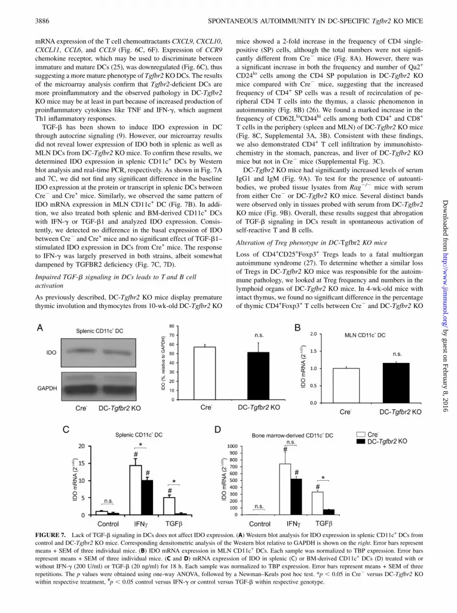
through autocrine signaling (9). However, our microarray resultsdid not reveal lower expression of IDO both in splenic as well asMLN DCs from DC-Tgfbr2 KO mice. To confirm these results, wedetermined IDO expression in splenic CD11c+ DCs by Westernblot analysis and real-time PCR, respectively. As shown in Fig. 7Aand 7C, we did not find any significant difference in the baselineIDO expression at the protein or transcript in splenic DCs betweenCre2 and Cre+ mice. Similarly, we observed the same pattern ofIDO mRNA expression in MLN CD11c+ DC (Fig. 7B). In addi-tion, we also treated both splenic and BM-derived CD11c+ DCswith IFN-g or TGF-b1 and analyzed IDO expression. Consis-tently, we detected no difference in the basal expression of IDObetween Cre2 and Cre+ mice and no significant effect of TGF-b1–stimulated IDO expression in DCs from Cre+ mice. The responseto IFN-g was largely preserved in both strains, albeit somewhatdampened by TGFBR2 deficiency (Fig. 7C, 7D).
Impaired TGF-b signaling in DCs leads to T and B cellactivation
As previously described, DC-Tgfbr2 KO mice display prematurethymic involution and thymocytes from 10-wk-old DC-Tgfbr2 KO
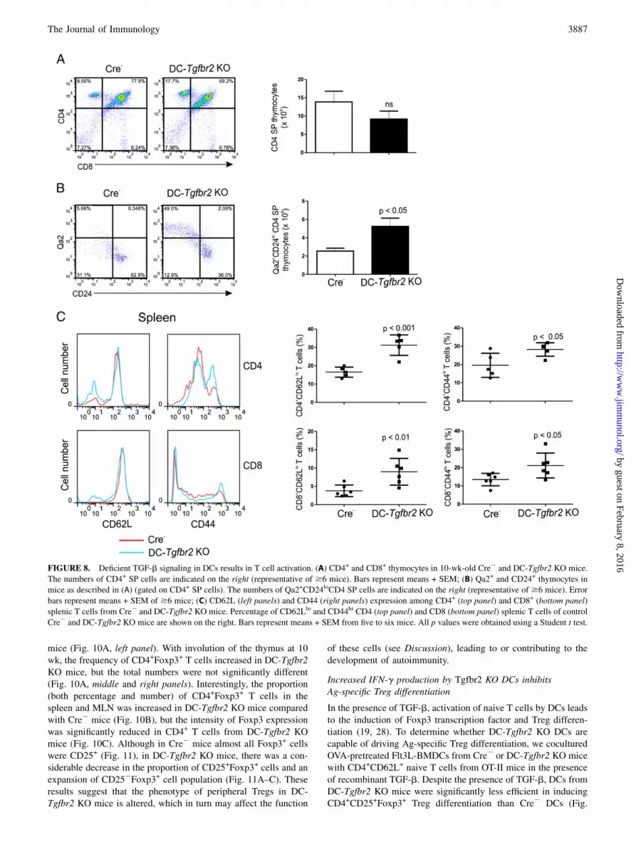
mice showed a 2-fold increase in the frequency of CD4 single-positive (SP) cells, although the total numbers were not signifi-cantly different from Cre2 mice (Fig. 8A). However, there wasa significant increase in both the frequency and number of Qa2+
CD24lo cells among the CD4 SP population in DC-Tgfbr2 KOmice compared with Cre2 mice, suggesting that the increasedfrequency of CD4+ SP cells was a result of recirculation of pe-ripheral CD4 T cells into the thymus, a classic phenomenon inautoimmunity (Fig. 8B) (26). We found a marked increase in thefrequency of CD62LloCD44hi cells among both CD4+ and CD8+
T cells in the periphery (spleen and MLN) of DC-Tgfbr2 KO mice(Fig. 8C, Supplemental 3A, 3B). Consistent with these findings,we also demonstrated CD4+ T cell infiltration by immunohisto-chemistry in the stomach, pancreas, and liver of DC-Tgfbr2 KOmice but not in Cre2 mice (Supplemental Fig. 3C).DC-Tgfbr2 KO mice had significantly increased levels of serum
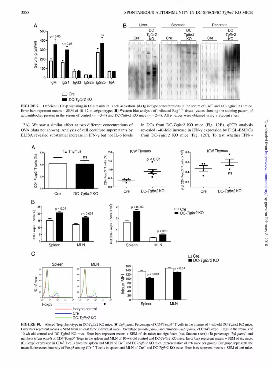
IgG1 and IgM (Fig. 9A). To test for the presence of autoanti-bodies, we probed tissue lysates from Rag2/2 mice with serumfrom either Cre2 or DC-Tgfbr2 KO mice. Several distinct bandswere observed only in tissues probed with serum from DC-Tgfbr2KO mice (Fig. 9B). Overall, these results suggest that abrogationof TGF-b signaling in DCs result in spontaneous activation ofself-reactive T and B cells.
Alteration of Treg phenotype in DC-Tgfbr2 KO mice
Loss of CD4+CD25+Foxp3+ Tregs leads to a fatal multiorganautoimmune syndrome (27). To determine whether a similar lossof Tregs in DC-Tgfbr2 KO mice was responsible for the autoim-mune pathology, we looked at Treg frequency and numbers in thelymphoid organs of DC-Tgfbr2 KO mice. In 4-wk-old mice withintact thymus, we found no significant difference in the percentageof thymic CD4+Foxp3+ T cells between Cre2 and DC-Tgfbr2 KO
FIGURE 7. Lack of TGF-b signaling in DCs does not affect IDO expression. (A) Western blot analysis for IDO expression in splenic CD11c+ DCs from
control and DC-Tgfbr2 KO mice. Corresponding densitometric analysis of the Western blot relative to GAPDH is shown on the right. Error bars represent
means + SEM of three individual mice. (B) IDO mRNA expression in MLN CD11c+ DCs. Each sample was normalized to TBP expression. Error bars
represent means + SEM of three individual mice. (C and D) mRNA expression of IDO in splenic (C) or BM-derived CD11c+ DCs (D) treated with or
without IFN-g (200 U/ml) or TGF-b (20 ng/ml) for 18 h. Each sample was normalized to TBP expression. Error bars represent means + SEM of three
repetitions. The p values were obtained using one-way ANOVA, followed by a Newman–Keuls post hoc test. *p , 0.05 in Cre2 versus DC-Tgfbr2 KO
within respective treatment, #p , 0.05 control versus IFN-g or control versus TGF-b within respective genotype.
3886 SPONTANEOUS AUTOIMMUNITY IN DC-SPECIFIC Tgfbr2 KO MICE
by guest on February 8, 2016http://w
ww
.jimm
unol.org/D
ownloaded from
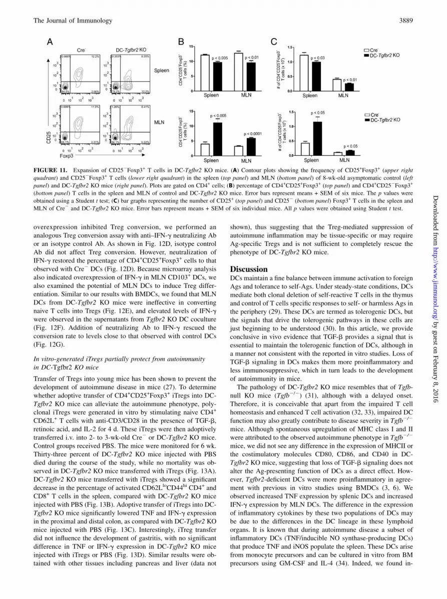
mice (Fig. 10A, left panel). With involution of the thymus at 10wk, the frequency of CD4+Foxp3+ T cells increased in DC-Tgfbr2KO mice, but the total numbers were not significantly different(Fig. 10A, middle and right panels). Interestingly, the proportion(both percentage and number) of CD4+Foxp3+ T cells in thespleen and MLN was increased in DC-Tgfbr2 KO mice comparedwith Cre2 mice (Fig. 10B), but the intensity of Foxp3 expressionwas significantly reduced in CD4+ T cells from DC-Tgfbr2 KOmice (Fig. 10C). Although in Cre2 mice almost all Foxp3+ cellswere CD25+ (Fig. 11), in DC-Tgfbr2 KO mice, there was a con-siderable decrease in the proportion of CD25+Foxp3+ cells and anexpansion of CD252Foxp3+ cell population (Fig. 11A–C). Theseresults suggest that the phenotype of peripheral Tregs in DC-Tgfbr2 KO mice is altered, which in turn may affect the function
of these cells (see Discussion), leading to or contributing to thedevelopment of autoimmunity.
Increased IFN-g production by Tgfbr2 KO DCs inhibitsAg-specific Treg differentiation
In the presence of TGF-b, activation of naive T cells by DCs leadsto the induction of Foxp3 transcription factor and Treg differen-tiation (19, 28). To determine whether DC-Tgfbr2 KO DCs arecapable of driving Ag-specific Treg differentiation, we coculturedOVA-pretreated Flt3L-BMDCs from Cre2 or DC-Tgfbr2 KO micewith CD4+CD62L+ naive T cells from OT-II mice in the presenceof recombinant TGF-b. Despite the presence of TGF-b, DCs fromDC-Tgfbr2 KO mice were significantly less efficient in inducingCD4+CD25+Foxp3+ Treg differentiation than Cre2 DCs (Fig.
FIGURE 8. Deficient TGF-b signaling in DCs results in T cell activation. (A) CD4+ and CD8+ thymocytes in 10-wk-old Cre2 and DC-Tgfbr2 KO mice.
The numbers of CD4+ SP cells are indicated on the right (representative of $6 mice). Bars represent means + SEM; (B) Qa2+ and CD24+ thymocytes in
mice as described in (A) (gated on CD4+ SP cells). The numbers of Qa2+CD24loCD4 SP cells are indicated on the right (representative of $6 mice). Error
bars represent means + SEM of $6 mice; (C) CD62L (left panels) and CD44 (right panels) expression among CD4+ (top panel) and CD8+ (bottom panel)
splenic T cells from Cre2 and DC-Tgfbr2 KO mice. Percentage of CD62Llo and CD44hi CD4 (top panel) and CD8 (bottom panel) splenic T cells of control
Cre2 and DC-Tgfbr2 KO mice are shown on the right. Bars represent means + SEM from five to six mice. All p values were obtained using a Student t test.
The Journal of Immunology 3887
by guest on February 8, 2016http://w
ww
.jimm
unol.org/D
ownloaded from
12A). We saw a similar effect at two different concentrations ofOVA (data not shown). Analysis of cell coculture supernatants byELISA revealed substantial increase in IFN-g but not IL-6 levels
in DCs from DC-Tgfbr2 KO mice (Fig. 12B). qPCR analysisrevealed ∼40-fold increase in IFN-g expression by Flt3L-BMDCsfrom DC-Tgfbr2 KO mice (Fig. 12C). To test whether IFN-g
FIGURE 9. Deficient TGF-b signaling in DCs results in B cell activation. (A) Ig isotype concentrations in the serum of Cre2 and DC-Tgfbr2 KO mice.
Error bars represent means + SEM of 10–12 mice/genotype; (B) Western blot analysis of indicated Rag2/2 tissue lysates showing the staining pattern of
autoantibodies present in the serum of control (n = 3–4) and DC-Tgfbr2 KO mice (n = 2–4). All p values were obtained using a Student t test.
FIGURE 10. Altered Treg phenotype in DC-Tgfbr2 KOmice. (A) Left panel, Percentage of CD4+Foxp3+ T cells in the thymus of 4-wk-old DC-Tgfbr2 KOmice.
Error bars represent means + SEM from at least three individual mice. Percentage (middle panel) and numbers (right panel) of CD4+Foxp3+ Tregs in the thymus of
10-wk-old control and DC-Tgfbr2 KO mice. Error bars represent means + SEM of six mice; not significant (ns), Student t test); (B) percentage (left panel) and
numbers (right panel) of CD4+Foxp3+ Tregs in the spleen and MLN of 10-wk-old control and DC-Tgfbr2 KO mice. Error bars represent means + SEM of six mice;
(C) Foxp3 expression in CD4+ T cells from the spleen and MLN of Cre2 and DC-Tgfbr2 KO mice (representative of$6 mice per group). Bar graph represents the
mean fluorescence intensity of Foxp3 among CD4+ T cells in spleen and MLN of Cre2 and DC-Tgfbr2 KO mice. Error bars represent means + SEM of $6 mice.
3888 SPONTANEOUS AUTOIMMUNITY IN DC-SPECIFIC Tgfbr2 KO MICE
by guest on February 8, 2016http://w
ww
.jimm
unol.org/D
ownloaded from
overexpression inhibited Treg conversion, we performed ananalogous Treg conversion assay with anti–IFN-g neutralizing Abor an isotype control Ab. As shown in Fig. 12D, isotype controlAb did not affect Treg conversion. However, neutralization ofIFN-g restored the percentage of CD4+CD25+Foxp3+ cells to thatobserved with Cre2 DCs (Fig. 12D). Because microarray analysisalso indicated overexpression of IFN-g in MLN CD103+ DCs, wealso examined the potential of MLN DCs to induce Treg differ-entiation. Similar to our results with BMDCs, we found that MLNDCs from DC-Tgfbr2 KO mice were ineffective in convertingnaive T cells into Tregs (Fig. 12E), and elevated levels of IFN-gwere observed in the supernatants from Tgfbr2 KO DC coculture(Fig. 12F). Addition of neutralizing Ab to IFN-g rescued theconversion rate to levels close to that observed with control DCs(Fig. 12G).
In vitro-generated iTregs partially protect from autoimmunityin DC-Tgfbr2 KO mice
Transfer of Tregs into young mice has been shown to prevent thedevelopment of autoimmune disease in mice (27). To determinewhether adoptive transfer of CD4+CD25+Foxp3+ iTregs into DC-Tgfbr2 KO mice can alleviate the autoimmune phenotype, poly-clonal iTregs were generated in vitro by stimulating naive CD4+
CD62L+ T cells with anti-CD3/CD28 in the presence of TGF-b,retinoic acid, and IL-2 for 4 d. These iTregs were then adoptivelytransferred i.v. into 2- to 3-wk-old Cre2 or DC-Tgfbr2 KO mice.Control groups received PBS. The mice were monitored for 6 wk.Thirty-three percent of DC-Tgfbr2 KO mice injected with PBSdied during the course of the study, while no mortality was ob-served in DC-Tgfbr2 KO mice transferred with iTregs (Fig. 13A).DC-Tgfbr2 KO mice transferred with iTregs showed a significantdecrease in the percentage of activated CD62LloCD44hi CD4+ andCD8+ T cells in the spleen, compared with DC-Tgfbr2 KO miceinjected with PBS (Fig. 13B). Adoptive transfer of iTregs into DC-Tgfbr2 KO mice significantly lowered TNF and IFN-g expressionin the proximal and distal colon, as compared with DC-Tgfbr2 KOmice injected with PBS (Fig. 13C). Interestingly, iTreg transferdid not influence the development of gastritis, with no significantdifference in TNF or IFN-g expression in DC-Tgfbr2 KO miceinjected with iTregs or PBS (Fig. 13D). Similar results were ob-tained with other tissues including pancreas and liver (data not
shown), thus suggesting that the Treg-mediated suppression ofautoimmune inflammation may be tissue-specific or may requireAg-specific Tregs and is not sufficient to completely rescue thephenotype of DC-Tgfbr2 KO mice.
DiscussionDCs maintain a fine balance between immune activation to foreignAgs and tolerance to self-Ags. Under steady-state conditions, DCsmediate both clonal deletion of self-reactive T cells in the thymusand control of T cells specific responses to self- or harmless Ags inthe periphery (29). These DCs are termed as tolerogenic DCs, butthe signals that drive the tolerogenic pathways in these cells arejust beginning to be understood (30). In this article, we provideconclusive in vivo evidence that TGF-b provides a signal that isessential to maintain the tolerogenic function of DCs, although ina manner not consistent with the reported in vitro studies. Loss ofTGF-b signaling in DCs makes them more proinflammatory andless immunosuppressive, which in turn leads to the developmentof autoimmunity in mice.The pathology of DC-Tgfbr2 KO mice resembles that of Tgfb-
null KO mice (Tgfb2/2) (31), although with a delayed onset.Therefore, it is conceivable that apart from the impaired T cellhomeostasis and enhanced T cell activation (32, 33), impaired DCfunction may also greatly contribute to disease severity in Tgfb2/2
mice. Although spontaneous upregulation of MHC class I and IIwere attributed to the observed autoimmune phenotype in Tgfb2/2
mice, we did not see any difference in the expression of MHCII orthe costimulatory molecules CD80, CD86, and CD40 in DC-Tgfbr2 KO mice, suggesting that loss of TGF-b signaling does notalter the Ag-presenting function of DCs as a direct effect. How-ever, Tgfbr2-deficient DCs were more proinflammatory in agree-ment with previous in vitro studies using BMDCs (3, 6). Weobserved increased TNF expression by splenic DCs and increasedIFN-g expression by MLN DCs. The difference in the expressionof inflammatory cytokines by these two populations of DCs maybe due to the differences in the DC lineage in these lymphoidorgans. It is known that during autoimmune disease a subset ofinflammatory DCs (TNF/inducible NO synthase-producing DCs)that produce TNF and iNOS populate the spleen. These DCs arisefrom monocyte precursors and can be cultured in vitro from BMprecursors using GM-CSF and IL-4 (34). Indeed, we found in-
FIGURE 11. Expansion of CD252Foxp3+ T cells in DC-Tgfbr2 KO mice. (A) Contour plots showing the frequency of CD25+Foxp3+ (upper right
quadrant) and CD252Foxp3+ T cells (lower right quadrant) in the spleen (top panel) and MLN (bottom panel) of 8-wk-old asymptomatic control (left
panel) and DC-Tgfbr2 KO mice (right panel). Plots are gated on CD4+ cells; (B) percentage of CD4+CD25+Foxp3+ (top panel) and CD4+CD252Foxp3+
(bottom panel) T cells in the spleen and MLN of control and DC-Tgfbr2 KO mice. Error bars represent means + SEM of six mice. The p values were
obtained using a Student t test; (C) bar graphs representing the number of CD25+ (top panel) and CD252 (bottom panel) Foxp3+ T cells in the spleen and
MLN of Cre2 and DC-Tgfbr2 KO mice. Error bars represent means + SEM of six individual mice. All p values were obtained using Student t test.
The Journal of Immunology 3889
by guest on February 8, 2016http://w
ww
.jimm
unol.org/D
ownloaded from
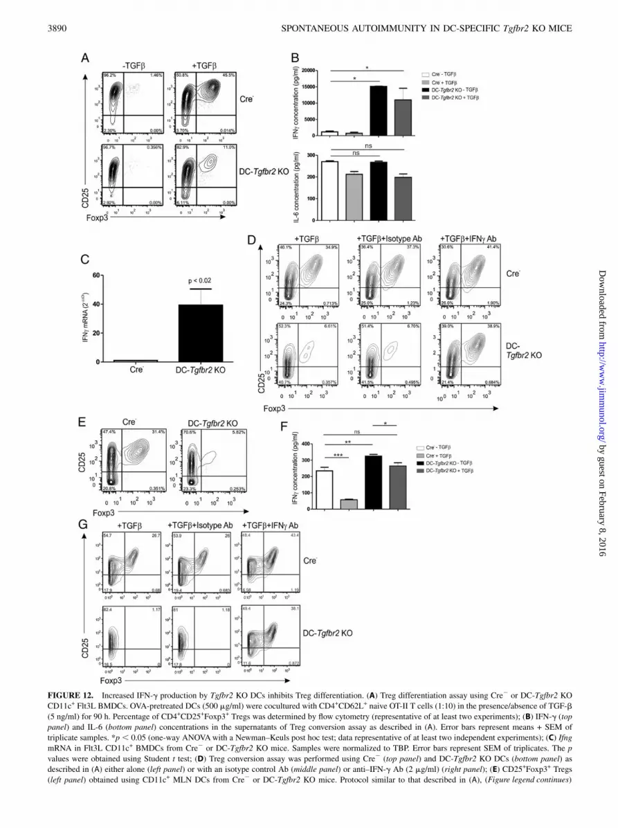
FIGURE 12. Increased IFN-g production by Tgfbr2 KO DCs inhibits Treg differentiation. (A) Treg differentiation assay using Cre2 or DC-Tgfbr2 KO
CD11c+ Flt3L BMDCs. OVA-pretreated DCs (500 mg/ml) were cocultured with CD4+CD62L+ naive OT-II T cells (1:10) in the presence/absence of TGF-b
(5 ng/ml) for 90 h. Percentage of CD4+CD25+Foxp3+ Tregs was determined by flow cytometry (representative of at least two experiments); (B) IFN-g (top
panel) and IL-6 (bottom panel) concentrations in the supernatants of Treg conversion assay as described in (A). Error bars represent means + SEM of
triplicate samples. *p , 0.05 (one-way ANOVAwith a Newman–Keuls post hoc test; data representative of at least two independent experiments); (C) Ifng
mRNA in Flt3L CD11c+ BMDCs from Cre2 or DC-Tgfbr2 KO mice. Samples were normalized to TBP. Error bars represent SEM of triplicates. The p
values were obtained using Student t test; (D) Treg conversion assay was performed using Cre2 (top panel) and DC-Tgfbr2 KO DCs (bottom panel) as
described in (A) either alone (left panel) or with an isotype control Ab (middle panel) or anti–IFN-g Ab (2 mg/ml) (right panel); (E) CD25+Foxp3+ Tregs
(left panel) obtained using CD11c+ MLN DCs from Cre2 or DC-Tgfbr2 KO mice. Protocol similar to that described in (A), (Figure legend continues)
3890 SPONTANEOUS AUTOIMMUNITY IN DC-SPECIFIC Tgfbr2 KO MICE
by guest on February 8, 2016http://w
ww
.jimm
unol.org/D
ownloaded from

creased TNF production in BMDCs differentiated using GM-CSFand IL-4, and these DCs were able to exacerbate T cell-mediatedcolitis. MLN CD103+ DCs (and CD1032 DC; data not shown), aswell as Flt3L-differentiated BMDC from DC-Tgfbr2 KO, showedelevated IFN-g expression without additional stimulation. Inter-estingly, microarray analysis of splenic DCs from DC-Tgfbr2 KOmice showed a clear IFN-g signature response, although expres-sion of IFN-g itself was not elevated. Similarly, splenic DCs fromCD11cdnR mice did not produce IFN-g even after stimulation withIL-12 and IL-18 (15). These two observations suggest differences
in IFN-g induction in the splenic and mucosal/LN DC lineagesand/or that DCs migrating to the spleen are primed by IFN-g at themucosal/parenchymal sites. This is also consistent with the factthat CD103+ DCs produce higher levels of active TGF-b (35),which may act in an autocrine or paracrine manner to suppressIFN-g expression, both in CD103+ and CD1032 DCs.Although TGF-b has been shown to induce expression of IDO
in DCs (9, 36), we did not find any significant differences in theexpression of IDO in either splenic DCs or MLN CD103+ DCs.Expression and activity of IDO protein in Tgfbr2 KO mucosal
except that DCs were cocultured with T cells (1:2) in the presence of 1 mg/ml OVA; (F) IFN-g expression (ELISA) in the supernatants of Treg conversion
assay with MLN DCs. *p , 0.05, **p , 0.01, ***p , 0.001 (one-way ANOVA with Newman–Keuls post hoc test). (G) Treg conversion assay was
performed using Cre2 (top panel) and DC-Tgfbr2 KO DCs (bottom panel) as described in (E) (DC/T cell ratio, 1:10) either alone (left panel), with an
isotype control (middle panel), or anti–IFN-g neutralizing Ab (2 mg/ml) (right panel). Representative data from one of three repetitions is shown.
FIGURE 13. Adoptive transfer of Tregs partially rescues the autoimmune phenotype of DC-Tgfbr2 KO mice. (A) Survival curve of Cre2 or DC-Tgfbr2
KO mice injected with PBS or adoptively transferred at 2–3 wk of age with Foxp3+ iTregs (2 3 106 cells/mouse) generated as described in Materials and
Methods; (B) percentage of CD4+ (left panel) and CD8+ (right panel) CD62LloCD44hi T cells in the spleen of recipient mice at the end of the experiment
described in (A); (C) cytokine mRNA expression in the proximal and distal colon of recipient mice as described in (A). Samples were normalized to TBP
expression. Error bars represent means + SEM of six to seven individual mice; (D) cytokine mRNA expression in the forestomach and glandular stomach of
recipient mice as described in (A). Error bars represent means + SEM of six to seven individual mice. *p , 0.05, **p , 0.01, ***p , 0.001 (one-way
ANOVA with a Newman–Keuls post hoc test).
The Journal of Immunology 3891
by guest on February 8, 2016http://w
ww
.jimm
unol.org/D
ownloaded from
DCs remain to be investigated. Lack of detectable changes in IDOexpression in MLN or splenic DCs may represent the net effect ofthe absence or reduction of stimulatory TGF-b signaling andcompensatory autocrine effects of elevated IFN-g, which is knownto induce IDO expression (37). In pDCs, TGF-b also controlsnonenzymatic cell signaling functions of IDO without affecting itsexpression levels (10). It is therefore plausible that this pathwaymay also be affected, thus leading to the loss of regulatory phe-notype in pDCs, a response skewed toward the canonical NF-kBpathway, more inflammatory state, and ultimately loss of tolerancein DC-Tgfbr2 KO mice.Immunosuppressive Tregs control T cell activation and prevent
the development of autoimmune disease. Expression of Foxp3 insecondary lymphoid CD4+ T cells was significantly decreased,along with a decrease in the frequency of CD4+CD25+Foxp3+
Tregs in DC-Tgfbr2 KO mice. Moreover, among the CD4+Foxp3+
T cells, we observed a dramatic increase in the proportion ofCD4+CD252Foxp3+ T cells, a population also reported recently inpatients with systemic lupus erythematosus (38, 39) and relapsingpatients with multiple sclerosis (40). The immunosuppressive po-tential of CD4+CD252Foxp3+ Tregs is not entirely clear. Func-tional analyses of this population from SLE patients revealed apartial loss of function (39, 41). Zelenay et al. (42) showed thatCD4+CD252Foxp3+ T cells constitute a reservoir of committedTregs that regain CD25 expression upon homeostatic proliferationin a lymphopenic host. However, the same group showed thatTregs identified as the CD45RBloCD252 subset failed to showsuppressive function in vitro when freshly isolated from mice (42).It has also been shown that Tregs with attenuated Foxp3 expres-sion have lower levels of CD25 and that these cells have a ten-dency to convert to Th2 type cells (43). On the basis of thesestudies, it is highly likely that the CD4+CD252Foxp3+ T cells inDC-Tgfbr2 KO mice are less immunosuppressive thereby con-tributing to the autoimmune pathology. Such alterations in Treghomoeostasis and spontaneous autoimmunity were not observedin CD11cdnR mice, suggesting that residual TGF-b signaling inDCs may have been sufficient to maintain self-tolerance underbasal conditions and that the consequences of impaired TGF-bsignaling in DCs may represent a continuum depending on thedegree of suppression. Future studies with DC-Tgfbr2 KO crossedwith Foxp3-RFP knockin mice should further clarify the natureand function of the CD4+CD252Foxp3+ T cells.We have also demonstrated that in vitro, DC-mediated Ag-
specific Treg conversion is impaired because of elevated produc-tion of IFN-g by Tgfbr2 KO DCs. However, we did not observe theexpansion of the CD4+CD252Foxp3+ population as we didin vivo. Therefore, the mechanisms leading to the potential loss offunction of Tregs in DC-Tgfbr2 KO mice remain unclear. Bidi-rectional DC–Treg interactions are required to maintain immu-nological tolerance (44). DCs induce Treg differentiation andproliferation through Ag-dependent and -independent interactionsbut in a cell–cell contact- and IL-2–dependent mechanism (45).However, the suppressive function of the expanded Tregs may stillrequire additional signals that remain unidentified. Adoptivetransfer of in vitro-generated polyclonal Foxp3+ iTregs into youngDC-Tgfbr2 KO mice prevented early mortality and partially res-cued the autoimmune phenotype with significant reduction in theproportion of activated T cells and protection from colitis. It didnot, however, protect from gastritis, pancreatitis, or hepatitis. Al-though the optimal timing of transfer and lifespan of the injectedTregs may be debatable, loss of their immunosuppressive functionin vivo in DC-Tgfbr2 KO mice cannot be ruled out. In contrast,iTregs are generally thought to suppress immune responses to en-vironmental, food allergens, and commensal microbiota, whereas
natural Tregs prevent autoimmunity by raising the threshold foractivation of immune response to self-Ags (46). Consistent withour observations, non–Ag-specific iTregs have been shown to beprotective in mouse models of colitis (46). In contrast, naturalTregs are selected in the thymus through MHCII-dependent TCRinteractions and may mediate suppression in an Ag-specificmanner. Ag-specific Tregs were required to prevent autoimmunegastritis in mice (47), which may explain persistent gastritis in ourTreg rescue model.This novel mouse model with DC-specific Tgfbr2 deletion
highlights the critical importance of TGF-b signaling in DCs inthe maintenance of immune homeostasis and in the prevention ofautoimmunity. However, these functions may be independent ofthe previously ascribed TGF-b control of Ag presentation andcostimulation. Although a mechanistic relationship between TGF-b signaling in DCs and Foxp3+ Treg responses still remains to beelucidated in detail, the phenotype of our novel mouse model canbe exploited to advance our understanding of the pathogenesis ofcomplex autoimmune disorders, including the in vivo expansionand function of CD252Foxp3+ Tregs.
AcknowledgmentsWe thank Drs. Tom Doetschman and Nicolas Larmonier for helpful dis-
cussions, Dr. David Besselsen for help with pathological evaluation,
Darya Alizadeh and Paula Campbell for assistance with flow cytometry
and flow sorting, and Dr. Vijay Radhakrishnan for proofreading help.
DisclosuresThe authors have no financial conflicts of interest.
References1. Li, M. O., Y. Y. Wan, S. Sanjabi, A. K. Robertson, and R. A. Flavell. 2006.
Transforming growth factor-beta regulation of immune responses. Annu. Rev.Immunol. 24: 99–146.
2. Grauer, O., P. Poschl, A. Lohmeier, G. J. Adema, and U. Bogdahn. 2007. Toll-like receptor triggered dendritic cell maturation and IL-12 secretion are neces-sary to overcome T-cell inhibition by glioma-associated TGF-b2. J. Neurooncol.82: 151–161.
3. Geissmann, F., P. Revy, A. Regnault, Y. Lepelletier, M. Dy, N. Brousse,S. Amigorena, O. Hermine, and A. Durandy. 1999. TGF-b1 prevents the noncognatematuration of human dendritic Langerhans cells. J. Immunol. 162: 4567–4575.
4. Ronger-Savle, S., J. Valladeau, A. Claudy, D. Schmitt, J. Peguet-Navarro,C. Dezutter-Dambuyant, L. Thomas, and D. Jullien. 2005. TGFb inhibits CD1dexpression on dendritic cells. J. Invest. Dermatol. 124: 116–118.
5. Yamaguchi, Y., H. Tsumura, M. Miwa, and K. Inaba. 1997. Contrasting effectsof TGF-b1 and TNF-a on the development of dendritic cells from progenitors inmouse bone marrow. Stem Cells 15: 144–153.
6. Siddiqui, K. R., S. Laffont, and F. Powrie. 2010. E-cadherin marks a subset ofinflammatory dendritic cells that promote T cell-mediated colitis. Immunity 32:557–567.
7. Ogata, M., Y. Zhang, Y. Wang, M. Itakura, Y. Y. Zhang, A. Harada,S. Hashimoto, and K. Matsushima. 1999. Chemotactic response toward che-mokines and its regulation by transforming growth factor-b1 of murine bonemarrow hematopoietic progenitor cell-derived different subset of dendritic cells.Blood 93: 3225–3232.
8. Sato, K., H. Kawasaki, H. Nagayama, M. Enomoto, C. Morimoto, K. Tadokoro,T. Juji, and T. A. Takahashi. 2000. TGF-b1 reciprocally controls chemotaxis ofhuman peripheral blood monocyte-derived dendritic cells via chemokinereceptors. J. Immunol. 164: 2285–2295.
9. Belladonna, M. L., C. Volpi, R. Bianchi, C. Vacca, C. Orabona, M. T. Pallotta,L. Boon, S. Gizzi, M. C. Fioretti, U. Grohmann, and P. Puccetti. 2008. Cuttingedge: autocrine TGF-b sustains default tolerogenesis by IDO-competent den-dritic cells. J. Immunol. 181: 5194–5198.
10. Pallotta, M. T., C. Orabona, C. Volpi, C. Vacca, M. L. Belladonna, R. Bianchi,G. Servillo, C. Brunacci, M. Calvitti, S. Bicciato, et al. 2011. Indoleamine 2,3-dioxygenase is a signaling protein in long-term tolerance by dendritic cells. Nat.Immunol. 12: 870–878.
11. Brenner, O., D. Levanon, V. Negreanu, O. Golubkov, O. Fainaru, E. Woolf, andY. Groner. 2004. Loss of Runx3 function in leukocytes is associated withspontaneously developed colitis and gastric mucosal hyperplasia. Proc. Natl.Acad. Sci. USA 101: 16016–16021.
12. Fainaru, O., E. Woolf, J. Lotem, M. Yarmus, O. Brenner, D. Goldenberg,V. Negreanu, Y. Bernstein, D. Levanon, S. Jung, and Y. Groner. 2004. Runx3regulates mouse TGF-b‑mediated dendritic cell function and its absence resultsin airway inflammation. EMBO J. 23: 969–979.
3892 SPONTANEOUS AUTOIMMUNITY IN DC-SPECIFIC Tgfbr2 KO MICE
by guest on February 8, 2016http://w
ww
.jimm
unol.org/D
ownloaded from

13. Laouar, Y., T. Town, D. Jeng, E. Tran, Y. Wan, V. K. Kuchroo, and R. A. Flavell.2008. TGF-b signaling in dendritic cells is a prerequisite for the control ofautoimmune encephalomyelitis. Proc. Natl. Acad. Sci. USA 105: 10865–10870.
14. Sanjabi, S., and R. A. Flavell. 2010. Overcoming the hurdles in using mousegenetic models that block TGF-b signaling. J. Immunol. Methods 353: 111–114.
15. Laouar, Y., F. S. Sutterwala, L. Gorelik, and R. A. Flavell. 2005. Transforminggrowth factor-b controls T helper type 1 cell development through regulation ofnatural killer cell interferon-g. Nat. Immunol. 6: 600–607.
16. Boomershine, C. S., A. Chamberlain, P. Kendall, A. R. Afshar-Sharif, H. Huang,M. K. Washington, W. E. Lawson, J. W. Thomas, T. S. Blackwell, andN. A. Bhowmick. 2009. Autoimmune pancreatitis results from loss of TGFbsignalling in S100A4-positive dendritic cells. Gut 58: 1267–1274.
17. Shevach, E. M. 2009. Mechanisms of foxp3+ T regulatory cell-mediated sup-pression. Immunity 30: 636–645.
18. Darrasse-Jeze, G., S. Deroubaix, H. Mouquet, G. D. Victora, T. Eisenreich,K. H. Yao, R. F. Masilamani, M. L. Dustin, A. Rudensky, K. Liu, andM. C. Nussenzweig. 2009. Feedback control of regulatory T cell homeostasis bydendritic cells in vivo. J. Exp. Med. 206: 1853–1862.
19. Chen, W., W. Jin, N. Hardegen, K. J. Lei, L. Li, N. Marinos, G. McGrady, andS. M. Wahl. 2003. Conversion of peripheral CD4+CD25‑ naive T cells to CD4+
CD25+ regulatory T cells by TGF-b induction of transcription factor Foxp3. J.Exp. Med. 198: 1875–1886.
20. Caton, M. L., M. R. Smith-Raska, and B. Reizis. 2007. Notch-RBP-J signalingcontrols the homeostasis of CD8‑ dendritic cells in the spleen. J. Exp. Med. 204:1653–1664.
21. Chytil, A., M. A. Magnuson, C. V. Wright, and H. L. Moses. 2002. Conditionalinactivation of the TGF-b type II receptor using Cre:Lox. Genesis 32: 73–75.
22. Mombaerts, P., J. Iacomini, R. S. Johnson, K. Herrup, S. Tonegawa, andV. E. Papaioannou. 1992. RAG-1‑deficient mice have no mature B andT lymphocytes. Cell 68: 869–877.
23. Xu, Y., Y. Zhan, A. M. Lew, S. H. Naik, and M. H. Kershaw. 2007. Differentialdevelopment of murine dendritic cells by GM-CSF versus Flt3 ligand hasimplications for inflammation and trafficking. J. Immunol. 179: 7577–7584.
24. Ohnmacht, C., A. Pullner, S. B. King, I. Drexler, S. Meier, T. Brocker, andD. Voehringer. 2009. Constitutive ablation of dendritic cells breaks self-tolerance ofCD4T cells and results in spontaneous fatal autoimmunity. J. Exp.Med. 206: 549–559.
25. Hadeiba, H., T. Sato, A. Habtezion, C. Oderup, J. Pan, and E. C. Butcher. 2008.CCR9 expression defines tolerogenic plasmacytoid dendritic cells able to sup-press acute graft-versus-host disease. Nat. Immunol. 9: 1253–1260.
26. Hakim, F. T., and R. E. Gress. 2007. Thymic involution: implications for self-tolerance. Methods Mol. Biol. 380: 377–390.
27. Sakaguchi, S., T. Yamaguchi, T. Nomura, and M. Ono. 2008. Regulatory T cellsand immune tolerance. Cell 133: 775–787.
28. Yamazaki, S., A. J. Bonito, R. Spisek, M. Dhodapkar, K. Inaba, andR. M. Steinman. 2007. Dendritic cells are specialized accessory cells along withTGF- for the differentiation of Foxp3+CD4+ regulatory T cells from peripheralFoxp3 precursors. Blood 110: 4293–4302.
29. Steinman, R. M., D. Hawiger, and M. C. Nussenzweig. 2003. Tolerogenicdendritic cells. Annu. Rev. Immunol. 21: 685–711.
30. Manicassamy, S., and B. Pulendran. 2011. Dendritic cell control of tolerogenicresponses. Immunol. Rev. 241: 206–227.
31. Shull, M. M., I. Ormsby, A. B. Kier, S. Pawlowski, R. J. Diebold, M. Yin,R. Allen, C. Sidman, G. Proetzel, D. Calvin, et al. 1992. Targeted disruption ofthe mouse transforming growth factor-b1 gene results in multifocal inflamma-tory disease. Nature 359: 693–699.
32. Li, M. O., S. Sanjabi, and R. A. Flavell. 2006. Transforming growth factor-bcontrols development, homeostasis, and tolerance of T cells by regulatory T cell-dependent and -independent mechanisms. Immunity 25: 455–471.
33. Marie, J. C., D. Liggitt, and A. Y. Rudensky. 2006. Cellular mechanisms of fatalearly-onset autoimmunity in mice with the T cell-specific targeting of trans-forming growth factor-b receptor. Immunity 25: 441–454.
34. Shortman, K., and S. H. Naik. 2007. Steady-state and inflammatory dendritic-cell development. Nat. Rev. Immunol. 7: 19–30.
35. Coombes, J. L., K. R. Siddiqui, C. V. Arancibia-Carcamo, J. Hall, C. M. Sun,Y. Belkaid, and F. Powrie. 2007. A functionally specialized population of mu-cosal CD103+ DCs induces Foxp3+ regulatory T cells via a TGF-b and retinoicacid-dependent mechanism. J. Exp. Med. 204: 1757–1764.
36. Belladonna, M. L., C. Orabona, U. Grohmann, and P. Puccetti. 2009. TGF-b andkynurenines as the key to infectious tolerance. Trends Mol. Med. 15: 41–49.
37. Mellor, A. L., and D. H. Munn. 2004. IDO expression by dendritic cells: tol-erance and tryptophan catabolism. Nat. Rev. Immunol. 4: 762–774.
38. Yang, H. X., W. Zhang, L. D. Zhao, Y. Li, F. C. Zhang, F. L. Tang, W. He, andX. Zhang. 2009. Are CD4+CD25‑Foxp3+ cells in untreated new-onset lupuspatients regulatory T cells? Arthritis Res. Ther. 11: R153.
39. Bonelli, M., A. Savitskaya, C. W. Steiner, E. Rath, J. S. Smolen, andC. Scheinecker. 2009. Phenotypic and functional analysis of CD4+CD25‑Foxp3+
T cells in patients with systemic lupus erythematosus. J. Immunol. 182: 1689–1695.
40. Fransson, M., J. Burman, C. Lindqvist, C. Atterby, J. Fagius, and A. Loskog.2010. T regulatory cells lacking CD25 are increased in MS during relapse.Autoimmunity 43: 590–597.
41. Horwitz, D. A. 2010. Identity of mysterious CD4+CD25‑Foxp3+ cells in SLE.Arthritis Res. Ther. 12: 101.
42. Zelenay, S., T. Lopes-Carvalho, I. Caramalho, M. F. Moraes-Fontes, M. Rebelo,and J. Demengeot. 2005. Foxp3+CD25‑ CD4 T cells constitute a reservoir ofcommitted regulatory cells that regain CD25 expression upon homeostatic ex-pansion. Proc. Natl. Acad. Sci. USA 102: 4091–4096.
43. Wan, Y. Y., and R. A. Flavell. 2007. Regulatory T-cell functions are subvertedand converted owing to attenuated Foxp3 expression. Nature 445: 766–770.
44. Lange, C., M. Durr, H. Doster, A. Melms, and F. Bischof. 2007. Dendritic cell-regulatory T-cell interactions control self-directed immunity. Immunol. Cell Biol.85: 575–581.
45. Zou, T., A. J. Caton, G. A. Koretzky, and T. Kambayashi. 2010. Dendritic cellsinduce regulatory T cell proliferation through antigen-dependent and -indepen-dent interactions. J. Immunol. 185: 2790–2799.
46. Curotto de Lafaille, M. A., and J. J. Lafaille. 2009. Natural and adaptive foxp3+
regulatory T cells: more of the same or a division of labor? Immunity 30: 626–635.
47. Nguyen, T. L., N. L. Sullivan, M. Ebel, R. M. Teague, and R. J. DiPaolo. 2011.Antigen-specific TGF-b‑induced regulatory T cells secrete chemokines, regulateT cell trafficking, and suppress ongoing autoimmunity. J. Immunol. 187: 1745–1753.
The Journal of Immunology 3893
by guest on February 8, 2016http://w
ww
.jimm
unol.org/D
ownloaded from