complex x chromosome rearrangement associated with multiorgan autoimmunity
TRANSCRIPT

CASE REPORT Open Access
Complex X chromosome rearrangementassociated with multiorgan autoimmunityIrén Haltrich1*†, Henriett Pikó2†, Horolma Pamjav3, Anikó Somogyi4, Antónia Völgyi3, Dezső David5, Artúr Beke6,Zoltán Garamvölgyi6, Eszter Kiss1, Veronika Karcagi2 and György Fekete1
Abstract
Background: Turner syndrome, a congenital condition that affects 1/2,500 births, results from absence or structuralalteration of the second sex chromosome. Turner syndrome is usually associated with short stature, gonadaldysgenesis and variable dysmorphic features.The classical 45,X karyotype accounts approximately for half of all patients, the remainder exhibit mosaicism orstructural abnormalities of the X chromosome. However, complex intra-X chromosomal rearrangements involvingmore than three breakpoints are extremely rare.
Results: We present a unique case of a novel complex X chromosome rearrangement in a young female patientpresenting successively a wide range of autoimmune diseases including insulin dependent diabetes mellitus,Hashimoto’s thyroiditis, celiac disease, anaemia perniciosa, possible inner ear disease and severe hair loss. For thegenetic evaluation, conventional cytogenetic analysis and FISH with different X specific probes were initially performed.The complexity of these results and the variety of autoimmune problems of the patient prompted us to identify theexact composition and breakpoints of the rearranged X as well as methylation status of the X chromosomes. The highresolution array-CGH (assembly GRCh37/hg19) detected single copy for the whole chromosome X short arm. Twodifferent sized segments of Xq arm were present in three copies: one large size of 80,3 Mb from Xq11.1 to Xq27.3region and another smaller (11,1 Mb) from Xq27.3 to Xq28 region. An 1,6 Mb Xq27.3 region of the long arm waspresent in two copies. Southern blot analysis identified a skewed X inactivation with ≈ 70:30 % ratios of methylated/unmethylated fragments. The G-band and FISH patterns of the rearranged X suggested the aspect of a restructuredi(Xq) chromosome which was shattered and fortuitously repaired. The X-STR genotype analysis of the family detectedthat the patient inherited intact maternal X chromosome and a rearranged paternal X chromosome.The multiple Xq breakages and fusions as well as inverted duplication would have been expected to cause a severeTurner phenotype. However, the patient lacks many of the classic somatic features of Turner syndrome, instead shepresented multiorgan autoimmune diseases.
Conclusions: The clinical data of the presented patient suggest that fragmentation of the i(Xq) chromosome elevatesthe risk of autoimmune diseases.
Keywords: Complex X chromosome rearrangement, Autoimmune disease, Turner syndrome, Type 1 diabetes mellitus,X inactivation disorder
* Correspondence: [email protected]†Equal contributors12nd Department of Pediatrics, Semmelweis University, Tűzoltó utca 7-9,1094 Budapest, HungaryFull list of author information is available at the end of the article
© 2015 Haltrich et al. This is an Open Access article distributed under the terms of the Creative Commons AttributionLicense (http://creativecommons.org/licenses/by/4.0), which permits unrestricted use, distribution, and reproduction inany medium, provided the original work is properly credited. The Creative Commons Public Domain Dedication waiver(http://creativecommons.org/publicdomain/zero/1.0/) applies to the data made available in this article, unless otherwise stated.
Haltrich et al. Molecular Cytogenetics (2015) 8:51 DOI 10.1186/s13039-015-0152-5

BackgroundTurner syndrome (TS) is a known genetic disorder associ-ated with complete or partial absence of one of the sexchromosomes (the X or Y chromosome) affecting aboutone out of 2500 liveborn girls. The most common pheno-typic features of TS patients are short stature and gonadaldysgenesis, they are at significantly increased risk of differ-ent congenital malformations and prone to develop grad-ually autoimmune disorders.The isochromosome X [i(X)(q10)], which consists of
two copies of the long arm with missing short arm is themost common structural rearrangement, occurring inabout 20 % of TS cases. By the currently applied tech-niques complex X chromosome rearrangements havingthree or more breakpoints are rarely seen in constitutionalkaryotype, even rarer are rearrangements occurring withina single chromosome. To date, only seven previousreports of complex X chromosomal rearrangement havebeen described [1–7].Here we report a novel complex X chromosome re-
arrangement in a young TS patient with type 1 diabetesmellitus (DM) and severe other endocrine complications.We delineated the highly rearranged derivative X chromo-some by G-banding and FISH analysis as well as arraycomparative genome hybridization (CGH; assemblyGRCh37/hg19). The presented X chromosome rearrange-ment can bring insight into the possible genetic mecha-nisms responsible for X-linked autoimmune disorder.
Case presentationThe patient was born from an uncomplicated pregnancy,on term per vias naturales with 2750 grams. She was thefirst child of a 20-year-old mother and a 23-year-old father.The mother was a joiner, the father dealt forest yields, andhe drank excessively by his late adolescence. He lost hissight because of alcohol use disorder and repeatedlyattempted suicide. He died premature, at age of 24 yearsbecause of alcohol induced central nervous system dam-age. The father’s parents are farmers; they are healthy,without any family history of alcoholism. The mother’sfather worked in a quarry and the mother was housewife.Apart from the female cousin suffering from type 1 dia-betes mellitus (DM), the family history is unremarkable.Genetic and autoimmune disorders, mental retardationhave never occurred. The patient’s mother had one miscar-riage and one healthy son with a second husband.At newborn age the patient presented no stigmata of
Turner syndrome. At the age of 17 years she wasreferred to cytogenetic laboratory due to her primaryamenorrhoea. Standard karyotype analysis had shown acomplex rearrangement of the one X chromosome.On examination, her height was 152 cm, her weight was
50 kg. Her short stature wasn’t conspicuous because par-ental grandparents, her mother’s sibling and several
cousins were at the height around of 155 cm. The pro-band’s predicted final height is 158, 5 cm, currently she is24 years old and 154 cm in height (<3rd percentile, growthrate - 2 SD).She had spontaneous breast development (Tanner
stage III-IV) but sparse axillary and pubic hair (Tannerstage II). Laboratory studies revealed the followingvalues: FSH level: 77.83 IU/L (normal women: >40 IU/l),LH level: 24.20 IU/L (normal women: >21 IU/l), E25 pg/ml (normal women: up to 375 pg/ml), confirmingprimary ovarian insufficiency (POF). Receiving estradioltherapy her secondary sexual characteristics have devel-oped and her menstruation was normal. Since ten years ofage, she has been suffering from hearing loss on both earswhich was resolved by a hearing aid. She had recurrent re-spiratory infections, she also suffered from pyelonephritis.At the age of 13 years after tooth extraction mandibularosteomyelitis developed, which was successfully treatedwith antibiotic therapy. At the age of 15 years she wasdiagnosed with type 1 DM and insulin substitutive treat-ment was started. In the same year anaemia perniciosawas diagnosed as the underlying cause of her anaemia.She received vitamin B12 regularly. Some month laterlevothyroxine therapy was introduced due to the diagnosisof Hashimoto thyroiditis with clinical hypothyroidism.Her thyroid function was normal under the therapy (TSH:3.17 mIU/l). At the same time she presented symptoms ofcoeliac disease. The immunhistochemistry examination ofgastric mucosa revealed an increased level of chro-mogranin A. The gastroscopy examination exploredneuroendocrine cell hyperplasy in the antral and corpusregions. When dydrogesterone was added to her hor-mone replacement therapy, she suffered from severehyperglycaemia and ketoacidosis and intensive care wasnecessary. The estradiol dydrogesterone therapy wastemporarily stopped due to insufficient carbohydratemetabolism. Initially, for two weeks an insulin pumpwas introduced to regulate her carbohydrate metabol-ism. Due to her inability to learn the use of the insulinpump (memory deficit) regular insulin therapy wascontinued. Without the hormone replacement therapythe patient had an amenorrhoea. Therefore, hormonereplacement therapy was newly added and well toler-ated without any ketoacidotic periods until today. Atthe age of 23 years gynaecological ultrasound examin-ation showed that the myometrium of the uterus isnormal (size of the uterus: PF: 32 mm, AP: 16 mm;endometrium: 4 mm.). Both ovaries were smaller ascompared with the size of normal reproductive-agedwomen (the right ovary with 9x6 mm, without any folli-cles; the left ovary with 10x7mm, without follicles) andsuspect for streak gonads and infertility. No free fluidin Douglas was detected. The patient’s other currentmedical problems include osteoporosis, eczema and
Haltrich et al. Molecular Cytogenetics (2015) 8:51 Page 2 of 11

severe hair loss. Her behaviour is conventional andsocial; a mild intellectual failure could be observed.
MethodsCytogenetic analysisChromosome analysis was performed on phytohem-agglutinin stimulated peripheral blood lymphocytecultures on metaphase cells with trypsin and WrightGiemsa stain. Fluorescence in Situ Hybridization (FISH)analysis was carried out on methanol/acetic acid-fixedsuspensions. Slides preparation for FISH was made ac-cording to standard techniques. X and Y centromere(CEP X and Y) specific probes as well as X, Y paintingprobe, SRY specific probes were used for evaluation ofexact genotype and to detect chromosome X copy num-ber. CEP X specific FISH probe was investigated also forbuccal and urine mucosa interphase cells of the patientin order to reveal the possible tissue specific X chromo-some mosaicism. To determine the composition of rear-ranged X chromosome, additional hybridizations wereeffectuated with X p arm red, Xq arm green, SHOX,XIST (Cytocell, United Kingdom) as well as subtelomereXq/Yq specific (ToTelVysion, Abbott, Germany) probes.Karyotypes and FISH results were described accordingto the International System for Human CytogeneticNomenclature (ISCN 2013).
Southern blot analysisFor X chromosome methylation status determinationFRAXA analysis was initiated. Genomic DNA from thepatient was isolated from peripheral lymphocytes by thesimple salting-out procedure. DNA was subjected torestriction enzyme digestion with EcoRI and the methy-lation sensitive enzyme EagI followed by Southern blotanalysis and hybridization using the DNA probe StB12.3[8]. Methylated normal alleles are not cut by methylationsensitive enzymes and give rise to the normal 5.2 KbEcoRI fragment. In unaffected females, two bands arevisible: a 2.8 Kb fragment corresponding to the unmethy-lated/active X and a 5.2 kb allele representing the methyl-ated/inactive X chromosome and the signal strength ofboth fragments are more or less equal. In case of FRAXApremutations or full mutations, methylated and unmethy-lated CGG expansions are indicated by the presence ofbands and/or smears above 5.2 Kb and 2.8 Kb,respectively.
Repeat primed PCR analysisRepeat primed (RP) PCRs were performed on 20-40 ngDNA according the supplied protocol from Asuragene.This protocol supports two different PCR formats, genespecific FMR1 PCR and CGG Repeat Primed (RP) PCR.The gene specific PCR contains two primers outside theCGG repeat in FMR1 gene, and the CGG Repeat Primed
PCR contains 3 primers: two outside and one inside theCGG repeat.
Array comparative genomic hybridizationTo ascertain the segmental composition of the rear-ranged X chromosome, a high resolution genomic scanusing ISCA plus design array of NimbleGene-Rochecontaining 1.4 M probes per subarray (assemblyGRCh37/hg19) was performed in the DNA samples ofthe patient. This CGX microarray provides a mean aver-age resolution of approximately 15-20 Kb. Array CGHanalysis was performed according to the manufacturer’sprotocol. The CGH protocol involves independent label-ling of the patient (test DNA) and the reference genomicDNA (Human Genomic DNA, Promega Madison, WIU.S.A.) with Cy3 and Cy5 dyes using a NimbleGeneDual-Color DNA Labelling Kit (Roche-NimbleGen Inc.).Co-hybridization of these DNAs to a NimbleGene CGHarray was performed for 72 h at 42 °C. The subarrays werescanned on NimbleGene MS 200 microarray scanner anddata were extracted and analysed using NimbleScan, Sig-nalMap and Deva 1.1 softwares (Roche NimbleGene Inc.).DNA CNVs were mentioned as gain or loss in a linear ra-tio, and the length of the variation was given in megabase(Mb) and base pair (bp) respectively.
Identification of the rearranged X chromosome originIn order to identify the origin of the rearranged Xchromosome, we have tested 12 X- chromosomal STR(Short Tandem Repeat) markers on the entire length ofthe X chromosome according to the manufacturer’sinstructions. The Investigator Argus X-12 Kit (Qiagen,Hilden, Germany) is a multiplex PRC kit for determin-ation of 12 X-STR loci simultaneously. The kit containsprimers for Amelogenin for gender determination,DXS7132, DXS7423, DXS8378, DXS10074, DXS10079,DXS10101, DXS10103, DXS10134, DXS10135, DXS10146,DXS10148, and HPRTB loci. The markers are clusteredinto 4 linkage groups (3 markers per group), and each setof 3 markers is closely linked and handled by a haplotypefor genotyping. These 12 X-STR loci are located in fourlinkage groups as followings: on Xp22: DXS8378 – DXS10135 – DXS10148; on Xq11.2-Xq12: DXS7132 – DXS10074 – DXS10079; on Xq26: HPRTB – DXS10101 –DXS10103 and on Xq28: DXS7423 – DXS10134 – DXS10146 loci. The PCR products were analyzed by capillaryelectrophoresis using an ABI 3130 Genetic Analyzer (LifeTechnologies, Foster City, CA). The fragment sizes andallele designations were determined by using the Gene-Mapper ID 3.2.1 (Life Technologies, Foster City, CA).
WAIS-IV intelligence testThe Wechsler Adult Intelligence Scale, Fourth Edition(WAIS-IV) [9] is a comprehensive clinical instrument
Haltrich et al. Molecular Cytogenetics (2015) 8:51 Page 3 of 11
for assessing intelligence of adults between the ages of16–90 years. The test is composed of 15 core and sup-plemental subtests which contribute to a compositescore that represents general intellectual ability (fullscale IQ, FSIQ) and scores in indices of specific cogni-tive areas: Verbal Comprehension Index (VCI), Percep-tual Reasoning Index (PRI), Working Memory Index(WMI), and Processing Speed Index (PSI).
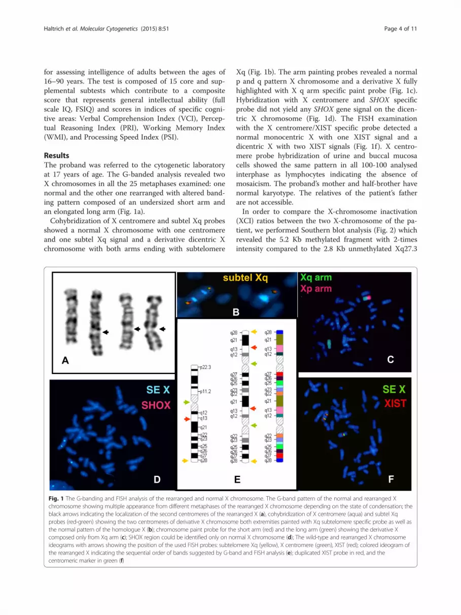
ResultsThe proband was referred to the cytogenetic laboratoryat 17 years of age. The G-banded analysis revealed twoX chromosomes in all the 25 metaphases examined: onenormal and the other one rearranged with altered band-ing pattern composed of an undersized short arm andan elongated long arm (Fig. 1a).Cohybridization of X centromere and subtel Xq probes
showed a normal X chromosome with one centromereand one subtel Xq signal and a derivative dicentric Xchromosome with both arms ending with subtelomere
Xq (Fig. 1b). The arm painting probes revealed a normalp and q pattern X chromosome and a derivative X fullyhighlighted with X q arm specific paint probe (Fig. 1c).Hybridization with X centromere and SHOX specificprobe did not yield any SHOX gene signal on the dicen-tric X chromosome (Fig. 1d). The FISH examinationwith the X centromere/XIST specific probe detected anormal monocentric X with one XIST signal and adicentric X with two XIST signals (Fig. 1f ). X centro-mere probe hybridization of urine and buccal mucosacells showed the same pattern in all 100-100 analysedinterphase as lymphocytes indicating the absence ofmosaicism. The proband’s mother and half-brother havenormal karyotype. The relatives of the patient’s fatherare not accessible.In order to compare the X-chromosome inactivation
(XCI) ratios between the two X-chromosome of the pa-tient, we performed Southern blot analysis (Fig. 2) whichrevealed the 5.2 Kb methylated fragment with 2-timesintensity compared to the 2.8 Kb unmethylated Xq27.3
Fig. 1 The G-banding and FISH analysis of the rearranged and normal X chromosome. The G-band pattern of the normal and rearranged Xchromosome showing multiple appearance from different metaphases of the rearranged X chromosome depending on the state of condensation; theblack arrows indicating the localization of the second centromeres of the rearranged X (a), cohybridization of X centromere (aqua) and subtel Xqprobes (red-green) showing the two centromeres of derivative X chromosome both extremities painted with Xq subtelomere specific probe as well asthe normal pattern of the homologue X (b); chromosome paint probe for the short arm (red) and the long arm (green) showing the derivative Xcomposed only from Xq arm (c); SHOX region could be identified only on normal X chromosome (d); The wild-type and rearranged X chromosomeideograms with arrows showing the position of the used FISH probes: subtelomere Xq (yellow), X centromere (green), XIST (red); colored ideogram ofthe rearranged X indicating the sequential order of bands suggested by G-band and FISH analysis (e); duplicated XIST probe in red, and thecentromeric marker in green (f)
Haltrich et al. Molecular Cytogenetics (2015) 8:51 Page 4 of 11

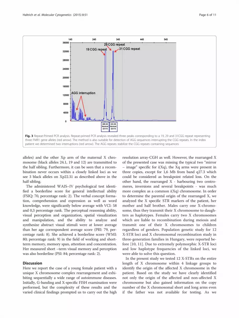
fragment (approx. with 70:30 % ratio), assuming thepresence of three FMR1 alleles with a skewed X inactiva-tion. In the contrary, for all controls we identified two Xchromosome normal alleles (with a 50:50 % value of the2,8 kb unmethylated and the 5.2 kb methylated frag-ments). In order to ascertain the presence of three Xqarms we performed repeat-primed PCR analysis whichrevealed three peaks corresponding to a 19, 29 and31CGG repeats, proving the existence of three FMR1genes (Fig. 3).Array-CGH (NimbleGene Array CGX 1.4 M; assembly
GRCh37/hg19) detected single copy for the wholechromosome X short arm (involving Xp11.1-22.33 regionand 60,697 - 58,554,898 bp). Chromosome Xq27.3 regionof the long arm flanked by 142,402,005 – 144,097,010 bpwas present in two copies. Two segments with differentsizes of Xq arm were present in three copies: one large of80,3 Mb ranging from Xq11.1 to Xq27.3 comprising of62,020,443 - 142,391,424 bp and another smaller one(11,1 Mb) from Xq27.3 to Xq28 region flanked by144,108,359-155,267,000 bp (Fig. 4). The G-banding pat-tern suggests that the rearranged X results from severalbreakages succeeded by segmental inverted duplication,deletion and reunion of different Xq segments. On thebases of array finding the breakpoint could be localized onthe Xq21.2, Xq22.2, Xq27.3 and Xq27.3-Xq28 regions(Fig. 4). These findings suggest that the short arm of the
abnormal X chromosome was totally lost and theremaining part was likely a structurally reorganized iso-chromosome of the long arm [i(Xq)] (Fig. 1e). Based on200 interphase cells X centromere analysis there was noevidence of mosaicism. Y chromosome whole paintingand SRY specific probes hybridization were negatives. Thepatient’s mother has normal karyotype; her father wasalready deceased.Based on the X-STR studies we have identified the ori-
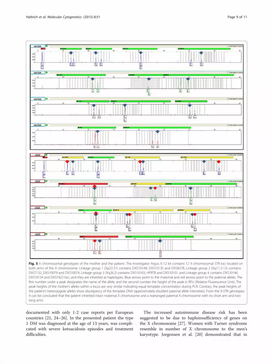
gin of the rearranged X chromosome: the alleles inheritedfrom the mother to the patient are highlighted in redcolour and the other ones on the second X chromosomeare highlighted in black (Table 1). Alleles originated fromthe father to the patient are highlighted in blue (Fig. 5, redarrows). As seen in the linked loci DXS10148-DXS10135-DXS8378 and their alleles (26.1, 22 and 10 in the patient)on Xp22.31, these are in red colour indicating only onecopy of X chromosome short arm (see the lower peakheights and blue arrows in Fig. 5 as well) and are inheritedfrom the mother. It is interesting to see the genotype ofthe maternal half sibling of the patient that the boy inher-ited a recombinant X chromosome from the mothershowing that a recombination occurred between Linkagegroup 1 (Xp22.31) and Linkage group 2 (Xq11.2-12) be-tween two the maternal X chromosomes (his electrophe-rogram is not included in Fig. 5). In other words, the sameXq arm of the mother in the case of the patient (red
Fig. 2 Southern blot analysis of the patient and of control females. EcoRI and EagI double digested DNA samples hybridized with radioactive-labelledStb12.3 probe were used for Southern blot analysis. Black arrows indicated the 2.8 kb unmethylated and the 5.2 kb methylated fragments. For theindex patient (red arrow) we can define two copies of 5.2 kb methylated fragments and one copy of 2.8 kb unmethylated fragment. For all controlswe identified two normal alleles (2,8 kb unmethylated and 5.2 kb methylated) characteristic for two normal X chromosomes
Haltrich et al. Molecular Cytogenetics (2015) 8:51 Page 5 of 11
alleles) and the other Xp arm of the maternal X chro-mosome (black alleles 24.1, 19 and 12) are transmitted tothe half sibling. Furthermore, it can be seen that a recom-bination never occurs within a closely linked loci as wesee 3 black alleles on Xp22.31 as described above in thehalf sibling.The administered WAIS–IV psychological test identi-
fied a borderline score for general intellectual ability(FSIQ: 70, percentage rank: 2). The verbal concept forma-tion, comprehension and expression as well as wordknowledge, were significantly below average with VCI: 58and 0,3 percentage rank. The perceptual reasoning ability,visual perception and organization, spatial visualizationand manipulation, and the ability to analyze andsynthesize abstract visual stimuli were at lower averagethan her age correspondent average score (PRI: 79, per-centage rank: 8). She achieved a borderline score (WMI:80; percentage rank: 9) in the field of working and short-term memory, memory span, attention and concentration.Her measured short –term visual memory and perceptionwas also borderline (PSI: 84; percentage rank: 2).
DiscussionHere we report the case of a young female patient with aunique X chromosome complex rearrangement and exhi-biting sequentially a wide range of autoimmune diseases.Initially, G-banding and X-specific FISH examination wereperformed, but the complexity of these results and thevaried clinical findings prompted us to carry out the high
resolution array-CGH as well. However, the rearranged Xof the presented case was missing the typical two “mirror– image” specific for i(Xq), the Xq arms were present inthree copies, except for 1,6 Mb from band q27.3 whichcould be considered as breakpoint related loss. On theother hand, the rearranged X - harbouring two centro-meres, inversions and several breakpoints - was muchmore complex as a common i(Xq) chromosome. In orderto determine the parental origin of the rearranged X, weanalyzed the X specific STR markers of the patient, hermother and half brother. Males carry one X-chromo-some, thus they transmit their X chromosome to daugh-ters as haplotypes. Females carry two X chromosomeswhich are liable to recombination during meiosis andtransmit one of their X chromosomes to childrenregardless of genders. Population genetic study for 12X-STR loci and X chromosomal recombination study inthree-generation families in Hungary, were reported be-fore [10, 11]. Due to extremely polymorphic X-STR lociand low haplotype frequencies of the linked loci, wewere able to solve this question.In the present study we tested 12 X-STRs on the entire
length of X chromosome within 4 linkage groups toidentify the origin of the affected X chromosome in thepatient. Based on the study we have clearly identifiednot only the origin of the affected and non-affected Xchromosome but also gained information on the copynumber of the X chromosomal short and long arms evenif the father was not available for testing. As we
Fig. 3 Repeat-Primed PCR analysis. Repeat-primed PCR analysis revealed three peaks corresponding to a 19, 29 and 31CGG repeat representingthree FMR1 gene alleles (red arrow). The method is also suitable for detection of AGG sequences interrupting the CGG repeats. In the indexpatient we determined two interruptions (red arrow). The AGG repeats stabilize the CGG repeats containing sequences
Haltrich et al. Molecular Cytogenetics (2015) 8:51 Page 6 of 11

described in Results (Table 1 and Fig. 5) on the basis ofthe peak heights of the mother’s and patient’s alleleswithin a locus and the X-STR genotypes detected it canbe concluded that the patient inherited an intact mater-nal X chromosome and a rearranged paternal X chromo-some with no short arm and two long arms.Recent studies [12] elucidated that most of the isodi-
centric X chromosomes [idic(Xq)] result from non-allelichomologous recombination between palindromic low copyrepeats and highly homologous palindromic LINE ele-ments. We could speculate that during the paternal meiosisthe preformed i(Xq) chromosome could have undergoneseveral breakage due to a catastrophic mutagen effect (se-vere alcohol abuse?) and this event was followed by wrongreunion route. The distal position of the two centromeresand the location of the XIST gene signals as well as thebanding pattern suggest that in our case the dicentric Xchromosome was initiated by centromere misdivision,multiple double-strand breakage, inversion of centromeric-Xq27.3 segment followed by a fortuitously recombinationprocess (Fig. 1e). The fact that other tissues as urine orbuccal epithelial cells did not show any signs of mosaicismsuggests that this aberrated X chromosome was generatedat early stage of meiosis during spermatogenesis.
Cytogenetically idic(Xq) chromosome cases had break-points in euchromatic sequences within a 7 Mb region ofproximal Xp arm and monocentric i(Xq) within centro-meric heterochromatin. On the bases of the array-CGHresults the breakpoint in our case was localized to theDXZ1 array junction which marks the beginning of thefunctional centromere and corresponds to the knownmonocentric i(Xq) breakpoint [12].To date, only seven previous reports of complex X
chromosomal rearrangements have been described [1–7].To the best of our knowledge this is the first complex Xchromosome rearrangement reported case associated withtype 1 DM and other four autoimmune diseases.Loss of genetic material from one of the sex chro-
mosomes causes the distinct clinical phenotype of TS:reduced adult height, gonadal dysgenesis, variable dys-morphic features. (low posterior hairline, webbed neck,broad chest, short fourth metacarpals). Congenitalheart defects, structural renal anomalies, hearing con-cerns, autoimmune diseases and cognitive deficits withparticular difficulties in visuo-spatial and memory areasare common in TS patients.The presented patient in our study has lack of the
typical Turner phenotype. She is 154 cm but her short
Fig. 4 Detailed analysis of the rearranged X chromosome using NimbleGene Array (assembly GRCh37/hg19). a The ideogram delineates genomicregions with the cytogenetic bands on normal X chromosome. b Panel “b” reveals the result of the array-CGH genotype. The CGX ISCA plus arrayshowed a 58.494 Mb loss (ChrXp22.33-p11.; one copy) and an 80.370 Mb three copies gain (ChrXq11.1-q27.3). Furthermore, there is a 1.6 Mb normaltwo copies region (ChrXq27.3) and an 11.1 Mb three copies gain (ChrXq27.3-q28). The red arrows represent the breakpoints. c Panel “c” indicatesgenomic regions with cytogenetic bands on the complex rearranged X chromosome of the patient. d Panel “d” represents genes and positions whichare affected in this patient. The blue rectangle indicates those genes which can be associated with immunodeficiency and the red rectangle representthe genes involved in POF phenotype. The red arrows represent the breakpoints
Haltrich et al. Molecular Cytogenetics (2015) 8:51 Page 7 of 11
height is not obvious because she has close relatives withsimilar height; dysmorphic features are absent, she israther an attractive young woman. Turner diagnosis wassuggested by POF, the concurrent health concerns suchas severe hearing loss, type 1 DM, hypothyreosis,anaemia perniciosa, celiac disease; severe hair loss(alopecia areata) and repeated severe infections werethe main health problems.TS patients usually have a typical neurocognitive pro-
file characterized by average to low-average full-scaleintelligence quotient (IQ). Intellectual disability isdefined as having an IQ score below 70; in our case theadministered WAIS-IV intelligence test identified aborderline score for the full-scale IQ (FSIQ: 70) andborderline score for the three supplemental subsetsindices (PRI, WMI, PSI); furthermore her “verbalintelligence” was well below average (VCI: 58).POF is defined as a primary ovarian defect charac-
terized by amenorrhea, low levels of gonadal andhigh levels of gonadotropin hormones. Deletions onthe X chromosome reduce both fertility and repro-ductive lifespan. Two loci on Xq have been postu-lated as the critical regions for POF: POF1 onXq27.3-q28 (OMIM #311360) and POF2 (POF2B onXq21.1q21.2; OMIM #300604 and POF2A onXq21.33; OMIM # 300511) [13, 14].
The breakpoints of the rearranged X chromosome pre-sented were adjacent to the POF critical regions. Thesefindings suggest that the breakpoint related lesions couldbe responsible for the patient’s POF. We assume that atthe Xq21.2 breakpoint two genes, POF1B and DACH2could be affected. These genes are located 700 kb apart,proximal to the centromere; POF1B is expressed duringearly ovarian development and DACH2 during earlyovarian follicular differentiation process. The interrup-tion of both genes by X-autosomal translocations wasdescribed in many patients with POF and Xq21.2 hasbeen postulated as critical region for infertility andreproductive lifespan. Furthermore, we presume that theother gene in question in the Xq21.33 breakpoint isDIAPH2 [NM_007309]. The product of this genebelongs to the diaphanous subfamily of the forminhomology family of proteins. This gene may play a rolein the development and normal function of the ovaries.Defects in this gene have been linked also to prematureovarian failure 2 (POF2).The presented patient suffered from severe bilateral
hearing loss which needed a hearing aid from six year ofage. Otologic disease is an important characteristic inTurner syndrome [15]. The associated hearing im-pairment has been described as both conductive andsensorineural, signifying both middle and inner earinvolvement. Recent studies suggested that there is acorrelation between frequency and severity of hearingproblems and karyotype: a higher occurrence of hearingproblems and an age-related deterioration of hearingthresholds is expected in children with X monosomy ori(Xq) compared to those with a mosaicism or otherstructural anomaly [16]. Based on these findings, the Xchromosome p arm loss may have some influence onthe development of hearing loss phenotype [17].Several studies reported an increased risk of auto-
immune diseases among women with Turner syndrome,most notably Hashimoto’s thyroiditis, diabetes mellitus,celiac disease, ulcerative colitis, Crohn’s disease, psoria-sis, idiopathic thrombocytopenic purpura, vitiligo andjuvenile rheumatoid arthritis [18–22]. Recent studiesdescribe a 4 to11-fold increased risk of -usually mild-diabetes in Turner syndrome patients compared tonormal females, with typically age of onset in the 30s or40s. They explain this high prevalence with the impairedglucose homeostasis in TS patients and with their higherglycemic response to oral glucose load and significantlylower insulin secretory response [19]. We need to pointout that despite of Danish registry study [23] whichfound an 11-fold increase in type 1 DM among TSpatients, TS patients seem to have an increased inci-dence of adult-onset, type 2 DM related to obesity and/or insulin resistance. Based on the literature search theassociation of TS and type 1 DM is very rare,
Table 1 X-chromosomal genotypes of the affected family
Haltrich et al. Molecular Cytogenetics (2015) 8:51 Page 8 of 11
documented with only 1-2 case reports per Europeancountries [21, 24–26]. In the presented patient the type1 DM was diagnosed at the age of 13 years, was compli-cated with severe ketoacidosis episodes and treatmentdifficulties.
The increased autoimmune disease risk has beensuggested to be due to haploinsufficiency of genes onthe X chromosome [27]. Women with Turner syndromeresemble in number of X chromosome to the men’skaryotype. Jorgensen et al. [20] demonstrated that in
Fig. 5 X chromosomal genotypes of the mother and the patient. The Investigator Argus X-12 kit contains 12 X-chromosomal STR loci located onboth arms of the X chromosome. Linkage group 1 (Xp22.31) contains DXS10148, DXS10135 and DXS8378, Linkage group 2 (Xq11.2-12) containsDXS7132, DXS10079 and DXS10074, Linkage group 3 (Xq26.2) contains DXS10103, HPRTB and DXS10101, and Linkage group 4 contains DXS10146,DXS10134 and DXS7423 loci, and they are inherited as haplotypes. Blue arrows point to the maternal and red arrows point to the paternal alleles. Thefirst number under a peak designates the name of the allele, and the second number the height of the peak in RFU (Relative Fluorescence Unit). Thepeak heights of the mother’s alleles within a locus are very similar indicating equal template concentration during PCR. Contrary, the peak heights ofthe patient’s heterozygote alleles show discrepancy of the template DNA (approximately doubled paternal allele intensities). From the X-STR genotypesit can be concluded that the patient inherited intact maternal X chromosome and a rearranged paternal X chromosome with no short arm and twolong arms
Haltrich et al. Molecular Cytogenetics (2015) 8:51 Page 9 of 11

Turner syndrome patients the standardized incidenceratios for autoimmune diseases with male predominanceare most pronounced, more than twice higher thanfemale predominant autoimmune diseases.The X chromosome contains the largest number of
immune-related genes of the human genome. The X-linked genes dosage is critical for maintenance or theloss of the immune tolerance. Immune related cellscannot be tolerated by self-antigens encoded by Xchromosome, thus triggering an autoimmune responsein target tissues [28].Regarding the putative influence of the type of X
chromosome rearrangement on clinical features, somestudies demonstrated a stronger association betweenautoimmune diseases and trisomy for Xq [19, 22].Jorgensen et al. [20] have been identified 3-fold in-creased risk for female- and 5-fold respectively formale–predominant autoimmune diseases in women withan i(Xq) karyotype. Particularly strong association wasfound between i(X) karyotype with ulcerative colitis,which was detected also in the presented patient. Hamzaet al. [22] reported a higher prevalence of autoimmunethyroiditis in Turner patients with i(Xq) than with 45,Xkaryotype. Bakalov et al. [19] demonstrated that haploin-sufficiency for Xp causes an increased risk for diabetes,and that the rate of DM is significantly higher (~43 %)in the i(Xq) patients. Comparing gene expression profileof 45,X and 46,X,i(Xq), they explained this higher risk byoverexpression of Xq genes that may provide a link to alarge number of autosomal genes involved in pancreaticislets and B-cell function. The Xq genes transcripts re-sult an increased IGF2, CRP and GAD level which sus-tain a proinflammatory state and low-grade chronicautoimmunity in individuals [19, 29]. Current studieshave been indicating a strong association of X –linkedgenes with autoimmune diseases risk and also the evi-dence of sex-specific effect of each of these genes in thesame disease. Several of these genes have an immune-related function or interact with autosomal genes inimmune-related pathways or are involved in regulationof apoptosis with role in autoimmune diseases [28, 30].Another aspect of the idic(Xq) chromosome is the pos-
sible perturbation of the random X inactivation throughwhich the gene dosage between male (XY) and female(XX) sexes are balanced. Inactive X chromosome ischaracterized by XIST RNA coating, condensation ofchromatin, methylation of CpG islands, post-translationalmodifications of histones. We determined the methylationstatus of the polymorphic FMR1 gene locus and we foundunequal methylated and unmethylated fragments sizeswhich refer to a skewed X chromosome inactivation.Skewed X chromosome inactivations have been describedin females with major X chromosome rearrangements[31]. The detected methylation shift could result in our
case a local escape from inactivation of genes which con-tributed also to the abnormal phenotype of the patient.
ConclusionsIn conclusion, the clinical data of the presented patientsuggest that fragmentation of the i(Xq) chromosome com-pounds the risk of autoimmune diseases. The skewed Xinactivation and perturbation of XIST-mediated silencingby the double XIST region of complex rearranged Xchromosome could be an important contributing mechan-ism to autoimmune disorders of the patient. Another fac-tor such as altered gene expression due to their disruptionor proximity to the breakpoints, transformed position ef-fect of genes caused by inversions and deletions could alsocontribute to development of multiorgan autoimmune syn-drome of the patient. Our case suggests that array-basedanalysis and X-inactivation studies might be useful comple-mentary clinical investigations to reveal the aetiology ofprimary amenorrhea and autoimmune diseases.
ConsentWritten informed consent was obtained from the patientfor publication of this case report, and accompanyingimages. A copy of the written consent is avaible by theEditor-in –chief of this journal.
AbbreviationsBp: Base pair(s); CGH: Comparative genomic hybridisation; DACH2: Homosapiens dachshund homolog 2 transcript variant 3 mRNA; DIAPH2: Homosapiens diaphanous-related formin 2 transcript variant 156, mRNA;DM: Diabetes mellitus; FMR1 and FMR2: Fragile X mental retardation 1 and 2genes; FRAXA: Fragile Site, Folic Acid Type, Rare, Fra(X)(q27.3) A;idic(Xq): Isodicentric chromosome of the long arm X; i(Xq): Isochromosomeof the long arm X; LG: Linkage Group; Mb: Megabase(s); POF: Prematureovarian failure; POF1B: Homo sapiens premature ovarian failure1B mRNA; RPPCR: Repeat primed polymerase chain reaction; SHOX: Homo sapiens shortstature homeobox transcript variant 1; SRY: Sex Determining Region Y; STR: ShortTandem Repeat; TS: Turner syndrome; WCP: Whole chromosome painting probe;XIST: Homo sapiens X inactive specific transcript non-coding RNA.
Competing interestsThe authors declare that they have no competing interests.
Authors’ contributionsIH and HP contributed equal to the design and coordination of this study aswell as to draft the manuscript. HP and AV performed X-STR genotypesanalysis of the family. GyF, AS, AB, ZG cared for the patient. IH and EKperformed cytogenetic and FISH examination. HP and VK performed thearray CGH, FRAXA Southern blot and FMR1 RP analyses. DD and GyF revisedthe manuscript critically for professional content. All authors read andapproved the final manuscript.
AcknowledgementsWe are grateful to Gergev Gyurgyinka, neuropsychologist in SemmelweisUniversity, 2nd Department of Paediatrics in Budapest, who completed andevaluated the WAIS-IV intelligence test. The authors wish to thank the patientand the mother who participated in this study.
Author details12nd Department of Pediatrics, Semmelweis University, Tűzoltó utca 7-9,1094 Budapest, Hungary. 2Department of Molecular Genetics andDiagnostics, National Center of Public Health, Budapest, Hungary. 3DNALaboratory, Institute of Forensic Medicine, Network of Forensic Science
Haltrich et al. Molecular Cytogenetics (2015) 8:51 Page 10 of 11
Institutes, Budapest, Hungary. 42nd Department of Medicine, SemmelweisUniversity, Budapest, Hungary. 5Department of Human Genetics,Organization National Institute of Health Dr Ricardo Jorge, Lisbon, Portugal.61st Department of Obstetrics and Gynecology, Semmelweis University,Budapest, Hungary.
Received: 8 April 2015 Accepted: 23 June 2015
References1. Grass FS, Schwartz RP, Deal JO, Parke Jr JC. Gonadal dysgenesis, intra-X
chromosome insertion, and possible position effect in an otherwise normalfemale. Clin Genet. 1981;20:28–35.
2. Letteire GS. Unique unbalanced X;X translocation (Xq22; p11.2) in a womanwith primary amenorrhea but without Ullrich-Turner syndrome. Am J MedGenet. 1995;59:414–6.
3. Hernando C, Plaja A, Catala V, Sarret EJ, Egozcue J, Fuster C. Primaryamenorrhea in a woman with a cryptic complex chromosomerearrangement involving the critical regions Xp11.2 and Xq24. Fertil Steril.2004;82:1666–71.
4. Ochalski ME, Engle N, Wakim A, Ravnan BJ, Hoffner L, Rajkovic A, et al.Complex X chromosome rearrangement delineated by array comparativegenome hybridization in a woman with premature ovarian insufficiency.Fertil Steril. 2011;95:2433. e9-2433.e15.
5. Auger J, Bonnet C, Valduga M, Philippe C, Bertolo-Houriez E, Beri-DexheimerM, et al. De novo complex X chromosome rearrangement unmaskingmaternally inherited CSF2RA deletion in a girl with pulmonary alveolarproteinosis. Am J Med Genet A. 2013;161A:2594–9.
6. Shchelochkov OA, Cooper ML, Ou Z, Peacock S, Yatsenko SA, Brown CW,et al. Mosaicism for r(X) and der(X)del(X)(p11.23)dup(X)(p11.21p11.22)provides insight into the possible mechanism of rearrangement. MolCytogenet. 2008;1:16. doi:10.1186/1755-8166-1-16. 1:16.
7. Kim MK, Seok HH, Kim YS, Chin MU, Sung SR, Lee WS, et al. Moleculargenetic and cytogenetic characterization of a partial Xp duplication and Xqdeletion in a patient with premature ovarian failure. Gene. 2014;534:54–9.
8. Rousseau F, Labelle Y, Bussières J, Lindsay C. The fragile X mentalretardation syndrome 20 years after the FMR1 gene discovery: anexpanding universe of knowledge. Clin Biochem Rev. 2011;32:135–62.
9. Wechsler D. Wechsler Adult Intellignece Scale: Technical and InterpretiveManual. 4th ed. San Antonio, TX, USA: Pearson Education, Inc; 2008.
10. Horváth G, Zalán A, Kis Z, Pamjav H. A genetic study of 12 X-STR lociin the Hungarian population. Forensic Sci Int Genet. 2012;6:e46–7.doi:10.1016/j.fsigen.2011.03.007.
11. Pamjav H, Kugler R, Zalán A, Völgyi A, Straky Z, Endrédy P, et al. Xchromosomal recombination study in three-generation families in Hungary.Forensic Sci Int Genet. 2012;6:e95–6. doi:10.1016/j.fsigen.2011.08.009.
12. Koumbaris G, Hatzisevastou-Loukidou H, Alexandrou A, Ioannides M,Christodoulou C, Fitzgerald T, et al. FoSTeS, MMBIR and NAHR at the humanproximal Xp region and the mechanisms of human Xq isochromosomeformation. Hum Mol Genet. 2011;20:1925–36.
13. Powell CM, Taggart RT, Drumheller TC, Wangsa D, Qian C, Nelson LM, et al.Molecular and cytogenetic studies of an X;autosome translocation in apatient with premature ovarian failure and review of the literature.Am J Med Genet. 1994;52:19–26.
14. Eggermann T, Meschede D, Schüler H, Palm S, Gläser D, Horsthemke B, et al.Premature ovarian failure associated with a small terminal Xq deletion:narrowing the POF1 region down to Xq27.2/Xq27.3-qter. Clin Genet.2005;67:434–7.
15. Gawron W, Wikiera B, Rostkowska-Nadolska B, Orendorz-Fraczkowska K,Noczyńska A. Evaluation of hearing organ in patients with Turner syndrome.Int J Pediatr Otorhinolaryngol. 2008;72:575–9.
16. Morimoto N, Tanaka T, Taiji H, Horikawa R, Naiki Y, Morimoto Y, et al.Hearing loss in Turner syndrome. J Pediatr. 2006;149:697–701.
17. Verver EJ, Freriks K, Thomeer HG, Huygen PL, Pennings RJ, der Velden AAA-v, et al. Ear and hearing problems in relation to karyotype in children withTurner syndrome. Hear Res. 2011;275:81–8.
18. Schoemaker MJ, Swerdlow AJ, Higgins CD, Wright AF, Jacobs PA, UnitedKingdom Clinical Cytogenetics Group. Mortality in women withTurnersyndrome in Great Britain: a national cohort study. J Clin Endocrinol Metab.2008;93:4735–42.
19. Bakalov VK, Cheng C, Zhou J, Bondy CA. X-chromosome gene dosage andthe risk of diabetes in Turner syndrome. J Clin Endocrinol Metab.2009;94:3289–96.
20. Jørgensen KT, Rostgaard K, Bache I, Biggar RJ, Nielsen NM, Tommerup N,et al. Autoimmune diseases in women with Turner's syndrome. ArthritisRheum. 2010;62:658–66.
21. Grossi A, Crinò A, Luciano R, Lombardo A, Cappa M, Fierabracci A.Endocrine autoimmunity in Turner syndrome. Ital J Pediatr. 2013;39:79.doi:10.1186/1824-7288-39-79.
22. Hamza RT, Raof NA, Abdallah KO. Prevalence of multiple forms ofautoimmunity in Egyptian patients with Turner syndrome: relation tokaryotype. J Pediatr Endocrinol Metab. 2013;26:545–50.
23. Gravholt CH, Juul S, Naeraa RW, Hansen J. Morbidity in Turner syndrome.J Clin Epidemiol. 1998;51:147–58.
24. Franzese A, De Filippo G, Argenziano A, Salerno MC. Turner syndrome andinsulin-dependent diabetes mellitus. Arch Pediatr. 1994;1:727–9.
25. Terano T, Fukuda K, Nakamura M, Takiguchi Y, Sakai Y, Hirai A. Diabeticketoacidosis associated with recurrent pulmonary edema andrhabdomyolysis in a patient with Turner's syndrome. Intern Med.2001;40:418–20.
26. Schmidt F, Kapellen TM, Wiegand S, Herbst A, Wolf J, Fröhlich-Reiterer EE,et al. Diabetes mellitus in children and adolescents with genetic syndromes.Exp Clin Endocrinol Diabetes. 2012;120:579–85.
27. Invernizzi P, Miozzo M, Selmi C, Persani L, Battezzati PM, Zuin M, et al. Xchromosome monosomy: a common mechanism for autoimmune diseases.J Immunol. 2005;175:575–8.
28. Bianchi I, Lleo A, Gershwin ME, Invernizzi P. The X chromosome andimmune associated genes. J Autoimmun. 2012;38:J187–92.
29. Bakalov VK, Gutin L, Cheng CM, Zhou J, Sheth P, Shah K, et al. Autoimmunedisorders in women with turner syndrome and women with karyotypicallynormal primary ovarian insufficiency. J Autoimmun. 2012;38:315–21.
30. Chang D, Gao F, Slavney A, Ma L, Waldman YY, Sams AJ, et al. Accountingfor eXentricities: analysis of the X chromosome in GWAS reveals X-linkedgenes implicated in autoimmune diseases. PLoS One. 2014;9:e113684.doi:10.1371/journal.pone.0113684.
31. Wong CC, Caspi A, Williams B, Houts R, Craig IW, Mill J. A longitudinal twinstudy of skewed X chromosome-inactivation. PLoS One. 2011;6:e17873.doi:10.1371/journal.pone.0017873.
Submit your next manuscript to BioMed Centraland take full advantage of:
• Convenient online submission
• Thorough peer review
• No space constraints or color figure charges
• Immediate publication on acceptance
• Inclusion in PubMed, CAS, Scopus and Google Scholar
• Research which is freely available for redistribution
Submit your manuscript at www.biomedcentral.com/submit
Haltrich et al. Molecular Cytogenetics (2015) 8:51 Page 11 of 11