accelerated central nervous system autoimmunity in baff-receptor-deficient mice
TRANSCRIPT

This article appeared in a journal published by Elsevier. The attachedcopy is furnished to the author for internal non-commercial researchand education use, including for instruction at the authors institution
and sharing with colleagues.
Other uses, including reproduction and distribution, or selling orlicensing copies, or posting to personal, institutional or third party
websites are prohibited.
In most cases authors are permitted to post their version of thearticle (e.g. in Word or Tex form) to their personal website orinstitutional repository. Authors requiring further information
regarding Elsevier’s archiving and manuscript policies areencouraged to visit:
http://www.elsevier.com/copyright

Author's personal copy
Accelerated central nervous system autoimmunity in BAFF-receptor-deficient mice
Susan S. Kim a,⁎, David P. Richman a, Scott S. Zamvil b,⁎⁎, Mark A. Agius a,c
a Department of Neurology and Neuroscience Graduate Program, University of California Davis, Davis, CA, United Statesb Department of Neurology and Program in Immunology, University of California, San Francisco, San Francisco, CA, United Statesc Veterans Administration Northern California Health Care System, Mather, CA, United States
a b s t r a c ta r t i c l e i n f o
Article history:Received 16 December 2010Received in revised form 2 April 2011Accepted 4 April 2011Available online 6 May 2011
Keywords:B-cell activating factorBAFF-receptorExperimental autoimmune encephalomyelitisMultiple sclerosisCentral nervous system inflammationMacrophagesT-cells
B cell activating factor (BAFF) is critical for B cell survival, a function that ismediated by BAFF receptor, (BAFF-R).The roleofBAFF (or BAFF-R) in themultiple sclerosismodel, experimental autoimmuneencephalomyelitis (EAE),was examinedusing BAFF-R-deficientmice. BAFF-Rdeficiency resulted in paradoxically increased severity of EAEinduced by myelin-oligodendrocyte glycoprotein (MOG) peptide 35–55. Inflammatory foci in BAFF-R-deficientmice comprised increased numbers of activated macrophages expressing BAFF and correlated with increasedBAFF secretion. Thus, BAFF-R may be important in EAE pathogenesis, possibly by influencing macrophagefunction through a mechanism that involves modulation of BAFF expression.
Published by Elsevier B.V.
1. Introduction
Multiple sclerosis [1] is an immune-mediated disease of the centralnervous system (CNS) [2], in which inflammation is thought to beregulated by both T cells and B cells [3]. The significance of B cells inthe pathogenesis of MS is highlighted by the benefit of B cell depletiontherapy with intravenous rituximab inMS patients. However, a recentphase II clinical trial of B cell-activating factor (BAFF) blockade withAtacicept, a recombinant TACI Ig fusion protein, led to worseningdisease activity in MS patients [4]. It is possible that the deleteriousresults of systemic anti-BAFF therapy may have derived from effectsnot just on the survival of B cells, which is mediated through B cellactivating factor receptor (BAFF-R) [5], but also on the regulation ofT cell, B cell and macrophage function, which may be mediated byother BAFF receptors including transmembrane activator calcium-modulator and cyclophilin ligand interactor (TACI), and B cellmaturation antigen (BCMA) [6]. Whereas BAFF is primarily expressedby myeloid cells and radioresistant stromal cells in the lymphoidorgans and spleen [7,8], BAFF receptors are expressed in B cells,subsets of T cells andmacrophages [9–12]. In particular, macrophages,
which are a major source of inducible BAFF, may become activatedupon stimulation with BAFF via BAFF-R, TACI or BCMA [11,12].
The current investigation addresses whether BAFF plays a role inregulating B cells, T cells and macrophages in the development of theT cell-dependent model of EAE in which Th17 activity and IL-23 arerequired [13]. BAFF-R-deficientmice,whichexpress noBAFF-R resultingin significantly reduced numbers of mature B cells [9], were used toexamine the effects of BAFF-R deficiency on susceptibility to EAE. Theresults demonstrate that, paradoxically, BAFF-R-deficient mice devel-oped an earlier onset and increased severity of p35–55-induced EAE,with more widespread inflammation in the spinal cord. The inflamma-tory foci in the CNS consisted of increased numbers of activatedmacrophages, many of which expressed BAFF, which correlated withincreased systemic BAFF secretion. In addition, there was a similarincrease in peripheral pro-inflammatory cytokines in BAFF-R-deficientmice with EAE. These findings suggest that BAFF-R-deficient micerespond to the lack of the receptor BAFF-R and relative B cell deficiencyby generating increased levels of BAFF, which may act through TACI orBCMA to promote pro-inflammatory T cell and macrophage responsesthat facilitate the development of CNS autoimmunity.
2. Materials and methods
2.1. Peptide
Rodent myelin oligodendrocyte glycoprotein (MOG) peptide 35–55(MEVGWYRSPFSRVVHLYRNGK) was synthesized and reverse-phasechromatographically purified by New England Peptide (Gardner, MA).
Journal of the Neurological Sciences 306 (2011) 9–15
⁎ Correspondence to: S. Kim, 1515 Newton Court, Room 510, Davis, CA 95618,United States. Tel.: +1 530 754 5013; fax: +1 530 754 5036.⁎⁎ Correspondence to: S. Zamvil, Department of Neurology, University of California,
San Francisco, 513 Parnassus Avenue, S-268, San Francisco, CA 94143, United States.Tel.: +1 415 502 7395; fax: +1 415 502 8512.
E-mail addresses: [email protected] (S.S. Kim), [email protected](S.S. Zamvil).
0022-510X/$ – see front matter. Published by Elsevier B.V.doi:10.1016/j.jns.2011.04.008
Contents lists available at ScienceDirect
Journal of the Neurological Sciences
j ourna l homepage: www.e lsev ie r.com/ locate / jns

Author's personal copy
2.2. Mice
Wild-type C57BL/6 mice were purchased from The JacksonLaboratory (JaxWest). Homozygous BAFF-R−/− (knockout) mice ona B6 background, which are deficient in BAFF-R, created using atargeting vector to produce a floxed BAFF-R allele by inserting lox Psites flanking exon 3 and 4 and a neomycin resistance cassette flankedby two FRT sites [9], were obtained from Yoshiteru Sasaki, (CBRInstitute, Harvard University, Boston, MA). BAFF-R deficient mice usedin the present study are distinct from A/WySnJ mice which have amutated BAFF-R but are still able to bind BAFF, and are characterizedby B cell maturational defects [14,15].
2.3. EAE induction and clinical analysis
Eight to 12 week old female C57BL/6, and BAFF-R KO mice wereadministered 300 μg of MOG peptide in 0.1 mL PBS emulsified in0.1 mL complete Freund's adjuvant (CFA) containing 4 mg/mL heat-inactivated Mycobacterium tuberculosis H37RA (Difco, Detroit, MI) viasubcutaneous flank injection on day 0, followed by 200 ng ofBordetella pertussis toxin (List Biological Laboratories) in 0.2 mL ofPBS administered intraperitoneally on day 0 and 48 h post immuni-zation. The mice were weighed and observed daily. Clinical scoring ofmotor weakness was graded on a 0–5 point scale as follows [16]:0=no clinical disease, 1=decreased tail tone, 2=mild monoparesisor paraparesis, 3=severe paraparesis, 4=paraplegia and or quad-raparesis, and 5=moribund or death. All experiments were performedin accordance with Institutional Animal Care and Use Committee(IACUC) guidelines at the University of California, Davis.
2.4. Histology
At 21 or 43 days post-immunization with MOG p35–55, BAFF-RKO and wild-type B6 mice were anesthetized by intraperitonealinjection of ketamine/xylazine prior to intracardiac perfusion withsaline followed by phosphate buffered 4% (w/v) paraformalde-hyde. Spinal cords and brains were removed and post-fixed atroom temperature with phosphate buffered 4% paraformaldehydefor 1 h. Spinal cord sections were transected in the mid-lumbar andcervico-thoracic regions, and brain sections were transected mid-sagitally. Tissue sections were processed for paraffin embedding andcryostat sectioning. Fixed brain and spinal cord tissues were embeddedin paraffin, sectioned into 6 μm sections, and stained with H&E toidentify inflammatory cells. Inflammatory foci (containing 10 or moreinflammatory cells per focus) were counted and areasmeasured (μm2),in the parenchyma and meninges of brain, lumbar and cervical spinalcord sections from eachmouse by an examiner in a blindedmanner. Forcryostat sectioning, fixed CNS tissues were cryoprotected by immersingtissues in 30% sucrose at 4 °C overnight, embedded in OCT cryostatmountingmedium (Sakura Finatek), frozen in liquid nitrogen cooled 2-methylbutane, sectioned (transversely for spinal cord, and sagitally forbrain) into 10 μm sections, and stained for immunofluorescence andconfocal microscopy.
2.5. Immunohistology
Primary antibodies used in this study include BAFF (rat anti-mousemAb, Abcam), activated macrophage/microglia marker IBA-1 (goatanti mouse polyclonal Ab, Abcam), CD3 (rabbit anti-mouse polyclonalAb, Abcam), and nuclear dye Hoechst 33258 (Invitrogen). Cryostatsections were immunostained with monoclonal and polyclonalprimary antibodies. Biotinylated secondary antibodies binding toantibodies against BAFF were detected with rhodamine-conjugatedstreptavidin (Jackson Immunoresearch Laboratories). FITC-labeledsecondary antibodies were used to detect IBA-1 and CD3 expression.Cryostat sections mounted on poly-L-lysine coated slides were
allowed to dry, and then pre-fixed with 0.5% paraformaldehyde,washed with phosphate buffered saline (PBS) pH 7.2, blocked with adonkey block solution containing 10% donkey serum (JacksonImmunoresearch Laboratories), MEM/HEPES (Invitrogen), 1% (v/v)penicillin (PCN)/strep (Invitrogen), and 0.05% (v/v) sodium azide for15 min at room temperature, and thenwashedwith PBS, blockedwithstrep–avidin and biotin (Vector Labs) blocking solutions, rinsed withPBS and incubated overnight with primary antibodies in donkey blocksolution at 4 °C. Cryostat sections were subsequently washed in PBS,incubated with secondary antibodies in donkey block solutioncontaining 10% donkey serum for 30 min at room temperature,then washed with PBS, incubated with strep–avidin rhodamine indonkey block solution for 15 min at room temperature, washed withPBS, and then were post-fixed with methanol at −20 °C, counter-stained with Hoechst nuclear stain (2 μg/mL in PBS) and mounted inVectashield.
2.6. Imaging and image analysis
H&E stained spinal cord sections were viewed with a Nikon E600(Tokyo, Japan) microscope and a 40× objective lens (Plan Apochro-mat, NA 0.95), and detected with a color CCD camera (LE-470,Optronics, Goleta, CA). Quantification of inflammatory foci was per-formed by counting the number of inflammatory foci and measuringareas of each focus using Stereoinvestigator (version 8, Micro BrightField, Inc., Williston, VT). Fluorescent images were captured byconfocal microscopy with a Zeiss LSM 510 META (Carl Zeiss,Germany) laser scanning confocal microscope and an oil DIC 63×objective lens (Plan Apochromat, NA 1.4). Three dimensional imagesof EAE spinal cord sections were generated by collecting a z-series ofconfocal images. To evaluate the proportion of macrophages amongHoechst positive cells in inflammatory foci, Volocity (version 4.0,Improvision Inc.) software was used to measure and analyze objectsin 3-dimensional images.
2.7. Splenocyte activation and cytokine analysis
Day 12 MOG peptide-immunized BAFF-R KO and wild-typecontrols were euthanized by CO2 inhalation. Spleens were removedfrom each group of mice, homogenized using a Dunce homogenizer,and filtered using a 100 μm mesh filter. Cell suspensions werecentrifuged for 10 min at 1000 ×g, washed in RPMI-5 containingRPMI 1640, 5% heat inactivated FBS, 10 mMHEPES, 1% (v/v) penicillin(PCN)/streptomycin (strep), 50 μM 2-mercaptoethanol. Cell pelletswere resuspended in RPMI-5, Ficoll Hypaque (Amersham PharmaciaBiotech) solution was layered under the media, and the suspensionwas centrifuged at 800 ×g for 15 min at room temperature. Cells werecollected from the Ficoll Hypaque interface, transferred to 50 mLconical tubes and washed in RPMI-5 media. Cell pellets wereresuspended in RPMI-10 media containing RPMI-1640, 10%heat inactivated FBS, 10 mM HEPES, 1% (v/v) PCN/strep, 50 μM 2-mercaptoethanol, and counted using a hemocytometer. Splenocytes(5×10⁵ cells/well) were cultured in flat-bottomed, 96-well plates inmedia (RPMI 1640 supplemented with 2 mM L-glutamine, 1 mMsodium pyruvate, 100 U mL−1 penicillin, 0.1 mg mL−1 streptomycin,0.5 μM2-mercaptoethanol and 10% FBS) in the presence of MOG p35–55 peptide (0, 10, 20, 50 μg mL−1) or media control. Cytokines weremeasured in the supernatants (at 48 h: IL-2, IL-6; 72 h: IFN-γ, TNF-α,BAFF; 96 h: IL-17; 120 h: IL-10) of cultured splenocytes using OPTEIAELISA sets (BD Pharmingen) or Quantikine ELISA KIT for BAFF (R&DSystems) as per protocol. Splenocytes were pooled from sevenanimals per group and wells were plated in triplicate. Data wereanalyzed using a SOFTmax ELISA software and were expressed asmeans±SEM of triplicate wells.
10 S.S. Kim et al. / Journal of the Neurological Sciences 306 (2011) 9–15
Author's personal copy
2.8. Statistical analysis
Data are shown as means±SEM. A t-test was used for parametricdata (for 2 groups) andMann–Whitney U statistics were used for non-parametric data (for 2 groups). Fisher's exact test was used todetermine whether differences in proportions were significant.P-valuesb0.05 were considered statistically significant.
3. Results
3.1. BAFF-R-deficient mice develop an earlier onset and increasedseverity of p35–55-induced EAE
Similar to BAFF-deficient mice, BAFF-R-deficient mice havereduced numbers of mature B cells [9]. Following immunization
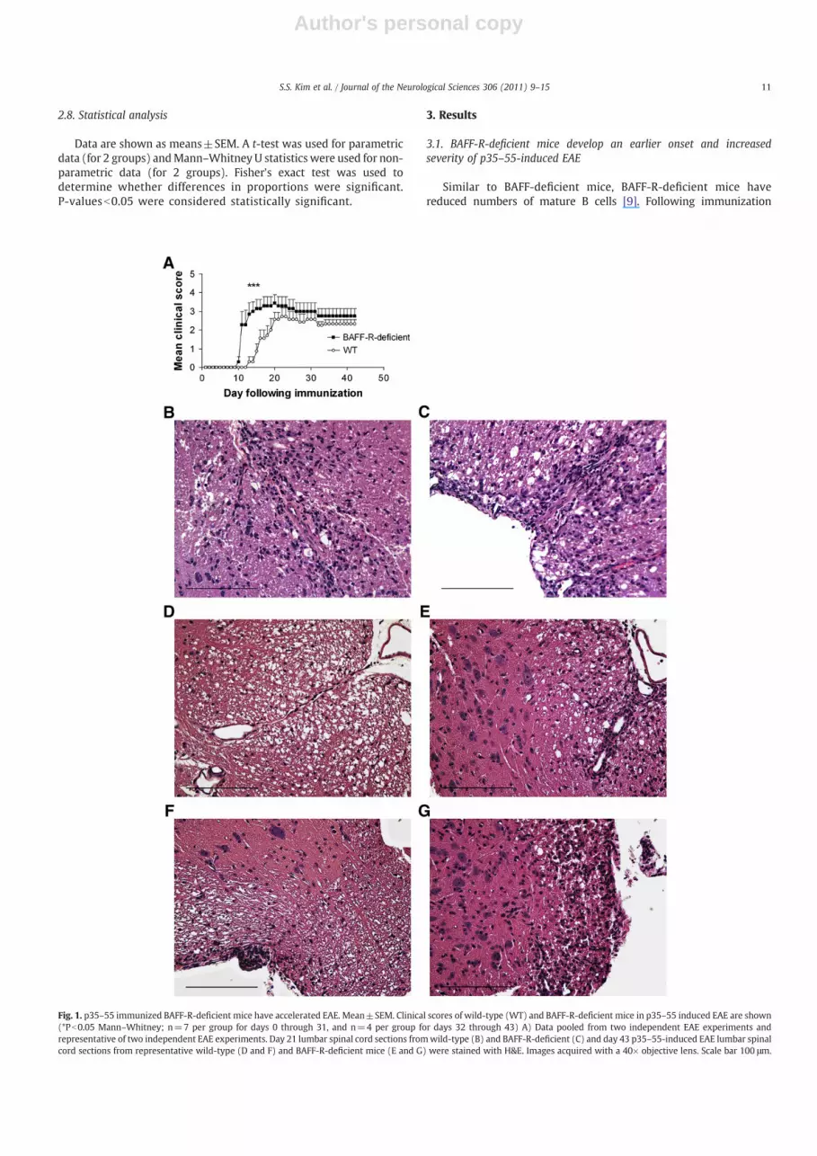
Fig. 1. p35–55 immunized BAFF-R-deficient mice have accelerated EAE. Mean±SEM. Clinical scores of wild-type (WT) and BAFF-R-deficient mice in p35–55 induced EAE are shown(*Pb0.05 Mann–Whitney; n=7 per group for days 0 through 31, and n=4 per group for days 32 through 43) A) Data pooled from two independent EAE experiments andrepresentative of two independent EAE experiments. Day 21 lumbar spinal cord sections fromwild-type (B) and BAFF-R-deficient (C) and day 43 p35–55-induced EAE lumbar spinalcord sections from representative wild-type (D and F) and BAFF-R-deficient mice (E and G) were stained with H&E. Images acquired with a 40× objective lens. Scale bar 100 μm.
11S.S. Kim et al. / Journal of the Neurological Sciences 306 (2011) 9–15

Author's personal copy
with p35–55, BAFF-R-deficient mice had an earlier onset of clinicaldisease and increased severity of acute EAE compared to wild-type[10] (Fig. 1A), (Table 1). Thus, BAFF-R expression is not required forp35–55-induced EAE and its absence results in more severe disease.
3.2. CNS inflammatory foci are increased in BAFF-R-deficient mice andhave an increased proportion of activated macrophages
The inflammatory foci were five-fold greater in area in the spinalcords of p35–55 immunized BAFF-R-deficient mice compared to p35–55 immunized wild-type controls during the chronic phase of EAE(43 days) (Fig. 1D–G, Table 2). In contrast, at 21 days, levels ofinflammation appeared similar (Fig. 1B and C). A possible explanationfor this may be that the histological measures used to gaugeinflammation, cross sectional area of and numbers of foci are notsufficiently sensitive to detect statistically significant differences earlyon in the course of EAE. To determine the cellular composition of thelesions, we examined histological sections with markers IBA-1 foractivated macrophages and CD3 for T cells. Compared with p35–55immunized wild-type controls, BAFF-R-deficient mice immunizedwith p35–55 had increased levels of IBA-1, which co-localized withcells expressing increased levels of BAFF in spinal cord infiltrates atboth 21 and 43 days (Fig. 2A–D). In contrast, while there was someincreased expression of the pan T-cell marker, (CD3) at 43 days inp35–55 immunized BAFF-R-deficient mice compared with p35–55immunized wild-type controls, it did not significantly colocalize withincreased BAFF expression. Furthermore, the increase in CD3expression was not as prominent as the elevation in IBA-1 expressionin p35–55 immunized BAFF-R-deficient mice (data not shown). Thus,the inflammatory foci in BAFF-R-deficient mice comprised largernumbers of activated macrophages many of which expressed BAFF.Moreover, BAFF-R deficiency resulted in a higher proportion ofactivated macrophages within inflammatory infiltrates comparedwith wild-type controls following p35–55 immunization (Fig. 2E),possibly via upregulation of other BAFF receptors in macrophages.
3.3. p35–55 immunization promotes increased pro-inflammatory T cellactivation in BAFF-R-deficient mice
Increased susceptibility to p35–55-induced EAE could also reflectchanges in T cell activation, Therefore, splenocyte culture superna-tants were assessed for Th1 cytokines (IL-2, IFN-γ and TNF-α), Th17cytokines (IL-17, IL-6), and Th2 cytokine (IL-10). BAFF-R-deficientmice immunized with p35–55 produced increased levels of IL-2, andIL-6 (Fig. 3A and B), as well as IFN-γ and TNF-α (Fig. 3C and D)
compared with similarly immunized wild-type controls. At the sametime, secretion of IL-10 was markedly reduced in BAFF-R-deficientmice (Fig. 3F). IL-17 production was similar for both groups (Fig. 3E).To address whether susceptibility to p35–55-induced EAE in BAFF-R-deficient mice could also reflect alterations in systemic BAFFregulation, splenocyte culture supernatants were examined for BAFFexpression. Splenocytes from BAFF-R-deficient mice produced two tofour-fold greater levels of secreted BAFF compared to wild-typeanimals, even when not stimulated by antigen (Fig. 3G).
4. Discussion
In a recent MS clinical trial of Atacicept, a recombinant TACI-Igfusion protein which binds BAFF and its associated ligand APRIL andfunctions to prevent BAFF ligand activation of its receptors, MSpatients developed worsening disease activity [4]. Our present studyof BAFF-R deficiency in which the absence of BAFF-R led to worseningCNS autoimmunity and inflammation may provide some insight intothe results of the Atacicept trial. BAFF is critical for the survival ofB cells [17]. It has been shown that BAFF-deficient mice havedecreased numbers of mature B cells and have impaired antibodyproduction [17]. BAFF-R-deficient mice have a phenotype similar toBAFF-deficient mice, showing reduced numbers of mature B cells anddisplaying impaired antigen dependent antibody responses [9]. Inparticular, BAFF-R-deficient mice have a significant decrease in thenumber of mature B2 cells and marginal zone B cells but have nosignificant reduction in the number of B1 cells, similar to BAFF-deficient mice, suggesting that the effect of BAFF on B cell survival ismediated through BAFF-R [9].
While it is evident that BAFF-R mediates the B cell survival effectsof BAFF, the results of the present study suggest that the increasedseverity of CNS inflammation observed in BAFF-R-deficient mice aremore likely to be attributable to the effects of BAFF on T cell regulationand changes in macrophage function than through the promotion ofB cell survival. In addition to their expression on B cells, BAFFreceptors are also expressed in T cells and macrophages, suggestingthat BAFFmay play a role in T cell regulation andmacrophage function[9–12]. In the model presented here, BAFF may exert its pro-inflammatory effects through the NF-Kappa B pathways [18,19] viaother BAFF receptors including TACI and BCMA. Increases in IL-6 andTNF-α in the setting of BAFF-R deficiency suggest increasedmacrophage activation possibly associated with elevated BAFFexpression.
Macrophages, which are a major producer of inducible BAFF,express all three BAFF receptors, BAFF-R, TACI and BCMA, in certain
Table 1Clinical and histological characteristics of EAE.
Incidence Mean day OF onseta Mean maximum disease scoreb Histological scorec,d
BAFF-R-deficient 7/7 11.4±0.4⁎ 3.4±0.43 (p=0.2994) 101±23×103 (n=29)⁎
C57BL/6 7/7 16.1±1.1 2.7±0.48 19.3±6.7×10³ (n=16)
a Mean day of disease onset of individual animals in each group±SEM.b Mean of maximum disease score of individual animals in each group±SEM.c Total area of parenchymal foci (μm2) in 43 day lumbar spinal cord samples.d n=number of foci.⁎ Pb0.001.
Table 2Comparison of inflammatory foci in 43 day lumbar spinal cord samples from wild-type and BAFF-R-deficient p35–55-induced EAE mice.
Parenchymal foci/section1 Meningeal foci/section2 Total area of parenchymalfoci3 (μm2)
Total area of meningealfoci4 (μm2)
BAFF-R-deficient 7±2 (n=5) 7.5±1.1 (n=5) 101±23×103 (n=29)⁎⁎ 17.8±4.4×10³ (n=30)⁎
Wild-type 5±1 (n=4) 3.5±2.3 (n=4) 19.3±6.7×10³ (n=16) 3.4±0.9×10³ (n=14)
Values are mean±SEM; 1, 2 n=number of sections; 3, 4 n=number of foci.⁎ Pb0.05.⁎⁎ Pb0.01.
12 S.S. Kim et al. / Journal of the Neurological Sciences 306 (2011) 9–15

Author's personal copy
contexts [11,12]. Furthermore, these receptors may be upregulated inthe presence of BAFF stimulation [11]. In the current study, BAFF-R-deficient mice were used to examine the effects of BAFF-R deficiencyon susceptibility to a T cell model of EAE. We have demonstrated thatBAFF-R is not required for EAE induced by immunizationwith p35–55.The observation that T cells from BAFF-R-deficient EAE mice, culturedwith p35–55, secrete increased levels of pro-inflammatory cytokinesimplies that priming is not impaired in BAFF-R-deficient mice, and ispossibly even enhanced. Consistent with the latter possibility is our
observation that BAFF-R-deficient mice exhibit an earlier onset andincreased severity of p35–55-induced EAE, along with increasedlevels of IBA-1 staining and increased proportions of IBA-1 positivemacrophageswithin inflammatory lesions at day 21, whichmay resultfrom increased macrophage activation via upregulation of other BAFFreceptors.
The observation that EAE ismore severe in BAFF-R-deficient animalssuggests that the absence of BAFF-R may be important in promotingEAE, possibly by influencing macrophage function. Increased levels of
Fig. 2. BAFF-R-deficient mice have increased levels of activated macrophages in CNS spinal cord inflammatory foci at 21 and 43 days following immunization. Three colorimmunofluorescence for IBA-1 (FITC-labeled), BAFF (rhodamine-labeled), andnuclei (Hoechst) shows elevated levels of activatedmacrophagemarker, IBA-1,which colocalizedwith BAFFexpression in inflammatory foci in day 21 p35–55-induced EAE lumbar spinal cord in BAFF-R-deficient (A) compared to p35–55 immunizedwild-type controls (B), andwhich colocalizedwith increased BAFF expression in inflammatory foci of 43 day p35–55-induced EAE spinal cord in BAFF-R-deficient (C) compared to p35–55 immunized wild-type controls (D). Imagesacquiredwith a 63× objective lens. Scale bar 50 μm(A and B) and 100 μm(C and D).Mean±SEM. Proportion of FITC-labeled IBA-1 cells amongHoechst positive cells in inflammatory fociof wild-type and BAFF-R-deficient EAE lumbar spinal cord sections are shown (E) (**Pb0.01, t-test, n=3 foci per group).
13S.S. Kim et al. / Journal of the Neurological Sciences 306 (2011) 9–15

Author's personal copy
BAFF in the CNS and expression of BAFF in-vitro in BAFF-R-deficientmice compared with p35–55 immunized wild-type controls suggestthat BAFF may contribute to chronic CNS inflammation and increasedCNS autoimmunity possibly through the activation of other BAFFreceptors in the absence of BAFF-R. A caveat is that the associationbetween systemic BAFF levels and EAE susceptibilitymaydependon thecontext of disease. For example, in the setting of intact BAFF-Rexpression, EAE susceptibility in SJL/J mice immunized with PLPp139–151 was associated with alterations in B cell homeostasis aswell as decreased serum BAFF levels at the peak of disease (14 days)while B10.Smice resistant to EAE inductionwere shown tohave anearlyincrease in serum BAFF levels at 7 days following immunization withPLP p139–151 but not at later time points, compared with PBS andadjuvant controls [20].
It is possible that BAFF-R can mediate its effects on macrophagefunction and T cell regulation in EAE independently of mature B cells.This may result in a bias toward pro-inflammatory cytokine responsesin p35–55 immunized BAFF-R-deficient mice comparedwith similarly
immunized wild-type controls. Alternatively, since B cells are criticalfor recovery in EAE, through an IL-10 dependent mechanism [21], thereduction in the number of B cells and the markedly reduced IL-10productionmay contribute to the impaired recovery in EAE in BAFF-R-deficientmice. In addition, the lack ofmature B cells may also promoteEAE induction by decreasing the proportion of CD4+CD25+FoxP3+
regulatory T cells, and by regulating macrophage and other antigenpresenting cell (APC) functions that enhance encephalitogenic pro-inflammatory cytokine responses [22]. Generating BAFF-R/B celldeficient (uMT) mice to determine the effects of combined BAFF-Rand B cell deficiencies on susceptibility to EAE may help to assessdirectly whether BAFF-R may affect T cell regulation and macrophagefunction independently of B cells.
Our observations appear to be relevant in explaining the unantic-ipated worsening of MS in patients treated with Atacicept. Systemicallydirected anti-BAFF therapy inMS such as in the Atacicept study [4] mayresult in worsening disease activity through various mechanisms.Treatment with Atacicept may have led to a paradoxically increased
Fig. 3. p35–55 immunization promotes increased splenocyte activation in BAFF-R-deficient mice. Secretion of IL-2, IL-6, IFN-γ, TNF-α, IL-17, IL-10 and BAFF from culturedsplenocytes (pg/mL), (A–G) derived from day 12 p35–55 immunized BAFF-R-deficient mice compared to p35–55 immunized wild-type controls. Cultured splenocytes werestimulated with p35–55 antigen (0, 10, 20 and 50 μg/mL). Cytokines were measured in the supernatants (at 48 h: IL-2, IL-6; 72 h: IFN-γ, TNF-α, BAFF; 96 h: IL-17; 120 h: IL-10) ofcultured splenocytes. Splenocytes were pooled from seven animals per group and wells were plated in triplicate. Data are representative of two independent experiments.
14 S.S. Kim et al. / Journal of the Neurological Sciences 306 (2011) 9–15
Author's personal copy
expression of BAFF systemically and in the CNS, aswell as an increase insystemic T-cell activation and enhanced pro-inflammatorymacrophagefunction, as is seen in the setting of BAFF-R deficiency in our study,resulting in theunexpectedworsening of CNS inflammationobserved inthose patients. In contrast, anti-BAFF therapies specifically targeting theCNS may play an important role in preventing the development ofchronic CNS inflammation and progression of disease in MS.
In summary, our findings indicate that BAFF-R is important in EAEpathogenesis. BAFF-R deficiency was associated with more severe EAE,elevated levels of BAFF, macrophage activation and greater myelin-reactive pro-inflammatory immune responses. Thepossibility that someof these effects may occur independently of B cells will require furtherinvestigation. Paradoxicalworseningof EAE through selective inhibitionof one member of BAFF receptors, suggests that activity of elevatedBAFF on other BAFF family receptors, may promote pro-inflammatorymyeloid and T cell responses in MS or other autoimmune diseases.
Acknowledgements
This study was supported by the University of California Davisresearch funds. We thank Timothy Vartanian (Weill Cornell MedicalCollege) for his helpful comments, TEVA Neuroscience for their kindsupport and Yoshiteru Sasaki (Harvard University) for providing theBAFF-R deficient mice. S.S.Z. was supported by grants from theNational Institute of Health (NIH) (RO1 AI073737, RO1 AI059709, andRO1 NS063008), the National Multiple Sclerosis Society (NMSS)(RG 3622 and 3913), Dana Foundation, Guthy Jackson CharitableFoundation and Maisin Foundation. M. Agius has received grantsupport from the NMSS, the California Myasthenia Gravis Foundationand an unrestricted research award from TEVA Neuroscience.
References
[1] Lyons JA, Ramsbottom MJ, Cross AH. Critical role of antigen-specific antibody inexperimental autoimmune encephalomyelitis induced by recombinant myelinoligodendrocyte glycoprotein. Eur J Immunol 2002 Jul;32(7):1905–13.
[2] Lassmann H, BruckW, Lucchinetti CF. The immunopathology of multiple sclerosis:an overview. Brain pathology (Zurich, Switzerland) 2007 Apr;17(2) 210–8.
[3] Weiner HL. Multiple sclerosis is an inflammatory T-cell-mediated autoimmunedisease. Arch Neurol 2004 Oct;61(10):1613–5.
[4] Rammohan KW, Shoemaker J. Emerging multiple sclerosis oral therapies.Neurology 2010 Jan 5;74(Suppl 1):S47–53.
[5] Thompson JS, Bixler SA, Qian F, Vora K, Scott ML, Cachero TG, et al. BAFF-R, a newlyidentified TNF receptor that specifically interacts with BAFF. Science 2001 Sep14;293(5537):2108–11.
[6] Gross JA, Johnston J, Mudri S, Enselman R, Dillon SR, Madden K, et al. TACI andBCMA are receptors for a TNF homologue implicated in B-cell autoimmunedisease. Nature 2000 Apr 27;404(6781):995–9.
[7] Nardelli B, Belvedere O, Roschke V, Moore PA, Olsen HS, Migone TS, et al. Synthesisand release of B-lymphocyte stimulator from myeloid cells. Blood 2001 Jan 1;97(1):198–204.
[8] Gorelik L, Gilbride K, Dobles M, Kalled SL, Zandman D, Scott ML. Normal B cellhomeostasis requires B cell activation factor production by radiation-resistantcells. J Exp Med 2003 Sep 15;198(6):937–45.
[9] Sasaki Y, Casola S, Kutok JL, Rajewsky K, Schmidt-Supprian M. TNF family memberB cell-activating factor (BAFF) receptor-dependent and -independent roles forBAFF in B cell physiology. J Immunol 2004 Aug 15;173(4):2245–52.
[10] Ng LG, Sutherland AP, Newton R, Qian F, Cachero TG, Scott ML, et al. B cell-activating factor belonging to the TNF family (BAFF)-R is the principal BAFFreceptor facilitating BAFF costimulation of circulating T and B cells. J Immunol2004 Jul 15;173(2):807–17.
[11] Chang SK, Arendt BK, Darce JR, Wu X, Jelinek DF. A role for BLyS in the activation ofinnate immune cells. Blood 2006 Oct 15;108(8):2687–94.
[12] Jeon ST, Kim WJ, Lee SM, Lee MY, Park SB, Lee SH, et al. Reverse signaling throughBAFF differentially regulates the expression of inflammatory mediators andcytoskeletal movements in THP-1 cells. Immunol Cell Biol 2010 Feb;88(2):148–56.
[13] Cua DJ, Sherlock J, Chen Y, Murphy CA, Joyce B, Seymour B, et al. Interleukin-23rather than interleukin-12 is the critical cytokine for autoimmune inflammation ofthe brain. Nature 2003 Feb 13;421(6924):744–8.
[14] Miller DJ, Hayes CE. Phenotypic and genetic characterization of a unique Blymphocyte deficiency in strain A/WySnJ mice. Eur J Immunol 1991 May;21(5):1123–30.
[15] Miller DJ, Hanson KD, Carman JA, Hayes CE. A single autosomal gene defectseverely limits IgG but not IgM responses in B lymphocyte-deficient A/WySnJmice. Eur J Immunol 1992 Feb;22(2):373–9.
[16] Weber MS, Prod'homme T, Youssef S, Dunn SE, Rundle CD, Lee L, et al. Type IImonocytes modulate T cell-mediated central nervous system autoimmunedisease. Nat Med 2007 Aug;13(8):935–43.
[17] Schiemann B, Gommerman JL, Vora K, Cachero TG, Shulga-Morskaya S, Dobles M,et al. An essential role for BAFF in the normal development of B cells through aBCMA-independent pathway. Science 2001 Sep 14;293(5537):2111–4.
[18] Sasaki Y, Derudder E, Hobeika E, Pelanda R, Reth M, Rajewsky K, et al. CanonicalNF-kappaB activity, dispensable for B cell development, replaces BAFF-receptorsignals and promotes B cell proliferation upon activation. Immunity 2006 Jun;24(6):729–39.
[19] Enzler T, Bonizzi G, Silverman GJ, Otero DC, Widhopf GF, Anzelon-Mills A, et al.Alternative and classical NF-kappa B signaling retain autoreactive B cells in thesplenic marginal zone and result in lupus-like disease. Immunity 2006 Sep;25(3):403–15.
[20] Lee-Chang C, Lefranc D, Salleron J, Faveeuw C, Allet C, Vermersch P, et al.Susceptibility to experimental autoimmune encephalomyelitis is associated withaltered B-cell subsets distribution and decreased serum BAFF levels. Immunol Lett2011 Mar 30;135(1–2):108–17.
[21] Fillatreau S, Sweenie CH, McGeachy MJ, Gray D, Anderton SM. B cells regulateautoimmunity by provision of IL-10. Nat Immunol 2002 Oct;3(10):944–50.
[22] Weber MS, Prod'homme T, Patarroyo JC, Molnarfi N, Karnezis T, Lehmann-HornK, et al. B-cell activation influences T-cell polarization and outcome of anti-CD20B-cell depletion in central nervous system autoimmunity. Ann Neurol 2010 Jul16.
15S.S. Kim et al. / Journal of the Neurological Sciences 306 (2011) 9–15