erg rearrangement is associated with prostate cancer
TRANSCRIPT

ERG Rearrangement Is Associated with Prostate Cancer-Related Death in Chinese Prostate Cancer PatientsMei Qi1., Xiaoqing Yang1., Fan Zhang2, Tao Lin3, Xiubin Sun4, Yanjiang Li5, Huiqing Yuan6, Yubo Ren7,
Juan Zhang8, Xiaomin Qin8, Bo Han1,8*
1 Department of Pathology, Shandong University Medical School, Jinan, China, 2 Department of Orthodontics, Shandong University School of Stomatology, Jinan, China,
3 Department of Surgery, The Central Hospital of Jinan, Jinan, China, 4 Department of Statistics, Shandong University School of Public Health, Jinan, China, 5 Department
of Urology, The Affiliated Hospital of Qingdao University Medical College, Qingdao, China, 6 Department of Biochemistry, Shandong University Medical School, Jinan,
China, 7 Department of Pathology, Liaocheng General Hospital, Liaocheng, China, 8 Department of Pathology, Shandong University Qilu hospital, Jinan, China
Abstract
Recently, ETS-related gene (ERG) gene rearrangements, phosphatase tensin homologue (PTEN) deletions and EGFR familyaberrations were characterized as potential biomarkers for prostate cancer (PCa) patient management. Although ERG generearrangement has been identified in approximately 50% of localized prostate cancers in western countries, the prognosticsignificance of this critical molecular event remains unknown in Chinese patients. Using fluorescence in situ hybridization(FISH) and immunohistochemistry, we evaluated ERG, PTEN and EGFR family aberrations in a cohort of 224 Chinese prostatecancer patients diagnosed in transurethral resection of the prostate (TUR-P). Overall, ERG rearrangement was detected in23.2% (44/190) cases, of which 54.5% (24/44) showed deletion of the 59end of ERG. PTEN deletion was identified in 10.8%(19/176) cases. Amplification of EGFR and HER2 genes was present in 1.1% (2/178) and 5.8% (10/173) of cases, respectively.Significant correlation between ERG rearrangement and PTEN deletion was identified in this cohort. EGFR and HER2aberrations occurred more frequently in PCas without ERG rearrangement than in those with ERG rearrangement, althoughthis did not reach statistical significance. Overall, ERG rearrangement was associated with pre-operative PSA values(P = 0.038) and cancer-related death (P = 0.02), but not with the age, clinical T stage, Gleason score, or Ki-67 labeling index(LI). Notably, multivariate analysis including known prognostic markers revealed ERG rearrangement was an independentprognostic factor (P = 0.022). Additionally, ERG rearrangement status was helpful to identify patients with poor prognosisfrom PCa group with low Ki-67 LI. In summary, we reported that ERG rearrangement was associated with cancer-relateddeath in Chinese PCa patients. Determination of ERG rearrangement status allows stratification of PCa patients into differentsurvival categories.
Citation: Qi M, Yang X, Zhang F, Lin T, Sun X, et al. (2014) ERG Rearrangement Is Associated with Prostate Cancer-Related Death in Chinese Prostate CancerPatients. PLoS ONE 9(2): e84959. doi:10.1371/journal.pone.0084959
Editor: Jindan Yu, Northwestern University, United States of America
Received August 15, 2013; Accepted November 20, 2013; Published February 7, 2014
Copyright: � 2014 Qi et al. This is an open-access article distributed under the terms of the Creative Commons Attribution License, which permits unrestricteduse, distribution, and reproduction in any medium, provided the original author and source are credited.
Funding: This research was supported by the National Natural Science Foundation of China (grant numbers 81072110 and 81171951); Research Foundation ofHealth Department of Shandong (grant number 2011HZ037). The funders had no role in study design, data collection and analysis, decision to publish, orpreparation of the manuscript.
Competing Interests: The authors have declared that no competing interests exist.
* E-mail: [email protected]
. These authors contributed equally to this work.
Introduction
Prostate cancer (PCa) is a heterogeneous disease with a variable
natural history [1,2]. It is estimated that only a small fraction of
patients suffers from potential life-threatening disease that requires
aggressive treatment. Currently, the established prognostic factors
(Gleason score, pathological stage and serum prostate-specific
antigen (PSA)) cannot precisely distinguish clinically aggressive
PCas from clinically indolent ones [3,4]. Thus, novel prognostic
biomarkers are urgently needed for PCa patient management.
Recently, recurrent gene fusions involving the ETS family of
transcription factors, ERG,ETV1, ETV4, ETV5 and ELK4, fused to
androgen-regulated gene TMPRSS2 or other upstream partners,
have been identified in the majority of PCas in western countries
[5–8]. Among these aberrations, ERG rearrangement, which
mostly results from TMPRSS2-ERG fusion, is the most prevalent
and occurs in approximately 50% of localized PCas [8]. As
TMPRSS2 and ERG are located ,3 Mb apart on chromosome 21,
the rearrangement between them occurs either through insertion
or by an interstitial deletion (EDel) [6]. TMPRSS2-ERG fusion
leads to over-expression of ERG, which may play a critical role in
PCa development [8]. To date, the prognostic significance of ERG
rearrangement in PCa remains controversial. Although several
studies have indicated that ERG rearrangement confers a worse
prognosis [9–12], others found either a favorable prognostic
association[13–17] or no association with clinical outcome [18–
20]. Of note, most of these data are from Caucasian patients in
western countries. Although the emerging data suggested the
distinct prevalence of ERG rearrangement in PCas among
different ethnic groups [21,22], survival analysis of ERG aberra-
tions is rare in Asian populations.
PTEN (phosphatase and tensin homolog deleted on chromo-
some 10) is a key tumor suppressor gene in PCa [23]. Deletion of
the PTEN occurs in 20–70% of PCas and has been linked to rapid
tumor progression and early recurrence [24]. Previously, we and
PLOS ONE | www.plosone.org 1 February 2014 | Volume 9 | Issue 2 | e84959
others reported the significant association between PTEN deletion
and ERG rearrangement both in localized and metastatic PCas
[25]. Recent clinical data have suggested that PTEN deletion and
ERG rearrangement could be used for prognostic stratification of
PCa patients [26].
The epidermal growth factor receptor (EGFR) and HER2
belong to the EGFR family and are known to regulate cell
proliferation, differentiation, angiogenesis, and survival. Amplifi-
cation and over-expression of EGFR and HER2 have been
described in PCa and associated with cancer progression, poor
prognosis or development of androgen independence [27]. Yet so
far, the link between ERG rearrangement and genetic aberrations
of EGFR and HER2 remains unclear.
The Ki-67 LI is a classical proliferation marker and has been
found to be a predictor of outcome for PCa patients treated with
radical prostatectomy [28,29] or radiotherapy. Ki-67 has emerged
as one of the global predictive markers of treatment outcome in
PCa patients.
The aim of the current study was to investigate whether ERG
rearrangement was associated with a more aggressive phenotype
of PCa. Herein, we systematically characterized the frequency and
prognostic significance of ERG rearrangement in a large cohort of
Chinese PCa patients (n.200). We further determined whether
the ERG rearrangement can be utilized as a prognostic indicator
and provide additional value in prognostic analysis. Additionally,
the relationship of ERG gene rearrangement with other molecular
markers, including PTEN deletion and genetic aberrations of
EGFR and HER2, was also investigated.
Materials and Methods
PatientsA total of 224 PCa patients who underwent tumor resection by
transurethral resection of prostate (TUR-P) were included in our
study. The tumor samples were obtained from Qilu Hospital of
Shandong University (Jinan, China), The Affiliated Hospital of
Qingdao University (Qingdao, China) and Liaocheng General
Hospital (Liaocheng, China) between 2003 and 2011. All of these
patients were hospitalized due to symptoms of lower tract urinary
obstruction. Eighty-five PCa patients in the current study had
transrectal ultrasound-guided prostate biopsy and 63.5% (54/85)
cases had peripheral zone cancer that extended into transition
zone. None of the patients received preoperative radiation or
androgen deprivation therapy. Anti-androgen flutamide therapy
was followed after surgery and follow-up data were available for
190 patients, ranging from 3 to 147 months (mean 47 months).
Because the number of non-PCa deaths (n = 17) was limited in this
cohort, the prostate cancer-related death approached the all-cause
mortality. The clinical and pathological characteristics of 190 PCa
cases in our cohort are summarized in Table 1. Three tissue
microarrays (TMAs) were assembled using a manual tissue
arrayer; for each case, two cores (1.0 mm in diameter) were taken
from each representative tumor focus and morphology was
verified by three pathologists (M.Q., B.H. and X.Y.). A 4 mm
section form each TMA was stained with H&E to verify the
presence of tumor in PCa cases. Detailed clinical and pathological
profile were obtained from medical records and maintained on a
secure relational database with TMA data. Informed written
consents were obtained from the PCa patients and this study was
approved by the Institutional Review Board at the school of
medicine of Shandong University and local ethics.
Fluorescence in situ Hybridization (FISH)A previously described dual-color interphase break-apart FISH
assay was performed to detect ERG rearrangement [25]. Bacterial
artificial chromosomes (BACs) were obtained from the BACPAC
Resource Center (Oakland, CA), and probes RP11-95I21 (59 to
ERG) and RP11-476D17 (39 to ERG) were prepared as described
[25]. The integrity and correct localization of all probes were
verified by hybridization to metaphase spreads of normal
peripheral lymphocytes. To detect PTEN deletion, the commer-
cially available DNA probes for cytoband 10q23 (Spectrum
Orange PTEN locus-specific probe) and region 10p11.1–q11.1
(Spectrum Green centromere of chromosome 10 probe) (LSI
PTEN/CEP 10; Vysis Inc. Des Plaines,IL, USA) for chromosome
identification were utilized. The PTEN genomic probe spans
368 kb and starts 166 kb from 59 end of the gene and extends
98 kb past the 39 end of the gene. Assessment of EGFR and HER2
gene aberrations was performed using the GLP EGFR/CSP 7
probe and GLP HER2/CSP17, respectively (GP Medical Tech-
nologies, Beijing, China).
Interphase FISH was performed as previously described
[25,30]. Slides were examined using an ImagingZ1 microscope
(Carl Zeiss, Oberkochen, Germany). FISH signals were scored
manually (1006oil immersion) in morphologically intact and non-
overlapping nuclei by two pathologists (B.H., and M.Q.), and a
minimum of 50 cancer cells from each site were recorded. Cancer
sites with very weak or no signals were recorded as insufficiently
hybridized. Cases lacking tumor tissue in all two cores were
excluded.
To validate deletion of PTEN and amplification of EGFR and
HER2, we utilized a previously documented method with minor
modification [25,31]. Briefly, based on hybridization in five
control cores (data not shown), hemizygous deletion of PTEN gene
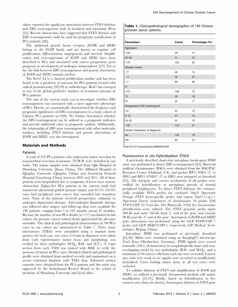
Table 1. Clinicopathological demographics of 190 Chineseprostate cancer patients.
Parameters Count Percentage (%)
Age(years)
,60 29 15
60–69 41 22
$70 120 63
Gleason scores
,7 26 14
= 7 70 37
.7 94 49
cT
#T2 138 73
T3 30 16
T4 22 11
Preoperative PSA levels(ng/ml)
#4 22 12
4–10 27 14
10–20 25 13
.20 116 61
Distant metastasis at diagnosis
No 150 79
Yes 40 21
doi:10.1371/journal.pone.0084959.t001
ERG Rearrangement in Chinese Prostate Cancer
PLOS ONE | www.plosone.org 2 February 2014 | Volume 9 | Issue 2 | e84959
was termed as .50% nuclei (mean63 standard deviations in non-
neoplastic controls) containing either one signal of locus probe and
$2 signals of reference probe (absolute deletion), or two signals of
locus probe and $4 signals of reference probe (relative deletion).
Homozygous deletion of PTEN was exhibited by the concurrent
lack of the both PTEN locus signals and the presence of control
signals in .30% of cells. Specimens were considered amplified for
EGFR when .10% of tumor cells displayed either EGFR: CEP 7
ratio .2 or countless tight clusters of signals of the locus probe (3–
5copies). EGFR copy number gain was defined as a low copy
number increase due to chromosome 7 polysomy. Similarly,
specimens were considered amplified for HER2 when .10% of
tumor cells displayed either HER2: CEP 17 ratio .2 or countless
tight clusters of signals of the locus probe (3–5copies). HER2 copy
number gains were defined as a low copy number increase due to
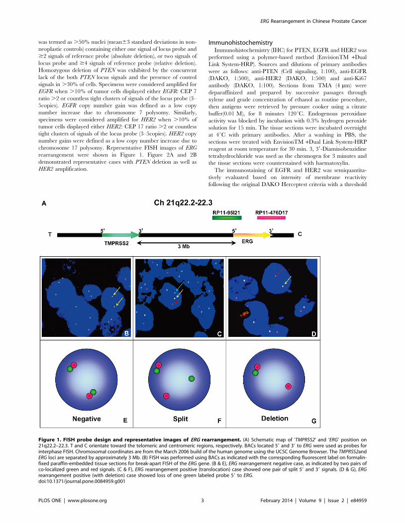
chromosome 17 polysomy. Representative FISH images of ERG
rearrangement were shown in Figure 1. Figure 2A and 2B
demonstrated representative cases with PTEN deletion as well as
HER2 amplification.
ImmunohistochemistryImmunohistochemistry (IHC) for PTEN, EGFR and HER2 was
performed using a polymer-based method (EnvisionTM +Dual
Link System-HRP). Sources and dilutions of primary antibodies
were as follows: anti-PTEN (Cell signaling, 1:100), anti-EGFR
(DAKO, 1:500), anti-HER2 (DAKO, 1:500) and anti-Ki67
antibody (DAKO, 1:100). Sections from TMA (4 mm) were
deparaffinized and prepared by successive passages through
xylene and grade concentration of ethanol as routine procedure,
then antigens were retrieved by pressure cooker using a citrate
buffer(0.01 M), for 8 minutes 120uC. Endogenous peroxidase
activity was blocked by incubation with 0.3% hydrogen peroxide
solution for 15 min. The tissue sections were incubated overnight
at 4uC with primary antibodies. After a washing in PBS, the
sections were treated with EnvisionTM +Dual Link System-HRP
reagent at room temperature for 30 min. 3, 39-Diaminobenzidine
tetrahydrochloride was used as the chromogen for 3 minutes and
the tissue sections were counterstained with haematoxylin.
The immunostaining of EGFR and HER2 was semiquantita-
tively evaluated based on intensity of membrane reactivity
following the original DAKO Herceptest criteria with a threshold
Figure 1. FISH probe design and representative images of ERG rearrangement. (A) Schematic map of ‘TMPRSS2’ and ‘ERG’ position on21q22.2–22.3. T and C orientate toward the telomeric and centromeric regions, respectively. BACs located 59 and 39 to ERG were used as probes forinterphase FISH. Chromosomal coordinates are from the March 2006 build of the human genome using the UCSC Genome Browser. The TMPRSS2andERG loci are separated by approximately 3 Mb. (B) FISH was performed using BACs as indicated with the corresponding fluorescent label on formalin-fixed paraffin-embedded tissue sections for break-apart FISH of the ERG gene. (B & E), ERG rearrangement negative case, as indicated by two pairs ofco-localized green and red signals. (C & F), ERG rearrangement positive (translocation) case showed one pair of split 59 and 39 signals. (D & G), ERGrearrangement positive (with deletion) case showed loss of one green labeled probe 59 to ERG.doi:10.1371/journal.pone.0084959.g001
ERG Rearrangement in Chinese Prostate Cancer
PLOS ONE | www.plosone.org 3 February 2014 | Volume 9 | Issue 2 | e84959

of 10% immunopositive cells. The scoring system was described
elsewhere [27]. Evaluation of PTEN was based on the cytoplasmic
staining intensity; the tumors were divided into three categories as
previously described [25]. Grade 2 showed increased or equal
staining intensity compared to the corresponding normal tissue;
grade 1 had decreased staining intensity, and grade 0 demon-
strated complete absence of staining. The Ki-67 labeling index (LI)
was defined as the fraction of tumor cells showing any nuclear Ki-
67 immunoreactivity and was considered high if 10% or more of
the tumor nuclei were stained. For this purpose, 100–200 tumor
cells were analyzed for each case. Representative immunohisto-
chemical images of Ki-67 were shown in Figure 2C.
Statistical AnalysisStatistical analyses were carried out using the Statistical Package
for Social Sciences, version 19.0 (SPSS), with a significance level of
0.05(two-tailed probability). Pearson’s x2 test and Fisher’s exact
test were used to evaluate the associations between ERG
rearrangement and clinico-pathologic variables as well as other
molecular aberrations. Kaplan-Meier analysis was utilized to
assess the prognostic value of ERG rearrangement in PCa patients.
The prognostic value of ERG rearrangement was further
determined in univariable and multivariable analysis, including
PSA values at diagnosis, Gleason score, clinical tumor stage,
distant metastasis, Ki-67 LI and EGFR family gene aberrations.
Results
Frequency of ERG Rearrangement, PTEN Deletion andEGFR Family Aberrations
Overall, ERG was rearranged in 23.2% (44/190) of Chinese
PCa patients, of which 54.5% (24/44) demonstrated deletion of
the 59end of ERG. Interestingly, two out of these 24 cases
demonstrated two copies of the 39-ERG signals, suggesting the
duplication of ERG rearrangement. PTEN deletion was identified
in 10.8% (19/176) of cases, with hemizygous and homozygous
deletions present in 12 of 19 (63.2%) and 7 of 19 (36.8%) cases,
respectively. Amplification of HER2 was identified in 10 of 173
(5.8%) tumors and polysomy of chromosome 17 was noted in 41 of
173 (23.8%) cases. By contrast, only 2 of 178 (1.1%) cases showed
amplification of EGFR with polysomy of chromosome 7 being
present in 18 of 178 (10.1%) tumors.
Relationships between ERG Rearrangement andClinicopathologic Variables
ERG gene rearrangement was significantly associated with
preoperative PSA levels in PCa patients (P = 0.038) (Table 2). The
incidence of ERG rearrangement was significantly lower in patients
with Low PSA level (,4 ng/ml) compared with those having
medium or high PSA levels. However, no significant correlation
Figure 2. Representative images for IHC staining and FISH analysis of PTEN, HER2 and Ki-67 expression in PCa. (A1–A3) FISH images ofundeleted, hemizygous and homozygous PTEN deletion in PCa. A1, PTEN deletion negative case showed both paired red signals (10q23/PTEN locus)and green signals in tumor cells. A2, Representative case with PTEN hemizygous deletion showed one red signals and pairs of green signals in tumorcells. A3, Representative case with PTEN homozygous deletion showed absence of red signals but retained pairs of green signals. For all assays, atleast 50 cancer cell nuclei were evaluated. (B1–B3) The detection of HER2 expression by IHC and FISH in PCa. B1, FISH analysis of representative casewithout HER2 amplification. B2, FISH analysis of case with HER2 amplification. B3, HER2 IHC staining shows complete membranous reactivity of strongintensity (3+) in tumor cells (original magnification,6200). (C1–C3) The Ki-67 staining by IHC in PCa cells. C1, No staining (0) of Ki-67 in tumor cells. C2,Low Ki-67 (LI,10%) nuclear positivity in tumor cells. C3, High Ki-67(LI$10%) nuclear positivity in tumor cells.doi:10.1371/journal.pone.0084959.g002
ERG Rearrangement in Chinese Prostate Cancer
PLOS ONE | www.plosone.org 4 February 2014 | Volume 9 | Issue 2 | e84959

was identified between ERG rearrangement and age, Gleason
score, clinical T stage, or distant metastasis at diagnosis.
Association of ERG Rearrangement with Other MolecularMarkers
As deletion of PTEN and amplifications of EGFR and HER2 are
relevant genomic aberrations in PCa, we next explored the
association of ERG rearrangement with these molecular events in
our cohort. As shown in Table 2, the ERG rearrangement was
present in approximately 63.2% (12/19) of PCa patients with
PTEN deletion (hemizygous or homozygous). Likewise, PTEN
deletion occurred more frequently in cases that harbored ERG
rearrangement (30.8%, 12/39) as compared with those ERG
rearrangement negative cases (5.1%, 7/137). Overall, a significant
association between PTEN deletion and ERG rearrangement was
observed in Chinese PCa cohort (P = 0.0008). Of note, 46/182
(25.2%) PCa cases revealed decreased PTEN protein expression
by immunohistochemistry. Concordance between PTEN deletion
status and PTEN protein expression was also identified in our
cohort (data not shown).
Amplification of EGFR was identified only in two PCa cases,
both of which were negative for ERG rearrangement. Similarly, 9
out of 10 (90.0%) PCa cases with HER2 amplification were absent
for ERG rearrangement. ERG rearrangement was more often
present in PCa cases without HER2 amplification (34/163, 20.9%)
than in HER2-amplified tumors (1/10, 10.0%) (P = 0.149).
Immunohistochemical overexpressions of EGFR and HER2
were identified in 17.6% (31/176) and 6.0% (11/181) of cases,
respectively. HER2 protein overexpression was significantly
correlated to amplification of HER2 (P,0.01). However, there
was no correlation between EGFR protein expression and gene
amplification (data not shown). ERG rearrangement was neither
associated with EGFR nor HER2 protein expression.
Survival Analysis of ERG Rearrangement in Relation toCancer-related Death
To determine whether the presence of ERG rearrangement was
a prognostic factor for PCa, we compared cancer-related death
rates between patients with or without ERG rearrangement. On
the basis of the Kaplan-Meier survival estimates, the group of
patients with ERG rearrangement had a much greater rate of
mortality than patients who lacked the gene rearrangement
(P = 0.02) (Figure 3).
ERG rearrangement status was shown to be a significant
prognostic predictor of prostate cancer-related survival [HR (95%
CI): 3.368 (1.261–8.955), P = 0.015] in univariate analysis
(Table 3). PSA values at diagnosis (P = 0.009), Gleason score
(P,0.001), clinical tumor stage (P = 0.011), distant metastasis
(P = 0.006), Ki-67 LI (P = 0.002), EGFR amplification (P = 0.023),
and HER2 amplification (P = 0.001) were also significantly related
to cancer-related survival in univariate analysis. Notably, in a
multivariate analysis that included known prognostic markers,
ERG rearrangement status remained a significant predictor
(P = 0.022) with a hazard ratio of 2.099 (95% CI: 1.112–3.962)
(Table 3).
Prognostic Relevance of ERG Rearrangement and Ki-67 LIWe next determined whether combining markers further
improved prognostic value. Since Ki-67 is a known strong
prognosticator in PCa and has independent predictive value for
cancer-related survival in our cohort, we directly compared the
prognostic effects of ERG rearrangement and Ki-67 LI in
combination. For this analysis, we grouped all cancers according
to their ERG status (not rearranged vs. rearranged) and the Ki-67
Label index status (LI ,10% vs LI.10%). Cox regression
analyses were therefore conducted using the group with low Ki-
Table 2. Association of clinicopathologic variables andmolecular biomarkers with ERG rearrangement.
Variable
ERG rearrangement according toFISH (%) P
Not rearranged(n/%) Rearranged (n/%)
All cases 146(76.8) 44(23.2)
age(years)
#65 21(67.8) 10(32.2) 0.173
.65 117(79.0) 31(21.0)
Pre PSA(ng/ml)
,4 15(93.8) 1(6.2) 0.038
4–10 11(64.7) 6(35.3)
.10 103(79.8) 26(20.2)
Gleason score
,7 17(73.9) 6(26.1) 0.430
7 51(76.1) 16(23.9)
.7 78(83.0) 16(17.0)
Clinical tumorstage
#cT2 101(78.3) 28(21.7) 0.607
$cT3 29(74.4) 10(25.6)
Metastasis
No 97(80.8) 23(19.1) 0.585
Yes 37(77.1) 11(22.9)
Ki-67
,10% 127(80.5) 31(19.5) 0.385
$10% 19(73.1) 7(26.9)
PTEN deletion
Not deleted 130(80.3) 27(19.7) 0.0008
Deleted 7(36.8) 12(63.2)
EGFRamplification
Not amplified 139(79.0) 37(21.0) 0.883
Amplified 2(100.0) 0(0.0)
EGFR IHC
0 and 1+ 120(82.3) 25(17.7) 0.779
2+ and 3+ 25(80.6) 6(19.7)
HER2amplification
Not amplified 129(79.1) 34(20.9) 0.671
Amplified 9(90.0) 1(10.0)
HER2 IHC
0 and 1+ 131(76.6) 40(23.4) 0.541
2+ and 3+ 11(100.0) 0(0.0)
Values not available for all 190 cases.doi:10.1371/journal.pone.0084959.t002
ERG Rearrangement in Chinese Prostate Cancer
PLOS ONE | www.plosone.org 5 February 2014 | Volume 9 | Issue 2 | e84959

67 LI and no ERG aberration as the reference. As shown in
Figure 4, the largest group, which comprised those who had no
ERG rearrangement and low Ki-67 LI, had a greater cancer-
related survival when compared with the three other groups.
Notably, the subset of patients with ERG rearrangement and high
Ki-67 LI had the worst cancer-related survival.
We further determined whether ERG rearrangement status
could be utilized in improving risk stratification of PCa patients
with low Ki-67 LI. Kaplan-Meier analysis showed that ERG
rearrangement status was a prognostic factor in the group of
patients with low Ki-67 LI (P = 0.019) (Figure 5A). The median
survival of PCa patients with and without ERG rearrangement was
69 and 89 months, respectively. However, ERG rearrangement
status lost its predictive value of outcome in those with high Ki-67
LI (Figure 5B). By contrast, ERG rearrangement status was not
helpful in identifying high-risk PCa patients with low Gleason
score (data not shown).
Discussion
This is one of the largest series of PCa patients (n.200) reported
so far in China analyzing ERG rearrangement. Our cohort
comprises men treated with TUR-P and all of the study patients
had symptoms of lower tract urinary obstruction, therefore
representing a select subgroup of clinically recognized PCas.
The patients with incidental PCas were excluded from our study.
Although more and more PSA-screed PCa patients have been
identified in western countries, there are limited data regarding the
clinical phenotype or natural history of PCa. Of note, our cohort
included a subset of patients with high grade PCas. This differed
from most Western patients who were found to have PCa due to
PSA screening and were often treated with radical prostatectomy.
Figure 3. Kaplan-Meier survival curves for PCa patients with and without ERG rearrangement. The cancer-related survival rates werecompared between patients with and without ERG rearrangement using the log-rank test.doi:10.1371/journal.pone.0084959.g003
Table 3. Univariate and multivariate analysis of variablesassociated with survival in PCa patients.
Parameter Univariate analysis Multivariate analysis
HR(95%CI) P HR(95%CI) P
age(years)a 0.588(0.328–1.053)
0.074 – –
Pre-PSA 0.601(0.410–0.880)
0.009 Nonsignificancance
Gleason score 2.297(1.455–3.625)
,0.001 4.680(2.020–10.483)
,0.001
Clincial tumorstage
2.011(1.177–3.435)
0.011 Nonsignificancance
Metastasis 2.106(1.240–3.577)
0.006 2.897(1.236–6.789)
0.014
Ki-67 2.592(1.435–4.682)
0.002 2.641(1.084–6.435)
0.019
HER2amplification
6.687(2.253–19.844)
0.001 Nonsignificancance
HER2 IHC 3.240(0.998–10.527)
0.05 Nonsignificancance
EGFRamplification
5.255(1.259–21.929)
0.023 Nonsignificancance
ERGrearrangement
3.368(1.261–8.955)
0.015 2.099(1.112–3.962)
0.022
HR = hazard ratio; CI = confidence interval; PSA = prostate-specific antigen.anot included in multivariate analysis.doi:10.1371/journal.pone.0084959.t003
ERG Rearrangement in Chinese Prostate Cancer
PLOS ONE | www.plosone.org 6 February 2014 | Volume 9 | Issue 2 | e84959

Overall, the frequency of ERG rearrangement was 23.2% in our
cohort and this was comparable with that previously reported by
Mao et al [21] and Ren et al [32] in Chinese PCa patients. In
consistent with these findings, Kimura et al [17] and Lee et al [33]
Figure 4. Kaplan-Meier curves illustrating cancer related survival among PCa patients. The patients were stratified by ERG rearrangementand Ki-67 LI in combination and log-rank test was performed.doi:10.1371/journal.pone.0084959.g004
Figure 5. Kaplan-Meier survival analysis of PCa patients in relation to ERG rearrangement status. (A) low ki-67 LI (,10%) subgroup, (B)high ki-67 LI ($10%) subgroup.doi:10.1371/journal.pone.0084959.g005
ERG Rearrangement in Chinese Prostate Cancer
PLOS ONE | www.plosone.org 7 February 2014 | Volume 9 | Issue 2 | e84959

reported the prevalence of TMPRSS2-ERG gene fusion was 16.3%
(15/92) in Japanese and 20.9% (53/254) in Korean PCa patients,
respectively. Previously, Mosquera et al [34] detected TMPRSS2-
ERG fusion in 100 Caucasian and non-Caucasian PCa patients
undergoing prostate biopsy. They reported that the incidence was
significantly different in Caucasians (44/85, 52%) and in non-
Caucasians (2/15, 13%). Most recently, using a multicolor FISH
assay, Magi-Galluzzi et al [35] found that TMPRSS2-ERG gene
fusion was present in 50% (21/42) of Caucasians, 31.3% (20/64)
of African-Americans and 15.9% (7/44) of Japanese (P = 0.003).
Collectively, these studies highlighted the low prevalence of
TMPRSS2-ERG gene fusions in PCa patients in Asia compared
with western countries and this disparity at least partially resulted
from different genetic background rather than the effects of
lifestyle or diet. Of note, the difference may also reflect previous
findings that the fusion is less common in transition zone tumors
(from which most tumors found in TUR-P samples) than in
peripheral zone tumors [17,36,37]. Additionally, cohort design
and consideration of multifocality might have impacts on the ERG
rearrangement frequency [38].
So far, the prognostic significance of ERG rearrangement in
PCa remains contradictory. A series of retrospective studies that
sought an association between TMPRSS2-ERG and outcome
following PSA screened radical prostatectomy gave mixed results.
Several published studies have shown that PCa patients with the
TMPRSS2-ERG gene fusion conferred a higher risk of recurrence,
whereas others reported a significant association with a favorable
prognosis or a null relationship with clinical outcome. Among
patients managed with watchful waiting, TMPRSS2-ERG seemed
to be associated with worse outcomes. In a meta-analysis including
227 men diagnosed with TUR-P, men with fusion-positive tumors
were 1.37 (95% CI, 0.53–3.51) times as likely to experience distant
metastases or die from PCa as those negative for the fusion [18].
Discrepancies in the reported prognostic significance of ERG
rearrangements can be due to cohort design (multifocality and
zonal origin of the tumor), fusion detection technique, and are also
liable to the primary end point of the study (i.e., biochemical
recurrence, overall survival). Therefore, further standardized
studies are needed to address this issue. In the current study, we
found that ERG rearrangement was significantly associated with
prostate cancer-related death in Chinese PCa patients. More
importantly, ERG rearrangement was suggested to be an
independent predictor of overall survival in multivariate analyses.
It is notable that biochemical recurrence is an imprecise predictor
of prostate cancer death. Although PSA might serve as a surrogate
endpoint for overall survival, the majority of men with PSA
biochemical failure will die of other causes. Ward et al found that
in a population of 3897 radical prostatectomy patients, only 8.3%
of the men with PSA biochemical failure died of PCa [39].
Therefore, prostate cancer-related death as the primary end point
might be more reliable for prognostic analysis. In total, our data
supported the concept that if left untreated or lack of initial
therapy, TMPRSS2-ERG PCa will run a more aggressive clinical
course than fusion-negative cancer.
To date, Ki-67 has been widely utilized as a prognostic
biomarker in malignancy including PCa. Its independent predic-
tive value for PCa related survival has been confirmed in our
study. In line with Antonarakis’s report [40], no significant
association between ERG rearrangement and Ki-67 LI status was
identified. One explanation is that ERG rearrangement has
different effects on proliferation and invasion in vitro, respectively,
Ki-67 is a well-known proliferation marker [41–43]. By contrast,
in 2008, Tomlins et al reported that alternation of ERG gene
expression significantly affect invasion in vitro but has no effect on
cellular proliferation [41]. However, when stratifying for Ki-67
status, ERG rearrangement was a prognostic factor for cancer-
related survival only in PCa patients with low Ki-67 LI. A major
clinical challenge in PCa management is the inability to readily
distinguish indolent from aggressive tumors in patients who
present with low Gleason grade, low tumor volume or low Ki-
67 LI. Our data suggested that determination of ERG rearrange-
ment status could be helpful in stratification of PCa patients with
low Ki-67 LI into different survival categories.
Although gene fusion is a key molecular event in PCa
development and TMPRSS2-ERG fusion may induce high grade
prostatic neoplasia (HGPIN), it is not sufficient to generate a fully
transformed phenotype in vitro and in vivo [41,44]. Several
independent groups have suggested ERG may cooperate with
other genetic aberrations to promote PCa development and
progression, such as PTEN haploinsufficiency, enhanced androgen
receptor (AR) signaling, overexpression of SOX9 and aberrant
phosphoinositide 3-kinase (PI3K) pathway [45–47]. Most recently,
TMPRSS2-ERG was shown to mediate Epithelial to Mesenchymal
Transition (EMT) through the induction of WNT signaling
pathway via FZD4 as well as ZEB1/ZEB2 axis [48,49].
Previously, we and others have suggested the significant associa-
tion between PTEN deletion and ERG rearrangement both in
localized and metastatic PCas in western countries. In this study,
we confirmed significant association between PTEN deletion and
ERG rearrangement in Chinese PCa cohort (P = 0.0008). Thus our
data highlighted a possible cooperative role of both ERG and
PTEN aberrations in a subset of Chinese PCa cases.
Genetic aberrations of HER2 and EGFR were associated with
advanced-stages disease, metastasis and shorten survival in PCa
progression. Previous studies have shown the rarity of EGFR/
HER2 amplifications in PCa. Schlomm et al [50] reported that
amplification of EGFR was present only in 6 of 2,446 PCa cases
(0.25%). Similarly, Baek et al [51] found no amplification of the
EGFR or HER2 genes in 66 PCa specimens. In our cohort,
amplification of HER2 was present in 5.8% of Chinese PCa cases.
Although not reaching statistic significance, ERG rearrangement
seemed to be more often present in PCa cases without HER2
amplification than in HER2-amplified tumors. Therefore, HER2
genetic aberration might play a role in a subset of Chinese PCa
patients without ERG rearrangement.
It should be noted that a small proportion of tumors showing
ERG arrangement may harbor a fusion between ERG and genes
other than TMPRSS2, including SLC45A3 or NDRG1. On the
other hand, it has been suggested that cancers harboring gene
fusions occurring by deletion have worse prognosis than those
occurring by translocation. However, we did not find significant
associations between ERG rearrangement by translocation or
positive by deletion cancers and outcomes in Chinese PCa
patients.
In total, for the first time, we reported that ERG rearrangement
was associated with cancer-related death in Chinese PCa patients.
Determination of ERG rearrangement status allows stratification of
PCa patients into different survival categories.
Author Contributions
Conceived and designed the experiments: BH. Performed the experiments:
MQ XY FZ TL YL. Analyzed the data: MQ XS. Contributed reagents/
materials/analysis tools: XY YL JZ HY YR XQ XS. Wrote the paper: MQ
BH.
ERG Rearrangement in Chinese Prostate Cancer
PLOS ONE | www.plosone.org 8 February 2014 | Volume 9 | Issue 2 | e84959

References
1. Shen MM, Abate-Shen C (2010) Molecular genetics of prostate cancer: new
prospects for old challenges. Genes Dev 24: 1967–2000.2. Lu-Yao GL, Albertsen PC, Moore DF, Shih W, Lin Y, et al. (2009) Outcomes of
localized prostate cancer following conservative management. JAMA 302: 1202–1209.
3. Albertsen PC, Hanley JA, Gleason DF, Barry MJ (1998) Competing risk analysis
of men aged 55 to 74 years at diagnosis managed conservatively for clinicallylocalized prostate cancer. JAMA 280: 975–980.
4. Barry MJ, Albertsen PC, Bagshaw MA, Blute ML, Cox R, et al. (2001)Outcomes for men with clinically nonmetastatic prostate carcinoma managed
with radical prostactectomy, external beam radiotherapy, or expectant
management: a retrospective analysis. Cancer 91: 2302–2314.5. Tomlins SA, Rhodes DR, Perner S, Dhanasekaran SM, Mehra R, et al. (2005)
Recurrent fusion of TMPRSS2 and ETS transcription factor genes in prostatecancer. Science 310: 644–648.
6. Perner S, Demichelis F, Beroukhim R, Schmidt FH, Mosquera JM, et al. (2006)TMPRSS2:ERG fusion-associated deletions provide insight into the heteroge-
neity of prostate cancer. Cancer Res 66: 8337–8341.
7. Helgeson BE, Tomlins SA, Shah N, Laxman B, Cao Q, et al. (2008)Characterization of TMPRSS2:ETV5 and SLC45A3:ETV5 gene fusions in
prostate cancer. Cancer Res 68: 73–80.8. Kumar-Sinha C, Tomlins SA, Chinnaiyan AM (2008) Recurrent gene fusions in
prostate cancer. Nat Rev Cancer 8: 497–511.
9. Nam RK, Sugar L, Yang W, Srivastava S, Klotz LH, et al. (2007) Expression ofthe TMPRSS2:ERG fusion gene predicts cancer recurrence after surgery for
localised prostate cancer. Br J Cancer 97: 1690–1695.10. Attard G, Clark J, Ambroisine L, Fisher G, Kovacs G, et al. (2008) Duplication
of the fusion of TMPRSS2 to ERG sequences identifies fatal human prostatecancer. Oncogene 27: 253–263.
11. Perner S (2010) [Dangerous liaisons in prostate cancer. Clinical and biological
implications of recurrent gene fusions]. Pathologe 31 Suppl 2: 121–125.12. Demichelis F, Fall K, Perner S, Andren O, Schmidt F, et al. (2007)
TMPRSS2:ERG gene fusion associated with lethal prostate cancer in a watchfulwaiting cohort. Oncogene 26: 4596–4599.
13. Saramaki OR, Harjula AE, Martikainen PM, Vessella RL, Tammela TL, et al.
(2008) TMPRSS2:ERG fusion identifies a subgroup of prostate cancers with afavorable prognosis. Clin Cancer Res 14: 3395–3400.
14. Winnes M, Lissbrant E, Damber JE, Stenman G (2007) Molecular geneticanalyses of the TMPRSS2-ERG and TMPRSS2-ETV1 gene fusions in 50 cases
of prostate cancer. Oncol Rep 17: 1033–1036.15. Hermans KG, Boormans JL, Gasi D, van Leenders GJ, Jenster G, et al. (2009)
Overexpression of prostate-specific TMPRSS2(exon 0)-ERG fusion transcripts
corresponds with favorable prognosis of prostate cancer. Clin Cancer Res 15:6398–6403.
16. Boormans JL, Porkka K, Visakorpi T, Trapman J (2011) Confirmation of theassociation of TMPRSS2(exon 0):ERG expression and a favorable prognosis of
primary prostate cancer. Eur Urol 60: 183–184.
17. Kimura T, Furusato B, Miki J, Yamamoto T, Hayashi N, et al. (2012)Expression of ERG oncoprotein is associated with a less aggressive tumor
phenotype in Japanese prostate cancer patients. Pathol Int 62: 742–748.18. Pettersson A, Graff RE, Bauer SR, Pitt MJ, Lis RT, et al. (2012) The
TMPRSS2:ERG rearrangement, ERG expression, and prostate canceroutcomes: a cohort study and meta-analysis. Cancer Epidemiol Biomarkers
Prev 21: 1497–1509.
19. FitzGerald LM, Agalliu I, Johnson K, Miller MA, Kwon EM, et al. (2008)Association of TMPRSS2-ERG gene fusion with clinical characteristics and
outcomes: results from a population-based study of prostate cancer. BMCCancer 8: 230.
20. Gopalan A, Leversha MA, Satagopan JM, Zhou Q, Al-Ahmadie HA, et al.
(2009) TMPRSS2-ERG gene fusion is not associated with outcome in patientstreated by prostatectomy. Cancer Res 69: 1400–1406.
21. Mao X, Yu Y, Boyd LK, Ren G, Lin D, et al. (2010) Distinct genomicalterations in prostate cancers in Chinese and Western populations suggest
alternative pathways of prostate carcinogenesis. Cancer Res 70: 5207–5212.
22. Miyagi Y, Sasaki T, Fujinami K, Sano J, Senga Y, et al. (2010) ETS family-associated gene fusions in Japanese prostate cancer: analysis of 194 radical
prostatectomy samples. Mod Pathol 23: 1492–1498.23. Di Cristofano A, Pandolfi PP (2000) The multiple roles of PTEN in tumor
suppression. Cell 100: 387–390.24. Bertram J, Peacock JW, Fazli L, Mui AL, Chung SW, et al. (2006) Loss of PTEN
is associated with progression to androgen independence. Prostate 66: 895–902.
25. Han B, Mehra R, Lonigro RJ, Wang L, Suleman K, et al. (2009) Fluorescence insitu hybridization study shows association of PTEN deletion with ERG
rearrangement during prostate cancer progression. Mod Pathol 22: 1083–1093.26. Yoshimoto M, Joshua AM, Cunha IW, Coudry RA, Fonseca FP, et al. (2008)
Absence of TMPRSS2:ERG fusions and PTEN losses in prostate cancer is
associated with a favorable outcome. Mod Pathol 21: 1451–1460.27. Di Lorenzo G, Tortora G, D’Armiento FP, De Rosa G, Staibano S, et al. (2002)
Expression of epidermal growth factor receptor correlates with disease relapse
and progression to androgen-independence in human prostate cancer. Clin
Cancer Res 8: 3438–3444.
28. Bubendorf L, Sauter G, Moch H, Schmid HP, Gasser TC, et al. (1996) Ki67labelling index: an independent predictor of progression in prostate cancer
treated by radical prostatectomy. J Pathol 178: 437–441.
29. Bettencourt MC, Bauer JJ, Sesterhenn IA, Mostofi FK, McLeod DG, et al.
(1996) Ki-67 expression is a prognostic marker of prostate cancer recurrenceafter radical prostatectomy. J Urol 156: 1064–1068.
30. Han B, Mehra R, Dhanasekaran SM, Yu J, Menon A, et al. (2008) Afluorescence in situ hybridization screen for E26 transformation-specific
aberrations: identification of DDX5-ETV4 fusion protein in prostate cancer.
Cancer Res 68: 7629–7637.
31. Korshunov A, Sycheva R, Gorelyshev S, Golanov A (2005) Clinical utility offluorescence in situ hybridization (FISH) in nonbrainstem glioblastomas of
childhood. Mod Pathol 18: 1258–1263.
32. Ren S, Peng Z, Mao JH, Yu Y, Yin C, et al. (2012) RNA-seq analysis of prostate
cancer in the Chinese population identifies recurrent gene fusions, cancer-
associated long noncoding RNAs and aberrant alternative splicings. Cell Res 22:806–821.
33. Lee K, Chae JY, Kwak C, Ku JH, Moon KC (2010) TMPRSS2-ERG gene
fusion and clinicopathologic characteristics of Korean prostate cancer patients.
Urology 76: 1268 e1267–1213.
34. Mosquera JM, Mehra R, Regan MM, Perner S, Genega EM, et al. (2009)
Prevalence of TMPRSS2-ERG fusion prostate cancer among men undergoingprostate biopsy in the United States. Clin Cancer Res 15: 4706–4711.
35. Magi-Galluzzi C, Tsusuki T, Elson P, Simmerman K, LaFargue C, et al. (2011)
TMPRSS2-ERG gene fusion prevalence and class are significantly different in
prostate cancer of Caucasian, African-American and Japanese patients. Prostate71: 489–497.
36. Falzarano SM, Navas M, Simmerman K, Klein EA, Rubin MA, et al. (2010)
ERG rearrangement is present in a subset of transition zone prostatic tumors.
Mod Pathol 23: 1499–1506.
37. Guo CC, Zuo G, Cao D, Troncoso P, Czerniak BA (2009) Prostate cancer of
transition zone origin lacks TMPRSS2-ERG gene fusion. Mod Pathol 22: 866–871.
38. Braun M, Scheble VJ, Menon R, Scharf G, Wilbertz T, et al. (2011) Relevance
of cohort design for studying the frequency of the ERG rearrangement in
prostate cancer. Histopathology 58: 1028–1036.
39. Ward JF, Blute ML, Slezak J, Bergstralh EJ, Zincke H (2003) The long-term
clinical impact of biochemical recurrence of prostate cancer 5 or more yearsafter radical prostatectomy. J Urol 170: 1872–1876.
40. Antonarakis ES, Keizman D, Zhang Z, Gurel B, Lotan TL, et al. (2012) An
immunohistochemical signature comprising PTEN, MYC, and Ki67 predicts
progression in prostate cancer patients receiving adjuvant docetaxel afterprostatectomy. Cancer 118: 6063–6071.
41. Tomlins SA, Laxman B, Varambally S, Cao X, Yu J, et al. (2008) Role of the
TMPRSS2-ERG gene fusion in prostate cancer. Neoplasia 10: 177–188.
42. Pathmanathan N, Balleine RL (2013) Ki67 and proliferation in breast cancer.
J Clin Pathol 66: 512–516.
43. Perner S, Mosquera JM, Demichelis F, Hofer MD, Paris PL, et al. (2007)
TMPRSS2-ERG fusion prostate cancer: an early molecular event associatedwith invasion. Am J Surg Pathol 31: 882–888.
44. Klezovitch O, Risk M, Coleman I, Lucas JM, Null M, et al. (2008) A causal rolefor ERG in neoplastic transformation of prostate epithelium. Proc Natl Acad
Sci U S A 105: 2105–2110.
45. Carver BS, Tran J, Gopalan A, Chen Z, Shaikh S, et al. (2009) Aberrant ERG
expression cooperates with loss of PTEN to promote cancer progression in theprostate. Nat Genet 41: 619–624.
46. Sun C, Dobi A, Mohamed A, Li H, Thangapazham RL, et al. (2008)TMPRSS2-ERG fusion, a common genomic alteration in prostate cancer
activates C-MYC and abrogates prostate epithelial differentiation. Oncogene 27:5348–5353.
47. Cai C, Wang H, He HH, Chen S, He L, et al. (2013) ERG induces androgenreceptor-mediated regulation of SOX9 in prostate cancer. J Clin Invest 123:
1109–1122.
48. Zong Y, Xin L, Goldstein AS, Lawson DA, Teitell MA, et al. (2009) ETS family
transcription factors collaborate with alternative signaling pathways to inducecarcinoma from adult murine prostate cells. Proc Natl Acad Sci U S A 106:
12465–12470.
49. Leshem O, Madar S, Kogan-Sakin I, Kamer I, Goldstein I, et al. (2011)
TMPRSS2/ERG promotes epithelial to mesenchymal transition through the
ZEB1/ZEB2 axis in a prostate cancer model. PLoS One 6: e21650.
50. Schlomm T, Kirstein P, Iwers L, Daniel B, Steuber T, et al. (2007) Clinicalsignificance of epidermal growth factor receptor protein overexpression and
gene copy number gains in prostate cancer. Clin Cancer Res 13: 6579–6584.
51. Baek KH, Hong ME, Jung YY, Lee CH, Lee TJ, et al. (2012) Correlation of AR,
EGFR, and HER2 Expression Levels in Prostate Cancer: Immunohistochemical
Analysis and Chromogenic In Situ Hybridization. Cancer Res Treat 44: 50–56.
ERG Rearrangement in Chinese Prostate Cancer
PLOS ONE | www.plosone.org 9 February 2014 | Volume 9 | Issue 2 | e84959