autoimmunity in membranous nephropathy targets aldose reductase and sod2
TRANSCRIPT
Autoimmunity in Membranous Nephropathy TargetsAldose Reductase and SOD2
Marco Prunotto,* Maria Luisa Carnevali,† Giovanni Candiano,* Corrado Murtas,†
Maurizio Bruschi,*‡ Emilia Corradini,§ Antonella Trivelli,* Alberto Magnasco,*Andrea Petretto,� Laura Santucci,*‡ Silvia Mattei,† Rita Gatti,¶ Francesco Scolari,**††
Peter Kador,‡‡ Landino Allegri,† and Gian Marco Ghiggeri*
*Division of Nephrology and Laboratory on Pathophysiology of Uremia and �Mass Spectrometry Core Facility,G. Gaslini Children Hospital, Genoa, Italy; ‡Renal Child Foundation, Genoa, Italy; Departments of †ClinicalMedicine, Nephrology and Health Sciences and §Pathology and Laboratory Medicine and ¶Department ofExperimental Medicine, Section of Histology, University of Parma, Parma, Italy; **Department of NephrologyUniversity of Brescia, Brescia, Italy; ††Division of Nephrology, Montichiari Hospital, Brescia, Italy; and ‡‡College ofPharmacy, University of Nebraska Medical Center, Omaha, Nebraska
ABSTRACTGlomerular targets of autoimmunity in human membranous nephropathy are poorly understood. Here,we used a combined proteomic approach to identify specific antibodies against podocyte proteins inboth serum and glomeruli of patients with membranous nephropathy (MN). We detected specificanti–aldose reductase (AR) and anti–manganese superoxide dismutase (SOD2) IgG4 in sera of patientswith MN. We also eluted high titers of anti-AR and anti-SOD2 IgG4 from microdissected glomeruli ofthree biopsies of MN kidneys but not from biopsies of other glomerulonephritides characterized by IgGdeposition (five lupus nephritis and two membranoproliferative glomerulonephritis). We identified bothantigens in MN biopsies but not in other renal pathologies or normal kidney. Confocal and immuno-electron microscopy (IEM) showed co-localization of anti-AR and anti-SOD2 with IgG4 and C5b-9 inelectron-dense podocyte immune deposits. Preliminary in vitro experiments showed an increase ofSOD2 expression on podocyte plasma membrane after treatment with hydrogen peroxide. In conclu-sion, our data support AR and SOD2 as renal antigens of human MN and suggest that oxidative stressmay drive glomerular SOD2 expression.
J Am Soc Nephrol 21: 507–519, 2010. doi: 10.1681/ASN.2008121259
Primary membranous nephropathy (MN) is a com-mon glomerular disease in humans with no univer-sally effective clinical therapy. Treatments are en-tirely empirical, and the disease evolves towardrenal failure in a significant number of patients.1,2
The presence of glomerular subepithelial immunedeposits is the distinctive pathologic feature of MN,thus supporting the concept of an immunologic or-igin. It is also known that inflammatory com-pounds such as complement, oxygen radicals,3,4 orintracellular protein kinase C�5 may participate,having a key role in disease progression.
In the past few decades, studies of experimentalmodels, with a particular emphasis on the Hey-
mann nephritis (HN) model,6–8 have led to the iden-tification of antigens of the autoantibody response inrats (megalin),7 mice (aminopeptidase A),9 and rab-bits (neutral endopeptidase [NEP]),10 –12 but lim-
Received December 12, 2008. Accepted October 19, 2009.
Published online ahead of print. Publication date available atwww.jasn.org.
M.L.C. and M.P. contributed equally to this work.
Correspondence: Dr. Gian Marco Ghiggeri, Laboratory on Patho-physiology of Uremia, G. Gaslini Children Hospital, Largo G.Gaslini, 5. 16148 Genova, Italy. Phone: ��39-010-380742; Fax:��39-010-395214; E-mail: [email protected]
Copyright � 2010 by the American Society of Nephrology
CLI
NIC
AL
RE
SEA
RC
H
CLINICAL RESEARCH www.jasn.org
J Am Soc Nephrol 21: 507–519, 2010 ISSN : 1046-6673/2103-507 507
ited data are available for humans. Moreover, megalin, whichis the target antigen in HN, is absent in human glomeruli, andthe LDL receptor, its human homolog, is only partially co-localized with MN IgG deposits.5,13–15 Seminal studies by De-biec et al.11,12 support the formation of immune depositsagainst NEP, in particular, cases with metallomembrane endo-peptidase mutations that lead to NEP deficiency and alloim-munization during pregnancy. More recently, Beck et al.16 re-ported the presence of specific IgG4 against the M-typephospholipase A2 receptor (PLA2R) in glomerular eluates andin plasma of a significant percentage of patients with MN, sug-gesting PLA2R is a major antigen in this disease.
Unequivocal identification of coexisting antigens in thepodocyte membrane and in subepithelial deposits is essentialfor any progression in the understanding of the mechanisms ofMN in humans. The aim of this study was to identify podocyteproteins recognized by circulating autoantibodies in patientswith MN, to define their expression in glomeruli, and to quan-tify the levels of specific antibodies in sera and in renal biopsies.Results provide first evidence for de novo expression of specificautoantibodies against aldose reductase (AR) and superoxidedismutase 2 (SOD2) in sera and glomeruli of patients withMN.
RESULTS
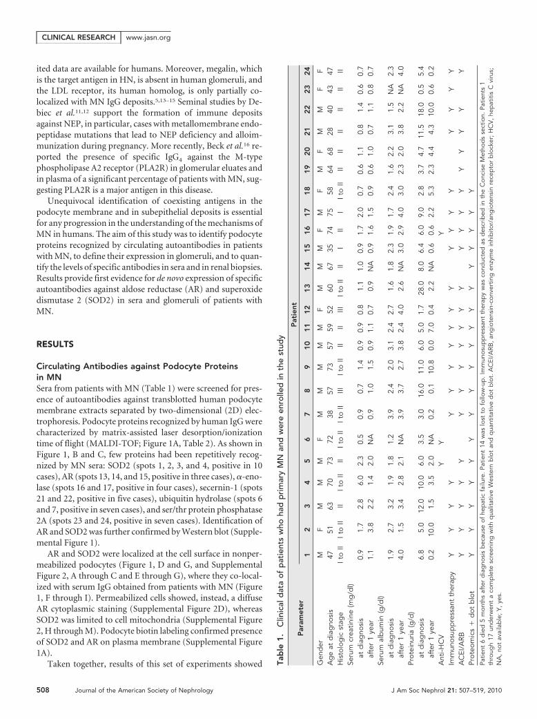
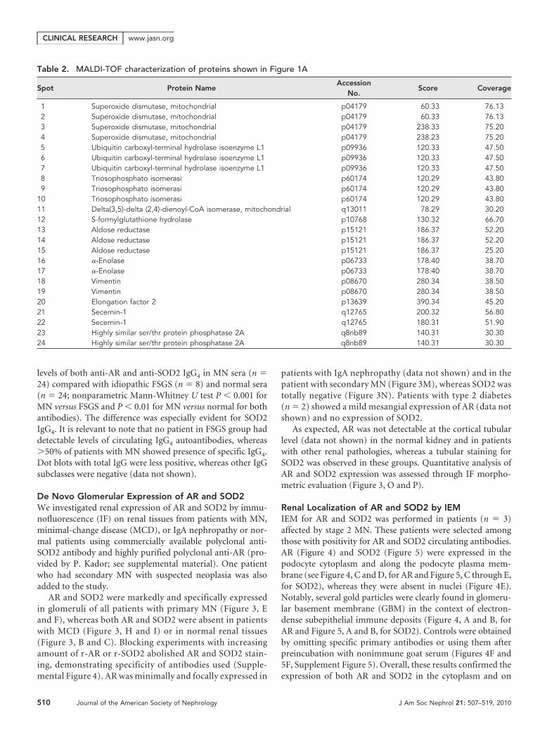
Circulating Antibodies against Podocyte Proteinsin MNSera from patients with MN (Table 1) were screened for pres-ence of autoantibodies against transblotted human podocytemembrane extracts separated by two-dimensional (2D) elec-trophoresis. Podocyte proteins recognized by human IgG werecharacterized by matrix-assisted laser desorption/ionizationtime of flight (MALDI-TOF; Figure 1A, Table 2). As shown inFigure 1, B and C, few proteins had been repetitively recog-nized by MN sera: SOD2 (spots 1, 2, 3, and 4, positive in 10cases), AR (spots 13, 14, and 15, positive in three cases), �-eno-lase (spots 16 and 17, positive in four cases), secernin-1 (spots21 and 22, positive in five cases), ubiquitin hydrolase (spots 6and 7, positive in seven cases), and ser/thr protein phosphatase2A (spots 23 and 24, positive in seven cases). Identification ofAR and SOD2 was further confirmed by Western blot (Supple-mental Figure 1).
AR and SOD2 were localized at the cell surface in nonper-meabilized podocytes (Figure 1, D and G, and SupplementalFigure 2, A through C and E through G), where they co-local-ized with serum IgG obtained from patients with MN (Figure1, F through I). Permeabilized cells showed, instead, a diffuseAR cytoplasmic staining (Supplemental Figure 2D), whereasSOD2 was limited to cell mitochondria (Supplemental Figure2, H through M). Podocyte biotin labeling confirmed presenceof SOD2 and AR on plasma membrane (Supplemental Figure1A).
Taken together, results of this set of experiments showed Tab
le1
.C
linic
ald
ata
ofp
atie
nts
who
had
pri
mar
yM
Nan
dw
ere
enro
lled
inth
est
udy
Par
amet
erP
atie
nt
12
34
56
78
910
11
12
13
14
15
16
17
18
19
20
21
22
23
24
Gen
der
MF
MM
MF
MM
MM
MF
MM
MF
MF
MF
MM
FF
Ag
eat
dia
gno
sis
4751
6370
7372
3857
7357
5952
6067
3574
7558
6468
2840
4347
His
tolo
gic
stag
eIt
oII
Ito
IIII
Ito
IIII
Ito
IIIt
oII
IIIIt
oII
IIII
IIIIt
oII
III
III
Ito
IIII
IIII
IIII
IISe
rum
crea
tinin
e(m
g/d
l)at
dia
gno
sis
0.9
1.7
2.8
6.0
2.3
0.5
0.9
0.7
1.4
0.9
0.9
0.8
1.1
1.0
0.9
1.7
2.0
0.7
0.6
1.1
0.8
1.4
0.6
0.7
afte
r1
year
1.1
3.8
2.2
1.4
2.0
NA
0.9
1.0
1.5
0.9
1.1
0.7
0.9
NA
0.9
1.6
1.5
0.9
0.6
1.0
0.7
1.1
0.8
0.7
Seru
mal
bum
in(g
/dl)
atd
iag
nosi
s1.
92.
73.
21.
91.
81.
23.
92.
42.
03.
12.
42.
71.
61.
82.
31.
91.
72.
41.
62.
23.
11.
5N
A2.
3af
ter
1ye
ar4.
01.
53.
42.
82.
1N
A3.
93.
72.
73.
82.
44.
02.
6N
A3.
02.
94.
03.
02.
32.
03.
82.
2N
A4.
0Pr
otei
nuria
(g/d
)at
dia
gno
sis
6.8
5.0
12.0
10.0
6.0
3.5
3.0
16.0
11.0
6.0
5.0
1.7
28.0
8.0
6.4
6.0
9.0
2.8
3.7
4.7
11.5
18.0
0.5
5.4
afte
r1
year
0.2
10.0
1.5
3.5
2.0
NA
0.2
0.1
10.8
0.0
7.0
0.4
2.2
NA
0.6
0.6
2.2
5.3
2.3
4.4
4.3
10.0
0.6
0.2
Ant
i-HC
VY
YY
Imm
unos
upp
ress
ant
ther
apy
YY
YY
YY
YY
YY
YY
YY
YY
YY
YA
CEI
/ARB
YY
YY
YY
YY
YY
YY
YY
YY
YY
YY
YPr
oteo
mic
s�
dot
blo
tY
YY
YY
YY
YY
YY
YY
YY
YY
YPa
tient
6d
ied
5m
onth
saf
ter
dia
gno
sis
bec
ause
ofhe
pat
icfa
ilure
.Pa
tient
14w
aslo
stto
follo
w-u
p.
Imm
unos
upp
ress
ant
ther
apy
was
cond
ucte
das
des
crib
edin
the
Con
cise
Met
hod
sse
ctio
n.Pa
tient
s1
thro
ugh
17un
der
wen
ta
com
ple
tesc
reen
ing
with
qua
litat
ive
Wes
tern
blo
tan
dq
uant
itativ
ed
otb
lot.
AC
EI/A
RB,
ang
iote
nsin
-con
vert
ing
enzy
me
inhi
bito
r/an
gio
tens
inre
cep
tor
blo
cker
;H
CV
,he
pat
itis
Cvi
rus;
NA
,no
tav
aila
ble
;Y,
yes.
CLINICAL RESEARCH www.jasn.org
508 Journal of the American Society of Nephrology J Am Soc Nephrol 21: 507–519, 2010
that MN sera contain IgG recognizing several podocyte anti-gens. AR and SOD2 analysis was extended to determine theirserum titer, renal expression, and co-localization within glo-merular structures with IgG4.
Serum Titer of Anti-AR and Anti-SOD2 AntibodiesAfter the observation in the previous section, we devised a newassay to assess the titer of circulating anti-AR and anti-SOD2antibodies in an amplified set of 24 MN sera. The assay was
based on dot-blot analysis with peroxidase detection ofIgG1 to 4 linked to membrane-fixed antigens. The assay (see theConcise Methods section and supplemental material), usingrecombinant AR (r-AR) or SOD2 (r-SOD2) spotted as anti-gens, proved to be sensitive to the nanogram level. Moreover,as expected, recombinant proteins were constituted by a singleband at 36 and 48 kD, respectively, and were recognized as asingle band by sera of patients with MN (Supplemental Figure3). Results shown in Figure 2 documented an increase in serum
Figure 1. Sera of MN patients show presence of antibodies against podocyte proteins. (A through C) 2D electrophoresis analysis ofpodocyte membrane extracts and Western blot analysis of MN serum for anti-podocyte antibodies. (A) Representative 2D map ofpodocyte membrane extracts stained by colloidal Coomassie. Numbers correspond to identified proteins as displayed in Table 2; spots1, 2, 3, and 4 correspond to SOD2, and spots 13, 14, and 15 correspond to AR. (B and C) Western blot analysis of MN serum foranti-podocyte antibodies. Sera of patients with MN were largely positive for SOD2 (B and C); only a few were positive for AR (C).Representative normal sera (n � 10) run in parallel to MN samples were negative and have been reported as control. (D through I)Expression of AR and SOD2 on cultured podocytes. Nonpermeabilized human podocyte cell line stained for AR (D) or SOD2 (G) andMN sera (10% in medium) incubated with cells (E and H). (F and I) Merged images. Arrows in merged images indicate the coexpressionof AR or SOD2 with MN IgG. Magnification, �630.
CLINICAL RESEARCHwww.jasn.org
J Am Soc Nephrol 21: 507–519, 2010 Renal Antigens in Membranous Nephropathy 509
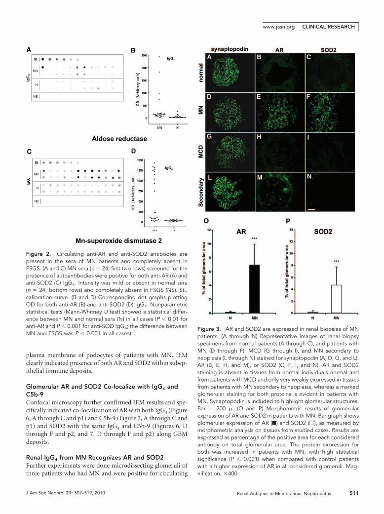
levels of both anti-AR and anti-SOD2 IgG4 in MN sera (n �24) compared with idiopathic FSGS (n � 8) and normal sera(n � 24; nonparametric Mann-Whitney U test P � 0.001 forMN versus FSGS and P � 0.01 for MN versus normal for bothantibodies). The difference was especially evident for SOD2IgG4. It is relevant to note that no patient in FSGS group haddetectable levels of circulating IgG4 autoantibodies, whereas�50% of patients with MN showed presence of specific IgG4.Dot blots with total IgG were less positive, whereas other IgGsubclasses were negative (data not shown).
De Novo Glomerular Expression of AR and SOD2We investigated renal expression of AR and SOD2 by immu-nofluorescence (IF) on renal tissues from patients with MN,minimal-change disease (MCD), or IgA nephropathy or nor-mal patients using commercially available polyclonal anti-SOD2 antibody and highly purified polyclonal anti-AR (pro-vided by P. Kador; see supplemental material). One patientwho had secondary MN with suspected neoplasia was alsoadded to the study.
AR and SOD2 were markedly and specifically expressedin glomeruli of all patients with primary MN (Figure 3, Eand F), whereas both AR and SOD2 were absent in patientswith MCD (Figure 3, H and I) or in normal renal tissues(Figure 3, B and C). Blocking experiments with increasingamount of r-AR or r-SOD2 abolished AR and SOD2 stain-ing, demonstrating specificity of antibodies used (Supple-mental Figure 4). AR was minimally and focally expressed in
patients with IgA nephropathy (data not shown) and in thepatient with secondary MN (Figure 3M), whereas SOD2 wastotally negative (Figure 3N). Patients with type 2 diabetes(n � 2) showed a mild mesangial expression of AR (data notshown) and no expression of SOD2.
As expected, AR was not detectable at the cortical tubularlevel (data not shown) in the normal kidney and in patientswith other renal pathologies, whereas a tubular staining forSOD2 was observed in these groups. Quantitative analysis ofAR and SOD2 expression was assessed through IF morpho-metric evaluation (Figure 3, O and P).
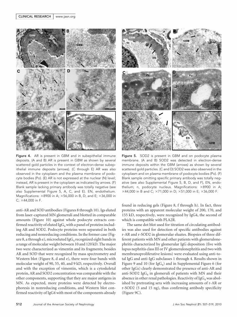
Renal Localization of AR and SOD2 by IEMIEM for AR and SOD2 was performed in patients (n � 3)affected by stage 2 MN. These patients were selected amongthose with positivity for AR and SOD2 circulating antibodies.AR (Figure 4) and SOD2 (Figure 5) were expressed in thepodocyte cytoplasm and along the podocyte plasma mem-brane (see Figure 4, C and D, for AR and Figure 5, C through E,for SOD2), whereas they were absent in nuclei (Figure 4E).Notably, several gold particles were clearly found in glomeru-lar basement membrane (GBM) in the context of electron-dense subepithelial immune deposits (Figure 4, A and B, forAR and Figure 5, A and B, for SOD2). Controls were obtainedby omitting specific primary antibodies or using them afterpreincubation with nonimmune goat serum (Figures 4F and5F, Supplement Figure 5). Overall, these results confirmed theexpression of both AR and SOD2 in the cytoplasm and on
Table 2. MALDI-TOF characterization of proteins shown in Figure 1A
Spot Protein NameAccession
No.Score Coverage
1 Superoxide dismutase, mitochondrial p04179 60.33 76.132 Superoxide dismutase, mitochondrial p04179 60.33 76.133 Superoxide dismutase, mitochondrial p04179 238.33 75.204 Superoxide dismutase, mitochondrial p04179 238.23 75.205 Ubiquitin carboxyl-terminal hydrolase isoenzyme L1 p09936 120.33 47.506 Ubiquitin carboxyl-terminal hydrolase isoenzyme L1 p09936 120.33 47.507 Ubiquitin carboxyl-terminal hydrolase isoenzyme L1 p09936 120.33 47.508 Triosophosphato isomerasi p60174 120.29 43.809 Triosophosphato isomerasi p60174 120.29 43.80
10 Triosophosphato isomerasi p60174 120.29 43.8011 Delta(3,5)-delta (2,4)-dienoyl-CoA isomerase, mitochondrial q13011 78.29 30.2012 S-formylglutathione hydrolase p10768 130.32 66.7013 Aldose reductase p15121 186.37 52.2014 Aldose reductase p15121 186.37 52.2015 Aldose reductase p15121 186.37 25.2016 �-Enolase p06733 178.40 38.7017 �-Enolase p06733 178.40 38.7018 Vimentin p08670 280.34 38.5019 Vimentin p08670 280.34 38.5020 Elongation factor 2 p13639 390.34 45.2021 Secernin-1 q12765 200.32 56.8022 Secernin-1 q12765 180.31 51.9023 Highly similar ser/thr protein phosphatase 2A q8nb89 140.31 30.3024 Highly similar ser/thr protein phosphatase 2A q8nb89 140.31 30.30
CLINICAL RESEARCH www.jasn.org
510 Journal of the American Society of Nephrology J Am Soc Nephrol 21: 507–519, 2010
plasma membrane of podocytes of patients with MN. IEMclearly indicated presence of both AR and SOD2 within subep-ithelial immune deposits.
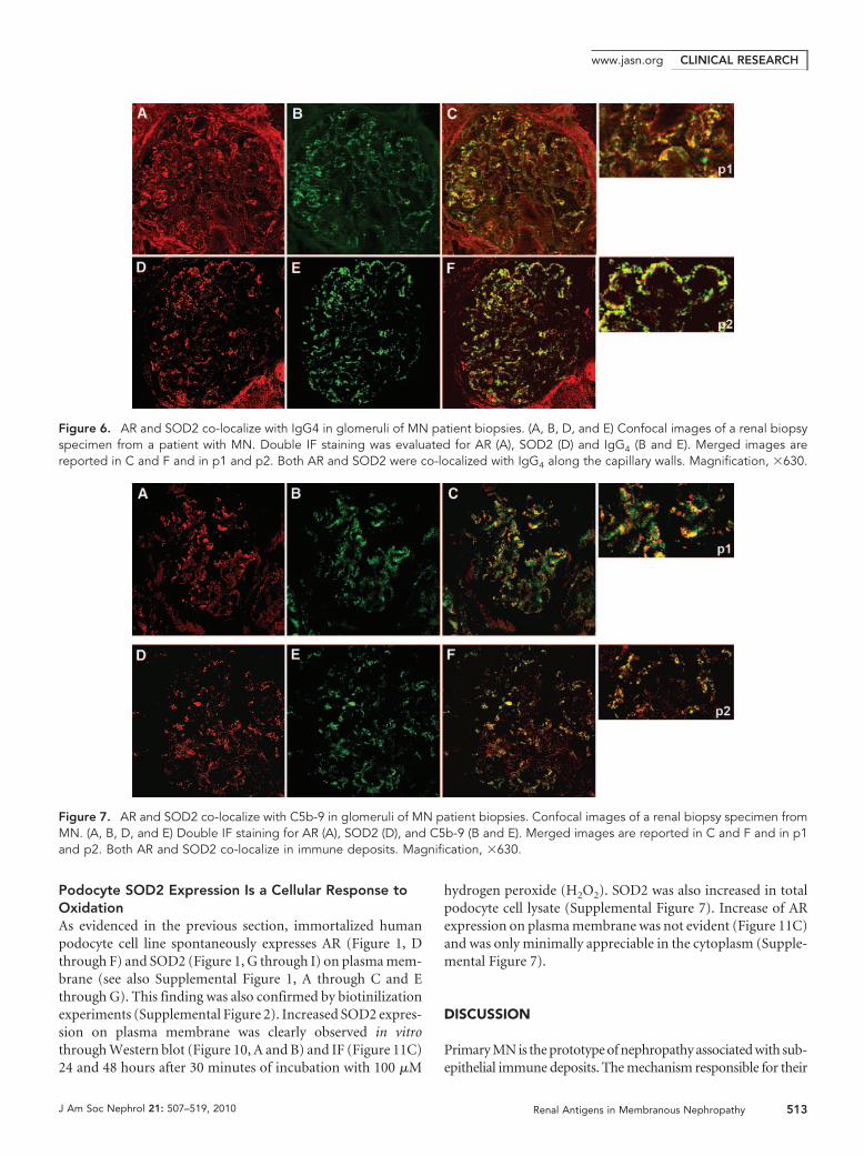
Glomerular AR and SOD2 Co-localize with IgG4 andC5b-9Confocal microscopy further confirmed IEM results and spe-cifically indicated co-localization of AR with both IgG4 (Figure6, A through C and p1) and C5b-9 (Figure 7, A through C andp1) and SOD2 with the same IgG4 and C5b-9 (Figures 6, Dthrough F and p2, and 7, D through F and p2) along GBMdeposits.
Renal IgG4 from MN Recognizes AR and SOD2Further experiments were done microdissecting glomeruli ofthree patients who had MN and were positive for circulating
Figure 3. AR and SOD2 are expressed in renal biopsies of MNpatients. (A through N) Representative images of renal biopsyspecimens from normal patients (A through C), and patients withMN (D through F), MCD (G through I), and MN secondary toneoplasia (L through N) stained for synaptopodin (A, D, G, and L),AR (B, E, H, and M), or SOD2 (C, F, I, and N). AR and SOD2staining is absent in tissues from normal individuals normal andfrom patients with MCD and only very weakly expressed in tissuesfrom patients with MN secondary to neoplasia, whereas a markedglomerular staining for both proteins is evident in patients withMN. Synaptopodin is included to highlight glomerular structures.Bar � 200 �. (O and P) Morphometric results of glomerularexpression of AR and SOD2 in patients with MN. Bar graph showsglomerular expression of AR (f) and SOD2 (�), as measured bymorphometric analysis on tissues from studied cases. Results areexpressed as percentage of the positive area for each consideredantibody on total glomerular area. The protein expression forboth was increased in patients with MN, with high statisticalsignificance (P � 0.001) when compared with control patientswith a higher expression of AR in all considered glomeruli. Mag-nification, �400.
Figure 2. Circulating anti-AR and anti-SOD2 antibodies arepresent in the sera of MN patients and completely absent inFSGS. (A and C) MN sera (n � 24, first two rows) screened for thepresence of autoantibodies were positive for both anti-AR (A) andanti-SOD2 (C) IgG4. Intensity was mild or absent in normal sera(n � 24, bottom rows) and completely absent in FSGS (NS). St.,calibration curve. (B and D) Corresponding dot graphs plottingOD for both anti-AR (B) and anti-SOD2 (D) IgG4. Nonparametricstatistical tests (Mann-Whitney U test) showed a statistical differ-ence between MN and normal sera (N) in all cases (P � 0.01 foranti-AR and P � 0.001 for anti-SOD IgG4; the difference betweenMN and FSGS was P � 0.001 in all cases).
CLINICAL RESEARCHwww.jasn.org
J Am Soc Nephrol 21: 507–519, 2010 Renal Antigens in Membranous Nephropathy 511
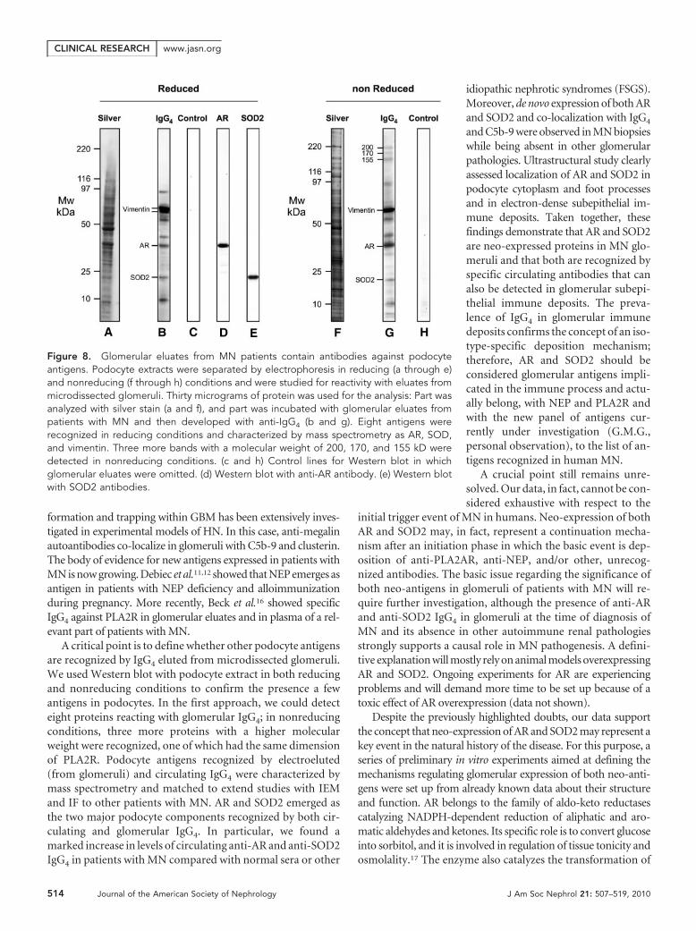
anti-AR and SOD antibodies (Figures 8 through 10). Igs elutedfrom laser-captured MN glomeruli and blotted in comparableamounts (Figure 10) against whole podocyte extracts con-firmed reactivity of eluted IgG4 with a panel of proteins includ-ing AR and SOD2. Podocyte proteins were separated in bothreducing and nonreducing conditions. In the former case (Fig-ure 8, a through e), microeluted IgG4 recognized eight bands ina range of molecular weight between 10 and 120 kD. The majortwo were characterized as vimentin and its fragments; besidesAR and SOD that were recognized by mass spectrometry andWestern blot (Figure 8, d and e), there were four bands withmolecular weight of 90, 55, 40, and 9 kD, respectively. Overalland with the exception of vimentin, which is a cytoskeletalprotein, AR and SOD2 concentration was comparable with theother components, supporting that they are major antigens inMN. As expected, more proteins were detected by electro-phoresis in nonreducing conditions, and Western blot con-firmed reactivity of IgG4 with most of the components already
found in reducing gels (Figure 8, f through h). In fact, threeproteins with an apparent molecular weight of 200, 170, and155 kD, respectively, were recognized by IgG4, the second ofwhich is compatible with PLA2R.
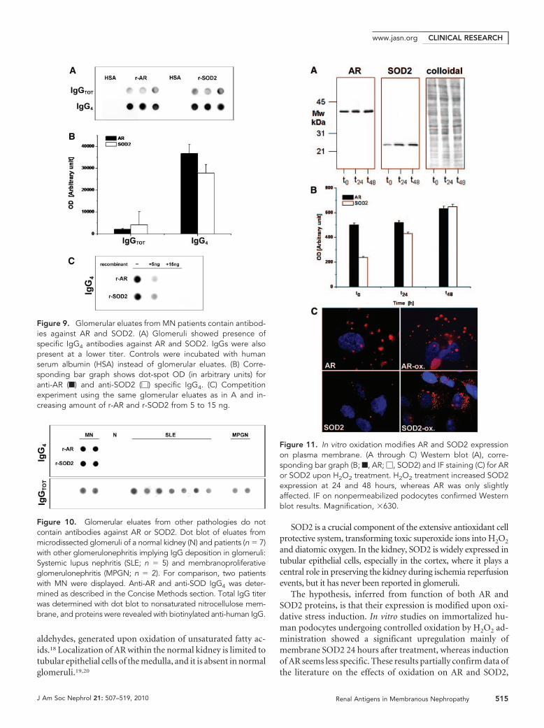
The same dot blot used for titration of circulating antibod-ies was also used for detection of specific antibodies againstr-AR and r-SOD2 in glomerular eluates. Biopsies of three dif-ferent patients with MN and other patients with glomerulone-phritis characterized by glomerular IgG deposition (five withlupus nephritis class III or IV glomerulonephritis and two withmembranoproliferative lesions) were evaluated using anti-to-tal IgG and anti-IgG subclasses 1 through 4. Results shown inFigure 9 and 10 (for IgG4) and in Supplemental Figure 6 (forother IgGs) clearly demonstrated the presence of anti-AR andanti-SOD2 IgG4 in glomeruli of patients with MN and theirabsence in other renal pathologies. Reactivity of IgG4 was abol-ished by pretreating sera with increasing amounts of r-AR orr-SOD2 (5 and 15 ng), thus confirming antibody specificity(Figure 9C).
Figure 4. AR is present in GBM and in subepithelial immunedeposits. (A and B) AR is present in GBM as shown by severalscattered gold particles in the context of electron-dense subep-ithelial immune deposits (arrows). (C through E) AR was alsoobserved in the cytoplasm and the plasma membrane of podo-cyte bodies (Po). (E) AR is not expressed at the nuclear (N) level;instead, AR is present in the cytoplasm as indicated by arrows. (F)Blank sample lacking primary antibody was totally negative (seealso Supplemental Figure 5, A, C, and E). EN, endothelium.Magnifications: �8900 in A; �56,000 in B, D, and E; �36,000 inC; �44,000 in F.
Figure 5. SOD2 is present in GBM and on podocyte plasmamembrane. (A and B) SOD2 was detected in electron-denseimmune deposits within the GBM (arrows) as shown by severalscattered gold particles. (C and D) SOD2 was also observed in thecytoplasm and on plasma membrane of podocyte bodies (Po). (F)Blank sample omitting specific primary antibody was totally neg-ative (see also Supplemental Figure 5, B, D, and F), EN, endo-thelium; n, podocyte nucleus. Magnifications: �8900 in A;�44,000 in B and C; �71,000 in D; �51,000 in E; �36,000 F.
CLINICAL RESEARCH www.jasn.org
512 Journal of the American Society of Nephrology J Am Soc Nephrol 21: 507–519, 2010
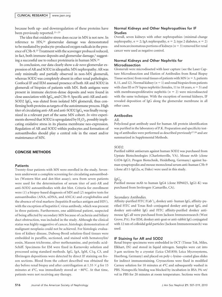
Podocyte SOD2 Expression Is a Cellular Response toOxidationAs evidenced in the previous section, immortalized humanpodocyte cell line spontaneously expresses AR (Figure 1, Dthrough F) and SOD2 (Figure 1, G through I) on plasma mem-brane (see also Supplemental Figure 1, A through C and Ethrough G). This finding was also confirmed by biotinilizationexperiments (Supplemental Figure 2). Increased SOD2 expres-sion on plasma membrane was clearly observed in vitrothrough Western blot (Figure 10, A and B) and IF (Figure 11C)24 and 48 hours after 30 minutes of incubation with 100 �M
hydrogen peroxide (H2O2). SOD2 was also increased in totalpodocyte cell lysate (Supplemental Figure 7). Increase of ARexpression on plasma membrane was not evident (Figure 11C)and was only minimally appreciable in the cytoplasm (Supple-mental Figure 7).
DISCUSSION
Primary MN is the prototype of nephropathy associated with sub-epithelial immune deposits. The mechanism responsible for their
Figure 6. AR and SOD2 co-localize with IgG4 in glomeruli of MN patient biopsies. (A, B, D, and E) Confocal images of a renal biopsyspecimen from a patient with MN. Double IF staining was evaluated for AR (A), SOD2 (D) and IgG4 (B and E). Merged images arereported in C and F and in p1 and p2. Both AR and SOD2 were co-localized with IgG4 along the capillary walls. Magnification, �630.
Figure 7. AR and SOD2 co-localize with C5b-9 in glomeruli of MN patient biopsies. Confocal images of a renal biopsy specimen fromMN. (A, B, D, and E) Double IF staining for AR (A), SOD2 (D), and C5b-9 (B and E). Merged images are reported in C and F and in p1and p2. Both AR and SOD2 co-localize in immune deposits. Magnification, �630.
CLINICAL RESEARCHwww.jasn.org
J Am Soc Nephrol 21: 507–519, 2010 Renal Antigens in Membranous Nephropathy 513
formation and trapping within GBM has been extensively inves-tigated in experimental models of HN. In this case, anti-megalinautoantibodies co-localize in glomeruli with C5b-9 and clusterin.The body of evidence for new antigens expressed in patients withMN is now growing. Debiec et al.11,12 showed that NEP emerges asantigen in patients with NEP deficiency and alloimmunizationduring pregnancy. More recently, Beck et al.16 showed specificIgG4 against PLA2R in glomerular eluates and in plasma of a rel-evant part of patients with MN.
A critical point is to define whether other podocyte antigensare recognized by IgG4 eluted from microdissected glomeruli.We used Western blot with podocyte extract in both reducingand nonreducing conditions to confirm the presence a fewantigens in podocytes. In the first approach, we could detecteight proteins reacting with glomerular IgG4; in nonreducingconditions, three more proteins with a higher molecularweight were recognized, one of which had the same dimensionof PLA2R. Podocyte antigens recognized by electroeluted(from glomeruli) and circulating IgG4 were characterized bymass spectrometry and matched to extend studies with IEMand IF to other patients with MN. AR and SOD2 emerged asthe two major podocyte components recognized by both cir-culating and glomerular IgG4. In particular, we found amarked increase in levels of circulating anti-AR and anti-SOD2IgG4 in patients with MN compared with normal sera or other
idiopathic nephrotic syndromes (FSGS).Moreover, de novo expression of both ARand SOD2 and co-localization with IgG4
and C5b-9 were observed in MN biopsieswhile being absent in other glomerularpathologies. Ultrastructural study clearlyassessed localization of AR and SOD2 inpodocyte cytoplasm and foot processesand in electron-dense subepithelial im-mune deposits. Taken together, thesefindings demonstrate that AR and SOD2are neo-expressed proteins in MN glo-meruli and that both are recognized byspecific circulating antibodies that canalso be detected in glomerular subepi-thelial immune deposits. The preva-lence of IgG4 in glomerular immunedeposits confirms the concept of an iso-type-specific deposition mechanism;therefore, AR and SOD2 should beconsidered glomerular antigens impli-cated in the immune process and actu-ally belong, with NEP and PLA2R andwith the new panel of antigens cur-rently under investigation (G.M.G.,personal observation), to the list of an-tigens recognized in human MN.
A crucial point still remains unre-solved. Our data, in fact, cannot be con-sidered exhaustive with respect to the
initial trigger event of MN in humans. Neo-expression of bothAR and SOD2 may, in fact, represent a continuation mecha-nism after an initiation phase in which the basic event is dep-osition of anti-PLA2AR, anti-NEP, and/or other, unrecog-nized antibodies. The basic issue regarding the significance ofboth neo-antigens in glomeruli of patients with MN will re-quire further investigation, although the presence of anti-ARand anti-SOD2 IgG4 in glomeruli at the time of diagnosis ofMN and its absence in other autoimmune renal pathologiesstrongly supports a causal role in MN pathogenesis. A defini-tive explanation will mostly rely on animal models overexpressingAR and SOD2. Ongoing experiments for AR are experiencingproblems and will demand more time to be set up because of atoxic effect of AR overexpression (data not shown).
Despite the previously highlighted doubts, our data supportthe concept that neo-expression of AR and SOD2 may represent akey event in the natural history of the disease. For this purpose, aseries of preliminary in vitro experiments aimed at defining themechanisms regulating glomerular expression of both neo-anti-gens were set up from already known data about their structureand function. AR belongs to the family of aldo-keto reductasescatalyzing NADPH-dependent reduction of aliphatic and aro-matic aldehydes and ketones. Its specific role is to convert glucoseinto sorbitol, and it is involved in regulation of tissue tonicity andosmolality.17 The enzyme also catalyzes the transformation of
Figure 8. Glomerular eluates from MN patients contain antibodies against podocyteantigens. Podocyte extracts were separated by electrophoresis in reducing (a through e)and nonreducing (f through h) conditions and were studied for reactivity with eluates frommicrodissected glomeruli. Thirty micrograms of protein was used for the analysis: Part wasanalyzed with silver stain (a and f), and part was incubated with glomerular eluates frompatients with MN and then developed with anti-IgG4 (b and g). Eight antigens wererecognized in reducing conditions and characterized by mass spectrometry as AR, SOD,and vimentin. Three more bands with a molecular weight of 200, 170, and 155 kD weredetected in nonreducing conditions. (c and h) Control lines for Western blot in whichglomerular eluates were omitted. (d) Western blot with anti-AR antibody. (e) Western blotwith SOD2 antibodies.
CLINICAL RESEARCH www.jasn.org
514 Journal of the American Society of Nephrology J Am Soc Nephrol 21: 507–519, 2010
aldehydes, generated upon oxidation of unsaturated fatty ac-ids.18 Localization of AR within the normal kidney is limited totubular epithelial cells of the medulla, and it is absent in normalglomeruli.19,20
SOD2 is a crucial component of the extensive antioxidant cellprotective system, transforming toxic superoxide ions into H2O2
and diatomic oxygen. In the kidney, SOD2 is widely expressed intubular epithelial cells, especially in the cortex, where it plays acentral role in preserving the kidney during ischemia reperfusionevents, but it has never been reported in glomeruli.
The hypothesis, inferred from function of both AR andSOD2 proteins, is that their expression is modified upon oxi-dative stress induction. In vitro studies on immortalized hu-man podocytes undergoing controlled oxidation by H2O2 ad-ministration showed a significant upregulation mainly ofmembrane SOD2 24 hours after treatment, whereas inductionof AR seems less specific. These results partially confirm data ofthe literature on the effects of oxidation on AR and SOD2,
Figure 10. Glomerular eluates from other pathologies do notcontain antibodies against AR or SOD2. Dot blot of eluates frommicrodissected glomeruli of a normal kidney (N) and patients (n � 7)with other glomerulonephritis implying IgG deposition in glomeruli:Systemic lupus nephritis (SLE; n � 5) and membranoproliferativeglomerulonephritis (MPGN; n � 2). For comparison, two patientswith MN were displayed. Anti-AR and anti-SOD IgG4 was deter-mined as described in the Concise Methods section. Total IgG titerwas determined with dot blot to nonsaturated nitrocellulose mem-brane, and proteins were revealed with biotinylated anti-human IgG.
Figure 9. Glomerular eluates from MN patients contain antibod-ies against AR and SOD2. (A) Glomeruli showed presence ofspecific IgG4 antibodies against AR and SOD2. IgGs were alsopresent at a lower titer. Controls were incubated with humanserum albumin (HSA) instead of glomerular eluates. (B) Corre-sponding bar graph shows dot-spot OD (in arbitrary units) foranti-AR (f) and anti-SOD2 (�) specific IgG4. (C) Competitionexperiment using the same glomerular eluates as in A and in-creasing amount of r-AR and r-SOD2 from 5 to 15 ng.
Figure 11. In vitro oxidation modifies AR and SOD2 expressionon plasma membrane. (A through C) Western blot (A), corre-sponding bar graph (B; f, AR; �, SOD2) and IF staining (C) for ARor SOD2 upon H2O2 treatment. H2O2 treatment increased SOD2expression at 24 and 48 hours, whereas AR was only slightlyaffected. IF on nonpermeabilized podocytes confirmed Westernblot results. Magnification, �630.
CLINICAL RESEARCHwww.jasn.org
J Am Soc Nephrol 21: 507–519, 2010 Renal Antigens in Membranous Nephropathy 515
because both up- and downregulation of these proteins havebeen previously reported.21–23
The idea that oxidative stress does occur in MN is not new. Inreference to HN,24 glomerular damage was demonstratedto be mediated by podocyte-produced oxygen radicals in the pres-ence of C5b-9.25 Treatment with the scavenger probucol reduced,in fact, both immune deposits and glomerular damage,3 suggest-ing a successful use to reduce proteinuria in human MN.26
In conclusion, our data clearly show a de novo glomerular ex-pression of AR and SOD2 in idiopathic MN nephropathy. AR wasonly minimally and partially observed in non-MN glomeruli,whereas SOD2 was completely absent in other renal pathologies.Confocal IF and IEM assessed presence of both AR and SOD2 inglomeruli of biopsies of patients with MN. Both antigens werepresent in immune electron-dense deposits and were found inclose association with IgG4 and C5b-9. Specific anti-AR and anti-SOD2 IgG4 was eluted from isolated MN glomeruli, thus con-firming both proteins as targets of the autoimmune process. Hightiter of circulating anti-AR and anti-SOD2 IgG4 was finally recog-nized in a relevant part of the same MN cohort. In vitro experi-ments showed that SOD2 is upregulated by H2O2, possibly impli-cating oxidative stress in its plasma membrane neo-expression.Regulation of AR and SOD2 within podocytes and formation ofautoantibodies should play a central role in the onset and/ormaintenance of MN.
CONCISE METHODS
PatientsTwenty-four patients with MN were enrolled in the study. Seven-
teen underwent a complete screening for circulating autoantibod-
ies (Western blot and dot-blot assay); sera from seven patients
were used for the determination of serum titer of anti-AR and
anti-SOD2 autoantibodies with dot blot. Criteria for enrollment
were (1) a biopsy-based diagnosis of MN and (2) negative tests for
autoantibodies (ANA, nDNA, and ANCA) and cryoglobulins and
the absence of viral markers (hepatitis B surface antigen and HIV),
with the exception of hepatitis C virus antibody, which was present
in three patients. Furthermore, one additional patient, suspected
of being affected by secondary MN because of cachexia and biliary
duct obstruction, was included in the study. Although the clinical
status was highly suggestive of cancer, histologic demonstration of
malignant neoplasia could not be achieved. For histologic evalua-
tion of kidney disease, Dubosq-Bresil solution-fixed tissues were
embedded in paraffin, sectioned, and stained with hematoxylin/
eosin, Masson trichrome, silver methenamine, and periodic acid-
Schiff. Specimens for EM were fixed in Karnovsky solution and
processed using standard methods. IgG, IgA, IgM, C1q, C3, and
fibrinogen depositions were detected by direct IF staining on fro-
zen sections. Blood from the cohort described was obtained the
day before renal biopsy and after centrifugation at 1.55 � g for 15
minutes at 4°C, was immediately stored at �80°C. At that time,
patients were not receiving any therapy.
Normal Kidneys and Other Nephropathies for IFStudiesOverall, seven kidneys with other nephropathies (minimal-change
nephropathy, n � 2; IgA nephropathy, n � 2; type 2 diabetes, n � 2)
and noncarcinomatous portions of kidneys (n � 3) removed for renal
cancer were used as negative control.
Normal Kidneys and Other Nephritis forMicrodissectionGlomeruli were microdisseted with laser capture (see the Laser Cap-
ture Microdissection and Elution of Antibodies from Renal Biopsy
Tissue section) from renal tissues of patients with MN (n � 3, patients
8, 11, and 12). Normal kidney (n � 1) and renal biopsies from patients
with class III or IV lupus nephritis (females, 11 to 18 years, n � 5) and
with membranoproliferative nephritis (n � 2) were microdissected
with the same technique. With the exception of normal kidneys, IF
revealed deposition of IgG along the glomerular membrane in all
other cases.
AntibodiesAR.Polyclonal goat antibody used for human AR protein identification
was purified in the laboratory of P.K. Preparation and specificity test-
ing of antibodies were performed as described previously27,28 and are
summarized in Supplemental Methods.
SOD2.Purified rabbit antiserum against human SOD2 was purchased from
Upstate Biotechnologies (Charlottesville, VA). Mouse mAb (clone
G1D4-IgG1; Progen Biotechnik, Heidelberg, Germany) against hu-
man synaptopodin and mouse monoclonal serum anti-human C5b-9
(clone aE11-IgG2a, �; Dako) were used in this study.
IgG4.Purified mouse mAb to human IgG4 (clone HP6025, IgG1-K) was
purchased from Invitrogen (Camarillo, CA).
Secondary Antibodies.Affinity-purified FITC F(ab�)2 donkey anti- human IgG, affinity-pu-
rified FITC and Texas Red– conjugated donkey anti-goat IgG, and
donkey anti-rabbit IgG and FITC affinity-purified donkey anti-
mouse IgG all were purchased from Jackson Immunoresearch (West
Grove, PA). For IEM, donkey anti-goat or anti-rabbit IgG conjugated
with 12 nm of colloidal gold particles (Jackson Immunoresearch) was
used.
IF Staining for AR and SOD2Renal biopsy specimens were embedded in OCT (Tissue Tek, Miles,
Elkhart, IN) and stored in liquid nitrogen. Samples were cut into
3-�m sections by a cryostat (Leica CM1850; Leica Mycrosystems,
Heerbrog, Germany) and placed on poly-L-lysine– coated glass slides
for indirect immunostaining. Cryosections were fixed in modified
Carnoy solution for 10 minutes at 4°C and subsequently washed in
PBS. Nonspecific binding was blocked by incubation in BSA 3% wt/
vol in PBS for 20 minutes at room temperature. Sections were then
CLINICAL RESEARCH www.jasn.org
516 Journal of the American Society of Nephrology J Am Soc Nephrol 21: 507–519, 2010
incubated for 2 hours at room temperature with primary goat anti-AR
or rabbit anti-SOD2 antibody diluted in PBS 1:150 or 1:50, respec-
tively.
FITC-conjugated donkey IgG anti-goat (1:50) and FITC-conju-
gated donkey IgG anti-rabbit (1:20) were used as secondary antibod-
ies. Competition experiments performed with anti-AR or anti-SOD2
antibodies preincubated for 4 hours at 37°C with an excess of AR or
SOD2 protein, respectively, gave negative results (Supplemental Fig-
ure 4). Negative controls were processed in parallel using an equiva-
lent concentration of a normal goat or rabbit antiserum as primary
antibody, respectively.
Co-localization and Confocal Microscopy AnalysisFor double-staining indirect IF and co-localization analysis, Carnoy
fixed cryosections were incubated with blocking solution (3% wt/vol
BSA in PBS) and in succession with FITC donkey anti-human IgG
F(ab�)2 (1:20) for 2 hours at room temperature. After additional PBS
washes, slides were then exposed to a diluted polyclonal goat serum
anti-AR (1:150) or rabbit serum anti-SOD2 (1:50) for 2 hours at room
temperature. The presence of AR and SOD2 proteins was revealed by
incubation of Texas Red– conjugated donkey IgG anti-goat and anti-
rabbit, respectively. IgG4 deposits were characterized using purified
anti-human IgG4 mouse mAb (Invitrogen).
For C5b-9 and AR or SOD2 co-localization, Carnoy fixed sections
were incubated in primary step with anti–C5b-9 mAb diluted (1:10)
for 2 hours at room temperature and revelated by FITC-conjugated
donkey IgG anti-mouse diluted (1:20) in PBS. AR and SOD2 were
detected as described already. Samples were observed using a confocal
system (LSM 510 Meta scan head integrated with the Axiovert 200 M
inverted microscope; Carl Zeiss, Jena, Germany) with a �63 oil ob-
jective. Image acquisition was carried out in multitrack mode, namely
through consecutive and independent optical pathways.
Electron MicroscopySmall fragments of the kidney biopsies were fixed in Karnovsky solu-
tion and processed according to standard methods omitting postfix-
ation in osmium tetroxide.29 Osmication of tissues was omitted to
avoid masking of antigenic sites as previously reported in the litera-
ture.30
After exposure with a blocking agent (BSA 3% wt/vol in PBS),
ultrathin sections were incubated overnight with anti-AR (diluted
1:200) and anti-SOD2 (diluted 1:50) antibodies. Immune reaction
was then revealed using donkey anti-goat or anti-rabbit IgG conju-
gated with 12 nm of colloidal gold particles.
The specificity and validity of the labeling were verified by substi-
tuting primary antibody with the same antibody preincubated with
nonimmune goat or rabbit serum (see Supplemental Figure 5) or
alternatively be substituting primary antibody with PBS. After coun-
terstaining with uranyl acetate and lead citrate, ultrathin sections were
examined and micrographs were taken using a Philips EM208 trans-
mission electron microscope (Philips Electron Optics, FEI, Eind-
hoven, Netherlands).
Cell CultureA human conditionally immortalized podocyte cell line31 was cul-
tured in RPMI 1640 supplemented with 10% inactivated FCS, insulin
transferrin selenium, 100 U/ml penicillin, and 100 mg/ml streptomy-
cin. Cells were expanded at 33°C. For IF, cells were plated in 6-cm
Petri dishes at a density of 3.5 ° 103 cells/cm2 and differentiated for 15
days at 37°C in 5% CO2/95% air. The same cell line was used for the
oxidation experiments (see the In Vitro Oxidation section).
Detection of Circulating Antibodies against PodocyteProteinsDetection of anti-podocyte antibodies in sera of patients with MN was
assessed by Western blot of podocyte cell line membrane extracts
separated by 2D electrophoresis (see supplemental material). Detailed
technique for transblot and detection is described in the Supplement
Methods section. For membrane preparation, subconfluent cells were
homogenized in cold hypotonic solution containing 5 mM phosphate
buffer (pH 7.4), 0.15% wt/vol n-Hexyl �-D-glucopyranoside (Calbio-
chem), and protease inhibitor cocktail. Homogenates were centri-
fuged at 14,000 rpm for 15 minutes, and supernatant containing cy-
toplasm proteins was discarded. Pellets were resuspended in 150 mM
NaCl, 0.5% vol/vol Triton X-100, 0.25% wt/vol natrium desoxy-
cholate, and 50 mM phosphate buffer (pH 7.4) and centrifuged at
14000 rpm for 15 minutes. Supernatants containing membrane pro-
teins were collected, and protein concentration was determined using
BCA assay.
2D Electrophoresis2D electrophoresis was performed in soft gels as described previ-
ously32 (see the Supplemental Methods section).
Gel/Membrane Staining and Image AnalysisAfter separation in SDS-PAGE gels, proteins were visualized by a dou-
ble-staining procedure: First, methyl-trichloroacetate–negative stain-
ing,33 then silver staining or Blue silver colloidal or Coomassie for
preparative mass spectrometry analysis (see the Supplemental Meth-
ods section).
Tryptic Digestion and Protein Identification by MALDI-TOFDetails of MALDI-TOF are given in the Supplemental Methods sec-
tion.
Dot-Blot Analysis for Circulating Anti-AR and Anti-SOD2 LevelsThe titer of circulating autoantibodies (IgG4) in sera of patients with
MN (n � 24), normal control subjects (n � 24), or children with
idiopathic nephrotic syndrome (n � 8) was determined with dot blot
using r-AR (300 ng) and r-SOD2 (300 ng) as immobilized antigens.
Purity of both recombinant proteins was confirmed by monodimen-
sional electrophoresis (Supplemental Figure 3). The technique for dot
blot is described in the Supplemental Methods section.
Laser Capture Microdissection and Elution ofAntibodies from Renal Biopsy TissueCryostatic sections (5 �m) of kidney tissue specimens were placed on
metal frame slides with thermoplastic membrane (Molecular Ma-
chines & Industries AG; Glattburg, Zurich, Switzerland), stained, and
CLINICAL RESEARCHwww.jasn.org
J Am Soc Nephrol 21: 507–519, 2010 Renal Antigens in Membranous Nephropathy 517
dehydrated using an Arcturus HistoGene, LCM Frozen Section Stain-
ing Kit (Arcturus Bioscience, Mountain View, CA) according to the
manufacturer’s instructions. Air-dried sections were then viewed
with the NIKON ECLIPSE-TE 2000 inverted microscope (Nikon-
Instruments, Sesto Fiorentino, Italy). Glomeruli were identified and
isolated with the Molecular Machines & Industries Cellcut LMD sys-
tem by focal melting of the membrane through laser activation. The
Molecular Machines & Industries Cellcut Laser Capture Microdissec-
tion system is equipped with a solid-state ultraviolet laser that guar-
antees precise cutting without damaging the tissue. High precision
xy-stage and CCD camera allow identification, documentation, and
dissection of multiple regions of interest from the same tissue speci-
men. For each specimen, a total of 25 to 30 glomeruli were microdis-
sected and removed sequentially in separate isolation cap (Nikon In-
struments) with special adhesive material in the lid.
After visual control of the completeness of dissection, captured
tissue was immersed in denaturation buffer and used for proteomic
analysis. Sections of human kidney derived from noncarcinomatous
portions of kidneys removed for renal carcinoma were used as nega-
tive control. Igs were recovered from glomeruli by means of acid
elution as described previously.34 Briefly, after washings with PBS
(0.01 M, pH 7.2) groups of 20 glomeruli were incubated overnight
with 0.02 M citrate buffer (pH 3.2) at 4°C in a humid atmosphere.
After removal of citrate buffer, 0.4 M NaOH was added to achieve a
pH of 7.2. Dot-blot analysis for anti-AR and anti-SOD2 IgG and IgG4
in glomerular eluates was performed as already described (see also the
Supplemental Methods section). Total IgGs were determined by the
same dot-blot assay in which 50 �l of sample had been applied to
nonsaturated nitrocellulose membranes and IgGs were revealed with
biotinylated anti-human IgG antibodies.
Cell StudiesFor IF, human podocyte cell lines were gently washed in RPMI/2%
HEPES buffer and fixed for 20 minutes at 37°C in 1% paraformalde-
hyde dissolved in RPMI/2% HEPES buffer. Details for cell culturing
and interaction with MN serum are given in the supplemental mate-
rial.
Podocyte Surface Biotin LabelingSubconfluent adherent cells were rinsed three times in ice-cold PBS,
and cell surfaces were labeled with the appropriate volume of 230 �M
EZ-linf sulfo-NHS-LC-LC-biotin (Pierce, Rockford, IL). The reac-
tion was performed for 5 minutes at 4°C. Cells were rinsed three times
in ice-cold TBS to quench and remove the excess of biotin reagent.
Membranes were prepared as described in the section Detection of
Circulating Antibodies against Podocyte Proteins. Podocyte mem-
brane extracts (200 �g) were run on a 2D electrophoresis as described
in the supplemental material and transferred to a nitrocellulose mem-
brane saturated in 3% wt/vol BSA in TBS and incubated with 50 pg/ml
Neutravidin in 1% wt/vol BSA in TBS-Tween 0.15% vol/vol (TBS-T).
Chemiluminescence was used for immunodetection. Images were
digitalized by VersaDoc 4000 (Bio-Rad, Hercules, CA) and analyzed
with QuantityOne and PDQuest software (Bio-Rad).
In Vitro OxidationFor in vitro oxidation, human podocyte cell line was maintained in RPMI
1640 medium with 10% vol/vol FCS, 20 mM glutamine, 100 IU/ml pen-
icillin, 100 IU/ml streptomycin, 5 �g/ml insulin, 5 �g/ml transferrin, and
5 ng/ml sodium selenite at 37°C in a 10% CO2 humidified incubator.
Cells at 70 to 80% confluence were rinsed twice with serum-deprived
RPMI 1640 and stimulated with 125 �M H2O2 for 0, 18, or 24 hours by
addition to the growth serum-deprived media. Cells were harvested after
the indicated times, rinsed three times with PBS, and lysed in 2D buffer
(see the two-dimensional electrophoresis section in the Supplemental
Methods). Membrane preparation was done following the same proce-
dure described already.
Statistical AnalysisNonparametric Mann-Whitney U test was used for comparison of
anti-AR and anti-SOD2 IgG4 serum titer in patients with MN versus
control subjects. Results were given as median. Parametric t test was
used for comparing results deriving from morphometric analysis of
renal AR and SOD2 expression. Results were give as means � SEM.
ACKNOWLEDGMENTS
This work was supported by the Italian Ministry of Health, by
E-RARE Project “PodoNet: EU Consortium for Clinical, Genetic and
Experimental Research into Hereditary Diseases of Podocyte,” and by
the Renal Child Foundation.
We are grateful to Dr. M.A. Saleem for giving us human podocyte
cell lines. We also acknowledge Fondazione Mara Wilma e Bianca
Querci for financial support to the project “Nuove evoluzioni sulla
multifattorialita della sindrome nefrosica.” We gratefully acknowledge
the support of the Foundation Guido Erluison for making available
the laser capture microdissection system. Data were critically dis-
cussed with Prof. Rosanna Gusmano. The manuscript was revised by
Miss Capurro.
DISCLOSURESNone.
REFERENCES
1. Wasserstein AG: Membranous glomerulonephritis. J Am Soc Nephrol8: 664–674, 1997
2. Kerjaschki D: Pathomechanisms and molecular basis of membranousglomerulopathy. Lancet 364: 1194–1196, 2004
3. Neale TJ, Ojha PP, Exner M, Poczewski H, Ruger B, Witztum JL, DavisP, Kerjaschki D: Proteinuria in passive Heymann nephritis is associatedwith lipid peroxidation and formation of adducts on type IV collagen.J Clin Invest 94: 1577–1584, 1994
4. Neale TJ, Ullrich R, Ojha P, Poczewski H, Verhoeven AJ, Kerjaschki D:Reactive oxygen species and neutrophil respiratory burst cytochromeb558 are produced by kidney glomerular cells in passive Heymannnephritis. Proc Natl Acad Sci U S A 90: 3645–3649, 1993
5. Rastaldi MP, Candiano G, Musante L, Bruschi M, Armelloni S, RimoldiL, Tardanico R, Sanna-Cherchi S, Ferrario F, Montinaro V, Haupt R,
CLINICAL RESEARCH www.jasn.org
518 Journal of the American Society of Nephrology J Am Soc Nephrol 21: 507–519, 2010
Parodi S, Carnevali ML, Allegri L, Camussi G, Gesualdo L, Scolari F,Ghiggeri GM: Glomerular clusterin is associated with PKC-alpha/betaregulation and good outcome of membranous glomerulonephritis inhumans. Kidney Int 70: 477–485, 2006
6. Kerjaschki D, Miettinen A, Farquhar MG: Initial events in the formationof immune deposits in passive Heymann nephritis: gp330-anti-gp330immune complexes form in epithelial coated pits and rapidly becomeattached to the glomerular basement membrane. J Exp Med 166:109–128, 1987
7. Kerjaschki D, Ullrich R, Diem K, Pietromonaco S, Orlando RA,Farquhar MG: Identification of a pathogenic epitope involved ininitiation of Heymann nephritis. Proc Natl Acad Sci U S A 89:11179 –11183, 1992
8. Heymann W, Hackel DB, Harwood S, Wilson SG, Hunter JL: Produc-tion of nephrotic syndrome in rats by Freund’s adjuvants and ratkidney suspensions. Proc Soc Exp Biol Med 100: 660–664, 1959
9. Assmann KJ, van Son JP, Dijkman HB, Koene RA: A nephritogenic ratmonoclonal antibody to mouse aminopeptidase A: Induction of mas-sive albuminuria after a single intravenous injection. J Exp Med 175:623–635, 1992
10. Ronco P, Allegri L, Brianti E, Chatelet F, Van Leer EH, Verroust P:Antigenic targets in epimembranous glomerulonephritis: Experimen-tal data and potential application in human pathology. Appl Pathol 7:85–98, 1989
11. Debiec H, Guigonis V, Mougenot B, Decobert F, Haymann JP, Bens-man A, Deschenes G, Ronco PM: Antenatal membranous glomerulo-nephritis due to anti-neutral endopeptidase antibodies. N Engl J Med346: 2053–2060, 2002
12. Debiec H, Nauta J, Coulet F, van der Burg M, Guigonis V, SchurmansT, de Heer E, Soubrier F, Janssen F, Ronco P: Role of truncatingmutations in MME gene in fetomaternal alloimmunisation and ante-natal glomerulopathies. Lancet 364: 1252–1259, 2004
13. Allegri L: Antigens in experimental models of membranous nephrop-athy: Are they involved in human disease? Nephrol Dial Transplant 12:1801–1804, 1997
14. Ronco P, Debiec H: Target antigens and nephritogenic antibodies inmembranous nephropathy: Of rats and men. Semin Immunopathol 29:445–458, 2007
15. Bruschi M, Candiano G, Murtas C, Prunotto M, Santucci L, CarnevaliML, Scolari F, Allegri L, Ghiggeri GM: Patients with primary membra-nous nephropathy lack auto-antibodies against LDL receptor, thehomologue of megalin in human glomeruli. NDT Plus sfp002, 2009
16. Beck LH Jr, Bonegio RG, Lambeau G, Beck DM, Powell DW, CumminsTD, Klein JB, Salant DJ: M-type phospholipase A2 receptor as targetantigen in idiopathic membranous nephropathy. N Engl J Med 361:11–21, 2009
17. Williamson JR, Chang K, Frangos M, Hasan KS, Ido Y, Kawamura T,Nyengaard JR, van den Enden M, Kilo C, Tilton RG: Hyperglycemicpseudohypoxia and diabetic complications. Diabetes 42: 801–813, 1993
18. Srivastava S, Spite M, Trent JO, West MB, Ahmed Y, Bhatnagar A:Aldose reductase-catalyzed reduction of aldehyde phospholipids.J Biol Chem 279: 53395–53406, 2004
19. Barski OA, Papusha VZ, Ivanova MM, Rudman DM, Finegold MJ:Developmental expression and function of aldehyde reductase in
proximal tubules of the kidney. Am J Physiol Renal Physiol 289:F200–F207, 2005
20. Terubayashi H, Sato S, Nishimura C, Kador PF, Kinoshita JH: Local-ization of aldose and aldehyde reductase in the kidney. Kidney Int 36:843–851, 1989
21. Spycher SE, Tabataba-Vakili S, O’Donnell VB, Palomba L, Azzi A:Aldose reductase induction: A novel response to oxidative stress ofsmooth muscle cells. FASEB J 11: 181–188, 1997
22. Son D, Kojima I, Inagi R, Matsumoto M, Fujita T, Nangaku M: Chronichypoxia aggravates renal injury via suppression of Cu/Zn-SOD: Aproteomic analysis. Am J Physiol Renal Physiol 294: F62–F72, 2008
23. Zhao W, Zhao T, Chen Y, Ahokas RA, Sun Y: Oxidative stress mediatescardiac fibrosis by enhancing transforming growth factor-beta1 inhypertensive rats. Mol Cell Biochem 317: 43–50, 2008
24. Heymann W, Lund HZ, Hackel DB: The nephrotic syndrome in rats;with special reference to the progression of the glomerular lesion andto the use of nephrotoxic sera obtained from ducks. J Lab Clin Med39: 218–224, 1952
25. Adler S, Baker PJ, Pritzl P, Couser WG: Detection of terminal comple-ment components in experimental immune glomerular injury. KidneyInt 26: 830–837, 1984
26. Haas M, Kerjaschki D, Mayer G: Lipid-lowering therapy in membra-nous nephropathy. Kidney Int Suppl 71: S110–S112, 1999
27. Sato S, Old S, Carper D, Kador PF: Purification and characterization ofrecombinant human placental and rat lens aldose reductases ex-pressed in Escherichia coli. Adv Exp Med Biol 372: 259–268, 1995
28. Herrmann RK, Kador PF, Kinoshita JH: Rat lens aldose reductase:Rapid purification and comparison with human placental aldose re-ductase. Exp Eye Res 37: 467–474, 1983
29. D’Adda T, Bertele A, Pilato FP, Bordi C: Quantitative electron micros-copy of endocrine cells in oxyntic mucosa of normal human stomach.Cell Tissue Res 255: 41–48, 1989
30. Bendayan M, Reddy MK, Hashimoto T, Reddy JK: Immunocytochem-ical localization of fatty acid metabolizing heat-stable and heat-labileenoyl-coenzyme A (CoA) hydratases in liver and renal cortex. J Histo-chem Cytochem 31: 509–516, 1983
31. Saleem MA, O’Hare MJ, Reiser J, Coward RJ, Inward CD, Farren T,Xing CY, Ni L, Mathieson PW, Mundel P: A conditionally immortalizedhuman podocyte cell line demonstrating nephrin and podocin expres-sion. J Am Soc Nephrol 13: 630–638, 2002
32. Bruschi M, Musante L, Candiano G, Ghiggeri GM, Herbert B, Anto-nucci F, Righetti PG: Soft immobilized pH gradient gels in proteomeanalysis: a follow-up. Proteomics 3: 821–825, 2003
33. Candiano G, Bruschi M, Musante L, Santucci L, Ghiggeri GM,Carnemolla B, Orecchia P, Zardi L, Righetti PG: Blue silver: A verysensitive colloidal Coomassie G-250 staining for proteome analysis.Electrophoresis 25: 1327–1333, 2004
34. Feltkamp TE, Boode JH: Elution of antibodies from biopsy tissue.J Clin Pathol 23: 629–631, 1970
Supplemental information for this article is available online at http://www.jasn.org/.
CLINICAL RESEARCHwww.jasn.org
J Am Soc Nephrol 21: 507–519, 2010 Renal Antigens in Membranous Nephropathy 519