role of perforin in controlling b-cell hyperactivity and humoral autoimmunity
TRANSCRIPT
IntroductionCytotoxic T lymphocyte (CTL) effector mechanismsinclude the Fas (APO-1/CD95)/FasL,perforin/granzyme, and the slower TNF-mediatedpathways (1–3). It has been suggested that the rapidlyresponding pathways segregate with their functionsuch that the perforin/granzyme pathway plays amajor role in viral clearance (4) and tumor surveillance(5), whereas the Fas/FasL pathway plays a pivotal rolein immunoregulation and homeostasis through dele-tion of activated cells of the immune system (6).Recent studies, however, have questioned thisdichotomy, as Fas-induced apoptosis has been shownto contribute to viral clearance (4, 7), and Fas-deficient(lpr) mice that were also perforin deficient had areduced survival compared with lpr mice (8). These lat-ter results raise the possibility that perforin may playa role in immunoregulation and homeostasis in addi-tion to its well-documented role in viral clearance.
A well-established model of immunoregulation is theparent-into-F1 model of graft-versus-host disease(GVHD) (reviewed in ref. 9). In this model, activationof both donor CD4+ and CD8+ T cells results in acuteGVHD characterized by donor antihost CTLs that
eliminate activated autoreactive host B cells. In addi-tion, activated donor T cells upregulate Fas/FasL (10)and are eliminated, presumably by activation-inducedcell death (AICD), such that by 4 weeks after injection,donor cells are barely detectable (11) and lymphocytehomeostasis is restored. By contrast, in chronic GVHD,anti-host CTL activity is minimal, Fas/FasL expressionon donor T cells is reduced, and continued activationof host B cells by donor CD4+ helper T cells occurs (10).The persistence of donor CD4 T cells coupled with thefailure to eliminate self-reactive B cells results in sus-tained autoantibody production and immune complexglomerulonephritis (9). Of note, reagents that selec-tively block donor CD8+ T cell development intomature antihost CTLs without attenuating donorCD4+ T cell maturation (e.g., anti–IL-2 mAb) promotethe evolution of chronic GVHD (12).
We have previously shown that antihost CTLs gener-ated in acute GVHD utilize both perforin and Fas path-ways in vitro (10). To address the in vivo role of perforinin the regulation of humoral autoimmunity, perforin-deficient (pfp) donor cells were used in a P→F1 combi-nation that normally results in acute GVHD. Our resultsindicate that although pfp donor T cells can upregulate
The Journal of Clinical Investigation | September 2000 | Volume 106 | Number 6 R39
Role of perforin in controlling B-cell hyperactivity and humoral autoimmunity
Andrei Shustov,1 Irina Luzina,1 Phuong Nguyen,1 John C. Papadimitriou,2
Barry Handwerger,1 Keith B. Elkon,3 and Charles S. Via1
1Research Service, Department of Veteran Affairs Medical Center and Division of Rheumatology and Clinical Immunology, and2Pathology Department, University of Maryland School of Medicine, Baltimore, Maryland, USA3Hospital for Special Surgery, Weill Medical College of Cornell University, New York, New York, USA
Address correspondence to: Charles S. Via, Division of Rheumatology and Clinical Immunology, MSTF 8-34, 10 S. Pine Street, University of Maryland School of Medicine Baltimore, Maryland 21201, USA. Phone: (410) 706-6474; Fax: (410) 706-0231; E-mail: [email protected].
Received for publication November 8, 1999, and accepted in revised form July 25, 2000.
To determine the role of perforin-mediated cytotoxic T lymphocyte (CTL) effector function inimmune regulation, we studied a well-characterized mouse model of graft-versus-host disease(GVHD). Induction of acute GVHD using perforin-deficient donor T cells (pfp→F1) initially result-ed in features of acute GVHD, e.g., engraftment of both donor CD4+ and CD8+ T cells, upregulationof Fas and FasL, production of antihost CTL, and secretion of both Th1 and Th2 cytokines. Despitefully functional FasL activity, pfp donor cells failed to totally eliminate host B cells, and, by 4 weeksof disease, cytokine production in pfp→F1 mice had polarized to a Th2 response. Pfp→F1 mice even-tually developed features of chronic GVHD, such as increased numbers of B cells, persistence ofdonor CD4 T cells, autoantibody production, and lupuslike renal disease. We conclude that in thesetting of B- and T-cell activation, perforin plays an important immunoregulatory role in the pre-vention of humoral autoimmunity through the elimination of both autoreactive B cells and ag-spe-cific T cells. Moreover, an ineffective initial CTL response can evolve into a persistent antibody-medi-ated response and, with it, the potential for sustained humoral autoimmunity.
This article may have been published online in advance of the print edition. The date of publication is available from the JCI website, http://www.jci.org. J. Clin. Invest. 106:R39–R47 (2000).
Online firstPUBLICATION
Online firstPUBLICATION

Fas/FasL and mature into antihost CTLs, the elimina-tion of host B cells and return to homeostasis throughthe reduction of donor T cells are defective. As a result,the balance between B-cell proliferation and eliminationis shifted to favor the development of a lupuslike disease.
MethodsMice. C57BL/6J wild-type (B6wt), perforin-deficientC57BL/6-Pfptm/Sdz (B6pfp) (4), DBA/2 and B6D2F1/J(BDF1) male mice, 6–8 weeks of age, were purchased fromThe Jackson Laboratories (Bar Harbor, Maine, USA).
GVHD induction. Single-cell suspensions of viable cells(trypan blue excluding) were prepared as described pre-viously (13) and used as a source of donor cells. Unlessotherwise noted, unirradiated BDF1 mice received 50× 106 donor B6wt (acute GVHD), 50 × 106 B6pfp (pfpGVHD), or 90 × 106 DBA/2 (chronic GVHD) spleencells intravenously, doses that reliably induce acute andchronic GVHD (14). Control mice consisted of unin-jected age- and sex-matched F1 mice.
RT-PCR. The coupled RT-PCR reaction was used asdescribed previously (15). Briefly, RNase-free plasticand water were used throughout the assay. Spleen cellsuspensions were homogenized in RNA-STAT-60 (Tel-Test, Friendswood, Texas, USA) at 1 mL/107 cells. RNAsamples were reverse transcribed with M-MLV-RT (LifeTechnologies Inc., Grand Island, New York, USA). FasLspecific primers were used for amplification asdescribed elsewhere (10). To verify that equal amountsof RNA were added in each RT-PCR reaction within anexperiment, primers for the housekeeping gene, hypox-anthine phosphoribosyl transferase (HPRT), wereamplified in each experiment for individual mice. Foreach gene product, the optimum number of cycles wasdetermined experimentally. The RT-PCR product wasquantitated by densitometry for individual mice andexpressed as a ratio to each respective HPRT value, andgroup means were calculated.
In vitro generation and measurement of cytokine produc-tion. Spleen cells from control or GVHD mice were test-ed for their ability to spontaneously produce thecytokines IFN-γand IL-10 in vitro. Spleen cells (4 × 10⁶
per well) were cultured in 24-well plates (Corning GlassWorks, Corning, New York, USA) without added stim-ulation in a total volume of 2 mL culture media asdescribed elsewhere (13), and supernatants were har-vested at 72 hours and frozen at –20°C until testing.The cytokine content of each supernatant was quanti-tated by ELISA using anti–IFN-γ or anti–IL-10 mAb(BD PharMingen, San Diego, California, USA). Super-natants from individual mice were tested in triplicateat four successive twofold dilutions, and individualmeans were calculated. OD values were converted tonanograms per milliliter or units per milliliter by com-parison to a standard curve using recombinant IFN-γand IL-10 as standards (PharMingen).
Flow cytometry analysis and engraftment studies. Spleencells were prepared as described elsewhere (13). Afterincubation with anti-murine Fc receptor mAb for 10
minutes, cells were stained with saturating concentra-tions of FITC-conjugated, biotin-conjugated, or PE-conjugated mAb against CD4, CD8, B220, H-2Kd, I-ad,I-ab, Fas (CD95), and Fas ligand purchased from BDPharMingen. Two- and three-color flow cytometry wasperformed using a FACScan flow cytometer (BectonDickinson Immunocytometry Systems, San Jose, Cali-fornia, USA). Lymphocytes were gated by forward andside scatter, and fluorescence data were collected on10,000 cells. Studies of donor T cells were performedon 5,000 gated cells. Donor T cells were identified asnegative for H-2K of the opposite parent and positivefor CD4 or CD8. Host B cells were identified as B220-positive, host I-a positive. Monocyte populations wereexcluded on the basis of forward and side scatter.
Detection of anti-host CTL activity ex vivo. Effector CTLactivity was tested using freshly harvested splenocyteswithout an in vitro sensitization period in a 4-hour 51Crrelease assay as described previously (10). Splenocytesfrom control F1, acute GVHD, and pfp GVHD micewere tested for their ability to lyse either (a) Fas-dullP815 cell-line target (H-2d, MHC class I positive, class IInegative) or (b) Fas-positive or Fas-negative L1210 cells(H-2d, MHC class I positive, class II negative). Using seri-al dilutions, effectors were tested in triplicate at foureffector/target ratios beginning at 100:1 (1.5 × 10⁶ effec-tors and 0.015 × 10⁶ targets per well). The percent lysiswas calculated according to the formula: [(cpm sample– cpm spontaneous)/(cpm maximum – cpm sponta-neous)] × 100%. Results are shown as the mean per centlysis ± SEM at a given E/T for each treatment group.
Serum anti-DNA antibody. Serum whole IgG and IgGisotype levels of anti-ssDNA were determined by ELISAas described previously (13). Briefly, microtiter plateswere coated with heat-denatured salmon sperm DNA,blocked with 2% BSA-PBS, and incubated with twofoldserial dilutions of experimental mouse sera beginning ata dilution of 1/40. The plates were then incubated withalkaline phosphatase–labeled anti-mouse IgG, IgG1 orIgG2a (Southern Biotechnology Associates, Inc., Birm-ingham, Alabama, USA) and OD was quantitated at 405nm. For each experiment, pooled murine MRL/lpr serawas tested in parallel and a standard curve constructedfor conversion of experimental sera OD values to units.An arbitrary value of 1,000 units was assigned toMRL/lpr sera at a dilution of 1/2,000. Serum anti-dsDNA was measured as described elsewhere (16) usingS1 nuclease–treated calf thymus DNA.
Renal tissue. For histopathology studies, renal tissue wasfixed in 10% phosphate buffered formalin, embedded inparaffin, and stained routinely with hematoxylin andeosin (H&E). For immunohistochemistry studies, kid-neys were embedded, fixed, mounted, and stained asdescribed previously (17). Nonspecific avidin bindingwas blocked with avidin-biotin blocking kit (Vector Lab-oratories Inc., Burlingame, California, USA) and anti-Fcreceptor mAb. Sections were incubated with biotin con-jugated anti-murine IgG1, IgG2a mAb (PharMingen) orhorseradish peroxidase–conjugated (HRP-conjugated)
R40 The Journal of Clinical Investigation | September 2000 | Volume 106 | Number 6

anti-murine complement C3 (C) mAb (ICN Pharmaceu-ticals, Inc., Costa Mesa, California, USA) followed byalkaline phosphatase-streptavidin conjugate (SouthernBiotechnology) and color developed with fast blueBB/naphthol-AS-MX phosphate (Sigma Chemical Co.,St. Louis, Missouri, USA). HRP-labeled anti-murine CmAb were visualized using AEC substrate kit for peroxi-dase (Vector Laboratories).
Assessment of glomerulonephritis. All slides were blindlyscored by a renal pathologist (J.C. Papadimitriou) semi-quantitatively using the following scale: 0 = normal/neg-ative; 1+ = mild; 2+ = moderate; and 3+ = severe. For H&Eslides, the following glomerular features were graded:mesangial hypercellularity, neutrophilic exudate, mem-brane thickness, crescents, and glomerular cell apoptosis.Interstitial features (venulitis and tubulitis) were alsograded, and a cumulative glomerular and interstitialseverity index was calculated for each individual mouse.For immunohistochemistry staining, slides were evalu-ated semiquantitatively, and the level of Ig or C deposi-tion was graded using the same scale.
Urine protein measurement. Proteinuria was assessedsemiquantitatively using urine dip sticks (Albustix;Bayer Diagnostics, Basingstoke, United Kingdom).
Statistical analysis. Statistical differences betweengroups were determined by Student’s t test.
Nonparametric data (glomerular score and urinaryprotein) was analyzed by the Mann-Whitney test.
ResultsB6pfp→F1 mice exhibit persistent survival of host B cells andautoantibody production. The data in Table 1 demon-strate that the induction of acute GVHD using B6wtdonor cells (wt→F1) is associated with a significantreduction of host B cells by day 14, which persiststhrough day 28. Elimination of host B cells in wt→F1mice is long lived and is still observed at 9–12 weeksafter parental cell transfer (Figure 1). By contrast,GVHD induction using an equal or greater number ofB6pfp donor T cells (pfp→F1) is associated with elim-ination of fewer host B cells at days 14 and 28 aftertransfer compared with acute GVHD mice (Table 1). By9 and 12 weeks after donor cell transfer, pfp→F1 micehave absolute numbers of B cells that are equal to orgreater than those of normal F1 mice (Figure 1), indi-cating that impaired elimination of host B cells inpfp→F1 mice is a stable, long-term defect. Because hostB cells are eliminated early in acute GVHD mice,autoantibody production is typically transient (13)andgone by day 28 (Table 1); however, the impaired elimi-nation of host B cells in pfp→F1 mice is associatedwith significantly elevated serum anti-ssDNA antibodylevels at both 14 and 28 days after disease induction(Table 1), indicating the presence of activated autore-active B cells.
Defective elimination of host B cells in pfp→F1 mice is notexplained by reduced donor CD8+ T cell engraftment. It hasbeen previously shown that elimination of host B cellsin this model of acute GVHD is mediated by donor
anti-host CD8+ CTLs (14, 18). Defective elimination ofhost B cells in pfp→F1 mice cannot be explained byreduced numbers of CD8+ T cells in the B6pfp donorinoculum as the percentage of CD8+ T cells in thedonor inocula from five independent experiments didnot differ significantly between B6wt and B6pfp mice(wt =7.1 ± 0.4% vs. pfp = 5.7% ± 0.8; n = 5, P = not sig-nificant [NS]). Second, defective elimination of host Bcells in pfp→F1 mice could not be corrected by either(a) adjusting the donor inocula such that equal num-bers of donor CD8+ T cells were injected (data notshown), or (b) injecting greater numbers of pfp donorT cells (Table 1, day 14).
We previously observed that the number of donorCD8+ T cells engrafted is a better correlate of acuteGVHD development than is the number of donorCD8+ T cells injected (13). Whereas the injection ofgreater numbers of B6pfp donor cells (75 × 106 pfp vs.50 × 106 wt) resulted in increased engraftment of donorCD8+ T cells in pfp→F1 compared with wt→F1 mice,elimination of host B cells in pfp→F1 mice was signif-icantly less efficient compared with wt → F1 mice(Table 1). Even the injection of twice as many pfp (1 ×10⁸) vs. wt (50 × 106) donor cells failed to eliminate hostsplenic B cells to levels observed in acute GVHD at twoweeks after injection (wt→F1 = 1.6 × 10⁶ ± 0.2 B cells vs.pfp→F1 =20.0 × 10⁶ ± 3.6 B cells; n = 5, P = 0.001).Although host B cells decreased substantially by 4weeks after transfer in pfp→F1 (7.1 × 10⁶ ± 1.9), B cellnumbers were still higher than the 2-week nadirobserved in wt→F1 typical of acute GVHD (P = 0.016).
Defective elimination of host B cells in pfp→F1 mice is notdue to preferential B-cell expansion. B-cell expansion in theP→F1 model has been shown to be driven solely byalloreactive donor CD4+ T cells that provide cognate
The Journal of Clinical Investigation | September 2000 | Volume 106 | Number 6 R41
Figure 1Long-term survival of host B cells in pfp GVHD is similar to that ofchronic GVHD mice. Splenic host B cells were quantitated by flowcytometry in acute GVHD (B6wt→F1), chronic GVHD (DBA→F1),and pfp GVHD (B6pfp→F1) mice at 9 and 12 weeks after parentalcell transfer. Values represent group mean cells per spleen × 10–6 ±SE (n = 4–5 mice per group). Similar results were seen at 6 and 8weeks after parental cell transfer.

help to host B cells (9, 19). To determine whether thegreater number of residual host B cells in pfp→F1 micereflects differential donor CD4-driven B-cell expansion(as opposed to differential CD8+ T cell elimination), wecompared the ability of B6wt or B6pfp CD4+ T cells(CD8-depleted) to drive host B-cell expansion. As
shown in Table 1, the injection of 5 ×10⁶ CD4+ T cells from B6wt or B6pfpdonors resulted in comparable expan-sion of host B cells (P = NS). Thus thepersistence of host B cells in pfp→F1mice (Table 1) is explained by defectiveB-cell elimination by pfp CD8 T cellsrather than an intrinsic effect of pfpCD4 T cells.
Fas/FasL-mediated CTL function is intact inpfp→F1 mice. We have previously report-ed that ex vivo antihost CTL activity inacute GVHD has both perforin- andFasL-mediated components and that Fasand FasL are upregulated on host B anddonor T cells, respectively, by day 10 afterparental cell transfer (10). To exclude thepossibility that perforin deficiency pro-duced a secondary effect on Fas or FasLregulation, phenotypic and functionalstudies were performed. Flow cytometryanalysis at 10 days after parental celltransfer revealed equivalent FasL upreg-ulation for both wt→F1 (mean channelfluorescence [mcf] = 11.2 ± 0.4; n = 5) andpfp→F1 (mcf = 9.7 ± 0.5; n = 5) micecompared with naive donor B6 spleencells (mcf = 6.8). Similarly, comparedwith control F1 B cells (mcf = 15.1 ± 0.9;
n = 5), equivalent upregulation of Fas was observed onhost B cells for both wt→F1 (mcf = 42.9 ± 3.2; n = 5) andpfp→F1 mice (mcf = 41.1 ± 1.6; n = 5). Similar resultswere seen at 14 days after transfer (data not shown). RT-PCR analysis of the appropriate cell subsets was concor-dant with these results (data not shown).
To ensure that engrafted pfp donor T cells were capa-ble of mediating apoptosis through the Fas pathway,splenocytes from wt→F1 or pfp→F1 mice were testedfor their ability to lyse the following H2-Kd targets: (a)Fas-negative L1210 cells, (b) Fas-transfected L1210 cells,and (c) Fas-dull P815 cells (Fas expression is shown inFigure 2, d–f). As shown in Figure 2, a–c, cells fromwt→F1 mice lysed all three targets, although lysis of Fas-negative targets was reduced compared with lysis ofL1210 Fas-positive cells, consistent with the loss of Fas-mediated pathway. Effectors from pfp→F1 mice lysedFas-positive but not Fas-negative L1210 targets or Fas-dull P815 targets, although in some experiments, signif-icant but low-level lysis of P815 by pfp→F1 splenocyteswas observed, consistent with low target cell Fas expres-sion (data not shown). Killing of Fas-positive targets bypfp→F1 mice was reduced to approximately half of thatof wt→F1 mice consistent with a loss of perforin-medi-ated CTL activity in pfp→F1 mice. These data indicatethat the antihost CTL effectors that arise in pfp→F1mice are fully functional through the Fas pathway.
Cytokine production in pfp→F1 mice initially resemblesacute GVHD but evolves to resemble chronic GVHD. The ini-tiation of acute GVHD is characterized by production
R42 The Journal of Clinical Investigation | September 2000 | Volume 106 | Number 6
Figure 2Splenocytes from B6pfp→F1 mice exhibit Fas-dependent antihostCTL activity. Antihost CTL activity from control BDF1 (open circles),B6wt→F1 (closed circles), and B6pfp→F1 (closed triangles) mice isshown for (a) L1210 Fas-positive, (b) P815 Fas-dull, and (c) L1210Fas-negative targets at four successive E/T ratios. Target expressionof murine Fas is shown in d–f. Isotype-matched irrelevant Ig was usedfor background staining (shaded histograms). Similar results wereobtained in an additional independent experiment.
Table 1Elimination of host B cells is impaired in B6pfp→F1 mice compared with B6wt→F1mice (acute GVHD) and results in humoral autoimmunity
Donor Host anti-ssDNA
Group CD4+ CD8+ B220+ Units
Day 10 Control F1 ND ND 28.3 ± 1.9 NTB6wt→F1 5.8 ± 0.2 8.8 ± 0.8 22.7 ± 2.0 NT(50 × 106)B6pfp→F1 7.4 ± 0.31A 16.1 ± 2.0A 58.4 ± 2.1A NT(50 × 106)
Day 14 Control F1 ND ND 47.8 ± 2.7 <5B6wt→F1 3.1 ± 0.5 4.9 ± 1.2 8.4 ± 3.8 109.8 ± 16.2(50 × 106)B6pfp→F1 3.1 ± 1.0 7.2 ± 0.4A 19.4 ± 1.0A 153.0 ± 34A
(75 × 106)Day 28 Control F1 ND ND 34.8 ± 1.0 8.8 ± 4
B6wt→F1 0.8 ± 0.2 1.3 ± 0.3 0.9 ± 0.3 <5(50 × 106)B6pfp→F1 1.8 ± 0.3A 0.9 ± 0.1 17.4 ± 6.1A 70.2 ± 1.4A
(50 × 106)Day 10 Control F1 ND ND 46.9 ± 5.3 NTCD8-depleted
CD4wt→F1 6.6 ± 0.7 ND 102.8 ± 9.2 NT(5 × 10⁶)CD4pfp→F1 5.0 ± 0.5 ND 115.0 ± 12.0 NT(5 × 10⁶)
Acute and pfp GVHD were induced using the donor cell numbers shown in parentheses. For theCD8 depletion studies, 5 × 10⁶ donor CD4+ T cells were injected into F1 mice. Splenocytes wereanalyzed by three-color flow cytometry at the times indicated for donor T-cell engraftment andpersistence of host B cells. Values represent mean absolute number of cells ± SEM × 10–6 (n = 5mice per group for all experiments). Anti-ssDNA antibody levels are expressed as units per milli-liter (see Methods). ND, not detectable above control F1 levels; NT, not tested. AP < 0.01, acuteGVHD versus pfp GVHD.
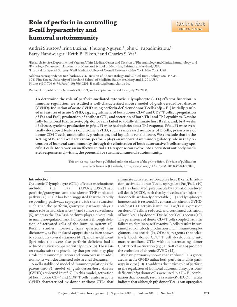
of cytokines important for cell-mediated immunity(IFN-γ) and antibody production (IL-10, IL-4), where-as the initiation of chronic GVHD is characterized bythe production of IL-4 and IL-10 and no detectableIFN-γ (13, 20). To determine whether cytokine produc-tion in pfp→F1 mice resembles that of acute or chron-ic GVHD, splenocyte cytokine mRNA expression wasevaluated by RT-PCR. As shown in Figure 3a, at day 10after parental cell transfer both wt→F1 and pfp→F1mice exhibited a three- to fourfold upregulation ofIFN-γand an approximately threefold upregulation ofIL-10, indicating that cytokine production duringGVHD initiation was similar for both pfp GVHD andacute GVHD mice. Interestingly, IL-4 production wasgreater in pfp GVHD mice at day 10.
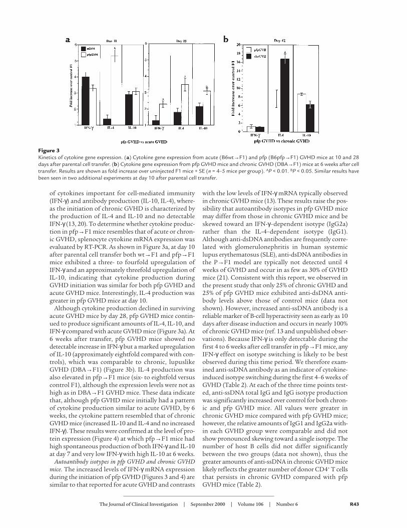
Although cytokine production declined in survivingacute GVHD mice by day 28, pfp GVHD mice contin-ued to produce significant amounts of IL-4, IL-10, andIFN-γcompared with acute GVHD mice (Figure 3a). At6 weeks after transfer, pfp GVHD mice showed nodetectable increase in IFN-γbut a marked upregulationof IL-10 (approximately eightfold compared with con-trols), which was comparable to chronic, lupuslikeGVHD (DBA→F1) (Figure 3b). IL-4 production wasalso elevated in pfp→F1 mice (six- to eightfold versuscontrol F1), although the expression levels were not ashigh as in DBA→F1 GVHD mice. These data indicatethat, although pfp GVHD mice initially had a patternof cytokine production similar to acute GVHD, by 6weeks, the cytokine pattern resembled that of chronicGVHD mice (increased IL-10 and IL-4 and no increasedIFN-γ). These results were confirmed at the level of pro-tein expression (Figure 4) at which pfp→F1 mice hadhigh spontaneous production of both IFN-γand IL-10at day 7 and very low IFN-γwith high IL-10 at 6 weeks.
Autoantibody isotypes in pfp GVHD and chronic GVHDmice. The increased levels of IFN-γ mRNA expressionduring the initiation of pfp GVHD (Figures 3 and 4) aresimilar to that reported for acute GVHD and contrasts
with the low levels of IFN-γ mRNA typically observedin chronic GVHD mice (13). These results raise the pos-sibility that autoantibody isotypes in pfp GVHD micemay differ from those in chronic GVHD mice and beskewed toward an IFN-γ–dependent isotype (IgG2a)rather than the IL-4–dependent isotype (IgG1).Although anti-dsDNA antibodies are frequently corre-lated with glomerulonephritis in human systemiclupus erythematosus (SLE), anti-dsDNA antibodies inthe P→F1 model are typically not detected until 4weeks of GVHD and occur in as few as 30% of GVHDmice (21). Consistent with this report, we observed inthe present study that only 25% of chronic GVHD and25% of pfp GVHD mice exhibited anti-dsDNA anti-body levels above those of control mice (data notshown). However, increased anti-ssDNA antibody is areliable marker of B-cell hyperactivity seen as early as 10days after disease induction and occurs in nearly 100%of chronic GVHD mice (ref. 13 and unpublished obser-vations). Because IFN-γ is only detectable during thefirst 4 to 6 weeks after cell transfer in pfp→F1 mice, anyIFN-γ effect on isotype switching is likely to be bestobserved during this time period. We therefore exam-ined anti-ssDNA antibody as an indicator of cytokine-induced isotype switching during the first 4–6 weeks ofGVHD (Table 2). At each of the three time points test-ed, anti-ssDNA total IgG and IgG isotype productionwas significantly increased over control for both chron-ic and pfp GVHD mice. All values were greater inchronic GVHD mice compared with pfp GVHD mice;however, the relative amounts of IgG1 and IgG2a with-in each GVHD group were comparable and did notshow pronounced skewing toward a single isotype. Thenumber of host B cells did not differ significantlybetween the two groups (data not shown), thus thegreater amounts of anti-ssDNA in chronic GVHD micelikely reflects the greater number of donor CD4+ T cellsthat persists in chronic GVHD compared with pfpGVHD mice (Table 2).
The Journal of Clinical Investigation | September 2000 | Volume 106 | Number 6 R43
Figure 3Kinetics of cytokine gene expression. (a) Cytokine gene expression from acute (B6wt→F1) and pfp (B6pfp→F1) GVHD mice at 10 and 28days after parental cell transfer. (b) Cytokine gene expression from pfp GVHD mice and chronic GVHD (DBA→F1) mice at 6 weeks after celltransfer. Results are shown as fold increase over uninjected F1 mice + SE (n = 4–5 mice per group). AP < 0.01. BP < 0.05. Similar results havebeen seen in two additional experiments at day 10 after parental cell transfer.
Glomerulonephritis in pfp GVHD mice. To determinewhether pfp GVHD mice developed a lupuslikeglomerulonephritis similar to that reported in chronicGVHD mice (22, 23), renal function and pathologicalanalysis of the kidneys were performed. Increased pro-teinuria (≥ 2+) was seen for both pfp→F1 and chronicGVHD mice at 4 weeks (pfp GVHD or chronic GVHDvs. control P < 0.001; n = 8–9 per group) and at 11 weeks(pfp GVHD or chronic GVHD vs. control P < 0.01; n =4 per group) after disease induction. By 12 weeks, threeof five chronic GVHD mice and one of five pfp→F1mice had features consistent with nephrotic syndrome(ascites and/or lipemic serum). There was no sponta-neous mortality in either group at this point.
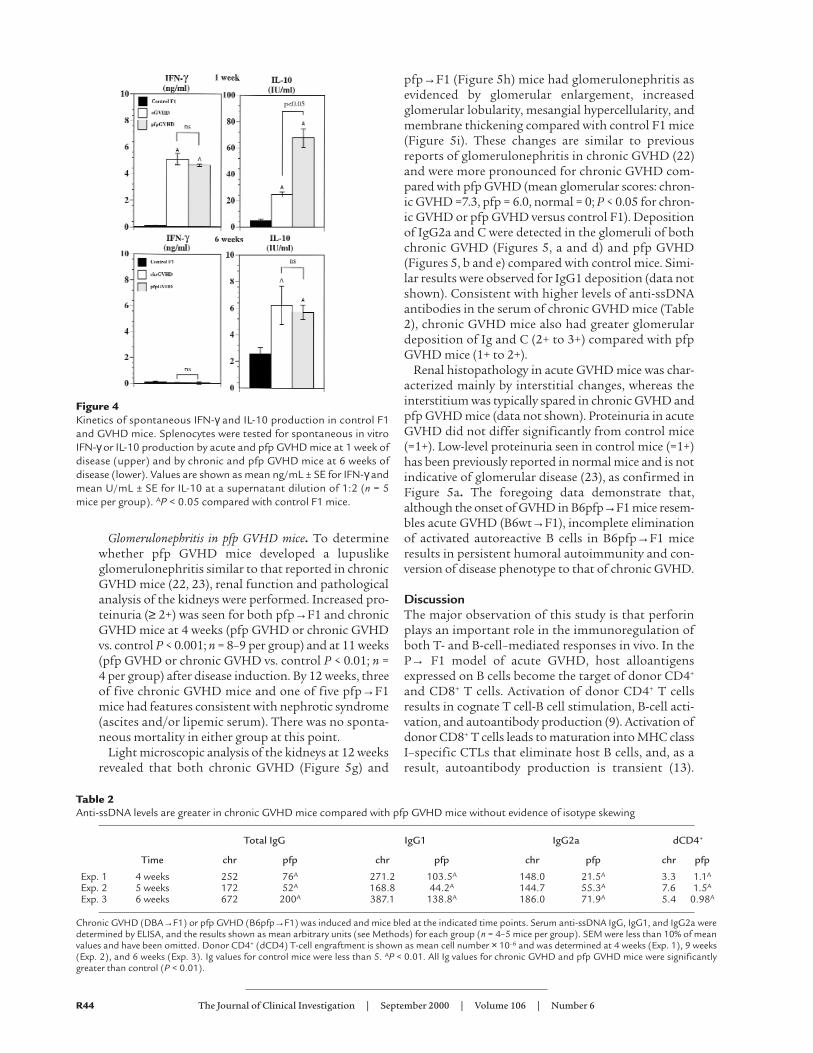
Light microscopic analysis of the kidneys at 12 weeksrevealed that both chronic GVHD (Figure 5g) and
pfp→F1 (Figure 5h) mice had glomerulonephritis asevidenced by glomerular enlargement, increasedglomerular lobularity, mesangial hypercellularity, andmembrane thickening compared with control F1 mice(Figure 5i). These changes are similar to previousreports of glomerulonephritis in chronic GVHD (22)and were more pronounced for chronic GVHD com-pared with pfp GVHD (mean glomerular scores: chron-ic GVHD =7.3, pfp = 6.0, normal = 0; P < 0.05 for chron-ic GVHD or pfp GVHD versus control F1). Depositionof IgG2a and C were detected in the glomeruli of bothchronic GVHD (Figures 5, a and d) and pfp GVHD(Figures 5, b and e) compared with control mice. Simi-lar results were observed for IgG1 deposition (data notshown). Consistent with higher levels of anti-ssDNAantibodies in the serum of chronic GVHD mice (Table2), chronic GVHD mice also had greater glomerulardeposition of Ig and C (2+ to 3+) compared with pfpGVHD mice (1+ to 2+).
Renal histopathology in acute GVHD mice was char-acterized mainly by interstitial changes, whereas theinterstitium was typically spared in chronic GVHD andpfp GVHD mice (data not shown). Proteinuria in acuteGVHD did not differ significantly from control mice(=1+). Low-level proteinuria seen in control mice (=1+)has been previously reported in normal mice and is notindicative of glomerular disease (23), as confirmed inFigure 5a. The foregoing data demonstrate that,although the onset of GVHD in B6pfp→F1 mice resem-bles acute GVHD (B6wt→F1), incomplete eliminationof activated autoreactive B cells in B6pfp→F1 miceresults in persistent humoral autoimmunity and con-version of disease phenotype to that of chronic GVHD.
DiscussionThe major observation of this study is that perforinplays an important role in the immunoregulation ofboth T- and B-cell–mediated responses in vivo. In theP→ F1 model of acute GVHD, host alloantigensexpressed on B cells become the target of donor CD4+
and CD8+ T cells. Activation of donor CD4+ T cellsresults in cognate T cell-B cell stimulation, B-cell acti-vation, and autoantibody production (9). Activation ofdonor CD8+ T cells leads to maturation into MHC classI–specific CTLs that eliminate host B cells, and, as aresult, autoantibody production is transient (13).
R44 The Journal of Clinical Investigation | September 2000 | Volume 106 | Number 6
Figure 4Kinetics of spontaneous IFN-γ and IL-10 production in control F1and GVHD mice. Splenocytes were tested for spontaneous in vitroIFN-γor IL-10 production by acute and pfp GVHD mice at 1 week ofdisease (upper) and by chronic and pfp GVHD mice at 6 weeks ofdisease (lower). Values are shown as mean ng/mL ± SE for IFN-γandmean U/mL ± SE for IL-10 at a supernatant dilution of 1:2 (n = 5mice per group). AP < 0.05 compared with control F1 mice.
Table 2Anti-ssDNA levels are greater in chronic GVHD mice compared with pfp GVHD mice without evidence of isotype skewing
Total IgG IgG1 IgG2a dCD4+
Time chr pfp chr pfp chr pfp chr pfp
Exp. 1 4 weeks 252 76A 271.2 103.5A 148.0 21.5A 3.3 1.1A
Exp. 2 5 weeks 172 52A 168.8 44.2A 144.7 55.3A 7.6 1.5A
Exp. 3 6 weeks 672 200A 387.1 138.8A 186.0 71.9A 5.4 0.98A
Chronic GVHD (DBA→F1) or pfp GVHD (B6pfp→F1) was induced and mice bled at the indicated time points. Serum anti-ssDNA IgG, IgG1, and IgG2a weredetermined by ELISA, and the results shown as mean arbitrary units (see Methods) for each group (n = 4–5 mice per group). SEM were less than 10% of meanvalues and have been omitted. Donor CD4+ (dCD4) T-cell engraftment is shown as mean cell number × 10–6 and was determined at 4 weeks (Exp. 1), 9 weeks(Exp. 2), and 6 weeks (Exp. 3). Ig values for control mice were less than 5. AP < 0.01. All Ig values for chronic GVHD and pfp GVHD mice were significantlygreater than control (P < 0.01).
Donor T cells expand during the first 2 weeks of diseasebut subsequently undergo significant depletion, pre-sumably by AICD, during weeks 2–4 (ref. 11 and Table1), and immune homeostasis is restored in the F1 ani-mal. The present study demonstrates that in this modelof in vivo cognate T-B stimulation, perforin deficiencypredisposes to persistent B-cell expansion, autoanti-body production, and a lupuslike disease. The mecha-nisms involved include defective elimination of autore-active host B cells, persistence of activated donor Tcells, and a cytokine switch from heterogeneous to apolarized Th2 response. Thus, perforin, like Fas (6)plays an important role in immunoregulation.
It should be noted that defective perforin-mediatedCTL effector function does not necessarily result inautoimmunity. Perforin-deficient mice have normallife spans (4) and histologically normal immune sys-tems (24). Although CD8+ T cell activation by alloanti-gen or virus is normal in pfp mice, CTL function in theform of viral clearance or induction of acute GVHDafter bone marrow transplantation (BMT) is impairedbut not totally abrogated (25, 26). For example, in amurine model of BMT in which recipient mice are irra-diated before donor cell transfer, Levy and coworkershave shown that pfp donor cells are capable of elimi-nating host B cells (27) and of inducing an acuteGVHD that is qualitatively similar to that seen withwild-type donor cells but is delayed on onset approxi-
mately twofold (26). In contrast, in the P→F1 model,our results demonstrate that pfp donor cells initiallyinduce a picture that resembles acute GVHD but by6–8 weeks, evolves into a picture that resembles chron-ic lupuslike GVHD. We postulate that it is the lack ofhost irradiation in the P→F1 model that is critical forthe evolution to humoral autoimmunity and chronicGVHD. Specifically, in the BMT model, irradiation ren-ders host B cells nonfunctional. B-cell function intransplanted mice is thus a measure of donor cellrepopulation. As a result, there is no possibility thathumoral autoimmunity can develop in this model as aresult of alloreactive donor T cells, providing cognatehelp to host B cells as described in the P→F1 model.
Moreover, our data demonstrating that pfp→F1 miceexhibit incomplete elimination of activated host B cellsand persistent IL-10 production provide a frameworkfor explaining the mechanism by which the initialacute GVHD picture in pfp→F1 mice evolves intohumoral autoimmunity and chronic GVHD. AlthoughIL-10 has pleiotropic effects, it has been classified as aTh2 cytokine based on its ability to serve as a costimu-lator of B-cell proliferation (28). It is also a majorinhibitor of Th1 functions such as CTL (28) and can beproduced by B cells, T cells, and macrophages (28) .Thus, the persisting activated autoreactive host B cellsin pfp GVHD mice may produce IL-10, which not onlycan provide a positive feedback loop leading to further
The Journal of Clinical Investigation | September 2000 | Volume 106 | Number 6 R45
Figure 5Kidneys from both pfp GVHD and chronic GVHD mice exhibit glomerulonephritis and deposition of IgG and C. Kidney sections from chron-ic GVHD (a, d, and g), pfp GVHD (b, e, and h), and control F1 (c, f, and i) mice were stained with anti-murine IgG2a (a, b, and c) and com-plement (d, e, and f) or H&E (g, h, and i) 12 weeks after donor cell transfer as described in Methods. Photographs were taken at × 200. Rep-resentative results are shown from a single mouse from each group (n = 3 per group).
B-cell activation, humoral autoimmunity, and IL-10production, but can also downregulate acute GVHDmechanisms by inhibiting CTL development.
Despite impaired CTL function, enhanced immuneresponses that could be attributed to a loss of negativeregulatory activities have not been described in pfpmice as they have for fas-deficient mice (6). Whereas, inan autoimmune prone MRL strain background, per-forin deficiency exacerbated disease manifestations (8),our results demonstrate that perforin plays a signifi-cant role in immunoregulation in the setting of normalFas/FasL function. Taken together, these observationsare consistent with the idea that, in addition to its rolein host defense against viruses, perforin is also impor-tant in controlling autoimmunity through the deletionof activated autoreactive B cells and activated ag-spe-cific Th cells. Whether this form of immunoregulationis executed by CD8+ T cells and/or natural killer cells iscurrently under investigation.
The results obtained have implications for the patho-genesis of humoral autoimmune diseases such as SLE.Increasing evidence indicates that autoantibody pro-duction in SLE is T cell driven (29, 30). Paradoxically, invitro T-cell function and CTL generation are widelydescribed as depressed in patients with SLE (31, 32). It isunclear at present whether impaired CTL function is asecondary effect, e.g., due to reduced IL-2 production orto increased IL-10 production seen in SLE (33, 34), orinstead reflects an intrinsic defect in a cytotoxic effectorpathway that predisposes to SLE. This question has beendifficult to resolve completely in humans. An advantageof the P→F1 model of lupus is that mice are immuno-logically normal before disease induction. Thus, primaryevents can be separated from secondary epiphenomena.Our results clearly show that a preexisting defect inCD8+ CTL function can lead to impaired elimination ofautoreactive B cells and lupuslike disease.
If the chronic GVHD model of lupus caused by defec-tive CTL activity as shown here shares a commonpathogenesis with human SLE, how could this arise?While it seems unlikely that human SLE is initiated byrecognition of MHC alloantigens as in the GVHDmodel, the idea cannot be totally excluded in view ofreports of persistent maternal-fetal microchimerism inpatients with humoral autoimmunity (35). One possi-bility is that a B-cell tropic virus, such as Epstein-Barrvirus, that infects B cells regardless of the antigenicspecificity through the complement receptor, CD21(36), activates an oligoclonal population of viral spe-cific CD4+ and CD8+ T cells. In either of these examplesof CD4+ Th cell stimulation by antigens expressed onB cells, individuals with inherited defects in one of thecytotoxic effector pathways would be unable to elimi-nate activated autoreactive B cells completely. As aresult, activated ag-specific CD4+ T cells could persistand eventually switch from an initial CTL-mediatedresponse to an exclusively B-cell stimulatory cytokine-driven response that would in turn promote the devel-opment of humoral autoimmunity. The permissive
defect in CTL could be at the level of reduced cell-medi-ated immunity–promoting cytokines (TNF-α),impaired effector function (perforin or fas pathways),or reduced precursor CTL frequencies, all of which havebeen associated with SLE (6, 14, 37–39).
AcknowledgmentsThis work was supported by NIH grants AR45482and R29 AI33882, a Department of Veterans AffairsMerit Review grant, and grants from the Marylandchapters of the Lupus Foundation and ArthritisFoundation. A. Shustov is recipient of an EngelicheffFellowship Award from the Maryland Chapter of theArthritis Foundation.
1. Rouvier, E., Luciani, M.F., and Golstein, P. 1993. Fas involvement inCa(2+)-independent T cell-mediated cytotoxicity. J. Exp. Med.177:195–200.
2. Millard, P.J., Henkart, M.P., Reynolds, C.W., and Henkart, P.A. 1984.Purification and properties of cytoplasmic granules from cytotoxic ratLGL tumors. J. Immunol. 132:3197–3204.
3. Clark, W.R., et al. 1995. Molecular pathways of CTL-mediated cytotoxi-city. Immunol. Rev. 146:33–44.
4. Kagi, D., et al. 1994. Cytotoxicity mediated by T cells and natural killercells is greatly impaired in perforin-deficient mice. Nature. 369:31–37.
5. van den Broek, M.E., et al. 1996. Decreased tumor surveillance in per-forin-deficient mice. J. Exp. Med. 184:1781–1790.
6. Cohen, P.L., and Eisenberg, R.A. 1992. The lpr and gld genes in systemicautoimmunity: life and death in the Fas lane. Immunol. Today.13:427–428.
7. Topham, D.J., Tripp, R.A., and Doherty, P.C. 1997. CD8+ T cells clearinfluenza virus by perforin or Fas-dependent processes. J. Immunol.159:5197–5200.
8. Peng, S.L., Moslehi, J., Robert, M.E., and Craft, J.E. 1998. Perforin pro-tects against autoimmunity in lupus-prone mice. J. Immunol.160:652–660.
9. Gleichmann, E., Pals, S.T., Rolink, A.G., Radaszkiewicz, T., and Gleich-mann, H. 1984. Graft-versus-host reactions: clues to the etiopathologyof a spectrum of immunological diseases. Immunol. Today. 5:324–332.
10. Shustov, A., Nguyen, P., Finkelman, F.D., Elkon, K.B., and Via, C.S. 1998.Differential expression of Fas and Fas ligand in acute and chronic graft-versus-host disease: up-regulation of Fas and Fas ligand requires CD8+T cell activation and IFN-gamma production. J. Immunol.161:2848–2855.
11. Hakim, F.T., Sharrow, S.O., Payne, S., and Shearer, G.M. 1991. Repopu-lation of host lymphohematopoietic systems by donor cells during graft-versus-host reaction in unirradiated adult F1 mice injected with parentallymphocytes. J. Immunol. 146:2108–2115.
12. Via, C.S., and Finkelman, F.D. 1993. Critical role of interleukin-2 in thedevelopment of acute graft-versus-host disease. Int. Immunol. 5:565–572.
13. Rus, V., Svetic, A., Nguyen, P., Gause, W.C., and Via, C.S. 1995. Kineticsof Th1 and Th2 cytokine production during the early course of acuteand chronic murine graft-versus-host disease. Regulatory role of donorCD8+ T cells. J. Immunol. 155:2396–2406.
14. Via, C.S., Sharrow, S.O., and Shearer, G.M. 1987. Role of cytotoxic T lym-phocytes in the prevention of lupus-like disease occurring in a murinemodel of graft-vs-host disease. J. Immunol. 139:1840–1849.
15. Svetic, A., et al. 1991. Cytokine gene expression after in vivo primaryimmunization with goat antibody to mouse IgD antibody. J. Immunol.147:2391–2397.
16. Handwerger, B.S., Storrer, C.E., Wasson, C.S., Movafagh, F., and Reich-lin, M. 1999. Further characterization of the autoantibody response ofPalmerston North mice. J. Clin. Immunol. 19:45–57.
17. Luzina, I.G., et al. 1999. Vasculitis in the Palmerston North mouse modelof lupus. Arthritis Rheum. 42:561–568.
18. Singh, J.N., Sabbadini, E., and Sehon, A.H. 1972. Cytotoxicity in graft-versus-host reaction. I. Role of donor and host spleen cells. J. Exp. Med.136:39–48.
19. Morris, S.C., Cheek, R.L., Cohen, P.L., and Eisenberg, R.A. 1990. Autoan-tibodies in chronic graft versus host result from cognate T-B interac-tions. J. Exp. Med. 171:503–517.
20. Garlisi, C.G., Pennline, K.J., Smith, S.R., Siegel, M.I., and Umland, S.P.1993. Cytokine gene expression in mice undergoing chronic graft-ver-sus-host disease. Mol. Immunol. 30:669–677.
21. Gleichmann, E., van Elven, E.H., and Van Der Veen, P.J.W. 1982. A sys-temic lupus erythematosus (SLE)-like disease in mice induced by
R46 The Journal of Clinical Investigation | September 2000 | Volume 106 | Number 6
abnormal T-B cell cooperation. Preferential formation of autoantibod-ies characteristic of SLE. Eur. J. Immunol. 12:152–159.
22. Bruijn, J.A., et al. 1988. Murine chronic graft-versus-host disease as amodel for lupus nephritis. Am. J. Pathol. 130:639–641.
23. Bielschowsky, M., and D’Ath, E.F. 1971. The kidneys of NZB-B1, NZO-B1, NZC-B1 and NZY-B1 mice. J. Pathol. 103:97–105.
24. Kojima, H., et al. 1994. Two distinct pathways of specific killing revealedby perforin mutant cytotoxic T lymphocytes. Immunity. 1:357–364.
25. Braun, M.Y., Lowin, B., Lars, F., Acha-Oreba, H., and Tschopp, J. 1996.Cytotoxic T cells deficient in both functional fas ligand and perforinshow residual cytolytic activity yet lose their capacity to induce lethalacute graft-versus-host disease. J. Exp. Med. 183:657–661.
26. Baker, M.B., Altman, N.H., Podack, E.R., and Levy, R.B. 1996. The role ofcell-mediated cytotoxicity in acute GVHD after MHC-matched allo-geneic bone marrow transplantation in mice. J. Exp. Med. 183:2645–2656.
27. Baker, M.B., Riley, R.L., Podack, E.R., and Levy, R.B. 1997. Graft-versus-host-disease-associated lymphoid hypoplasia and B cell dysfunction isdependent upon donor T cell-mediated Fas-ligand function, but not per-forin function. Proc. Natl. Acad. Sci. USA. 94:1366–1371.
28. Moore, K.W., O’Garra, A., de Waal Malefyt, R., Vieira, P., and Mosmann,T.R. 1993. Interleukin-10. Annu. Rev. Immunol. 11:165–190.
29. Burlingame, R.W., Rubin, R.L., Balderas, R.S., and Theofilopoulos, A.N.1993. Genesis and evolution of antichromatin autoantibodies in murinelupus implicates T-dependent immunization with self antigen. J. Clin.Invest. 91:1687–1696.
30. Mohan, C., Adams, S., Stanik, V., and Datta, S.K. 1993. Nucleosome: amajor immunogen for pathogenic autoantibody-inducing T cells oflupus. J. Exp. Med. 177:1367–1381.
31. Tsokos, G.C., and Balow, J.E. 1981. Cytotoxic responses to alloantigensin systemic lupus erythematosus. J. Clin. Immunol. 1:208–216.
32. Stohl, W. 1995. Impaired polyclonal T cell cytolytic activity: a possiblerisk factor for systemic lupus erythematosus. Arthritis Rheum.38:506–516.
33. Tsokos, G.C. 1992. Lymphocyte abnormalities in human lupus. Clin.Immunol. Immunopathol. 63:7–9.
34. Georgescu, L., Vakkalanka, R.K., Elkon, K.B., and Crow, M.K. 1997. Inter-leukin-10 promotes activation-induced cell death of SLE lymphocytesmediated by Fas ligand. J. Clin. Invest. 100:2622–2633.
35. Nelson, J.L. 1998. Microchimerism and autoimmune disease. N. Engl. J.Med. 338:1224–1225.
36. James, J.A., et al. 1997. An increased prevalence of Epstein-Barr virusinfection in young patients suggests a possible etiology for systemiclupus erythematosus. J. Clin. Invest. 100:3019–3026.
37. Horwitz, D.A., and Jacob, C.O. 1994. The cytokine network in the patho-genesis of systemic lupus erythematosus and possible therapeutic impli-cations. Springer Semin. Immunopathol. 16:181–200.
38. Stohl, W., et al. 1997. Impaired nonrestricted cytolytic activity in systemiclupus erythematosus. Arthritis Rheum. 40:1130–1137.
39. Jacob, C.O., and McDevitt, H.O. 1988. Tumour necrosis factor-alpha inmurine autoimmune ‘lupus’ nephritis. Nature. 331:356–358.
The Journal of Clinical Investigation | September 2000 | Volume 106 | Number 6 R47