adult neurogenesis in the decapod crustacean brain: a hematopoietic connection?
TRANSCRIPT

Adult neurogenesis in the decapod crustacean brain: Ahematopoietic connection?
Barbara S. Beltz, Yi Zhang, Jeanne L. Benton, and David C. SandemanNeuroscience Program, Wellesley College, Wellesley, MA 02481
AbstractNew neurons are produced and integrated into circuits in the adult brains of many organisms,including crustaceans. In some crustacean species, the 1st- generation neuronal precursors reside ina niche exhibiting characteristics analogous to mammalian neurogenic niches. However, unlikemammalian niches where several generations of neuronal precursors coexist, the lineage ofprecursor cells in crayfish is spatially separated allowing the influence of environmental andendogenous regulators on specific generations in the neuronal precursor lineage to be defined.Experiments also demonstrate that the 1st-generation neuronal precursors in the crayfishProcambarus clarkii are not self-renewing. A source external to the neurogenic niche musttherefore provide cells that replenish the 1st-generation precursor pool, because although thesecells divide and produce a continuous efflux of 2nd-generation cells from the niche, the populationof 1st-generation niche precursors is not diminished with growth and aging. In vitro studies showthat cells extracted from the hemolymph, but not other tissues, are attracted to and incorporatedinto the neurogenic niche, a phenomenon that appears to involve serotonergic mechanisms. Wepropose that in crayfish, the hematopoietic system may be a source of cells that replenish the nichecell pool. These and other studies reviewed here establish decapod crustaceans as model systemsin which the processes underlying adult neurogenesis, such as stem cell origins andtransformation, can be readily explored. Studies in diverse species where adult neurogenesisoccurs will result in a broader understanding of fundamental mechanisms and how evolutionaryprocesses may have shaped the vertebrate/mammalian condition.
Keywordsneurogenic niche; serotonin; bromodeoxyuridine; hematopoietic system; Procambarus clarkii
I. A stem cell niche in the crustacean brainNeuronal proliferation in most regions of the decapod crustacean brain ceases in the periodaround hatching when the embryonic precursor cells (neuroblasts) die (Harzsch, 2003). Theexception to this is in the central olfactory pathway where mitotic activity continuesthroughout life (Schmidt, 1997; Schmidt and Harzsch, 1999; Harzsch et al., 1999). Adultneurogenesis also occurs in the visual pathway (Sullivan and Beltz, 2005b), though this hasbeen studied in much less detail. In the olfactory pathway, life-long neurogenesis is foundamong the sensory (Steullet et al., 2000), local (Cluster 9) and projection (Cluster 10)neurons (Figure 1) (Schmidt and Harzsch, 1999). Each Cluster 9 neuron innervates theprimary olfactory processing areas, the olfactory lobes (OLs), and higher order processingareas, the accessory lobes (ALs). The Cluster 10 projection neurons innervate either the OL
*Corresponding author: Barbara S. Beltz, Postal address: Neuroscience Program, Wellesley College, 106 Central Street, Wellesley,MA 02481, Telephone: 781-283-3048, Fax: 781-283-3642, [email protected].
NIH Public AccessAuthor ManuscriptEur J Neurosci. Author manuscript; available in PMC 2012 September 1.
Published in final edited form as:Eur J Neurosci. 2011 September ; 34(6): 870–883. doi:10.1111/j.1460-9568.2011.07802.x.
NIH
-PA Author Manuscript
NIH
-PA Author Manuscript
NIH
-PA Author Manuscript

or AL and olfactory globular tract neuropil (Sullivan et al., 2000), and their axons projectvia the olfactory globular tract (OGT) to neuropil regions in the lateral protocerebrum(Sullivan and Beltz, 2001). The AL is involved in higher-order integration of olfactory,visual and mechanosensory information (Sandeman et al., 1995; Sullivan and Beltz, 2005b).
For many years after the discovery of adult neurogenesis in the crustacean brain, the sourceof the adult-born neurons was a mystery. This situation changed, however, when antibodiesagainst glutamine synthetase (GS), which labels astrocytes and some neuronal stem cells invertebrates, revealed a structure on the ventral surface of the crayfish (Procambarus clarkii)brain that extended between the lateral (LPZ) and medial (MPZ) proliferation zones inClusters 9 and 10 (Figure 2A) (Sullivan et al., 2005; 2007a). In roughly the center of thisstructure was a swollen region enclosing what appeared to be a hollow cavity. Short-survivaltime studies using the S-phase marker bromodeoxyuridine (BrdU), in conjunction withimmunocytochemistry for GS, labeled a discontinuous stream of BrdU labeled cells lyingalong the GS-labeled structure between Clusters 9 and 10 (Figure 2B). Some of thesefeatures in P. clarkii have been confirmed in studies by others (Song et al., 2007; 2009). Inaddition, similar structures have been found in the Australian crayfish (Cherax destructor),clawed lobster (Homarus americanus) and green crab (Carcinus maenus) (Sullivan et al.,2007b). The same structure had in fact been discovered and described in much earlierstudies in separate members of all the decapod crustaceans (Bazin and Demeusy 1968;Bazin 1970a, 1970b; Sandeman et al., 2011) but without any indication of its function.
Lucifer yellow dye was injected into the swollen region in Procambarus clarkii, revealing astructure filled with bipolar cells (Figure 2C). Studies using the cell cycle marker MCM2–7(Cvetic and Walter, 2006) indicated that these are in G1 of the cell cycle except when theyare progressing through S to M phase (Sullivan et al., 2007a), suggesting that these cells arenot terminally differentiated. Injection of dextran-ruby dye (MW 3000) into the dorsal arterythat supplies the brain (Figure 2D) or into the pericardial cavity (Benton et al., 2010; 2011)demonstrated that the cavity in the center of this cluster of cells is confluent with thevasculature. Studies using double nucleoside labeling further showed that proliferating(BrdU-labeled) cells migrate from the cluster of cells towards the proliferation zones inClusters 9 and 10 (Sullivan et al., 2007a), where they divide at least once more beforedifferentiating (Sullivan et al., 2005a). As in the higher vertebrates, the precursor cellsresiding in the crayfish niche have glial properties and act as both precursor and supportcells, the processes of the niche cells guiding the migration of the daughter cells towardsClusters 9 and 10 (see model, Figure 3). Based on these and other features, we proposed thatthe swollen region constitutes a neurogenic niche containing primary neuronal precursors,with a connection to the blood vascular system via the central cavity (Sullivan et al., 2005;2007a). Many of these features but with a somewhat different overall organization, have alsobeen described in the spiny lobster, Panulirus argus, where a neurogenic niche containinguniquely-endowed, persistent neuroblasts appears to support adult neurogenesis (Schmidt,2007).
Within two weeks of the final cell divisions in Clusters 9 and 10, cell counts in BrdU cellsurvival studies reveal a period of cell death; the surviving cells differentiate into neuronsover the next 2–4 weeks (Panulirus argus: Schmidt, 2001; Cherax destructor: Kim andBeltz, 2008; Kim, 2009). The resulting neurons express transmitters that are appropriate forCluster 9 (allatostatin, orkokinin) and 10 (FMRFamide) neurons (Figure 4) (Sullivan andBeltz, 2007a), and innervate the olfactory and accessory lobes as do the mature local andprojection interneurons in these regions (Figure 5) (Sullivan and Beltz, 2005b).
Beltz et al. Page 2
Eur J Neurosci. Author manuscript; available in PMC 2012 September 1.
NIH
-PA Author Manuscript
NIH
-PA Author Manuscript
NIH
-PA Author Manuscript

II. The neurogenic niche: common strategies in diverse organisms1. Neurogenic niches: organizational parallels and distinctions
While neuronal stem cell niches in different organisms do have some characteristicproperties (see below), there are many parallels across a broad phyletic range. Among theseare the glial characteristics of the primary neuronal precursor (stem) cells (but see theexception in zebrafish, Kaslin et al., 2009), a close association between the niche and thevasculature, the presence of a specialized extracellular matrix and basal lamina, directedmigration of progeny, and regulation by environmental and endogenous cues (Doetsch et al.,1999; Sullivan et al., 2007a;b; Tavazoie et al., 2008; Kriegstein and Alvarez-Buylla, 2009;Pathania et al., 2010). In addition, the presence of a period of cell death following neuronalproliferation and the time required for the new neurons to begin differentiation (~2–4 weeks)are features of adult neurogenesis that are shared between vertebrate (e.g., van Praag et al.,2002) and crustacean (Panulirus argus, Schmidt, 2001; Cherax destructor, Kim et al., 2009)systems. Many of the organizational properties of the cellular machinery maintaining adultneurogenesis appear, therefore, to be shared by widely disparate taxa. These parallelssuggest common strategies for the generation of new neurons in adult brains that may haveresulted from shared ancestral origins or evolutionary convergence.
The 1st-generation neuronal precursors—In the mammalian brain, adult-bornneurons are incorporated into the olfactory bulb and hippocampus; the neurogenic nichescontaining the neuronal precursor lineages are located in the subventricular and subgranularzones, respectively. The 1st- generation neuronal precursor cells (neural stem cells) displaytypical astroglial markers such as GFAP and glutamine synthetase, in addition to stem cellmarkers such as nestin. As a group, however, the astroglia are a heterogeneous population,with some serving as neurogenic cells and others as quiescent stem cells (Ma et al., 2008;Riquelme et al., 2008; Kriegstein and Alvarez-Buylla, 2009). It has been proposed that thequiescent astroglia may interact, and may also retain the capacity to become neurogenic inthe presence of appropriate signals (Nyfeler et al., 2005). Likewise, it is not known whetherall niche cells in the crayfish brain have the capacity to become neuronal precursors,although this possibility is implicit in some proposed models (Zhang et al., 2009; Ayub et al,2011) and is supported by four lines of evidence: Firstly, all niche cells label with the G1-phase marker MCM2–7, except when they are progressing through S to M phase (Sullivan etal., 2007a). This indicates that, although resting in G1, the niche cells are actively in the cellcycle and not terminally differentiated cells. Secondly, all niche cells, including those in Mphase, label immunocytochemically for the glial marker glutamine synthetase, indicatingthat all the niche cells share this molecular property (Zhang et al., 2009). Thirdly,examination of semi-thin sections shows that, with very few exceptions, the niche cells haveidentical cellular characteristics (Zhang et al., 2009). Finally, the studies of Ayub et al.(2011) demonstrate that the number of S-phase (BrdU-labeled) precursors in the niche isregulated by environmental enrichment, suggesting the existence of a quiescent pool ofprecursor cells whose status in the cell cycle can be modulated by local conditions.
Vascularization of neurogenic niches—The vascular system has been receivingincreasing attention as a common feature of all niches, including those in the nervous system(Tavazoie et al., 2008). In both the subventricular (SVZ) and subgranular (SGZ) zones thatsupport adult neurogenesis of olfactory bulb and hippocampal neurons, respectively, thestem cells lie in close proximity to a rich plexus of blood vessels. Endothelial cells, whichare known to regulate stem cell self-renewal and neurogenesis via a cadre of secretedfactors, are emerging as critical components of neural stem cell niches (Shen et al., 2004,2008; Riquelme et al., 2008). Blood vessels also are conduits for circulating hormones andcytokines released from distant sources (Lennington et al., 2003). In addition, the associated
Beltz et al. Page 3
Eur J Neurosci. Author manuscript; available in PMC 2012 September 1.
NIH
-PA Author Manuscript
NIH
-PA Author Manuscript
NIH
-PA Author Manuscript

extracellular matrix appears to provide a means of cell anchoring in the niche (Shen et al.,2008), as well as creating a supportive microenvironment and architecture (Riquelme et al.,2008).
While details of how the blood vasculature interacts with the crustacean neurogenic nicheare not known, some form of communication via the vascular cavity has been demonstratedin two different crayfish species using injection of labeled dextrans directly into the dorsalartery that vascularizes the brain (Figure 2D) (Procambarus clarkii, Sullivan et al., 2005;2007a; Cherax destructor, Sandeman et al., 2009), or into the pericardial cavity (Benton etal., 2010; 2011). The blood system also is closely associated with the niche in Panulirusargus (Schmidt, 2007). As reviewed below, a variety of cues regulate the numbers of nichecells and the rate of neurogenesis in crustaceans, and ongoing studies are defining how thevascular connections with the niche may support these actions.
Directed migration of neuronal precursors—In the mammalian brain, 3rd generationprecursors (called neuroblasts) migrate away from the subventricular zone towards theolfactory bulb along the rostral migratory stream (RMS) by means of chain migration; tensof thousands of cells travel this path to the olfactory bulb daily (Lois et al., 1996), followinga route created by astrocytes that then communicate with the migrating cells and regulatetheir migration (Bolteus and Bordey, 2004). The RMS is deeply embedded in the brain, andtherefore it has been difficult to observe the migratory behavior of these cells in livingorganisms. Recently, magnetic resonance imaging (MRI) techniques in rats (Shapiro et al.,2006; Vreys et al., 2010) and two-photon time-lapse imaging in acute slices of mouse brain(Nam et al., 2007) have been used to examine the dynamic behavior of cells migrating alongthis pathway; these studies have shown that cell motility in the RMS is more complex thanpreviously thought, involving multiple cell types, behaviors, speeds and directions. Neuronalprecursors in the hippocampus also migrate, although over very short distances comparedwith the RMS, which can be 5 mm in length in the adult mouse. In addition, adultneurogenesis is prevalent in a number of brain regions in fish, and these cells are oftenguided by radial glial fibers (Zupanc et al., 2005; Zupanc, 2006). In the crustacean brain, theprimary neuronal precursor cells residing in the niche appear to serve as both procursor andsupport cells, with their long glutamine synthetase- and lissencephaly 1 (LIS1)-labeledprocesses forming a tract along which the 2nd generation precursors travel. Like the rostralmigratory stream in mammals, the migratory streams in the crayfish brain are relatively long---up to 1 mm in adult crayfish, and presumably even longer in larger species. Unlike theRMS, however, the cells traversing the streams are few in number (<15 cells) (Zhang et al.,2009).
Distinctive characteristics of the crayfish neurogenic niche—Potentiallyimportant differences in the properties of neurogenic niches and neuronal precursor lineagesemerge from comparisons among diverse species. For example, neuronal stem cells inmammals produce both glia and neurons (Gage, 2002), a feature that is distinct from theniche precursors in the crustacean brain, which appear to produce only neurons (Harzsch etal., 1999; Sullivan and Beltz, 2005a). Second, the crustacean niche contains fewer cells andis organizationally less complex than vertebrate neurogenic niches, where multiple precursorgenerations coexist and relationships among precursor cell types have not been directlydemonstrated (Zhao et al., 2008). In the crayfish, neuronal precursor cell generations arespatially segregated from each other; only 1st-generation progenitors are found in the niche,2nd-generation precursors along the migratory streams; 3rd- and later generations are locatedin the proliferation zones in Clusters 9 and 10 (see Figure 3). This spatialcompartmentalization of the neuronal precursor lineage allows easy assessment ofquantitative changes in precursor cell generations. Finally, and perhaps most surprising, isthe lack of evidence for asymmetric divisions of the 1st-generation neuronal precursors in
Beltz et al. Page 4
Eur J Neurosci. Author manuscript; available in PMC 2012 September 1.
NIH
-PA Author Manuscript
NIH
-PA Author Manuscript
NIH
-PA Author Manuscript

the crustacean niche (Zhang et al., 2009; Benton et al., 2011). While the niche precursorcells are, functionally speaking, neuronal stem cells, they appear not to adhere to the basicdefinition of a stem cell, i.e. possessing the capacity for self-renewal. The corollary of thisfinding, given that the crustacean neurogenic niche is not exhausted over time (and, in fact,the niche cell population expands as animals grow and age; Zhang et al., 2009), is that theseprimary neuronal precursors must be replenished from a source extrinsic to the niche. (Theissues of self-renewal and replenishment are discussed further in sections 4 and 5).
Studies of adult neurogenesis in diverse species therefore highlight both similarities anddifferences in the neurogenic niches and underlying processes that produce adult-bornneurons. The use of a variety of organisms to examine mechanisms of adult neurogenesiswill provide a broader view and a deeper understanding of the basic principles underlyingthese phenomena, and could also reveal important factors that may have shaped thevertebrate/mammalian condition.
2. Molecular conservationGlutamine synthetase antibodies label the 1st generation neuronal precursorcells—Glutamine synthetase (GS) is an enzyme that converts glutamate to glutamine and isfound predominantly in the nervous system, kidneys and liver in vertebrates (Lemieux et al.,1976; Gebhardt et al., 2007). In the nervous system, GS is a marker of astrocytes and stemcells (Wen et al., 2007; 2009). In the brain of the crayfish P. clarkii, GS immunoreactivity isfound in two types of cells: the neuronal precursor cells in the niche and migratory streams,and glia in other parts of the brain. The somata of GS-immunopositive cells in theneurogenic niche are tightly packed. Each has a short process extending to the vascularcavity in the center of the niche (Figure 2C), and a long process extending towardinterneuronal clusters 9 and 10 and along which proliferating neuronal precursor cellsmigrate (Figure 2B). In contrast, GS-immunopositive glia outside the niche bear shortprocesses and are separate from each other. These distinct labeling patterns led us to askwhether the GS antibody labels the same protein in the neurogenic niche as in the rest of thecrayfish brain; additionally, we wanted to determine if the GS is enzymatically active and todefine the GS-sequence(s).
GS activity in homogenates of the crayfish brain has been tested by a glutamyl-transferassay. Accumulation of glutamyl-hydroxamate, the product of the assay, was observed overtime and could be blocked with an inhibitor of GS activity, L-methionine-S-sulfoximine(Zhang and Beltz, 2010). A complete nucleotide sequence of GS mRNA from total braintissue was determined using primers based on sequence homology to lobster (Linser et al.,1997) and other species. This analysis confirmed that GS in the crayfish brain is highlyhomologous to the GS sequence found in other species. In situ hybridization with DIG–labeled riboprobes from this sequence was performed on crayfish brains. The antisenseprobe recognized glial cells in several brain regions, but not the cells comprising theneurogenic niche and migratory stream. However, Western blots with anti-GS antibodyreveal two bands in the crayfish brain, a 45 kDa dominant band that is similar in size withthe known GS in other species, and a novel 37 kDa band (Zhang and Beltz, 2010). It ispossible that these two bands represent GS splicing variants, an issue currently beinginvestigated, as is the molecular identity of the GS immunoreactivity in the niche andstreams.
LIS1 labels the niche and migratory streams—Lissencephaly1 (LIS1) is a dynein-binding protein that is involved in neuronal migration in vertebrate systems (Morris et al.,1998; Tsai et al., 2005, 2007). In the crayfish brain, an antibody against the LIS1 proteinlabels the cytoplasm of all cells in the niche and streams (Figure 6) (Zhang et al., 2009).
Beltz et al. Page 5
Eur J Neurosci. Author manuscript; available in PMC 2012 September 1.
NIH
-PA Author Manuscript
NIH
-PA Author Manuscript
NIH
-PA Author Manuscript

Punctate staining was observed in the cytoplasm of the niche precursor cells and in theirprocesses composing the stream; LIS1 co-labels with GS in these areas and is particularlyintense near the vascular cavity (Figure 6C, D). No labeling was seen after LIS1 antibodywas pre-absorbed with LIS1 blocking peptide (Santa Cruz, sc-7577p). Western blotsrevealed an immunopositive band (between 37–50 kDa) in brain preparations from crayfishof three different sizes/ages (lanes 1–3; Figure 6A); this molecular weight is close to the sizeof the mammalian LIS1 protein (lane 4, mouse brain tissue, ~45 kDa). The presence of LIS1immunoreactivity in the migratory streams and niche in the crayfish brain implicates thisprotein in the translocation of the neuronal precursor cells. We have explored the mode ofmigration by considering shape changes of cells in the streams and observed that the nucleiof migrating cells elongate and undergo deformation consistent with nucleokineticmovement: a leading process is extended in the direction of migration, followed by thesaltatory forward movement of the nucleus (Zhang et al., 2009). This motion strategy isemployed by newborn cells migrating along radial glial processes in the cerebellum and bymigrating cortical neurons (Samuels and Tsai, 2004; Martini and Valdeolmillos, 2010). Thehypothesis that 2nd-generation neuronal precursors in the crayfish brain migrate usingnucleokinesis is being directly investigated with live imaging of ventral brain slices andisolated niche-stream preparations.
3. Regulation of adult neurogenesis by environmental and endogenous factorsThe production of adult-born neurons in the crustacean brain, as in vertebrates, is a highlyregulated process. Modulatory factors include the quality of the living conditions(Kempermann et al., 1997; Sandeman and Sandeman, 2000; Scotto-Lomassese et al., 2000),hormonal cycles (Rasika et al., 1994; Harrison et al., 2001), diet (Lindqvist et al., 2006;Beltz et al., 2007; Stangl and Thuret, 2009), physical activity (van Praag et al., 2005; vanPraag, 2009), seasonality (Barnea and Nottebohm, 1994; Hansen and Schmidt, 2001), theday-night cycle (Huang et al., 1998; Goergen et al., 2002; Jacobs, 2002), serotonin (Gould,1999; Brezun and Daszuta, 2000; Beltz et al., 2001; Benton et al., 2008), and nitric oxide(Moreno-Lopez et al., 2004; Matarredona et al., 2005; Benton et al., 2007; Torroglosa et al.,2007). These factors influence the rate and timing of neuronal proliferation and survival in avariety of organisms, and the up- or down-regulation of these processes is generallyconsistent between crustacean and mammalian species. Hence, factors that regulate adultneurogenesis in the crustacean brain are essentially the same as those discussed in mammals(for reviews see Beltz and Sandeman, 2003; Kempermann, 2006; Sullivan et al., 2007b;Pathania et al., 2010), indicating that not only the process of adult neurogenesis but alsomechanisms related to its regulation, are shared across a broad phyletic range.
III. Exploiting the unique advantages of the neurogenic system in adultcrustaceans
In the brains of crayfish and clawed lobsters, the system generating adult-born neuronsprovides a particularly attractive experimental model for exploring fundamental mechanismsunderlying adult neurogenesis. First, the neurogenic niche and migratory streams are locatedsuperficially on the ventral surface of the brain, just beneath the sheath that encloses thebrain. Therefore, these are highly accessible, unlike the niches of vertebrates that are burieddeep within the brain. Second, the neurogenic niche and migratory streams continue tofunction when brains are dissected and placed in organ culture, a situation that we haveexploited to examine local influences of serotonin and other factors, in the absence ofpossible indirect actions of distant tissues (Benton et al., 2008; Zhang et al., 2011). Third,the 1st-, 2nd- and 3rd- generation precursors in the lineage producing adult-born neurons arespatially separated (see Figure 3; Sullivan et al., 2007b), and each of these generations in theneuronal lineage can be readily assessed separately from others; this situation is very
Beltz et al. Page 6
Eur J Neurosci. Author manuscript; available in PMC 2012 September 1.
NIH
-PA Author Manuscript
NIH
-PA Author Manuscript
NIH
-PA Author Manuscript
different from the subgranular and subventricular zone niches in the mammalian brain whereseveral generations of precursor cells coexist and their lineage relationships are complex(Zhao et al., 2008). Adult neurogenesis in the crustacean brain therefore provides aremarkably convenient model system in which to explore the sequence of cellular andmolecular events leading to the production of new neurons in adult brains. Below we reviewfive areas where studies of adult neurogenesis in the crustacean brain have provided uniqueinsights.
1. Does the genesis of olfactory sensory neurons drive central neurogenesis in the adultbrain?
It has been proposed in vertebrate and non-vertebrate organisms that the life-long addition ofolfactory interneurons to the central olfactory pathway and the turnover of olfactoryreceptors neurons (ORNs) in the periphery may be functionally related, with new olfactoryinterneurons added to accommodate the ingrowing axons of the new ORNs (Lledo andSaghatelyan, 2005; Lindsey and Tropepe, 2006). This hypothesis was tested by examiningneuronal proliferation in the olfactory system of spider crabs, Libinia emarginata, whichstop molting after a terminal, maturational molt (Carlisle, 1957; Hinsch, 1972). The additionof new cuticular sensillae such as aesthetascs, as well as ORNs to the olfactory organ, isdependent on molting. If, as proposed, the continuous addition of olfactory interneurons is amechanism by which the central olfactory pathway accommodates the addition of ORNs, itwould follow that the proliferation of olfactory interneurons in L. emarginata would notcontinue beyond the terminal molt. A comparison of neuronal proliferation in immature andmature L. emarginata therefore enabled an examination of the interdependence of centraland peripheral neurogenesis in the olfactory pathway. These studies showed that thecontinuous addition of ORNs in L. emarginata ceases at the terminal molt, but that theproliferation and differentiation of olfactory interneurons continues in mature animals(Sullivan and Beltz, 2005c), demonstrating that continuous proliferation of ORNs is not arequirement for life-long neurogenesis among interneurons in the central olfactory pathwayin this organism. A test of this hypothesis was only possible because of special features inthe life cycle of this crustacean species, but the conclusions may nevertheless be relevant tophylogenetically more advanced species where the contributions of ORNs to the productionof central olfactory neurons cannot be readily dissociated.
2. Environmental enrichment alters the functionality of the entire neuronal precursor celllineage
Sandeman and Sandeman (2000) assessed the numbers of BrdU-labeled cells in Clusters 9and 10 in sexually undifferentiated crayfish (Cherax destructor) maintained in enriched(e.g., large spaces, communal living, plants) and deprived (e.g., small spaces, isolation)living conditions, and demonstrated that the quality of the living environment influencesneurogenesis in the crustacean brain. Recent studies have built on this foundation byexploiting the spatial separation of the 1st, 2nd and 3rd generations of neuronal precursorcells (Sullivan et al., 2007a, b) in the crayfish brain, to determine which of these may bealtered by environmental conditions (Ayub et al., 2011). In addition we used two sizes ofcrayfish to examine the size/age dependency of environmental influence. Neurogenesis inthe brains of both sexually undifferentiated (carapace length 4 mm, CL4) and sexuallydifferentiated (but not yet sexually active) crayfish (CL10) is enhanced by environmentalenrichment as previously demonstrated (Sandeman and Sandeman, 2000). However,environmental enrichment also increases the rate of BrdU incorporation into 1st-generationneuronal precursors residing in the neurogenic niche, indicating that the cell cycle time ofthese stem cells decreases (Ayub et al., 2011). Further, the number of BrdU-labeled (2nd-generation) cells in the migratory streams was greater in the CL10 enriched crayfish relativeto those maintained in a deprived environment, but this effect was not observed in the CL4
Beltz et al. Page 7
Eur J Neurosci. Author manuscript; available in PMC 2012 September 1.
NIH
-PA Author Manuscript
NIH
-PA Author Manuscript
NIH
-PA Author Manuscript

crayfish. Overall, these data demonstrate consistent influences of environmental conditionson the cell cycle rate of the 1st-generation neuronal precursors, and revealed effects on 2ndgeneration migratory precursors that may be age dependent. The fact that animals living inenriched conditions have significantly higher numbers of primary neuronal precursor cellsactively in S-phase (BrdU-labeled) in the niche relative to animals reared in deprivedconditions, also demonstrates that the neuronal precursor cells are not single uniquelyendowed progenitors, but rather comprise a population of cells that can be recruited into thecell cycle in response to local conditions (Ayub et al., 2011).
3. The influence of serotonin on adult neurogenesis is lineage-dependentSerotonin (5-HT) is a potent regulator of adult neurogenesis in both the crustacean brain(Benton et al., 2008), and in the mammalian brain (Brezun and Dazuta, 1999; 2000). Herewe could exploit the spatial separation of the neuronal precursor lineage of adult-bornneurons in the crayfish (Procambarus clarkii) to determine which generation(s) isinfluenced by serotonin, and to identify and localize serotonin receptor subtypes underlyingthese effects (Zhang et al., 2011). RT-PCR shows that mRNAs of serotonin receptorshomologous to mammalian subtypes 1A and 2B are expressed in P. clarkii brain (referred tohere as 5-HT1α and 5-HT2β). In situ hybridization with antisense riboprobes reveals strongexpression of these mRNAs in several brain regions, including cell clusters 9 and 10 whereadult-born neurons reside. Antibodies generated against the crustacean forms of thesereceptors (Sosa et al., 2004; Clark et al., 2004) do not bind to the 1st-generation neuronalprecursors in the neurogenic niche or their daughters as they migrate, but do label these 2nd
generation precursors as they approach the Clusters 9 and 10 proliferation zones. Likeserotonin, administration of the P. clarkii 5-HT1α-specific agonist quipazine maleate salt(QMS) increases the number of BrdU-labeled cells in Cluster 10; the P. clarkii 5-HT2β-specific antagonist methiothepin mesylate salt (MMS) suppresses neurogenesis in thisregion. However, serotonin, QMS and MMS do not alter BrdU incorporation into the 1st-generation niche precursor cells or their migratory daughters. Our results demonstrate thatthe influences of serotonin on neurogenesis are confined to the late 2nd-generationprecursors and their descendants. Further, the distribution of 5-HT1α and 5-HT2β mRNAsand protein indicate that these serotonergic effects are exerted directly on specificgenerations of neuronal precursors. Taken together, these results suggest that the influenceof serotonin on adult neurogenesis in the crustacean brain is lineage dependent, and that 5-HT1α and 5-HT2β receptors underlie these effects (Zhang et al., 2011).
Therefore, in contrast to the effect of environmental enrichment, which can alter cell cycleactivity in all precursor cell generations (Ayub et al., 2011), serotonin influences BrdUincorporation only into the late 2nd and later generations of cells in the proliferation zones ofClusters 9 and 10. These effects are correlated with the expression of serotonin receptors inthese precursor generations (Zhang et al., 2011). The more extensive influence ofenvironment on the neuronal precursor cell lineage likely reflects the many physiologicalchanges that occur in response to living conditions, where many variables are introduced.
4. 1st-generation neuronal precursors: cell divisions and self-renewal capacityStudies exploring the mode of division of the niche precursors in crayfish revealed that thesecells undergo geometrically symmetrical divisions (Zhang et al., 2009). These divisionsoften occur where the streams emerge from the niche, or in the initial segments of thestreams (Figure 7). The cytoplasm of the dividing/migratory cells is GS-labeled (Figs. 2A, Band 7, inset), a characteristic feature of the niche cells, thus confirming the ancestry of thesecells, although it is not known if all niche cells are competent to become neuronalprecursors. However, as elaborated above, the common cytological features of the nichecells (Zhang et al., 2009), their labeling with antibodies against the G1-phase marker
Beltz et al. Page 8
Eur J Neurosci. Author manuscript; available in PMC 2012 September 1.
NIH
-PA Author Manuscript
NIH
-PA Author Manuscript
NIH
-PA Author Manuscript

MCM2–7 (Sullivan et al., 2007a) and the fact that the size of the pool of S-phase cells canbe regulated (Ayub et al., 2011), suggest that many (if not all) niche cells are competent tobecome neuronal precursor cells.
Bromodeoxyuridine (BrdU; S phase) and phosphohistone-H3 (M phase) labelingdemonstrates that the G2 phase of the cell cycle in the 1st generation niche precursor cells isshort, as these cells label with both markers during a 6-hr span (Figure 7, insets). However,the time to complete the entire cell cycle is relatively long (>48 hrs) in the niche precursorcells (Benton et al., 2011). This cycle time was calculated based on the fact that the streams,which are the only egress from the niche, have never been seen to contain more than 10–121abeled cells and that these cells require 5–7 days to complete their migration to theproliferation zones (Sullivan et al., 2007a). Further, the numbers of cells in the niche remainstable for each size/age of crayfish (Zhang et al., 2010). Therefore, it is possible to deducethe rate of 1st generation precursors emerging from the niche using long (5-day, based onthe minimum migration time) BrdU incubations, to reveal all cells that have emerged fromthe niche (the source) and entered the streams (the sink) over the minimum migration period.A second approach comparing the numbers of labeled cells in the streams following 1, 3 and5-day BrdU incubations confirms that the 1st generation niche precursors require a minimumof 48 hours to complete a full cell cycle. Because each side of the niche generally contains asingle active precursor cell, this means that the maximum output of the niche is 1 cell perday, except in the case of specific regulators (e.g., enriched environment; Ayub et al., 2011)that increase the activity of these precursors cells beyond “control” levels.
The geometrically symmetrical niche cell divisions generally occur near the emergence ofthe migratory streams and pairs of cells are frequently observed along the migratory route,indicating that both daughter cells exit from the niche (Zhang et al., 2011). These datatherefore suggest that the outcome of these mitoses is unlike the self-renewing divisions thatare generally associated with stem cells where one daughter remains at the site of division toreplenish the stem cell pool, and the other pursues a different fate. Several approaches havebeen used to directly test the self-renewal capacity of the 1st generation neuronal precursorcells residing in the niche. Among these, pulse-chase BrdU-EdU (5-ethynyl-2’-deoxyuridine) studies were conducted in P. clarkii. In planning these experiments, the long(>36 hours) clearing time following a 24-hour BrdU exposure (Benton et al., 2011) and thecycle time (>48 hours) of the 1st-generation niche precursor cells were taken into account.Animals were maintained in pond water for 3.5 or 7 days between the 24-hour BrdU and 6-hour EdU exposures, and killed immediately after the EdU exposure. Such experimentsresult in only EdU-labeled cells in the niche; all BrdU labeling is confined to the streams(3.5 days) and/or proliferation zones (3.5 and 7 days) (Figure 8; Benton et al., 2011). Thesedata demonstrate that the earlier-labeled (BrdU, red label) group of cells has moved awayfrom the niche, and that none are left behind to divide and maintain the niche population,confirming our earlier suspicions (Zhang et al., 2009).
A second line of evidence pertinent to self-renewal capacity comes from the incubation ofanimals in 10−9 M serotonin, which is known to stimulate neurogenesis in these animals(Beltz et al., 2001; Benton and Beltz, 2001; Benton et al., 2008; Zhang et al., 2011). Thistreatment increases the number of BrdU-labeled cells in the Cluster 10 proliferation zonesbut does not alter the number of S phase (BrdU-labeled) cells in the niche or streams (Zhanget al., 2011). However, the total number of cells in the niche in serotonin-treated animalsincreases by >20% in 24 hrs (Benton et al., 2010; 2011). This highly significant increase(ANOVA, p < 0.0001) in the total number of cells in the absence of additional cell divisionsamong the niche cells, suggests that the additional cells are being recruited to the niche froman outside source. Whether or not all of these recruited cells are competent to become
Beltz et al. Page 9
Eur J Neurosci. Author manuscript; available in PMC 2012 September 1.
NIH
-PA Author Manuscript
NIH
-PA Author Manuscript
NIH
-PA Author Manuscript

neuronal precursors, the potential for an important relationship between the hematopoieticand neurogenic systems is clear from these findings, which are discussed further below.
A crucial question to address is why the number of 1st-generation niche precursor cells doesnot diminish over time, as these divide and migrate away. The data above suggest thatneuronal precursors must be recruited to the niche from a source extrinsic to the nichebecause the niche precursor cells do not undergo self-renewing divisions. That the numberof niche cells can increase without additional mitoses following exposure of animals toserotonin is an intriguing finding that may relate directly to the attraction of specific celltypes to the niche (Benton et al., 2010; 2011).
5. Neuronal precursors from a non-neuronal source?To explore possible relationships between the niche and other tissues, several cell typeswere isolated from their respective tissues and labeled with the fluorescent markerCellTracker™ Green CMFDA (CTG; Invitrogen). Labeled cells were then introduced intoculture dishes containing freshly dissected, desheathed crayfish brains, followed by a 6-hrincubation period at 18°C. The distribution of labeled cells in each culture dish wassubsequently visualized to determine whether cells showed any affinity for the brains and/orassociated niches.
Of the various cell types tested, most remained evenly distributed in the culture dishes andshowed no particular affinity for the brains or niches (Table 1). For instance, in brainsincubated with CTG-labeled cells from hepatopancreas, 10 out of 11 niches were devoid oflabeled cells in the vascular cavity and niche. In contrast, cells extracted from thehemolymph showed a remarkable affinity for the niche. CTG-labeled circulating hemocyteswere found in the vascular cavities in 77% of niches with which they were co-cultured(Figure 9A). They were also found among the precursor cells in the niches (Figure 9B), andsome of these cells labeled for glutamine synthetase (Benton et al., 2010; submitted), amarker of the 1st generation niche precursor cells in these crayfish (see Figures 2A, B and7).
Serotonin and the neurogenic niche—Our studies described above suggested thatserotonin may serve as a signal to attract cells to the niche. Immunocytochemical techniqueswere used to explore this possibility and to determine whether the niche contains serotonin.Serotonin immunoreactivity is indeed present as a discontinuous rim around the vascularcavity (Benton et al., 2010; 2011). We do not know the cellular origins of this structure, norhow it may be associated with the niche precursor cells because none of the niche cells werethemselves labeled.
Because increased serotonin levels lead to an increase in the niche precursor cell populationin vivo and because serotonin is localized around the vascular cavity, we further examinedthe relationship between cells circulating in the vasculature and the niche. This was done byintroducing serotonin or methiothepin mesylate salt (MMS, a P. clarkii-specific serotonin 2βreceptor antagonist; Spitzer et al., 2008), into brain-hemocyte co-cultures. Our hypothesiswas that if serotonergic mechanisms are involved in the attraction between hemocytes andthe niche in our co-cultures, then serotonin or molecules that would interfere withserotonergic mechanisms might alter the behavior of the cell type responsible for increasesin the number of niche cells. In practice we found labeled cells in only 17% of the niches/vascular cavities in brain-hemocyte co-cultures to which 10−9 M serotonin was added. Thisis a significantly lower rate of cell incorporation into the niche than brain-niche co-culturesincubated in normal medium without serotonin (Table 1). Similarly, in brain-hemocyte co-cultures to which 10−8 M MMS (10−8 M) was added to the normal culture medium, CTG-labeled cells were found in only 5 of the 16 vascular cavities, and no CTG-labeled cells were
Beltz et al. Page 10
Eur J Neurosci. Author manuscript; available in PMC 2012 September 1.
NIH
-PA Author Manuscript
NIH
-PA Author Manuscript
NIH
-PA Author Manuscript

found in the niche cell clusters (Table 1). These results demonstrate a change in cellbehavior as a result of exposure to serotonin or MMS, suggesting that serotonergic signalingmechanisms may at least partly explain the affinity between the CTG-labeled hemocytes andthe niche. This conclusion is reinforced by the finding that the attraction of CTG-labeledcells for the niche is severely reduced in brains of crayfish pretreated withparachlorophenylalanine (PCPA), an inhibitor of serotonin synthesis (Table 1).
It follows that if serotonin is acting directly to attract cells to the niche, then somecirculating hemocytes should contain serotonin receptors. RT-PCR indeed shows thatmRNAs of serotonin receptor subtypes 1α and 2β are present not only in the nervous system,but also in hematopoietic tissue (HPT) and hemocytes (Benton et al., 2010; 2011). InWestern blots, antibodies against crustacean 5-HT1α and 5-HT2β receptors label proteinsaround 37kDa and 80kDa, respectively.
Glutamine Synthetase Immunoreactivity: A Marker of Neuronal PrecursorCells and a Subtype of Hemocytes—Some of the CTG-labeled cells that wereattracted to the niche were immunoreactive for glutamine synthetase (Benton et al., 2011), amarker of the 1st generation neuronal precursors in the procambarid neurogenic niche. Wetherefore asked whether any cells circulating specifically in the hemolymph label for thismarker. Indeed, one type of circulating cell labels immunocytochemically with GSantibodies (Ayub et al., 2011; Benton et al., 2010; 2011) a finding that has been confirmedby Western blot analysis (Zhang and Beltz, 2010). The GS-immunoreactive cells have finecytoplasmic granules and often have pseudopodia and/or processes, consistent with thephenotype of semi-granular hemocytes, which have been proposed by others to be thecrustacean hematopoietic stem cell (Chaga et al., 1995; Johansson et al., 2000). Furtherstudies of specific types of hemocytes and their behavior relative to the niche will be greatlyfacilitated by work done in the laboratories of Irene and Kenneth Söderhäll, where hemocyteproduction and maturation have been examined in a closely related crayfish, Pacifastacusleniusculus. They have developed methods for isolating hematopoietic stem cells from P.leniusculus (Söderhäll et al., 2003), separated specific classes of hemocytes in viablecondition (Söderhäll and Smith, 1983; Lin et al., 2008), identified lineage marker proteinsfor hematopoietic, semi-granular and granular cells (Wu et al., 2008), and demonstrated thatsemi-granular and granular hemocytes emerge from separate hematopoietic lineages (Wu etal., 2008). Further, their studies show that injection of crayfish with microbialpolysaccharides (Wu et al., 2008) or a crustacean cytokine in the prokineticin family(Söderhäll et al., 2005; Lin et al., 2010) causes rapid release of hemocytes fromhematopoietic tissues, phenomena that make possible in vivo tests of several aspects of ourcurrent model.
IV. A new synthesis and a novel hypothesisThe data described above have led to a revision and expansion of our original model of adultneurogenesis in crayfish (Figure 3), which assumed that the primary precursors residing inthe niche are self-renewing (Sullivan et al., 2007b), and we are now testing the alternativehypothesis that hematopoietic stem cells may be replenishing the niche cell pool (Benton etal., 2010; 2011). In our current model (Figure 10) of adult neurogenesis we propose thatstem cells released from hematopoietic tissues are attracted to the niche where they aretransformed into 1st-generation neuronal precursor cells. These aspects of our model arepresently hypothetical.
Further, we propose that the 1st generation neuronal precursor cells in the niche that label forglutamine synthetase divide symmetrically to produce daughters, both of which migratealong processes of the niche cells towards cell cluster 9 or 10 (Sullivan et al., 2007a; Zhang
Beltz et al. Page 11
Eur J Neurosci. Author manuscript; available in PMC 2012 September 1.
NIH
-PA Author Manuscript
NIH
-PA Author Manuscript
NIH
-PA Author Manuscript

et al, 2009). In these cell clusters, the 2nd generation neuronal precursors divide at least oncemore before differentiating into neurons (Sullivan et al., 2005b; 2007b). These aspects of themodel are supported by several types of data that have been peer-reviewed, published andare described above.
There are several important implications of the proposed model. Perhaps most critical is theunderlying suggestion that the neurogenic niche, rather than providing an environment fornurturing long-lived stem cells, may instead be providing an environment where pluripotentstem cells may transform into 1st-generation neuronal precursors; the resulting niche cellsare functionally equivalent to neuronal stem cells in mammals in terms of their position inthe neuronal precursor lineage, but they do not meet the criterion of self-renewal. Instead,our model suggests that “pre-stem” cells (that, before their transformation, are not "neural"cells at all) circulating in the vasculature will be renewed by hematopoietic tissues. Alsoimplicit in this model is the idea that the transformation from circulating hemocyte to 1st-generation neuronal precursor would involve molecular changes typical of a mesenchymal-epithelial transition, a process that would be supported by the niche. The inclusion of thepresent data into current theoretical frameworks may therefore require the revision andexpansion of our definitions for “neuronal stem cells” and “stem cell niche”, terms that wereproposed well before direct tests of many of the proposed features. For example, the firstdemonstration of self-renewal of mammalian neuronal stem cells in vivo was obtainedrelatively recently (Suh et al., 2007) and long after the stem cell definition was proposedbased primarily on in vitro and non-vertebrate studies (Zhao et al., 2008). We thereforesuggest that the crayfish neurogenic niche may function as a "transformational niche" bychanging mesenchymal cells derived from hematopoietic tissues into epithelial cells thatfunction as 1st-generation neuronal precursors.
There are additional reasons to suspect a close relationship between the hematopoieticsystem and the niche in crayfish: (1) The crayfish niche is a discrete organ. Althoughcontained within the brain sheath, it is physically isolated from the nearby brain tissues.However, the niche apparently does communicate with the vascular system via the vascularcavity (e.g., Figure 2D) and the extracellular milieu, as the niche is not bounded by amembrane. The migratory streams connecting the niche with the proliferation zones havebeen shown to support migration only away from the niche (Sullivan et al., 2007a; Benton etal., 2011), and it is therefore unlikely that they participate in the maintenance or renewal ofthe niche cell population. By means of the vascular connection or the extracellular milieusurrounding the niche, the hematopoietic system therefore has the opportunity to interactwith the niche via its production of circulating cells. (2) A small population of hemocyteslabels immunocytochemically for glutamine synthetase (GS), a marker of the niche stemcells (Benton et al., 2010; 2011). Further, the proportion of GS-immunoreactive hemocytesincreases after 2–3 weeks of environmental enrichment, a treatment that also promotesneurogenesis in these animals (Ayub et al., 2011). (3) Hemocytes express 5-HT1α and 5-HT2β receptors (Western blot and PCR analysis) (Benton et al., 2011). Cells that expand theniche pool in response to serotonin would be expected to express these receptors, if theserotonergic effect is a direct one. The finding that addition of serotonin or MMS to theculture alters the affinity of vascular cells for the niche is also consistent with the presenceof these receptors on hemocytes. (4) Niche precursor cells have the same large dimensionsand unusual chromatin pattern that are seen in some circulating and perivascular cells thatare thought to be of hematopoietic origin, and indeed semi-thin sections show one cell type(Type III) that has a stalk connecting the cell body to the niche (Zhang et al., 2009). Thisfeature is highly reminiscent of CTG-labeled hemocytes that insert into the niche, althoughthe semi-thin sections in our previous studies were prepared from freshly dissected and fixedbrains from untreated crayfish.
Beltz et al. Page 12
Eur J Neurosci. Author manuscript; available in PMC 2012 September 1.
NIH
-PA Author Manuscript
NIH
-PA Author Manuscript
NIH
-PA Author Manuscript

V. Significance and ConclusionsThe details that have emerged from our study of the crayfish neurogenic niche are of interestfor several reasons. First, they provide an example of 1st-generation neuronal precursor cellsthat apparently do not undergo self-renewing divisions, indicating that these primaryneuronal precursors in adult crayfish must be replenished from a source external to theneurogenic niche. We have demonstrated that in co-cultures, hemocytes have an affinity forthe niche, and that some of these cells extend processes and express glutamine synthetase, asdo the 1st generation neuronal precursors that comprise the niche. A small percentage ofcells extracted from the hemolymph of crayfish also label with the niche cell markerglutamine synthetase. The findings that serotonin treatment increases the niche cellpopulation, that serotonin is localized to the rim of the vascular cavity, and that serotoninreceptors are found in hemocytes, are consistent with the behavior of these cells in niche co-cultures, where flooding the culture dish with serotonin or the 5-HT2β receptor antagonistMMS interferes with their affinity for the niche. Given these findings, the hematopoieticsystem appears to be a very likely source of primary neuronal precursor cells in crayfish.Should this be the case, we still do not know whether cells recruited into the niche aretransformed into resident bipolar niche cells that will eventually become primary neuronalprecursors (an "assembly line" or “inventory” model), or whether the recruited cells becometransformed immediately into 1st generation neuronal precursors (a "conveyor belt" or “just-in-time” model). The first scenario would suggest that the niche cells represent progressivestages in the transformation and activation as 1st-generation precursors. The second, thatnewly recruited cells may be fast-tracked through the niche, escorted by the niche cells, asthey progress through the cell cycle and divide. In either case, however, the studies of Ayubet al. (2011) show that the numbers of niche cells actively engaged in the cell cycle can bemodulated, presumably in response to local signals that are, in turn, influenced by theanimal's living conditions. Therefore, the 1st generation neuronal precursors cannot be single(or a few) specially-endowed cells, but rather are a dynamic population whose proliferativepotential is regulated. Experiments designed to differentiate between the “inventory” and“just-in-time” models are currently underway. In addition, our studies will focus on theapparent transformation of hemocytes from mesenchymal to epithelial niche cells, and on invivo studies to track labeled hemocytes over periods of several days or weeks, which willallow us to observe and test their transdifferentiation potential.
The proposal that hematopoietic stem cells may play a central role in adult neurogenesis isnot new. Mammalian bone marrow cells have a proclivity to migrate to the brain wheninfused into a host animal (Eglitis and Mezey, 1997; Kopen et al., 1999; Brazelton et al.,2000). In studies where bone marrow cells were grafted into the lateral ventricle theymigrated throughout the brain, including areas undergoing active postnatal neurogenesis.The descendants of these cells expressed a variety of glial and neuronal markers (Egletis andMezey, 1997; Mezey et al., 2000; Mahmood et al., 2001; Chen et al., 2001), and in somestudies these cells developed the characteristics of astrocytes (Kopen et al., 1999). In vitro,bone marrow cells have been induced by various means to form neurons (Sanchez-Ramos,2002; Sanchez-Ramos et al., 1998, 2000, 2001; Kohyama et al., 2001) and in one study thebone marrow-derived neurons responded to depolarizing stimuli, showing a rapid andreversible calcium increase in response to acetylcholine, a response characteristic of neurons(Kohyama et al., 2001). While the identities of specific types of bone marrow cells that arethe source of neurons and glia in these studies is not always clear, other experimentsspecifically used hematopoietic cells, and found that in the brain these could acquire neuralfeatures and express neural genes (Goolbsy et al., 2003; Gottschling et al., 2007). However,other studies suggest that cell fusion may account for the acquisition of such broadproperties by stem cells (Moorshead et al., 2002; Wagers et al., 2002; Wells, 2002; Coyne et
Beltz et al. Page 13
Eur J Neurosci. Author manuscript; available in PMC 2012 September 1.
NIH
-PA Author Manuscript
NIH
-PA Author Manuscript
NIH
-PA Author Manuscript

al., 2006). Nevertheless, the idea that cells derived from bone marrow can transdifferentiateinto neuronal and glial precursors in response to signals in the brain persists in the literature.
Our current results show that the 1st generation neuronal precursor cells comprising aneurogenic niche in the adult crayfish brain are not self-renewing and that their numbers arenot depleted as their daughters migrate away. The corollary of these findings is that theseneuronal precursor cells must be replenished from a source extrinsic to the niche. Our datapoint to the hematopoietic system as one possible source of neuronal precursor cells, afinding that, if confirmed in further studies, has profound mechanistic, developmental andevolutionary implications.
AcknowledgmentsThe authors thank P. Carey and V. LePage for care of the animals used in these studies. The work was supported byNIH R01 MH67157, NSF IBN 0344448 and 0091092, NSF IOS 0818259, NSF DBI 0922895 and a BrachmanHoffman Fellowship from Wellesley College.
Abbreviations
5-HT 5-hydroxytryptamine; serotonin
AL accessory lobe
BrdU bromodeoxyuridine
CL carapace length
CTG CellTracker™ Green CMFDA
EdU 5-ethynyl-2’-deoxyuridine
GS glutamine synthetase
LIS1 lissencephaly1
LPZ lateral proliferation zone
MMS methiothepin mesylate salt
MPZ medial proliferation zone
MRI magnetic resonance imaging
OL olfactory lobe
OGT olfactory globular tract
ORN olfactory receptor neuron
PCPA parachlorophenylalanine
QMS quipazine maleate salt
RMS rostral migratory stream
SGZ subgranular zone
SVZ subventricular zone
References CitedAyub N, Benton JL, Zhang Y, Beltz BS. Environmental enrichment influences neuronal stem cells in
the adult crayfish brain. Dev. Neurobiol. 2011 epub ahead of print, PMID 21192037.
Beltz et al. Page 14
Eur J Neurosci. Author manuscript; available in PMC 2012 September 1.
NIH
-PA Author Manuscript
NIH
-PA Author Manuscript
NIH
-PA Author Manuscript

Barnea A, Nottebohm F. Seasonal recruitment of hippocampal neurons in adult free-ranging black-capped chickadees. Proc. Natl. Acad. Sci. USA. 1994; 91:11217–11221. [PubMed: 7972037]
Bazin F. Étude comparée de l’organe deutocérébral des Macroures Reptantia et des Anomoures(Crustacés Décapodes). Arch. Zool. Exp. Gen. 1970a; 111:245–264.
Bazin F. Les organs deutocérébraux chez deux Crustacés Décapodes Macroures Reptantia: Panulirusregius de Brito Capello, Scyllarus arctus (L.). B. Soc. Zool. Fr. 1970b; 96:87–92.
Bazin F, Demeusy N. Existance d’organes intracérébraux énigmatiques chez le Crustacé DécapodeCarcinus maenas (L.). C. R. Acad. Sci. 1968; 267:356–358.
Beltz BS, Sandeman DC. Regulation of life-long neurogenesis in the decapod crustacean brain.Arthropod Struct. Dev. 2003; 32:39–60. [PubMed: 18088995]
Beltz BS, Benton JL, Sullivan JM. Transiaent uptake of serotonin by newborn olfactory projectionneurons. Proc. Natl. Acad. Sci. USA. 2001; 98:12730–12735. [PubMed: 11675504]
Beltz BS, Tlusty MF, Benton JL, Sandeman DC. Omega-3 fatty acids upregulate adult neurogenesis.Neurosci. Lett. 2007; 415:154–158. [PubMed: 17240063]
Benton J, Beltz BS. Effects of embryonic serotonin depletion in olfactory interneurons in lobsters. J.Neurobiol. 2001; 46:193–205. [PubMed: 11169505]
Benton JL, Goergen EM, Rogan SC, Beltz BS. Hormonal and synaptic influences of serotonin on adultneurogenesis. Gen. Comp. Endocrin. 2008; 158:183–190.
Benton JL, Sandeman DC, Beltz BS. Nitric oxide in the crustacean brain: regulation of neurogenesisand morphogenesis in the developing olfactory pathway. Dev. Dyn. 2007; 236:3047–3060.[PubMed: 17948307]
Benton JL, Zhang Y, Kirkhart CR, Sandeman DC, Beltz BS. Primary neuronal precursors in thecrayfish brain: self-renewal or replenishment? Soc. Neurosci. Abstr. 2010; 36 233.1.
Benton JL, Zhang Y, Kirkhart CR, Sandeman DC, Beltz BS. Primary neuronal precursors in adultcrayfish brain: replenishment from a non-neuronal source. BMC Neuroscience. 2011 in press.
Bolteus AJ, Bordey A. GABA release and uptake regulate neuronal precursor migration in thepostnatal subventricular zone. J. Neurosci. 2004; 24:7623–7631. [PubMed: 15342728]
Brazelton TR, Rossi FM, Keshet GI, Blau HM. From marrow to brain: expression of neuronalphenotypes in adult mice. Science. 2000; 290:1775–1779. [PubMed: 11099418]
Brezun JM, Daszuta A. Depletion in serotonin decreases neurogenesis in the dentate gyrus and thesubventricular zone of adult rats. Neuroscience. 1999; 89:999–1002. [PubMed: 10362289]
Brezun JM, Daszuta A. Serotonin may stimulate granule cell proliferation in the adult hippocampus, asobserved in rats grafted with foetal raphe neurons. Eur. J. Neurosci. 2000; 12:391–396. [PubMed:10651896]
Carlisle DB. On the hormonal inhibition of molting in decapod Crustacea. II. Terminal anecdysis incrabs. J. Marine Biol. Ass. UK. 1957; 36:291–307.
Chaga O, Lignell M, Söderhäll K. The haematopoietic cells of the freshwater crayfish Pacifasticusleniusculus. Anim Biol. 1995; 4:59–70.
Chen J, Sanberg PR, Li Y, Wang L, Lu M, Willing AE, Sanchez-Ramos J, Chopp M. Intravenousadministration of human umbilical cord blood reduces behavioral deficits after stroke in rats.Stroke. 2001; 32:2682–2688. [PubMed: 11692034]
Clark MC, Dever TE, Dever JJ, Xu P, Rehder V, Sosa MA, Baro DJ. Arthropod 5-HT2 receptors: aneurohormonal receptor in decapod crustaceans that displays agonist independent activity resultingfrom an evolutionary alteration to the DRY motif. J. Neurosci. 2004; 24:3421–3435. [PubMed:15056722]
Coyne TM, Marcus AJ, Woodbury D, Black IB. Marrow stromal cells transplanted to the adult brainare rejected by an inflammatory response and transfer donor labels to host neurons and glia. StemCells. 2006; 24:2483–2492. [PubMed: 16873764]
Cvetic CA, Walter JC. Getting a grip on licensing: mechanism of stable Mcm2–7 loading ontoreplication origins. Mol. Cell. 2006; 21:143–144. [PubMed: 16427002]
Doetsch F. A niche for adult neural stem cells. Curr. Opin. Genet. Dev. 2003; 13:543–550. [PubMed:14550422]
Beltz et al. Page 15
Eur J Neurosci. Author manuscript; available in PMC 2012 September 1.
NIH
-PA Author Manuscript
NIH
-PA Author Manuscript
NIH
-PA Author Manuscript

Doetsch F, Caille I, Lim DA, Garcia-Verdugo JM, Alvarez-Buylla A. Subventricular zone astrocytesare neural stem cells in the adult mammalian brain. Cell. 1999; 97:703–716. [PubMed: 10380923]
Eglitis MA, Mezey E. Hematopoietic cells differentiate into both microglia and macroglia in the brainsof adult mice. Proc. Natl. Acad. Sci. USA. 1997; 94:4080–4085. [PubMed: 9108108]
Gage FH. Neurogenesis in the adult brain. J. Neurosci. 2002; 22:612–613. [PubMed: 11826087]Gebhardt R, Baldysiak-Figiel A, Krugel V, Ueberham E, Gaunitz F. Hepatocellular expression of
glutamine synthetase: an indicator of morphogen actions as master regulators of zonation in adultliver. Prog. Histochem. Cytochem. 2007; 41:201–266. [PubMed: 17368308]
Goergen EM, Bagay LA, Rehm K, Benton JL, Beltz BS. Circadian control of neurogenesis. J.Neurobiol. 2002; 53:90–95. [PubMed: 12360586]
Goolsby J, Marty MC, Heletz D, Chiappelli J, Tashko G, Yarnell D, Fishman PS, Dhib-Jalbut S, BeverCT Jr, Pessac B. Hematopoietic progenitors express neural genes. Proc. Natl. Acad. Sci. USA.2003; 100:14926–14931. [PubMed: 14634211]
Gottschling S, Eckstein V, Saffrich R, Jonas A, Uhrig M, Krause U, Seckinger A, Miesala K, HorschK, Straub BK, Ho AD. Primitive and committed human hematopoietic progenitor cells interactwith primary murine neural cells and are induced to undergo self-renewing cell divisions. Exp.Hematol. 2007; 35:1858–1871. [PubMed: 17697743]
Gould E. Serotonin and hippocampal neurogenesis. Neuropsychopharmacology. 1999; 21:46S–51S.[PubMed: 10432488]
Hansen A, Schmidt M. Neurogenesis in the central olfactory pathway of the adult shore crab Carcinusmaenas is controlled by sensory afferents. J. Comp. Neurol. 2001; 441:223–233. [PubMed:11745646]
Harrison PJ, Cate HS, Swanson ES, Derby CD. Postembryonic proliferation in the spiny lobsterantennular epithelium: rate of genesis of olfactory receptor neurons is dependent on molt stage. JNeurobiol. 2001; 47:51–66. [PubMed: 11257613]
Harzsch S. Ontogeny of the ventral nerve cord in malacostracan crustaceans: a common plan forneuronal development in Crustacea, Hexapoda and other Arthropoda? Arthropod Struct. Dev.2003; 32:17–37. [PubMed: 18088994]
Harzsch S, Miller J, Benton J, Beltz B. From embryo to adult: persistent neurogenesis and apoptoticcell death shape the lobster deutocerebrum. J. Neurosci. 1999; 19:3472–3485. [PubMed:10212307]
Hinsch GW. Some factors controlling reproduction in the spider crab, Libinia emarginata. Biol. Bull.1972; 143:358–366. [PubMed: 4637908]
Huang S, Sato S. Progenitor cells in the adult zebrafish nervous system express a Brn-1-related POUgene, tai-ji. Mech. Dev. 1998; 71:23–35. [PubMed: 9507055]
Jacobs BL. Adult brain neurogenesis and depression. Brain Behav. Immun. 2002; 16:602–609.[PubMed: 12401475]
Johansson MW, Keyser P, Sritunyalucksana K, Söderhäll K. Crustacean hemocytes andhaematopoiesis. Aquaculture. 2000; 191:45–52.
Kaslin J, Ganz J, Geffarth M, Grandel H, Hans S, Brand M. Stem cells in the adult zebrafishcerebellum: initiation and maintenance of a novel stem cell niche. J.Neurosci. 2009; 29:6142–6153. [PubMed: 19439592]
Kempermann, G. Adult Neurogenesis. New York: Oxford University Press; 2006.Kempermann G, Kuhn HG, Gage FH. More hippocampal neurons in adult mice living in an enriched
environment. Nature. 1997; 386:493–495. [PubMed: 9087407]Kim, YF. Honors thesis. Wellesley, MA: Wellesley College; 2009. Differentiation and survival of
adult-born neurons in the brain of Cherax destructor.Kim YF, Beltz BS. Adult neurogenesis: quantitative analysis of proliferation, survival and rate of
differentiation of newborn neurons in the brain of the crayfish, Cherax destructor. Soc. Neurosci.Abstr. 2008; 34 423.26.
Kohyama J, Abe H, Shimazaki T, Koizumi A, Nakashima K, Gojo S, Taga T, Okano H, Hata J,Umezawa A. Brain from bone: efficient "meta-differentiation" of marrow stroma derived matureosteoblasts to neurons with Noggin or a demethylating agent. Differentiation. 2001; 68:235–244.[PubMed: 11776476]
Beltz et al. Page 16
Eur J Neurosci. Author manuscript; available in PMC 2012 September 1.
NIH
-PA Author Manuscript
NIH
-PA Author Manuscript
NIH
-PA Author Manuscript

Kopen GC, Prockop DJ, Phinney DG. Marrow stromal cells migrate throughout forebrain andcerebellum, and they differentiate into astrocytes after injection into neonatal mouse brains. ProcNatl Acad Sci USA. 1999; 96:10711–10716. [PubMed: 10485891]
Kriegstein A, Alvarez-Buylla A. The glial nature of embryonic and adult neural stem cells. Ann. Rev.Neurosci. 2009; 32:149–184. [PubMed: 19555289]
Lanz H, Tsutsumi V, Arechiga H. Morphological and biochemical characerization of Procambarusclarkii blood cells. Dev. Comp. Immunol. 1993; 17:389–397. [PubMed: 8270091]
Lemieux G, Baverel G, Vinay P, Wadoux P. Glutamine synthetase and glutamyltransferase in thekidney of man, dog and rat. Am. J. Physiol. 1976; 231:1068–1073. [PubMed: 10736]
Lennington JB, Yang Z, Conover JC. Neural stem cells and the regulation of adult neurogenesis.Reprod. Biol. Endocrin. 2003; 1:99.
Lin X, Söderhäll K, Söderhäll I. Transglutaminase activity in the hematopoietic tissue of a crustacean,Pacifastacus leniusculus, importance in hemocyte homeostasis. BMC Immunol. 2008; 9:58.[PubMed: 18840279]
Lin X, Novotny M, Söderhäll K, Söderhäll I. Ancient cytokines -the role of astakines as hematopoieticgrowth factors. J. Biol. Chem. 2010; 285:28577–28586. [PubMed: 20592028]
Lindqvist A, Mohapel P, Bouter B, Frielingsdorf H, Pizzo D, Brundin P, Erlanson-Albertsson C. High-fat diet impairs hippocampal neurogenesis in male rats. Eur. J. Neurol. 2006; 13:1385–1388.[PubMed: 17116226]
Lledo PM, Saghatelyan A. Integrating new neurons into the adult olfactory bulb: joining the network,life-death decisions, and the effects of sensory experience. Trends Neurosci. 2005; 28:248–254.[PubMed: 15866199]
Lindsey BW, Tropepe V. A comparative framework for understanding the biological principles ofadult neurogenesis. Prog. Neurobiol. 2006; 80:281–307. [PubMed: 17218052]
Linser PJ, Trapido-Rosenthal HG, Orona E. Glutamine synthetase is a glial-specific marker in theolfactory regions of the lobster (Panulirus argus) nervous system. Glia. 1997; 20:275–283.[PubMed: 9262232]
Lois C, Garcia-Verdugo JM, Alvarez-Buylla A. Chain migration of neuronal precursors. Science.1996; 271:978–981. [PubMed: 8584933]
Ma, D-K.; Ming, G-L.; Gage, FH.; Song, H. Neurogenic niches in the adult mammalian brain. Chapter11 in Adult Neurogenesis. Cold Spring Harbor, NY: Cold Spring Harbor Laboratory Press; 2008.
Mahmood A, Lu D, Wang L, Li Y, Lu M, Chopp M. Treatment of traumatic brain injury in female ratswith intravenous administration of bone marrow stromal cells. Neurosurgery. 2001; 49:1196–1203. [PubMed: 11846913]
Martini FJ, Valdeolmillos M. Actomyosin contraction at the cell rear drives nuclear translocation inmigrating cortical interneurons. J. Neurosci. 2010; 30:8660–8670. [PubMed: 20573911]
Matarredona ER, Murillo-Carretero M, Moreno-Lopez B, Estrada C. Role of nitric oxide insubventricular zone neurogenesis. Brain Res. Rev. 2005; 49:355–366. [PubMed: 16111562]
Mezey E, Chandross KJ, Harta G, Maki RA, McKercher SR. Turning blood into brain: cells bearingneuronal antigens generated in vivo from bone marrow. Science. 2000; 290:1779–1782. [PubMed:11099419]
Moreno-Lopez B, Romero-Grimaldi C, Noval JA, Murillo-Carretero M, Matarredona ER, Estrada C.Nitric oxide is a physiological inhibitor of neurogenesis in the adult mouse subventricular zoneand olfactory bulb. J. Neurosci. 2004; 24:85–95. [PubMed: 14715941]
Morshead CM, Benveniste P, Iscove NN, van der Kooy D. Hematopoietic competence is a rareproperty of neural stem cells that may depend on genetic and epigenetic alterations. Nat Med.2002; 8:268–273. [PubMed: 11875498]
Morris NR, Efimov VP, Xiang X. Nuclear migration, nucleokinesis and lissencephaly. Trends CellBiol. 1998; 8:467–470. [PubMed: 9861667]
Nam SC, Kim Y, Dryanovski D, Walker A, Goings G, Woolfrey K, Kang SS, Chu C, Chenn A,Erdelyi F, Szabo G, Hockberger P, Szele FG. Dynamic features of postnatal subventricular zonecell motility: a two-photon time-lapse study. J. Comp. Neurol. 2007; 505:190–208. [PubMed:17853439]
Beltz et al. Page 17
Eur J Neurosci. Author manuscript; available in PMC 2012 September 1.
NIH
-PA Author Manuscript
NIH
-PA Author Manuscript
NIH
-PA Author Manuscript

Nyfelar Y, Kirch RD, Mantei N, Leone DP, Radtke F, Suter U, Taylor V. Jagged 1 signals in thepostnatal subventricular zone are required for neural stem cell self-renewal. EMBO J. 2005;24:3504–3515. [PubMed: 16163386]
Pathania M, Yan LD, Bordey A. A symphony of signals conducts early and late stages of adultneurogenesis. Neuropharmacology. 2010; 58:865–876. [PubMed: 20097213]
Rasika S, Nottebohm F, Alvarez-Buylla A. Testosterone increases the recruitment and/or survival ofnew high vocal center neurons in adult female canaries. Proc. Natl. Acad. Sci. USA. 1994;91:7854–7858. [PubMed: 8058723]
Riquelme PA, Drapeau E, Doetsch F. Brain micro-ecologies: neural stem cell niches in the adultmammalian brain. Philos. T. Roy. Soc. B. 2008; 363:123–137.
Samuels BA, Tsai LH. Nucleokinesis illuminated. Nat. Neurosci. 2004; 7:1169–1170. [PubMed:15508010]
Sanchez-Ramos JR. Neural Cells Derived from Adult Bone Marrow and Umbilical Cord Blood. J.Neurosci. Res. 2002; 69:880–893. [PubMed: 12205681]
Sanchez-Ramos JR, Cardozo-Pelaez F, Song S. Differentiation of neuron-like cells from bone marrowstromal cells. Mov. Disord. 1998; 13 Suppl:122.
Sanchez-Ramos J, Song S, Cardozo-Pelaez F, Hazzi C, Stedeford T, Willing A, Freeman TB, SaportaS, Janssen W, Patel N. Adult bone marrow stromal cells differentiate into neural cells in vitro.Exp. Neurol. 2000; 164:247–256. [PubMed: 10915564]
Sanchez-Ramos JR, Song S, Kamath SG, Zigova T, Willing A, Cardozo-Pelaez F, Stedeford T, ChoppM, Sanberg PR. Expression of neural markers in human umbilical cord blood. Exp. Neurol. 2001;171:109–115. [PubMed: 11520125]
Sandeman DC, Bazin F, Beltz BS. Adult neurogenesis: evolutionary parallels in crustaceans andmammals. Arthropod Struct. Dev. 2011 in press.
Sandeman DC, Benton JL, Beltz BS. An identified serotonergic neuron regulates neurogenesis in thecrayfish brain. Dev. Neurobiol. 2009; 69:530–545. [PubMed: 19373861]
Sandeman D, Beltz B, Sandeman R. Crayfish brain interneurons that converge with serotonin giantcells in accessory lobe glomeruli. J. Comp. Neurol. 1995; 352:263–279. [PubMed: 7721994]
Sandeman DC, Sandeman RE, Derby C, Schmidt M. Morphology of the brain of crayfish, crabs, andspiny lobsters: a common nomenclature for homologous structures. Biol. Bull. 1992; 183:304–326.
Sandeman R, Sandeman D. "Impoverished" and "enriched" living conditions influence theproliferation and survival of neurons in crayfish brain. J. Neurobiol. 2000; 45:215–226. [PubMed:11077426]
Sanes, DH.; Reh, TA.; Harris, WA. Development of the Nervous System. Oxford, U.K: ElsevierAcademic Press; 2006.
Schmidt M. Continuous neurogenesis in the olfactory brain of adult shore crabs, Carcinus maenas.Brain Res. 1997; 762:131–143. [PubMed: 9262167]
Schmidt M. Neuronal differentiation and long-term survival of newly generated cells in the olfactorymidbrain of the adult spiny lobster, Panulirus argus. J. Neurobiol. 2001; 48:181–203. [PubMed:11466706]
Schmidt M. Identification of putative neuroblasts at the base of adult neurogenesis in the olfactorymidbrain of the spiny lobster, Panulirus argus. J. Comp. Neurol. 2007; 503:64–84. [PubMed:17480012]
Schmidt M, Harzsch S. Ontogeny of the ventral nerve cord in malacostracan crustaceans: a commonplan for neuronal development in Crustacea, Hexapoda and other Arthropoda? Arthropod Struct.Dev. 1999; 32:17–37.
Scotto Lomassese S, Strambi C, Strambi A, Charpin P, Augier R, Aouane A, Cayre M. Influence ofenvironmental stimulation on neurogenesis in the adult insect brain. J. Neurobiol. 2000; 45:162–171. [PubMed: 11074462]
Shapiro EM, Gonzalez-Perez O, Garcia-Verdugo M, Alvarez-Buylla A, Koretsky AP. Magneticresonance imaging of the migration of neuronal precursors generated in theadult rodent brain.Neuroimage. 2006; 32:1150–1157. [PubMed: 16814567]
Beltz et al. Page 18
Eur J Neurosci. Author manuscript; available in PMC 2012 September 1.
NIH
-PA Author Manuscript
NIH
-PA Author Manuscript
NIH
-PA Author Manuscript
Shen Q, Goderie SK, Jin L, Karanth N, Sun Y, Abramova N, Vincent P, Pumiglia K, Temple S.Endothelial cells stimulate self-renewal and expand neurogenesis of neural stem cells. Science.2004; 304:1338–1340. [PubMed: 15060285]
Shen Q, Wang Y, Kokovay E, Lin G, Chuang S-M, Goderie SK, Roysam B, Temple S. Adult SVZstem cells lie in a vascular niche: A quantitative analysis of niche cell-cell interactions. Cell StemCell. 2008; 3:3289–3300.
Söderhäll I, Kim Y-A, Jiravanichpaisal P, Lee S-Y, Söderhäll K. An ancient role for a prokineticindomain in invertebrate hematopoiesis. J. Immunol. 2005; 174:6153–6160. [PubMed: 15879111]
Söderhäll I, Bangyeekhun E, Mayo S, Söderhäll L. Hemocyte production and maturation in aninvertebrate animal; proliferation and gene expression in hematopoietic stem cells of Pacifastacusleniusculus. Dev. Comp. Immunol. 2003; 27:661–672. [PubMed: 12798363]
Söderhäll K, Smith VJ. Separation of the haemocyte populations of Carcinus maenas and otherdecapods, and prophenoloxidase distribution. Dev. Comp. Immunol. 1983; 7:229–239. [PubMed:6409683]
Song CK, Johnstone LM, Schmidt M, Derby CD, Edwards DH. Social domination increases neuronalsurvival in the brain of juvenile crayfish Procambarus clarkii. J. Exp. Biol. 2007; 210:1311–1324.[PubMed: 17401115]
Song CK, Johnstone LM, Edwards DH, Derby CD, Schmidt M. Cellular basis of neurogenesis in thebrain of crayfish, Procambarus clarkii: neurogenic complex in the olfactory midbrain fromhatchlings to adults. Arth. Struct. Dev. 2009; 38:339–360.
Sosa MA, Spitzer N, Edwards DH, Baro DJ. A crustacean serotonin receptor: cloning and distributionin the thoracic ganglia of crayfish and freshwater prawn. J. Comp. Neurol. 2004; 473:526–537.[PubMed: 15116388]
Spitzer N, Edwards DH, Baro DJ. Conservation of structure, signaling and pharmacology between twoserotonin receptor subtypes from decapod crustaceans, Panulirus interruptus and Procambarusclarkii. J. Exp. Biol. 2008; 211:92–105. [PubMed: 18083737]
Stangl D, Thuret S. Impact of diet on adult hippocampal neurogenesis. Genes Nutr. 2009; 4:271–282.[PubMed: 19685256]
Steullet P, Cate HS, Derby CD. A spatiotemporal wave of turnover and functional maturation ofolfactory receptor neurons in the spiny lobster Panulirus argus. J. Neurosci. 2000; 20:3282–3294.[PubMed: 10777792]
Sullivan JM, Beltz BS. Neural pathways connecting the deutocerebrum and lateral protocerebrum inthe brains of decapod crustaceans. J. Comp. Neurol. 2001; 441:9–22. [PubMed: 11745632]
Sullivan JM, Beltz BS. Newborn cells in the adult crayfish brain differentiate into distinct neuronaltypes. J. Neurobiol. 2005a; 65:157–170. [PubMed: 16114027]
Sullivan JM, Beltz BS. Integration and segregation of inputs to higher-order neuropils of the crayfishbrain. J. Comp. Neurol. 2005b; 48:118–126.
Sullivan JM, Beltz BS. Adult neurogenesis in the central olfactory pathway in the absence of receptorneuron turnover in Libinia emarginata. Eur. J. Neurosci. 2005c; 22:2397–2402. [PubMed:16307582]
Sullivan JM, Benton JL, Beltz BS. Serotonin depletion in vivo inhibits the branching of olfactoryprojection neurons in the lobster deutocerebrum. J. Neurosci. 2000; 20:7716–7721. [PubMed:11027233]
Sullivan JM, Benton JL, Sandeman DC, Beltz BS. Adult neurogenesis: a common strategy acrossdiverse species. J. Comp. Neurol. 2007a; 500:574–584. [PubMed: 17120293]
Sullivan JM, Sandeman DC, Beltz BS. Characterization of a putative stem/progenitor cell niche in thebrain of an adult invertebrate, the crayfish Procambarus clarkii. Soc. Neurosci. Abstr. 2005; 31366.4.
Sullivan JM, Sandeman DC, Benton JL, Beltz BS. Adult neurogenesis and cell cycle regulation in thecrustacean olfactory pathway: from glial precursors to differentiated neurons. J. Mol. Histol.2007b; 38:527–542. [PubMed: 17624620]
Suh H, Consiglio A, Ray J, Sawai T, D’Amour KA, Gage FH. In vivo fate analysis reveals themultipotent and self-renewal capacities of Sox2+ neural stem cells in the adult hippocampus. CellStem Cell. 2007; 1:515–528. [PubMed: 18371391]
Beltz et al. Page 19
Eur J Neurosci. Author manuscript; available in PMC 2012 September 1.
NIH
-PA Author Manuscript
NIH
-PA Author Manuscript
NIH
-PA Author Manuscript

Tavazoie M, Van der Veken L, Silva-Vargas V, Louissaint M, Colonna L, Zaidi B, Garcia-VerdugoJM, Doetsch F. A specialized vascular niche for adult neural stem cells. Cell Stem Cell. 2008;3:279–288. [PubMed: 18786415]
Torroglosa A, Murillo-Carretero M, Romero-Grimaldi C, Matarredona ER, Campos-Caro A, EstradaC. Nitric oxide decreases subventricular zone stem cell proliferation by inhibition of epidermalgrowth factor receptor and phosphoinositide-3-kinase/Akt pathway. Stem Cells. 2007; 25:88–97.[PubMed: 16960136]
Tsai LH, Gleeson JG. Nucleokinesis in neuronal migration. Neuron. 2005; 46:383–388. [PubMed:15882636]
Tsai JW, Bremner KH, Vallee RB. Dual subcellular roles for LIS1 and dynein in radial neuronalmigration in live brain tissue. Nat. Neurosci. 2007; 10:970–979. [PubMed: 17618279]
van Praag H. Exercise and the brain: something to chew on. Trends Neurosci. 2009; 32:283–290.[PubMed: 19349082]
van Praag H, Shubert T, Zhao C, Gage FH. Exercise enhances learning and hippocampal neurogenesisin aged mice. J. Neurosci. 2005; 25:8680–8685. [PubMed: 16177036]
van Praag H, Schinder AF, Christie BR, Toni N, Palmer TD, Gage FH. Functional neurogenesis in theadult hippocampus. Nature. 2002; 415:1030–1034. [PubMed: 11875571]
Vreys R, Velde GV, Krylychkina O, Vellema M, Verhoye M, Timmermans JP, Baekelandt V, Van derLinder A. MRI visualization of endogenous neural progenitor cell migration along the RMS inthe adult mouse brain: Validation of various MPIO labeling strategies. Neuroimage. 2010; 492094-2013.
Wagers AJ, Sherwood RI, Christensen JL, Weissman IL. Little evidence for developmental plasticityof adult hematopoietic stem cells. Science. 2002; 297:2256–2259. [PubMed: 12215650]
Wells WA. Is transdifferentiation in trouble? J. Cell Biol. 2002; 157:15–18. [PubMed: 11916984]Wen CM, Cheng YH, Huang YF, Wang CS. Isolation and characterization of a neural progenitor cell
line from tilapia brain. Comp. Biochem. Physiol. A Mol. Integr. Physiol. 2007; 149:167–180.[PubMed: 18096421]
Wen CM, Huang JY, Ciou JH, Kao YL, Cheng YH. Immunochemical and molecular characterizationof GBC4 as a tanycyte-like cell line derived from grouper brain. Comp. Biochem. Physiol. AMol. Integr. Physiol. 2009; 153:191–201. [PubMed: 19248839]
Wu C, Söderhäll I, Kim Y-A, Liu H, Söderhäll K. Hemocyte-lineage marker proteins in a crustacean,the freshwater crayfish, Pacifastacus leniusculus. Proteomics. 2008; 8:4226–4235. [PubMed:18814328]
Zhang Y, Beltz BS. Glutamine synthetase, a functionally active enzyme in the crayfish brain. Soc.Neurosci. Abstr. 2010; 36 737.14.
Zhang Y, Allodi S, Sandeman DC, Beltz BS. Adult neurogenesis in the crayfish brain: proliferation,migration and possible origin of precursor cells. Dev. Neurobiol. 2009; 69:415–436. [PubMed:19294644]
Zhang Y, Benton JL, Beltz BS. 5-HT receptors mediate lineage-dependent effects of serotonin on adultneurogenesis. Neural Dev. 2011; 6:2. [PubMed: 21205292]
Zhao C, Deng W, Gage FH. Mechanisms and functional implications of adult neurogenesis. Cell.2008; 132:645–660. [PubMed: 18295581]
Zupanc GKH. Neurogenesis and neuronal regeneration in the adult fish brain. J. Comp. Physiol. A.2006; 192:649–670.
Zupanc GKH, Hinsch K, Gage FH. Proliferation, migration, neuronal differentiation, and long-termsurvival of new cells in the adult zebrafish brain. J. Comp. Neurol. 2005; 488:290–319.[PubMed: 15952170]
Beltz et al. Page 20
Eur J Neurosci. Author manuscript; available in PMC 2012 September 1.
NIH
-PA Author Manuscript
NIH
-PA Author Manuscript
NIH
-PA Author Manuscript

Figure 1.A. Diagram of the eureptantian (crayfish/lobster) brain including the optic ganglia, andshowing the locations of the proto-, trito- and deutocerebral neuropils. The soma clusters 9and 10 (circled), locations of neurogenesis in the adult brain, flank two prominent neuropilregions of the deutocerebrum, the olfactory (OL) and accessory (AL) lobes. (Names of brainareas are according to Sandeman et al., 1992). B. Bromodeoxyuridine (BrdU)-labeled nucleiin the migratory stream (MS), the lateral proliferation zone (LPZ) and the adjacent cluster 10(CL10). C. BrdU-labeled cells in the migratory stream, the medial proliferation zone (MPZ)and cluster 9 (CL9) which is separated from the MPZ by a stream of migrating cells. Scalebars: B, C, 20µm. (Figure from Sullivan et al., 2007b).
Beltz et al. Page 21
Eur J Neurosci. Author manuscript; available in PMC 2012 September 1.
NIH
-PA Author Manuscript
NIH
-PA Author Manuscript
NIH
-PA Author Manuscript

Figure 2.The proliferative system maintaining adult neurogenesis in the crayfish (Procambarusclarkii) brainA. Both the LPZ and MPZ are contacted by the processes of a population ofglial cells immunoreactive to glutamine synthetase (green). The somata of these cells form acluster, the neurogenic niche, on the ventral surface of the brain. B. Left side of the brain ofP. clarkii labeled immunocytochemically for the S-phase marker BrdU (green). Labeledcells are found in the lateral proliferation zone (LPZ) contiguous with cluster 10 and in themedial proliferation zone (MPZ) near cluster 9. The two zones are linked by a chain oflabeled cells in a migratory stream that originates in the oval region labeled “niche”.Labeling for Drosophila synapsin (blue) and propidium iodide (red) is shown. C. Niche cells(green), labeled by intracellular injection of Lucifer yellow, have short processes(arrowheads) projecting to the vascular cavity (arrow) and longer fibers (double arrowheads)that fasciculate together to form the tracts projecting to the LPZ and MPZ. (blue: glutaminesynthetase; red: propidium iodide). D. The vascular connection of the cavity in the centre of
Beltz et al. Page 22
Eur J Neurosci. Author manuscript; available in PMC 2012 September 1.
NIH
-PA Author Manuscript
NIH
-PA Author Manuscript
NIH
-PA Author Manuscript

the glial soma cluster was demonstrated by injecting a dextran dye into the cerebral artery.The cavity, outlined in green by its reactivity to an antibody to Elav, contains the dextrandye (red) which is also contained within a larger blood vessel that runs along beneath theniche. Propidium iodide (blue) labeling of the glial cell nuclei is also shown. The insetshows the dextran-filled vasculature in the olfactory (OL) and accessory (AL) lobes on theleft side of the brain. Scale bars: A, 100 µm; B, 75 µm; C, D, 20 µm. (Images A, C and Dfrom Sullivan et al., 2007a; B from Benton et al., 2011).
Beltz et al. Page 23
Eur J Neurosci. Author manuscript; available in PMC 2012 September 1.
NIH
-PA Author Manuscript
NIH
-PA Author Manuscript
NIH
-PA Author Manuscript

Figure 3.Model summarizing a view of events leading to the production of new olfactoryinterneurons in adult crayfish (from Sullivan et al., 2007b). Neuronal precursor (1st
generation) cells reside within a neurogenic niche where they undergo mitosis. Theirdaughters (2nd generation precursors) migrate along tracts created by the fibers of the nichecells, towards either the LPZ or the MPZ. At least one more division will occur in the LPZand MPZ before the progeny (3rd and subsequent generations) differentiate into neurons.(Image from Zhang et al., 2011).
Beltz et al. Page 24
Eur J Neurosci. Author manuscript; available in PMC 2012 September 1.
NIH
-PA Author Manuscript
NIH
-PA Author Manuscript
NIH
-PA Author Manuscript

Figure 4.A. BrdU (blue) and SIFamide (green) immunolabeling in cluster 10 six months after theexposure of P. clarkii to BrdU. Double-labeled cells are indicated by the arrow- heads. Theinset shows a higher magnification image of the soma of a double-labeled neuron. B. Doublelabeling of a soma for BrdU (red) and orcokinin (green) in cluster 9 of an animal exposed 6months previously to BrdU. C. Double labeling of a cluster 9 soma for BrdU (red) andallatostatin-like peptide (blue). Scale bars: A, 100 µm; B, 20 µm; inset in B, 10 µm; C, D, 15µm. (Images from Sullivan et al., 2007a).
Beltz et al. Page 25
Eur J Neurosci. Author manuscript; available in PMC 2012 September 1.
NIH
-PA Author Manuscript
NIH
-PA Author Manuscript
NIH
-PA Author Manuscript

Figure 5.A. The left side of a brain of the crayfish Cherax destructor in which dextran was applied tothe accessory lobe. The dextran (green) enters neurons that have their terminals in theaccessory lobe and labels the corresponding cell bodies and axons. From this it is clear thatboth projection neurons in cluster 10, and local interneurons in cluster 9, have theirterminals in the accessory lobes and the axons from the projection neurons lie in theolfactory globular tract. B. Cluster 10 cell bodies from an animal that was exposed to BrdUfor 12 days and then maintained in fresh pond water for 4 months. At this stage the animalwas killed and dextran fluorescein 3000 MW was applied to the accessory lobe. Cellslabeled red indicate that they passed through the cell cycle in the presence of BrdU. Cells
Beltz et al. Page 26
Eur J Neurosci. Author manuscript; available in PMC 2012 September 1.
NIH
-PA Author Manuscript
NIH
-PA Author Manuscript
NIH
-PA Author Manuscript

labeled green indicate that they have terminals in the accessory lobe but did not pass througha cell cycle in the presence of BrdU. Double-labeled cells (orange) are cells that passedthrough a cell cycle in the presence of BrdU and have differentiated into neurons with theirterminals in the accessory lobe. C. Cluster 10 cell bodies with BrdU (blue) and crustacean-SIFamide (green) label six months after being exposed to BrdU. Double-labeled cells, greenand blue (arrowheads). Crustacean-SIFamide immunoreactivity is known to be expressed inolfactory interneurons in P. clarkii (Yasuda-Kamatani and Yasuda, 2006) and the presenceof double labeling indicates that these cells were born in the adult animal and havedifferentiated into olfactory interneurons. Scale bars: A, 10µm; B, C, 20µm; C inset, 10µm.(Images from Sullivan and Beltz, 2005a).
Beltz et al. Page 27
Eur J Neurosci. Author manuscript; available in PMC 2012 September 1.
NIH
-PA Author Manuscript
NIH
-PA Author Manuscript
NIH
-PA Author Manuscript

Figure 6.Precursor cells in the niche express LIS1 protein. A. Western blot for LIS1 on proteinpreparations from the P. clarkii brain (lane 1, 35mm carapace length (CL); lane 2, 16mmCL; lane 3, 8mm CL and adult mouse brain (lane 4). B. Whole mount brains were double-labeled for BrdU (red) and LIS1 (green) and counterstained with propidium iodide (blue),and optically sectioned with the confocal microscope. The white box outlines the niche,which is magnified in C and D. C. LIS1 staining is found throughout the niche cells andtheir fibers that form the stream, but is particularly strong immediately around the vascularcavity (white arrow). D. The same magnification as in C, but combining all threefluorescence channels in a single image. Scare bars: B, 200 µm; C, D, 50 µm. (Images fromZhang et al., 2009).
Beltz et al. Page 28
Eur J Neurosci. Author manuscript; available in PMC 2012 September 1.
NIH
-PA Author Manuscript
NIH
-PA Author Manuscript
NIH
-PA Author Manuscript
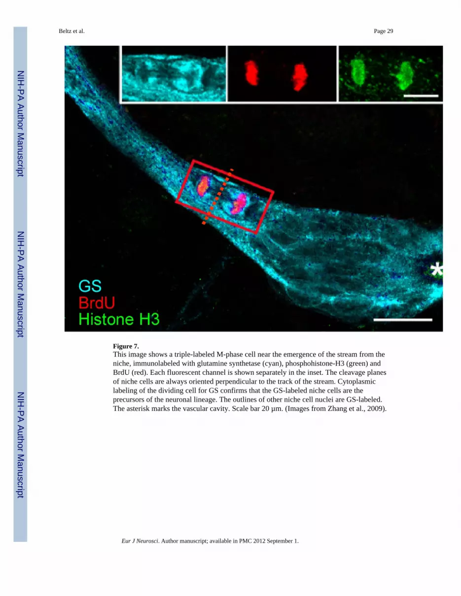
Figure 7.This image shows a triple-labeled M-phase cell near the emergence of the stream from theniche, immunolabeled with glutamine synthetase (cyan), phosphohistone-H3 (green) andBrdU (red). Each fluorescent channel is shown separately in the inset. The cleavage planesof niche cells are always oriented perpendicular to the track of the stream. Cytoplasmiclabeling of the dividing cell for GS confirms that the GS-labeled niche cells are theprecursors of the neuronal lineage. The outlines of other niche cell nuclei are GS-labeled.The asterisk marks the vascular cavity. Scale bar 20 µm. (Images from Zhang et al., 2009).
Beltz et al. Page 29
Eur J Neurosci. Author manuscript; available in PMC 2012 September 1.
NIH
-PA Author Manuscript
NIH
-PA Author Manuscript
NIH
-PA Author Manuscript

Figure 8.A. Live crayfish were incubated in BrdU for 24 hours and then maintained in fresh pondwater for 7 days. Just before sacrifice, they were treated with EdU (green) for 6 hours. Fixedbrains also were labeled immunocytochemically for glutamine synthetase (blue) to revealthe niche and streams. S phase cells in the niche and streams are labeled only with EdU,demonstrating that migration is uni-directional (away from the niche) and that niche celldivisions are not self-renewing, as no BrdU-labeled cells remain in the niche. B. Highermagnification image of the LPZ. The BrdU-labeled cells which were labeled first in thesequence are found only in the proliferation zones (MPZ and LPZ) and not in the niche.Arrow indicates direction of migration. Scale bars: A, 100 µm; B, 50 µm. (Images fromBenton et al., 2011).
Beltz et al. Page 30
Eur J Neurosci. Author manuscript; available in PMC 2012 September 1.
NIH
-PA Author Manuscript
NIH
-PA Author Manuscript
NIH
-PA Author Manuscript

Figure 9.CellTracker™-labeled cells (CTG-labeled cells) are found in the vascular cavity and in, andon, the neurogenic niche itself. A. Projections of confocal stacked images of the neurogenicniche. Position of a CTG-labeled cell in the vascular cavity with all fluorescence channelsmerged. Ai. (Left, center, right panels) Glutamine synthetase (GS, left) immunoreactivity inthe niche; propidium iodide (PI, center) labeling all cell nuclei, with arrow pointing to thenucleus of the CTG-labeled cell; CTG-labeled cell (right). B. Other CTG-labeled cells haveinteresting morphological characteristics, two of which can be seen just outside the niche(arrowheads); one has a long process extending into the niche (arrow), and another CTG-labeled cell inserts on the outer margin of the niche. Bi. Top, middle, and bottom panelsrepresent the separate channels: GS outlining the niche (top); PI revealing cell nuclei,arrowheads pointing to the respective CTG-labeled cells in B (middle); and CTG-labeledcells with arrow pointing to the fine process coming from the CTG-labeled cell (bottom).Scale bars: A, 50 µm; Ai, 20 µm; B, 20 µm; Bi, 10 µm. (Images from Benton et al., 2011).
Beltz et al. Page 31
Eur J Neurosci. Author manuscript; available in PMC 2012 September 1.
NIH
-PA Author Manuscript
NIH
-PA Author Manuscript
NIH
-PA Author Manuscript

Figure 10.Our current model of adult neurogenesis in the crayfish P. clarkii, beginning withhematopoietic tissue, the release of hematopoietic stem cells, their attraction to the niche andtransformation into niche cells. These aspects of our model are hypothetical. The niche cells(1st generation neuronal precursors), which label for glutamine synthetase, dividesymmetrically to produce daughters that migrate along processes of the niche cells towardscell cluster 9 or 10. These 2nd generation neuronal precursors divide at least once more inthe proliferation zones in the cell clusters, before differentiating into neurons. These aspectsof the model are supported by several types of data, reviewed in this paper.
Beltz et al. Page 32
Eur J Neurosci. Author manuscript; available in PMC 2012 September 1.
NIH
-PA Author Manuscript
NIH
-PA Author Manuscript
NIH
-PA Author Manuscript

NIH
-PA Author Manuscript
NIH
-PA Author Manuscript
NIH
-PA Author Manuscript
Beltz et al. Page 33
TABLE 1
The relationship between the neurogenic niche and dissociated, CellTracker™ Green (CTG)-labeled cellsfrom four crayfish tissues (green gland, hepatopancreas, hematopoietic tissue and hemolymph) was examinedin co-cultured, desheathed brains. Complete exposure to the niche/ventral brain surface appears to be essentialfor the niche region to attract CGT-labeled hemocytes in vitro, because no cells are attracted to the niche whensheathed brains are placed in co-culture with CTG-labeled hemocytes. The number and the percentage of totalniches containing CTG+ cells appear in the second and third columns. The Chi-square test of independencewas used to test the significance of these findings; the result is chi square (7, N=111) =34.8885, p < 0.001,indicating that the number of niches containing CTG+ cells was significantly dependent on the cell type. Theattraction of hemocytes to the niche relative to other cell types is therefore of statistical significance.
1. Dissociated cells from crayfishtissues labeled with CTG.
Total numberof niches
Percent of nicheswith CTG labeled
cells
Green Gland 10 0%
Hepatopancreas 11 0%
Hematopoietic Tissue 12 9%
Hemocytes 30 77%
2. Changes in cell culture mediumwith CTG labeled hemocytes
Hemocytes + 5-HT×10−9 M 12 17%
Hemocytes + MMS ×10−8 M 16 31%
3. Treatment prior to co-culture,culture + CTG labeled hemocytes
5-day PCPA pretreatment 8 12.5%
Sheath left on dissected brain 12 0%
Eur J Neurosci. Author manuscript; available in PMC 2012 September 1.