chapter 4 results and discussion
TRANSCRIPT

CHAPTER 4
RESULTS AND DISCUSSION
4.1 Collection
During collection and observation from October 2004 to October 2005 in four
sites, 118 samples were collected in Nothern Thailand, covering 40 families of host
plant, 76 samples were collected in Chiang Mai Province, 30 samples were collected
in Chiang Rai Province, 5 samples were collected in Uttradit Province and 7 samples
were collected in Phetchabun Province respectively.
In Chiang Mai Province, 73 species of cercosporoid fungi covering 5 genera
were found. Furthermore, 33 species of cercosporoid fungi of 3 genera were found in
Chiang Rai Province, 5 species of cercosporoid fungi of 2 genera were found in
Uttradit Province, and 7 species of cercosporoid fungi of 2 genera were found in
Phetchabun Province. The complete results were listed in the table 1.
4.2 Observation and identification
In this study, totally 118 species of the Cercospora and allied genera were
found in 90 genera of plants belonging to 40 families (Table 1).
ÅÔ¢ÊÔ·¸Ô ìÁËÒÇÔ·ÂÒÅÑÂàªÕ§ãËÁèCopyright by Chiang Mai UniversityA l l r i g h t s r e s e r v e d
ÅÔ¢ÊÔ·¸Ô ìÁËÒÇÔ·ÂÒÅÑÂàªÕ§ãËÁèCopyright by Chiang Mai UniversityA l l r i g h t s r e s e r v e d

93
Table 1 New Record of Cercospora and allied genera in Thailand.
Family Name Cercosporoid Genera Scientific Name
Acanthaceae Cercospora andrographidicola Andrographis paniculata3
Cercospora barlericola Barleria cristata3
Cercospora strobilanthis Strobilanthes sp.1
Cercospora sp. Phlogacanthus curviflorus2
Pseudocercospora rhinacanthi Rhinacanthus nasutus1
Asclepiadaceae Pseudocercospora sp. Raphistemma pulchellum1
Cercospora celosiae Celosia argentea 2
Cercospora celosiae Celosia argentea var. cristata2
Apocynaceae Pseudocercospora plumeriae Plumeria acuminate2
Araceae Cercospora richardiicola Zantedeschia sp.4
Araliaceae Pseudocercospora sp. Trevesia palmate2
Asteraceae Cercospora gerberae Gerbera jamesonii2
Balsaminaceae Cercospora balsaminiana Impatiens balsamina4
Pseudocercospora balsaminae Impatiens balsamina1
Basellaceae Cercospora basellae-albae Basella alba1
Bignoniaceae Pseudocercospora tecomae-
heterophyllae
Tecoma stans1
Boraginacare Cercospora ehretiicola Ehretia microphylla2
Caprifoliaceae Pseudocercospora viburnigena Viburnum prunifolium2
Cruciferae Cercospora brassicicola Brassica pekinensis1
Cercospora brassicicola Brassica campestris var. chinensis1
Cercospora brassicicola Brassica rapa1
Cercospora beticola Spinacia oleraceal1
ÅÔ¢ÊÔ·¸Ô ìÁËÒÇÔ·ÂÒÅÑÂàªÕ§ãËÁèCopyright by Chiang Mai UniversityA l l r i g h t s r e s e r v e d
ÅÔ¢ÊÔ·¸Ô ìÁËÒÇÔ·ÂÒÅÑÂàªÕ§ãËÁèCopyright by Chiang Mai UniversityA l l r i g h t s r e s e r v e d

94
Table 1 (continued)
Family Name Cercosporoid Genera Scientific Name
Cruciferae Cercospora brassicicola Brassica oleracea2
Cercospora brassicicola Brassica juncea1
Cercospora brassicicola Brassica alboglabra1
Chenopodiaceae Cercospora beticola Beta vulgaris var. alba1
Combretaceae Pseudocercospora quisqualidis Quisqualis indica2
Compositae Cercospora bidentis Bidens pilosa4
Cercospora eupatorii Eupatorium adenophorum1
Cercospora virgaureae Eupatorium adenophorum1
Cercospora helianthicola Helianthus annuus1
Cercospora lactucae-sativae Lactuca sativa var. longifolia1
Cercospora lactucae-sativae Lactuca sativa var. crispa1
Cercospora sp. Melampodium paludosum1
Cercospora mikaniicola Mikania cordata1
Passalora tithoniae Tithonia diversifolia1
Cercospora tagetis-erectae Tagetes erecta2
Cercospora tridacis-procumbentis Tridax procumbens1
Convolvulaceae Cercospora ipomoeae Ipomoea nil2
Cercospora ipomoeae Ipomoea aquatica2
Cercospora merremiae Merremia vitifolia2
Cucurbitaceae Cercospora cucurbitacea Cucurbita moschata1
Cercospora citrullina Coccinia grandis1
Cercospora citrullina Sechium edule2
Dioscoreaceae Pseudocercospora carbonacea Dioscorea glabra var. glabra1
ÅÔ¢ÊÔ·¸Ô ìÁËÒÇÔ·ÂÒÅÑÂàªÕ§ãËÁèCopyright by Chiang Mai UniversityA l l r i g h t s r e s e r v e d
ÅÔ¢ÊÔ·¸Ô ìÁËÒÇÔ·ÂÒÅÑÂàªÕ§ãËÁèCopyright by Chiang Mai UniversityA l l r i g h t s r e s e r v e d

95
Table 1 (continued)
Family Name Cercosporoid Genera Scientific Name
Dioscoreaceae Pseudocercospora contraria Dioscorea alata2
Euphorbiaceae Cercospora analyphae Acalypha wilkesiana4
Cercospora analyphae Acalypha indica1
Cercospora sp. Bridelia ovata2
Cercospora pulcherrimae Euphorbia pulcherrima2
Cercospora jatrophigena Jatropha curcas2
Cercospora manihobae Manihot esculenta2
Cercospora phyllanthi Phyllanthus sp.3
Cercospora ricinella Ricinus communis1
Pseudocercospora melanolepidis Mallotus pierrei3
Elaeanaceae Cercospora elaeagni Elaeagnus latifolia2
Elaeocarpaceae Cercospora sp. Elaeocarpus hygrophilus2
Hydrangeaceae Cercospora hydrangeae Hydrangea macrophylla1
Leguminosae Cercospora arachidicola Arachis hypogaea1
Pseudocercospora stizolobii Mucuna bracteata1
Cercospora psophocarpicola Psophocarpus letragonlobus2
Passalora arachidicola Arachis hypogaea2
Cercospora bauhiniae-variegatae Bauhinia racemosa1
Passalora aenea Cassia angustifolia1
Cercospora centrosematis Centrosema pubescens1
Pseudocercospora dalbergiae Dalbergia stipulacea1
Cercospora kikuchii Glycine max1
Cercospora leucaenae Leucaena leucocephalade2
ÅÔ¢ÊÔ·¸Ô ìÁËÒÇÔ·ÂÒÅÑÂàªÕ§ãËÁèCopyright by Chiang Mai UniversityA l l r i g h t s r e s e r v e d
ÅÔ¢ÊÔ·¸Ô ìÁËÒÇÔ·ÂÒÅÑÂàªÕ§ãËÁèCopyright by Chiang Mai UniversityA l l r i g h t s r e s e r v e d
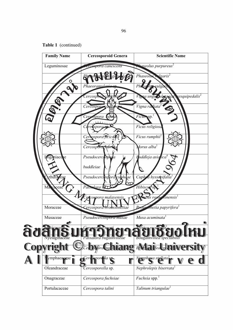
96
Table 1 (continued)
Family Name Cercosporoid Genera Scientific Name
Leguminosae Cercospora canescens Phaseolus purpureus2
Phaeoisariopsis griseola Phaseolus vulgaris2
Phaeoramularia sp. Phaseolus vulgaris1
Cercospora canescens Vigna unguiculata var. sesquipedalis2
Cercospora canescens Vigna radiata2
Cercospora rufula Ficus sp.1
Cercospora sp. Ficus religiosa1
Cercospora ficicola Ficus rumphii1
Cercospora morina Morus alba1
Loganiaceae Pseudocercospora
buddleiae
Buddleja asiatica4
Lythuraceae Pseudocercospora cupheae Cuphea hyssopifalia1
Malvaceae Passalora sp. Hibiscus sp.1
Cercospora malayensis Hibiscus rosae-sinensis1
Moraceae Cercospora broussonetiae Broussonetia papyrifera1
Musaceae Pseudocercospora musae Musa acuminata1
Myricaceae Cercospora myrti Myrica esculenta1
Nyctaginaceae Passalora bugainvilleae Bougainvillea spectabilis1
Cercospora bugainvilleae Bougainvillea spectabilis1
Nymphaeaceae Cercosporella sp. Nymphaea stellata1
Oleandraceae Cercosporella sp. Nephrolepis biserrata1
Onagraceae Cercospora fuchsiae Fuchsia spp.1
Portulacaceae Cercospora talini Talinum triangulae1
ÅÔ¢ÊÔ·¸Ô ìÁËÒÇÔ·ÂÒÅÑÂàªÕ§ãËÁèCopyright by Chiang Mai UniversityA l l r i g h t s r e s e r v e d
ÅÔ¢ÊÔ·¸Ô ìÁËÒÇÔ·ÂÒÅÑÂàªÕ§ãËÁèCopyright by Chiang Mai UniversityA l l r i g h t s r e s e r v e d

97
Table 1 (continued)
Family Name Cercosporoid Genera Scientific Name
Rosaceae Cercospora fragariae Fragaria sp.1
Cercospora rosicola Rosa sp.1
Rubiaceae Pseudocercospora sp. Haldina cordifolia3
Cercospora coffeicola Coffea arabica1
Pseudocercospora ixorae Ixora congesta2
Saururaceae Pseudocercospora houttuyniae Houttuynia cordata1
Solanaceae Pseudocercospora sp. Solanum trilobatum1
Cercospora beticola Spinacia oleracea1
Passalora sp. Lycopersicon esculentumcer var.
asiforme1
Cercospora physalidis Capsicum frutescens4
Cercospora capsici Capsicum annuum1
Cercospora lycopersici Lycopersicon esculentum var.
pyriforme1
Cercospora capsicigena Capsicum annuum var.
acuminatum1
Cercospora sp. Solanum pseudocapsicum1
Cercospora sp. Solanum wrightii2
Pseudocercospor solani-
melongenicola
Solanum melongena1
Cercospora physalidis-
angulatae
Physalis angulata1
Cercospora nicotianicola Nicotiana tabacum1
ÅÔ¢ÊÔ·¸Ô ìÁËÒÇÔ·ÂÒÅÑÂàªÕ§ãËÁèCopyright by Chiang Mai UniversityA l l r i g h t s r e s e r v e d
ÅÔ¢ÊÔ·¸Ô ìÁËÒÇÔ·ÂÒÅÑÂàªÕ§ãËÁèCopyright by Chiang Mai UniversityA l l r i g h t s r e s e r v e d

98
Table 1 (continued)
Family Name Cercosporoid Genera Scientific Name
Umbelliferae Cercospora apii Apium graveolens1
Verbenaceae Cercospora volkameriae Clerodendrum paniculatum1
Cercospora volkameriae. Clerodendrum indicum1
Passalora gmelinae-arboreae Gmelina arborea1
Pseudocercospora viticicola Vitex quinata1
Cercospora lantanae-camarae Lantana camera2
Cercospora tectonae Tectona grandis1
Cercospora holmskioldiae Holmskioldia sanginea1
1 = Chiang Mai Province, 2 = Chiang Rai Province, 3 = Uttradit Province, 4 = Phetchabun
Province.
In this study, 92 species of plants were recorded as new hosts (Table 2).
Furthermore, the data led to new species based on the recent publications on
Cercosporoid classification (Table 3).
ÅÔ¢ÊÔ·¸Ô ìÁËÒÇÔ·ÂÒÅÑÂàªÕ§ãËÁèCopyright by Chiang Mai UniversityA l l r i g h t s r e s e r v e d
ÅÔ¢ÊÔ·¸Ô ìÁËÒÇÔ·ÂÒÅÑÂàªÕ§ãËÁèCopyright by Chiang Mai UniversityA l l r i g h t s r e s e r v e d

99
Table 2 New record of Cercospora and allied genera in Thailand.
Family Name Cercosporoid Genera Scientific Name
Acanthaceae Cercospora andrographidicola Andrographis paniculata
Cercospora barlericola Barleria cristata
Cercospora strobilanthis Strobilanthes sp.
Cercospora sp. Phlogacanthus curviflorus
Asclepiadaceae Pseudocercospora sp. Raphistemma pulchellum
Apocynaceae Pseudocercospora plumeriae Plumeria acuminate
Araceae Cercospora callae richardiicola Zantedeschia sp.
Araliaceae Pseudocercospora sp. Trevesia palmata
Balsaminaceae Cercospora balsaminiana Impatiens balsamina
Pseudocercospora balsaminae Impatiens balsamina
Basellaceae Cercospora basellae-albae Basella alba
Bignoniaceae Pseudocercospora tecomae-
heterophyllae
Tecoma stans
Boraginacare Cercospora ehretiicola Ehretia microphylla
Caprifoliaceae Pseudocercospora viburnigena Viburnum prunifolium
Cruciferae Cercospora brassicicola Brassica pekinensis
Cercospora brassicicola Brassica campestris var. chinensis
Cercospora brassicicola Brassica rapa
Cercospora beticola Spinacia oleraceal
Cruciferae Cercospora brassicicola Brassica juncea
Cercospora brassicicola Brassica oleracea
Cercospora brassicicola Brassica alboglabra
Chenopodiaceae Cercospora beticola Beta vulgaris var. alba
ÅÔ¢ÊÔ·¸Ô ìÁËÒÇÔ·ÂÒÅÑÂàªÕ§ãËÁèCopyright by Chiang Mai UniversityA l l r i g h t s r e s e r v e d
ÅÔ¢ÊÔ·¸Ô ìÁËÒÇÔ·ÂÒÅÑÂàªÕ§ãËÁèCopyright by Chiang Mai UniversityA l l r i g h t s r e s e r v e d

100
Table 2 (continued)
Family Name Cercosporoid Genera Scientific Name
Combretaceae Pseudocercospora quisqualidis Quisqualis indica
Compositae Cercospora bidentis Bidens pilosa
Cercospora eupatorii Eupatorium adenophorum
Cercosporella virgaureae Eupatorium adenophorum
Pseudocercospora eupatorii Eupatorium odoratum
Cercospora lactucae-sativae Lactuca sativa var. crispa
Cercospora sp. Melampodium paludosum
Cercospora mikaniicola Mikania cordata
Passalora tithoniae. Tithonia diversifolia
Cercospora tagetis-erectae Tagetes erecta
Cercospora lactucae-sativae Lactuca sativa var.
longifolia
Convolvulaceae Cercospora ipomoeae Ipomoea nil
Cercospora ipomoeae Ipomoea aquatica
Cercospora merremiae. Merremia vitifolia
Cucurbitaceae Cercospora cucurbitacea Cucurbita moschata
Cercospora citrullina Sechium edule
Dioscoreaceae Pseudocercospora carbonacea Dioscorea glabra var.
glabra
Pseudocercospora contraria Dioscorea alata
Euphorbiaceae Cercospora analyphae Acalypha wilkesiana
Cercospora sp. Bridelia ovata
ÅÔ¢ÊÔ·¸Ô ìÁËÒÇÔ·ÂÒÅÑÂàªÕ§ãËÁèCopyright by Chiang Mai UniversityA l l r i g h t s r e s e r v e d
ÅÔ¢ÊÔ·¸Ô ìÁËÒÇÔ·ÂÒÅÑÂàªÕ§ãËÁèCopyright by Chiang Mai UniversityA l l r i g h t s r e s e r v e d

101
Table 2 (continued)
Family Name Cercosporoid Genera Scientific Name
Euphorbiaceae Cercospora jatrophigena Jatropha curcas
Cercospora pulcherrimae Euphorbia pulcherrima
Cercospora manihobae Manihot esculenta
Cercospora phyllanthi Phyllanthus sp.
Pseudocercospora melanolepidis Mallotus pierrei
Elaeanaceae Cercospora elaeagni Elaeagnus latifolia
Elaeocarpaceae Cercospora sp. Elaeocarpus hygrophilus
Leguminosae Cercospora arachidicola Arachis hypogaea
Passalora arachidicola Arachis hypogaea
Cercospora bauhiniae-variegatae Bauhinia racemosa
Cercospora centrosematis Centrosema pubescens
Pseudocercospora dalbergiae Dalbergia stipulacea
Passalora aenea Cassia angustifolia
Cercospora leucaenae Leucaena leucocephalade
Pseudocercospora stizolobii Mucuna bracteata
Phaeoisariopsis griseola Phaseolus vulgaris
Phaeoramularia sp. Phaseolus vulgaris
Loganiaceae Pseudocercospora buddleiae Buddleja asiatica
Lythraceae Pseudocercospora cupheae Cuphea hyssopifalia
Malvaceae Passalora sp. Hibiscus sp.
Moraceae Cercospora broussonetiae Broussonetia papyrifera
Cercospora rufula Ficus sp.
Cercospora sp. Ficus religiosa
ÅÔ¢ÊÔ·¸Ô ìÁËÒÇÔ·ÂÒÅÑÂàªÕ§ãËÁèCopyright by Chiang Mai UniversityA l l r i g h t s r e s e r v e d
ÅÔ¢ÊÔ·¸Ô ìÁËÒÇÔ·ÂÒÅÑÂàªÕ§ãËÁèCopyright by Chiang Mai UniversityA l l r i g h t s r e s e r v e d
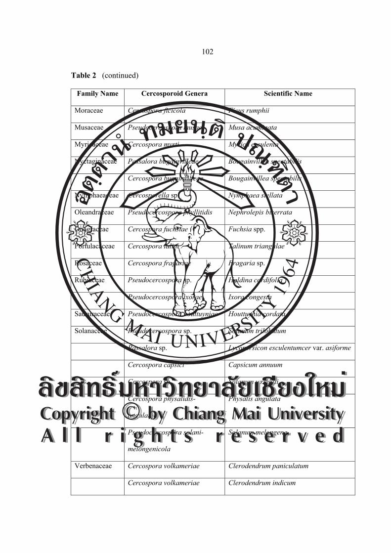
102
Table 2 (continued)
Family Name Cercosporoid Genera Scientific Name
Moraceae Cercospora ficicola Ficus rumphii
Musaceae Pseudocercospora musae Musa acuminata
Myricaceae Cercospora myrti Myrica esculenta
Nyctaginaceae Passalora bugainvilleae Bougainvillea spectabilis
Cercospora bugainvilleae Bougainvillea spectabilis
Nymphaeaceae Cercosporella sp. Nymphaea stellata
Oleandraceae Pseudocercospora phyllitidis Nephrolepis biserrata
Onagraceae Cercospora fuchsiae Fuchsia spp.
Portulacaceae Cercospora talini Talinum triangulae
Rosaceae Cercospora fragariae Fragaria sp.
Rubiaceae Pseudocercospora sp. Haldina cordifolia
Pseudocercospora ixorae Ixora congesta
Saururaceae Pseudocercospora houttuyniae Houttuynia cordata
Solanaceae Pseudocercospora sp. Solanum trilobatum
Passalora sp. Lycopersicon esculentumcer var. asiforme
Cercospora capsici Capsicum annuum
Cercospora sp. Solanum wrightii
Cercospora physalidis-
angulatae
Physalis angulata
Pseudocercospora solani-
melongenicola
Solanum melongena
Verbenaceae Cercospora volkameriae Clerodendrum paniculatum
Cercospora volkameriae Clerodendrum indicum
ÅÔ¢ÊÔ·¸Ô ìÁËÒÇÔ·ÂÒÅÑÂàªÕ§ãËÁèCopyright by Chiang Mai UniversityA l l r i g h t s r e s e r v e d
ÅÔ¢ÊÔ·¸Ô ìÁËÒÇÔ·ÂÒÅÑÂàªÕ§ãËÁèCopyright by Chiang Mai UniversityA l l r i g h t s r e s e r v e d

103
Table 2 (continued)
Family Name Cercosporoid Genera Scientific Name
Verbenaceae Passalora gmelinae-arboreae Gmelina arborea
Cercospora tectonae Tectona grandis
Cercospora holmskioldiae Holmskioldia sanginea
Pseudocercospora viticicola Vitex quinata
Table 3 New record of Cercospora and allied genera in the world.
Family Name Cercosporoid Genera Scientific Name
Acanthaceae Cercospora sp. Phlogacanthus curviflorus
Asclepiadaceae Pseudocercospora sp. Raphistemma pulchellum
Araliaceae Pseudocercospora sp. Trevesia palmate
Compositae Cercospora sp. Melampodium paludosum
Euphorbiaceae Cercospora sp. Bridelia ovata
Elaeocarpaceae Cercospora sp. Elaeocarpus hygrophilus
Leguminosae Phaeoramularia sp. Phaseolus vulgaris
Moraceae Cercospora sp. Ficus religiosa
Pseudocercospora fici Ficus rumphii
Nymphaeaceae Cercosporella sp. Nymphaea stellata
Oleandraceae Pseudocercospora
phyllitidis
Nephrolepis biserrata
Rubiaceae Pseudocercospora sp. Haldina cordifolia
Solanaceae Pseudocercospora sp. Solanum trilobatum
Cercospora sp. Solanum pseudocapsicum
Passalora sp. Lycopersicon esculentumcer var. asiforme
ÅÔ¢ÊÔ·¸Ô ìÁËÒÇÔ·ÂÒÅÑÂàªÕ§ãËÁèCopyright by Chiang Mai UniversityA l l r i g h t s r e s e r v e d
ÅÔ¢ÊÔ·¸Ô ìÁËÒÇÔ·ÂÒÅÑÂàªÕ§ãËÁèCopyright by Chiang Mai UniversityA l l r i g h t s r e s e r v e d

104
Cercospora and allied genera are described based on the family name of the
host plants.
Family Acanthaceae
Cercospora barlericola Payak and Thirum. (barlericola), Indian Phytopathol. 2: 191.
1949.
= Cercospora barleriae-cristatae Govindu and Thirum., Sydowia 10:
273. (1956) 1957.
Leaf spots amphigenous, circular to subcircular, 3.00-4.00 mm in diameter, distinct on
the upper surface, greyish-brown to dark brown. Stromata distinct, usually
epiphyllous, then erumpent, olive-brown, 27.06-41.82 µm in diameter. Conidiophores
emerging from the upper part of stromata, dark brown, densely fasciculate, simple,
straight or slightly curved, 1-6-septate, (17.22-)22.14-61.50(-68.88)×(2.46-)3.69-
4.92(-6.88) µm, with indistinct and unthickened conidial scars. Conidia obclavate,
straight, smooth, hyaline, with thickened and truncate hilum, (29.52-)50.43-93.48
(-123.00)×(2.46-)4.42-4.92(-5.41) µm, 5-13-septate (Figure 17 and 18).
Figure 17 Photograph of Cercospora barlericola on Barleria cristata: A. Symptom,
B. Conidiophores and C. Conidia.
A B C
ÅÔ¢ÊÔ·¸Ô ìÁËÒÇÔ·ÂÒÅÑÂàªÕ§ãËÁèCopyright by Chiang Mai UniversityA l l r i g h t s r e s e r v e d
ÅÔ¢ÊÔ·¸Ô ìÁËÒÇÔ·ÂÒÅÑÂàªÕ§ãËÁèCopyright by Chiang Mai UniversityA l l r i g h t s r e s e r v e d

105
Figure 18 Drawing of Cercospora barlericola on Barleria cristata:
A. Conidiophores and B. Conidia (scale bar = 40 µm).
Material examined: Thailand, Sak Yai National Park, A. Nam Pad, Uttradit
Province. Leaves of Barleria cristata, November 25, 2004, C. Nakashima (CN) and J.
Meeboon (JM), (CMU MH 014), CMU MH 001.
Known distribution: India and Jamaica (Crous and Braun, 2003).
Notes: This is the first record from Thailand, type species was in Banares Hindu
University, India; Barleria cristata; M. M. Payak; December 9, 1949. Several Indian
collections from IMI on Barleria spp. have been examined and proved to belong to a
single variable Cercospora s.str. species indistinguishable from C. apii s. lat.
Cercospora sp.
Leaf spots distinct, circular-subcircular, dark brown, blackish-brown border,
3.00-5.00 mm in diameter, often confluent. Fruit bodies amphigenous. Stromata
brown, amphigenous, mostly hypophyllous, 12.30-24.60 µm in diameter.
Conidiophores arising from stromata, hyaline to brown, straight or sinuous, 1-9-
septate, fasciculate, 14.76-118.08×3.69-4.92 µm, with thickened conidial scars.
Conidia pale olivaccous, obclavate, 27.06-108.24×1.23-4.92 µm, 3-12-septate, hilum
conspicuously thickened and darkened (Figure 19 and 20).
A B
ÅÔ¢ÊÔ·¸Ô ìÁËÒÇÔ·ÂÒÅÑÂàªÕ§ãËÁèCopyright by Chiang Mai UniversityA l l r i g h t s r e s e r v e d
ÅÔ¢ÊÔ·¸Ô ìÁËÒÇÔ·ÂÒÅÑÂàªÕ§ãËÁèCopyright by Chiang Mai UniversityA l l r i g h t s r e s e r v e d

106
Figure 19 Photograph of Cercospora sp. on Phlogacanthus curviflorus:
A. Symptom, B. Conidiophores and C. Conidia.
Figure 20 Drawing of Cercospora sp. on Phlogacanthus curviflorus:
A. Conidiophores and B. Conidia (scale bar = 40 µm).
Material examined: Thailand, Wiang Pa Pao, Chiang Rai Province. Leaves of
Phlogacanthus curviflorus, November 25, 2004, JM CMU MH 114.
Known distribution: Known only in Thailand.
Notes: This is the first record of Phlogacanthus curviflorus cause by Cercospora sp.
from Thailand.
A B C
A B
ÅÔ¢ÊÔ·¸Ô ìÁËÒÇÔ·ÂÒÅÑÂàªÕ§ãËÁèCopyright by Chiang Mai UniversityA l l r i g h t s r e s e r v e d
ÅÔ¢ÊÔ·¸Ô ìÁËÒÇÔ·ÂÒÅÑÂàªÕ§ãËÁèCopyright by Chiang Mai UniversityA l l r i g h t s r e s e r v e d

107
Pseudocercospora rhinacanthi (Höhn.) Deighton. Pap.140: 152. 1976.
≡ Cercospora rhinacanthi Höhn. (rhynacanthi). Sitzungsber. Kaiserl.
Akad. Wiss., Math.-Naturwiss. Cl., Wien. 121: 414. 1912. [T: Kabàt
and Bubàk, Fungi imp. exs. 847, e.g., BPI440801, IMI 89002
(slide); HBG; W].
≡ Cercosporina rhinacanthi (Höhn.) Sacc., Syll. Fung. 25: 917. 1931.
Leaf spots visible on both surfaces, distinct, circular-subcircular, 1.00-4.00
mm in diameter, dark brown, centre whitish grey surrounded by raised yellowish
brown border line, on the lower surface brown to yellowish brown margin. Stromata
small, globular to subglobular. Conidiophores in a loose to dense fasicle, emerging
through stomata and the cuticle, dark brown, uniform in colour, straight to mildly
curved, slightly geniculate or geniculate-sinuous, 1-7-septate, (19.51-)19.51-39.02
(-43.90)×(2.43-)4.87- 4.87(-4.87) µm, conidial scars inconspicuous. Conidia solitary,
filiform or narrowly obclavate, straight, hyaline, 3-7-septate, non-constricted at the
septa, subacute at the apex, obconically truncate at the base, (31.70-)39.02-58.53
(-65.85)×(3.65-)4.87-4.87(-4.87) µm, hilum unthickened and not darkened
(Figure 21 and 22).
ÅÔ¢ÊÔ·¸Ô ìÁËÒÇÔ·ÂÒÅÑÂàªÕ§ãËÁèCopyright by Chiang Mai UniversityA l l r i g h t s r e s e r v e d
ÅÔ¢ÊÔ·¸Ô ìÁËÒÇÔ·ÂÒÅÑÂàªÕ§ãËÁèCopyright by Chiang Mai UniversityA l l r i g h t s r e s e r v e d

108
Figure 21 Photograph of Pseudocercospora rhinacanthi on Rhinacanthus nasutus:
A. Symptom, B. Conidiophores and C. Conidia.
Figure 22 Drawing of Pseudocercospora rhinacanthi on Rhinacanthus nasutus:
A. Conidiophores and B. Conidia (scale bar = 40 µm).
Material examined: Thailand, Chiang Mai University, Chiang Mai Province. Leaves
of Rhinacanthus nasutus, November 25, 2004, JM CMU MH 002.
Known distribution: China, Indonesia, Java, Philippines and Thailand (Crous and
Braun, 2003).
Notes: Chandrasrikul (1962) reported Cercospora rhinacanthi in Rhinacanthus
nasutus in a preliminary host list of plant diseases in Thailand.
A B
C A B
ÅÔ¢ÊÔ·¸Ô ìÁËÒÇÔ·ÂÒÅÑÂàªÕ§ãËÁèCopyright by Chiang Mai UniversityA l l r i g h t s r e s e r v e d
ÅÔ¢ÊÔ·¸Ô ìÁËÒÇÔ·ÂÒÅÑÂàªÕ§ãËÁèCopyright by Chiang Mai UniversityA l l r i g h t s r e s e r v e d

109
Cercospora andrographidicola S. Q. Chen and P. K. Chi (andrographicala), J. S.
China Agric. Univ. 11: 61. 1990; also in Chi, Fungal diseases of cultivated medicinal
plants in Guangdong Province: 94. 1994. [T: Herbarium, S. China Agric. Univ.,
Guangzhou].
Leaf spots amphigenous, deep blackish-brown, subcircular to irregular, 2.00-
5.00 mm in diameter, distinct on the upper surface, brown to dark brown with out
definite margins on the lower surface. Stromata lacking to small, rudimentary to
poorly developed. Conidiophores arranged in a loose fascicle, arising from stromata,
straight or flexuous, simple or branched, brown to dark brown or paler towards the
apex, 1-9-septate, (12.30-)22.14-73.80(-83.64)×3.69-4.92 µm, with thickened conidial
scars. Conidia obclavate to acicular, straight, smooth, hyaline, with thickened and
truncate hilum, (19.68-)36.90-93.48(-172.20)×2.46-4.92 µm, 3-15-septate (Figure 23
and 24).
Figure 23 Photograph of Cercospora andrographidicola on
Andrographis paniculata: A. Symptom, B. Conidiophores and
C. Conidia.
A B C
ÅÔ¢ÊÔ·¸Ô ìÁËÒÇÔ·ÂÒÅÑÂàªÕ§ãËÁèCopyright by Chiang Mai UniversityA l l r i g h t s r e s e r v e d
ÅÔ¢ÊÔ·¸Ô ìÁËÒÇÔ·ÂÒÅÑÂàªÕ§ãËÁèCopyright by Chiang Mai UniversityA l l r i g h t s r e s e r v e d

110
Figure 24 Drawing of Cercospora andrographidicola on Andrographis paniculata:
A. Conidiophores and B. Conidia (scale bar = 40 µm).
Material examined: Thailand, Sak Yai National Park, Uttradit Province. Leaves of
Andrographis paniculata, November 25, 2004, JM CMU MH 003.
Known distribution: China (Crous and Braun, 2003).
Notes: Cercospora andrographidicola has been previously recorded in China. This is
the first record of this fungus in Thailand.
Cercospora strobilanthis Chidd. (strobilanthidis), Mycopathol. Mycol. Appl. 17: 71.
1962. [T: IMI 83190].
Leaf spots amphigenous, subcircular, grey in the centre, usually surrounded by
a rather undulate greyish brown, 1.50-3.00 mm in diameter. Stromata lacking to
small, rudimentary to poorly developed, 10.00-15.00 µm in diameter. Conidiophores
arranged in a loose fascicle, arising from stromata, straight or flexuous, simple or
branched, brown to dark brown or paler towards the apex, 1-9-septate, 12.30-
B
ÅÔ¢ÊÔ·¸Ô ìÁËÒÇÔ·ÂÒÅÑÂàªÕ§ãËÁèCopyright by Chiang Mai UniversityA l l r i g h t s r e s e r v e d
ÅÔ¢ÊÔ·¸Ô ìÁËÒÇÔ·ÂÒÅÑÂàªÕ§ãËÁèCopyright by Chiang Mai UniversityA l l r i g h t s r e s e r v e d

111
83.64×3.69-4.92 µm, with thickened conidial scars. Conidia obclavate, straight,
smooth, hyaline, with thickened and truncate hilum, 19.68-172.20×2.46-4.92 µm,
3-15-septate (Figure 25 and 26).
Figure 25 Photograph of Cercospora strobilanthis on Strobilanthes sp.:
A. Symptom, B. Conidiophores and C. Conidia.
Figure 26 Drawing of Cercospora strobilanthis on Strobilanthes sp.:
A. Conidiophores and B. Conidia (scale bar = 40 µm).
Material examined: Thailand, Chiang Mai University, Chiang Mai Province. Leaves
of Strobilanthes sp., November 25, 2004, JM CMU MH 004.
Known distribution: India (Crous and Braun, 2003).
A B C
A B ÅÔ¢ÊÔ·¸Ô ìÁËÒÇÔ·ÂÒÅÑÂàªÕ§ãËÁèCopyright by Chiang Mai UniversityA l l r i g h t s r e s e r v e d
ÅÔ¢ÊÔ·¸Ô ìÁËÒÇÔ·ÂÒÅÑÂàªÕ§ãËÁèCopyright by Chiang Mai UniversityA l l r i g h t s r e s e r v e d
112
Notes: Strobilanthes sp. has been previously recorded in India. This is the first record
of this fungus in Thailand. A true Cercospora s.str is distinct from C. apii s. lat.
Family Amaranthaceae
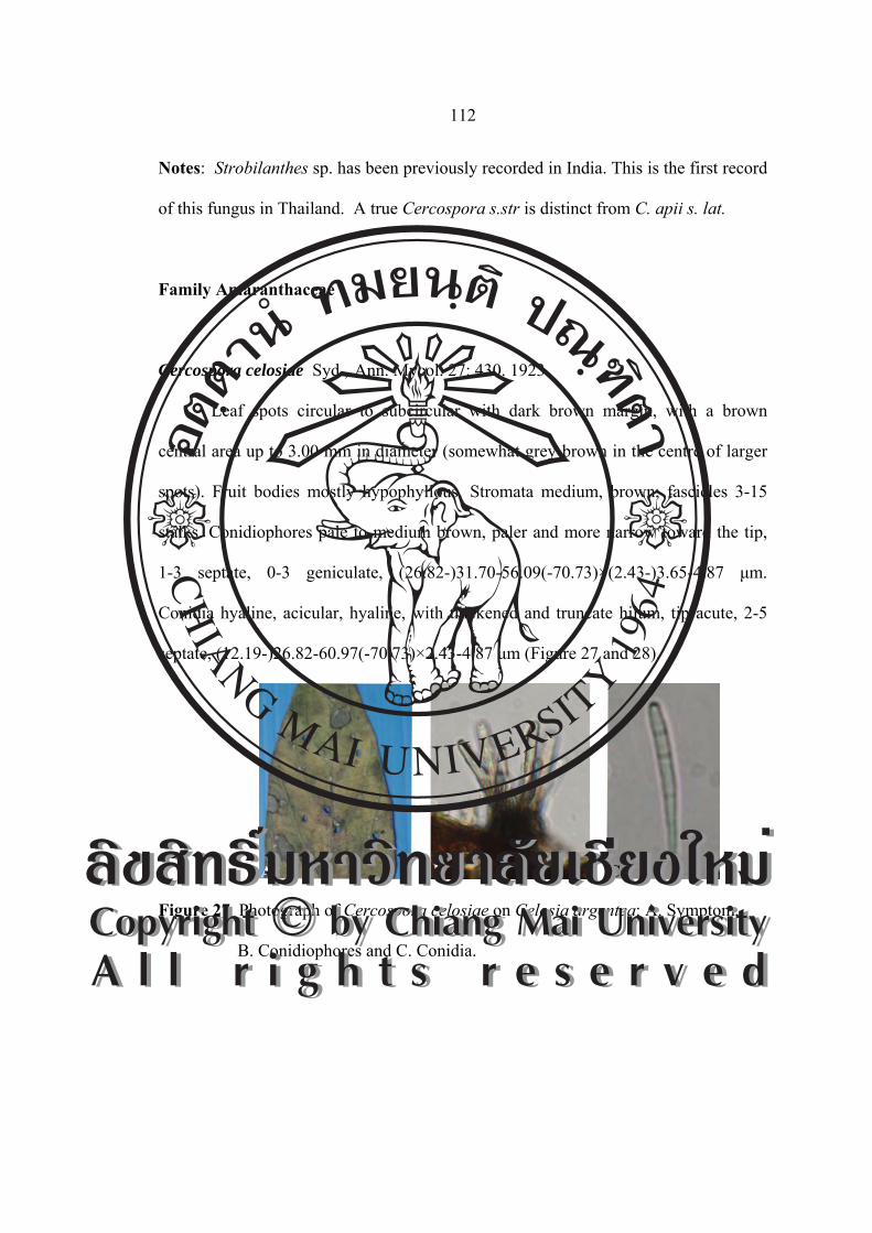
Cercospora celosiae Syd., Ann. Mycol. 27: 430. 1923.
Leaf spots circular to subcircular with dark brown margin, with a brown
central area up to 3.00 mm in diameter (somewhat grey-brown in the centre of larger
spots). Fruit bodies mostly hypophyllous. Stromata medium, brown, fascicles 3-15
stalks. Conidiophores pale to medium brown, paler and more narrow toward the tip,
1-3 septate, 0-3 geniculate, (26.82-)31.70-56.09(-70.73)×(2.43-)3.65-4.87 μm.
Conidia hyaline, acicular, hyaline, with thickened and truncate hilum, tip acute, 2-5
septate, (12.19-)26.82-60.97(-70.73)×2.43-4.87 μm (Figure 27 and 28).
Figure 27 Photograph of Cercospora celosiae on Celosia argentea: A. Symptom,
B. Conidiophores and C. Conidia.
A B C ÅÔ¢ÊÔ·¸Ô ìÁËÒÇÔ·ÂÒÅÑÂàªÕ§ãËÁèCopyright by Chiang Mai UniversityA l l r i g h t s r e s e r v e d
ÅÔ¢ÊÔ·¸Ô ìÁËÒÇÔ·ÂÒÅÑÂàªÕ§ãËÁèCopyright by Chiang Mai UniversityA l l r i g h t s r e s e r v e d
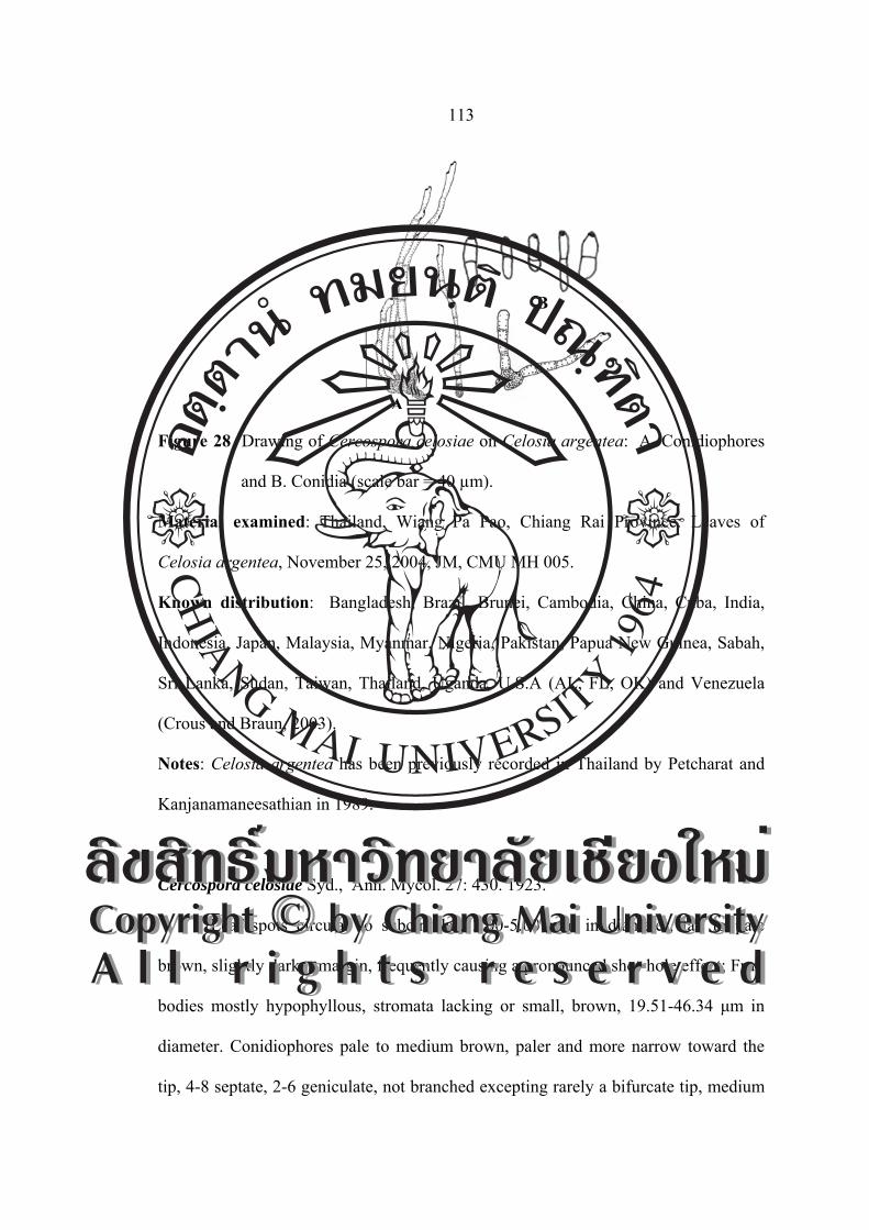
113
Figure 28 Drawing of Cercospora celosiae on Celosia argentea: A. Conidiophores
and B. Conidia (scale bar = 40 µm).
Material examined: Thailand, Wiang Pa Pao, Chiang Rai Province. Leaves of
Celosia argentea, November 25, 2004, JM, CMU MH 005.
Known distribution: Bangladesh, Brazil, Brunei, Cambodia, China, Cuba, India,
Indonesia, Japan, Malaysia, Myanmar, Nigeria, Pakistan, Papua New Guinea, Sabah,
Sri Lanka, Sudan, Taiwan, Thailand, Uganda, U.S.A (AL, FL, OK) and Venezuela
(Crous and Braun, 2003).
Notes: Celosia argentea has been previously recorded in Thailand by Petcharat and
Kanjanamaneesathian in 1989.
Cercospora celosiae Syd., Ann. Mycol. 27: 430. 1923.
Leaf spots circular to subcircular, 2.00-5.00 mm in diameter, tan to pale
brown, slightly darker margin, frequently causing a pronounced shot-hole effect; Fruit
bodies mostly hypophyllous, stromata lacking or small, brown, 19.51-46.34 μm in
diameter. Conidiophores pale to medium brown, paler and more narrow toward the
tip, 4-8 septate, 2-6 geniculate, not branched excepting rarely a bifurcate tip, medium
A
B
ÅÔ¢ÊÔ·¸Ô ìÁËÒÇÔ·ÂÒÅÑÂàªÕ§ãËÁèCopyright by Chiang Mai UniversityA l l r i g h t s r e s e r v e d
ÅÔ¢ÊÔ·¸Ô ìÁËÒÇÔ·ÂÒÅÑÂàªÕ§ãËÁèCopyright by Chiang Mai UniversityA l l r i g h t s r e s e r v e d

114
sized spore scar, (75.60-)109.75-158.53(-195.12)×4.87 μm. Conidia hyaline, acicular
to obclavate, with thickened hilum, straight to curved, base truncate, tip acute, 4-10
septate, (34.14-)48.78-131.70(-180.48)×(2.43-)2.43-2.43(-4.87) μm (Figure 29 and
30).
Figure 29 Photograph of Cercospora celosiae on Celosia argentea var. cristata:
A. Symptom, B. Conidiophores and C. Conidia.
Figure 30 Drawing of Cercospora celosiae on Celosia argentea var. cristata: A.
Conidiophores and B. Conidia (scale bar = 40 µm).
Material examined: Thailand, Wiang Pa Pao, Chiang Rai Province. Leaves of
Celosia argentea var. cristata, November 25, 2004, JM CMU MH 006.
Known distribution: Bangladesh, Brazil, Brunei, Cambodia, China, Cuba, India,
Indonesia, Japan, Malaysia, Myanmar, Nigeria, Pakistan, Papua New Guinea, Sabah,
A B C
A B
ÅÔ¢ÊÔ·¸Ô ìÁËÒÇÔ·ÂÒÅÑÂàªÕ§ãËÁèCopyright by Chiang Mai UniversityA l l r i g h t s r e s e r v e d
ÅÔ¢ÊÔ·¸Ô ìÁËÒÇÔ·ÂÒÅÑÂàªÕ§ãËÁèCopyright by Chiang Mai UniversityA l l r i g h t s r e s e r v e d

115
Sri Lanka, Sudan, Taiwan, Thailand, Uganda, U.S.A (AL, FL, OK) and Venezuela
(Crous and Braun, 2003).
Notes: Celosia argentea var. cristata. has been previously recorded by Niranam in
1960 refer to Sontirat (1980). A true Cercospora s.str. is distinct from C. apii s. lat.
The above description is based on Chupp (1954).
Family Asclepiadaceae
Pseudocercospora sp.
Leaf spots amphigenous, distinct, subcircular to irregular, 2-8 mm in diameter,
sometimes confluent, brown or grey-brown, later with a greyish or whitish centre,
with reddish brown margin on the upper surface, paler on the lower surface. Fruit
bodies amphigenous. Stromata medium, rudimentary to slightly developed, irregular,
dark brown, composed of a few swollen, brown hyphal cells. Conidiophores mostly
numerous in a dense fascicle emerging through a stoma and the cuticle, paler brown
or paler towards the apex, straight to slightly curved, 1-5 times mildly geniculate, not
branched, 1-3-septate, (19.51-)19.51-39.02(-43.90)×(2.43-)4.87-4.87 μm. Conidia
pale olivaceous, smooth, usually obclavate-cylindric, substraight or more usually
slightly curved, subobtuse or obtuse at the apex, 3-7-septate, (31.70-)39.02-58.53
(-65.85)×(3.65-)4.87-4.87 μm (Figure 31 and 32).
ÅÔ¢ÊÔ·¸Ô ìÁËÒÇÔ·ÂÒÅÑÂàªÕ§ãËÁèCopyright by Chiang Mai UniversityA l l r i g h t s r e s e r v e d
ÅÔ¢ÊÔ·¸Ô ìÁËÒÇÔ·ÂÒÅÑÂàªÕ§ãËÁèCopyright by Chiang Mai UniversityA l l r i g h t s r e s e r v e d

116
Figure 31 Photograph of Pseudocercospora sp. on Raphistemma pulchellum:
A. Symptom, B. Conidiophores and C. Conidia.
Figure 32 Drawing of Pseudocercospora sp.on Raphistemma pulchellum:
A. Conidiophores and B. Conidia (scale bar = 40 µm).
Material examined: Thailand, Chiang Mai University, Chiang Mai Province. Leaves
of Raphistemma pulchellum, November 25, 2004, JM CMU MH 007.
Known distribution: Known only Thailand.
Notes: This is the first record of this fungus in Thailand.
B A
A B C
ÅÔ¢ÊÔ·¸Ô ìÁËÒÇÔ·ÂÒÅÑÂàªÕ§ãËÁèCopyright by Chiang Mai UniversityA l l r i g h t s r e s e r v e d
ÅÔ¢ÊÔ·¸Ô ìÁËÒÇÔ·ÂÒÅÑÂàªÕ§ãËÁèCopyright by Chiang Mai UniversityA l l r i g h t s r e s e r v e d

117
Family Apocynaceae
Cercospora plumeriae Chupp, A monograph of the fungus genus Cercospora: 49.
1954. [T: CUP].
≡ Pseudocercospora plumeriae (Chupp) Tak. Kobay., Nishijima and C.
Nakash., Mycoscience 39: 188. 1998.
Leaf spots amphigenous, scattered to confluent, distinct, subcircular to
angular, 2.00-10.00 mm in diameter, grey to greyish white with dark brown to
purplish brown margin on the upper surface, pale brown to olivaceous brown on the
lower surface. Fruit bodies amphigenous. Stromata lacking to medium, rudimentary to
slightly developed, irregular, dark brown, 36.58-121.95 μm in diameter, composed of
a few swollen, brown hyphal cells. Conidiophores 5-15 in a divergent fascicle,
emerging through stomatal openings and the cuticle, paler brown or paler towards the
apex, straight to slightly curved, 1-5 times mildly geniculate, not branched, 2-8-
septate, (48.78-)78.04-109.75(-121.95)×(4.87-)4.87-4.87(-7.31) μm, conidial scars
conspicuous, apical or on shoulders of conidiogenous cells caused by geniculation.
Conidia solitary, acicular to filiform, substraight to moderately curved or even
undulate, hyaline, 4-11-septate, non-constriced at the septa, acute to obtuse at the
apex, truncate at the base, (51.21-)85.36-97.56(-236.58)×4.87-7.31 μm, hilum
conspicuously thickened and darkened (Figure 33 and 34).
ÅÔ¢ÊÔ·¸Ô ìÁËÒÇÔ·ÂÒÅÑÂàªÕ§ãËÁèCopyright by Chiang Mai UniversityA l l r i g h t s r e s e r v e d
ÅÔ¢ÊÔ·¸Ô ìÁËÒÇÔ·ÂÒÅÑÂàªÕ§ãËÁèCopyright by Chiang Mai UniversityA l l r i g h t s r e s e r v e d

118
Figure 33 Photograph of Cercospora plumeriae on Plumeria acuminate:
A. Symptom, B. Conidiophores and C. Conidia.
Figure 34 Drawing of Cercospora plumeriae on Plumeria acuminate:
A. Conidiophores and B. Conidia (scale bar = 40 µm).
Material examined: Thailand, Wiang Pa Pao, Chiang Rai Province. Leaves of
Plumeria acuminate, November 25, 2004, JM CMU MH 008.
Known distribution: Bangladesh, India, Indonesia, Japan, Malaysia, Myanmar,
Philippines, Trinidad, Tobago and U.S.A (FL) (Crous and Braun, 2003).
Notes: Hitherto known species in Thailand. This is the first record of this fungus in
Thailand.
A B C
A
B
ÅÔ¢ÊÔ·¸Ô ìÁËÒÇÔ·ÂÒÅÑÂàªÕ§ãËÁèCopyright by Chiang Mai UniversityA l l r i g h t s r e s e r v e d
ÅÔ¢ÊÔ·¸Ô ìÁËÒÇÔ·ÂÒÅÑÂàªÕ§ãËÁèCopyright by Chiang Mai UniversityA l l r i g h t s r e s e r v e d

119
Family Araceae
Cercospora richardiicola G. F. Atk. ‘richardiaecola’, J. Elisha Mitchell Sci. Soc. 8:
51 (1892). (Cercospora apii sensu lato, Crous and Braun 2003).
Leaf spots scattered, 1.00-7.00 mm in diameter, later confluent and
zonate, distinct, circular to subcircular, center tan to brown with purple brown margin.
Fruit bodies amphigenous. Stromata pale brown, 9.84-44.28 µm in diameter.
Conidiophores pale brown at the base and paler to apex, loose to dense fascicule, with
distinct and small conidial scars at the tip, straight, (17.22-)-24.60-56.58
(-76.26)×2.46-6.15 µm. Conidia solitary, acicular, straight to mildly curved, hyaline,
smooth, thickened and truncate basal end, (27.06-)41.82-88.56(-172.20)×(2.46-)3.69-
4.92 µm, 3-12 septate, obtuse at the apex (Figure 35 and 36).
Figure 35 Photograph of Cercospora richardiicola on Zantedeschia sp.:
A. Symptom, B. Conidiophores and C. Conidia.
A B C
ÅÔ¢ÊÔ·¸Ô ìÁËÒÇÔ·ÂÒÅÑÂàªÕ§ãËÁèCopyright by Chiang Mai UniversityA l l r i g h t s r e s e r v e d
ÅÔ¢ÊÔ·¸Ô ìÁËÒÇÔ·ÂÒÅÑÂàªÕ§ãËÁèCopyright by Chiang Mai UniversityA l l r i g h t s r e s e r v e d
120
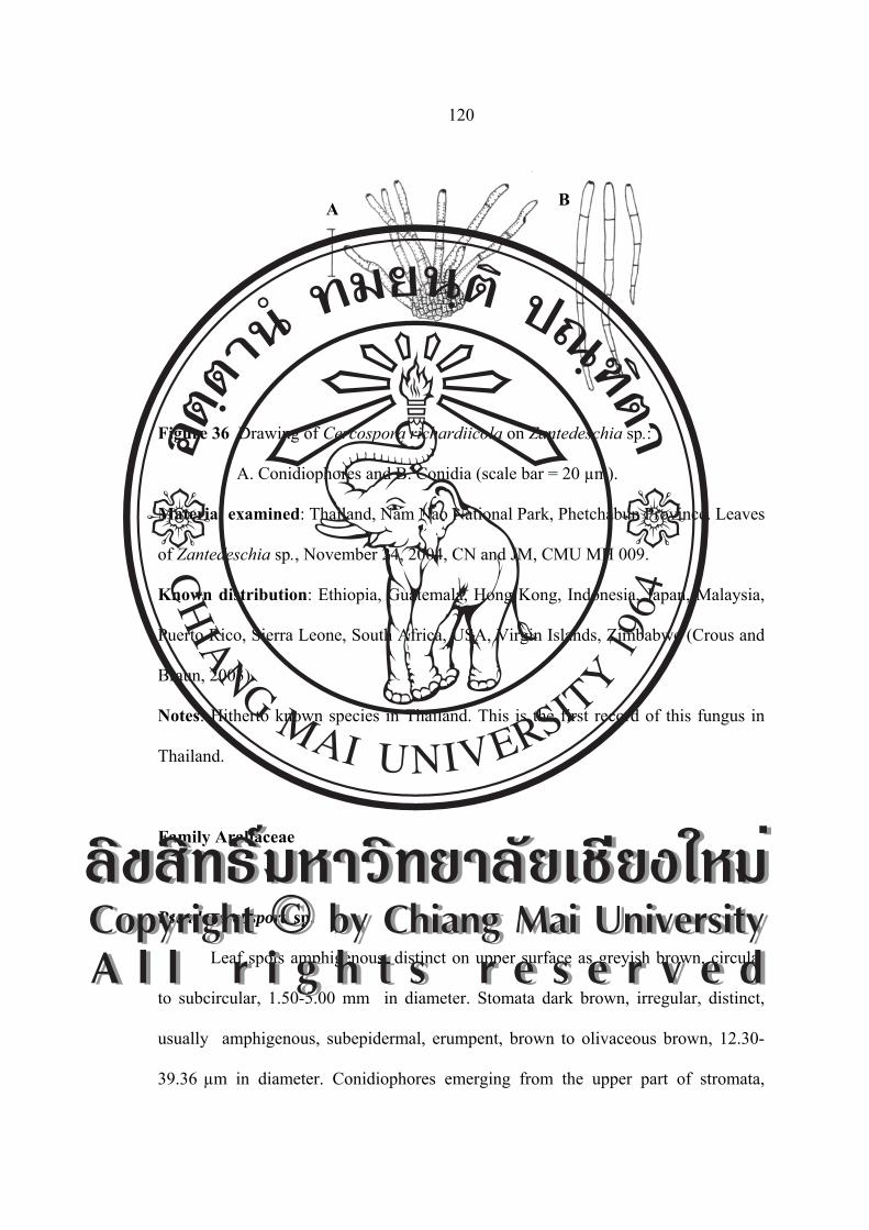
Figure 36 Drawing of Cercospora richardiicola on Zantedeschia sp.:
A. Conidiophores and B. Conidia (scale bar = 20 µm).
Material examined: Thailand, Nam Nao National Park, Phetchabun Province. Leaves
of Zantedeschia sp., November 24, 2004, CN and JM, CMU MH 009.
Known distribution: Ethiopia, Guatemala, Hong Kong, Indonesia, Japan, Malaysia,
Puerto Rico, Sierra Leone, South Africa, USA, Virgin Islands, Zimbabwe (Crous and
Braun, 2003).
Notes: Hitherto known species in Thailand. This is the first record of this fungus in
Thailand.
Family Araliaceae
Pseudocercospora sp.
Leaf spots amphigenous, distinct on upper surface as greyish brown, circular
to subcircular, 1.50-5.00 mm in diameter. Stomata dark brown, irregular, distinct,
usually amphigenous, subepidermal, erumpent, brown to olivaceous brown, 12.30-
39.36 µm in diameter. Conidiophores emerging from the upper part of stromata,
A B
ÅÔ¢ÊÔ·¸Ô ìÁËÒÇÔ·ÂÒÅÑÂàªÕ§ãËÁèCopyright by Chiang Mai UniversityA l l r i g h t s r e s e r v e d
ÅÔ¢ÊÔ·¸Ô ìÁËÒÇÔ·ÂÒÅÑÂàªÕ§ãËÁèCopyright by Chiang Mai UniversityA l l r i g h t s r e s e r v e d

121
greenish brown, densely fasciculate, simple, straight, sinuous or slightly curved, 2-9-
septate, (21.95-)41.46-126.82(139.02)×(3.65-)4.87-4.87 µm, conidial scars
inconspicuous. Conidia solitary, obclavate-cylindric to obclavate, straight to mildly
curved, pale olivaceous brown, 5-10-septate, non-constricted, occationally mildly
constricted at the septa, subobtuse to broadly rounded at the apex, obconically
truncate at the base, (47.56-)51.21-90.24(-117.07)×2.43-4.87 µm, hilum unthickened
and not darkened (Figure 37 and 38).
Figure 37 Photograph of Pseudocercospora sp. on Trevesia palmata: A. Symptom,
B. Conidiophores and C. Conidia.
Figure 38 Drawing of Pseudocercospora sp. on Trevesia palmata: A. Conidiophores
and B. Conidia (scale bar = 40 µm).
Material examined: Thailand, Wiang Pa Pao, Chiang Rai Province. Leaves of
Trevesia palmata, December 6, 2005, JM CMU MH 010.
A B C
AB
ÅÔ¢ÊÔ·¸Ô ìÁËÒÇÔ·ÂÒÅÑÂàªÕ§ãËÁèCopyright by Chiang Mai UniversityA l l r i g h t s r e s e r v e d
ÅÔ¢ÊÔ·¸Ô ìÁËÒÇÔ·ÂÒÅÑÂàªÕ§ãËÁèCopyright by Chiang Mai UniversityA l l r i g h t s r e s e r v e d

122
Known distribution: Known only Thailand.
Notes: Hitherto known species in Thailand. This is the first record of this fungus in
Thailand.
Family Balsaminaceae
Cercospora balsaminiana J. M. Yen and Lim, Cah. Pacifique 14: 91. 1970.
Leaf spots circular, 1.50-5.50 mm in diameter, white center and dark purple to
brown margin. Fruit bodies amphigenous. Stromata lacking to medium, rudimentary
to slightly developed, irregular, dark brown. Conidiophores 5-15 in a divergent
fascicle, emerging through stomatal openings and the cuticle, paler brown or paler
towards the apex, straight to slightly curved, 1-5 times mildly geniculate, not
branched, 2-5-septate, (13.00-)18.20-44.20(-65.00)×(2.60-)5.20-7.80 μm. Conidial
scars large, conspicuous, apical or on shoulders of conidiogenous cells caused by
geniculation. Conidia solitary, acicular to obclavate, substraight to moderately curved
or even undulate, hyaline, 4-16-septate, non-constriced at the septa, acute to obtuse at
the apex, truncate at the base, (52.00-)65.00-130.00(-234.00)×(1.30-)2.60-5.20(-7.80)
μm, hilum conspicuously thickened, darkened (Figure 39 and 40).
ÅÔ¢ÊÔ·¸Ô ìÁËÒÇÔ·ÂÒÅÑÂàªÕ§ãËÁèCopyright by Chiang Mai UniversityA l l r i g h t s r e s e r v e d
ÅÔ¢ÊÔ·¸Ô ìÁËÒÇÔ·ÂÒÅÑÂàªÕ§ãËÁèCopyright by Chiang Mai UniversityA l l r i g h t s r e s e r v e d
123
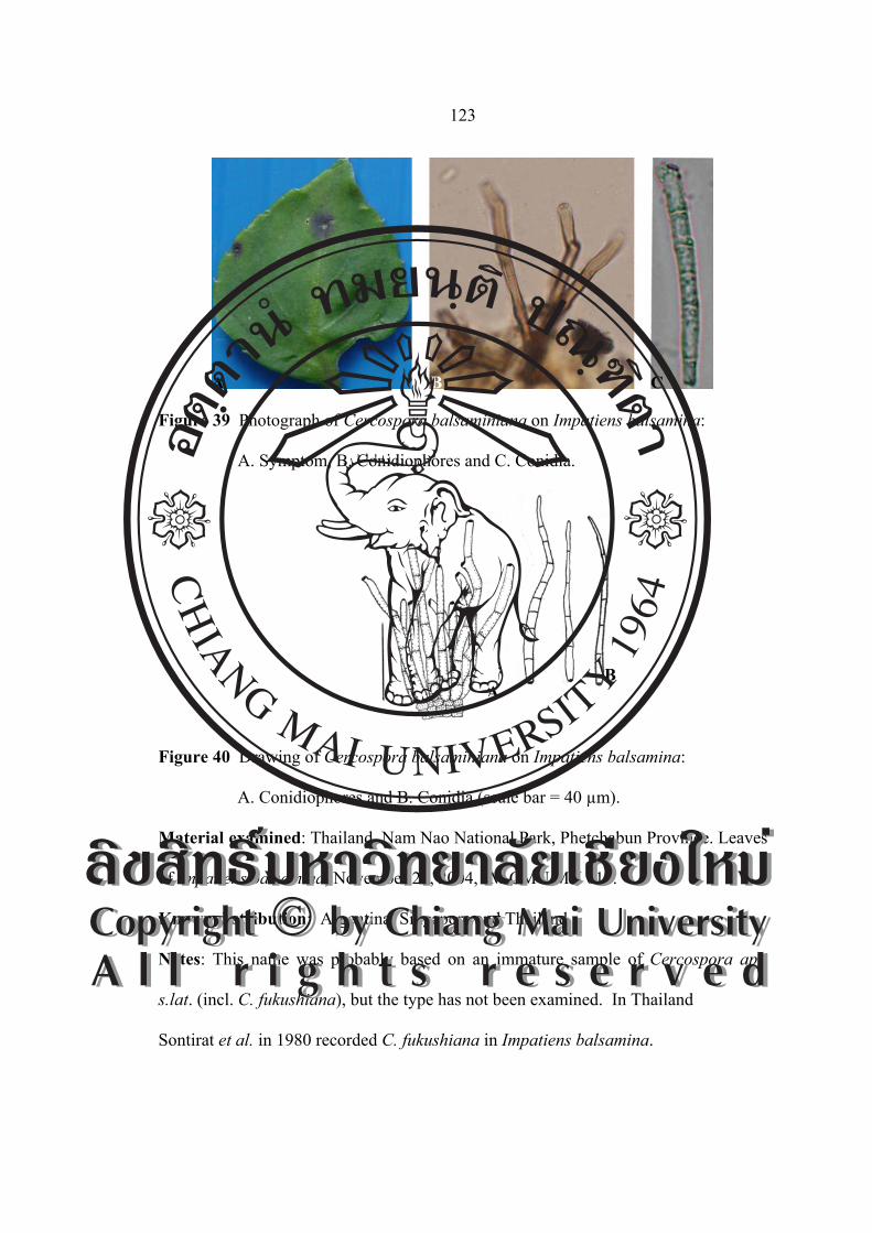
Figure 39 Photograph of Cercospora balsaminiana on Impatiens balsamina:
A. Symptom, B. Conidiophores and C. Conidia.
Figure 40 Drawing of Cercospora balsaminiana on Impatiens balsamina:
A. Conidiophores and B. Conidia (scale bar = 40 µm).
Material examined: Thailand, Nam Nao National Park, Phetchabun Province. Leaves
of Impatiens balsamina, November 24, 2004, JM CMU MH 011.
Known distribution: Argentina, Singapore and Thailand.
Notes: This name was probably based on an immature sample of Cercospora apii
s.lat. (incl. C. fukushiana), but the type has not been examined. In Thailand
Sontirat et al. in 1980 recorded C. fukushiana in Impatiens balsamina.
AB
A B C
ÅÔ¢ÊÔ·¸Ô ìÁËÒÇÔ·ÂÒÅÑÂàªÕ§ãËÁèCopyright by Chiang Mai UniversityA l l r i g h t s r e s e r v e d
ÅÔ¢ÊÔ·¸Ô ìÁËÒÇÔ·ÂÒÅÑÂàªÕ§ãËÁèCopyright by Chiang Mai UniversityA l l r i g h t s r e s e r v e d

124
Pseudocercospora balsaminae (Syd.) Deighton Mycol. Papers 140: 139 (1976)
═ Cercoseptoria balsaminae Syd., Annls Mycol. 33: 69 (1935)
Leaf spots amphigenous, orbicular to irregular, distinct, 2.00-6.00 mm in
diameter, pale brown to dingy grey in the center, with a dark purple border. Stromata
medium, subglobular, brown to dark brown, 7.00-19.00 µm in diameter, composed of
a few brown hyphal cells. Conidiophores 6-10 in a loose fascicle, emerging through
stomatal openings or erupent through the cuticle, olivaceous brown to brown,
irregular in width, short, not geniculate, not branched, short, slightly narrower at the
apical portion, euseptate, 1-3-septate, (12.30-)17.22-34.44(-63.96)×(4.92-)4.92-4.92
(-7.38) µm, conidial scars inconspicuous. Conidia solitary, obclavate, subhyaline, 3-4-
septate, euseptate, constricted at the septa, subobtuse to subacute at the apex,
obconically truncate to truncate at the base, (9.84-)22.14-29.52(-54.12)×(4.92-)4.92-
7.38(-9.84) µm (Figure 41 and 42).
Figure 41 Photograph of Pseudocercospora balsaminae on Impatiens balsamina:
A. Symptom, B. Conidiophores and C. Conidia.
A B C
ÅÔ¢ÊÔ·¸Ô ìÁËÒÇÔ·ÂÒÅÑÂàªÕ§ãËÁèCopyright by Chiang Mai UniversityA l l r i g h t s r e s e r v e d
ÅÔ¢ÊÔ·¸Ô ìÁËÒÇÔ·ÂÒÅÑÂàªÕ§ãËÁèCopyright by Chiang Mai UniversityA l l r i g h t s r e s e r v e d

125
Figure 42 Drawing of Pseudocercospora balsaminae on Impatiens balsamina:
A. Conidiophores and B. Conidia (scale bar = 40 µm).
Material examined: Thailand, Doi Suthep-Pui National Park, Chiang Mai Province.
Leaves of Impatiens balsamina, October 3, 2004, JM, CMU MH 012.
Known distribution: Worldwide where the host is growing (Crous and Braun,
2003).
Notes: This is the first record of this fungus in Thailand. In Korea was recorded by
Kim and Shin (1999d). They provided the detailed description and illustration for this
fungus based on Korean collection.
Guo and Liu (1992) described the following characters of the present fungus:
Secondary mycelium superficial; conidiophores arranged in loose to dense fascicle or
arising singly from the external mycelial hyphae, 0-2-septate, 6.50-4.00×2.50-4.00
µm, conidia narrowly obclavate, 3-11-septate, 25.00-90.00×1.50-3.00 µm. Therefore,
the Korean collections match well with Chinese description (Guo and Liu, 1992; Guo
and Hsied, 1995), though the secondary mycelium was not observed in all Korean
specimens.
A
B
ÅÔ¢ÊÔ·¸Ô ìÁËÒÇÔ·ÂÒÅÑÂàªÕ§ãËÁèCopyright by Chiang Mai UniversityA l l r i g h t s r e s e r v e d
ÅÔ¢ÊÔ·¸Ô ìÁËÒÇÔ·ÂÒÅÑÂàªÕ§ãËÁèCopyright by Chiang Mai UniversityA l l r i g h t s r e s e r v e d

126
Several species of Cercospora and allied genera have been known on I.
balsamina, namely Cercospora fukushiana (Marsuura) W. Yamam., C. balsamiana J.
M. Yen and Lim, Cercoseptoria balsaminicola (J. M. Yen and Lim) J. M. Yen,
Passalora campi-silii (Speg.) U. Braun, and Pseudocercospora nojimai (Togashi and
Katsuki) Y. L. Guo and X. J. Liu. C. balsaminicola is confusable with this species,
but differs in several respects: Stromata large, well-developed, 20.00-60.00 µm in
diameter, conidiophores arranged in dense fascicles, oncegeniculate, much shorter and
somewhat narrower, 10.00-27.60×2.50-3.60 µm, conidia filiform, somewhat longer
and in diameter, 45.60-132.00×2.00-2.50 µm, C. balsamiana is clearly distinguishable
from it by having epiphyllous fructification, aseptate conidiophores, very long (66.00-
307.00 µm in length) and 5-17-septate conidia, P. campi-silii on I. noli-tamgere was
described with conidia which are broadly obclavate-subcylindric, 4.00-7.00 µm in
diameter, and 1-6-septate. Deighton (1976) believed that this species is the same as C.
nojimai Tagashi and Pseudocercospora nojimai Katsuki, but according to the original
description, the latter species has epiphyllous fructification, 0-1-septate
conidiophores, and elongate-obclavate conidia, P. nojimai, described and illustrated
from Korea by Kim and Shin (1999a) is confusable with the present fungus, but
distinguished from the latter species as follows: secondary mycelium developed,
conidiophores and conidia somewhat in diameter.
Family Basellaceae
Cercospora basellae-albae R. K. Srivast., S. Narayan and A. K. Srivast., Indian
Phytopathol. 47: 229. 1994. [T: HCIO 30880].
ÅÔ¢ÊÔ·¸Ô ìÁËÒÇÔ·ÂÒÅÑÂàªÕ§ãËÁèCopyright by Chiang Mai UniversityA l l r i g h t s r e s e r v e d
ÅÔ¢ÊÔ·¸Ô ìÁËÒÇÔ·ÂÒÅÑÂàªÕ§ãËÁèCopyright by Chiang Mai UniversityA l l r i g h t s r e s e r v e d
127
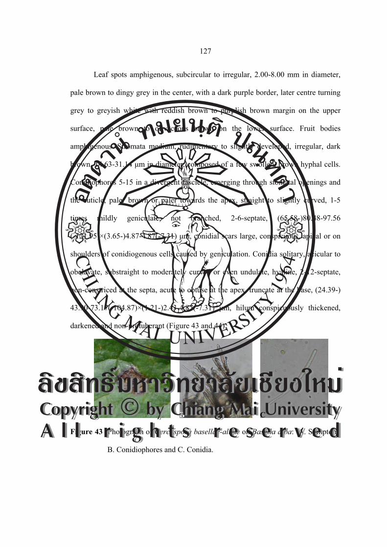
Leaf spots amphigenous, subcircular to irregular, 2.00-8.00 mm in diameter,
pale brown to dingy grey in the center, with a dark purple border, later centre turning
grey to greyish white with reddish brown to purplish brown margin on the upper
surface, pale brown to olivaceous brown on the lower surface. Fruit bodies
amphigenous. Stromata medium, rudimentary to slightly developed, irregular, dark
brown, 14.63-31.14 μm in diameter, composed of a few swollen, brown hyphal cells.
Conidiophores 5-15 in a divergent fascicle, emerging through stomatal openings and
the cuticle, paler brown or paler towards the apex, straight to slightly curved, 1-5
times mildly geniculate, not branched, 2-6-septate, (65.58-)80.48-97.56
(-121.95)×(3.65-)4.87-4.87(-7.31) μm, conidial scars large, conspicuous, apical or on
shoulders of conidiogenous cells caused by geniculation. Conidia solitary, acicular to
obclavate, substraight to moderately curved or even undulate, hyaline, 2-12-septate,
non-constriced at the septa, acute to obtuse at the apex, truncate at the base, (24.39-)
43.90-73.17(-104.87)×(1.21-)2.43-4.87(-7.31) μm, hilum conspicuously thickened,
darkened and non-protuberant (Figure 43 and 44).
Figure 43 Photograph of Cercospora basellae-albae on Basella alba: A. Symptom,
B. Conidiophores and C. Conidia.
A B C
ÅÔ¢ÊÔ·¸Ô ìÁËÒÇÔ·ÂÒÅÑÂàªÕ§ãËÁèCopyright by Chiang Mai UniversityA l l r i g h t s r e s e r v e d
ÅÔ¢ÊÔ·¸Ô ìÁËÒÇÔ·ÂÒÅÑÂàªÕ§ãËÁèCopyright by Chiang Mai UniversityA l l r i g h t s r e s e r v e d
128
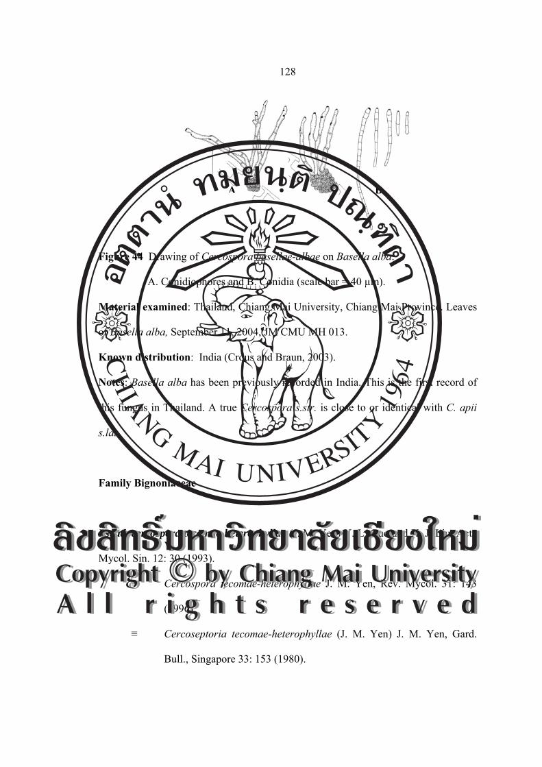
Figure 44 Drawing of Cercospora basellae-albae on Basella alba:
A. Conidiophores and B. Conidia (scale bar = 40 µm).
Material examined: Thailand, Chiang Mai University, Chiang Mai Province. Leaves
of Basella alba, September 11, 2004, JM CMU MH 013.
Known distribution: India (Crous and Braun, 2003).
Notes: Basella alba has been previously recorded in India. This is the first record of
this fungus in Thailand. A true Cercospora s.str. is close to or identical with C. apii
s.lat.
Family Bignoniaceae
Pseudocercospora tecomae-heterophyllae (J. M. Yen) Y. L. Guo and X. J. Liu, Acta
Mycol. Sin. 12: 30 (1993).
≡ Cercospora tecomae-heterophyllae J. M. Yen, Rev. Mycol. 31: 143
(1996).
≡ Cercoseptoria tecomae-heterophyllae (J. M. Yen) J. M. Yen, Gard.
Bull., Singapore 33: 153 (1980).
A B
ÅÔ¢ÊÔ·¸Ô ìÁËÒÇÔ·ÂÒÅÑÂàªÕ§ãËÁèCopyright by Chiang Mai UniversityA l l r i g h t s r e s e r v e d
ÅÔ¢ÊÔ·¸Ô ìÁËÒÇÔ·ÂÒÅÑÂàªÕ§ãËÁèCopyright by Chiang Mai UniversityA l l r i g h t s r e s e r v e d

129
Leaf spots amphigenous, very small, irregular, not vein limited, scattered
coalescing to large spot, pale brown to brown with indefinite margins. Fruit bodies
amphigenous. Stromata lacking or well-developed, amphigenous, 14.76-36.90 µm in
diameter, brown. External hyphae developed on both leaf surface. Conidiophores
arising from stromata or external hyphae, brown, solitary to densely fasciculate,
simple, straight or slightly curved, 0-4-septate, (9.84-)19.68-49.20(-59.04)×2.46 µm,
with distinct and unthickened conidial scars. Conidia acicular to obclavate, straight,
smooth, hyaline to pale colored, with unthickened and truncate basal end, acute at the
tip, (24.60-)27.06-49.20(-63.96)×(2.46-)2.46-2.46(-4.92) µm, 3-7-septate (Figure 45
and 46).
Figure 45 Photograph of Pseudocercospora tecomae-heterophyllae on
Tecoma stans: A. Symptom, B. Conidiophores and C. Conidia.
A B C
ÅÔ¢ÊÔ·¸Ô ìÁËÒÇÔ·ÂÒÅÑÂàªÕ§ãËÁèCopyright by Chiang Mai UniversityA l l r i g h t s r e s e r v e d
ÅÔ¢ÊÔ·¸Ô ìÁËÒÇÔ·ÂÒÅÑÂàªÕ§ãËÁèCopyright by Chiang Mai UniversityA l l r i g h t s r e s e r v e d
130
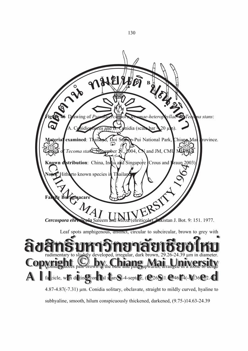
Figure 46 Drawing of Pseudocercospora tecomae-heterophyllae on Tecoma stans:
A. Conidiophores and B. Conidia (scale bar = 20 µm).
Material examined: Thailand, Doi Suthep-Pui National Park, Chiang Mai Province.
Leaves of Tecoma stans, November 21, 2004, CN and JM, CMU MH 014.
Known distribution: China, India and Singapore (Crous and Braun 2003).
Notes: Hitherto known species in Thailand.
Family Boraginacare
Cercospora ehretiicola Saleem and Mirza (ehriticola), Pakistan J. Bot. 9: 151. 1977.
Leaf spots amphigenous, distinct, circular to subcircular, brown to grey with
blackish brown margins, 1.00-3.00 mm in diameter. Stromata small to medium,
rudimentary to slightly developed, irregular, dark brown, 29.26-24.39 µm in diameter.
Conidiophores pale brown at the base and paler upwards, arranged in a loose to dense
fascicle, with distinct conidial scars, 2-4-septate, (29.26-)41.46-46.34(-85.36)×(2.43-)
4.87-4.87(-7.31) µm. Conidia solitary, obclavate, straight to mildly curved, hyaline to
subhyaline, smooth, hilum conspicuously thickened, darkened, (9.75-)14.63-24.39
AB
ÅÔ¢ÊÔ·¸Ô ìÁËÒÇÔ·ÂÒÅÑÂàªÕ§ãËÁèCopyright by Chiang Mai UniversityA l l r i g h t s r e s e r v e d
ÅÔ¢ÊÔ·¸Ô ìÁËÒÇÔ·ÂÒÅÑÂàªÕ§ãËÁèCopyright by Chiang Mai UniversityA l l r i g h t s r e s e r v e d

131
(-34.14)×(2.43-)4.87-4.87(-7.31) µm, 1-4-septate, acute to obtuse at the apex, truncate
at the base (Figure 47 and 48).
Figure 47 Photograph of Cercospora ehretiicola on Ehretia microphylla:
A. Symptom, B. Conidiophores and C. Conidia.
Figure 48 Drawing of Cercospora ehretiicola on Ehretia microphylla:
A. Conidiophores and B. Conidia (scale bar = 40 µm).
A B C
A
B
ÅÔ¢ÊÔ·¸Ô ìÁËÒÇÔ·ÂÒÅÑÂàªÕ§ãËÁèCopyright by Chiang Mai UniversityA l l r i g h t s r e s e r v e d
ÅÔ¢ÊÔ·¸Ô ìÁËÒÇÔ·ÂÒÅÑÂàªÕ§ãËÁèCopyright by Chiang Mai UniversityA l l r i g h t s r e s e r v e d

132
Material examined: Thailand, Wiang Pa Pao, Chiang Rai Province. Leaves of
Ehretia microphylla, November 11, 2005, JM CMU MH 015.
Known distribution: Pakistan (Crous and Braun, 2003).
Notes: This is the first record of this fungus in Thailand. A true Cercospora s.str. is
close to C. apii s. lat. Saleem and Mirza found Cercospora ehretiicola on Ehretia
acuminata in 1977.
Family Caprifoliaceae
Pseudocercospora viburnigena U. Braun and Crous, Mycol. Progress 1(1): 23. 2002.
≡ Cercospora tinea Sacc., Michelia 1: 268. 1878. [T: Sacc., Mycoth.
ven. 1252, e.g., HAL], non Pseudocercospora tinea Y. L. Guo and
W. H. Hsieh, 1994.
≡ Cercoseptoria tinea (Sacc.) Deighton, Mycol. Pap. 140: 167. 1976
≡ Stigmina tinea (Sacc.) M. B. Ellis, More dematiaceous hyphomycetes:
118. 1976.
≡ Stigmina tinea (Sacc.) M. B. Ellis, Mycol. Pap. 151: 1. 1983 (comb.
superfl.).
Leaf spots amphigenous, suborbicular to irregular, with a slightly raised black
margin, mostly 2.00-8.00 mm in diameter, often confluent, dark brown to blackish,
center greyish white to brown, margin dark brown to blackish on the upper surface,
brown to dark brown on the lower surface. Fruit bodies epigenous. Stromata small.
Conidiophore loosly fasciculate, moderately olivaceous brown to pale brown, paler
towards the apex, 3-15-septate, rarely geniculate, not branched, apex bluntly rounded
ÅÔ¢ÊÔ·¸Ô ìÁËÒÇÔ·ÂÒÅÑÂàªÕ§ãËÁèCopyright by Chiang Mai UniversityA l l r i g h t s r e s e r v e d
ÅÔ¢ÊÔ·¸Ô ìÁËÒÇÔ·ÂÒÅÑÂàªÕ§ãËÁèCopyright by Chiang Mai UniversityA l l r i g h t s r e s e r v e d

133
to subtruncate or conic, uniform in colour and width, (63.41-)92.68-214.63
(-239.02)×(3.65-)4.87-7.31 µm, conidial scars inconspicuous. Conidia pale
olivaceous, cylindric or cylindric-obclavate, straight to mildly curved, rarely slightly
curved, subobtuse to subacute at the apex, obcomic to obconically truncate at the base
(17.07-)53.65-187.80(-204.87)×(4.87-)4.87-6.09(-7.31) µm, hilum inconspicuous
(Figure 49 and 50).
Figure 49 Photograph of Pseudocercospora viburnigena on Viburnum prunifolium:
A. Symptom, B. Conidiophores and C. Conidia.
Figure 50 Drawing of Pseudocercospora viburnigena on Viburnum prunifolium:
A. Conidiophores and B. Conidia (scale bar = 40 µm).
Material examined: Thailand, Wiang Pa Pao, Chiang Rai Province. Leaves of
Viburnum prunifolium, February 11, 2004, JM CMU MH 016.
A B C
A B ÅÔ¢ÊÔ·¸Ô ìÁËÒÇÔ·ÂÒÅÑÂàªÕ§ãËÁèCopyright by Chiang Mai UniversityA l l r i g h t s r e s e r v e d
ÅÔ¢ÊÔ·¸Ô ìÁËÒÇÔ·ÂÒÅÑÂàªÕ§ãËÁèCopyright by Chiang Mai UniversityA l l r i g h t s r e s e r v e d

134
Known distribution: Germany, Great Britain, Italy, Portugal and Spain (Crous and
Braun, 2003)
Notes: Hitherto known species in Thailand.
Family Chenopodiaceae
Cercospora beticola Sacc., Nuovo Giorn. Bot. Ital. 8: 189. 1876. [T: PAD].
≡ Cercosporina beticola (Sacc.) K. Nakata, T. Nakajima and K.
Katimoto, Rep. Agric. Korea 6. 1915.
= Fusisporium betae Desm., Ann. Sci. Net., Bot., 2 Ser., 19: 434. 1843.
= Fusarium betae (Desm.) Sacc., Michelia 2: 132.1880.
= Pionnotes betae (Desm.) Sacc., Syll. Fung. 4: 726. 1886.
= Cercospora betae A. B. Frank ex Sacc., Syll. Fung. 10: 637. 1892.
= Cercospora longissima Cooke and Eills, Grevillea 17: 65. 1889.
= Cercospora flagelliformis Ellis and Halst., New Jersey Agric. Coll.
Exp. Sta., Annual Rep. 11: 355. 1890. [T: NY].
= Cercospora anthelmintica G. F. Atk., J. Elisha Mitchell Sci. Soc. 8:
48. 1892. [T: CUP].
= Cercospora spinaciae Oudem., Ned. Kruidk. Arch. III, 2: 324.1900.
= Cercospora chenopodiicola Bres., Hedwigia 39: 328. 1900. [T:
Krieger, Fungi sax. 1631, e.g., HAL, LEP].
= Cercosporina spinaciicola Sacc., Nuovo Giorn. Bot. Ital., N. S., 22:
73. 1915.
= Cercospora beticola var. poonensis Chidd., Sydowia 13: 153. 1959
(nom. inval.). [T: BPI; HCIO; IMI]. (= Cercospora apii s.lat.).
ÅÔ¢ÊÔ·¸Ô ìÁËÒÇÔ·ÂÒÅÑÂàªÕ§ãËÁèCopyright by Chiang Mai UniversityA l l r i g h t s r e s e r v e d
ÅÔ¢ÊÔ·¸Ô ìÁËÒÇÔ·ÂÒÅÑÂàªÕ§ãËÁèCopyright by Chiang Mai UniversityA l l r i g h t s r e s e r v e d

135
Leaf spots scattered, often confluent, circular, 2.00-5.00 mm in diameter, grey
in colour, later centre becoming whitish grey, reddish brown or purplish brown at the
margin. Fruit bodies amphigenous, but chiefly epiphyllous. Stromata rudimentary to
slightly developed. Conidiophores 3-14 in a divergent fascicle, pale brown at the base,
paler upwards, nearly subhyaline at the apex, 2-5-septate, not branched, straight to 1-3
times geniculate towards the apex, weakly to mildly attenuated, (52.00-)83.20-
187.20(-208.00)×(3.90-)3.90-5.20(-7.80) µm, conidial scars small, mostly less than 2
µm in diameter, conspicuous, thickened and darkened, apical or on shoulders of
conidiogenous cells caused by geniculation, usually concentrated in the apical portion.
Conidia solitary, acicular, straight to mildly curved, hyaline, 3-15-septate, non-
constricted at the septa, obtuse to subacute at the apex, subtruncate at the base, greatly
variable in length, (52.00-)65.00-135.20(-145.60)×(2.60-)3.90-5.20(-7.80) µm, hilum
conspicuously thickened, darkened and non-protuberant (Figure 51 and 52).
Figure 51 Photograph of Cercospora beticola on Beta vulgaris var. alba:
A. Symptom, B. Conidiophores and C. Conidia.
A B C ÅÔ¢ÊÔ·¸Ô ìÁËÒÇÔ·ÂÒÅÑÂàªÕ§ãËÁèCopyright by Chiang Mai UniversityA l l r i g h t s r e s e r v e d
ÅÔ¢ÊÔ·¸Ô ìÁËÒÇÔ·ÂÒÅÑÂàªÕ§ãËÁèCopyright by Chiang Mai UniversityA l l r i g h t s r e s e r v e d

136
Figure 52 Drawing of Cercospora beticola on Beta vulgaris var. alba:
A. Conidiophores and B. Conidia (scale bar = 40 µm).
Material examined: Thailand, Mae Jo, Chiang Mai Province. Leaves of
Beta vulgaris var. alba, August 19, 2004, JM, CMU MH 017.
Known distribution: Nearly throughout the world wherever sugar beet and chard are
cultivated, including China, Japan and Korea (Crous and Braun, 2003).
Notes: This is the first record of Cercospora beticola in Thailand. Nakata and
Takimoto (1928) first reported this leaf spot fungus from Korea on Beta vulgaris var.
saccharifera (sugar beet). Shin and Braun (1993) listed this fungal species on
B. vulgaris var. alba (chard). Detailed description and illustration based on Korean
collection of this species was provided by Kim and Shin (1998b). The leaf spot
disease of chard associated with this fungus is one of the limiting factors for
successful harvest. Though the sugar beet is not cultivated in Korea, this host species
planted in the experimental plot of Crop Research Sation, Suwon, was found to be
infected by this fungus.
A
B
ÅÔ¢ÊÔ·¸Ô ìÁËÒÇÔ·ÂÒÅÑÂàªÕ§ãËÁèCopyright by Chiang Mai UniversityA l l r i g h t s r e s e r v e d
ÅÔ¢ÊÔ·¸Ô ìÁËÒÇÔ·ÂÒÅÑÂàªÕ§ãËÁèCopyright by Chiang Mai UniversityA l l r i g h t s r e s e r v e d

137
The conidiophores in collection are longer (18.00-85.00 µm) and in diameter
(3.50-6.00 µm) than those described by other authors (Chupp, 1954; Hsieh and Goh,
1990), but the measurementas of conidiophores are usually of little taxonomic
importance, since these structures are very variable. Most earlier authors (Chupp,
1954; Hsieh and Goh, 1990) described acicular conidia with acute apices. The conidia
of our collections are, however, obtuse to subacute at the apex.
Family Combretaceae
Pseudocercospara quisqualidia (Narain and B. S. Mehrotra) Z. D. Jing and P. K. Chi,
J. S. China Agr. Univ. 15: 19. 1994 and in Chi, Fungal diseases of cultivated
medicinal plants in Guangdong Province: 100. 1994.
≡ Cercospora quisqualidis Narain and B. S. Mehrotra, Sydowia
24: 327. (1970) 1971. [T: BPI].
Leaf spots amphigenous, scattered to confluent, circular to angular, 1.00-6.00
mm in diameter, at first appearing yellowish brown to deep brown, later centre
becoming greyish white to greyish brown with reddish brow margins, finally turning
dingy to dark grey with definite margin. Fruit bodies amphigenous, but abundantly
hypophyllous, punctiform. Mycelium internal, hyphae septate, branched, hyaline,
2.00-3.00 µm in diameter. Secondary mycelium external, hyphae emerging from the
stomata, olivaceous to pale brown, septate, branched, bearing secondary
conidiophores laterally and terminally. Stromata absent. Conidiophores loosely
fasciculate at stomatal openings, or sometimes emerging through the cuticle,
olivaceous brown, fairly uniform in colour and width, 1-8 times mildly geniculate
ÅÔ¢ÊÔ·¸Ô ìÁËÒÇÔ·ÂÒÅÑÂàªÕ§ãËÁèCopyright by Chiang Mai UniversityA l l r i g h t s r e s e r v e d
ÅÔ¢ÊÔ·¸Ô ìÁËÒÇÔ·ÂÒÅÑÂàªÕ§ãËÁèCopyright by Chiang Mai UniversityA l l r i g h t s r e s e r v e d
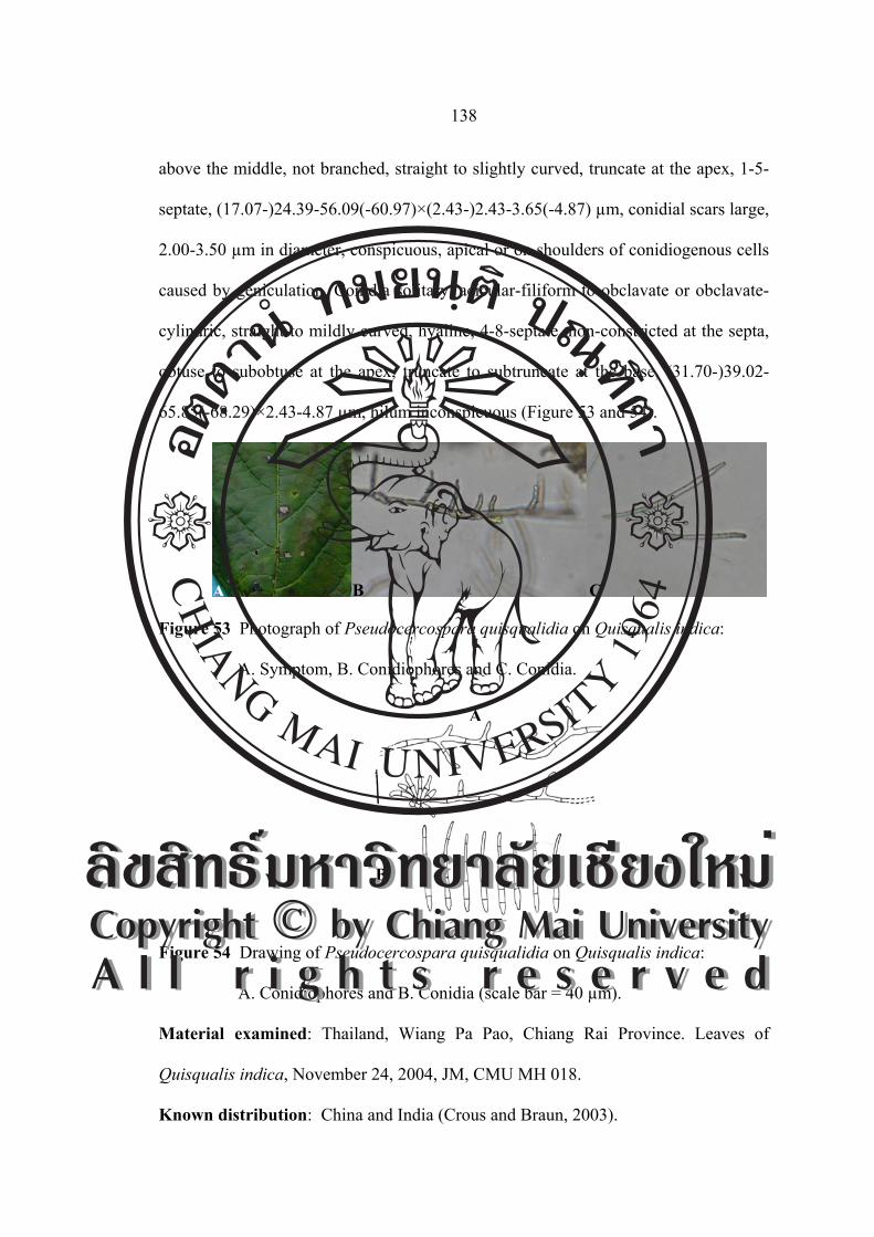
138
above the middle, not branched, straight to slightly curved, truncate at the apex, 1-5-
septate, (17.07-)24.39-56.09(-60.97)×(2.43-)2.43-3.65(-4.87) µm, conidial scars large,
2.00-3.50 µm in diameter, conspicuous, apical or on shoulders of conidiogenous cells
caused by geniculation. Conidia solitary, acicular-filiform to obclavate or obclavate-
cylindric, straight to mildly curved, hyaline, 4-8-septate, non-constricted at the septa,
obtuse to subobtuse at the apex, truncate to subtruncate at the base, (31.70-)39.02-
65.85(-68.29)×2.43-4.87 µm, hilum inconspicuous (Figure 53 and 54).
Figure 53 Photograph of Pseudocercospara quisqualidia on Quisqualis indica:
A. Symptom, B. Conidiophores and C. Conidia.
Figure 54 Drawing of Pseudocercospara quisqualidia on Quisqualis indica:
A. Conidiophores and B. Conidia (scale bar = 40 µm).
Material examined: Thailand, Wiang Pa Pao, Chiang Rai Province. Leaves of
Quisqualis indica, November 24, 2004, JM, CMU MH 018.
Known distribution: China and India (Crous and Braun, 2003).
A B C
A
B ÅÔ¢ÊÔ·¸Ô ìÁËÒÇÔ·ÂÒÅÑÂàªÕ§ãËÁèCopyright by Chiang Mai UniversityA l l r i g h t s r e s e r v e d
ÅÔ¢ÊÔ·¸Ô ìÁËÒÇÔ·ÂÒÅÑÂàªÕ§ãËÁèCopyright by Chiang Mai UniversityA l l r i g h t s r e s e r v e d

139
Notes: This is the first record of Pseudocercospara quisqualidia in Thailand.
Family Asteraceae
Cercospora gerberae Chupp and Viégas Bol. Da Soc. Brasil. De Agron. 8: 27. 1945.
[T: CUP 39884; IACM 3958].
Leaf spots suborbicular to irregular, 1.00-20.00 mm in diameter, at first
olivaceous, then turning dark until the spots are almost black, later coalescing to from
large blotches covering much of the leaf surface, dark brown, surrouned by a purplish
black margin, the dead tissues may drop out leaving the leaf with holes. Fruit bodies
amphigenous but chiefly hypophyllous. Stromata a few brown cells or up to 50.00 μm
in diameter, subglobose, dark brown. Conidiphores in dense divergent fascicles of 10-
35, emerging through the stomata, pale to medium brown, paler and more narrow
toward the tip, uniform in colour, straight, not branched, 2-6-septate, 0-3 geniculate,
subtruncate at apex, (34.14-)51.21-92.68(-104.87)×4.87-7.31 μm, conidial scare
conspicuously thickened. Conidia hyaline, acicular, straight to slightly curved,
indistinctly multi-septate, acute at the apex, truncate at the base with a thickened
hilum, (58.53-)63.41-146.34(-182.92)×(2.43-)4.87-4.87 µm (Figure 55 and 56).
Figure 55 Photograph of Cercospora gerberae on Gerbera jamesonii: A. Symptom,
B. Conidiophores and C. Conidia.
A B C
ÅÔ¢ÊÔ·¸Ô ìÁËÒÇÔ·ÂÒÅÑÂàªÕ§ãËÁèCopyright by Chiang Mai UniversityA l l r i g h t s r e s e r v e d
ÅÔ¢ÊÔ·¸Ô ìÁËÒÇÔ·ÂÒÅÑÂàªÕ§ãËÁèCopyright by Chiang Mai UniversityA l l r i g h t s r e s e r v e d

140
Figure 56 Drawing of Cercospora gerberae on Gerbera jamesonii:
A. Conidiophores and B. Conidia (scale bar = 40 µm).
Material examined: Thailand, Wiang Pa Pao, Chiang Rai Province. Leaves of
Gerbera jamesonii, March 9, 2004, JM CMU MH 019.
Known distribution: Australia (NSW), Bangladesh, Bermuda, Brazil, British
Solomon Islands, Brunei, Cuba, Cambodia, Ghana, Hong Kong, India, Indonesia,
Iran, Jamaica, Kenya, Malawi, Malaysia, Pakistan, Philippines, Puerto Rico, Sierra
Leone, Singapore, Taiwan, Tanzania, Thailand, Uganda, U.S.A and Virgin Islands
(Crous and Braun, 2003).
Notes: The first mount made showed a few conidia with appendages, like those of
Centrospora on Carum, Viola, and Apium, but such appendages later could not be
found in fairly numerous mounts. It there is considered a Cercospora. Type species
are Est. Exp. De Agriculture, Belo Horizonte, Minas Geraes, Brazil; Gerbera
jamesonii; Carlos Tomas de Almeida, 3958; April 14, 1939.
A
B
ÅÔ¢ÊÔ·¸Ô ìÁËÒÇÔ·ÂÒÅÑÂàªÕ§ãËÁèCopyright by Chiang Mai UniversityA l l r i g h t s r e s e r v e d
ÅÔ¢ÊÔ·¸Ô ìÁËÒÇÔ·ÂÒÅÑÂàªÕ§ãËÁèCopyright by Chiang Mai UniversityA l l r i g h t s r e s e r v e d

141
Family Compositae
Passalora tithonia (R. E. D. Baker and W. T. Dale) U. Braun and Crous, comb. nov.
≡ Cercospora tithoniae R. E. D. Baker and Dale, Mycol. Pap. 33: 106.
1951. [T: IMI 24506].
≡ Phaeoramularia tithoniae (R. E. D. Baker and W. T. Dale) Deighton,
in Ellis, More dematiaceous hyphomycetes: 319. 1976.
≡ Cercospora tithoniae Chidd., Mycopathol. Mycol. Appl. 17: 80. 1962
(nom. illeg.). [T: BPI 441950], homonym of C. tithoniae R. E. D.
Baker and W. T. Dale, 1951.
≡ Cercospora tithonicola J. M. Yen (tithonicola) Rev. Mycol. 31: 144
1966 (nom. nov.).
Leaf spots amphigenous, scattered to confluent, usually vein-limited, angular
to irregular in shape, brown to greyish-brown in colour, 2.00-15.00 mm in diameter,
coalescing and covering the whole surface of the leaf. Stromata medium, breaking
throught epidermis, brown to olive-brown in colour, 34.44-49.20 µm in diameter,
with out external mycelia. Conidiophores emerging from the upper part of stromata,
brown to olive-brown at the basal part, hyaline to pale at the tip, with small conidial
scars, 1-8-septate, (9.84-)19.68-78.72(-86.10)×2.46-4.92 µm. Conidia obclavate,
subcylindric, olive-brown, straight, smooth, with small thickened hilum, (22.14-)
24.60-51.66(-73.80)×(3.69-)4.92-4.92(-7.38) µm, 1-6-septate (Figure 57 and 58).
ÅÔ¢ÊÔ·¸Ô ìÁËÒÇÔ·ÂÒÅÑÂàªÕ§ãËÁèCopyright by Chiang Mai UniversityA l l r i g h t s r e s e r v e d
ÅÔ¢ÊÔ·¸Ô ìÁËÒÇÔ·ÂÒÅÑÂàªÕ§ãËÁèCopyright by Chiang Mai UniversityA l l r i g h t s r e s e r v e d

142
Figure 57 Photograph of Passalora tithonia on Tithonia diversifolia: A. Symptom,
B. Conidiophores and C. Conidia.
Figure 58 Drawing of Passalora tithonia on Tithonia diversifolia: A. Conidiophores
and B. Conidia (scale bar = 40 µm).
Material examined: Thailand, Queen Sirikit Botanical Garden, Chiang Mai Province.
Leaves of Tithonia diversifolia, November 20, 2004, CN and JM, CMU MH 020.
Known distribution: Barbados, Cuba, Hong Kong, India, Ivory Coast, Mauritius,
Singapore, Taiwan, Trinidad and Tobago (Crous and Braun, 2003).
Notes: This is the first record of Passalora tithonia in Thailand.
A B C
AB
ÅÔ¢ÊÔ·¸Ô ìÁËÒÇÔ·ÂÒÅÑÂàªÕ§ãËÁèCopyright by Chiang Mai UniversityA l l r i g h t s r e s e r v e d
ÅÔ¢ÊÔ·¸Ô ìÁËÒÇÔ·ÂÒÅÑÂàªÕ§ãËÁèCopyright by Chiang Mai UniversityA l l r i g h t s r e s e r v e d
143
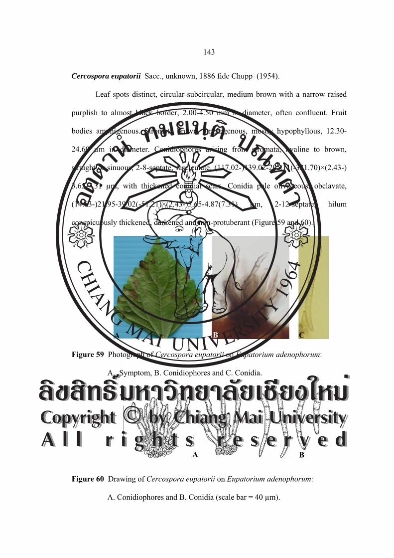
Cercospora eupatorii Sacc., unknown, 1886 fide Chupp (1954).
Leaf spots distinct, circular-subcircular, medium brown with a narrow raised
purplish to almost black border, 2.00-4.50 mm in diameter, often confluent. Fruit
bodies amphigenous. Stromata brown, amphigenous, mostly hypophyllous, 12.30-
24.60 µm in diameter. Conidiophores arising from stromata, hyaline to brown,
straight or sinuous, 2-8-septate, fasciculate, (117.02-)139.02-295.11(-331.70)×(2.43-)
3.65-7.31 µm, with thickened conidial scars. Conidia pale olivaceous, obclavate,
(14.63-)21.95-39.02(-51.21)×(2.43-)3.65-4.87(7.31) µm, 2-12-septate, hilum
conspicuously thickened, darkened and non-protuberant (Figure 59 and 60).
Figure 59 Photograph of Cercospora eupatorii on Eupatorium adenophorum:
A. Symptom, B. Conidiophores and C. Conidia.
Figure 60 Drawing of Cercospora eupatorii on Eupatorium adenophorum:
A. Conidiophores and B. Conidia (scale bar = 40 µm).
A B C
A B
ÅÔ¢ÊÔ·¸Ô ìÁËÒÇÔ·ÂÒÅÑÂàªÕ§ãËÁèCopyright by Chiang Mai UniversityA l l r i g h t s r e s e r v e d
ÅÔ¢ÊÔ·¸Ô ìÁËÒÇÔ·ÂÒÅÑÂàªÕ§ãËÁèCopyright by Chiang Mai UniversityA l l r i g h t s r e s e r v e d

144
Material examined: Thailand, Queen Sirikit Botanical Garden, Chiang Mai Province.
Leaves of Eupatorium adenophorum, November 20, 2004, JM, CMU MH 115.
Known distribution: Nepal and U.S.A (FL) (Crous and Braun, 2003).
Notes: This is the first record of Cercospora eupatorii found on Eupatorium
adenophorum in Thailand. Chupp (1954) was unable to locate a description or
specimen of this species.
Cercosporella virgaureae (Thüm.) Allesch., Hedwigia 34: 286. 1895.
≡ Ramularia virgaureae Thüm., Fungi austr., No. 1072, 1874. [T:
Thüm., Fungi austr. 1074, e.g., HAL].
≡ Ovularia virgaureae (Thüm.) Sacc., Syll. Fung. 4: 142. 1886.
≡ Cercospora virgaureae (Thüm.) Ellis and Everh. J. Mycol. 5: 69.
1889.
≡ Cercospora virgaureae (Thüm.) Allesch., in Oudem., Ned. Kruidk.
Arch., Ser. 3, 2: 315. 1901.
= Cercospora cana Sacc., Nuovo Giorn. Bot. Ital. 8: 188. 1876. [T: BPI
420584; PAD].
≡ Cercosporella cana (Sacc.) Sacc., Michelia 2: 20. 1880.
≡ Septocylindrium canum (Sacc.) J. Schroet., in Cohn, Kryptog. -F1.
Schles., 3: 493. 1897.
= Fusidium canum Pass., in Thüm., Mycoth. Univ., No. 378. 1876.
[T: Thüm., Mycoth. Univ. 378, eg., HAL].
= Cercospora fulvescens Sacc., Nuovo Giorn. Bot. Ital. 8: 189.1876.
[T: PAD].
ÅÔ¢ÊÔ·¸Ô ìÁËÒÇÔ·ÂÒÅÑÂàªÕ§ãËÁèCopyright by Chiang Mai UniversityA l l r i g h t s r e s e r v e d
ÅÔ¢ÊÔ·¸Ô ìÁËÒÇÔ·ÂÒÅÑÂàªÕ§ãËÁèCopyright by Chiang Mai UniversityA l l r i g h t s r e s e r v e d

145
= Cercospora canadensis A. B. Frank. 1878, unknow fide chupp (1954,
p. 126).
= Cercospora griseёlla Peck, Rep. (Annual) New York State Mus.
Nat. Hist. 33: 29. 1880. [T: NYS].
= Cercosporella reticulate Peck, Rep. (Annual) New York State Mus.
Nat. Hist. 34: 47. 1881. [T: NYS].
≡ Cercospora reticulate (Peck) Ellis and Everh., J. Mycol. 1: 61. 1885.
= Cercospora grindeliae Ellis and Everh., Proc. Acad. Nat Sci.
Philadelphia 47: 439. 1895. [T: NY].
= Cercosporella asterina Speg., Anales Mus. Nac. Hist. Nat. Buenos
Aires 6: 335. 1899. [T: LPS].
= Cercospora virgaureae Oudem., Ned. Kruidk. Arch., Ser. 3, 2: 315.
1901. [T: L].
= Cercosporella ontariensis Sacc., Ann. Mycol. 11: 551. 1913.
[T: PAD].
= Cercosporella dearnessii Bubák and Sacc., Ann. Mycol. 11: 552.
1913. [T: BPI 429648; PAD].
= Ramularia erigerontis Gonz. Frag., Bol. Acad. Ci. Exact., Madrid 5,
15: 39. 1917. [T: MA].
= Cercosporella cana var. gracilis Davis, Trans. Wisconsin Acad. Sci.
19: 675. 1919. [T: BPI 420617; WIS].
= Cercospora viminei Tehon, Mycologia 16: 141. 1924. [T: ILLS].
= Cercosporella eupatorii Sawada, rep. Gov. Agric. Res. Inst. Taiwan
86: 160. 1943 (nom. inval).
ÅÔ¢ÊÔ·¸Ô ìÁËÒÇÔ·ÂÒÅÑÂàªÕ§ãËÁèCopyright by Chiang Mai UniversityA l l r i g h t s r e s e r v e d
ÅÔ¢ÊÔ·¸Ô ìÁËÒÇÔ·ÂÒÅÑÂàªÕ§ãËÁèCopyright by Chiang Mai UniversityA l l r i g h t s r e s e r v e d

146
≡ Cercosporella eupatorii Sawada ex Goh and W.H. Hsieh, in Hsieh and
Goh, Cercospora and similar fungi from Taiwan: 70. 1990. [T: NTU-
PPE, herb. Sawada].
= Ramularia erigerontis-annui Sawada, Bull. Gov. Forest. Exp. Stst.
Tokyo 105: 86. 1958.
= Cercosporella curva Diedicke, (JE) fide Braun (1995a).
= Cercospora foliosa Ellis and Kellerm., unknown fide Chupp (1954).
Leaf spots circular to subcircular, vein-limited, 2.00-5.50 mm in diameter, on
the upper surface, at first indefinite yellowish discolourations, on the lower surface,
pale greenish, yellowish to brown, later greyish brown to brown without definite
margin. Fruit bodies hypophyllous, appearing as white to pale tan patches, similar to
symptoms of downy mildews, without define margin. Mycelium internal, hyphae
septate, branched, hyaline. Stromata small, but well-developed, composed of several
swollen brown hyphal cells. Conidiophores 5-11 in a divergent fascicle, emerging
from stomatal openings, hyaline, 0-2-septate, straight to mildly sinuous, sometimes
apically swollen due to compact conidial scars, geniculate in the upper portion, not
branched, (9.84-)12.30-27.06(-39.36)×2.46-4.92 μm, conidial scars small,
conspicuous, apical or on small shoulders of the upper plants caused by geniculation.
Conidia solitary, filiform to obclavate, straight to mildly curved, hyaline to subhyaline
due to dense cytoplasm, 1-3-septate, non-constricted at the septa, obtuse to subobtuse
at the apex, long obconic to obconically truncate at the base, (12.30-)24.60-41.82
(-49.20)×2.46-4.92(7.38) μm, hilum conspicuously thickened and non-protuberant
(Figure 61 and 62).
ÅÔ¢ÊÔ·¸Ô ìÁËÒÇÔ·ÂÒÅÑÂàªÕ§ãËÁèCopyright by Chiang Mai UniversityA l l r i g h t s r e s e r v e d
ÅÔ¢ÊÔ·¸Ô ìÁËÒÇÔ·ÂÒÅÑÂàªÕ§ãËÁèCopyright by Chiang Mai UniversityA l l r i g h t s r e s e r v e d

147
Figure 61 Photograph of Cercosporella virgaureae on Eupatorium adenophorum:
A. Symptom, B. Conidiophores and C. Conidia.
Figure 62 Drawing of Cercosporella virgaureae on Eupatorium adenophorum: A.
Conidiophores and B. Conidia (scale bar = 40 µm).
Material examined: Thailand, Inthanon National Park, Chiang Mai Province. Leaves
of Eupatorium adenophorum, November 22, 2004, JM, CMU MH 021.
Known distribution: Worldwide, including Abkhasia (Transcaucasia), Argentina,
Austria, Brazil, Bulgaria, Canada, China, Colombia, Czech Republ., Denmark,
Estonia, Finland, France, Germany, Greece, Hungary, Italy, Japan, Kazakhstan,
Kirghizia, Korea, Latvia, Netherlands, Norway, Poland, Puerto Rico, Romania,
Russia (Asian part), Slovakia, Spain, Sweden, Switzerland, Turkmenistan, Ukraine,
A B C
A B
ÅÔ¢ÊÔ·¸Ô ìÁËÒÇÔ·ÂÒÅÑÂàªÕ§ãËÁèCopyright by Chiang Mai UniversityA l l r i g h t s r e s e r v e d
ÅÔ¢ÊÔ·¸Ô ìÁËÒÇÔ·ÂÒÅÑÂàªÕ§ãËÁèCopyright by Chiang Mai UniversityA l l r i g h t s r e s e r v e d

148
U.S.A (CO, DE, ID, IL, KS, LA, MS, MT, NC, NE, NJ, OK, OR, TX, WA, WI),
Uzbekistan, Taiwan and Virgin Islands (Crous and Braun, 2003).
Notes: This is the first record of Cercosporella virgaureae found on Eupatorium
adenophorum in Thailand. Detailed description and illustration based on Korean
materials of this species were provided by Kim and Shin (1998c).
Gilman and Archer (1929) mentioned various synonyms of C. virgaureae
(Thüm.) Allesch. including Cercospora virgaureae Thüm., Ramularia virgaureae
Thüm., Cercospora virgaureae Oudem., Septoria virgaureae Oudem., Cercospora
reticulate Peck, Cercosporella ontariensis Sace., Cercosporella dearnessii Bub. and
Sacc. and Ramularia tenuis Davis. They remarked that further study of Cercosporella
cana on Erigeron will probably result in the combination of this form with that on
Solidago.
Deighton (1973) reported that the two species, C. cana and C. virgaureae,
cannot be distinguished by their conidia which vary considerably in length, width and
septation. Therefore, he added C. cana as a synonym of C. virgaureae. C. asterina
differs only in that its Fruit bodies are mostly epiphyllous, and R. asteris is
distinguished from his collection by having usually shorter, narrower conidia (15.00-
80.00×3.00-5.00 μm).
Cercospora fulvescens is only very young material of Cercosporella
virgaureae as described by Lindau (1907). Braum (1995) explained that young
conidia of C.virgaureae are similar to Ramularia erigerontis. Chupp (1954) found no
colour of Cercospora grindeliae and suggested it be considered as Cercosporella.
Therefore, Braun (1998) reduced this species to synonymy with C. virgaureae.
ÅÔ¢ÊÔ·¸Ô ìÁËÒÇÔ·ÂÒÅÑÂàªÕ§ãËÁèCopyright by Chiang Mai UniversityA l l r i g h t s r e s e r v e d
ÅÔ¢ÊÔ·¸Ô ìÁËÒÇÔ·ÂÒÅÑÂàªÕ§ãËÁèCopyright by Chiang Mai UniversityA l l r i g h t s r e s e r v e d

149
Cercospora bidentis Tharp, Mycologia 9: 108. 1917. [T: BPI433647].
= Cercospora bidentis Marchal and Steyaert, Bull. Soc. Roy. Bot.
Belgique61: 167. 1929 (nom. illegit.)
= Cercospora bidentis-pilosae Sawada, Rep. Gov. Agrric. Res. Inst.
Taiwan 85: 98. 1943 (nom. inval.)
Leaf spots are circular to subcircular, brown, centre with pale yellowish brown
margins, 0.50-2.50 mm in diameter. Fruit bodies amphigenous, but abundantly
epiphyllous. Stromata small to large, dark brown to brown, subglobular to globular,
19.68-29.52 µm in diameter. Conidiophores arranged in a loose to dense fascicle,
arising from stromata, emerging though the stromata or erumpent through the cuticle,
brown or paler towards the apex, slightly curved, with distinct conidial scars, 1-3-
septate, (9.84-)12.30-31.98(-39.36)×(2.46-)3.69-4.92 µm. Conidia solitary, obclavate
to subcylindric-obclavate, straight to mildly curved, hyaline to subhyaline, smooth,
hilum conspicuously thickened, 15.99-46.74×2.95-4.92 µm, 2-4-septate, subobtuse to
obtuse at the apex, truncate to subtruncate at the base (Figure 63 and 64).
Figure 63 Photograph of Cercospora bidentis on Bidens pilosa: A. Symptom,
B. Conidiophores and C. Conidia.
A B C
ÅÔ¢ÊÔ·¸Ô ìÁËÒÇÔ·ÂÒÅÑÂàªÕ§ãËÁèCopyright by Chiang Mai UniversityA l l r i g h t s r e s e r v e d
ÅÔ¢ÊÔ·¸Ô ìÁËÒÇÔ·ÂÒÅÑÂàªÕ§ãËÁèCopyright by Chiang Mai UniversityA l l r i g h t s r e s e r v e d

150
Figure 64 Drawing of Cercospora bidentis on Bidens pilosa: A. Conidiophores and
B. Conidia (scale bar = 40 µm).
Material examined: Thailand, Nam Nao National Park, Phetchabun Province. Leaves
of Bidens pilosa, November 24, 2004, CN and JM, CMU MH 022.
Known distribution: In diameter spread in the tropics and subtropics, American,
Samoa, Brazil, China, Congo, Cuba, Ghana, India, Indonesia, Japan, Kenya,
Malaysia, Malawi, Mauritius, Myanmar, Nepal, Nigeria, Panama, Papua, New
Guinea, Solomon Islands, South, Africa, Sudan, Taiwan, Tanzania, Tongo, Trinidad,
Tobago, Venezuela, U.S.A (FL, TX, WI) and Zimbabwe (Crous and Braun, 2003).
Notes: This is the first record of Cercospora bidentis found on Bidens pilosa in
Thailand. A true Cercospora s.str. is close to or identical with C. apii s. lat.
Cercospora eupatorii Sacc., unknown, 1886 fide Chupp (1954).
Leaf spots distinct, circular-subcircular, medium brown with a narrow raised
purplish to almost black border, 2.00-4.00 mm in diameter, often confluent. Fruit
bodies amphigenous. Stromata brown, amphigenous, mostly hypophyllous, 12.30-
24.60 µm in diameter. Conidiophores arising from stromata, hyaline to brown,
straight or sinuous, 1-9-septate, fasciculate, 14.76-118.08×3.69-4.92 µm, with
AB
ÅÔ¢ÊÔ·¸Ô ìÁËÒÇÔ·ÂÒÅÑÂàªÕ§ãËÁèCopyright by Chiang Mai UniversityA l l r i g h t s r e s e r v e d
ÅÔ¢ÊÔ·¸Ô ìÁËÒÇÔ·ÂÒÅÑÂàªÕ§ãËÁèCopyright by Chiang Mai UniversityA l l r i g h t s r e s e r v e d

151
thickened conidial scars. Conidia pale olivaccous, obclavate, 29.12-102.45×1.70-4.00
µm, 3-12-septate, hilum conspicuously thickened, darkened (Figure 65 and 66).
Figure 65 Photograph of Cercospora eupatorii on Eupatorium odorata:
A. Symptom, B. Conidiophores and C. Conidia.
Figure 66 Drawing of Cercospora eupatorii on Eupatorium odorata: A.
Conidiophores and B. Conidia (scale bar = 40 µm).
Material examined: Thailand, Nam Nao National Park, Phetchabun Province. Leaves
of Eupatorium odorata, November 24, 2004, CN and JM, CMU MH 023.
Known distribution: Cuba, U.S.A (AL, IN, NJ, NY). Long Island, Alabama, New
Jersey and India (Crous and Braun, 2003).
Notes: This is the first record of Cercospora eupatorii found on Eupatorium odorata
in Thailand. This species, the Fruit bodies not being effuse and occurring on both leaf
surfaces.
A B C
A B
ÅÔ¢ÊÔ·¸Ô ìÁËÒÇÔ·ÂÒÅÑÂàªÕ§ãËÁèCopyright by Chiang Mai UniversityA l l r i g h t s r e s e r v e d
ÅÔ¢ÊÔ·¸Ô ìÁËÒÇÔ·ÂÒÅÑÂàªÕ§ãËÁèCopyright by Chiang Mai UniversityA l l r i g h t s r e s e r v e d

152
Cercospora helianthicola Chupp and Viégas Bol. da Soc. Brasil. deagron. 8: 29. 1945
≡ Passalora helianthi (Ellis and Everh.) U. Braun and Crous, comb.
nov.
≡ Cercospora helianthi Ellis and Everh., J. Mycol. 3: 20. 1887. [T; NY].
≡ Mycovellosiella helianthi (Ellis and Everh.) U. Braun, Schlechtendalia
2: 2. 1999.
Leaf spots distinct on upper surface, irregularly angular, minute to large part
of leaf surface, very dark reddish brown. Fruit bodies amphigenous, stromata lacking
or a few brown cells, fascicles 2-8 stalks, conidiophores pale to medium brown, paler
and more narrow toward the tip, longer ones tortuous to multigeniculate, rarely
branched, subtruncate tip, 1-4 septate, (18.20-)26.00-65.00(-83.20)×(2.60-)3.90-5.20
(-7.80) μm. Some fascicle have only short conidiophores; conidia hyaline, acicular,
variously adcurved, 2-8-septate, base truncate, tip acute, (18.20-)28.60-59.80
(-72.80)×2.60-5.20 μm (Figure 67 and 68).
Figure 67 Photograph of Cercospora helianthicola on Helianthus annuus:
A. Symptom, B. Conidiophores and C. Conidia.
A B C
ÅÔ¢ÊÔ·¸Ô ìÁËÒÇÔ·ÂÒÅÑÂàªÕ§ãËÁèCopyright by Chiang Mai UniversityA l l r i g h t s r e s e r v e d
ÅÔ¢ÊÔ·¸Ô ìÁËÒÇÔ·ÂÒÅÑÂàªÕ§ãËÁèCopyright by Chiang Mai UniversityA l l r i g h t s r e s e r v e d

153
Figure 68 Drawing of Cercospora helianthicola on Helianthus annuus:
A. Conidiophores and B. Conidia (scale bar = 40 µm).
Material examined: Thailand, Doi Suthep-Pui National Park, Chiang Mai Province.
Leaves of Helianthus annuus, November 30, 2004, JM, CMU MH 024.
Known distribution: Brunei, China, India, Russia, South Africa, Thailand and
U.S.A (FL, IA, IL, KS, MO, NJ, OH, TX, WI) (Crous and Braun, 2003).
Notes: Petcharat and Kanjanamaneesathian (1989) recorded this fungus in Thailand.
Type species, est. exp. De Agriculture, Belo Horizonte, Minas Geraes, Brazil,
Helianthus annuus, Carlos Tomaz de Almeida, No. 3984, March 24, 1940.
Cercospora lactucae-sativae Sawads Formosa Agr. Res. Inst. Rept. 35: 111 (1928)
= Cercospora longispora Cugini ex Trav., Malpighis 17: 217 (1902),
non C. logispora Peck (1884)
= Cercospora longissima Trav., Malpighia 17: correzione
(correctionship) p. 217 (1903), non C. longissima Cooke and Ellis
(1889)
= Cercospora lactucae Stev., J. Dept. Agr. Puerto Rico 1: 105 (1917)
non C. lactucae Henn (1902)
A
B
ÅÔ¢ÊÔ·¸Ô ìÁËÒÇÔ·ÂÒÅÑÂàªÕ§ãËÁèCopyright by Chiang Mai UniversityA l l r i g h t s r e s e r v e d
ÅÔ¢ÊÔ·¸Ô ìÁËÒÇÔ·ÂÒÅÑÂàªÕ§ãËÁèCopyright by Chiang Mai UniversityA l l r i g h t s r e s e r v e d

154
= Cercospora lactucae Welles, Phytopathology 13: 289 (1923) non
C. lactucae Henn (1902), non C. lactucae Stev. (1917)
= Cercospora ixeridis-chinensis Sawada, Formosa Agric. Res. Inst. Rept.
86: 171 (1943) (nomen non rite publicatum, sine descriptione latina)
= Cercospora lactucae-indicae Sawada, Formosa Agric. Res. Inst. Rept.
86: 172 (1943) (nomen non rite publicatum, sine descriptione latina)
Leaf spots scattered, often confluent, distinct, circular to subcircular, often
zonate, small to fairly large, 3.00-10.00 mm in diameter, at first appearing as
watersoaked specks, then becoming dull brown to dingy grey in the centre, finally
turning greyish brown, without definite margin. Fruit bodies amphigenous, but chiefly
hypophyllous. Mycelium internal, hyphae septate, branched, pale olivaceous.
Stromata absent to rudimentary or weakly developed, composed of several brown
swollen hyphal cells. Conidiophores 4-10 in a divergent fascicle, sometimes solitary,
arranged over stomata, pale olivaceous brown throughout or paler upwards, 3-6-
septate, 1-3 times slightly geniculate near the middle, not branched, (98.80-)106.60-
174.20(-223.60)×(2.60-)3.90-5.20(-7.80) µm, conidial scars large, conspicuous, apical
or on small shoulders of conidiogenous cells caused by geniculation. Conidia solitary,
acicular to filiform or even obclavate, straight or mildly curved, hyaline, 5-16-septate,
non-constricted at the septa, mostly subacute at the apex, but obtuse in shorter
conidia, truncate to subtruncate at the base, very variable in length, (65.00-)72.80-
169.00(-202.80)×(3.90-)5.20-9.10(-11.70) µm, hilum conspicuously thickened,
darkened (Figure 69 and 70).
ÅÔ¢ÊÔ·¸Ô ìÁËÒÇÔ·ÂÒÅÑÂàªÕ§ãËÁèCopyright by Chiang Mai UniversityA l l r i g h t s r e s e r v e d
ÅÔ¢ÊÔ·¸Ô ìÁËÒÇÔ·ÂÒÅÑÂàªÕ§ãËÁèCopyright by Chiang Mai UniversityA l l r i g h t s r e s e r v e d
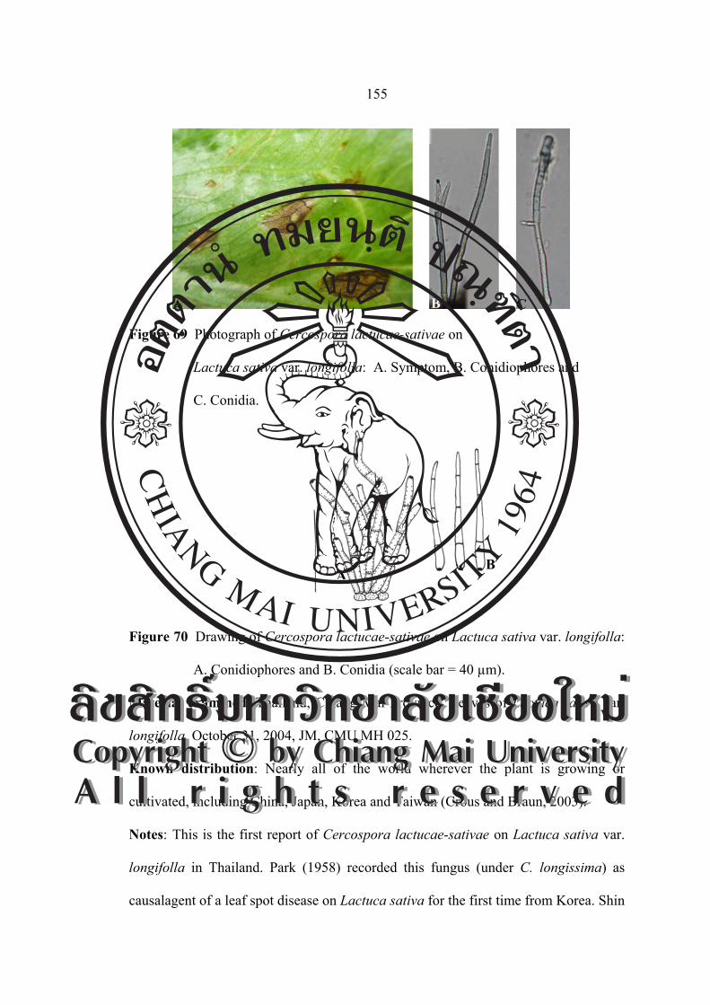
155
Figure 69 Photograph of Cercospora lactucae-sativae on
Lactuca sativa var. longifolla: A. Symptom, B. Conidiophores and
C. Conidia.
Figure 70 Drawing of Cercospora lactucae-sativae on Lactuca sativa var. longifolla:
A. Conidiophores and B. Conidia (scale bar = 40 µm).
Material examined: Thailand, Chiang Mai Province. Leaves of Lactuca sativa var.
longifolla, October 31, 2004, JM, CMU MH 025.
Known distribution: Nearly all of the world wherever the plant is growing or
cultivated, including China, Japan, Korea and Taiwan (Crous and Braun, 2003).
Notes: This is the first report of Cercospora lactucae-sativae on Lactuca sativa var.
longifolla in Thailand. Park (1958) recorded this fungus (under C. longissima) as
causalagent of a leaf spot disease on Lactuca sativa for the first time from Korea. Shin
A B C
AB
ÅÔ¢ÊÔ·¸Ô ìÁËÒÇÔ·ÂÒÅÑÂàªÕ§ãËÁèCopyright by Chiang Mai UniversityA l l r i g h t s r e s e r v e d
ÅÔ¢ÊÔ·¸Ô ìÁËÒÇÔ·ÂÒÅÑÂàªÕ§ãËÁèCopyright by Chiang Mai UniversityA l l r i g h t s r e s e r v e d

156
and Braun (1993) listed the second Korean record. The detailed description and
illustration based on Korean collection of this species were provided by Kim and Shin
(1998b).
The conidiophores of C. longissima-sativae are formed in a loose fascicle, and
the conidia are rather stout, with more septa than those of C. beticola. Chupp (1954),
in his monograph, used the name C. longissima (Cugini in Herb.) Sacc. For this
species, but this name is a younger homonym of C. longissima Cook and Ellis.
Cercospora lactucae-sativae Sawads Formosa Agr. Res. Inst. Rept. 35: 111 (1928)
= Cercospora longispora Cugini ex Trav., Malpighia 17: 217. 1902
(nom. illeg.), homonym of C. longispora Peck, 1884.
≡ Cercospora longissima Trav., Malpighia 17: correzione
(correction slip) to p. 217. 1903 (nom. illeg.), homonym of C.
longissima Cooke and Ellis, 1889.
≡ Cercospora longisima Cugini ex Sacc., Syll. Fung. 18: 607. 1906
(nom. illeg.), homonym of C. longissima Cooke and Ellis, 1889.
= Cercospora lactucae J. A. Stev., J. Dept. Agric. Puerto Rico 1: 105.
1917 (nom.illeg.) [T: BPI 437770, 437771], homonym of C.lactucae
Henn., 1902.
= Cercospora lactucae Welles, Phytopathology 13: 289. 1923 (nom.
illeg., Art. 53.1), homonym of C. lactucae Henn., 1902.
= Cercospora ixeridis-chinensis Sawada, Rep. Gov. Agric. Res. Inst.
Taiwan 86: 171. 1943 (nom. inval.). [T: NTU-PPE, herb. Sawada]
ÅÔ¢ÊÔ·¸Ô ìÁËÒÇÔ·ÂÒÅÑÂàªÕ§ãËÁèCopyright by Chiang Mai UniversityA l l r i g h t s r e s e r v e d
ÅÔ¢ÊÔ·¸Ô ìÁËÒÇÔ·ÂÒÅÑÂàªÕ§ãËÁèCopyright by Chiang Mai UniversityA l l r i g h t s r e s e r v e d

157
= Cercospora lactucae-indicae Sawada, Rep. Gov. Agric. Res. Inst.
Taiwan 86: 172. 1943 (nom. inval.). [T: NTU-PPE, herb. Sawada].
Leaf spots circular to oval, 2.00-5.00 mm in diameter, visible as concentric
zones resembling a frog eye, grey brown and dingy at centre and surrounded by a pale
tan halo, then bordered by a narrow raised dark brown line. Fruit bodies amphigenous,
but chiefly hypophyllous. Mycelium internal; hyphae septate, branched, pale
olivaceous. Stromata absent to rudimentary or weakly developed, composed of
several brown swollen hyphal cells. Conidiophores 4-10 in a divergent fascicle,
sometimes solitary, arranged over stomata, pale olivaceous brown throughout or paler
upwards, 3-9-septate, not branched, (41.46-)53.65-141.46(-185.36)×2.43-4.87 µm,
conidial scars large, conspicuous, apical or on small shoulders of conidiogenous cells
caused by geniculation. Conidia solitary, acicular to filiform or even obclavate,
straight or mildly curved, hyaline, 2-9-septate, non-constricted at the septa, mostly
subacute at the apex, but obtuse in shorter conidia, truncate to subtruncate at the base,
very variable in length, (31.70-)39.02-102.43(-136.58)×(2.43-)4.87-4.87(-7.31) µm,
hilum conspicuously thickened, darkened (Figure 71 and 72).
Figure 71 Photograph of Cercospora lactucae-sativae on Lactuca sativa var. crispa:
A. Symptom, B. Conidiophores and C. Conidia.
A B C
ÅÔ¢ÊÔ·¸Ô ìÁËÒÇÔ·ÂÒÅÑÂàªÕ§ãËÁèCopyright by Chiang Mai UniversityA l l r i g h t s r e s e r v e d
ÅÔ¢ÊÔ·¸Ô ìÁËÒÇÔ·ÂÒÅÑÂàªÕ§ãËÁèCopyright by Chiang Mai UniversityA l l r i g h t s r e s e r v e d

158
Figure 72 Drawing of Cercospora lactucae-sativae on Lactuca sativa var. crispa:
A. Conidiophores and B. Conidia (scale bar = 40 µm).
Material examined: Thailand, Chiang Mai Province. Leaves of Lactuca sativa var.
crispa, October 31, 2004, JM, CMU MH 026.
Known distribution: Worldwide, including Angola, Bangladesh, Barbados, Brazil,
Brunei, Cambodia, China, Colombia, Cuba, Dominican Republ., EI Salvador,
Guatemala, Guatemala, Guadeloupe, Hong Kong, India, Indonesia, Italy, Iran, Ivory
Coast, Jamaica, Japan, Korea, Laos, Lebanon, Libye, Malaysia, Martinique,
Mauritius, Morocco, Mozambique, New Caledonia, Nigeria, Philippines, Puerto Rico,
Romania, Russia (European part), Saudi Arabia, Sierra Leone, Somalia, South Africa,
Spain, Saint Kitts and Nevis, Sudan, Taiwan, Tanzania, Togo, Trinidad and Tobago,
Zambia and Zimbabwe (Crous and Braun, 2003).
Notes: This is the first report of Cercospora lactucae-sativae on Lactuca sativa var.
crispa in Thailand. A true Cercospora s.str. is close to C. apii s. lat.
Cercospora mikaniicola F. Stevens (mikaniacola), Trans. Illinois Acad. Sci. 10: 213.
1917. [T: BPI 438505; ILL 14865; IMI 120992; PC].
AB
ÅÔ¢ÊÔ·¸Ô ìÁËÒÇÔ·ÂÒÅÑÂàªÕ§ãËÁèCopyright by Chiang Mai UniversityA l l r i g h t s r e s e r v e d
ÅÔ¢ÊÔ·¸Ô ìÁËÒÇÔ·ÂÒÅÑÂàªÕ§ãËÁèCopyright by Chiang Mai UniversityA l l r i g h t s r e s e r v e d

159
= Cercospora mikaniae-cordatae J. M. Yen, Rev. Mycol. 30: 183. 1965.
[T: PC].
Leaf spots circular, 0.50-7.50 mm in diameter, greyish at center, on the upper
surface with a raised line border, on lower surface with a in diameter dark area in
which the Fruit bodies is found. Stromata lacking to medium, 14.76-39.36 μm.
Conidiophores pale to medium olivaceous brown, uniform in colour and in diameter,
multiseptate, not branced, 1-10 abruptly geniculate, rarely near tip rachislike, minute
spore scar at small rounded tip, 2-10-septate, (49.20-)118.08-154.98(-177.12)×4.92-
4.92(-7.38) μm. Conidia obclavate, subhyaline to very pale olivaceous, straight to
slightly curved, long obconically truncate base, tip obtuse to subobtuse, sometimes
catenulate, 3-11-septate, (54.12-)61.50-108.24(-135.30)×(2.46-)4.92-7.38(-8.61) μm
(Figure 73 and 74).
Figure 73 Photograph of Cercospora mikaniicola on Mikania cordata: A. Symptom,
B. Conidiophores and C. Conidia.
A B C ÅÔ¢ÊÔ·¸Ô ìÁËÒÇÔ·ÂÒÅÑÂàªÕ§ãËÁèCopyright by Chiang Mai UniversityA l l r i g h t s r e s e r v e d
ÅÔ¢ÊÔ·¸Ô ìÁËÒÇÔ·ÂÒÅÑÂàªÕ§ãËÁèCopyright by Chiang Mai UniversityA l l r i g h t s r e s e r v e d

160
Figure 74 Drawing of Cercospora mikaniicola on Mikania cordata:
A. Conidiophores and B. Conidia (scale bar = 40 µm).
Material examined: Thailand, Doi Suthep-Pui National Park, Chiang Mai Province.
Leaves of Mikania cordata, October 31, 2004, JM, CMU MH 027.
Known distribution: American Samoa, Agentina, Bangladesh, Brazil, Cuba,
Colombia, Fiji, Guadalcanal, Hong Kong, India, Jamaica, Malaysia, Niue, Pakistan,
Papua New Guinea, Puerto Rico, Samoa, Sierra Leone, Singapore, Solomon Islands,
Tuvalu, U.S.A and Vanuatu (Crous and Braun, 2003).
Notes: This is the first report of Cercospora mikaniicola on Mikania cordata in
Thailand. This species belongs in Cercospora s. str., and it is distinct from C. apii
s.lat. Definite leaf spots and the long conidiophores separate this from the other
species on Mikania. For a further description see Mycologia 23: 397. 1931. Key, page
154. Type species is in Utuado, Puerto Rico; Mikania sp.; F. L. Stevens, No. 7922;
July 7. 1915.
A
B
ÅÔ¢ÊÔ·¸Ô ìÁËÒÇÔ·ÂÒÅÑÂàªÕ§ãËÁèCopyright by Chiang Mai UniversityA l l r i g h t s r e s e r v e d
ÅÔ¢ÊÔ·¸Ô ìÁËÒÇÔ·ÂÒÅÑÂàªÕ§ãËÁèCopyright by Chiang Mai UniversityA l l r i g h t s r e s e r v e d

161
Cercospora tridacis-procumbentis Govindu and Thirum., Sydowia 7: 49. 1953.
[T: IMI 55520].
= Cercospora apii s.lat.
Leaf spots circular, suborbicular to irregular, without distinct margin, 2.00-
9.00 mm in diameter, dull grey to greyish brown, sometimes confluent. Stromata
lacking or a few brown cells. Conidiophores solitary or in fascicles of 2-5, pale to
medium olivaceous brown, subhyaline at the apex, 2-8-septate, not branched,
geniculate, conidial scars thickened, (46.80-)62.40-117.00(-124.80)×(2.60-)2.60-
5.20(-7.80) μm. Conidia cylindric, subhyaline to very pale olivaceous, straight to
slightly curved, 5-13-septate, long obconically truncate base, tip obtuse to subobtuse,
sometimes catenulate, (72.80-)88.40-158.60(-223.60)×(2.60-)2.60-5.20(-6.50) μm
(Figure 75 and 76).
Figure 75 Photograph of Cercospora tridacis-procumbentis on Tridax procumbens:
A. Symptom, B. Conidiophores and C. Conidia.
A B C ÅÔ¢ÊÔ·¸Ô ìÁËÒÇÔ·ÂÒÅÑÂàªÕ§ãËÁèCopyright by Chiang Mai UniversityA l l r i g h t s r e s e r v e d
ÅÔ¢ÊÔ·¸Ô ìÁËÒÇÔ·ÂÒÅÑÂàªÕ§ãËÁèCopyright by Chiang Mai UniversityA l l r i g h t s r e s e r v e d

162
Figure 76 Drawing of Cercospora tridacis-procumbentis on Tridax procumbens:
A. Conidiophores and B. Conidia (scale bar = 40 µm).
Material examined: Thailand, Chiang Mai Province. Leaves of Tridax procumbens,
October 31, 2004, JM, CMU MH 028.
Known distribution: Australia, Cuba, Ghana, Guinea, India, Kenya, Kiribati,
Myanmar, Nauru, Nigeria, Papua New Guinea, Sierra Leone, Solomon Islands, South
Africa, Tanzania, Togo and Tuvalu (Crous and Braun, 2003).
Notes: Cercospora tridacis-procumbentis was recorded by Sontirat et al. 1991 in
Thailand.
Cercospora sp.
Leaf spots amphigenous, distinct, subcircular to angular, yellowish brown with
blackish brown margins, 2.00-7.00 mm in diameter. Stromata small to medium,
rudimentary to slightly develped, irregular, dark brown, 14.00-40.00 µm in diameter.
Conidiophores pale brown at the base and paler upwards, arranged in a loose to dense
fascicle, with distinct conidial scars, 1-7-septate, (31.70-)39.02-70.73(-87.80)×4.87-
7.31 µm. Conidia solitary, obclavate, straight to mildly curved, hyaline to subhyaline,
smooth, hilum conspicuously thickened, darkened, (19.51-)31.70-99.99
A B
ÅÔ¢ÊÔ·¸Ô ìÁËÒÇÔ·ÂÒÅÑÂàªÕ§ãËÁèCopyright by Chiang Mai UniversityA l l r i g h t s r e s e r v e d
ÅÔ¢ÊÔ·¸Ô ìÁËÒÇÔ·ÂÒÅÑÂàªÕ§ãËÁèCopyright by Chiang Mai UniversityA l l r i g h t s r e s e r v e d

163
(-126.82)×(3.65-)4.87-4.87 µm, 2-10-septate, acute to obtuse at the apex, truncate at
the base (Figure 77 and 78).
Figure 77 Photograph of Cercospora sp. on Melampodium paludosum:
A. Symptom, B. Conidiophores and C. Conidia.
Figure 78 Drawing of Cercospora sp. on Melampodium paludosum:
A. Conidiophores and B. Conidia (scale bar = 40 µm).
Material examined: Thailand, Doi Suthep-Pui National Park, Chiang Mai Province.
Leaves of Melampodium paludosum, October 31, 2004, JM, CMU MH 029.
Known distribution: Known only Thailand.
Notes: This is the first report of Cercospora sp. on Melampodium paludosum in
Thailand.
A B C
A B
ÅÔ¢ÊÔ·¸Ô ìÁËÒÇÔ·ÂÒÅÑÂàªÕ§ãËÁèCopyright by Chiang Mai UniversityA l l r i g h t s r e s e r v e d
ÅÔ¢ÊÔ·¸Ô ìÁËÒÇÔ·ÂÒÅÑÂàªÕ§ãËÁèCopyright by Chiang Mai UniversityA l l r i g h t s r e s e r v e d
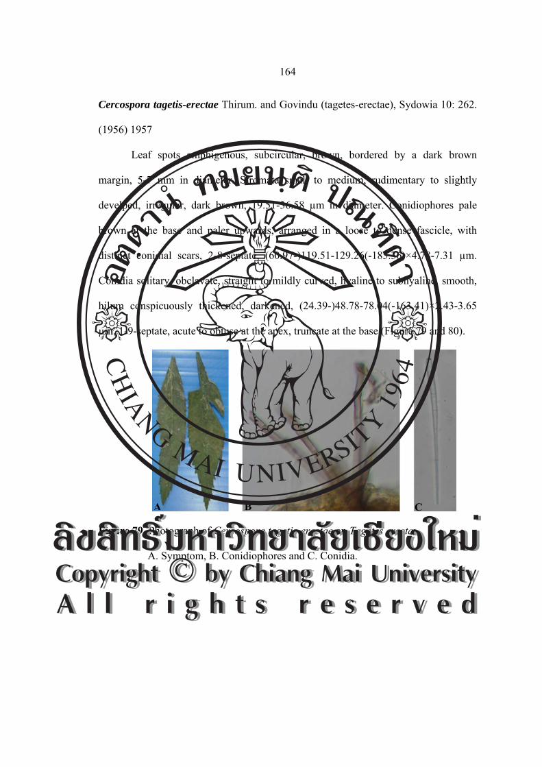
164
Cercospora tagetis-erectae Thirum. and Govindu (tagetes-erectae), Sydowia 10: 262.
(1956) 1957
Leaf spots amphigenous, subcircular, brown, bordered by a dark brown
margin, 5-7 mm in diameter. Stromata small to medium, rudimentary to slightly
develped, irregular, dark brown, 19.51-36.58 µm in diameter. Conidiophores pale
brown at the base and paler upwards, arranged in a loose to dense fascicle, with
distinct conidial scars, 2-8-septate, (60.97-)119.51-129.26(-185.36)×4.78-7.31 µm.
Conidia solitary, obclavate, straight to mildly curved, hyaline to subhyaline, smooth,
hilum conspicuously thickened, darkened, (24.39-)48.78-78.04(-163.41)×2.43-3.65
µm, 1-9-septate, acute to obtuse at the apex, truncate at the base (Figure 79 and 80).
Figure 79 Photograph of Cercospora tagetis-erectae on Tagetes erecta:
A. Symptom, B. Conidiophores and C. Conidia.
A B C
ÅÔ¢ÊÔ·¸Ô ìÁËÒÇÔ·ÂÒÅÑÂàªÕ§ãËÁèCopyright by Chiang Mai UniversityA l l r i g h t s r e s e r v e d
ÅÔ¢ÊÔ·¸Ô ìÁËÒÇÔ·ÂÒÅÑÂàªÕ§ãËÁèCopyright by Chiang Mai UniversityA l l r i g h t s r e s e r v e d

165
Figure 80 Drawing of Cercospora tagetis-erectae on Tagetes erecta:
A. Conidiophores and B. Conidia (scale bar = 40 µm).
Material examined: Thailand, Doi Suthep-Pui National Park, Wiang Pa Pao, Chiang
Rai Province. Leaves of Tagetes erecta, October 31, 2004, JM, CMU MH 030.
Known distribution: India (Crous and Braun, 2003).
Notes: This is the first report of Cercospora tagetis-erectae on Tagetes erecta in
Thailand.
Family Convolvulaaceae
Cercospora convolvuli Tracy and Earle, Bull. Torrey Bot. Club. 28: 187. 1901.
[T: NY].
≡ Passalora convolvuli (Tracy and Earle) U. Braun and Crous. comb.
nov.
A
B
A
ÅÔ¢ÊÔ·¸Ô ìÁËÒÇÔ·ÂÒÅÑÂàªÕ§ãËÁèCopyright by Chiang Mai UniversityA l l r i g h t s r e s e r v e d
ÅÔ¢ÊÔ·¸Ô ìÁËÒÇÔ·ÂÒÅÑÂàªÕ§ãËÁèCopyright by Chiang Mai UniversityA l l r i g h t s r e s e r v e d

166
≡ Mycovellosiella convolvuli (Tracy and Earle) U. Braun, in Braum and
Melnik, Trudy Bot. Inst. im. Komarova 20: 52. 1997.
= Cercospora elongata Sorok., Rev. Mycol. 12: 54. 1890 (nom. illeg.),
homonym of C. elongata Peck, 1880.
≡ Cercospora sorokinii Sace., Syll. Fung. 19: 255. 1910 (nom. nov.).
Leaf spots scattered or confluent, distinct, circular, often zonate, small to fairly
large, 3.50-4.50 mm in diameter, centre pale tan to dingy grey, with dark or purplish
brown margin, sometimes surrounded by a narrow dark brown margin, later greyish
brown due to abundant frutification of the fungus. Fruit bodies amphigenous, but
abundantly hypophyllous. Mycelium internal; hyphal septate, branched, hyaline.
Stromata rudimenrary to slightly to slightly developed, composed of several brown
hyphal cells, 12.19-24.39 µm. Conidiophores 4-12 in a divergent fascicle, uniformly
subhyaline to hyaline, straight to mildly sinuous, 1-4-septate, (29.26-)46.34-51.21
(-73.17)×4.87-7.31 µm, conidial scars conspicuous. Conidia solitary, acicular,
occasionally obclavate, straight to mildly curved, gradually narrowed towards the tip,
hyaline, 4-9-septate, non-constricted at the septa, subacute at the apex, truncate at the
base, very variable in length, (85.36-)122.19-146.34(-175.60)×4.87 µm, hilum
conspicuously thickened, darkened (Figure 81 and 82).
ÅÔ¢ÊÔ·¸Ô ìÁËÒÇÔ·ÂÒÅÑÂàªÕ§ãËÁèCopyright by Chiang Mai UniversityA l l r i g h t s r e s e r v e d
ÅÔ¢ÊÔ·¸Ô ìÁËÒÇÔ·ÂÒÅÑÂàªÕ§ãËÁèCopyright by Chiang Mai UniversityA l l r i g h t s r e s e r v e d

167
Figure 81 Photograph of Cercospora convolvuli on Ipomoea aquatica: A. Symptom,
B. Conidiophores and C. Conidia.
Figure 82 Drawing of Cercospora convolvuli on Ipomoea aquatica:
A. Conidiophores and B. Conidia (scale bar = 40 µm).
Material examined: Thailand, Wiang Pa Pao, Chiang Rai Province. Leaves of
Ipomoea aquatica, October 2, 2005, JM, CMU MH 032.
A B C
A
B
ÅÔ¢ÊÔ·¸Ô ìÁËÒÇÔ·ÂÒÅÑÂàªÕ§ãËÁèCopyright by Chiang Mai UniversityA l l r i g h t s r e s e r v e d
ÅÔ¢ÊÔ·¸Ô ìÁËÒÇÔ·ÂÒÅÑÂàªÕ§ãËÁèCopyright by Chiang Mai UniversityA l l r i g h t s r e s e r v e d
168
Known distribution: Brazil, Georgia, Italy (Sardinia), Lithuania, U.S.A (LA, MO,
WI) and Uzbekistan (Crous and Braun, 2003).
Notes: This is the first report of Cercospora convolvuli on Ipomoea aquatica in
Thailand. The synonymy of C. Elongata needs to be confirmed.
Cercospora ipomoeae G. Winter, Hedwigia 26: 34. 1887. [T: Rabenh., Fungi eur.
3585, in numerous herbaria, e.g., B, HAL, HBG].
= Cercospora viridula Ellis and Everh., Mycol. 5: 70 (1889)
= Cercospora alabamensis G. F. Atk., J. Elisha Mitchell Sci. Soc.
8: 51(1892)
= Cercospora stuckertiana Syd. and P. Syd., Mem. Herb. Boissier
8(4): 2(1900)
= Cercospora ipomoeae-illustriae Chidd., Ind. Phytopath. 12: 114 (1959)
= Cercospora ipomoeae-pes-caprae J. M. Yen and Lim, Bull. Soc.
Mycol. Fr. 86 (3): 747(1970)
Leaf spots scattered or confluent, distinct, circular, often zonate, small to fairly
large, 1.00-3.00 mm in diameter, centre pale tan to dingy grey, with dark or purplish
brown margin, sometimes surrounded by a narrow dark brown margin, later greyish
brown due to abundant frutification of the fungus. Fruit bodies amphigenous, but
abundantly hypophyllous. Mycelium internal, hyphal septate, branched, hyaline.
Stromata rudimenrary to slightly to slightly developed, composed of several brown
hyphal cells. Conidiophores 4-12 in a divergent fascicle, usually pale olivaceous
brown throughout or sometimes paler towards the apex, 2-4-septate, (19.51-)34.14-
68.29(-87.80)×(3.65-)4.87-4.87(-6.09) µm, conidial scars conspicuous, apical or on
ÅÔ¢ÊÔ·¸Ô ìÁËÒÇÔ·ÂÒÅÑÂàªÕ§ãËÁèCopyright by Chiang Mai UniversityA l l r i g h t s r e s e r v e d
ÅÔ¢ÊÔ·¸Ô ìÁËÒÇÔ·ÂÒÅÑÂàªÕ§ãËÁèCopyright by Chiang Mai UniversityA l l r i g h t s r e s e r v e d
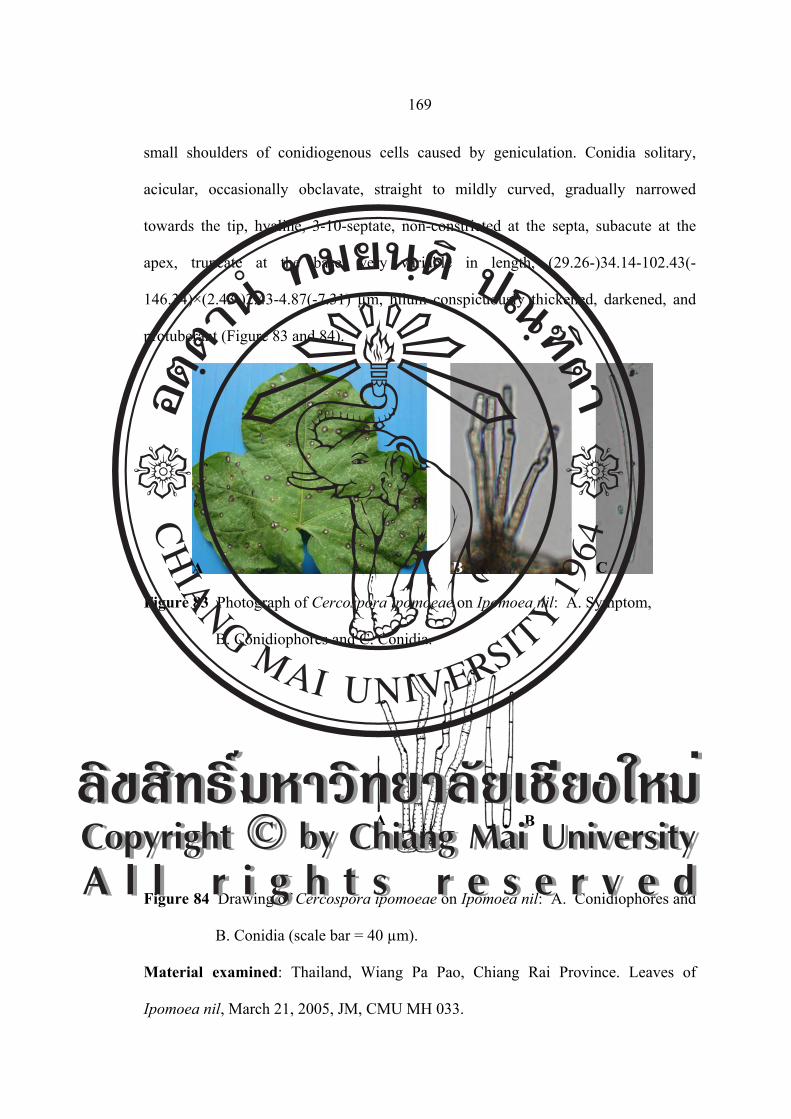
169
small shoulders of conidiogenous cells caused by geniculation. Conidia solitary,
acicular, occasionally obclavate, straight to mildly curved, gradually narrowed
towards the tip, hyaline, 3-10-septate, non-constricted at the septa, subacute at the
apex, truncate at the base, very variable in length, (29.26-)34.14-102.43(-
146.34)×(2.43-)2.43-4.87(-7.31) µm, hilum conspicuously thickened, darkened, and
protuberant (Figure 83 and 84).
Figure 83 Photograph of Cercospora ipomoeae on Ipomoea nil: A. Symptom,
B. Conidiophores and C. Conidia.
Figure 84 Drawing of Cercospora ipomoeae on Ipomoea nil: A. Conidiophores and
B. Conidia (scale bar = 40 µm).
Material examined: Thailand, Wiang Pa Pao, Chiang Rai Province. Leaves of
Ipomoea nil, March 21, 2005, JM, CMU MH 033.
A B C
A B ÅÔ¢ÊÔ·¸Ô ìÁËÒÇÔ·ÂÒÅÑÂàªÕ§ãËÁèCopyright by Chiang Mai UniversityA l l r i g h t s r e s e r v e d
ÅÔ¢ÊÔ·¸Ô ìÁËÒÇÔ·ÂÒÅÑÂàªÕ§ãËÁèCopyright by Chiang Mai UniversityA l l r i g h t s r e s e r v e d

170
Known distribution: Worldwide, including American Samoa, Antigua and Barbuda,
Argentina, Australia, Barbados, Brazil, Brunei, China, Cook Islands, Costa Rica,
Cuba, Fiji, Guam, Hong Kong, India, Indonesia, Italy, Ivory Coast, Jamaica, Japan,
Kenya, Kiribati, Korea, Malaysia, Marshall Islands, Mauritius, Myanmar, New
Caledonia, New Zealand, Pakistan, Panama, Papua New Guinea, Puerto Rico, Samoa,
Sierra Leone, Solomon Island, Sudan, Venezuala, Taiwan, Tanzania, U.S.A (AL, GA,
FL, HI, IL, IN, KS, MO, NC, NE, NJ, OH, TX, VA) and Vanuatu (Crous and Braun,
2003).
Notes: This is the first report of Cercospora ipomoeae on Ipomoea nil in Thailand.
Family Brassicaceae
Cercospora brassicicola Henn., Bot. Jahrb. Syst. 37: 166. 1906. [T: B; LEP].
= Cercospora brassicae-campestris Rangel, Arq. Mus.Nac., Rio de
Janeiro 18: 163. 1917.
≡ Cercosporina brassicae-campestris (Rangel) Sacc., Syll. Fung. 25:
899. 1931.
= Cercospora brassicae-junceae Sawada (brassicae-yunciae), Special
Publ. Coll. Agric. Natl. Taiwan Univ. 8: 212. 1959 (nom. nud.).
[T: NYU-PPE, herb. Sawada].
= Cercospora bloxami auct. Sensu E. Young, Mycologia 8: 43. 1916.
Leaf spots amphigenous, scattered to confluent, indistinct, subcircular to
irregular, fairly large, 3.00-6.00 mm in diameter, pale brown to tan centres with
indefinite margins. Fruit bodies amphigenous. Stromata small, rudimentary to slightly
ÅÔ¢ÊÔ·¸Ô ìÁËÒÇÔ·ÂÒÅÑÂàªÕ§ãËÁèCopyright by Chiang Mai UniversityA l l r i g h t s r e s e r v e d
ÅÔ¢ÊÔ·¸Ô ìÁËÒÇÔ·ÂÒÅÑÂàªÕ§ãËÁèCopyright by Chiang Mai UniversityA l l r i g h t s r e s e r v e d

171
developed, subglobular, brown to dark brown, composed of several brown hyphal
cells. Conidiophores 2-15 in a loose fascicle, olivaceous brown or paler towards the
upper portion, substraight to slightly curved, usually not branched, but very rarely
branched, 1-4 times mildly geniculate, 2-6-septate, (41.46-)63.41-131.70
(-163.41)×(2.43-)3.65-4.87(-7.31) µm, conspicuous, apical or on shoulders of
conidiogenous cells caused by geniculation. Conidia solitary, obclavate, straight to
mildly curved or somewhat undulate in long ones, hyaline, 5-10-septate, non-
constricted at the septa, obtuse to subobtuse at the apex, truncate to subtruncate at the
base, very variable in length, (47.56-)51.21-87.80(-97.56)×2.43-4.87 µm, hilum
conspicuously thickened, darkened and non-protuberant (Figure 85 and 86).
Figure 85 Photograph of Cercospora barlericola on Brarssica pekinensis:
A. Symptom, B. Conidiophores and C. Conidia.
A B C
ÅÔ¢ÊÔ·¸Ô ìÁËÒÇÔ·ÂÒÅÑÂàªÕ§ãËÁèCopyright by Chiang Mai UniversityA l l r i g h t s r e s e r v e d
ÅÔ¢ÊÔ·¸Ô ìÁËÒÇÔ·ÂÒÅÑÂàªÕ§ãËÁèCopyright by Chiang Mai UniversityA l l r i g h t s r e s e r v e d

172
Figure 86 Drawing of Cercospora barlericola on Brarssica pekinensis:
A. Conidiophores and B. Conidia (scale bar = 40 µm).
Material examined: Thailand, Chiang Mai University, Chiang Mai Province. Leaves
of Brarssica pekinensis, October 31, 2004, JM, CMU MH 034.
Known distribution: Worldwide, including Angola, Armenia, Australia, Belarus,
Brazil, China, Colombia, Cuba, Dominican Republ., Estonia, Great Britain, India,
Indonesia, Jamaica, Japan, Kazakhstan, Kenya, Korea, Latvia, Lithuania, Malaysia,
Malawi, Mauritius, Myanmar, Nigeria, Niue, Papua, New Guinea, Peru, Philippines,
Puerto Rico, Russia, Sierra Leone, South Africa, Solomon Islands, Somalia, Sri
Lanka, Sudan, Taiwan, Tanzania, Togo, Trinidad, Tobago, Togo, Uganda and
Ukraine and U.S.A (AL, CA, DE, FL, GA, HI, IL, IN, LA, MS, NC, NH, NJ, OK,
TX, VA) (Crous and Braun, 2003).
Notes: This is the first report of Cercospora brassicicola on Brarssica pekinensis in
Thailand. A true Cercospora s.str. is close to C. apii s. lat.
A
B
ÅÔ¢ÊÔ·¸Ô ìÁËÒÇÔ·ÂÒÅÑÂàªÕ§ãËÁèCopyright by Chiang Mai UniversityA l l r i g h t s r e s e r v e d
ÅÔ¢ÊÔ·¸Ô ìÁËÒÇÔ·ÂÒÅÑÂàªÕ§ãËÁèCopyright by Chiang Mai UniversityA l l r i g h t s r e s e r v e d

173
Cercospora brassicicola Henn., Bot. Jahrb. Syst. 37: 166. 1906. [T: B; LEP].
= Cercospora brassicae-campestris Rangel, Arq. Mus. Nac., Rio de
Janeiro 18: 163. 1917.
≡ Cercosporina brassicae-campestris (Rangel) Sacc., Syll. Fung. 25:
899. 1931.
= Cercospora brassicae-junceae Sawada (brassicae-yunciae), Special
Publ. Coll. Agric. Natl. Taiwan Univ. 8: 212. 1959 (nom. nud.).
[T: NYU-PPE, herb. Sawada].
= Cercospora bloxami auct. Sensu E. Young, Mycologia 8: 43. 1916.
Leaf spots amphigenous, visible on both surfaces, irregularly suborbicular,
2.00-7.00 mm in diameter, pale greenish brown to yellowish yellowish grey at the
center, with a brown raised margin. Fruit bodies amphigenous. Mycelium internal,
hyphae septate, branched, hyaline. Stromata small, rudimentary to slightly developed,
subglobular, brown to dark brown, composed of several brown hyphal cells.
Conidiophores loosely fasciculate at the cuticle, olivaceous brown or paler towards
the upper portion, substraight to slightly curved, subtruncate at the apex, 2-12-septate,
(31.70-)34.14-80.48(-97.56)×(2.43-)3.65-4.87 µm, conidial scars large, conspicuous,
apical or on shoulders of conidiogenous cells caused by geniculation. Conidia hyaline,
acicular, straight to curved, obtuse to subobtuse at the apex, truncate to subtruncate at
the base, very variable in length, 4-14-septate, (26.82-)43.90-114.63(-148.77)×(2.43-)
3.65-4.87(-6.09) µm, hilum conspicuously thickened, darkened and non-protuberant
(Figure 87 and 88).
ÅÔ¢ÊÔ·¸Ô ìÁËÒÇÔ·ÂÒÅÑÂàªÕ§ãËÁèCopyright by Chiang Mai UniversityA l l r i g h t s r e s e r v e d
ÅÔ¢ÊÔ·¸Ô ìÁËÒÇÔ·ÂÒÅÑÂàªÕ§ãËÁèCopyright by Chiang Mai UniversityA l l r i g h t s r e s e r v e d

174
Figure 87 Photograph of Cercospora barlericola on Brassica campestris:
A. Symptom, B. Conidiophores and C. Conidia.
Figure 88 Drawing of Cercospora barlericola on Brassica campestris:
A. Conidiophores and B. Conidia (scale bar = 40 µm).
Material examined: Thailand, Chiang Mai University, Chiang Mai Province. Leaves
of Brassica campestris, October 15, 2004, JM, CMU MH 035.
Known distribution: Worldwide, including Angola, Armenia, Australia, Belarus,
Brazil, China, Colombia, Cuba, Dominican Republ., Estonia, Great Britain, India,
Indonesia, Jamaica, Japan, Kazakhstan, Kenya, Korea, Latvia, Lithuania, Malaysia,
Malawi, Mauritius, Myanmar, Nigeria, Niue, Papua, New Guinea, Peru, Philippines,
Puerto Rico, Russia, Sierra Leone, South Africa, Solomon Islands, Somalia, Sri
Lanka, Sudan, Taiwan, Tanzania, Togo, Trinidad and Tobago, Togo, Uganda,
A B C
A
B
ÅÔ¢ÊÔ·¸Ô ìÁËÒÇÔ·ÂÒÅÑÂàªÕ§ãËÁèCopyright by Chiang Mai UniversityA l l r i g h t s r e s e r v e d
ÅÔ¢ÊÔ·¸Ô ìÁËÒÇÔ·ÂÒÅÑÂàªÕ§ãËÁèCopyright by Chiang Mai UniversityA l l r i g h t s r e s e r v e d

175
Ukraine and U.S.A (AL, CA, DE, FL, GA, HI, IL, IN, LA, MS, NC, NH, NJ, OK,
TX, VA) (Crous and Braun, 2003).
Notes: This is the first report of Cercospora brassicicola. on Brassica campestris in
Thailand. A true Cercospora s.str. is close to C. apii s. lat.
Cercospora brassicicola Henn., Bot. Jahrb. Syst. 37: 166. 1906. [T: B; LEP].
= Cercospora brassicae-campestris Rangel, Arq. Mus. Nac., Rio de
Janeiro 18: 163. 1917.
≡ Cercosporina brassicae-campestris (Rangel) Sacc., Syll. Fung. 25:
899. 1931.
= Cercospora brassicae-junceae Sawada (brassicae-yunciae), Special
Publ. Coll. Agric. Natl. Taiwan Univ. 8: 212. 1959 (nom. nud.).
[T: NYU-PPE, herb. Sawada].
= Cercospora bloxami auct. sensu E. Young, Mycologia 8: 43. 1916.
Leaf spots angular, scattered or aggregated and coalescing in larger areas, at
first yellowish green but gradually changing to greyish brown with indefinite margins.
Fruit bodies epiphyllous. Mycelium internal, hyphae septate, branched, hyaline.
Stromata small, rudimentary to slightly developed, subglobular, brown to dark brown,
8.00-15.00 µm in diameter, composed of several brown hyphal cells. Conidiophores
2-11 in a loose fascicle, pale olivaceous to medium brown or paler towards the upper
portion, substraight to slightly curved, usually not branched, but very rarely branched,
1-5 times mildly geniculate, 2-7-septate, (59.04-)76.26-147.60(-159.90)×(3.69-)4.92-
6.15(-7.38) µm, conidial scars thickened, apical or on shoulders of conidiogenous
cells caused by geniculation. Conidia hyaline, acicular to filiform, shorter ones
ÅÔ¢ÊÔ·¸Ô ìÁËÒÇÔ·ÂÒÅÑÂàªÕ§ãËÁèCopyright by Chiang Mai UniversityA l l r i g h t s r e s e r v e d
ÅÔ¢ÊÔ·¸Ô ìÁËÒÇÔ·ÂÒÅÑÂàªÕ§ãËÁèCopyright by Chiang Mai UniversityA l l r i g h t s r e s e r v e d
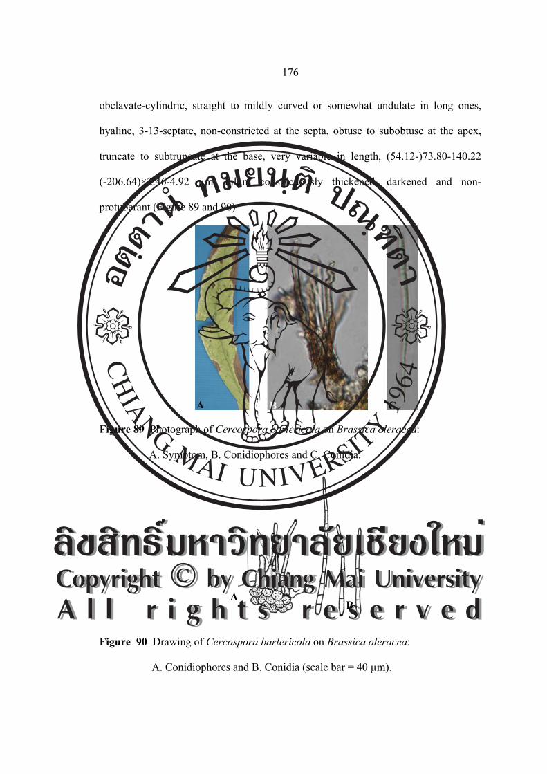
176
obclavate-cylindric, straight to mildly curved or somewhat undulate in long ones,
hyaline, 3-13-septate, non-constricted at the septa, obtuse to subobtuse at the apex,
truncate to subtruncate at the base, very variable in length, (54.12-)73.80-140.22
(-206.64)×2.46-4.92 µm, hilum conspicuously thickened, darkened and non-
protuberant (Figure 89 and 90).
Figure 89 Photograph of Cercospora barlericola on Brassica oleracea:
A. Symptom, B. Conidiophores and C. Conidia.
Figure 90 Drawing of Cercospora barlericola on Brassica oleracea:
A. Conidiophores and B. Conidia (scale bar = 40 µm).
AB
A B C
ÅÔ¢ÊÔ·¸Ô ìÁËÒÇÔ·ÂÒÅÑÂàªÕ§ãËÁèCopyright by Chiang Mai UniversityA l l r i g h t s r e s e r v e d
ÅÔ¢ÊÔ·¸Ô ìÁËÒÇÔ·ÂÒÅÑÂàªÕ§ãËÁèCopyright by Chiang Mai UniversityA l l r i g h t s r e s e r v e d

177
Material examined: Thailand, Wiang Pa Pao, Chiang Rai Province. Leaves of
Brassica oleracea, October 19, 2005, JM, CMU MH 036.
Known distribution: Worldwide (Crous and Braun, 2003).
Notes: This is the first report of Cercospora brassicicola on Brassica oleracea in
Thailand.
Cercospora beticola Sacc., Nuovo Giorn. Bot. Ital. 8: 189. 1876. [T: PAD].
≡ Cercosporina beticola (Sacc.) K. Nakata, T. Nakajima and K.
Katimoto, Rep. Agric. Korea 6. 1915.
= Fusisporium betae Desm., Ann. Sci. Net., Bot., 2Ser., 19: 434. 1843.
= Fusarium betae (Desm.) Sacc., Michelia 2: 132. 1880.
= Pionnotes betae (Desm.) Sacc., Syll. Fung. 4: 726. 1886.
= Cercospora betae A. B. Frank ex Sacc., Syll. Fung. 10: 637. 1892.
= Cercospora longissima Cooke and Eills, Grevillea 17: 65. 1889.
[T: K].
= Cercospora flagelliformis Ellis and Halst., New Jersey Agric. Coll.
exp. Sta., Annual Rep. 11: 355. 1890. [T: NY].
= Cercospora anthelmintica G. F. Atk., J. Elisha Mitchell Sci. Soc. 8: 48.
1892. [T: CUP].
= Cercospora spinaciae Oudem., Ned. Kruidk. Arch. III, 2: 324. 1900.
[T: L].
= Cercospora chenopodiicola Bres., Hedwigia 39: 328. 1900.
[T: Krieger, Fungi sax. 1631, e.g., HAL, LEP].
= Cercosporina spinaciicola Sacc., Nuovo Giorn. Bot. Ital., N. S.,
ÅÔ¢ÊÔ·¸Ô ìÁËÒÇÔ·ÂÒÅÑÂàªÕ§ãËÁèCopyright by Chiang Mai UniversityA l l r i g h t s r e s e r v e d
ÅÔ¢ÊÔ·¸Ô ìÁËÒÇÔ·ÂÒÅÑÂàªÕ§ãËÁèCopyright by Chiang Mai UniversityA l l r i g h t s r e s e r v e d

178
22: 73. 1915.
= Cercospora beticola var. Poonensis Chidd., Sydowia 13: 153. 1959
(nom. inval.). [T: BPI; HCIO; IMI]. (= Cercospora apii s.lat.).
Leaf spots scattered, often confluent, circular, 2.00-4.00 mm in diameter, at
first appearing dirty brown with definite dark brown border, later centre becoming
whitish grey, finally turning reddish brown or purplish brown at the margin. Fruit
bodies amphigenous, but chiefly epiphyllous. Mycelium internal, hyphae septate,
branched, hyaline. Stromata rudimentary to slightly developed. Conidiophores
densely fasciculate, pale brown at the base, paler upwards, nearly subhyaline at the
apex, 1-4-septate, rarely geniculate towards the apex, weakly to mildly attenuated,
29.52-98.40×2.46-3.69 µm, conidial scars conspicuous, thickened and darkened,
apical or on shoulders of conidiogenous cells caused by geniculation, usually
concentrated in the apical portion. Conidia solitary, filiform to acicular, straight to
mildly curved, hyaline, 3-10-septate, non-constricted at the septa, obtuse to subacute
at the apex, subtruncate at the base, greatly variable in length, 36.90-93.48×2.46-3.69
µm, hilum conspicuously thickened, darkened. (Figure 91 and 92).
Figure 91 Photograph of Cercospora beticola on Spinacia oleracea: A. Symptom,
B. Conidiophores and C. Conidia.
A B C
ÅÔ¢ÊÔ·¸Ô ìÁËÒÇÔ·ÂÒÅÑÂàªÕ§ãËÁèCopyright by Chiang Mai UniversityA l l r i g h t s r e s e r v e d
ÅÔ¢ÊÔ·¸Ô ìÁËÒÇÔ·ÂÒÅÑÂàªÕ§ãËÁèCopyright by Chiang Mai UniversityA l l r i g h t s r e s e r v e d

179
Figure 92 Drawing of Cercospora beticola on Spinacia oleracea: A. Conidiophores
and B. Conidia (scale bar = 40 µm).
Material examined: Thailand, Doi Suthep-Pui National Park, Chiang Mai Province.
Leaves of Spinacia oleracea, October 10, 2005, JM and Iman Hidayat, CMU MH
037.
Known distribution: Worldwide, including Afghanistan, Algeria, Angola, Armenia,
Antigua and Barbuda, Argentina, Australia, Austria, Bangladesh, Barbados, Belarus,
Belgium, Bermuda, Bosnia and Herzegovina, Brazil, Bulgaria, Cambodia, Canada,
Chile, China, Colombia, Congo, Costa Rica, Croatia, Cuba, Cyprus, Czech Republ.,
Denmark, Dominican Republ., Egypt, El Salvador, Estonia, Ethiopia, France,
Georgia, Germany, Great Britain, Greece, Guatemala, Guyana, Haiti, Honduras, Hong
Kong, Hungary,India, Indonesia, Israel, Italy, Ivory Coast, Iran, Iraq, Ireland,
Jamaica, Japan, Jordan, Kazakhstan, Kenya, Kirghizia, Korea, Latvia, Laos, Lebanon,
Libya, Lithuania, Macedonia, Madagascar, Malawi, Malaysia, Malta, Mauritius,
Mexico, Morocco, Mozambique, Nepal, Netherlands, New Calwdonia, New Zealand,
Nicaragua, Niger, Nigeria, Norway, Oman, Pakistan, Panama, Papua New Guinea,
Peru, Philippines, Poland, Portugal, Puerto Rico, Romania, Russia, Saudi Arabia,
Senegal, Sierra Leone, Slovakia, Slovenia, Somalia, South Africa, Spain, Sri Lanka,
AB
ÅÔ¢ÊÔ·¸Ô ìÁËÒÇÔ·ÂÒÅÑÂàªÕ§ãËÁèCopyright by Chiang Mai UniversityA l l r i g h t s r e s e r v e d
ÅÔ¢ÊÔ·¸Ô ìÁËÒÇÔ·ÂÒÅÑÂàªÕ§ãËÁèCopyright by Chiang Mai UniversityA l l r i g h t s r e s e r v e d

180
Saint Vincent and the Grenadines, Sudan, Sweden, Switzerland, Syria, Taiwan,
Tanzania, Togo, Trinidad, Tobago, Tunisia, Turkey, Ukraine, Uruguay, U.S.A (AL,
AR, AZ, CA, CO, Easterm states, FL, HI, IA, IL, IN, MA, MD, MI, MN, MS, MT,
NC, ND, NE, NM, OH, OK, SD, TX, WI, WY), Venezuela, Virgin Islands, Yemen,
Yugoslavia, Zambia and Zimbabwe (Crous and Braun, 2003).
Notes: This is the first report of Cercospora beticola on Spinacia oleracea in
Thailand. Numerous additional hosts have been reported in the literature.
Cercospora brassicicola Henn., Bot. Jahrb. Syst. 37: 166. 1906. [T: B; LEP].
= Cercospora brassicae-campestris Rangel, Arq. Mus. Nac., Rio de
Janeiro 18: 163. 1917.
≡ Cercosporina brassicae-campestris (Rangel) Sacc., Syll. Fung.
25: 899. 1931.
= Cercospora brassicae-junceae Sawada (brassicae-yunciae), Special
Publ. Coll. Agric. Natl. Taiwan Univ. 8: 212. 1959 (nom. nud.).
[T: NYU-PPE, herb. Sawada].
= Cercospora bloxami auct. Sensu E. Young, Mycologia 8: 43. 1916.
Leaf spots epiphyllous, distinct, circular to subcircular, 2.00-6.00 mm in
diameter, pale yellowish to tan centres with dark brown margins. Fruit bodies
amphigenous. Stromata small, composed of several brown hyphal cells.
Conidiophores 2-7 in a loose fascicle, olivaceous brown or paler towards the upper
portion, substraighr to slightly curved, usually not branched, but very rarely branched,
1-8 times mildly geniculate, 1-3-septate, (17.07-)21.95-51.21(-68.29)×(3.65-)4.87-
4.87(-7.31) µm, conidial scars large. Conidia acicular-filiform, hyaline, 3-14-septate,
ÅÔ¢ÊÔ·¸Ô ìÁËÒÇÔ·ÂÒÅÑÂàªÕ§ãËÁèCopyright by Chiang Mai UniversityA l l r i g h t s r e s e r v e d
ÅÔ¢ÊÔ·¸Ô ìÁËÒÇÔ·ÂÒÅÑÂàªÕ§ãËÁèCopyright by Chiang Mai UniversityA l l r i g h t s r e s e r v e d

181
non-constricted at the septa, obtuse to subobtuse at the apex, truncate to subtruncate at
the base, very variable in length, (29.26-)39.02-51.21(-58.53)×4.87-7.31 µm, hilum
conspicuously thickened, darkened. (Figure 93 and 94).
Figure 93 Photograph of Cercospora brassicicola on Brassica rapa: A. Symptom,
B. Conidiophores and C. Conidia.
Figure 94 Drawing of Cercospora brassicicola on Brassica rapa: A. Conidiophores
and B. Conidia (scale bar = 40 µm).
Material examined: Thailand, Faculty of Agriculture, Chiang Mai University,
Chiang Mai Province. Leaves of Brassica rapa, November 9, 2005, JM, CMU MH
038.
A B C
A
B
ÅÔ¢ÊÔ·¸Ô ìÁËÒÇÔ·ÂÒÅÑÂàªÕ§ãËÁèCopyright by Chiang Mai UniversityA l l r i g h t s r e s e r v e d
ÅÔ¢ÊÔ·¸Ô ìÁËÒÇÔ·ÂÒÅÑÂàªÕ§ãËÁèCopyright by Chiang Mai UniversityA l l r i g h t s r e s e r v e d

182
Known distribution: Worldwide including Angola, Armenia, Australia, Belarus,
Brazil, China, Colombia, Cuba, Dominican Republ., Estonia, Great Britain, India,
Indonesia, Jamaica, Japan, Kazakhstan, Kenya, Korea, Latvia, Lithuania, Malaysia,
Malawi, Mauritius, Myanmar, Nigeria, Niue, Papua, New Guinea, Peru, Philippines,
Puerto Rico, Russia, Sierra Leone, South Africa, Solomon Islands, Somalia,Sri Lanka,
Sudan, Taiwan, Tanzania, Togo, Trinidad and Tobago, Togo, Uganda, Ukraine and
U.S.A (AL, CA, DE, FL, GA, HI, IL, IN, LA, MS, NC, NH, NJ, OK, TX, VA) (Crous
and Braun, 2003).
Notes: This is the first report of Cercospora brassicicola on Brassica rapa in
Thailand. A true Cercospora s.str. is close to C. apii s. lat.
Cercospora brassicicola Henn., Bot. Jahrb. Syst. 37: 166. 1906. [T: B; LEP].
= Cercospora brassicae-campestris Rangel, Arq. Mus. Nac., Rio de
Janeiro 18: 163 (1917).
≡ Cercosporina brassicae-campestris (Rangel) Sacc., Syll. Fung. 25: 899
(1931).
= Cercospora brassicae-junceae Sawada, Special Publ. Coll. Agric. Natl.
Taiwan Univ. 8: 212 (1959).
= Cercospora bloxami auct. sensu E.Young, Mycologia 8: 43 (1916).
Leaf spots white to grayish white at the center with brown border, distinct,
circular to subcircular, 1.00-9.00 mm in diameter. Fruit bodies amphigenous.
Stromata pale brown, composed of a few large brown cells or large and loose hyphal
masses, 12.30-41.82 µm in diameter. Conidiophores loose fascicule, pale brown, pale
to apex, simple, occasionally branched, straight or geniculate, with thickened conidial
ÅÔ¢ÊÔ·¸Ô ìÁËÒÇÔ·ÂÒÅÑÂàªÕ§ãËÁèCopyright by Chiang Mai UniversityA l l r i g h t s r e s e r v e d
ÅÔ¢ÊÔ·¸Ô ìÁËÒÇÔ·ÂÒÅÑÂàªÕ§ãËÁèCopyright by Chiang Mai UniversityA l l r i g h t s r e s e r v e d

183
scars at shoulder, 0-4-septate, (14.76-)22.14-63.96(-83.64)×3.69-6.15 µm. Conidia
acicular, straight to mildly curved, hyaline, smooth, thickened and truncate basal end,
(19.68-)39.36-137.76(-184.50)×(1.23-)2.46-4.92(-5.41) µm, 1-10-septate. (Figure 95
and 96).
Figure 95 Photograph of Cercospora brassicicola on Brassica juncea:
A. Symptom, B. Conidiophores and C. Conidia.
Figure 96 Drawing of Cercospora brassicicola on Brassica juncea:
A. Conidiophores and B. Conidia (scale bar = 20 µm).
A
B
A B C
ÅÔ¢ÊÔ·¸Ô ìÁËÒÇÔ·ÂÒÅÑÂàªÕ§ãËÁèCopyright by Chiang Mai UniversityA l l r i g h t s r e s e r v e d
ÅÔ¢ÊÔ·¸Ô ìÁËÒÇÔ·ÂÒÅÑÂàªÕ§ãËÁèCopyright by Chiang Mai UniversityA l l r i g h t s r e s e r v e d

184
Material examined: Thailand, Doi Suthep-Pui National Park, Chiang Mai Province.
Leaves of Brassica juncea, November 21, 2004, CN and JM, CMU MH 039.
Known distribution: Worldwide, including Angola, Armenia, Australia, Belarus,
Brazil, China, Colombia, Cuba, Dominican Republ., Estonia, Great Britain, India,
Indonesia, Jamaica, Japan, Kazakhstan, Kenya, Korea, Latvia, Lithuania, Malaysia,
Malawi, Mauritius, Myanmar, Nigeria, Niue, Papua, New Guinea, Peru, Philippines,
Puerto Rico, Russia, Sierra Leone, South Africa, Solomon Islands, Somalia, Sri
Lanka, Sudan, Taiwan, Tanzania, Togo, Trinidad, Tobago, Togo, Uganda, Ukraine
and U.S.A (AL, CA, DE, FL, GA, HI, IL, IN, LA, MS, NC, NH, NJ, OK, TX, VA)
(Crous and Braun, 2003).
Notes: Hitherto Known. This is the first report of Cercospora brassicicola on
Brassica juncea in Thailand. A true Cercospora s.str. is close to C. apii s. lat.
Cercospora brassicicola Henn., Bot. Jahrb. Syst. 37: 166. 1906. [T: B; LEP].
= Cercospora brassicae-campestris Rangel, Arq. Mus. Nac., Rio de
Janeiro 18: 163. 1917.
≡ Cercosporina brassicae-campestris (Rangel) Sacc., Syll. Fung.
25: 899. 1931.
= Cercospora brassicae-juncea Sawada (brassicae-yunciae), Special
Publ. Coll. Agric. Natl. Taiwan Univ. 8: 212. 1959 (nom. nud.).
[T: NYU-PPE, herb. Sawada].
= Cercospora bloxami auct. sensu E. Young, Mycologia 8: 43. 1916.
Leaf spots white to grey at the center with brown border, distinct, subcircular
to irregular, 1.00-9.00 mm in diameter. Stromata small, composed of a few swollen,
ÅÔ¢ÊÔ·¸Ô ìÁËÒÇÔ·ÂÒÅÑÂàªÕ§ãËÁèCopyright by Chiang Mai UniversityA l l r i g h t s r e s e r v e d
ÅÔ¢ÊÔ·¸Ô ìÁËÒÇÔ·ÂÒÅÑÂàªÕ§ãËÁèCopyright by Chiang Mai UniversityA l l r i g h t s r e s e r v e d

185
brown hyphal cells. 12.30-41.82 µm in diameter. Conidiophores arising from the
upper part of stromata, densely fascicular, pale brown, simple, straight with thickened
conidial scars at shoulder, 1-5-septate, (29.26-)31.70-75.60(-126.82)×(2.43-)4.87-
4.87(-7.31) µm. Conidia obclavate to acicular, straight to mildly curved, hyaline,
smooth, hilum conspicuously thickened, darkened, 3-9-septate, (24.39-)39.02-87.80
(-109.75)×4.87-7.31 µm, 3-10-septate (Figure 97 and 98).
Figure 97 Photograph of Cercospora brassicicola on Brassica alboglabra:
A. Symptom, B. Conidiophores and C. Conidia.
Figure 98 Drawing of Cercospora brassicicola on Brassica alboglabra:
A B C
A
B
ÅÔ¢ÊÔ·¸Ô ìÁËÒÇÔ·ÂÒÅÑÂàªÕ§ãËÁèCopyright by Chiang Mai UniversityA l l r i g h t s r e s e r v e d
ÅÔ¢ÊÔ·¸Ô ìÁËÒÇÔ·ÂÒÅÑÂàªÕ§ãËÁèCopyright by Chiang Mai UniversityA l l r i g h t s r e s e r v e d
186
A. Conidiophores and B. Conidia (scale bar = 40 µm).
Material examined: Thailand, Faculty of Agriculture, Chiang Mai University,
Chiang Mai Province. Leaves of Brassica alboglabra, January 19, 2005, JM, CMU
MH 040.
Known distribution: Worldwide, including Angola, Armenia, Australia, Belarus,
Brazil, China, Colombia, Cuba, Dominican Republ., Estonia, Great Britain, India,
Indonesia, Jamaica, Japan, Kazakhstan, Kenya, Korea, Latvia, Lithuania, Malaysia,
Malawi, Mauritius, Myanmar, Nigeria, Niue, Papua, New Guinea, Peru, Philippines,
Puerto Rico, Russia, Sierra Leone, South Africa, Solomon Islands, Somalia, Sri
Lanka, Sudan, Taiwan, Tanzania, Togo, Trinidad and Tobago, Togo, Uganda,
Ukraine and U.S.A (AL, CA, DE, FL, GA, HI, IL, IN, LA, MS, NC, NH, NJ, OK,
TX, VA) (Crous and Braun, 2003).
Notes: This is the first report of Cercospora brassicicola on Brassica alboglabra in
Thailand. A true Cercospora s.str. is close to C. apii s. lat.
Family Cucurbitaceae
Cercospora citrullina Cooke Grevillea 12: 31 (1883)
= Cercospora cucurbitae Ellis and Everh., J. Mycol. 4: 3. 1883.
= Cercospora sechii J. A. Stev., Puerto Rico Agric. exp. Sta. Rep. 1917-
1918: 137. 1919.
= Cercospora momordicae McRae, Ann. Cryptog. Exot. 2: 267. 1929.
= Cercospora trichosanthis McRae, Ann. Cryptig. Exot. 2: 270. 1929.
= Cercospora luffae Hara, Diseases of cultivated plants: 228. 1928.
ÅÔ¢ÊÔ·¸Ô ìÁËÒÇÔ·ÂÒÅÑÂàªÕ§ãËÁèCopyright by Chiang Mai UniversityA l l r i g h t s r e s e r v e d
ÅÔ¢ÊÔ·¸Ô ìÁËÒÇÔ·ÂÒÅÑÂàªÕ§ãËÁèCopyright by Chiang Mai UniversityA l l r i g h t s r e s e r v e d

187
= Cercospora chardoniana Chupp, Monogr. Univ. Puerto Rico, B,
2: 245. 1934.
= Cercospora momordicae Mend., Philipp. J. Sci. 75: 173. 1941.
(mom. illeg.), homonym of C. momordicae McRae, 1929.
= Cercospora momordicae Sawada, Rep. Gov. Agric. Res. Inst. Taiwan
86: 173. 1943 (nom. inval.), homonym of C. momordicae McRae,
1929. (=Cercospora apii s.lat.).
Leaf spots amphigenous, scattered to confluent, distinct, circular to irregular,
1.00-8.00 mm in diameter, when coalescent, initially appearing pale brown to tan,
later centre becoming greyish brown to greyish white yellowish brown to dark brown
or sometimes purplish margins. Fruit bodies amphigenous, but mostly epiphyllous.
Sromata small, rudimentary to poorly developed, brown to dark brown, sub-globular
to irregular, 24.39-48.78 μm in diameter, composed of a few brown hyphal cells.
Conidiophores 3-18 in a divergent fascicle, mostly emerging through the cuticle, or
sometimes from stromata, pale olivaceous brown or sometimes paler towards the
apex, irregular in width, straight to slightly curved, narrowly attenuated towards the
apical portion of young conidiophores, 1-6 times abruptly or mildly geniculate,
usually not branched, but occationally branched, 1-9-septate, very variable in length,
(65.85-)70.73-92.68(-99.99)×4.87-4.87(-6.09) μm, conidial scars large, apical or on
shoulders of conidiogenous cells caused by geniculation. Conidia solitary, acicular-
filiform to obclavate-cylindric, straight to mildly curved, hyaline, 2-18-septate, non-
constricted at the septa, obtuse to subobtuse at the apex, truncate to subtruncate at the
base, very variable in length, (15.85-)112.19-165.85(-202.43)×(2.43-)3.65-4.87 μm,
hilum conspicuously thickened, darkened and non-protuberant (Figure 99 and 100).
ÅÔ¢ÊÔ·¸Ô ìÁËÒÇÔ·ÂÒÅÑÂàªÕ§ãËÁèCopyright by Chiang Mai UniversityA l l r i g h t s r e s e r v e d
ÅÔ¢ÊÔ·¸Ô ìÁËÒÇÔ·ÂÒÅÑÂàªÕ§ãËÁèCopyright by Chiang Mai UniversityA l l r i g h t s r e s e r v e d

188
Figure 99 Photograph of Cercospora citrullina on Coccinia grandis: A. Symptom,
B. Conidiophores and C. Conidia.
Figure 100 Drawing of Cercospora citrullina on Coccinia grandis:
A. Conidiophores and B. Conidia (scale bar = 40 µm).
Material examined: Thailand, Faculty of Agriculture, Chiang Mai University,
Chiang Mai Province. Leaves of Coccinia grandis, January 19, 2005, JM, CMU MH
041.
Known distribution: Wordwide, where the host plants are cultivated or growing,
including American Samoa, Argentina, Austria, Banggladesh, Barbados, Belize,
Bolivia, Brazil, Brunei, Bulgaria, Cambodia, Canada, Chile, China, Cook Island,
Costa Rica, Cuba, Czech Republ., Denmark, Dominican Republ., El Salvador,
A B C
A
B
ÅÔ¢ÊÔ·¸Ô ìÁËÒÇÔ·ÂÒÅÑÂàªÕ§ãËÁèCopyright by Chiang Mai UniversityA l l r i g h t s r e s e r v e d
ÅÔ¢ÊÔ·¸Ô ìÁËÒÇÔ·ÂÒÅÑÂàªÕ§ãËÁèCopyright by Chiang Mai UniversityA l l r i g h t s r e s e r v e d
189
Ethiopia, Fiji, French Polynesia, Gabon, Georgia, Germany, Ghana, Great Britain,
Greece, Guam, Hong Kong, India, Indonesia, Iran, Iraq, Ireland, Israel, Israel, Italy,
Jamaica, Japan, Kenya, Korea, Laos, Malawi, Malaysia, Mauritius, Mexico, Morocco,
Myanmar, Nepal, Netherlands, New Caledonia, New Zea-land, Nicaragua, Nigeria,
Norway, Pakistan, Panama, Papua New Guinea, Peru, Philippines, Pitcairn Island,
Poland, Puerto Rico, Romania, Russia, Samoa, Saudi Arabia, Solomon Islands,
Somalia, South Africa, Sri Lanka, Sweden, Switzerland, Sudan, Taiwan, Tanzania,
Thailand, Togo, Tonga, Trinidad and Tobago, Uganda, Ukraine, UK, U.S.A (AL, DE,
FL, GA, HI, IA, IL, IN, KS, MD, NC, NJ, NY, OH, OK, SC, SD, TX, WI, WV),
Vanuatu, Venezuela, Virgin Islands, Zambia and Zimbabwe (Crous and Braun, 2003).
Notes: Cercospora citrullina has been previously recorded in Thailand (Petcharat and
Kanjanamaneesathian, 1989)
Cercospora cucurbitacea Ellis and B.T. Galloway, U.S.D.A. Dept. Bull. 1366: 40.
1926 (nom. nud.).
Leaf spots visible on both surfaces, irregular, 2.00-10.00 mm in diameter,
often angular, greyish white, usually but not always with a narrow brown margin and
sometime with narrow brown line zonation. Fruit bodies amphigenous, but mostly
epiphyllous, densely distributed all over the spot. Stromata lacking to small,
rudimentary to poorly developed, brown to dark brown, sub-globular to irregular,
3.00-9.00 μm in diameter, composed of a few brown hyphal cells. Conidiophores 3-
12 in a divergent fascicle, mostly emerging through the cuticle, pale olivaceous brown
or sometimes paler towards the apex, straight to slightly curved, 1-5 times abruptly or
mildly geniculate, usually not branched, but occationally branched, 2-5-septate, very
ÅÔ¢ÊÔ·¸Ô ìÁËÒÇÔ·ÂÒÅÑÂàªÕ§ãËÁèCopyright by Chiang Mai UniversityA l l r i g h t s r e s e r v e d
ÅÔ¢ÊÔ·¸Ô ìÁËÒÇÔ·ÂÒÅÑÂàªÕ§ãËÁèCopyright by Chiang Mai UniversityA l l r i g h t s r e s e r v e d

190
variable in length, (-31.70-)48.78-104.87(-121.95)×(2.43-)3.65-6.09(-7.31) μm,
conidial scars conspicuous, apical or on shoulders of conidiogrnous cells caused by
geniculation. Conidia solitary, acicular-filiform to obclavate-cylindric, straight to
mildly curved, hyaline, 4-9-septate, non-constricted at the septa, obtuse to subobtuse
at the apex, truncate to subtruncate at the base, (26.82-)60.97-139.02(-170.73)×(2.43-)
3.65-6.09(-7.31) μm, hilum conspicuously thickened, darkened, and non-protuberant
(Figure 101 and 102).
Figure 101 Photograph of Cercospora cucurbitacea on Cucurbita moschata:
A. Symptom, B. Conidiophores and C. Conidia.
Figure 102 Drawing of Cercospora cucurbitacea on Cucurbita moschata:
A. Conidiophores and B. Conidia (scale bar = 40 µm).
A B C
A
B
ÅÔ¢ÊÔ·¸Ô ìÁËÒÇÔ·ÂÒÅÑÂàªÕ§ãËÁèCopyright by Chiang Mai UniversityA l l r i g h t s r e s e r v e d
ÅÔ¢ÊÔ·¸Ô ìÁËÒÇÔ·ÂÒÅÑÂàªÕ§ãËÁèCopyright by Chiang Mai UniversityA l l r i g h t s r e s e r v e d
191
Material examined: Thailand, Faculty of Agriculture, Chiang Mai University,
Chiang Mai Province. Leaves of Cucurbita moschata, January 19, 2005, JM, CMU
MH 042.
Known distribution: U.S.A (AL) and Zimbabwe (Crous and Braun, 2003).
Notes: This is the first report of Cercospora cucurbitacea on Cucurbita moschata in
Thailand.
Cercospora citrullina Cooke Grevillea 12: 31 (1883)
= Cercospora cucurbitae Ellis and Everh., J. Mycol. 4: 3. 1883.
= Cercospora sechii J. A. Stev., Puerto Rico Agric. exp. Sta. Rep.
1917-1918: 137. 1919.
= Cercospora momordicae McRae, Ann. Cryptog. Exot. 2: 267. 1929.
= Cercospora trichosanthis McRae, Ann. Cryptig. Exot. 2: 270. 1929.
= Cercospora luffae Hara, Diseases of cultivated plants: 228. 1928.
= Cercospora chardoniana Chupp, Monogr. Univ. Puerto Rico, B,
2: 245. 1934.
= Cercospora momordicae Mend., Philipp. J. Sci. 75: 173. 1941.
(mom. illeg.), homonym of C. momordicae McRae, 1929.
= Cercospora momordicae Sawada, Rep. Gov. Agric. Res. Inst. Taiwan
86: 173. 1943 (nom. inval.), homonym of C. momordicae McRae,
1929. (=Cercospora apii s.lat.).
Leaf spots amphigenous, distinct to irregular, 2.00-7.00 mm in diameter, at
first greenish yellow, they turn greyish yellow, finally becoming dark grey in colour.
Fruit bodies amphigenous, but mostly epiphyllous. Sromata very large, brown to dark
ÅÔ¢ÊÔ·¸Ô ìÁËÒÇÔ·ÂÒÅÑÂàªÕ§ãËÁèCopyright by Chiang Mai UniversityA l l r i g h t s r e s e r v e d
ÅÔ¢ÊÔ·¸Ô ìÁËÒÇÔ·ÂÒÅÑÂàªÕ§ãËÁèCopyright by Chiang Mai UniversityA l l r i g h t s r e s e r v e d

192
brown, sub-globular to irregular. Conidiophores 3-11 in a divergent fascicle,
olivaceous brown or sometimes paler towards the apex, straight, 2-6-septate, (21.95-)
41.46-85.36(-104.87)×4.87-6.09(-7.31) μm, conidial scars conspicuous. Conidia
obclavate, hyaline, 3-10-septate, straight to mildly curved, obtuse to subobtuse at the
apex, truncate to subtruncate at the base, very variable in length, (29.26-)34.14-
102.43(-146.34)×2.43-4.87(-7.31) μm, hilum conspicuously thickened and darkened
(Figure 103 and 104).
Figure 103 Photograph of Cercospora citrullina on Sechium edule: A. Symptom,
B. Conidiophores and C. Conidia.
Figure 104 Drawing of Cercospora citrullina on Sechium edule: A. Conidiophores
and B. Conidia (scale bar = 40 µm).
A B
A B C
ÅÔ¢ÊÔ·¸Ô ìÁËÒÇÔ·ÂÒÅÑÂàªÕ§ãËÁèCopyright by Chiang Mai UniversityA l l r i g h t s r e s e r v e d
ÅÔ¢ÊÔ·¸Ô ìÁËÒÇÔ·ÂÒÅÑÂàªÕ§ãËÁèCopyright by Chiang Mai UniversityA l l r i g h t s r e s e r v e d

193
Material examined: Thailand, Wiang Pa Pao, Chiang Rai Province. Leaves of
Sechium edule, December 19, 2005, JM, CMU MH 043.
Known distribution: Wordwide where the host plants are cultivated or growing,
including American Samoa, Argentina, Austria, Banggladesh, Barbados, Belize,
Bolivia, Brazil, Brunei, Bulgaria, Cambodia, Canada, Chile, China, Cook Island,
Costa Rica, Cuba, Czech Republ., Denmark, Dominican Republ., El Salvador,
Ethiopia, Fiji, French Polynesia, Gabon, Georgia, Germany, Ghana, Great Britain,
Greece, Guam, Hong Kong, India, Indonesia, Iran, Iraq, Ireland, Israel, Israel, Italy,
Jamaica, Japan, Kenya, Korea, Laos, Malawi, Malaysia, Mauritius, Mexico, Morocco,
Myanmar, Nepal, Netherlands, New Caledonia, New Zea-land, Nicaragua, Nigeria,
Norway, Pakistan, Panama, Papua New Guinea, Peru, Philip-pines, Pitcairn Island,
Poland, Puerto Rico, Romania, Russia, Samoa, Saudi Arabia, Solomon Islands,
Somalia, South Africa, Sri Lanka, Sweden, Switzerland, Sudan, Taiwan, Tanzania,
Togo, Tonga, Trinidad and Tobago, Uganda, Ukraine, UK, U.S.A (AL, DE, FL, GA,
HI, IA, IL, IN, KS, MD, NC, NJ, NY, OH, OK, SC, SD, TX, WI, WV), Vanuatu,
Venezuela, Virgin Islands, Zambia and Zimbabwe (Crous and Braun, 2003).
Notes: This is the first report of Cercospora citrullina on Sechium edule in Thailand.
Family Dioscoreaceae
Pseudocercospora carbonacea (L. E. Miles) N. Pons and B. Sutton, Mol. Pap. 160:
26. 1988.
≡ Cercospora carbonacea L. E. Miles, Trans. Illinois Acad. Sci. 10: 255.
1917. [T: IMI 255657; NY].
ÅÔ¢ÊÔ·¸Ô ìÁËÒÇÔ·ÂÒÅÑÂàªÕ§ãËÁèCopyright by Chiang Mai UniversityA l l r i g h t s r e s e r v e d
ÅÔ¢ÊÔ·¸Ô ìÁËÒÇÔ·ÂÒÅÑÂàªÕ§ãËÁèCopyright by Chiang Mai UniversityA l l r i g h t s r e s e r v e d

194
Leaf spots on the surface scattered to confluent, distinct, angular, dark brown,
without definite margins, 2.00-19.00 mm in diameter. Stromata moderately
developed, emerging through stomata or erumpent through the cuticle, 24.60-63.96
µm in diameter. Conidiophores arising from the upper part of stromata, densely
fascicular, pale brown, with indistinct conidial scars, 2-8-septate, (22.14-)29.52-81.18
(-100.86)×(2.46-)2.95-4.92 µm. Conidia solitary, cylindric-obclavate, straight to
mildly curved, hyaline to subhyaline, smooth, hilum small thickened, darkened,
(43.05-)56.58-110.70(-130.38)×2.46-4.92 µm, 4-14-septate, obtuse at the apex,
subtruncate or obconically truncate at the base (Figure 105 and 106).
Figure 105 Photograph of Pseudocercospora carbonacea on
Dioscorea glabra var. glabra: A. Symptom, B. Conidiophores and
C. Conidia.
A B C
ÅÔ¢ÊÔ·¸Ô ìÁËÒÇÔ·ÂÒÅÑÂàªÕ§ãËÁèCopyright by Chiang Mai UniversityA l l r i g h t s r e s e r v e d
ÅÔ¢ÊÔ·¸Ô ìÁËÒÇÔ·ÂÒÅÑÂàªÕ§ãËÁèCopyright by Chiang Mai UniversityA l l r i g h t s r e s e r v e d

195
Figure 106 Drawing of Pseudocercospora carbonacea on
Dioscorea glabra var. glabra: A. Conidiophores and B. Conidia
(scale bar = 40 µm).
Material examined: Thailand, Queen Sirikit Botanical Garden, Chiang Mai Province.
Leaves of Dioscorea glabra var. glabra, November 20, 2004, CN and JM, CMU MH
044.
Known distribution: Thailand, Barbados, Brabados, Brazil, Canada, Cuba,
Dominican Republ., Ethiopia, French Antilles, Ghana, Grenada, Guinea, Haiti, India,
Indonesia, Jamaica, Myanmar, Nigeria, Panama, Puerto Rico, Sierra Leone, Saint
Lucia, Saint Vincent and the Grenadines, Tanzania, Togo, Trinidad, Tobago,
Venezuela and Virgin Islands (Crous and Braun, 2003).
Notes: This is the first report of Pseudocercospora carbonacea on Dioscorea glabra
var. glabra in Thailand.
Pseudocercospora contraria (Syd. and P. Syd.) Deighton, Mycol. Pap. 140: 30. 1976.
≡ Cercospora contraria Syd. and P. Syd., Ann. Mus. Congo, Bot., Ser.
V, 3: 21. 1909. [T: B; BR; IMI 91059]
AB
ÅÔ¢ÊÔ·¸Ô ìÁËÒÇÔ·ÂÒÅÑÂàªÕ§ãËÁèCopyright by Chiang Mai UniversityA l l r i g h t s r e s e r v e d
ÅÔ¢ÊÔ·¸Ô ìÁËÒÇÔ·ÂÒÅÑÂàªÕ§ãËÁèCopyright by Chiang Mai UniversityA l l r i g h t s r e s e r v e d

196
= Cercospora wildemanii Syd. and P. Syd., Ann. Mus. Congo, Bot., Ser.
V, 3: 21. 1909. [T: BR; IMI 90862].
Teleomorph.: Mycosphaerella contraria Hansf., Proc. Linn. Soc. London
153: 22. 1941.
Leaf spots amphigenous, distinct, circular to irregular, 2.00-8.00 mm in
diameter brownish grey to dingy grey, centre greyish brown with dark brow margins.
Fruit bodies body epiphyllous. Mycelium internal, hyphae septate, branched, hyaline,
2.00-3.50 µm in diameter. Stromata medium, blackish brown, 12.00-45.00 µm in
diameter, composed of several swollen hyphal cells. Conidiophores fascicle, emerging
through the cuticle, olivaceous brown to brown throughout, straight to slightly curved,
usually not geniculate, 3-5-septate, (24.39-)31.70-85.36(-117.07)×(3.65-)4.87-4.87
(-7.31) µm, conidial scars inconspicuous. Conidia solitary, cylindric-obclavate to
cylindric, straight to substraight, subhyaline to very pale olivaceous brown, 3-10-
septate, non-constricted at the septa, obtuse to subobtuse at the apex, obconically
truncate to subtruncate at the base, (46.34-)48.78-85.36(-124.38)×3.65-4.87 µm,
hilum unthickened, not darkened (Figure 107 and 108).
.
Figure 107 Photograph of Pseudocercospora contraria on Dioscorea alata:
A. Symptom, B. Conidiophores and C. Conidia.
A B C
ÅÔ¢ÊÔ·¸Ô ìÁËÒÇÔ·ÂÒÅÑÂàªÕ§ãËÁèCopyright by Chiang Mai UniversityA l l r i g h t s r e s e r v e d
ÅÔ¢ÊÔ·¸Ô ìÁËÒÇÔ·ÂÒÅÑÂàªÕ§ãËÁèCopyright by Chiang Mai UniversityA l l r i g h t s r e s e r v e d

197
Figure 108 Drawing of Pseudocercospora contraria on Dioscorea alata:
A. Conidiophores and B. Conidia (scale bar = 40 µm).
Material examined: Thailand, Doi Suthep-Pui National Park, Chiang Mai Province.
Leaves of Dioscorea alata, November 28, 2005, JM, CMU MH 045.
Known distribution: Brazil, Cameroon, China, Congo, Ghana, Guinea, India,
Indonesia, Japan, Java, Korea, Nigeria, Sierra Leone, Sudan, Tanzania, Togo and
Uganda (Crous and Braun, 2003).
Notes: This is the first report of Pseudocercospora contraria on Dioscorea alata in
Thailand. The previous record of this species from Korea was made by Kim and Shin
(2001). They provided the detailed description and illustration for the Korean
material. The taxonomy of cercosporoid taxa on Dioscorea is complicated. Deighton
(1976) described Pseudocercospora contraria as follows: Fructification mostly
epiphyllous; conidiophores 30 or more in a dense fascicle, up to 65.00 µm along,
conidia cylindric to obcalvate, 2-22-septate, 35.00-114.00×4.00-7.00 µm, commonly
5.00-6.00 µm in diameter.
Chinese material (Hsieh and Guo, 1990) possessed chiefly hypophyllous
fructification. P. ubi (Racib) Deighton and P. cylindrata (Chupp and Linder) Pons and
A
B
ÅÔ¢ÊÔ·¸Ô ìÁËÒÇÔ·ÂÒÅÑÂàªÕ§ãËÁèCopyright by Chiang Mai UniversityA l l r i g h t s r e s e r v e d
ÅÔ¢ÊÔ·¸Ô ìÁËÒÇÔ·ÂÒÅÑÂàªÕ§ãËÁèCopyright by Chiang Mai UniversityA l l r i g h t s r e s e r v e d

198
B. Sutton on Dioscorea are distinct from this species. Therefore, the Korean
collection is very is close to P. contraria. Although the conidia are somewhat
narrower, less septate and guttulate, these features are only of minor taxonomic value,
and within the variation of this fungus. C. contralia (Chupp, 1954) was described as
having conidiophores in dense fascicles, 10.00-50.00×4.50-5.00 µm, and cylindric,
occasionally catenate conidia, 20.00-120.00×5.00-8.00 µm.
C. wildemanii was reported (Chupp, 1954) as follows: Conidiophore aseptate,
5.00-25.00×4.00-6.00 µm, conidia straight to strongly curved, 40.00-120.00×5.00-
6.50 µm. However, Deighton (1976) reallocated the two Cercospora species into
Pseudocercospora as synonyms of this species.
Family Euphorbiaceae
Cercospora analyphae Peck, Rep. (Annual) New York State Mus. Nat. Hist. 34: 48.
1881. [T: DAOM; NYS].
= Cercospora acalypharum Tharp, Mycologia 9: 106. 1917.
[T: BPI432369].
≡ Cercosporina acalypharum (Tharp) Sacc., Syll. Fung. 25: 902. 1931.
Leaf spots distinct, angular to irregular, blackish-brown, 4.00-11.00 mm in
diameter, frequently confluent. Fruit bodies amphigenous, mainly hypophyllous.
Stromata brown, composed of a few brown cells, 12.30-24.60 µm in diameter.
Conidiophores arising from stromata, brown, straight to slightly curved or geniculate,
1-9-septate, (14.76-)24.60-100.86(-118.08)×(3.69-)4.92-4.92 µm, with thickened
conidial scars. Conidia hyaline, acicular, (27.06-)29.52-93.98(-108.24)×(1.23-)2.46-
A B C
ÅÔ¢ÊÔ·¸Ô ìÁËÒÇÔ·ÂÒÅÑÂàªÕ§ãËÁèCopyright by Chiang Mai UniversityA l l r i g h t s r e s e r v e d
ÅÔ¢ÊÔ·¸Ô ìÁËÒÇÔ·ÂÒÅÑÂàªÕ§ãËÁèCopyright by Chiang Mai UniversityA l l r i g h t s r e s e r v e d

199
3.69(-4.92) µm, 6-12-septate, conspicuously thickened and truncate basal end, acute
tip (Figure 109 and 110).
Figure 109 Photograph of Cercospora analyphae on Acalypha wikesiana:
A. Symptom, B. Conidiophores and C. Conidia.
Figure 110 Drawing of Cercospora analyphae on Acalypha wikesiana:
A. Conidiophores and B. Conidia (scale bar = 40 µm).
Material examined: Thailand, Nam Nao National Park, Phetchabun Province. Leaves
of Acalypha wikesiana, November 24, 2004, CN and JM, CMU MH 046.
A B C
AB
A B C
ÅÔ¢ÊÔ·¸Ô ìÁËÒÇÔ·ÂÒÅÑÂàªÕ§ãËÁèCopyright by Chiang Mai UniversityA l l r i g h t s r e s e r v e d
ÅÔ¢ÊÔ·¸Ô ìÁËÒÇÔ·ÂÒÅÑÂàªÕ§ãËÁèCopyright by Chiang Mai UniversityA l l r i g h t s r e s e r v e d

200
Known distribution: Brazil, China, Cuba, India, Jamaica, Japan, Solomon Islands,
U.S.A (AL, DE, Eastern states, FL, NY, OK, OR, TX, WI) and Venezuela (Crous and
Braun, 2003).
Notes: Hitherto known. This is the first report of Cercospora analyphae on Acalypha
wikesiana in Thailand.
Cercospora transvalensis Syd., Ann. Mycol. 33: 237. 1935. [T: PREM 26002].
Leaf spots distinct, at first dark brown, later become greyish at center, with a
dark brown margin, 2.00-12.00 mm in diameter, often confluent. Fruit bodies
amphigenous. Stromata brown, amphigenous, mostly epiphyllous, 12.00-24.00 µm in
diameter. Conidiophores arising from stromata, hyaline to brown, straight or sinuous,
2-4-septate, fasciculate, (63.96-)73.80-83.64(-91.02)×4.92-4.92(-6.15) µm, with
thickened conidial scars. Conidia pale olivaceous, obclavate, (29.52-)44.28-78.72
(-86.10)×2.46-3.69(-4.92) µm, 2-11-septate, hilum conspicuously thickened and
darkened (Figure 111 and 112).
Figure 111 Photograph of Cercospora transvalensis on Acalypha indica:
A. Symptom, B. Conidiophores and C. Conidia.
A B C
ÅÔ¢ÊÔ·¸Ô ìÁËÒÇÔ·ÂÒÅÑÂàªÕ§ãËÁèCopyright by Chiang Mai UniversityA l l r i g h t s r e s e r v e d
ÅÔ¢ÊÔ·¸Ô ìÁËÒÇÔ·ÂÒÅÑÂàªÕ§ãËÁèCopyright by Chiang Mai UniversityA l l r i g h t s r e s e r v e d

201
Figure 112 Drawing of Cercospora transvalensis on Acalypha indica:
A. Conidiophores and B. Conidia (scale bar = 40 µm).
Material examined: Thailand, Queen Sirikit Botanic Garden, Chiang Mai University,
Chiang Mai Province. Leaves of Acalypha indica, September 23, 2005, JM, CMU
MH 047.
Known distribution: India and South Africa (Crous and Braun, 2003).
Notes: The first record of this species from Thailand was made by Petcharat and
Kanjanamaneesathian (1989).
Pseudocerpora melanolepidis Goh and W. H. Hsieh, Trans. Mycol. Soc. Republ.
China 2: 132.1987.
≡ Cercospora melanolepidis Sawada, Taiwan Agric. Rev. 38: 698. 1942
(nom. inval.). [T: BPI438248, 438249; NTU-PPE, herb. Sawada].
Leaf spots numerous, scattered, distinct, circular-subcircular, centre whitish
grey surrounded by raised dark brown border line, 3.00-9.00 mm in diameter.
Stromata amphigenous, intra or subepidermal, then erumpent, brown, 24.60-44.28 µm
in diameter. Conidiophores arranged in a loose fascicle, arising from stromata, brown
to dark brown or paler towards the apex, sightly to slightly curved, not branched, 1-3-
septate, fasciculate, (12.30-)17.22-36.90(-59.40)×2.46-4.92 µm, with indistinct and
A
B
ÅÔ¢ÊÔ·¸Ô ìÁËÒÇÔ·ÂÒÅÑÂàªÕ§ãËÁèCopyright by Chiang Mai UniversityA l l r i g h t s r e s e r v e d
ÅÔ¢ÊÔ·¸Ô ìÁËÒÇÔ·ÂÒÅÑÂàªÕ§ãËÁèCopyright by Chiang Mai UniversityA l l r i g h t s r e s e r v e d

202
unthickened conidial scars. Conidia cylindrical to obclavate, straight or curved,
occasionally sigmoid, with truncate and unthickened basal end, and with obconical
tip, (22.14-)34.44-73.80(-98.40)×(2.95-)4.92-4.92 µm, smooth, 2-9-septate (Figure
113 and 114).
Figure 113 Photograph of Cercospora barlericola on Mallotus pierrei:
A. Symptom, B. Conidiophores and C. Conidia.
Figure 114 Drawing of Cercospora barlericola on Mallotus pierrei:
A. Conidiophores and B. Conidia (scale bar = 40 µm).
Material examined: Thailand, Sak Yai National Park, Uttradit Province. Leaves of
Mallotus pierrei, November 25, 2004, CN and JM, CMU MH 048.
Known distribution: China and Taiwan (Crous and Braun, 2003).
A B
A B C
ÅÔ¢ÊÔ·¸Ô ìÁËÒÇÔ·ÂÒÅÑÂàªÕ§ãËÁèCopyright by Chiang Mai UniversityA l l r i g h t s r e s e r v e d
ÅÔ¢ÊÔ·¸Ô ìÁËÒÇÔ·ÂÒÅÑÂàªÕ§ãËÁèCopyright by Chiang Mai UniversityA l l r i g h t s r e s e r v e d

203
Notes: This is the first report of Pseudocerpora melanolepidis on Mallotus pierrei in
Thailand.
Cercospora ricinella Sacc. and Berl., Atti Reale Ist. Ven. Sci. Lett. Art., 6, Ser. 3:
721. 1885. [T: PAD].
≡ Cercosporina ricinella (Sacc. and Berl.) Speg., Anales Mus. Nac. Hist.
Nat. Buenos Aires 20: 429. 1910.
= Cercospora albido-maculans G. Winter, Hedwigia 24: 202.1885; also
in J. Mycol.1: 124. 1885.
= Cercospora ricini Speg., Anales Mus. Nac. Hist. Nat. Buenos Aires
Ser. 2.3: 343. 1899. [T: LPS937].
Leaf spots amphigenous, scattered, circular to subcircular, white to yellow
with brown margins, 0.50-2.00 mm in diameter. Stromata medium, rudimentary to
slightly develped, irregular, dark brown, 20.00-60.00 µm in diameter. Conidiophores
5-15 in a divergent fascicle, emerging slightly curved, 1-5 time mildly geniculate,
with distinct conidial scars, apical or on shoulders of conidiogenous cells caused by
geniculation, 2-7-septate, (30.00-)40.00-95.00(-105.00)×(3.75-)5.00-5.00(-7.50) µm.
Conidia solitary, aciculate to filiform, substraight to moderately curved or even
undulate, hyaline to pale olive to brown, smooth, hilum conspicuously thickened,
(15.00-) 30.00-117.50(-130.00)×3.75-5.00(-6.25) µm, 1-8-septate, acute to obtuse at
the apex, truncate at the base (Figure 115 and 116).
ÅÔ¢ÊÔ·¸Ô ìÁËÒÇÔ·ÂÒÅÑÂàªÕ§ãËÁèCopyright by Chiang Mai UniversityA l l r i g h t s r e s e r v e d
ÅÔ¢ÊÔ·¸Ô ìÁËÒÇÔ·ÂÒÅÑÂàªÕ§ãËÁèCopyright by Chiang Mai UniversityA l l r i g h t s r e s e r v e d

204
Figure 115 Photograph of Cercospora ricinella on Ricinus communis: A. Symptom,
B. Conidiophores and C. Conidia.
Figure 116 Drawing of Cercospora ricinella on Ricinus communis:
A. Conidiophores and B. Conidia (scale bar = 40 µm).
Material examined: Thailand, Inthanon National Park, Chiang Mai Province. Leaves
of Ricinus communis, November 22, 2004, CN and JM, CMU MH 049.
Known distribution: Worldwide, including Angola, Argentina, Australia,
Bangladesh, Barbados, Brazil, Bulgaria, Cambodia, China, Colombia, Cuba,
Dominican Republ., Egypt., El Salvador, Ethiopia, French Polynesia, Georgia, Ghana,
Guatemala, Haiti, India, Indonesia, Iraq, Jamaica, Japan, Kazakhstan, Kenya, Korea,
Malawi, Malaysia, Mauritius, Morocco, Mozambique, Myanmar, Nepal, New
Caledonia, Nigeria, Pakistan, Panama, Philippines, Puerto Rico, Russia (European
part), Sierra Leone, Somalia, South Africa, Sri Lanka, Sudan, Tahiti,Taiwan,
A
B
A B C
ÅÔ¢ÊÔ·¸Ô ìÁËÒÇÔ·ÂÒÅÑÂàªÕ§ãËÁèCopyright by Chiang Mai UniversityA l l r i g h t s r e s e r v e d
ÅÔ¢ÊÔ·¸Ô ìÁËÒÇÔ·ÂÒÅÑÂàªÕ§ãËÁèCopyright by Chiang Mai UniversityA l l r i g h t s r e s e r v e d

205
Tanzania, Togo, Trinidad and Tobago, Uganda, Ukrain, U.S.A (CA, FL, Gulf states,
MO), Vanuatu, Venezuela and Zimbabwe (Crous and Braun, 2003).
Notes: The first record of this species from Thailand was made by Petcharat and
Kanjanamaneesathian (1989). A true Cercospora s.str. is close to or identical with C.
apii s.lat. An Indian collection of this species, deposited at HAL, was characterized
by forming conidia in short chains. Nakata and Takimoto (1928) and Park (1958)
listed this fungus from Korea. The detailed morphologica description and illustration
for this species based on Korean material were provided by Kim and Shin (1999f).
The leaf spot disease caused by this fungus is very common during the rainy season in
Korea.
Chupp (1954) published this species with the following characters: Stromata
lacking to 50.00 µm in diameter, conidiophores arranged in dense fascicles, 1-2 times
geniculate, conidia acicular to obclavate or cylindric. Katsuki (1965) described
somewhat shorter (28.00-70.00 µm long), 0-1-septate conidiophores, acicular to
obclavate conidia with subconical-truncate bases. Bagynarayana et al. (1991) reported
that an Indian collection possesses pigmented conidiophores and colourless, partly
catenate conidia. These characters are, however, somewhat variable in this species.
Therefore, the Korean collections are in agreement with these previous descriptions.
Cercospora pulcherrimae Tharp, Mycologia 9: 114. 1917. [T: BPI 440049].
≡ Cercosporina pulcherrima (Tharp) Sacc., Syll. Fung. 25: 902. 1931.
= Cercosporina pulcherrimae var. minima Tharp, Mycologia 9: 114.
1917. [T: BPI].
≡ Cercosporina pulcherrima var. minima (Tharp) Sacc., Syll. Fung.
ÅÔ¢ÊÔ·¸Ô ìÁËÒÇÔ·ÂÒÅÑÂàªÕ§ãËÁèCopyright by Chiang Mai UniversityA l l r i g h t s r e s e r v e d
ÅÔ¢ÊÔ·¸Ô ìÁËÒÇÔ·ÂÒÅÑÂàªÕ§ãËÁèCopyright by Chiang Mai UniversityA l l r i g h t s r e s e r v e d

206
25: 903. 1931.
= Cercosporina euphorbiae Fukui, Bull. Liberal Arts Dept. Mie Univ.
6: 121. 1951.
Leaf spots circular to subcircular, 2.00-7.00 mm. in diameter, greyish brown,
center tan to grey, border dark reddish brown. Fruit bodies amphigenous. Stromata
lacking or small, dark, 36.58-121.95 μm in diameter, somewhat larger on upper than
on lower leaf surface, mostly 2-6 diverging stalks. Conidiophores pale to medium
olivaceous brown, uniform in colour and width, sometimes paler and attenuated
toward the apex, (48.78-)78.04-109.75(-121.95)×4.87-4.87(-7.31) μm, 2-8-septate,
rarely branched, 0-3 geniculate, conidial scars thickened conspicuously. Conidia
obclavate, hyaline, straight to curved, 4-11-septate, truncate at the base with a
thickened hilum, acute to subacute at the apex, (51.21-)65.85-97.56(-236.58)×4.87-
7.31 μm (Figure 117 and 118).
Figure 117 Photograph of Cercospora pulcherrimae on Euphorbia pulcherrima:
A. Symptom, B. Conidiophores and C. Conidia.
A B C ÅÔ¢ÊÔ·¸Ô ìÁËÒÇÔ·ÂÒÅÑÂàªÕ§ãËÁèCopyright by Chiang Mai UniversityA l l r i g h t s r e s e r v e d
ÅÔ¢ÊÔ·¸Ô ìÁËÒÇÔ·ÂÒÅÑÂàªÕ§ãËÁèCopyright by Chiang Mai UniversityA l l r i g h t s r e s e r v e d

207
Figure 118 Drawing of Cercospora pulcherrimae on Euphorbia pulcherrima:
A. Conidiophores and B. Conidia (scale bar = 40 µm).
Material examined: Thailand, Wiang Pa Pao, Chiang Rai Province. Leaves of
Euphorbia pulcherrima, November 25, 2004, JM, CMU MH 050.
Known distribution: Barbados, Brazil, Brunei, Chaina, Dominican Republ.,
Ethiopia, Hong Kong, India, Indonesia, Jamaica, Japan, Malaysia, Myanmar, Panama,
Sabah, Sierra Leone, Sudan, Taiwan, Tanzania, Uganda, U.S.A (FL, MO, OK, SD,
TX, WI), Zambia and Zimbabwe (Crous and Braun, 2003).
Notes: This is the first report of Cercospora pulcherrimae on Euphorbia pulcherrima
in Thailand. The differences between these two does not seem so marked as Tharp
(1916) suggested in his description. The hyaline acicular conidia separate this species
from the others on Euphorbia. Type species, Austin, Texas; Euphobia pulcherrima;
B. C. Tharp; January 29, 1916; (C. pulcherrimae minima) Victoria, Texas;
E. pulcherrima; H. C. Millender; October 18, 1915 (Chupp).
Cercospora jatrophigena U. Braun, Fungal Diversity 7: 51. 2001. [T: IMI 337609].
Leaf spots distinct, irregular. Fruit bodies amphigenous, but more abundant on
the upper surface, effuse, greyish, without definite border, usually confluent and
covering a large area of the leaf surface, 1.00-6.00 mm in diameter. Stromata slightly
AB
ÅÔ¢ÊÔ·¸Ô ìÁËÒÇÔ·ÂÒÅÑÂàªÕ§ãËÁèCopyright by Chiang Mai UniversityA l l r i g h t s r e s e r v e d
ÅÔ¢ÊÔ·¸Ô ìÁËÒÇÔ·ÂÒÅÑÂàªÕ§ãËÁèCopyright by Chiang Mai UniversityA l l r i g h t s r e s e r v e d

208
to moderately developed, dark brown, 12.19-36.58 μm in diameter. Conidiophores 2-
9 in a fascicle, pale olivaceous brown at the base and paler upwards, 1-6-septate,
(41.46-)56.09-107.31(-124.38)×(3.65-)4.87-4.87 μm, conically narrowed at the apex;
conidial scars small but conspicuous, apical or on shoulders of conidiogenous cells
caused by geniculation. Conidia acicular, subhyaline to pale olivaceous, obclavate-
cylindric, straight to slightly curved, constricted at the septa, 3-12 septate, subobtuse
to obtuse at the apex, truncate or obconically truncate at the base, (26.82-)40.24-
103.65(-178.04)×(1.21-)2.43-2.43(-3.65) µm, hilum conspicuously thickened,
darkened and non-protuberant (Figure 119 and 120).
Figure 119 Photograph of Cercospora jatrophigena on Jatropha curcas:
A. Symptom, B. Conidiophores and C. Conidia.
A B C
A B C
ÅÔ¢ÊÔ·¸Ô ìÁËÒÇÔ·ÂÒÅÑÂàªÕ§ãËÁèCopyright by Chiang Mai UniversityA l l r i g h t s r e s e r v e d
ÅÔ¢ÊÔ·¸Ô ìÁËÒÇÔ·ÂÒÅÑÂàªÕ§ãËÁèCopyright by Chiang Mai UniversityA l l r i g h t s r e s e r v e d

209
Figure 120 Drawing of Cercospora jatrophigena on Jatropha curcas:
A. Conidiophores and B. Conidia (scale bar = 40 µm).
Material examined: Thailand, Doi Suthep-Pui National Park, Chiang Mai Province.
Leaves of Jatropha curcas, November 25, 2004, JM, CMU MH 051.
Known distribution: India (Crous and Braun, 2003).
Notes: This is the first report of Cercospora jatrophigena on Jatropha curcas in
Thailand. Previous report listed Cercospora jatrophigena on Jatropha sp. in India
(Braun, 2001)
Cercospora phyllanthi Chupp (phyllanthae), J. Dept. Agric. Porto Rico 15: 12. 1931.
[T: CUP; IMI 73464].
≡ Pseudocercospora phyllanthi (Chupp) Deighton, Mycol. Pap.
140: 150. 1976.
≡ Cercospora phyllanthina Petr. and Cif., Ann. Mycol. 30: 329. 1932.
[T: BPI 439631; IMI 74751a; W].
A
B
ÅÔ¢ÊÔ·¸Ô ìÁËÒÇÔ·ÂÒÅÑÂàªÕ§ãËÁèCopyright by Chiang Mai UniversityA l l r i g h t s r e s e r v e d
ÅÔ¢ÊÔ·¸Ô ìÁËÒÇÔ·ÂÒÅÑÂàªÕ§ãËÁèCopyright by Chiang Mai UniversityA l l r i g h t s r e s e r v e d

210
= Cercospora phyllanthi Sawada, Rep. Gov. Agric. Res. Inst. Taiwan
85: 118. 1943 (nom. illeg.). [T: IMI 72709 (slide)], homonym of C.
phyllanthi Chupp, 1931.
= Cercospora phyllanthi-pentandri J. M. Yen and Gilles, Bull. Soc.
Mycol. France 90: 318. (1974) 1975. [T: IMI 212939 (paratype); PC].
= Pseudocercospora phyllanthi-pentandri (J. M. Yen and Gilles) J. M.
Yen, Bull. Soc. Mycol. France 94: 388. 1979.
= Cercospora pakistanica S. A. Khan and Kamal, Mycopath. Mycol.
Appl. 21: 112. 1963. [T: IMI 98168].
= Cercospora phyllanthicola J. M. Yen. Rev. Mycol. 30: 186. 1965
(nom. illeg.) [T: PC], homonym of C. phyllanthicola S. A. Khan and
Kamal, 1963
≡ Cercospora phyllanthi-niruri J. M. Yen, Rev. Mycol. 32: 192. 1967.
= Pseudocercospora phyllanthi-niruri (J. M. Yen) J. M. Yen, Gard.
Bull., Singapore 33: 181. 1980.
Leaf spots distinct, circular-subcircular, centre whitish grey surrounded by
raised blackish-brown border line, 7.00-12.00 mm in diameter, often confluent. Fruit
bodies amphigenous. Stromata small to large, dark brown to brown, subglobular to
globular, 14.76-73.80 µm in diameter. Conidiophores arranged in a loose to dense
fascicle, arising from stromata, brown to dark brown or paler towards the apex,
sightly to slightly curved, not branched, 1-10-septate, fasciculate, (14.76-)19.68-
95.94(-132.84)×2.46(-4.92)-4.92 µm, with distinct and unthickened conidial scars.
Conidia pale olivaceous, obclavate, (22.14-)34.44-105.78(-127.92)×2.46-4.92 µm, 2-
17-septate, hilum conspicuously thickened and darkened (Figure 121 and 122).
ÅÔ¢ÊÔ·¸Ô ìÁËÒÇÔ·ÂÒÅÑÂàªÕ§ãËÁèCopyright by Chiang Mai UniversityA l l r i g h t s r e s e r v e d
ÅÔ¢ÊÔ·¸Ô ìÁËÒÇÔ·ÂÒÅÑÂàªÕ§ãËÁèCopyright by Chiang Mai UniversityA l l r i g h t s r e s e r v e d

211
Figure 121 Photograph of Cercospora phyllanthi on Phyllanthus sp.: A. Symptom,
B. Conidiophores and C. Conidia.
Figure 122 Drawing of Cercospora phyllanthi on Phyllanthus sp.: A. Conidiophores
and B. Conidia (scale bar = 40 µm).
Material examined: Thailand, Sak Yai National Park, A. Muang, Uttradit Province.
Leaves of Phyllanthus sp., November 25, 2004, JM and Nakashima, CMU MH 052.
Known distribution: Brazil, China, Dominican Republ., Ghana, India, Ivory Coast,
Jamaica, Myanmar, Pakistan, Puerto Rico, Sierra Leone, Singapore, Taiwan and
Uganda (Crous and Braun, 2003).
Notes: This is the first report of Cercospora phyllanthi on Phyllanthus sp. in
Thailand. Chupp (1954) placed Cercospora phyllanthina Petrak, Cercospora
phyllanthi Saw. and Cercospora phyllanthi Hansf. under the name Cercospora
AB
A B C
ÅÔ¢ÊÔ·¸Ô ìÁËÒÇÔ·ÂÒÅÑÂàªÕ§ãËÁèCopyright by Chiang Mai UniversityA l l r i g h t s r e s e r v e d
ÅÔ¢ÊÔ·¸Ô ìÁËÒÇÔ·ÂÒÅÑÂàªÕ§ãËÁèCopyright by Chiang Mai UniversityA l l r i g h t s r e s e r v e d

212
entebbensis Deighton (1959) placed Cercospora phyllanthi Hansf. under the name
Cercospora entebbensis Deighton. Yen (1967) reported Cercospora phyllanthi-niruri
L. and Cercospora phyllanthi-pentandri Yen and Gilles on Phyllanthus pentandrus L.
in 1974.
Guo and Hsieh (1995) examined Sawada’s Cercospora phyllanthi Chupp
collected from Taiwan and Cercospora phyllanthi-pentandri Yen and Gilles collected
from the Ivory Cost by Gilles, and Cercospora phyllanthi-niruri Yen collected from
India by Das. They found to be very similar except in Cercospora phyllanthi-niruri
Yen, the conidiophores are longer (up to 110.00 μm) and regard Pseudocercospora
phyllanthi-niruri (Yen) Yen and Pseudocercospora phyllanthi-pentandri (Yen and
Gilles) Yen as synonyms of Pseudocercospora phyllanthi (Chupp) Deighton.
Cercospora manihobae Viegas, Bol. Soc. Brasil. Agron. 8: 38. 1945. [T: IACM].
Leaf spots amphigenous, scattered to confluent, distince, circular to
subcircular, 1-10 mm in diameter, or up to 20 mm when coalescent, initially
appearing pale brown to brown, later centre becoming greyish brown to dingy grey
with reddsh brown or blackish brown margins. Fruit bodies amphigenous, but
abundantly epiphyllous. Stromata lacking to small, rudimentary to slightly developed,
subglobular to globular, brown to dark brown, 14.63-48.78 µm in diameter, composed
of a few swollen hyphal cells. Conidiophores 3-15 in a loose fascicle, emerging
through the cuticle or occasionally arising from substomstal stromata, brown to deep
brown throughout, irregular in width, straight to slightly curved, 1-3 times geniculate,
2-5-septate, obtuse to subtruncate at the apex, 2-7-septate, (34.14-)46.68-85.36
(-136.58)×(3.65-)4.87-4.87(-7.31) µm, conidial scars large, conspicuous, apical or on
ÅÔ¢ÊÔ·¸Ô ìÁËÒÇÔ·ÂÒÅÑÂàªÕ§ãËÁèCopyright by Chiang Mai UniversityA l l r i g h t s r e s e r v e d
ÅÔ¢ÊÔ·¸Ô ìÁËÒÇÔ·ÂÒÅÑÂàªÕ§ãËÁèCopyright by Chiang Mai UniversityA l l r i g h t s r e s e r v e d

213
shoulders of conidiogenous cells caused by geniculation. Conidia solitary, acicular to
obclavate-cylindrec, substraight to mildly curved, hyaline, 2-10-septate, non-
constricted at the septa, subacute to obtuse at the apex, subtruncate to truncate,
(18.29-)29.26-39.02(-73.17)×(1.21-)2.43-3.65(-4.87) µm, hilum conspicuously
thickened, darkened, and non-protuberant (Figure 123 and 124).
Figure 123 Photograph of Cercospora manihobae on Manihot esculenta:
A. Symptom, B. Conidiophores and C. Conidia.
Figure 124 Drawing of Cercospora manihobae on Manihot esculenta:
A. Conidiophores and B. Conidia (scale bar = 40 µm).
Material examined: Thailand, Wiang Pa Pao, Chiang Rai Province. Leaves of
Manihot esculenta, November 18, 2005, JM, CMU MH 053.
Known distribution: Angola, Antigua and Barbuda, Australia, Barbados, Brazil,
Brunei, Cambodia, China, Colombia, Conta Rica, Cuba, Dominican Republ., Fiji, El
A
B
A B C
ÅÔ¢ÊÔ·¸Ô ìÁËÒÇÔ·ÂÒÅÑÂàªÕ§ãËÁèCopyright by Chiang Mai UniversityA l l r i g h t s r e s e r v e d
ÅÔ¢ÊÔ·¸Ô ìÁËÒÇÔ·ÂÒÅÑÂàªÕ§ãËÁèCopyright by Chiang Mai UniversityA l l r i g h t s r e s e r v e d
214
Salvador, French Polynesia, Gabon, Ghana, Haiti, India, Indonesia, Ivory Coast,
Jamaica, Kenya, Madagascar, Malawi, Malaysia, Mauritius, New Caledonia, Nigeria,
Palau, Panama, Peru, Philippines, Puerto Rico, Sierra Leone, Singapore, Somalia,
Solomon Islands, South Africa, Sri Lanka, Sudan, Suriname, Taiwan, Tanzania,
Timor, Togo, Trinidad, Tobago, Uganda, U.S.A (FL, HI, TX), Vanuatu, Venezuela,
Virgin Islands, Wallis and Futuna Islands and Zimbabwe.
Notes: This is the first report of Cercospora manihobae on Manihot esculenta in
Thailand. The first record of Cercospora cassavae from Thailand was made by
Chandrasrikul (1962) but now Cercospora cassavae transferred to Passalora (Crous
and Braun, 2003)
Cercospora sp.
Leaf spots orbicular, 3.00-10.00 mm in diameter, tan, grey, or white center
with a reddish brown to almost black margin, sometimes distinctly zonate, paler on
the lower surface. Fruit bodies amphigenous. Stromata medium, globular, dark brown.
Conidiophores 3-30 in a fascicle, pale olivaceous to medium brown, slightly paler
near the apex, sometimes branched, multiseptate, geniculate, truncate at the apex, 2-6-
septate, (65.85-)80.48-97.56(-121.95)×(3.65-)4.87-4.87(-7.31) μm. Conidia hyaline,
acicular to obclavate, straight to mildly curved, 2-12-septate, acute at the apex
truncate or subtruncate at the base, (24.39-)43.90-73.17(-104.87)×(1.21-)2.43-4.87
(-7.31) μm (Figure 125 and 126).
ÅÔ¢ÊÔ·¸Ô ìÁËÒÇÔ·ÂÒÅÑÂàªÕ§ãËÁèCopyright by Chiang Mai UniversityA l l r i g h t s r e s e r v e d
ÅÔ¢ÊÔ·¸Ô ìÁËÒÇÔ·ÂÒÅÑÂàªÕ§ãËÁèCopyright by Chiang Mai UniversityA l l r i g h t s r e s e r v e d

215
Figure 125 Photograph of Cercospora sp. on Bridelia ovata: A. Symptom,
B. Conidiophores and C. Conidia.
Figure 126 Drawing of Cercospora sp. on Bridelia ovata: A. Conidiophores and B.
Conidia (scale bar = 40 µm).
Material examined: Thailand, Queen Sirikit Botanic Garden, Chiang Mai Province.
Leaves of Bridelia ovata, November 18, 2005, JM, CMU MH 054.
Known distribution: Known only Thailand.
Notes: This is the first report of Cercospora sp. on Bridelia ovata in Thailand and a
new host plant in the world.
A B C
A
B
ÅÔ¢ÊÔ·¸Ô ìÁËÒÇÔ·ÂÒÅÑÂàªÕ§ãËÁèCopyright by Chiang Mai UniversityA l l r i g h t s r e s e r v e d
ÅÔ¢ÊÔ·¸Ô ìÁËÒÇÔ·ÂÒÅÑÂàªÕ§ãËÁèCopyright by Chiang Mai UniversityA l l r i g h t s r e s e r v e d

216
Family Elaeocarpaceae
Cercospora sp.
Leaf spots amphigenous, distinct, circular to subcircular, 1.00-8.00 mm in
diameter, pale brown to brown, later centre becoming greyish brown to dingy grey
brown or blackish brown margins. Fruit bodies amphigenous. Stromata medium,
rudimentary to slightly developed, subglobular to globular, brown, 12.19-43.90 µm in
diameter, composed of a few swollen hyphal cells. Conidiophores 3-12 in a loose
fascicle, emerging through the cuticle, brown to deep brown throughout, irregular in
width, straight to slightly curved, 1-5 times geniculate, usually 1-2 times abruptly
geniculate, not branched, 1-3-septate, obtuse to subtruncate at the apex, (13.41-)
17.07-21.95(-31.70)×(2.43-)3.65-4.87 µm. Conidia solitary, acicular to obclavate-
cylindrec, substraight to mildly curved, hyaline, 1-4-septate, non-constricted at the
septa, subacute to obtuse at the apex, subtruncate to truncate, (10.97-)14.63-24.39
(-31.70)×1.21-2.43(-4.87) µm, hilum conspicuously thickened, darkened, and non-
protuberant (Figure 127 and 128).
Figure 127 Photograph of Cercospora sp. on Ellaeocarpus hygrophilus:
A. Symptom, B. Conidiophores and C. Conidia.
A B C
ÅÔ¢ÊÔ·¸Ô ìÁËÒÇÔ·ÂÒÅÑÂàªÕ§ãËÁèCopyright by Chiang Mai UniversityA l l r i g h t s r e s e r v e d
ÅÔ¢ÊÔ·¸Ô ìÁËÒÇÔ·ÂÒÅÑÂàªÕ§ãËÁèCopyright by Chiang Mai UniversityA l l r i g h t s r e s e r v e d

217
Figure 128 Drawing of Cercospora sp. on Ellaeocarpus hygrophilus:
A. Conidiophores and B. Conidia (scale bar = 40 µm).
Material examined: Thailand, Wiang Pa Pao, Chiang Rai Province. Leaves of
Ellaeocarpus hygrophilus, November 18, 2005, JM, CMU MH 055.
Known distribution: Known only Thailand.
Notes: This is the first report of Cercospora sp. on Ellaeocarpus hygrophilus in
Thailand and a new host plant in the world.
Family Elaeanaceae
Cercospora elaeagni Head and F. A. Wolf.
≡ Cercospora elaeagni (Head and F. A. Wolf) Sacc., Syll. Fung.
25: 901. 1931.
Leaf spots circular, 2.00-4.00 mm in diameter, tan with dark greyish brown
centre, surrounded by a definite dark brown border. Fruit bodies amphigenous, but
abundantly epiphyllous. Stromata rudimentary to slightly developed, subglobular,
brown to dark brown, 21.95-60.97 µm in diameter, composed of a few swollen hyphal
A
B
ÅÔ¢ÊÔ·¸Ô ìÁËÒÇÔ·ÂÒÅÑÂàªÕ§ãËÁèCopyright by Chiang Mai UniversityA l l r i g h t s r e s e r v e d
ÅÔ¢ÊÔ·¸Ô ìÁËÒÇÔ·ÂÒÅÑÂàªÕ§ãËÁèCopyright by Chiang Mai UniversityA l l r i g h t s r e s e r v e d

218
cells. Conidiophores 3-15 in a loose fascicle, emerging through the cuticle or
occasionally arising from substomstal stromata, irregular in width, straight to slightly
curved, 1-3-septate, obtuse at the apex, (9.75-)14.63-19.51(-48.78)×2.43-3.65(-4.87)
µm, conidial scars large, conspicuous. Conidia obclavate-cylindrec, substraight,
hyaline, 1-6-septate, non-constricted at the septa, subacute at the apex, (12.19-) 23.17-
34.14(39.02)×2.43-2.43(-3.65) µm, hilum conspicuously thickened, darkened and
non-protuberant (Figure 129 and 130).
Figure 129 Photograph of Cercospora elaeagni on Elaeagnus latifolia:
A. Symptom, B. Conidiophores and C. Conidia.
Figure 130 Drawing of Cercospora elaeagni on Elaeagnus latifolia:
A. Conidiophores and B. Conidia (scale bar = 40 µm).
Material examined: Thailand, Wiang Pa Pao, Chiang Rai Province. Leaves of
Elaeagnus latifolia, November 18, 2005, JM, CMU MH 056.
A B
A B C
ÅÔ¢ÊÔ·¸Ô ìÁËÒÇÔ·ÂÒÅÑÂàªÕ§ãËÁèCopyright by Chiang Mai UniversityA l l r i g h t s r e s e r v e d
ÅÔ¢ÊÔ·¸Ô ìÁËÒÇÔ·ÂÒÅÑÂàªÕ§ãËÁèCopyright by Chiang Mai UniversityA l l r i g h t s r e s e r v e d

219
Known distribution: Iran and U.S.A (FL, MS, OK, TX) (Crous and Braun, 2003).
Notes: This is the first report of Cercospora elaeagni on Elaeagnus latifolia in
Thailand. A true Cercospora s.str. is close to or probably identical with C. apii s. lat.
Chupp (1954) reduced C. carrii to synonym with C. elaeagni, which is, however, a
distinct species placed of Pseudocercospora.
Family Hydrangeaceae
Cercospora hydrangeae Ellis and Everh., J. Elisha Mitch. Soc. 8: 52. 1892.
[T: BPI437266; NY].
= Cercosporina hydrangeicola Speg., Anales Mus. Nac. Buenos Aires
20: 426. 1910. [T: IMI247005 (slide); LPS4050].
≡ Cercospora hydrangeicola (Speg.) Vassiljevsky, in Vassiljevsky and
Karakulin, Fungi imperfecti parasitici. 1. Hyphomycetes: 339. 1937.
= Cercospora hydrangeana Tharp, Mycologia 9: 110. 1917.
[T: BPI437266].
≡ Cercosporina hydrangeana (Tharp) Sacc., Syll. Fung. 25: 915. 1931.
= Cercospora arborscentis Tehon and E. Daniels, Mycologia 17: 246.
1925. [T: BPI 433019; ILLS599]. (=Cercospora apii s.lat. fide Braun,
2000, p. 76)
Leaf spots amphigenous, distinct, subcircular, 2.00-12.00 mm in diameter,
brown to brown, centre dingy grey blackish brown margins. Fruit bodies
amphigenous, but abundantly epiphyllous. Stromata small, rudimentary to slightly
developed, brown to dark brown. Conidiophores 2-6 in a loose fascicle, emerging
ÅÔ¢ÊÔ·¸Ô ìÁËÒÇÔ·ÂÒÅÑÂàªÕ§ãËÁèCopyright by Chiang Mai UniversityA l l r i g h t s r e s e r v e d
ÅÔ¢ÊÔ·¸Ô ìÁËÒÇÔ·ÂÒÅÑÂàªÕ§ãËÁèCopyright by Chiang Mai UniversityA l l r i g h t s r e s e r v e d

220
through the cuticle or occasionally arising from substomstal stromata, brown, slightly
curved, 1-10 times geniculate, subtruncate at the apex, 3-9-septate, (23.40-)31.20-
78.00(-158.60)×2.60-3.90 µm, conidial scars small, conspicuous, apical or on
shoulders of conidiogenous cells caused by geniculation. Conidia obclavate, mildly
curved, hyaline, 3-18-septate, non-constricted at the septa, subacute at the apex,
subtruncate to truncate, (39.00-) 78.00-249.60(-293.80)×2.60-5.20(-7.80) µm, hilum
conspicuously thickened, darkened (Figure 131 and 132).
Figure 131 Photograph of Cercospora hydrangeae on Hydrangea macrophylla:
A. Symptom, B. Conidiophores and C. Conidia.
Figure 132 Drawing of Cercospora hydrangeae on Hydrangea macrophylla:
A. Conidiophores and B. Conidia (scale bar = 40 µm).
AB
A B C
ÅÔ¢ÊÔ·¸Ô ìÁËÒÇÔ·ÂÒÅÑÂàªÕ§ãËÁè
Copyright by Chiang Mai UniversityA l l r i g h t s r e s e r v e d
ÅÔ¢ÊÔ·¸Ô ìÁËÒÇÔ·ÂÒÅÑÂàªÕ§ãËÁèCopyright by Chiang Mai UniversityA l l r i g h t s r e s e r v e d
221
Material examined: Thailand, Doi Suthep-Pui National Park, Chiang Mai Province.
Leaves of Hydrangea macrophylla, November 21, 2004, JM, CMU MH 057.
Known distribution: Argentina, Borneo, Brazil, Brunei, China, Hong Kong, India,
Iran, Japan, Korea, Malawi, Malaysia, Myanmar, Nigeria, Philippines, Puerto Rico,
Romania, Russia (Asian part), Sabah, Sierra, Leone, Singapore, Taiwan, U.S.A (AL,
FL, IL, MS, OK, SE states, TX), Virgin Islands and Zimbabwe (Crous and Braun,
2003).
Notes: The first record of this species from Thailand was made by Petcharat and
Kanjanamaneesathian (1989) but they did not made the detailed morphological
description and illustration for this fungus. In Korea this species was recorded by Kim
and Shin (1999). They were identify this fungus based on Korean collection. Yen
(1967) described the following characters of this fungus. Fructification amphigenous;
stomata forming globular to irregular; conidiophores tortuous, 2-20 in fascicles, but
sometime solitary, 48.00-160.00×4.50-7.00 µm, conidia acicular to obclavate-
acicular, 84.00-176.00×3.60-4.80 µm.
Several species of Cercospora were also recorded on Hydrangea, including
C. obtegens Syd. and P. Syd., C. holobrunnea J. M. Yen, C. hyalofilispora J. M. Yen,
C. katonegensis J. M. Yen, C. triseptispora J. M. Yen, and C. yakushimensis Togashi
and Katsuki, C. obtegens (Chupp, 1954) differs by its much longer (40.00-300.00 µm
in length) conidiophores and undulate conidia, C. yakushimensis (Togashi and
Karsuki, 1952) is distinguished by possessing mostly hypophyllous fructification,
non-geniculate conidiophores, pigmented and somewhat shorter (50.00-65.00 µm
long) conidia. Furthermore, Yen and Lim (1980) redeposited C. holobrunnea,
ÅÔ¢ÊÔ·¸Ô ìÁËÒÇÔ·ÂÒÅÑÂàªÕ§ãËÁèCopyright by Chiang Mai UniversityA l l r i g h t s r e s e r v e d
ÅÔ¢ÊÔ·¸Ô ìÁËÒÇÔ·ÂÒÅÑÂàªÕ§ãËÁèCopyright by Chiang Mai UniversityA l l r i g h t s r e s e r v e d

222
C. hyalofilispora and C. triseptispora into Mycovellosiella, and C. katonegensis into
Pseudocercospora. Therefore, the Korean collections agree well with C. hydrangeae.
Family Leguminosae
Cercospora arachidicola Hori Nishigahara Agric. Expt. Stat. Tokyo Ann. Rept. p. 26
(1917)
= Cercospora arachidis var.macrospora Maffei, Riv. Pat. Veget.
12: 7(1992)
Teleomorph: Mycosphaerella arachidis. Deighton, Trans. Brit. Mycol. Soc.
50: 328 (1967)
Leaf spots scattered, rarely confluent, distinct, circular or subcircular to
irregular, small to fairly large, 1.00-3.00 mm in diameter, dark to greyish brown
surrounded by blackish brown margin. Fruit bodies amphigenous, on young spots
chiefly epiphyllous. Stromata slightly to moderately developed, dark brown.
Conidiophores golden brown, pale olivaceous brown at the base and paler upwards,
straight or 1-2 times mildly geniculate in the upper portion, not branched, 1-3-septate,
(13.41-)17.07-21.95(-31.70)×(2.43-)3.65-4.87 μm, conically narrowed at the apex,
conidial scars small but conspicuous, apical or on shoulders of conidiogenous cells
caused by geniculation. Conidia subhyaline to pale olivaceous, obclavate to cylindric,
straight to slightly curved, constricted at the septa, 1-4-septate, subobtuse to obtuse at
the apex, truncate or obconically truncate at the base, (10.97-)17.07-24.39
(-31.70)×1.21-2.43(-4.87) μm, hilum conspicuously thickened, darkened and non-
protuberant (Figure 133 and 134).
ÅÔ¢ÊÔ·¸Ô ìÁËÒÇÔ·ÂÒÅÑÂàªÕ§ãËÁèCopyright by Chiang Mai UniversityA l l r i g h t s r e s e r v e d
ÅÔ¢ÊÔ·¸Ô ìÁËÒÇÔ·ÂÒÅÑÂàªÕ§ãËÁèCopyright by Chiang Mai UniversityA l l r i g h t s r e s e r v e d

223
Figure 133 Photograph of Cercospora arachidicola on Arachis hypogaea:
A. Symptom, B. Conidiophores and C. Conidia.
Figure 134 Drawing of Cercospora arachidicola on Arachis hypogaea:
A. Conidiophores and B. Conidia (scale bar = 40 µm).
Material examined: Thailand, Doi Suthep-Pui National Park, Chiang Mai Province.
Leaves of Arachis hypogaea, November 21, 2004, JM, CMU MH 058.
Known distribution: Nearly throughout the world wherever the crop is cultivated,
including China, Japan, Korea and Taiwan (Crous and Braun, 2003).
Notes: The first record of this species from Thailand was made by Sontirat (1980).
Park (1961) recorded this fungus for the first time from Korea and Shin and Braun
(1996) included it in a paper dealing with Korean cercosporoid fungi. Detailed
description and illustration based on Korean collection of this species were provided
by Kim and Shin (1998a).
AB
A B C
ÅÔ¢ÊÔ·¸Ô ìÁËÒÇÔ·ÂÒÅÑÂàªÕ§ãËÁèCopyright by Chiang Mai UniversityA l l r i g h t s r e s e r v e d
ÅÔ¢ÊÔ·¸Ô ìÁËÒÇÔ·ÂÒÅÑÂàªÕ§ãËÁèCopyright by Chiang Mai UniversityA l l r i g h t s r e s e r v e d

224
The less dense fascicle, the mildly geniculate conidiophores and the truncate
or obconically truncate bases of the conidia separate it from other cercosporoid
species on hosts of this family, especially from Passalora personata (Berk. and Curt.)
S. A. Khan and K. Kamal [≡Cercosporidium personata (Berk. and Curt.) Ellis and
Everh.] which has cylindric conidia, 5.00-10.00 μm in diameter. This species grows
often together with P. personata on the same leaves. Microscopically, it is very
different from the latter one.
Passalora arachidicola (Hori) U. Braun, New Zealand J. Bot. 37: 303. 1999.
≡ Cercospora arachidicola Hori, Rep. (Annual) Nishigahara Agric. Exp.
Sta. Tokyo: 26. 1917.
= Cercospora arachidis var. macrospora Maffei, Riv. Patol. Veg.
12: 7. 1922.
Teleomorph: Mycosphaerella arachidis Deighton, Trans. Brit. Mycol. Soc.
50: 328. 1967.
Leaf spots orbicular, deep brown or blackish-brown, 2.00-7.00 mm in
diameter, with or without a narrow yellow margin, sometimes numerous on a leaf and
confluent. Fruit bodies mostly epiphyllous on larger spots, appearing as discrete large
deep olivaceous pustules densely and evenly distributed over the spot. Secondary
mycelium absent. Stromata well-developed, 80.48-158.53 μm in diameter.
Conidiophores very numerous in dense fascicle, pale olivaceous, smooth, slightly or
strongly geniculate, straight or slightly curved, not branched, 0-3-septate, (14.63-)
24.39-31.70(-39.02)×4.87-7.31 μm, conidial scars conspicuously thickened, usually
situated on rounded shoulders or at the end of a short peg-like projection. Conidia,
ÅÔ¢ÊÔ·¸Ô ìÁËÒÇÔ·ÂÒÅÑÂàªÕ§ãËÁèCopyright by Chiang Mai UniversityA l l r i g h t s r e s e r v e d
ÅÔ¢ÊÔ·¸Ô ìÁËÒÇÔ·ÂÒÅÑÂàªÕ§ãËÁèCopyright by Chiang Mai UniversityA l l r i g h t s r e s e r v e d

225
subhyaline, concolourous with the conidiophores, subcylindrical, long fusiform-
obclavate, or obclavate, sometimes beaked, rarely clavate, usually straight or very
slightly curved, usually very finely rough-walled, obtuse or broadly rounded at the
apex, shortly tapered at the base to the conspicuously thickened hilum, usually not
constricted but some times slightly constricted at the septa, (29.26-)39.02-41.46
(-46.34)×(2.43-)7.31-7.31(-9.75) µm, 1-6-septate (Figure 135 and 136).
Figure 135 Photograph of Passalora arachidicola on Arachis hypogaea:
A. Symptom, B. Conidiophores and C. Conidia.
Figure 136 Drawing of Passalora arachidicola on Arachis hypogaea:
A. Conidiophores and B. Conidia (scale bar = 40 µm).
A
B
A B C
ÅÔ¢ÊÔ·¸Ô ìÁËÒÇÔ·ÂÒÅÑÂàªÕ§ãËÁèCopyright by Chiang Mai UniversityA l l r i g h t s r e s e r v e d
ÅÔ¢ÊÔ·¸Ô ìÁËÒÇÔ·ÂÒÅÑÂàªÕ§ãËÁèCopyright by Chiang Mai UniversityA l l r i g h t s r e s e r v e d
226
Material examined: Thailand, Wiang Pa Pao, Chiang Rai Province. Leaves of
Arachis hypogaea, November 21, 2004, JM, CMU MH 059.
Known distribution: In diameterly distributed with host, including Afghanistan,
Angola, Argentina, Australia, Bangladesh, Benin, Bolivia, Brazil, Brunei, Burkina
Faso. China, Cuba, Cameroon, Colombia, Comoros, Congo, Cuba, Dominican
Republ., El Salvador, Fiji, Gabon, Gambia, Ghana, Guatemala, Guinea, Guyana,
Hong Kong, India, Indonesia, Ivory Coast, Jamaica, Japan, Kenya, Korea, Laos,
Lebanon, Libya, Madagascar, Malawi, Malaysia, Mali, Mauritius, Mexico,
Mozambique, Myanmar, Nepal, New Caledonia, Nicaragua, Niger, Nigeria, Pakistan,
Panama, Papua New Guinea, Philippines, Puerto Rico, Sabah, Senegal, Sierra Leone,
Solomon Islands, Somalia, South Africa, Sudan, Suriname, Taiwan, Tanzania, Togo,
Uganda, U.S.A (AL, AR, FL, GA, HI, NC, OK, TX, VA), Uruguay, Venezuela,
Vietnam, Zambia and Zimbabwe (Crous and Braun, 2003).
Notes: This is the first report of Passalora arachidicola on Arachis hypogaea in
Thailand. This fungus is a common pathogen which causes destructive disease where
peanuts are grown intensively.
Cercospora bauhiniae-variegatae R. C. Rajak, Curr. Sci. 51: 1022. 1982. [T: IMI
225289]. (=Cercospora apii s.lat.).
Leaf spots amphigenous, brown to greyish brown, with dark brown marginal
line or border, size and variable, usually 2.00-56.00 mm in diameter, irregular,
Stromata well-developed, subglobular, dark brown, 19.68-78.72 µm in diameter.
Conidiophores in a loose to dense fasicle, mostly arising from substomatal atroma,
dark brown, with indistinct conidial scars, 1-12-septate, (14.76-)49.20-118.08
ÅÔ¢ÊÔ·¸Ô ìÁËÒÇÔ·ÂÒÅÑÂàªÕ§ãËÁèCopyright by Chiang Mai UniversityA l l r i g h t s r e s e r v e d
ÅÔ¢ÊÔ·¸Ô ìÁËÒÇÔ·ÂÒÅÑÂàªÕ§ãËÁèCopyright by Chiang Mai UniversityA l l r i g h t s r e s e r v e d

227
(-221.40)×4.92 µm. Conidia solitary, obclavate, straight to mildly curved, hyaline to
subhyaline, smooth, hilum conspicuously thickened, darkened, (24.60-)29.52-59.04
(-83.64)×2.46 µm, 5-12-septate, obtuse at the apex, subtruncate or obconically
truncate at the base (Figure 137 and 138).
Figure 137 Photograph of Cercospora bauhiniae-variegatae on Bauhinia racemosa:
A. Symptom, B. Conidiophores and C. Conidia.
Figure 138 Drawing of Cercospora bauhiniae-variegatae on Bauhinia racemosa:
A. Conidiophores and B. Conidia (scale bar = 40 µm).
Material examined: Thailand, Doi Suthep-Pui National Park, Chiang Mai Province.
Leaves of Bauhinia racemosa, November 21, 2004, CN and JM, CMU MH 060.
Known distribution: India (Crous and Braun, 2003).
Notes: This is the first record of Cercospora bauhiniae-variegatae on Bauhinia
racemosa in Thailand.
A
B
A B C
ÅÔ¢ÊÔ·¸Ô ìÁËÒÇÔ·ÂÒÅÑÂàªÕ§ãËÁèCopyright by Chiang Mai UniversityA l l r i g h t s r e s e r v e d
ÅÔ¢ÊÔ·¸Ô ìÁËÒÇÔ·ÂÒÅÑÂàªÕ§ãËÁèCopyright by Chiang Mai UniversityA l l r i g h t s r e s e r v e d
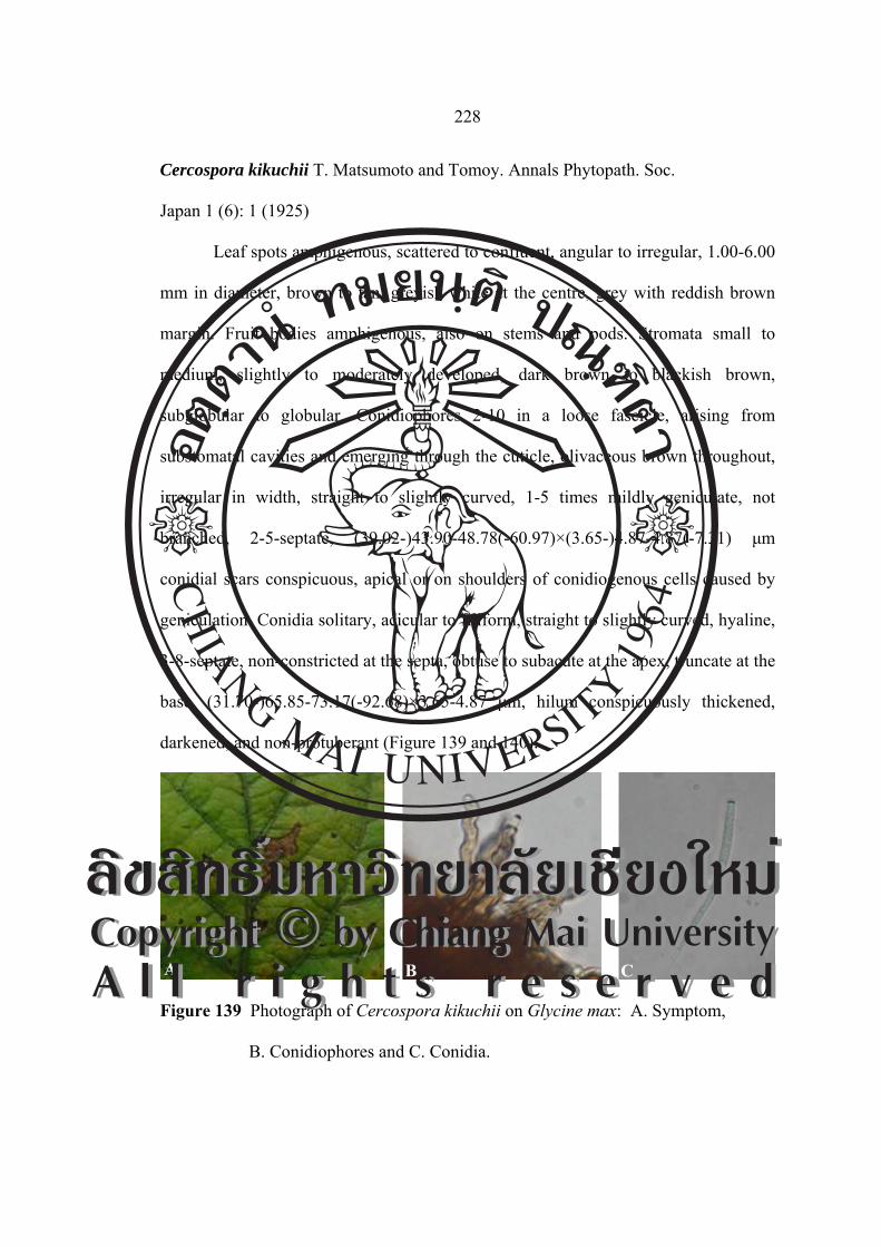
228
Cercospora kikuchii T. Matsumoto and Tomoy. Annals Phytopath. Soc.
Japan 1 (6): 1 (1925)
Leaf spots amphigenous, scattered to confluent, angular to irregular, 1.00-6.00
mm in diameter, brown to tan, greyish white at the centre, grey with reddish brown
margin. Fruit bodies amphigenous, also on stems and pods. Stromata small to
medium, slightly to moderately developed, dark brown to blackish brown,
subglobular to globular. Conidiophores 2-10 in a loose fascicle, arising from
substomatal cavities and emerging through the cuticle, olivaceous brown throughout,
irregular in width, straight to slightly curved, 1-5 times mildly geniculate, not
branched, 2-5-septate, (39.02-)43.90-48.78(-60.97)×(3.65-)4.87-4.87(-7.31) μm
conidial scars conspicuous, apical or on shoulders of conidiogenous cells caused by
geniculation. Conidia solitary, acicular to filiform, straight to slightly curved, hyaline,
3-8-septate, non-constricted at the septa, obtuse to subacute at the apex, truncate at the
base, (31.70-)65.85-73.17(-92.68)×3.65-4.87 μm, hilum conspicuously thickened,
darkened, and non-protuberant (Figure 139 and 140).
Figure 139 Photograph of Cercospora kikuchii on Glycine max: A. Symptom,
B. Conidiophores and C. Conidia.
A B C
ÅÔ¢ÊÔ·¸Ô ìÁËÒÇÔ·ÂÒÅÑÂàªÕ§ãËÁèCopyright by Chiang Mai UniversityA l l r i g h t s r e s e r v e d
ÅÔ¢ÊÔ·¸Ô ìÁËÒÇÔ·ÂÒÅÑÂàªÕ§ãËÁèCopyright by Chiang Mai UniversityA l l r i g h t s r e s e r v e d

229
Figure 140 Drawing of Cercospora kikuchii on Glycine max: A. Conidiophores and
B. Conidia (scale bar = 40 µm).
Material examined: Thailand, Chiang Mai University, Chiang Mai Province. Leaves
of Glycine max, November 21, 2004, JM, CMU MH 061.
Known distribution: Worldwide where the host is cultivated, including China, Japan,
Korea, Taiwan and Thailand (Crous and Braun, 2003).
Notes: The first record of this species from Thailand was made by Sontirat et al.
(1979). Nakata and Takimoto (1928), Park et al. (1978) and Oh and Kwon (1981)
listed this fungus from Korea. The detailed description and illustration for this species
based on Korean collection were provided by Kim and Shin (1998c).
Chupp (1954) recorded several Cercospora species on Glycine and related
host genera. Passalora sojina (Hara) H. D. Shin and U.Braun (≡Cercospora sojina
Hara) is clearly distinguishable. Cercospora canescens Ellis and G. Martin is
morphologically very akin to the present fungus, but different from it by having
condiophores arranged in dense fascicles and amphigenous Fruit bodies.
Pseudocercospora cruenta (Sacc.) Deighton (Deighton, 1976) differs from this
species as follows: Fructification mostly hypophyllous; conidiophores rarely
A
B
ÅÔ¢ÊÔ·¸Ô ìÁËÒÇÔ·ÂÒÅÑÂàªÕ§ãËÁèCopyright by Chiang Mai UniversityA l l r i g h t s r e s e r v e d
ÅÔ¢ÊÔ·¸Ô ìÁËÒÇÔ·ÂÒÅÑÂàªÕ§ãËÁèCopyright by Chiang Mai UniversityA l l r i g h t s r e s e r v e d

230
branched, 0-3-septate, 10.00-70.00×3.00-6.00 μm, conidia obclavate to cylindric,
40.00-150.00×2.50-5.00 μm. P. glycines (Cook) Deighton differs from this fungus in
many respects. Fructification abundantly epiphyllous; conidiophores arranged in very
dense fascicle, 0-1-septate, 10.00-25.00×1.50-3.00 μm, Conidia narrowly obclavate,
30.00-100.00×1.50-2.50 μm.
Phaeoisariopsis griseola (Sacc.) Ferraris, Ann. Mycol. 7: 273. 1909.
≡ Isariopsis griseola Sacc., Michelia 1: 273. 1878.
= Cercospora griseola (Sacc.) Ragunath. and K. Ramakr., J. Madras
Univ. 35-36: 11. (1965-1966) 1968.
= Graphium laxum Ellis, Bull. Torrey Bot. Club 8: 64. 1881. [T: NY].
≡ Isariopsis laxa (Ellis) Sacc., Syll. Fung. 4. 631. 1886.
= Phaeosariopsis laxa (Ellis) S. C. Jong and E. F. Morris, Mycopathol.
Mycol. Appl. 34: 269. 1968.
= Cercospora solimanii Speg. (solimani), Anales Soc. Ci. Argent. 16:
167. 1883. [T: LPS918].
= Cercospora columnaris Ellis and Everh. (columnare), Proc. Acad. Nat.
Sci. Philadelphia 46: 380. 1894. [T: NY].
≡ Pseudocercospora colummaris (Ellis and Everh.) J. M. Yen, in Ten
and Lim, Gard. Bull., Singapore 33: 172. 1980.
= Arthrobotryum puttemansii Henn., Hedwigia 41: 309. 1902.
= Cercospora stuhlmannii Henn., Bot. Jahrb. Syst. 33: 40. 1904.
Leaf spots distinct, angular, irregular when coalescing, usually vein limited,
sometimes also affecting pods, 1.00-5.00 mm in diameter, at first appearing yellowish
ÅÔ¢ÊÔ·¸Ô ìÁËÒÇÔ·ÂÒÅÑÂàªÕ§ãËÁèCopyright by Chiang Mai UniversityA l l r i g h t s r e s e r v e d
ÅÔ¢ÊÔ·¸Ô ìÁËÒÇÔ·ÂÒÅÑÂàªÕ§ãËÁèCopyright by Chiang Mai UniversityA l l r i g h t s r e s e r v e d

231
brown with weak margin and then turning brown to dark brown with well-developed
margin on petioles and young stems, later very light brownish grey arising from
stomatal openings, finally centre becoming dirty greyish brown with blackish dots
caused by heavy fungal fructification. Fruit bodies amphigenous, but mostly
hypophyllous, blackish brown to blackish grey. Stromata well-developed, formed as
brown hyphal aggregations, globose to lacrymoid. Conidiophores 6-18 in a densely
synnematous fascicle, subhyaline to olivaceous brown throughout or paler upwards,
2-7-septate, usually straight, but sometimes apically 1-3 times mildly geniculate or
sinuous, not branched, very long and slender, 170.00-400.00×3.00-5.50 µm, conidial
scars small but conspicuous, apical or on shoulders of conidiogenous cells caused by
geniculation. Conidia solitary, obclavate to cylindric-obclavate, sometimes slightly
constricted at some septa, subobtuse to obtuse at the apex, obconically truncate to
tounded at the base, 26.00-90.00×3.50-7.00 µm, hilum slightly thickened, darkened,
and non-protuberant (Figure 141 and 142).
Figure 141 Photograph of Phaeoisariopsis griseola on Phaseolus vulgaris:
A. Symptom, B. Conidiophores and C. Conidia.
A B C
ÅÔ¢ÊÔ·¸Ô ìÁËÒÇÔ·ÂÒÅÑÂàªÕ§ãËÁèCopyright by Chiang Mai UniversityA l l r i g h t s r e s e r v e d
ÅÔ¢ÊÔ·¸Ô ìÁËÒÇÔ·ÂÒÅÑÂàªÕ§ãËÁèCopyright by Chiang Mai UniversityA l l r i g h t s r e s e r v e d

232
Figure 142 Drawing of Phaeoisariopsis griseola on Phaseolus vulgaris:
A. Conidiophores and B. Conidia (scale bar = 40 µm).
Material examined: Thailand, Wiang Pa Pao, Chiang Rai Province, Phaseolus
vulgaris, November 21, 2004, JM, CMU MH 062.
Known distribution: Worldwide, including Angola, Argentina, Armenia, Australia,
Austria, Bhutan, Brazil, Bulgaria, Burundi, Cameroon, Canada, China, Colombia,
Congo, Costa Rica, Croatia, Cuba, Dominican Republ., Ecuador, E1 Salvador,
Ethiopia, Fiji, France, Georgia, Germany, Ghana, Great Britain, Greece, Guatemala,
Haiti, Hungary, Jamaica, Japan, India, Indonesia, Iran, Ireland, Israel, Italy, Ivory
Coast, Jamaica, Japan, Kenya, Korea, Laos, Latvia, Malawi, Madagascar, Malaysia,
Mauritius, Mexico, Mozambique, Nepal, Netherlands, Netherland Antilles, New
Caledonia, New Caledonia, New Zealand, Nicaragua, Nigeria, Norfolk Island,
Panama, Papua New Guinea, Paraguay, Peru, Philippines, Poland, Portugal, Puerto
Rico, Reunion, Romania, Russia, Rwanda, Saint Helena, Senegal, Sierra, Leone,
Singapore, Slovenia, Solomon Island, Somalia, South Africa, Spain, Sudan,
Suriname, Swaziland, Taiwan, Tanzania, Trinidad and Tobago, Turkey, Uganda,
Ukraine, U.S.A (CT, DE, Eastern states, FL, HI, IN, MA, MD, ME, MI, MS, NC,
A
B
ÅÔ¢ÊÔ·¸Ô ìÁËÒÇÔ·ÂÒÅÑÂàªÕ§ãËÁèCopyright by Chiang Mai UniversityA l l r i g h t s r e s e r v e d
ÅÔ¢ÊÔ·¸Ô ìÁËÒÇÔ·ÂÒÅÑÂàªÕ§ãËÁèCopyright by Chiang Mai UniversityA l l r i g h t s r e s e r v e d

233
NH, NJ, NY, OK, PA, SC, TX, VA, WI), Vanuatu, Venezuela, Vigin Islands,
Yugoslavia, Zambia and Zimbabwe (Crous and Braun, 2003).
Notes: The first record of this species from Thailand was made by Giatgong (1980).
Leaf lesions are the most conspicuous, and start as small, brown or grey spots that
develop into angular, necrotic lesions delimited by vascular strands. Lesions
eventually enlarge, coalesce and cause defoliation of plants. Most crop losses result
from premature defoliation. Circular to elliptical red-brown lesions can develop on
pods, whereas browning of stems has also been attributed to this disease.
Nakata and Takimoto (1928), Park (1958), and Shin and Braun (1993) listed
this fungus from Korea. Detailed description and illustration based on Korean
collections of this species were provided by Kim and Shin (1998b). The leaf spot
disease caused by this fungus occurs in diameterly and is responsible for premature
defoliation in Korea.
There are various concepts of the taxonomy of Phaeoisariopsis. Deighton
(1990) considered the synnematous structure of the conidiomata in this genus to be of
little taxonomic value. He proposed to confine this genus to Phaeoisariopsis griseola
and some allied species with non-geniculate conidiogenous cells, and to reallocate the
“geniculate” species to Passalora.
However, Braun (1995a, 1995b) discussed the taxonomy of Passalora and
Phaeoisariopsis and proposed to maintain the broad concept of the latter genus. The
formation of synnemata is considered to be a good generic feature, whereas the degree
of geniculation of conidiogenous cells is proposed to be used as a characteristic for
the separation of species, since these are gradual transition from non to slightly
geniculate.
ÅÔ¢ÊÔ·¸Ô ìÁËÒÇÔ·ÂÒÅÑÂàªÕ§ãËÁèCopyright by Chiang Mai UniversityA l l r i g h t s r e s e r v e d
ÅÔ¢ÊÔ·¸Ô ìÁËÒÇÔ·ÂÒÅÑÂàªÕ§ãËÁèCopyright by Chiang Mai UniversityA l l r i g h t s r e s e r v e d

234
African and Andean races are known for this pathogen. Deighton (1990) also
refers to records from different hosts.
Phaeoramularia sp.
Leaf spots amphogenous, distinct, scattered to confluent, sometimes vein
limited, angular to irregular, 2.00-11.00 mm in diameter, greyish brown to greyish
white, light brown centre with dark purplish brown to dark brown margins. Fruting
bodies epiphyllous. Stromata lacting to small. Conidiophore 2-6 in loose fascicle,
emerging though stomata, pale olivaceous brown near the basal portion, paler towords
the apex, straight to geniculate sinuous, not branched, 1-4 septate, (36.58-)70.73-
90.24(-136.36)×3.65-4.87 µm, conidial scars small and somewhat conspicuous, apical
or on small shoulders of conidiogenous cells caused by geniculation. Conidia solitary
to catenate, occasionally in branched chains, subcylindric to ellipsoid, straight,
hyaline, 0-1-septate, non-constricted at the septa, obtuse or shortly obconic to
subacute at both ends, (20.87-)26.34-35.98(-39.02)×1.21-2.43 µm, hilum small,
somewhat conspicuously thickened, somewhat darkened, and non-protuberant (Figure
143 and 144).
ÅÔ¢ÊÔ·¸Ô ìÁËÒÇÔ·ÂÒÅÑÂàªÕ§ãËÁèCopyright by Chiang Mai UniversityA l l r i g h t s r e s e r v e d
ÅÔ¢ÊÔ·¸Ô ìÁËÒÇÔ·ÂÒÅÑÂàªÕ§ãËÁèCopyright by Chiang Mai UniversityA l l r i g h t s r e s e r v e d

235
Figure 143 Photograph of Phaeoramularia sp. on Phaseolus vulgaris: A. Symptom,
B. Conidiophores and C. Conidia.
Figure 144 Drawing of Phaeoramularia sp. on Phaseolus vulgaris:
A. Conidiophores and B. Conidia (scale bar = 40 µm).
Material examined: Thailand, Chiang Mai University, Chiang Mai Province. Leaves
of Phaseolus vulgaris, November 21, 2004, JM, CMU MH 063.
Known distribution: Known only from type locality, Thailand.
Notes: This is the first record of Phaeoramularia sp. on Phaseolus vulgaris in
Thailand.
A
B
A
B C
ÅÔ¢ÊÔ·¸Ô ìÁËÒÇÔ·ÂÒÅÑÂàªÕ§ãËÁèCopyright by Chiang Mai UniversityA l l r i g h t s r e s e r v e d
ÅÔ¢ÊÔ·¸Ô ìÁËÒÇÔ·ÂÒÅÑÂàªÕ§ãËÁèCopyright by Chiang Mai UniversityA l l r i g h t s r e s e r v e d

236
Cercospora canescens Ellis and G. Martin, Amer. Naturalist 16: 1003. 1882.
[T: NY].
≡ Cercosporiopsis canescens (Ellis and G. Martin) Miura, Flora of
Manchuria and East Mongolia 3: 529. 1928.
= Cercospora vignicaulis Tehon, Mycologia 29: 436. 1937. [T: ILLS].
(=Cercospora apii s.lat).
Leaf spot suborbicular to irregular, pale brown, tan or dingy center, with a
reddish brown to ferruginous margin, 3.00-14.00 mm in diameter. Fruit bodies
amphigenous, but more abundant on lower surface, sometimes also present on stems,
cotyledon, and dying pods in effuse black to grey patches. Stromata small, 24.39-
39.02 μm. Conidiophores densely fasciculate, pale to medium dark brown, fairly
uniform in colour and width, 1-5-septate, geniculate, rarely branched, truncate at the
apex, (24.39-)31.70-36.58(-53.65)×4.87-7.31 μm, conidial scars conspicuously
thickened. Conidia hyaline, acicular, straight to curved, indistinctly multiseptate,
acute at the apex, truncate at the base with a thickened hilum, 1-6-septate, (19.51-)
24.39-29.26(-43.90)×4.87 μm (Figure 145 and 146).
Figure 145 Photograph of Cercospora canescens on Phaseolus purpureus:
A. Symptom, B. Conidiophores and C. Conidia.
A B C
ÅÔ¢ÊÔ·¸Ô ìÁËÒÇÔ·ÂÒÅÑÂàªÕ§ãËÁèCopyright by Chiang Mai UniversityA l l r i g h t s r e s e r v e d
ÅÔ¢ÊÔ·¸Ô ìÁËÒÇÔ·ÂÒÅÑÂàªÕ§ãËÁèCopyright by Chiang Mai UniversityA l l r i g h t s r e s e r v e d
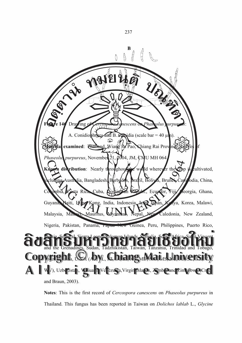
237
Figure 146 Drawing of Cercospora canescens on Phaseolus purpureus:
A. Conidiophores and B. Conidia (scale bar = 40 µm).
Material examined: Thailand, Wiang Pa Pao, Chiang Rai Province. Leaves of
Phaseolus purpureus, November 21, 2004, JM, CMU MH 064.
Known distribution: Nearly throughout the world wherever the crop is cultivated,
including Australia, Bangladesh, Barbados, Brazil, Bolivia, Brunei, Cambodia, China,
Colombia, Costa Rica, Cuba, Dominican Republ., Ecuador, Fiji, Georgia, Ghana,
Guyana, Haiti, Hong Kong, India, Indonesia, Iran, Japan, Kenya, Korea, Malawi,
Malaysia, Malawi, Mauritus, Myanmar, Nepal, New Caledonia, New Zealand,
Nigeria, Pakistan, Panama, Papua New Guinea, Peru, Philippines, Puerto Rico,
Russia, Senegal, Sierra Leone, Solomon Islands, Somalia, South Africa, Saint Vincent
and the Grenadines, Sudan, Tadzhikistan, Taiwan, Tanzania, Trinidad and Tobago,
Togo, Uganda, U.S.A (AL, FL, HI, IA, IL, KS, MD, MO, MS, NC, NJ, SC, TX, VA,
WV), Uzbekistan, Vanuatu, Venezuela,Virgin Islands, Zambia and Zimbabwe (Crous
and Braun, 2003).
Notes: This is the first record of Cercospora canescens on Phaseolus purpureus in
Thailand. This fungus has been reported in Taiwan on Dolichos lablab L., Glycine
A
B
ÅÔ¢ÊÔ·¸Ô ìÁËÒÇÔ·ÂÒÅÑÂàªÕ§ãËÁèCopyright by Chiang Mai UniversityA l l r i g h t s r e s e r v e d
ÅÔ¢ÊÔ·¸Ô ìÁËÒÇÔ·ÂÒÅÑÂàªÕ§ãËÁèCopyright by Chiang Mai UniversityA l l r i g h t s r e s e r v e d

238
max (L.) Merr., Vigna radiata (L.) R. Wilczek (Phaseolus radiatus (L.) Wilcz var.
typicus Prain). Vigna sinensis (L.) Endl. ex Hassk. and V. sinensis (L.) Endl. var.
catiang Nakai. Cercospora canescens was recorded as a member of Korean
cercosporoid fungi from Phaseolus radiatus (Park, 1958; Chung et al., 1977; Kwon
and Oh, 1981) and P. angularis (Shin and Braun, 1993). C. cruenta
(≡ Pseudocercospora cruenta) was reported on P. vulgaris (Nakata and Takimoto,
1928; Park, 1958; Chung et al., 1977) and P. angularis (Nakata and Takimoto, 1928;
Park, 1958) from Korea. C. cruenta (Nakata and Takimoto, 1928; Park, 1958) on
P. angularis is well-known as a causal agent of brown leaf spot in red beans and in
accordance with this species. The identification of the specimen concerned can,
however, not be proven, since it is not preserved. Recently, Kim and Shin (1998c)
provided the detailed description and illustration based on Korean materials from P.
angularis for this fungal species.
C. canescens on P. trilobus from India (Thirumalachar and Govindu, 1957)
possesses narrow conidiophores (2.80-4.20 μm) and conidia (2.40-3.50 μm).
C. canescens on Heylandia sp. described by Bagyanarayana et al. (1991) from Indian
collection is characterized by having conidiophores up to 180.00×4.00-6.00 μm,
solitary or in small fascicles, and conidia 50.00-120.00×2.50-4.00 μm, short and
narrow. The conidiophores and conidia are very variable and the conidiophores are
formed in relatively loose fascicles. The characters of conidiophores and conidia are,
however, generally variable in species of Cercospora s. str.
Deighton (1976) distinguished C. canescens from C. phaseolina by much
longer conidia with multisepta and roundly truncate bases. C. chamaecristae on
Cassia chamaecrista is characterized by having small stromata, 0-2-septate
ÅÔ¢ÊÔ·¸Ô ìÁËÒÇÔ·ÂÒÅÑÂàªÕ§ãËÁèCopyright by Chiang Mai UniversityA l l r i g h t s r e s e r v e d
ÅÔ¢ÊÔ·¸Ô ìÁËÒÇÔ·ÂÒÅÑÂàªÕ§ãËÁèCopyright by Chiang Mai UniversityA l l r i g h t s r e s e r v e d

239
conidiophores, narrowly obclavate, subhyaline to olivaceous conidia, 15.00-
55.00×5.00-7.00 μm (Chupp, 1954; Brown and Morgan-Jones, 1977). Brown and
Morgan-Jones (1977) noted this species to be an atypical Cercospora. Therefore,
Braun (Braum and Melnik, 1997) reallocated this species to Passalora.
Passalora centrosematis N. Pons, U. Braun and Crous, sp. nov.
≡ Cercospora centrosematis Chupp and A. S. Mull. (centrosemae), Bol.
Soc. Venez. Ci. Nat. 8: 40. 1942 (nom. inval.).
Leaf spot distinct, irregular, brown spots with definite dark brown border on
both sides of the leaf. Stromata medium to large. Conidiophores densely fasciculate,
medium dark brown, fairly uniform in colour and width, 1-3-septate, not branched,
truncate at the apex, (29.26-)31.70-56.09(-63.41)×(2.43-)3.65-6.09(-12.19) μm,
conidial scars conspicuously thickened. Conidia subhyaline, obclavate, obclavate-
cylindric, obtuse at the base with a small thickened hilum, 21.95-34.14(-46.34)×2.43-
4.87(-7.31) μm. 2-5-septate (Figure 147 and 148).
Figure 147 Photograph of Passalora centrosematis on Centrosema pubescens:
A. Symptom, B. Conidiophores and C. Conidia.
A B C
ÅÔ¢ÊÔ·¸Ô ìÁËÒÇÔ·ÂÒÅÑÂàªÕ§ãËÁèCopyright by Chiang Mai UniversityA l l r i g h t s r e s e r v e d
ÅÔ¢ÊÔ·¸Ô ìÁËÒÇÔ·ÂÒÅÑÂàªÕ§ãËÁèCopyright by Chiang Mai UniversityA l l r i g h t s r e s e r v e d

240
Figure 148 Drawing of Passalora centrosematis on Centrosema pubescens:
A. Conidiophores and B. Conidia (scale bar = 40 µm).
Material examined: Thailand, Queen Sirikit Botanic Garden, Chiang Mai Province.
Leaves of Centrosema pubescens, November 21, 2004, JM, CMU MH 065.
Known distribution: Philippines, Puerto Rico and Venezuela (Crous and Braun,
2003).
Notes: This is the first record of Cercospora canescens on Passalora centrosematis in
Thailand. A typical Passalora with conspicuous conidiogenous loci and pigmented
conidia. C. centrosematis is an invalid name published without Latin description. A
full English description was published by Chupp (1954).
Cercospora canescens Ellis and G. Martin, Amer. Naturalist 16: 1003. 1882. [T: NY].
≡ Cercosporiopsis canescens (Ellis and G. Martin) Miura, Flora of
Manchuria and East Mongolia 3: 529. 1928.
= Cercospora vignicaulis Tehon, Mycologia 29: 436. 1937. [T: ILLS].
(=Cercospora apii s.lat).
Leaf spot suborbicular to irregular, pale brown, tan or dingy center, with a
reddish brown to ferruginous margin, 3.00-15.00 mm in diameter. Fruit bodies
amphigenous, but more abundant on lower surface, sometimes also present on stems,
A
B
ÅÔ¢ÊÔ·¸Ô ìÁËÒÇÔ·ÂÒÅÑÂàªÕ§ãËÁèCopyright by Chiang Mai UniversityA l l r i g h t s r e s e r v e d
ÅÔ¢ÊÔ·¸Ô ìÁËÒÇÔ·ÂÒÅÑÂàªÕ§ãËÁèCopyright by Chiang Mai UniversityA l l r i g h t s r e s e r v e d

241
cotyledon, and dying pods in effuse black to grey patches. Stromata small, 21.95-
48.78 µm. Conidiophores densely fasciculate, pale to medium dark brown, fairly
uniform in colour and width, 2-3-septate, geniculate, rarely branched, truncate at the
apex, (48.78-)56.09-60.97(-75.60)×(3.65-)4.87-4.87 μm. Conidial scars conspicuously
thickened. Conidia hyaline, acicular, straight to curved, 3-9-septate, acute at the apex,
truncate at the base with a thickened hilum, (42.68-)53.65-80.48(-102.43)×2.43
(-3.65)-3.65 μm (Figure 149 and 150).
Figure 149 Photograph of Cercospora canescens on Vigna unguiculata var.
sesquipedalis: A. Symptom, B. Conidiophores and C. Conidia.
A B C
ÅÔ¢ÊÔ·¸Ô ìÁËÒÇÔ·ÂÒÅÑÂàªÕ§ãËÁèCopyright by Chiang Mai UniversityA l l r i g h t s r e s e r v e d
ÅÔ¢ÊÔ·¸Ô ìÁËÒÇÔ·ÂÒÅÑÂàªÕ§ãËÁèCopyright by Chiang Mai UniversityA l l r i g h t s r e s e r v e d

242
Figure 150 Drawing of Cercospora canescens on
Vigna unguiculata var. sesquipedalis: A. Conidiophores and B. Conidia
(scale bar = 40 µm).
Material examined: Thailand, Wiang Pa Pao, Chiang Rai Province. Leaves of Vigna
unguiculata var. sesquipedalis, November 25, 2005, JM, CMU MH 066.
Known distribution: Worldwide, including Afghanistan, Angola, Argentina,
Australia, Azerbaijan, Banglndesh, Barbados, Bolivia, Brazil, Brunei, Combodia,
Canda, China, Colobia, Cube, Dominican Republ., Egypt, E1 Salvador, Ethiopia, Fiji,
Georgia, Ghana, Grenada, Guatemala, Guyana, Haiti, Honduras, Hong Kong, India,
Indonesia, Iran, Iraq, Italy, Jamaica, Japan, Korea, Liberia, Malawi, Malaysia,
Mauritius, Mexico, Mosambique, Myanmar, Nepal, New Caledonia, Niger, Nigeria,
Pakistan, Panama, Papua, New Guinea, Peru, Philippines, Puerto Rico, Russia,
Rwanda, Saint Lucia, Saint Vincent and the Grenadines, Samoa, Saudi Arabia,
Senegal, Sierra Leone, Singapore, Solomon Islands, Somalia, South Africa, Sri
Lanka, Sudan, Suriname, Taiwan, Tanzania, Thailand, Togo, Tonga, Trinidad,
Tobago, Uganda, U.S.A (AL, AR, DE, Easterm states, FL, GA, HI, IA, IL, KS, LA,
A
B
ÅÔ¢ÊÔ·¸Ô ìÁËÒÇÔ·ÂÒÅÑÂàªÕ§ãËÁèCopyright by Chiang Mai UniversityA l l r i g h t s r e s e r v e d
ÅÔ¢ÊÔ·¸Ô ìÁËÒÇÔ·ÂÒÅÑÂàªÕ§ãËÁèCopyright by Chiang Mai UniversityA l l r i g h t s r e s e r v e d

243
MD, MO, MS, NC, NJ, OK, SC, TN, TX, VA, WI), Venezuela, Virgin Islands,
Zambia and Zimbabwe (Crous and Braun, 2003).
Notes: The first record of this species from Thailand was made by Pitakpraivan et al.
(1994).
Pseudocercospora stizolobii (Syd. and P.Syd.) Deighton, Mycol. Pap. 140: 153. 1976.
≡ Cercospora stizolobii Syd. and P. Syd., Ann. Mycol. 11: 270. 1913.
[T: S, BPI 441666, 441672; S].
= Cercospora lussoniensis Sacc., Ann. Mycol. 12: 314. 1914. [T: B].
= Cercospora mucunae-capitatae Sawada, Rep. Gov. Agric. Res. Inst.
Taiwan 85: 116. 1943 (nom. inval.).
Leaf spots orbicular, 1.00-5.00 mm in diameter, often confluent and become
irregular, tan with dark greyish brown center, surrounded by a definite dark reddish
raised border. Fruit bodies amphigenous, but chiefly hypophyllous. Secondary
mycelium absent. Stromata small or up to 34.00 μm in diameter, dark brown.
Conidiophores 6-15 in a fascicle, subhyaline to pale olivaceous brown, uniform in
colour, substraight to undulate or tortuous sometimes, irregular in width, 0-3
indistinctly septate, rarely branched, rarely once abruptly geniculate, conically
rounded at the apex, 17.22-44.39×2.46-3.93 μm, conidial scars unthickened. Conidia
subhyaline to pale olivaceous, cylindric to cylindric-clavate, longer ones may be
obclavate, straight to mildly curved, indistinctly 1-9 septate, subobtuse to rounded at
the apex, long obconic to obconically truncate at the base, 9.84-51.52×2.46-4.53 μm,
hilum unthickened and inconspicuous (Figure 151 and 152).
ÅÔ¢ÊÔ·¸Ô ìÁËÒÇÔ·ÂÒÅÑÂàªÕ§ãËÁèCopyright by Chiang Mai UniversityA l l r i g h t s r e s e r v e d
ÅÔ¢ÊÔ·¸Ô ìÁËÒÇÔ·ÂÒÅÑÂàªÕ§ãËÁèCopyright by Chiang Mai UniversityA l l r i g h t s r e s e r v e d

244
Figure 151 Photograph of Pseudocercospora stizolobii on Mucuna bracteata:
A. Symptom, B. Conidiophores and C. Conidia.
Figure 152 Drawing of Pseudocercospora stizolobii on Mucuna bracteata:
A. Conidiophores and B. Conidia (scale bar = 40 µm).
Material examined: Thailand, Doi Suthep-Pui National Park, Chiang Mai Province.
Leaves of Mucuna bracteata, November 21, 2004, CN and JM, CMU MH 067.
Known distribution: In diameter spread, including Australia, Barbados, Brazil,
Cambodia, China, Colombia,Cuba, Fiji, Gabon, Ghana, Guatemala, Guyana, Haiti,
Hong Kong, India, Jamaica, Japan, Java, Malawi, Nigeria, Nepal, Panama, Papua
New Guinea, Philippines, Puerto Rico, Sierra Leone, South Africa, Saint Vincent and
the Grenadines, Taiwan, Togo, Trinidad, Tobago, U.S.A (FL, MS, NC), Venezula,
Virgin Islands, Zambia and Zimbabwe (Crous and Braun, 2003).
A B
A B C
ÅÔ¢ÊÔ·¸Ô ìÁËÒÇÔ·ÂÒÅÑÂàªÕ§ãËÁèCopyright by Chiang Mai UniversityA l l r i g h t s r e s e r v e d
ÅÔ¢ÊÔ·¸Ô ìÁËÒÇÔ·ÂÒÅÑÂàªÕ§ãËÁèCopyright by Chiang Mai UniversityA l l r i g h t s r e s e r v e d

245
Notes: This is the first record of Pseudocercospora stizolobii on Mucuna bracteata in
Thailand.
Passalora aenea (Cif.) U. Braun and Crous, comb. nov.
≡ Berteromyces aeneus Cif., Sydowia 8: 267. 1954. [T: IMI 81080,
neotype, selected here].
= Cercospora cassiae Henn., Bull. Herb. Boissier 1: 121. 1893 [T: B,
IMI 88979, K, LEP, W], non Passalora cassiae Syd., 1939.
≡ Cercosporidium cassiae (Henn.) Deighton, Mycol. Pap.
112: 66. 1967.
≡ Phaeoisariopsis cassiae (Henn). Arx, Proc. K. Ned. Akad. Wet., C
86: 43. 1983.
≡ Passalora cassiae (Henn.) Poonam Srivast., J. Living World 1: 114.
1994 (nom. inval. et. illeg.), homonym of Passalora cassiae Syd.,
1939.
≡ Passalora cassiae (Henn.) U. Braun, Mikol. Fitopatol. 30: 6. 1996
(nom. illeg.), homonym of Passalora cassiae Syd., 1939.
= Cercospora cassiicola Roum. (cassiaecola), Fungi sel. exs., No. 4486.
1888 (nom. nud.). [T: LEP].
Leaf spots distinct, subcircular to irregular, brown, 2.00-11.00 mm in
diameter. Stromata well-developed, composed of swollen brown hyphal cells, 55.00-
237.00 µm in diameter. Conidiophores very numerous in adense fascicle, straight to
somewhat sinuous, uniformly pale yellowish brown to brown, with conidial scars
minute but conspicuous, 0-4-septate, (27.50-)35.00-57.50(-75.00)×(2.50-)5.00-10.00
ÅÔ¢ÊÔ·¸Ô ìÁËÒÇÔ·ÂÒÅÑÂàªÕ§ãËÁèCopyright by Chiang Mai UniversityA l l r i g h t s r e s e r v e d
ÅÔ¢ÊÔ·¸Ô ìÁËÒÇÔ·ÂÒÅÑÂàªÕ§ãËÁèCopyright by Chiang Mai UniversityA l l r i g h t s r e s e r v e d

246
(-12.50) µm, apical or on small shoulders of conidiogenous cells caused by
geniculation, concentrated at the apical cell. Conidia solitary, cylindric-obclavate to
obclavate, straight to mildly curved, (20.00-)32.50-47.50(-50.00)×(7.50-)10.00-
(12.50) µm, 2-4-septate, obtuse to subobtuse at the apex, obconically truncate to
subtruncate at the base, hilum slightly thickened, darkened (Figure 153 and 154).
Figure 153 Photograph of Passalora aenea on Cassia angustifolia: A. Symptom,
B. Conidiophores and C. Conidia.
Figure 154 Drawing of Passalora aenea on Cassia angustifolia: A. Conidiophores
and B. Conidia (scale bar = 40 µm).
Material examined: Thailand, Doi Suthep-Pui National Park, Chiang Mai Province.
Leaves of Cassia angustifolia, November 21, 2004, JM, CMU MH 068.
Known distribution: Brazil, Barbados, Colombia, Ethiopia, India, Jamaica,
Tanzania, Uganda, U.S.A (MO) and Zambia (Crous and Braun, 2003).
Notes: This is the first record of Passalora aenea on Cassia angustifolia in Thailand.
A B
A B C
ÅÔ¢ÊÔ·¸Ô ìÁËÒÇÔ·ÂÒÅÑÂàªÕ§ãËÁèCopyright by Chiang Mai UniversityA l l r i g h t s r e s e r v e d
ÅÔ¢ÊÔ·¸Ô ìÁËÒÇÔ·ÂÒÅÑÂàªÕ§ãËÁèCopyright by Chiang Mai UniversityA l l r i g h t s r e s e r v e d

247
Pseudocercospora dalbergiae (S. H. Sun) J. M. Yen, Bull. Soc. Mycol. France
94: 386. (1978) 1979.
≡ Cercospora dalbergiae S. H. Sun, J. Agric. Forest. Taiwan 4: 179.
1955.
≡ Cercoseptoria dalbergiae (S. H. Sun) J. M. Yen, Bull Soc. Mycol.
France 97: 91. 1981.
Leaf spots amphigenous, distinct, subcircular to irregular, 1.00-4.50 mm in
diameter, dark brown, without definite margins. Stromata medium, slightly to well-
developed, olivaceous brown, subglobular to angular, may up to 30.00 µm in
diameter. Conidiophores arranged in a loose to dense fasicle, usually erumpent
throught the cuticle, hyaline to subhyaline or sometimes very pale olivaceous brown
throught, uniform in width, straight to slightly curved, sometime once geniculate,
conidial scars inconspicuous, 0-2-septate, (9.84-)12.30-14.76(-17.22)×2.46-2.46
(-2.95) µm. Conidia long obclavate to cylindric, hyaline to subhyaline, hilum
unthickened, not darkened, (14.76-)27.06-56.58(-73.80)×2.46-3.69 µm, 2-8-septate,
subobtuse to obtuse at the apex, truncate to subtruncate at the base
(Figure 155 and 156).
Figure 155 Photograph of Pseudocercospora dalbergiae on Dalbergia stipulacea:
A. Symptom, B. Conidiophores and C. Conidia.
A B C
ÅÔ¢ÊÔ·¸Ô ìÁËÒÇÔ·ÂÒÅÑÂàªÕ§ãËÁèCopyright by Chiang Mai UniversityA l l r i g h t s r e s e r v e d
ÅÔ¢ÊÔ·¸Ô ìÁËÒÇÔ·ÂÒÅÑÂàªÕ§ãËÁèCopyright by Chiang Mai UniversityA l l r i g h t s r e s e r v e d

248
Figure 156 Drawing of Pseudocercospora dalbergiae on Dalbergia stipulacea:
A. Conidiophores and B. Conidia (scale bar = 40 µm).
Material examined: Thailand, Doi Suthep-Pui National Park, Chiang Mai Province.
Leaves of Dalbergia stipulacea, November 21, 2004, CN and JM, CMU MH 069.
Known distribution: China and Taiwan (Crous and Braun, 2003).
Notes: This is the first record of Pseudocercospora dalbergiae on Dalbergia
stipulacea in Thailand.
Cercospora psophocarpicola J. M. Yen, Bull. Soc. Mycol. France 83: 339. 1967.
Leaf spots amphigenous, scattered, circular to subcircular, white to yellow
with brown margins, 1.00-15.00 mm in diameter. Stromata medium, rudimentary to
slightly develped, irregular, dark brown, 9.75-29.26 µm in diameter. Conidiophores 2-
10 in a divergent fascicle, emerging slightly curved, 1-5 time mildly geniculate, with
distinct conidial scars, apical or on shoulders of conidiogenous cells caused by
geniculation, 2-6-septate, (39.02-)48.78-56.09(-78.04)×4.87-7.31 µm. Conidia
solitary, aciculate to filiform , substraight to moderately curved or even undulate,
hyaline to pale olive-brown, smooth, hilum conspicuously thickened, (51.21-)65.85-
104.87(-163.41)×2.43-4.87 µm, 6-15-septate, acute to obtuse at the apex, truncate at
the base (Figure 157 and 158).
A B
ÅÔ¢ÊÔ·¸Ô ìÁËÒÇÔ·ÂÒÅÑÂàªÕ§ãËÁèCopyright by Chiang Mai UniversityA l l r i g h t s r e s e r v e d
ÅÔ¢ÊÔ·¸Ô ìÁËÒÇÔ·ÂÒÅÑÂàªÕ§ãËÁèCopyright by Chiang Mai UniversityA l l r i g h t s r e s e r v e d

249
Figure 157 Photograph of Cercospora psophocarpicola on
Psophocarpus tetragonolobus: A. Symptom, B. Conidiophores and
C. Conidia.
Figure 158 Drawing of Cercospora psophocarpicola on
Psophocarpus tetragonolobus: A. Conidiophores and B. Conidia
(scale bar = 40 µm).
Material examined: Thailand, Wiang Pa Pao, Chiang Rai Province. Leaves of
Psophocarpus tetragonolobus, November 21, 2004, JM, CMU MH 070.
Known distribution: Myanmar, Nigeria and Singapore (Crous and Braun, 2003).
Notes: This is the first record of Cercospora psophocarpicola on Psophocarpus
tetragonolobus in Thailand. The type material of this species is the same collection as
A
B
A B C
ÅÔ¢ÊÔ·¸Ô ìÁËÒÇÔ·ÂÒÅÑÂàªÕ§ãËÁèCopyright by Chiang Mai UniversityA l l r i g h t s r e s e r v e d
ÅÔ¢ÊÔ·¸Ô ìÁËÒÇÔ·ÂÒÅÑÂàªÕ§ãËÁèCopyright by Chiang Mai UniversityA l l r i g h t s r e s e r v e d

250
the type of C. psophocarpi (mixed collection). There is abundant Fruit bodies of the
latter species, but conidiophores and conidia of C. psophocarpicola have not been
found.
Cercospora canescens Ellis and G. Martin, Amer. Naturalist 16: 1003. 1882.
≡ Cercosporiopsis canescens (Ellis and G. Martin) Miura, Flora of
Manchuria and East Mongolia 3: 529. 1928.
= Cercospora vignicaulis Tehon, Mycologia 29: 436. 1937. [T: ILLS].
(=Cercospora apii s.lat).
Leaf spot suborbicular to irregular, pale brown, tan or dingy center, with a
reddish brown to ferruginous margin, 3.00-15.00 mm in diameter. Fruit bodies
amphigenous, but more abundant on lower surface, sometimes also present on stems,
cotyledon, and dying pods in effuse black to grey patches. Stromata small to medium.
Conidiophores densely fasciculate, pale to medium dark brown, fairly uniform in
colour and width, 1-4-septate, geniculate, rarely branched, truncate at the apex,
(75.60-)109.75-158.53(-195.12)×4.87 μm, conidial scars conspicuously thickened.
Conidia hyaline, acicular, straight to curved, indistinctly multiseptate, acute at the
apex, truncate at the base with a thickened hilum, (34.14-)48.78-131.70
(-180.48)×2.43-2.43(-4.87) μm (Figure 159 and 160).
ÅÔ¢ÊÔ·¸Ô ìÁËÒÇÔ·ÂÒÅÑÂàªÕ§ãËÁèCopyright by Chiang Mai UniversityA l l r i g h t s r e s e r v e d
ÅÔ¢ÊÔ·¸Ô ìÁËÒÇÔ·ÂÒÅÑÂàªÕ§ãËÁèCopyright by Chiang Mai UniversityA l l r i g h t s r e s e r v e d

251
Figure 159 Photograph of Cercospora canescens on Vigna radiata: A. Symptom,
B. Conidiophores and C. Conidia.
Figure 160 Drawing of Cercospora canescens on Vigna radiata: A. Conidiophores
and B. Conidia (scale bar = 40 µm).
Material examined: Thailand, Wiang Pa Pao, Chiang Rai Province. Leaves of Vigna
radiata, October 25, 2005, JM, CMU MH 071.
Known distribution: Nearly throughout the world wherever the crop is cultivated,
including Australia, Bangladesh, Barbados, Brazil, Bolivia, Brunei, Cambodia, China,
Colombia, Costa Rica, Cuba, Dominican Republ., Ecuador, Fiji, Georgia, Ghana,
Guyana, Haiti, Hong Kong, India, Indonesia, Iran, Japan, Kenya, Korea, Malawi,
Malaysia, Malawi Mauritus, Myanmar, Nepal, New Caledonia, New Zealand,
Nigeria, Pakistan, Panama, Papua New Guinea, Peru, Philippines, Puerto Rico,
Russia, Saint Vincent, Senegal, Sierra Leone, Solomon Islands, Somalia, South
A B C
A B
A B C
ÅÔ¢ÊÔ·¸Ô ìÁËÒÇÔ·ÂÒÅÑÂàªÕ§ãËÁèCopyright by Chiang Mai UniversityA l l r i g h t s r e s e r v e d
ÅÔ¢ÊÔ·¸Ô ìÁËÒÇÔ·ÂÒÅÑÂàªÕ§ãËÁèCopyright by Chiang Mai UniversityA l l r i g h t s r e s e r v e d

252
Africa, the Grenadines, Sudan, Tadzhikistan, Taiwan, Tanzania, Thailand, Trinidad
and Tobago, Togo, Uganda, U.S.A (AL, FL, HI, IA, IL, KS, MD, MO, MS, NC, NJ,
SC, TX, VA, WV), Uzbekistan, Vanuatu, Venezuela, Virgin Islands, Zambia and
Zimbabwe (Crous and Braun, 2003).
Notes: The first record of this species from Thailand was made by Niranam refer to
Sontirat et al. (1980). This fungus has been reported in Taiwan on Dolichos lablab L.,
Glycine max (L.) Merr., Vigna radiata (L.)R. Wilczek (Phaseolus radiatus (L.) Wilcz
var. typicus Prain). Vigna sinensis (L.) Endl. ex Hassk. and V. sinensis (L.) Endl. var.
catiang Nakai. Cercospora canescens was recorded as a member of Korean
cercosporoid fungi from Phaseolus radiatus (Park, 1958; Chung et al., 1977; Kwon
and Oh, 1981) and P. angularis (Shin and Braun, 1993). C. cruenta
(≡Pseudocercospora cruenta) was reported on P. vulgaris (Nakata and Takimoto,
1928; Park, 1958; Chung et al., 1977) and P. angularis (Nakata and Takimoto, 1928;
Park, 1958) from Korea. C. cruenta (Nakata and Takimoto, 1928; Park, 1958) on
P. angularis is well-known as a causal agent of brown leaf spot in red beans and in
accordance with this species. The identify of the specimen concerned can, however,
not be proven, since it is not preserved. Kim and Shin (1998c) provided the detailed
description and illustration based on Korean materials from P. angularis for this
fungal species.
C. canescens on P. trilobus from India (Thirumalachar and Govindu, 1957)
possesses narrow conidiophores (2.80-4.20 μm) and conidia (2.40-3.50 μm).
C. canescens on Heylandia sp. described by Bagyanarayana et al. (1991) from Indian
collection is characterized by having conidiophores up to 180.00×4.00-5.50 μm,
solitary or in small fascicles, and conidia 50.00-120.00×2.50-4.00 μm, short and
ÅÔ¢ÊÔ·¸Ô ìÁËÒÇÔ·ÂÒÅÑÂàªÕ§ãËÁèCopyright by Chiang Mai UniversityA l l r i g h t s r e s e r v e d
ÅÔ¢ÊÔ·¸Ô ìÁËÒÇÔ·ÂÒÅÑÂàªÕ§ãËÁèCopyright by Chiang Mai UniversityA l l r i g h t s r e s e r v e d
253
narrow. The conidiophores and conidia are very variable and the conidiophores are
formed in relatively loose fascicles. The characters of conidiophores and conidia are,
however, generally variable in species of Cercospora s. str.
Deighton (1976) distinguished C. canescens from C. phaseolina by much
longer conidia with multisepta and roundly truncate bases. C. chamaecristae on
Cassia chamaecrista is characterized by having small stromata, 0-2-septate
conidiophores, narrowly obclavate, subhyaline to olivaceous conidia, 15.00-
55.00×5.00-7.00 μm (Chupp, 1954; Brown and Morgan Jone, 1977). Brown and
Morgan-Jones (1977) noted this species to be an atypical Cercospora. Therefore,
Braum and Melnik (1997) reallocated this species to Passalora.
Cercospora leucaenae A. N. Shukla and P. C. Sharma, Indian Forester 110: 1066.
1984. [T: IMI 284468].
Leaf spots on the surface scattered to confluent, distinct, circular to
subcircular, brown, centre yellowish white to brown with dark brown margin, 1.00-
7.00 mm in diameter. Stromata moderately developed, emerging through stomata or
erumpent through the cuticle, 17.07-48.78 µm in diameter. Conidiophores pale brown
at the base and paler upwards, arranged in a loose to dense fascicle, with distinct
conidial scars, 3-8-septate, (43.90-)90.24-99.99(-114.63)×(3.65-)4.87-4.87(-6.09) µm.
Conidia solitary, obclavate to cylindric-obclavate, straight to mildly curved, hyaline to
subhyaline, smooth, hilum conspicuously thickened, darkened, (24.39-)60.97-78.04
(-109.75)×2.43-3.65(-4.87) µm, 3-16-septate, obtuse at the apex, subtruncate or
obconically truncate at the base (Figure 161 and 162).
ÅÔ¢ÊÔ·¸Ô ìÁËÒÇÔ·ÂÒÅÑÂàªÕ§ãËÁèCopyright by Chiang Mai UniversityA l l r i g h t s r e s e r v e d
ÅÔ¢ÊÔ·¸Ô ìÁËÒÇÔ·ÂÒÅÑÂàªÕ§ãËÁèCopyright by Chiang Mai UniversityA l l r i g h t s r e s e r v e d

254
Figure 161 Photograph of Cercospora leucaenae on Leucaena leucocephalade:
A. Symptom, B. Conidiophores and C. Conidia.
Figure 162 Drawing of Cercospora leucaenae on Leucaena leucocephalade:
A. Conidiophores and B. Conidia (scale bar = 40 µm).
Material examined: Thailand, Wiang Pa Pao, Chiang Rai Province. Leaves of
Leucaena leucocephalade, November 21, 2004, JM, CMU MH 072.
Known distribution: India (Crous and Braun, 2003).
A
B
A B C
ÅÔ¢ÊÔ·¸Ô ìÁËÒÇÔ·ÂÒÅÑÂàªÕ§ãËÁèCopyright by Chiang Mai UniversityA l l r i g h t s r e s e r v e d
ÅÔ¢ÊÔ·¸Ô ìÁËÒÇÔ·ÂÒÅÑÂàªÕ§ãËÁèCopyright by Chiang Mai UniversityA l l r i g h t s r e s e r v e d

255
Notes: Cercospora leucaenae has been previously recorded in India. This is the first
record of this fungus in Thailand. A true Cercospora s.str. is close to or indentical
with C. apii s. lat.
Family Loganiaceae
Pseudocercospora buddleiae (W. Yamam.) Goh and W. H. Hsieh, Trans. Mycol. Soc.
Republ. China 2: 114. 1987.
≡ Cercospora buddleiae W. Yamam., Trans. Nat. Hist. Soc. Formosa 26:
279. 1936. [T: NTU-PPE].
≡ Pseudocercospora buddleiae (Yamam.) X. J. Liu and Y. L. Guo,
Mycosystema 2: 230. 1989 (comb. superfl.).
Leaf spots distinct, angular to irregular, vein limited, brown, scattered, later
confluent, 1.50-5.50 mm in diameter. Fruit bodies amphigenous. Stomata mainly
epiphyllous, distinct, brown, 12.30-39.36 µm in diameter. External hyphae developed
on under leaf surface. Conidiophores arising from stromata or external hyphae,
brown, densely fascicule, straight, sinuous or slightly curved, frequently geniculate at
head, rough, 2-11-septate, (24.60-)49.20-182.04(-214.02)×2.46-4.92 µm. Conidial
scars denticulate. Conidia solitary, cylindrical to obclavate, rough, straight to mildly
curved, pale olivaceous brown, 2-7-septate, obtuse at apex, obconically truncated at
the basal end, (19.68-) 29.52-54.12(-91.02)×(2.46-)3.69-4.92 µm (Figure 163 and
164).
ÅÔ¢ÊÔ·¸Ô ìÁËÒÇÔ·ÂÒÅÑÂàªÕ§ãËÁèCopyright by Chiang Mai UniversityA l l r i g h t s r e s e r v e d
ÅÔ¢ÊÔ·¸Ô ìÁËÒÇÔ·ÂÒÅÑÂàªÕ§ãËÁèCopyright by Chiang Mai UniversityA l l r i g h t s r e s e r v e d

256
Figure 163 Photograph of Pseudocercospora buddleiae on Buddleja asiatica:
A. Symptom, B. Conidiophores and C. Conidia.
Figure 164 Drawing of Pseudocercospora buddleiae on Buddleja asiatica:
A. Conidiophores and B. Conidia (scale bar = 20 µm).
Material examined: Thailand, Nam Nao National Park, Phetchabun Province. Leaves
of Buddleja asiatica, November 24, 2004, CN and JM, CMU MH 073.
Known distribution: China, India, Japan, Philippines and Taiwan (Crous and Braun,
2003).
A B C
A
B
ÅÔ¢ÊÔ·¸Ô ìÁËÒÇÔ·ÂÒÅÑÂàªÕ§ãËÁèCopyright by Chiang Mai UniversityA l l r i g h t s r e s e r v e d
ÅÔ¢ÊÔ·¸Ô ìÁËÒÇÔ·ÂÒÅÑÂàªÕ§ãËÁèCopyright by Chiang Mai UniversityA l l r i g h t s r e s e r v e d

257
Notes: This is the first report of Pseudocercospora buddleiae on Buddleja asiatica in
Thailand.
Family Lythraceae
Pseudocercospora cupheae (Syd.) U. Braun, Schlechtendalia 2: 14. 1999.
≡ Cercospora cupheae Syde., Ann. Mycol. 37: 428. 1939. [T: Syd.,
Fungi exot. exs. 1241, e.g., HBG].
Leaf spots distinct, circular to subcircular, brown, scattered, later confluent,
angular to irregular, 1.50-5.50 mm in diameter. Stomata distinct, usually
amphigenous, subepidermal, erumpent, brown to olivaceous brown, 12.30-39.36 µm
in diameter. Conidiophores emerging from the upper part of stromata, greenish
brown, densely fasciculate, simple, straight, sinuous or slightly curved, 4-8-septate,
(75.60-)109.75-158.53(-195.12)×4.87 µm, conidial scars inconspicuous. Conidia
solitary, obclavate-cylindric to obclavate, straight to mildly curved, pale olivaceous
brown, 4-10-septate, non-constricted, occationally midly constricted at the septa,
subobtuse to broadly rounded at the apex, obconically truncate at the base, (34.14-)
48.78-131.70(-180.48)×2.43 µm, hilum unthicked, not darkened (Figure 165 and
166).
ÅÔ¢ÊÔ·¸Ô ìÁËÒÇÔ·ÂÒÅÑÂàªÕ§ãËÁèCopyright by Chiang Mai UniversityA l l r i g h t s r e s e r v e d
ÅÔ¢ÊÔ·¸Ô ìÁËÒÇÔ·ÂÒÅÑÂàªÕ§ãËÁèCopyright by Chiang Mai UniversityA l l r i g h t s r e s e r v e d

258
Figure 165 Photograph of Pseudocercospora cupheae on Cuphea hyssopifalia:
A. Symptom, B. Conidiophores and C. Conidia.
Figure 166 Drawing of Pseudocercospora cupheae on Cuphea hyssopifalia:
A. Conidiophores and B. Conidia (scale bar = 40 µm).
Material examined: Thailand, Sirikit Botanic Garden, Chiang Mai Province. Leaves
of Cuphea hyssopifalia, November 21, 2004, JM, CMU MH 074.
Known distribution: Known only Thailand.
Notes: This is the first report of Pseudocercospora cupheae on Cuphea hyssopifalia
in Thailand.
A B C
A
B
ÅÔ¢ÊÔ·¸Ô ìÁËÒÇÔ·ÂÒÅÑÂàªÕ§ãËÁèCopyright by Chiang Mai UniversityA l l r i g h t s r e s e r v e d
ÅÔ¢ÊÔ·¸Ô ìÁËÒÇÔ·ÂÒÅÑÂàªÕ§ãËÁèCopyright by Chiang Mai UniversityA l l r i g h t s r e s e r v e d

259
Family Malvaceae
Pseudocercospora abelmoschi (Ellis and Everh.) Deighton, Mycol. Pap. 140: 138
(1976). (Figs. 2, 3)
≡ Cercospora abelmoschi Ellis and Everh., J. Inst. Jamaica 1: 347
(1893).
= Cercospora hibisci Tracy and Earle, Bull. Torrey Bot. Club 22: 179
(1895).
= Cercospora hibisci-manihotis Henn., Hedwigia 43: 146 (1904).
Leaf spots distinct, circular to subcircular, 1.50-9.00 mm in diameter, brown to
dark brown with blackish brown border. Fruit bodies amphigenous. Stromata small,
12.30-27.06 µm. Conidiophores loose to dense fascicule, brown to pale brown,
straight to geniculate, rough, tortuous, multi septated, (12.30-)19.68-46.74
(-56.58)×2.46-4.92 µm, with unthickened and slightly swelled scars. Conidia solitary,
ovoid to obclavate, straight or mildly curved, rough, pale to pale olivaceous brown, 4-
6 septate, obtuse to acute at the apex, obconically truncated and unthickened at basal
end, (17.22-)22.14-61.50(-66.42)×3.69-4.92 µm (Figure 167 and 168).
Figure 167 Photograph of Pseudocercospora abelmoschi on Hibiscus sp.: A.
Symptom, B. Conidiophores and C. Conidia.
A B C
ÅÔ¢ÊÔ·¸Ô ìÁËÒÇÔ·ÂÒÅÑÂàªÕ§ãËÁèCopyright by Chiang Mai UniversityA l l r i g h t s r e s e r v e d
ÅÔ¢ÊÔ·¸Ô ìÁËÒÇÔ·ÂÒÅÑÂàªÕ§ãËÁèCopyright by Chiang Mai UniversityA l l r i g h t s r e s e r v e d

260
Figure 168 Drawing of Pseudocercospora abelmoschi on Hibiscus sp.: A.
Conidiophores and B. Conidia (scale bar = 20 µm).
Material examined: Thailand, Queen Sirikit Botanic Garden, Chiang Mai Province.
Leaves of Hibiscus sp., November 20, 2004, CN and JM, CMU MH 075.
Known distribution: Worldwide. (Crous and Braun, 2003).
Notes: Hitherto known species in Thailand.
Cercospora malayensis F. Stevens and Solheim, Mycologia 23: 394. 1931.
[T: ILL 14824].
= Cercospora hibisci-esculenti Sawada (nom. nud.) fide Hsieh and Goh
(1990, p. 219).
= Cercospora hibisci-sabdariffae Sawada, Special Publ. Coll. Agric.
Natl. Taiwan Univ. 8: 220. 1959 (nom. nud.).
(= Cercospora apii s. lat.).
Leaf spots orbicular to irregular, 3.00-8.00 mm in diameter, tan to dingy grey,
usually with a purple or red border. Fruit bodies amphigenous. Stromata none or
24.60-73.80 µm in diameter. Conidiophores 5-20 in a compact or divergent fascicle,
pale to dark olivaceous brown, multiseptate, straight, or 2-5 geniculate, not branched,
A
B
ÅÔ¢ÊÔ·¸Ô ìÁËÒÇÔ·ÂÒÅÑÂàªÕ§ãËÁèCopyright by Chiang Mai UniversityA l l r i g h t s r e s e r v e d
ÅÔ¢ÊÔ·¸Ô ìÁËÒÇÔ·ÂÒÅÑÂàªÕ§ãËÁèCopyright by Chiang Mai UniversityA l l r i g h t s r e s e r v e d

261
fairly uniform in colour and width, (4.92-)9.84-19.68(-24.60)×2.46-3.69(-4.92) µm.
Conidia hyaline, acicular, straight to curved, indistinctly multiseptate, subobtuse at the
apex, truncate to subtruncate at the base, (36.90-)36.90-81.18(-100.86)×1.23-2.95
(-3.69)µm (Figure 169 and 170).
Figure 169 Photograph of Cercospora malayensis on Hibiscus rosae-sinensis:
A. Symptom, B. Conidiophores and C. Conidia.
Figure 170 Drawing of Cercospora malayensis on Hibiscus rosae-sinensis:
A. Conidiophores and B. Conidia (scale bar = 40 µm).
Material examined: Thailand, Doi Suthep-Pui National Park, Chiang Mai Province.
Leaves of Hibiscus rosae-sinensis, November 21, 2005, JM, CMU MH 076.
Known distribution: Brazil, Brunei, Cambodia, China, Cuba, El Salvador, Ethiopia,
Fiji, Ghana, India, Indonesia, Iran, Jamaica, Japan, Korea, Malaysia, Mauritius,
Nepal, New Caledonia, Nigeria, Niue, Pakistan, Papua New Guinea, Philippines,
AB
A B C
ÅÔ¢ÊÔ·¸Ô ìÁËÒÇÔ·ÂÒÅÑÂàªÕ§ãËÁèCopyright by Chiang Mai UniversityA l l r i g h t s r e s e r v e d
ÅÔ¢ÊÔ·¸Ô ìÁËÒÇÔ·ÂÒÅÑÂàªÕ§ãËÁèCopyright by Chiang Mai UniversityA l l r i g h t s r e s e r v e d

262
Senegal, Sierra Leone, Singapore, Solomon Islands, South Africa. Sudan, Taiwan,
Tanzania, Togo, Trinidad and Tobago, Uganda, U.S.A (AL, FL, GA, HI, MO, NJ,
OK, SE states, TX, VA), Venezuela, Zambia and Zimbabwe (Crous and Braun,
2003).
Notes: This is the first report of Cercospora malayensis on Hibiscus rosae-sinensis in
Thailand. A true Cercospora s.str. is close to C. apii s. lat., but distinct by having
obclavate-cylindrical conidia with obconically truncate bases.
Park (1967) and Shin and Braun (1993) listed this leaf spot fungus on Althaea
rosea from Korea. Detailed description and illustration based on Korean material of
this species were provided by Kim and Shin (1998a).
According to Yen and Sun (1983), Cercospora althaeigena on A. rosae is
different from this fungus in having coloured obclavate or cylindric to cylindric
conidia. They also recorded Cercospora althaeicola on A. rosae, which differs from
the present species in its indistinct leaf spots and cylindric conidia.
However, subhyaline and obclavate-cylindric have also been observed in the
present Korean collections, and indistinct leaf spots are present on the lower surface
in these specimens. The conidial shape and pigmentation is very variable in
C. althaeina and C. althaeicola are reduced to synonymy with this species.
Family Moraceae
Cercospora broussonetiae Y. L. Guo and L. Xu, Mycosystema 21: 181. 2002.
[T: HMA 81404].
≡ Pseudocercospora broussonetiae (Chupp and Linder, Mycologia )
ÅÔ¢ÊÔ·¸Ô ìÁËÒÇÔ·ÂÒÅÑÂàªÕ§ãËÁèCopyright by Chiang Mai UniversityA l l r i g h t s r e s e r v e d
ÅÔ¢ÊÔ·¸Ô ìÁËÒÇÔ·ÂÒÅÑÂàªÕ§ãËÁèCopyright by Chiang Mai UniversityA l l r i g h t s r e s e r v e d

263
29: 27. 1937. [T: BPI 433862; CUP 39252].
Leaf spots amphigenous, distinct, subcircular to angular, yellowish brown with
blackish brown margins, 3.00-8.00 mm in diameter. Stromata small to medium,
rudimentary to slightly develped, irregular, dark brown, 14.50-41.50 µm in diameter.
Conidiophores pale brown at the base and paler upwards, arranged in a loose to dense
fascicle, with distinct conidial scars, 5-10-septate, (158.53-)241.46-250.46
(-292.68)×4.87-4.87(-6.09) µm. Conidia solitary, obclavate, straight to mildly curved,
hyaline to subhyaline, smooth, hilum conspicuously thickened, darkened, (14.63-)
28.04-48.78(-82.19)×(2.43-)3.65-4.87(-6.09) µm, 3-7-septate, acute to obtuse at the
apex, truncate at the base (Figure 171 and 172).
Figure 171 Photograph of Cercospora broussonetiae on Broussonetia papyrifera:
A. Symptom, B. Conidiophores and C. Conidia.
Figure 172 Drawing of Cercospora broussonetiae on Broussonetia papyrifera:
A. Conidiophores and B. Conidia (scale bar = 40 µm).
A B
A B C
ÅÔ¢ÊÔ·¸Ô ìÁËÒÇÔ·ÂÒÅÑÂàªÕ§ãËÁèCopyright by Chiang Mai UniversityA l l r i g h t s r e s e r v e d
ÅÔ¢ÊÔ·¸Ô ìÁËÒÇÔ·ÂÒÅÑÂàªÕ§ãËÁèCopyright by Chiang Mai UniversityA l l r i g h t s r e s e r v e d

264
Material examined: Thailand, Mae Jo, Chiang Mai Province. Leaves of Broussonetia
papyrifera, September 1, 2005, JM, CMU MH 078.
Known distribution: China (Crous and Braun, 2003).
Notes: This is the first report of Cercospora broussonetiae on
Broussonetia papyrifera in Thailand. A true Cercospora s.str. is close to or identical
with C. apii s. lat.
Cercospora sp.
Leaf spots scattered, rarely confluent, distinct, circular or subcircular to
irregular, small to fairly large, 3.00-5.00 mm in diameter, dark brown, on the upper
surface surrounded by blackish brown margin. Fruit bodies amphigenous. Stromata
slightly to moderately developed, dark brown. Conidiophores 3-6 in a fascicle, pale
brown at the base and paler upwards, straight geniculate in the upper portion, not
branched, 2-5-septate, (46.80-)78.00-150.80(-161.20)×(2.60-)3.90-5.20 μm, conically
narrowed at the apex, conidial scars small but conspicuous, apical or on shoulders of
conidiogenous cells caused by geniculation. Conidia subhyaline to pale olivaceous,
obclavate to cylindric, straight to slightly curved, constricted at the septa, 3-12-
septate, subobtuse to obtuse at the apex, truncate or obconically truncate at the base,
(23.40-)49.40-52.00(-78.00)×(2.60-)5.20-5.20 μm, hilum conspicuously thickened,
darkened and non-protuberant (Figure 173 and 174).
ÅÔ¢ÊÔ·¸Ô ìÁËÒÇÔ·ÂÒÅÑÂàªÕ§ãËÁèCopyright by Chiang Mai UniversityA l l r i g h t s r e s e r v e d
ÅÔ¢ÊÔ·¸Ô ìÁËÒÇÔ·ÂÒÅÑÂàªÕ§ãËÁèCopyright by Chiang Mai UniversityA l l r i g h t s r e s e r v e d

265
Figure 173 Photograph of Cercospora sp. on Ficus religiosa: A. Symptom,
B. Conidiophores and C. Conidia.
Figure 174 Drawing of Cercospora sp. on Ficus religiosa: A. Conidiophores and
B. Conidia (scale bar = 40 µm).
Material examined: Thailand, Chiang Mai University, Chiang Mai Province. Leaves
of Ficus religiosa, December 3, 2005, JM, CMU MH 116.
Known distribution: Known only in Thailand.
Notes: This is the first report of Cercospora sp. on Ficus religiosa in Thailand and in
the world.
A B C
A
B
ÅÔ¢ÊÔ·¸Ô ìÁËÒÇÔ·ÂÒÅÑÂàªÕ§ãËÁèCopyright by Chiang Mai UniversityA l l r i g h t s r e s e r v e d
ÅÔ¢ÊÔ·¸Ô ìÁËÒÇÔ·ÂÒÅÑÂàªÕ§ãËÁèCopyright by Chiang Mai UniversityA l l r i g h t s r e s e r v e d

266
Cercospora rufula Syd., Ann. Mycol. 21: 91. 1923.
Leaf spots subcircular to irregular, 3.00-6.00 mm in diameter, pale brown to
dingy grey at center, with a darker margin. Fruit bodies amphigenous. Stromata
lacking, non fasciculate. Conidiophores pale to very pale olivaceous brown, uniform
in colour, irregular in width, 1-3-septate, rarely geniculate, straight to variously
curved or bent, (29.52-)36.90-51.66(-61.50)×4.92 µm. Conidia pale to very pale
olivaceous, cylindric-obclavate, straight to mildly curved, 3-5-septate, base long
obconically truncate, tip obtuse to conic, 24.60-27.06(-31.98)×2.46 µm (Figure 175
and 176).
Figure 175 Photograph of Cercospora rufula on Ficus sp.: A. Symptom,
B. Conidiophores and C. Conidia.
A B C
ÅÔ¢ÊÔ·¸Ô ìÁËÒÇÔ·ÂÒÅÑÂàªÕ§ãËÁèCopyright by Chiang Mai UniversityA l l r i g h t s r e s e r v e d
ÅÔ¢ÊÔ·¸Ô ìÁËÒÇÔ·ÂÒÅÑÂàªÕ§ãËÁèCopyright by Chiang Mai UniversityA l l r i g h t s r e s e r v e d
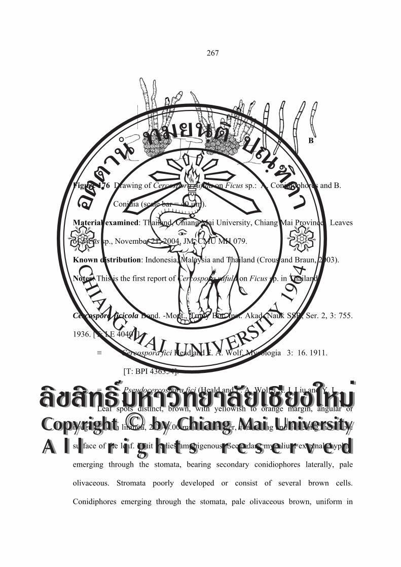
267
Figure 176 Drawing of Cercospora rufula on Ficus sp.: A. Conidiophores and B.
Conidia (scale bar = 40 µm).
Material examined: Thailand, Chiang Mai University, Chiang Mai Province. Leaves
of Ficus sp., November 21, 2004, JM, CMU MH 079.
Known distribution: Indonesia, Malaysia and Thailand (Crous and Braun, 2003).
Notes: This is the first report of Cercospora rufula on Ficus sp. in Thailand
Cercospora ficicola Bond. -Mont., Trudy Bot. Inst. Akad. Nauk SSR, Ser. 2, 3: 755.
1936. [T: LE 40401].
≡ Cercospora fici Head and F. A. Wolf, Mycologia 3: 16. 1911.
[T: BPI 436354].
= Pseudocercospora fici (Heald and F. A. Wolf,) X. J. Liu and Y. L.
Leaf spots distinct, brown, with yellowish to orange margin, angular or
irregular, vein limited, 2.00-8.00 mm in diameter, coalescing and covering the whole
surface of the leaf. Fruit bodies amphigenous. Secondary mycelium external: hyphae
emerging through the stomata, bearing secondary conidiophores laterally, pale
olivaceous. Stromata poorly developed or consist of several brown cells.
Conidiphores emerging through the stomata, pale olivaceous brown, uniform in
AB
ÅÔ¢ÊÔ·¸Ô ìÁËÒÇÔ·ÂÒÅÑÂàªÕ§ãËÁèCopyright by Chiang Mai UniversityA l l r i g h t s r e s e r v e d
ÅÔ¢ÊÔ·¸Ô ìÁËÒÇÔ·ÂÒÅÑÂàªÕ§ãËÁèCopyright by Chiang Mai UniversityA l l r i g h t s r e s e r v e d

268
colour, not branched erect or slightly tortuous, septate and geniculate, conidial scars
conspicuously thickened, 1-4-septate, (19.51-) 21.95-48.78(-60.97)×2.43 μm. Conidia
cylindric, subhyaline to pale olivaceous, straight or slightly curved, 4-11-septate,
rounded at the apex, obconically truncate at the base, catenulate end of the conidia
holding a conspicuously thickened hilum, (58.53-)65.85-109.75(-117.07)×3.65-4.87
μm (Figure 177 and 178).
Figure 177 Photograph of Cercospora ficicola on Ficus rumphii: A. Symptom, B.
Conidiophores and C. Conidia.
Figure 178 Drawing of Cercospora ficicola on Ficus rumphii: A. Conidiophores and
B. Conidia (scale bar = 40 µm).
Material examined: Thailand, Chiang Mai University, Chiang Mai Province. Leaves
of Ficus rumphii, December 3, 2005, JM, CMU MH 080.
Known distribution: Known only Thailand.
A B C
A
B
ÅÔ¢ÊÔ·¸Ô ìÁËÒÇÔ·ÂÒÅÑÂàªÕ§ãËÁèCopyright by Chiang Mai UniversityA l l r i g h t s r e s e r v e d
ÅÔ¢ÊÔ·¸Ô ìÁËÒÇÔ·ÂÒÅÑÂàªÕ§ãËÁèCopyright by Chiang Mai UniversityA l l r i g h t s r e s e r v e d

269
Notes: This is the first report of Pseudocercospora fici on Ficus rumphii in Thailand
and in the world.
Cercospora morina Chupp, a monograph of the fungus genus Cercospora: 400.
[T: CUP-MG 897]
Leaf spots irregular, vein-limited, scattered, extending from margin to midrib,
bounded on the sides by the veins, greyish brown to dingy grey, Fruit bodies slightly
darkening some of the areas, amphigenous, but more abundant on the upper leaf
surface. Stromata small, very dark brown. Conidiophore medium dark brown, paler
toward the tip, uniform in width, multiseptate, straight to curved, not branched, 2-5
geniculate, large spore scar at the subtruncate tip, (34.14-)41.46-78.04(-99.99)×4.87-
6.09 µm. Conidia hyaline, acicular, straight to curved, indistinctly multiseptate, base
truncate, tip acute, (51.21-)65.85-114.63(-170.73)×(3.65-)4.87-4.87 µm (Figure 179
and 180).
Figure 179 Photograph of Cercospora morina on Morus alba: A. Symptom,
B. Conidiophores and C. Conidia.
A B C
ÅÔ¢ÊÔ·¸Ô ìÁËÒÇÔ·ÂÒÅÑÂàªÕ§ãËÁèCopyright by Chiang Mai UniversityA l l r i g h t s r e s e r v e d
ÅÔ¢ÊÔ·¸Ô ìÁËÒÇÔ·ÂÒÅÑÂàªÕ§ãËÁèCopyright by Chiang Mai UniversityA l l r i g h t s r e s e r v e d
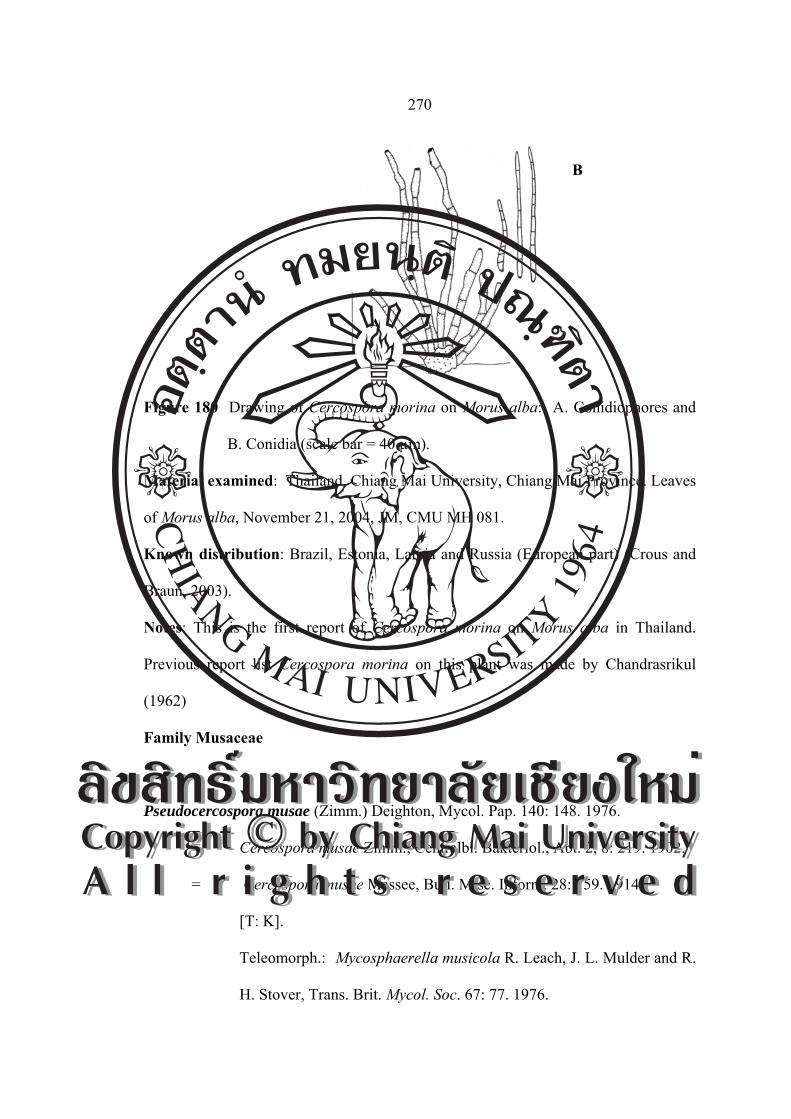
270
Figure 180 Drawing of Cercospora morina on Morus alba: A. Conidiophores and
B. Conidia (scale bar = 40 µm).
Material examined: Thailand, Chiang Mai University, Chiang Mai Province. Leaves
of Morus alba, November 21, 2004, JM, CMU MH 081.
Known distribution: Brazil, Estonia, Latvia and Russia (European part) (Crous and
Braun, 2003).
Notes: This is the first report of Cercospora morina on Morus alba in Thailand.
Previous report list Cercospora morina on this plant was made by Chandrasrikul
(1962)
Family Musaceae
Pseudocercospora musae (Zimm.) Deighton, Mycol. Pap. 140: 148. 1976.
≡ Cercospora musae Zimm., Centralbl. Bakteriol., Abt. 2, 8: 219. 1902.
= Cercospora musae Massee, Bull. Misc. Inform. 28: 159. 1914.
[T: K].
Teleomorph.: Mycosphaerella musicola R. Leach, J. L. Mulder and R.
H. Stover, Trans. Brit. Mycol. Soc. 67: 77. 1976.
A
B
ÅÔ¢ÊÔ·¸Ô ìÁËÒÇÔ·ÂÒÅÑÂàªÕ§ãËÁèCopyright by Chiang Mai UniversityA l l r i g h t s r e s e r v e d
ÅÔ¢ÊÔ·¸Ô ìÁËÒÇÔ·ÂÒÅÑÂàªÕ§ãËÁèCopyright by Chiang Mai UniversityA l l r i g h t s r e s e r v e d

271
≡ Mycosphaerella musicola R. Leach, Trop. Agric. 18: 92. 1941
(nom. nud.).
Leaf spots numerous, irregular, vein-limited but without definite margin, dark
brown, 3.00-8.00 mm in diameter. Stromata amphigenous, intra or subepidermal, then
erumpent, brown, 24.60-44.28 µm in diameter. Conidiophores arranged in a loose
fascicle, arising from stromata, brown, paler towards the apex, sightly to slightly
curved, not branched, 1-3-septate, fasciculate, 12.30-59.40×2.46-4.92 µm, 27.32×2.83
µm on average, with indistinct and unthickened conidial scars. Conidia cylindrical to
obclavate, straight or curved, occasionally sigmoid, with truncate and unthickened
basal end, and with obconical tip, 22.14-98.40×2.95-4.92 µm, smooth, 2-9-septate
(Figure 181 and 182).
Figure 181 Photograph of Pseudocercospora musae on Musa acuminate:
A. Symptom, B. Conidiophores and C. Conidia.
A B C
ÅÔ¢ÊÔ·¸Ô ìÁËÒÇÔ·ÂÒÅÑÂàªÕ§ãËÁèCopyright by Chiang Mai UniversityA l l r i g h t s r e s e r v e d
ÅÔ¢ÊÔ·¸Ô ìÁËÒÇÔ·ÂÒÅÑÂàªÕ§ãËÁèCopyright by Chiang Mai UniversityA l l r i g h t s r e s e r v e d

272
Figure 182 Drawing of Pseudocercospora musae on Musa acuminate:
A. Conidiophores and B. Conidia (scale bar = 40 µm).
Material examined: Thailand, Doi Suthep-Pui National Park, Chiang Mai Province.
Leaves of Musa acuminate, November 21, 2004, JM, CMU MH 082.
Known distribution: In diameterly distributed, including American Samoa, Angola,
Antigua and Barbuda, Argentina, Australia, Bahamas, Barbados, Belau, Belau,
Belize, Bolivia, Brazil, Brunei, Bhutan, Cambodia, Cameroon, Cape Verde, Cayman
Islands, China, Colombia, Congo, Cook Islands, Costa Rica, Cuba, Dominican
Republ., Ecuador, Egypt, E1 Salvador, Ethiopia, Fiji, France, Guiana, French
Polynesia, Guiana, French Polynesia, Gabon, Ghana, Grenada, Guadeloupe, Guam,
Guatemala, Guinea, Guinea-Bisiau, Guyana, Haiti, Honduras, Hong Kong, India,
Indonesia, Ivory Coast, Jamaica, Kenva, Kiribati, Laos,Madagascar, Malawi,
Malaysia, Martinique, Mauritius, Mexico, Micronesia, Montserrat, Mozambique,
Nepal, New Caledonia, Nicaragua, Nigeria, Niue, Norfolk Island, Panama, Papua
New Guinea, Peru, Philippines, Puerto Rico, Saint Kitts and Nevis, Saint Lucia, Saint
Vincent and the Grenadines, Samoa, São Toné e Príncipe, Sierra Leone, Solomon
Islands, Somalia, South Africa, Sri Lanka, Suriname, Taiwan, Tanzania, Togo, Tonga,
A
B
ÅÔ¢ÊÔ·¸Ô ìÁËÒÇÔ·ÂÒÅÑÂàªÕ§ãËÁèCopyright by Chiang Mai UniversityA l l r i g h t s r e s e r v e d
ÅÔ¢ÊÔ·¸Ô ìÁËÒÇÔ·ÂÒÅÑÂàªÕ§ãËÁèCopyright by Chiang Mai UniversityA l l r i g h t s r e s e r v e d

273
Trinidad and Tobago, Tuvalu, Uganda, U.S.A (FL, HI), Vanuatu, Venezuela,
Vietnam, Wallis and Futuna Islands,Yemen, Zambia and Zimbabwe (Crous and
Braun, 2003).
Notes: This is the first report of Pseudocercospora musae on Musa acuminata in
Thailand.
Family Myricaceae
Cercospora myriactidis Sawada, Rep. Gov. Agric. Res. Inst. Taiwan 85: 117. 1943
(nom. inval.). [T: NTU-PPE, herb. Sawada].
Leaf spots orbicular, often zonate, 0.50-5.00 mm in diameter, pale brown to
dingy grey at center, with a darker margin. Fruit bodies amphigenous. Stromata
lacking or a few darker brown cells. Conidiophores 2-14 in a divergent fascicle,
medium brown, paler towards the apex, uniform in width, straight, 1-3-septate, not
branched, truncate at the apex, (13.00-)15.60-24.70(-31.20)×2.60-5.20 μm, conidia
hyaline, acicular, straight to curved, 2-5-septate, acute to subacute at the apex,
truncate at the base, (23.40-)37.70-53.30(-57.20)×2.60-5.20 μm (Figure 183 and 184).
Figure 183 Photograph of Cercospora myriactidis on Myrica esculenta:
A. Symptom, B. Conidiophores and C. Conidia.
A B C
ÅÔ¢ÊÔ·¸Ô ìÁËÒÇÔ·ÂÒÅÑÂàªÕ§ãËÁèCopyright by Chiang Mai UniversityA l l r i g h t s r e s e r v e d
ÅÔ¢ÊÔ·¸Ô ìÁËÒÇÔ·ÂÒÅÑÂàªÕ§ãËÁèCopyright by Chiang Mai UniversityA l l r i g h t s r e s e r v e d
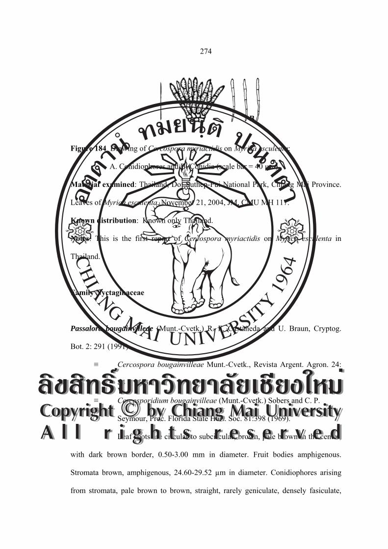
274
Figure 184 Drawing of Cercospora myriactidis on Myrica esculenta:
A. Conidiophores and B. Conidia (scale bar = 40 µm).
Material examined: Thailand, Doi Suthep-Pui National Park, Chiang Mai Province.
Leaves of Myrica esculenta, November 21, 2004, JM, CMU MH 117.
Known distribution: Known only Thailand.
Notes: This is the first report of Cercospora myriactidis on Myrica esculenta in
Thailand.
Family Nyctaginaceae
Passalora bougainvilleae (Munt.-Cvetk.) R. F. Castañeda and U. Braun, Cryptog.
Bot. 2: 291 (1991)
≡ Cercospora bougainvilleae Munt.-Cvetk., Revista Argent. Agron. 24:
84 (1957).
≡ Cercosporidium bougainvilleae (Munt.-Cvetk.) Sobers and C. P.
Seymour, Proc. Florida State Hort. Soc. 81:398 (1969).
Leaf spots are circular to subcircular, brown, pale brown in the center,
with dark brown border, 0.50-3.00 mm in diameter. Fruit bodies amphigenous.
Stromata brown, amphigenous, 24.60-29.52 µm in diameter. Conidiophores arising
from stromata, pale brown to brown, straight, rarely geniculate, densely fasiculate,
AB
ÅÔ¢ÊÔ·¸Ô ìÁËÒÇÔ·ÂÒÅÑÂàªÕ§ãËÁèCopyright by Chiang Mai UniversityA l l r i g h t s r e s e r v e d
ÅÔ¢ÊÔ·¸Ô ìÁËÒÇÔ·ÂÒÅÑÂàªÕ§ãËÁèCopyright by Chiang Mai UniversityA l l r i g h t s r e s e r v e d

275
with distinct and thickened conidial scars, 1-4-septate, (17.22-)27.06-56.58
(-86.10)×4.92 µm. Conidia ovoid to obclavate, brown, straight, with thickened and
truncate basal end, dull head, (31.98-)71.34-93.48(-98.40)×2.46(-4.92)-4.92 µm, 1-4-
septate (Figure 185 and 186).
Figure 185 Photograph of Passalora bugainvilleae on Bougainvillea spectabilis:
A. Symptom, B. Conidiophores and C. Conidia.
Figure 186 Drawing of Passalora bugainvilleae on Bougainvillea spectabilis:
A. Conidiophores and B. Conidia (scale bar = 40 µm).
Material examined: Thailand, Doi Suthep-Pui National Park, Chiang Mai Province.
Leaves of Bougainvillea spectabilis, November 21, 2004, CN and JM, CMU MH 083.
Known distribution: Argentina, Brasil, Brunei, China, Cuba, El Salvador, India,
Indonesia, Jamaica, Japan, USA and Venezuela (Crous and Braun 2003).
A B C
AB
ÅÔ¢ÊÔ·¸Ô ìÁËÒÇÔ·ÂÒÅÑÂàªÕ§ãËÁèCopyright by Chiang Mai UniversityA l l r i g h t s r e s e r v e d
ÅÔ¢ÊÔ·¸Ô ìÁËÒÇÔ·ÂÒÅÑÂàªÕ§ãËÁèCopyright by Chiang Mai UniversityA l l r i g h t s r e s e r v e d

276
Notes: This is the first record of the circular leaf spot of Bougainvillea spectabilis
caused by Passalora bugainvilleae from Thailand.
Cercospora bougainvilleae Munt. -Cvetk., Revista Argent. Agron. 24: 84. 1957.
≡ Passalora bugainvilleae (Munt. -Cvetk.) R.F. Castañeda and U.
Braum, Cryptog. Bot. 2: 291. 1991.
≡ Cercosporidium bougainvilleae (Munt. -Cvetk.) Sobers and C. P.
Seymour, Proc. Florida State Hort. Soc. 81: 398 (1968) 1969.
Leaf spots circular to angular, 2.00-8.00 mm in diameter, pale to medium dark
brown, the older spots with a pale center or with concentric rings and a dark line
margin; Fruit bodies chiefly epiphyllous, stromata a few cells to 24.00 μm in
diameter. dark brown, fascicles 3-20 diverging stalks, conidiophores pale to medium
dark brown, paler and sometimes more narrow toward the tip, 3-8-septate, slightly
branced, 0-2 geniculate or undulate, straight to curved, medium scar at the
subtruncate tip, (41.60-)44.20-104.00(-137.80)×(2.60-)3.90-5.20(-7.80) μm. Conidia
hyaline, acicular to obclavate, straight to mildly curved, 2-8-septate, base truncate to
long obconically truncate, tip subacute to subobtuse, (18.20-)23.40-57.20
(-72.80)×5.20-6.50 μm (Figure 187 and 188).
ÅÔ¢ÊÔ·¸Ô ìÁËÒÇÔ·ÂÒÅÑÂàªÕ§ãËÁèCopyright by Chiang Mai UniversityA l l r i g h t s r e s e r v e d
ÅÔ¢ÊÔ·¸Ô ìÁËÒÇÔ·ÂÒÅÑÂàªÕ§ãËÁèCopyright by Chiang Mai UniversityA l l r i g h t s r e s e r v e d

277
Figure 187 Photograph of Cercospora bougainvilleae on Bougainvillea spectabilis:
A. Symptom, B. Conidiophores and C. Conidia.
Figure 188 Drawing of Cercospora bougainvilleae on Bougainvillea spectabilis:
A. Conidiophores and B. Conidia (scale bar = 40 µm).
Material examined: Thailand, Doi Suthep-Pui National Park, Chiang Mai Province.
Leaves of Bougainvillea spectabilis, November 21, 2004, JM, CMU MH 084.
Known distribution: China and India (Crous and Braun, 2003).
Notes: This is the first record of Bougainvillea spectabilis caused by Passalora
bugainvilleae from Thailand.
AB
A B C
ÅÔ¢ÊÔ·¸Ô ìÁËÒÇÔ·ÂÒÅÑÂàªÕ§ãËÁèCopyright by Chiang Mai UniversityA l l r i g h t s r e s e r v e d
ÅÔ¢ÊÔ·¸Ô ìÁËÒÇÔ·ÂÒÅÑÂàªÕ§ãËÁèCopyright by Chiang Mai UniversityA l l r i g h t s r e s e r v e d

278
Family Nymphaeaceae
Cercosporella sp.
Leaf spots are subcircular to irregular, usually vein-limited, brown to dark
brown, centre with pale yellowish brown margins, 1.00-4.00 mm in diameter. Fruit
bodies amphigenous, but abundantly epiphyllous. Stromata medium, well-developed,
subglobular to globular, dark brtown. Conidiophores arising through the cuticle,
hyaline to brown, straight to slightly curved, densely fasiculate and solitary, with
indistinct conidial scars, 1-2-septate, 4.92-24.60×2.46-4.92 µm. Conidia aciculate to
filiform, subhyaline to pale olive-brown, straight, smooth, with unthickened hilum,
36.90-100.86×1.23-3.69 µm, 2-7-septate, with truncate and thin basal end (Figure 189
and 190).
Figure 189 Photograph of Cercosporella sp. on Nymphaea stellata: A. Symptom,
B. Conidiophores and C. Conidia.
A B C
ÅÔ¢ÊÔ·¸Ô ìÁËÒÇÔ·ÂÒÅÑÂàªÕ§ãËÁèCopyright by Chiang Mai UniversityA l l r i g h t s r e s e r v e d
ÅÔ¢ÊÔ·¸Ô ìÁËÒÇÔ·ÂÒÅÑÂàªÕ§ãËÁèCopyright by Chiang Mai UniversityA l l r i g h t s r e s e r v e d

279
Figure 190 Drawing of Cercosporella sp. on Nymphaea stellata: A. Conidiophores
and B. Conidia (scale bar = 40 µm).
Material examined: Thailand, Doi Suthep-Pui National Park, Chiang Mai Province.
Leaves of Nymphaea stellata, November 21, 2004, JM, CMU MH 085.
Known distribution: Known only Thailand.
Notes: This is the first record of Nymphaea stellata caused by Cercosporella sp. from
Thailand.
Family Oleandraceae
Pseudocercospora phyllitidis (H. H. Hume) U. Braun and Crous, Mycosphaerella and
its anamorphs: 321 (2003).
≡ Cercospora phyllitidis H. H. Hume, Bull. Torrey Bot Club 27: 577
(1900).
Leaf spots are subcircular to irregular, usually vein-limited, pale brown to
brown with pale yellowish halo, 1.00-4.00 mm in diameter. Fruit bodies
amphigenous. Stromata epiphyllous, 24.00-74.00 µm in diameter, brown. External
AB
ÅÔ¢ÊÔ·¸Ô ìÁËÒÇÔ·ÂÒÅÑÂàªÕ§ãËÁèCopyright by Chiang Mai UniversityA l l r i g h t s r e s e r v e d
ÅÔ¢ÊÔ·¸Ô ìÁËÒÇÔ·ÂÒÅÑÂàªÕ§ãËÁèCopyright by Chiang Mai UniversityA l l r i g h t s r e s e r v e d

280
hyphae developed. Conidiophores arising from stromata or external hyphae, pale
brown, geniculate to slightly curved, densely fasciculate or solitary, with indistinct
conidial scars, 1-2 septate, 4.00-34.00×2.00-3.00 µm. Conidia acicular to obclavate,
pale colored to pale olivaceous brown, straight to mildly curved, smooth, with
unthickened and truncate basal end, 36.00-118.00×1.80-3.70 µm, 2-10-septate (Figure
191 and 192).
Figure 191 Photograph of Pseudocercospora phyllitidis on Nephrolepis biserrata:
A. Symptom, B. Conidiophores and C. Conidia.
Figure 192 Drawing of Pseudocercospora phyllitidis on Nephrolepis biserrata:
A. Conidiophores and B. Conidia (scale bar = 20 µm).
Material examined: Thailand, Doi Suthep-Pui National Park, Chiang Mai Province.
Leaves of Nephrolepis biserrata, November 21, 2004, JM, CMU MH 086.
A B C
A
B
ÅÔ¢ÊÔ·¸Ô ìÁËÒÇÔ·ÂÒÅÑÂàªÕ§ãËÁèCopyright by Chiang Mai UniversityA l l r i g h t s r e s e r v e d
ÅÔ¢ÊÔ·¸Ô ìÁËÒÇÔ·ÂÒÅÑÂàªÕ§ãËÁèCopyright by Chiang Mai UniversityA l l r i g h t s r e s e r v e d

281
Known distribution: Canada, Great Britain, India, Puerto Rico, USA and Virgin
Islands (Crous and Braun, 2003) .
Notes: This is the first record of Nephrolepis biserrata caused by Pseudocercospora
phyllitidis from Thailand.
Family Onagraceae
Cercospora fuchsiae Chupp and A. S. Mull., Bol. Soc. Venez. Ci. Nat. 8: 45. 1942
(nom.inval.). [T: CUP-VZ3540]. (=Cercospora apii s.lat.).
Leaf spots circular to angular, 2.00-8.00 mm in diameter, pale to medium dark
brown, the older spots with a pale center or with concentric rings and a drak line
margin, Fruit bodies chiefly epiphyllous. Stromata dark brown. Conidiophore pale to
medium dark brown, paler and sometimes more narrow towards the tip, plainly
multiseptate, slightly branched, 0-2 geniculate or undulate, straight to curved, medium
spre scar at the subtruncate tip, (14.76-)24.60-49.20(-68.88)×(3.69-)4.92-4.92 μm.
Conidia hyaline, acicular to obclavate, straight to mildly curved, indistinctly
multiseptate, base truncate to long obconically truncate, tip subacute to subobtuse,
(22.14-)36.90-61.50(-73.80)×2.46-4.92 μm (Figure 193 and 194).
ÅÔ¢ÊÔ·¸Ô ìÁËÒÇÔ·ÂÒÅÑÂàªÕ§ãËÁèCopyright by Chiang Mai UniversityA l l r i g h t s r e s e r v e d
ÅÔ¢ÊÔ·¸Ô ìÁËÒÇÔ·ÂÒÅÑÂàªÕ§ãËÁèCopyright by Chiang Mai UniversityA l l r i g h t s r e s e r v e d
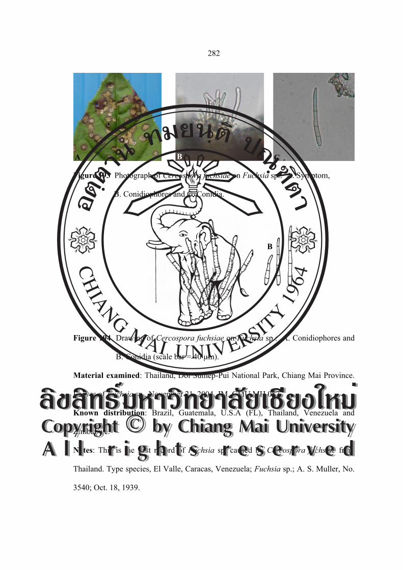
282
Figure 193 Photograph of Cercospora fuchsiae on Fuchsia sp.: A. Symptom,
B. Conidiophores and C. Conidia.
Figure 194 Drawing of Cercospora fuchsiae on Fuchsia sp.: A. Conidiophores and
B. Conidia (scale bar = 40 µm).
Material examined: Thailand, Doi Suthep-Pui National Park, Chiang Mai Province.
Leaves of Fuchsia sp., November 21, 2004, JM, CMU MH 087.
Known distribution: Brazil, Guatemala, U.S.A (FL), Thailand, Venezuela and
Zimbabwe.
Notes: This is the first record of Fuchsia sp. caused by Cercospora fuchsiae from
Thailand. Type species, El Valle, Caracas, Venezuela; Fuchsia sp.; A. S. Muller, No.
3540; Oct. 18, 1939.
A B C
AB
ÅÔ¢ÊÔ·¸Ô ìÁËÒÇÔ·ÂÒÅÑÂàªÕ§ãËÁèCopyright by Chiang Mai UniversityA l l r i g h t s r e s e r v e d
ÅÔ¢ÊÔ·¸Ô ìÁËÒÇÔ·ÂÒÅÑÂàªÕ§ãËÁèCopyright by Chiang Mai UniversityA l l r i g h t s r e s e r v e d
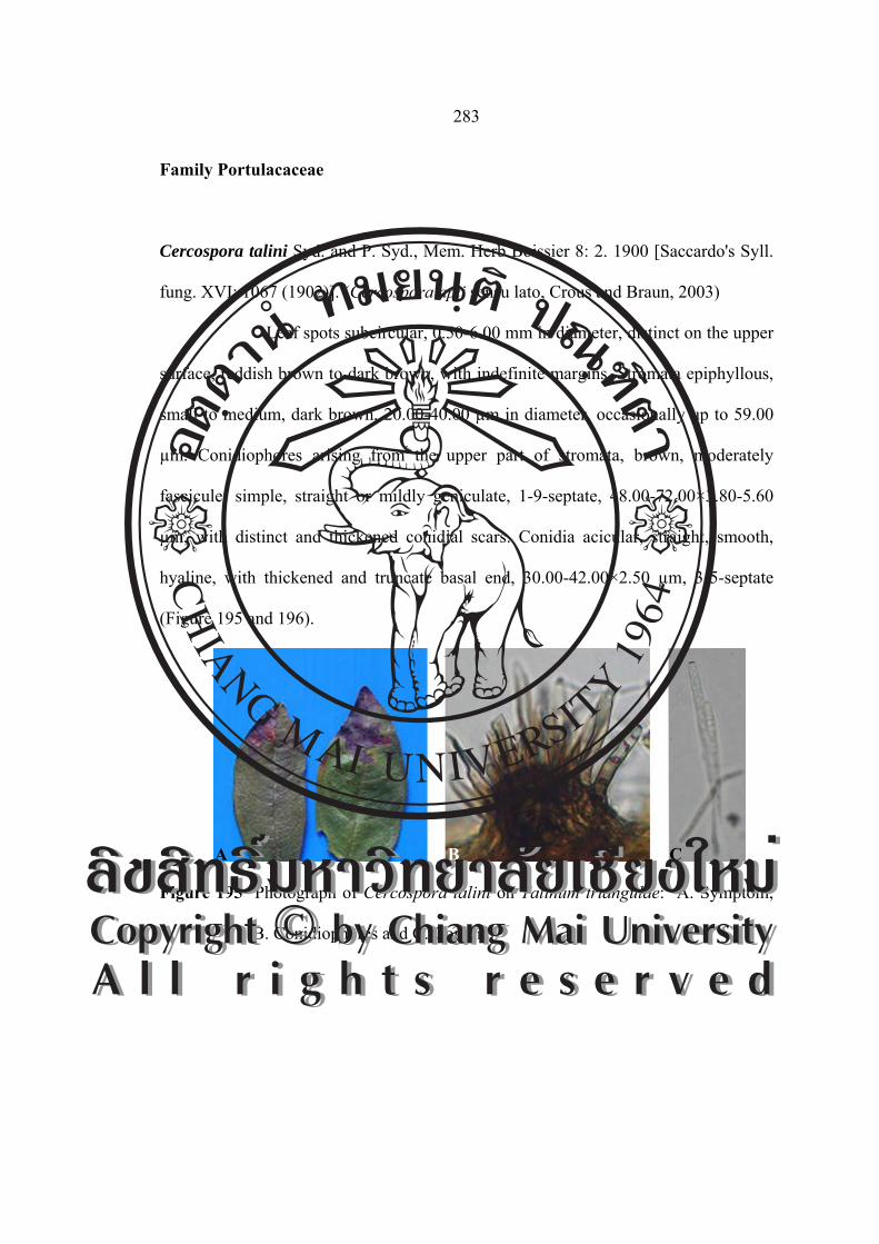
283
Family Portulacaceae
Cercospora talini Syd. and P. Syd., Mem. Herb Boissier 8: 2. 1900 [Saccardo's Syll.
fung. XVI: 1067 (1902)]. (Cercospora apii sensu lato, Crous and Braun, 2003)
Leaf spots subcircular, 0.50-6.00 mm in diameter, distinct on the upper
surface, reddish brown to dark brown, with indefinite margins. Stromata epiphyllous,
small to medium, dark brown, 20.00-40.00 µm in diameter, occasionally up to 59.00
µm. Conidiophores arising from the upper part of stromata, brown, moderately
fascicule, simple, straight or mildly geniculate, 1-9-septate, 48.00-72.00×3.80-5.60
µm, with distinct and thickened conidial scars. Conidia acicular, straight, smooth,
hyaline, with thickened and truncate basal end, 30.00-42.00×2.50 µm, 3-5-septate
(Figure 195 and 196).
Figure 195 Photograph of Cercospora talini on Talinum triangulae: A. Symptom,
B. Conidiophores and C. Conidia.
A B C ÅÔ¢ÊÔ·¸Ô ìÁËÒÇÔ·ÂÒÅÑÂàªÕ§ãËÁèCopyright by Chiang Mai UniversityA l l r i g h t s r e s e r v e d
ÅÔ¢ÊÔ·¸Ô ìÁËÒÇÔ·ÂÒÅÑÂàªÕ§ãËÁèCopyright by Chiang Mai UniversityA l l r i g h t s r e s e r v e d

284
Figure 196 Drawing of Cercospora talini on Talinum triangulae: A. Conidiophores
and B. Conidia (scale bar = 40 µm).
Material examined: Thailand, Doi Suthep-Pui National Park, Chiang Mai Province.
Leaves of Talinum triangulae, November 21, 2004, CN and JM, CMU MH 088.
Known distribution: Argentina and Venezuela (Chupp, 1954).
Notes: Present species are first report from Asian country in this report.
Family Rosaceae
Cercospora fagariae Lobik, Bolezni Rast. 17: 195. 1928 (nom. dub.). [T: LE].
Leaf spots circular, 1.00-3.00 mm in diameter, white center, dark purple
border, sometimes indistinct on lower surface, Fruit bodies amphigenous. Stromata a
few dark cells to 50 μm in diameter, dark brown to almost black. Conidiophore
fascicles 2-20 stalks, pale olivaceous brown, almost hyaline tips, 2-4-septate, not
branched, curved, undulate or 1-3 abruptly geniculate, large spore scar at subtruncate
tip, (12.30-)17.22-31.98(-46.74)×2.46-4.92 μm. Conidia hyaline to subhyaline,
acicular to obclavate, shorter ones may be cylindric, straight to mildly curved,
AB
ÅÔ¢ÊÔ·¸Ô ìÁËÒÇÔ·ÂÒÅÑÂàªÕ§ãËÁèCopyright by Chiang Mai UniversityA l l r i g h t s r e s e r v e d
ÅÔ¢ÊÔ·¸Ô ìÁËÒÇÔ·ÂÒÅÑÂàªÕ§ãËÁèCopyright by Chiang Mai UniversityA l l r i g h t s r e s e r v e d

285
indistinctly multiseptate, base truncate to long obconically truncate, tip subacute to
subobtuse, (14.76-)24.60-31.98(-49.20)×2.46 μm (Figure 197 and 198).
Figure 197 Photograph of Cercospora fagariae on Fragaria sp.: A. Symptom,
B. Conidiophores and C. Conidia.
Figure 198 Drawing of Cercospora fagariae on Fragaria sp.: A. Conidiophores and
B. Conidia (scale bar = 40 µm).
Material examined: Thailand, Chiang Mai University, Chiang Mai Province. Leaves
of Fragaria sp., October 8, 2004, JM, CMU MH 089.
Known distribution: Azerbaijan, Ethiopia, Kazakhstan, Malawi, Russia (European
part), Thailand and U.S.A (LA).
Notes: This is the first record of Fragaria sp. caused by Cercospora fagariae from
Thailand. The description of Chupp (1954), based on American material, dose not
agree with that of Lobik (1928). Braun and Melnik (1997) only found material of
A B C
A B A
ÅÔ¢ÊÔ·¸Ô ìÁËÒÇÔ·ÂÒÅÑÂàªÕ§ãËÁèCopyright by Chiang Mai UniversityA l l r i g h t s r e s e r v e d
ÅÔ¢ÊÔ·¸Ô ìÁËÒÇÔ·ÂÒÅÑÂàªÕ§ãËÁèCopyright by Chiang Mai UniversityA l l r i g h t s r e s e r v e d

286
Ramularia grevilleana on the type. See also C. vexans for differences between the
two species on Fragaria. They have been unable to procure the Lovik type to
compare with the Plakidas collection, but Lobik’s description fits very closely the
Louisiana fungus.
Cercospora rosicola
Leaf spots circular, 0.50-3.00 mm in diameter, white center, dark purple
border, sometimes indistinct on lower surface, Fruit bodies amphigenous; stromata a
few dark cells to 60.00 μm in diameter, dark brown to almost black. Conidiophore
fascicles 5-15 stalks, subhyaline, almost hyaline tips, uniforme in diameter, 1-4-
septate, not branced, curved, 24.39-77.87×2.43 μm. Conidia hyaline to subhyaline,
obclavate to cylindric, straight to mildly curved, 3-4-septate, base truncate to long
obconically truncate, tip subacute to subobtuse, 24.39-114.63×2.43-7.31 μm (Figure
199 and 200).
Figure 199 Photograph of Cercospora rosicola on Rosa sp.: A. Symptom,
B. Conidiophores and C. Conidia.
A B C
ÅÔ¢ÊÔ·¸Ô ìÁËÒÇÔ·ÂÒÅÑÂàªÕ§ãËÁèCopyright by Chiang Mai UniversityA l l r i g h t s r e s e r v e d
ÅÔ¢ÊÔ·¸Ô ìÁËÒÇÔ·ÂÒÅÑÂàªÕ§ãËÁèCopyright by Chiang Mai UniversityA l l r i g h t s r e s e r v e d

287
Figure 200 Drawing of Cercospora rosicola on Rosa sp.: A. Conidiophores and
B. Conidia (scale bar = 40 µm).
Material examined: Thailand, Wiang Pa Pao, Chiang Rai Province. Leaves of
Rosa sp., December 3, 2004, JM, CMU MH 090.
Known distribution: Iran (Crous and Braun, 2003).
Notes: This is the first record of Rosa sp. caused by Cercospora rosicola from
Thailand.
Family Rubiaceae
Cercopora coffeicola Berk. and Cooke, Grevillea 9: 99. 1881. [T: K].
= Cercospora coffeae Zimm., Ber. Land-Forstw.Deutsch-Ostafr.
2: 35. 1904.
= Cercospora herrerana Farneti, Atti Ist. Bot. Univ. Pavia, Ser. 2,
9: 37. 1911.
Teleomorph: Mycosphaerella coffeicola (Cooke) J. A. Stev. and
Wellman, J. Wash. Acad. Sci. 34: 262. 1944.
≡ Sphaerella coffeicola Cooke, Grevillea 9: 11. 1880.
A
B
ÅÔ¢ÊÔ·¸Ô ìÁËÒÇÔ·ÂÒÅÑÂàªÕ§ãËÁèCopyright by Chiang Mai UniversityA l l r i g h t s r e s e r v e d
ÅÔ¢ÊÔ·¸Ô ìÁËÒÇÔ·ÂÒÅÑÂàªÕ§ãËÁèCopyright by Chiang Mai UniversityA l l r i g h t s r e s e r v e d
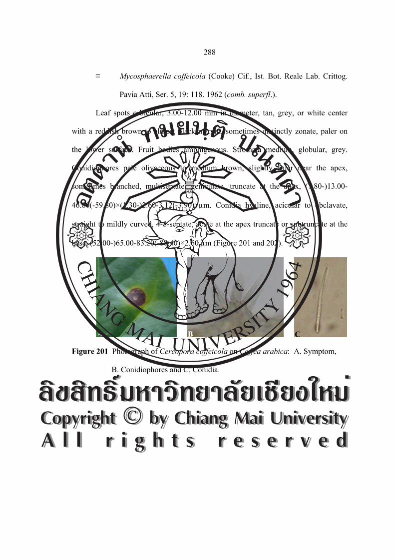
288
≡ Mycosphaerella coffeicola (Cooke) Cif., Ist. Bot. Reale Lab. Crittog.
Pavia Atti, Ser. 5, 19: 118. 1962 (comb. superfl.).
Leaf spots orbicular, 3.00-12.00 mm in diameter, tan, grey, or white center
with a reddish brown to almost black margin, sometimes distinctly zonate, paler on
the lower surface. Fruit bodies amphigenous. Stromata medium, globular, grey.
Conidiophores pale olivaceous to medium brown, slightly paler near the apex,
sometimes branched, multiseptate, geniculate, truncate at the apex, (7.80-)13.00-
46.80(-59.80)×(1.30-)2.60-3.12(-3.90) μm. Conidia hyaline, acicular to obclavate,
straight to mildly curved, 4-8-septate, acute at the apex truncate or subtruncate at the
base, (52.00-)65.00-83.20(-88.40)×2.60 μm (Figure 201 and 202).
Figure 201 Photograph of Cercopora coffeicola on Coffea arabica: A. Symptom,
B. Conidiophores and C. Conidia.
A B C
ÅÔ¢ÊÔ·¸Ô ìÁËÒÇÔ·ÂÒÅÑÂàªÕ§ãËÁèCopyright by Chiang Mai UniversityA l l r i g h t s r e s e r v e d
ÅÔ¢ÊÔ·¸Ô ìÁËÒÇÔ·ÂÒÅÑÂàªÕ§ãËÁèCopyright by Chiang Mai UniversityA l l r i g h t s r e s e r v e d

289
Figure 202 Drawing of Cercopora coffeicola on Coffea arabica: A. Conidiophores
and B. Conidia (scale bar = 40 µm).
Material examined: Thailand, Chiang Mai University, Chiang Mai Province. Leaves
of Coffea arabica, November 1, 2005, JM, CMU MH 091.
Known distribution: In diameterly distributed, including American, Samoa, Angola,
Australia, Brazil, Brunei, Cambodia, China, Colombia, Congo, Costa Rica, Cuba,
Dominican Republ., E1 Salvador, Ethiopia, Fiji, French Guiana, French Polynesia,
Gabon, Ghana, Guadeloupe, Guyana Haiti, India, Indonesia, Ivory Coast, Jamaica,
Japan, Kenya, Laos, Madagascar, Malawi, Martinque, Mauritius, Micronesia,
Mosambique, Myanmar, Nepal, New Caledonia, Nigeria, Panama, Papua New
Guinea, Peru, Philippines, Puerto Rico, Samoa, Sierra Leone, Somalia, South Africa,
Sudan, Surinam, Taiwan, Tanzania, Thailand, Togo, Trinidad, Tobago, Uganda,
U.S.A (FL, HI), Vanuatu, Venezuela, Yemen and Zimbabwe (Crous and Braun,
2003).
Notes: The first record of this species from Thailand was made by Chandrasrikul
(1962).
AB
ÅÔ¢ÊÔ·¸Ô ìÁËÒÇÔ·ÂÒÅÑÂàªÕ§ãËÁèCopyright by Chiang Mai UniversityA l l r i g h t s r e s e r v e d
ÅÔ¢ÊÔ·¸Ô ìÁËÒÇÔ·ÂÒÅÑÂàªÕ§ãËÁèCopyright by Chiang Mai UniversityA l l r i g h t s r e s e r v e d

290
Pseudocercospora sp.
Leaf spots are subcircular to irregular, brown to dark brown, reddish brown to
dark brown, with indefinite margins, 1.00-14.00 mm in diameter. Fruit bodies
amphigenous, but abundantly epiphyllous. Stromata medium, 22.14-41.82 µm in
diameter, well-developed, subglobular to globular, dark brtown. Conidiophores
arising through the cuticle, hyaline to brown, straight to slightly curved, densely
fasiculate and solitary, with indistinct conidial scars, 2-7-septate, (14.76-)19.68-
46.74(-56.58)×(1.23-)2.46-4.42(-4.92) µm. Conidia aciculate to filiform , subhyaline
to pale olive-brown, straight, smooth, with unthickened hilum, (17.22-)27.06-
(49.20)×(2.46-)2.95-4.92 µm, 1-5-septate, with truncate and thin basal end (Figure
203 and 204).
Figure 203 Photograph of Pseudocercospora sp. on Haldina cordifolia:
A. Symptom, B. Conidiophores and C. Conidia.
A B C
ÅÔ¢ÊÔ·¸Ô ìÁËÒÇÔ·ÂÒÅÑÂàªÕ§ãËÁèCopyright by Chiang Mai UniversityA l l r i g h t s r e s e r v e d
ÅÔ¢ÊÔ·¸Ô ìÁËÒÇÔ·ÂÒÅÑÂàªÕ§ãËÁèCopyright by Chiang Mai UniversityA l l r i g h t s r e s e r v e d

291
Figure 204 Drawing of Pseudocercospora sp. on Haldina cordifolia:
A. Conidiophores and B. Conidia (scale bar = 40 µm).
Material examined: Thailand, Sak Yai National Park, Uttradit Province Province.
Leaves of Haldina cordifolia, November 25, 2004, JM and Nakashima, CMU MH
092.
Known distribution: Known only Thailand.
Notes: This is the first record of Haldina cordifolia cause by Pseudocercospora sp.
from Thailand and the world.
Pseudocercospora ixorae (Solheim) Deighton, Mycol. Pap. 140: 145. 1976.
≡ Cercospora ixorae Solheim, Indian J. Agric. Sci. 3: 15. 1933.
= Cercospora ixorae W. Yamam., J. Soc. Trop. Agric. 6: 602. 1934
(nom. illeg.), homonym of C. ixorae Solheim, 1933.
Leaf spots suborbicular, 4.00-16.00 mm in diameter, confluent and become
irregular, at first yellowish or orangy, then become tan to pale brown, finally the
center turns greyish, surrounded by darker lines and a distinct dark brown margin,
zonate in appearance. Fruit bodies amphigenous but more abundant on the lower leaf
surface, especially on the dark margin of the spots. Stromata subglobular, dark brown,
15.00-45.00 µm in diameter, erumpent. Conidiophores densely fasciculate, subhyaline
AB
ÅÔ¢ÊÔ·¸Ô ìÁËÒÇÔ·ÂÒÅÑÂàªÕ§ãËÁèCopyright by Chiang Mai UniversityA l l r i g h t s r e s e r v e d
ÅÔ¢ÊÔ·¸Ô ìÁËÒÇÔ·ÂÒÅÑÂàªÕ§ãËÁèCopyright by Chiang Mai UniversityA l l r i g h t s r e s e r v e d

292
to very pale olivaceous brown, 1-4-septate, irregular in width, not branched, rarely
geniculate, substraight to variously bent, conically rounded at the apex, (24.39-)31.70-
92.68(-104.87)×(2.43-)4.87-4.87(-7.31) µm. Conidia subhyline to very pale
olivaceous, cylindric or longer ones filiform, straight to mildly curved, indistinctly 3-
10-septate, tip subobtuse to narrowly rounded, truncate at the base, (24.39-)34.14-
95.12 (-114.63)×(2.43-)3.65-7.31 µm (Figure 205 and 206).
Figure 205 Photograph of Pseudocercospora ixorae on Ixora congesta:
A. Symptom, B. Conidiophores and C. Conidia.
Figure 206 Drawing of Pseudocercospora ixorae on Ixora congesta:
A. Conidiophores and B. Conidia (scale bar = 40 µm).
A B C
A
B
ÅÔ¢ÊÔ·¸Ô ìÁËÒÇÔ·ÂÒÅÑÂàªÕ§ãËÁèCopyright by Chiang Mai UniversityA l l r i g h t s r e s e r v e d
ÅÔ¢ÊÔ·¸Ô ìÁËÒÇÔ·ÂÒÅÑÂàªÕ§ãËÁèCopyright by Chiang Mai UniversityA l l r i g h t s r e s e r v e d
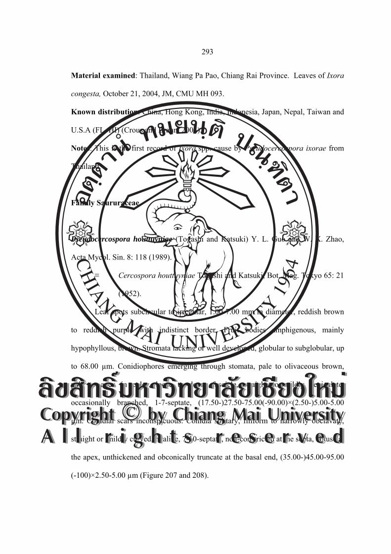
293
Material examined: Thailand, Wiang Pa Pao, Chiang Rai Province. Leaves of Ixora
congesta, October 21, 2004, JM, CMU MH 093.
Known distribution: China, Hong Kong, India, Indonesia, Japan, Nepal, Taiwan and
U.S.A (FL, HI) (Crous and Braun, 2003).
Notes: This is the first record of Ixora spp. cause by Pseudocercospora ixorae from
Thailand.
Family Saururaceae
Pseudocercospora houttuyniae (Togashi and Katsuki) Y. L. Guo and W. X. Zhao,
Acta Mycol. Sin. 8: 118 (1989).
≡ Cercospora houttuyniae Togashi and Katsuki, Bot. Mag. Tokyo 65: 21
(1952).
Leaf spots subcircular to irregular, 1.00-7.00 mm in diameter, reddish brown
to reddish purple with indistinct border. Fruit bodies amphigenous, mainly
hypophyllous, brown. Stromata lacking or well developed, globular to subglobular, up
to 68.00 µm. Conidiophores emerging through stomata, pale to olivaceous brown,
pale to apex in color, loose to dense fascicle, straight to mildly geniculate,
occasionally branched, 1-7-septate, (17.50-)27.50-75.00(-90.00)×(2.50-)5.00-5.00
µm. Conidial scars inconspicuous. Conidia solitary, filiform to narrowly obclavate,
straight or mildly curved, hyaline, 7-10-septate, non-constricted at the septa, obtuse at
the apex, unthickened and obconically truncate at the basal end, (35.00-)45.00-95.00
(-100)×2.50-5.00 µm (Figure 207 and 208).
ÅÔ¢ÊÔ·¸Ô ìÁËÒÇÔ·ÂÒÅÑÂàªÕ§ãËÁèCopyright by Chiang Mai UniversityA l l r i g h t s r e s e r v e d
ÅÔ¢ÊÔ·¸Ô ìÁËÒÇÔ·ÂÒÅÑÂàªÕ§ãËÁèCopyright by Chiang Mai UniversityA l l r i g h t s r e s e r v e d
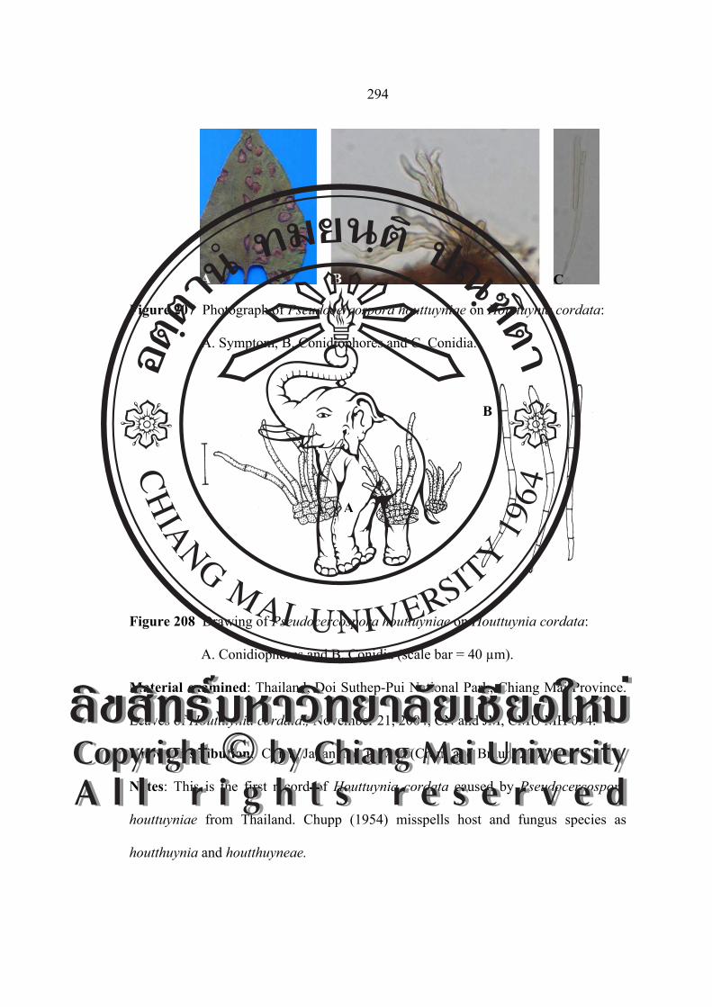
294
Figure 207 Photograph of Pseudocercospora houttuyniae on Houttuynia cordata:
A. Symptom, B. Conidiophores and C. Conidia.
Figure 208 Drawing of Pseudocercospora houttuyniae on Houttuynia cordata:
A. Conidiophores and B. Conidia (scale bar = 40 µm).
Material examined: Thailand, Doi Suthep-Pui National Park, Chiang Mai Province.
Leaves of Houttuynia cordata., November 21, 2004, CN and JM, CMU MH 094.
Known distribution: China, Japan and Taiwan (Crous and Braun, 2003).
Notes: This is the first record of Houttuynia cordata caused by Pseudocercospora
houttuyniae from Thailand. Chupp (1954) misspells host and fungus species as
houtthuynia and houtthuyneae.
A B C
A
B
ÅÔ¢ÊÔ·¸Ô ìÁËÒÇÔ·ÂÒÅÑÂàªÕ§ãËÁèCopyright by Chiang Mai UniversityA l l r i g h t s r e s e r v e d
ÅÔ¢ÊÔ·¸Ô ìÁËÒÇÔ·ÂÒÅÑÂàªÕ§ãËÁèCopyright by Chiang Mai UniversityA l l r i g h t s r e s e r v e d

295
Family Solanaceae
Cercospora capsicigena Bhartiya, R. Dubey and S. K. Singh, Indian Phytopathol.
53(3): 149. 2000. [T: GPU (Gorakhpur University, Dept. of Botany, India) 8010,
HCIO 42379]. (= Cercospora apii s. lat.).
Leaf spots hypophyllous, distinct, subcircular to irregular, yellowish brown
with blackish brown margins, 1.00-7.00 mm in diameter. Stromata small to medium,
rudimentary to slightly developed, irregular, dark brown, 13.00-40.00 µm in diameter.
Conidiophores pale brown at the base and paler upwards, arranged in a loose to dense
fascicle, with distinct conidial scars, 2-10-septate, 49.20-177.12×4.92-7.38 µm.
Conidia solitary, obclavate, straight to mildly curved, hyaline to subhyaline, smooth,
hilum conspicuously thickened, darkened, 54.12-135.30×2.46-8.61 µm, 3-11-septate,
acute to obtuse at the apex, truncate at the base (Figure 209 and 210).
Figure 209 Photograph of Cercospora capsicigena on
Capsicum annuum var. acuminatum: A. Symptom, B. Conidiophores and
C. Conidia.
A B C
ÅÔ¢ÊÔ·¸Ô ìÁËÒÇÔ·ÂÒÅÑÂàªÕ§ãËÁèCopyright by Chiang Mai UniversityA l l r i g h t s r e s e r v e d
ÅÔ¢ÊÔ·¸Ô ìÁËÒÇÔ·ÂÒÅÑÂàªÕ§ãËÁèCopyright by Chiang Mai UniversityA l l r i g h t s r e s e r v e d

296
Figure 210 Drawing of Cercospora capsicigena on
Capsicum annuum var. acuminatum: A. Conidiophores and B. Conidia
(scale bar = 40 µm).
Material examined: Thailand, Doi Suthep-Pui National Park, Chiang Mai Province.
Leaves of Capsicum annuum var. acuminatum, November 21, 2004, JM, CMU MH
095.
Known distribution: India and Thailand (Crous and Braun, 2003).
Notes: The first record of this species from Thailand was made by Sontirat et al.
(1980)
Cercospora capsici Heald and Wolf Mycologia 3: 15. 1911
Leaf spots circular to subcircular, sometimes zonate, 2.00-11.00 mm in
diameter, brown to yellowish brown, finally with minute to fairly large grey center,
Fruit bodies amphigenous. Stromata 17.50-35.00 μm in diameter. Conidiophore
A
B
ÅÔ¢ÊÔ·¸Ô ìÁËÒÇÔ·ÂÒÅÑÂàªÕ§ãËÁèCopyright by Chiang Mai UniversityA l l r i g h t s r e s e r v e d
ÅÔ¢ÊÔ·¸Ô ìÁËÒÇÔ·ÂÒÅÑÂàªÕ§ãËÁèCopyright by Chiang Mai UniversityA l l r i g h t s r e s e r v e d

297
fascicles 2-20 stalks, pale to medium dark olivaceous brown, paler and more narrow
toward the tip, plainly 3-6-septate, almost never branched, straight, medium to large
spore scar at truncate tip, 25.00-150.00×3.00-5.00 μm, base sometimes slightly in
diameter, conidia hyaline, acicular, rarely obclavate, straight to mildly curved, 5-14-
septate, base truncate to subtruncate, tip subobtuse to subacute, 30.00-200.00×2.50-
4.00 μm (Figure 211 and 212).
Figure 211 Photograph of Cercospora capsici on Capsicum annuum: A. Symptom,
B. Conidiophores and C. Conidia.
Figure 212 Drawing of Cercospora capsici on Capsicum annuum: A. Conidiophores
and B. Conidia (scale bar = 40 µm).
A B C
A B
ÅÔ¢ÊÔ·¸Ô ìÁËÒÇÔ·ÂÒÅÑÂàªÕ§ãËÁèCopyright by Chiang Mai UniversityA l l r i g h t s r e s e r v e d
ÅÔ¢ÊÔ·¸Ô ìÁËÒÇÔ·ÂÒÅÑÂàªÕ§ãËÁèCopyright by Chiang Mai UniversityA l l r i g h t s r e s e r v e d
298
Material examined: Thailand, Doi Suthep-Pui National Park, Chiang Mai Province.
Leaves of Capsicum annuum, November 21, 2004, JM, CMU MH 096.
Known distribution: Worldwide (Crous and Braun, 2003).
Notes: This is the first record of Capsicum annuum caused by Cercospora capsici
from Thailand. Kovachevski (Zeitschr. Pflanzenkr. 48: 321. 1938) after studying
C. capsici Mar. and Stey. concluded that this Cercospora on Capsicum was really
Cladosporium. It is true that the species which Marchal and Steyaert refer to Shin and
Kim (2001) described as well as the one Unamuno (Bol. Soc. Espanola Hist. Nat. 32:
161. 1932) named C. capsici has effuse olivaceous growth like a Cladosporium; but
the conidiophores are in dense fascicles, and the conidia elongate, so that they do not
agree with Kovachevski that the effuse type with coloured conidia is a Cladosporium,
but agree with Castellani (Riv. Agric. Subtrop. Trop. 42: 20. 1948) who says it is a
true Cercospora and gives the name C. unamunoi.
Cercospora physalidis Ellis, Amer. Naturalist 16: 810 (1882), emend. Braun and
Melnik, Trudy Bot. Inst. im V. L. Komarova 20: 79 (1997).
≡ Cercosporina physalidis (Ellis) Miura, South Manch. Railway
Co. Agric. Rept. 27: 525 (1928).
= Cercospora solanicola G. F. Atk., J. Elisha Mitchell Sci. Soc. 8:53
(1892).
= Cercospora nicotianae Ellis and Everh., Prroc. Acad. Sci.
Philadelphia 45: 170 (1893).
= Cercospora phyalidicola Ellis and Barthol., Erythea 4: 28 (1896).
ÅÔ¢ÊÔ·¸Ô ìÁËÒÇÔ·ÂÒÅÑÂàªÕ§ãËÁèCopyright by Chiang Mai UniversityA l l r i g h t s r e s e r v e d
ÅÔ¢ÊÔ·¸Ô ìÁËÒÇÔ·ÂÒÅÑÂàªÕ§ãËÁèCopyright by Chiang Mai UniversityA l l r i g h t s r e s e r v e d

299
= Cercospora physalidicola Speg., Anales Mus. Nac. Buenos Aires Ⅱ,
3: 342 (1899)(nom. illeg.).
= Cercospora raciborskii Sacc. and Syd, Syll. Fung. 16: 1070 (1902).
= Cercosporina physalidicola Speg. Anales Mus. Nac. Hist. Nat. Buenos
Aires 20: 426 (1910).
= Cercosporina daturicola Speg. Anales Mus. Nac. Hist. Nat. Buenos
Aires 20: 425 (1910).
≡ Cercospora daturicola (Speg.) Vassiljevsky, in Vassiljevsky and
Karakulin, Fungi imperfecti parasitici. 1. Hyphomycetes: 247 (1937).
≡ Cercospora daturicola (Speg.) W. W. Ray, Mycologia 36: 175 (1944).
= Cercospora capsici Heald and W. A. Wolf, Mycologia 3: 15 (1911).
= Cercospora abchasica Siemaszko, Izv. Severo-Kavkazsk. Muz. (Tiflis)
12: 26 (1919).
= Cercospora melongenae Welles, Phytopathology 12: 63 (1922).
= Cercospora atropae Kvashn., Izv. Severo-Kavkazsk. Kraev. Stantsii
Zashch. Rast. 4: 37 (1928).
= Cercosporina petuniae Saito, Trans. Tottori Soc. Agric. Sci. 3:271
(1931).
≡ Cercospora petuniae (Saito) Chupp and A. S. Mull., Ceiba 1: 176
(1950)(nom. illeg.).
= Cercospora petuniae A. S. Mull. and Chupp, Arq. Inst. Biol. Veg. Rio
de Janeiro 3: 96 (1936)(nom. inval.).
= Cercospora petuniae Sandu and Sarea, in Sandu et al., Lucr. Sti. Inst.
Agron. 1962: 94 (1962)(nom. illeg.).
ÅÔ¢ÊÔ·¸Ô ìÁËÒÇÔ·ÂÒÅÑÂàªÕ§ãËÁèCopyright by Chiang Mai UniversityA l l r i g h t s r e s e r v e d
ÅÔ¢ÊÔ·¸Ô ìÁËÒÇÔ·ÂÒÅÑÂàªÕ§ãËÁèCopyright by Chiang Mai UniversityA l l r i g h t s r e s e r v e d
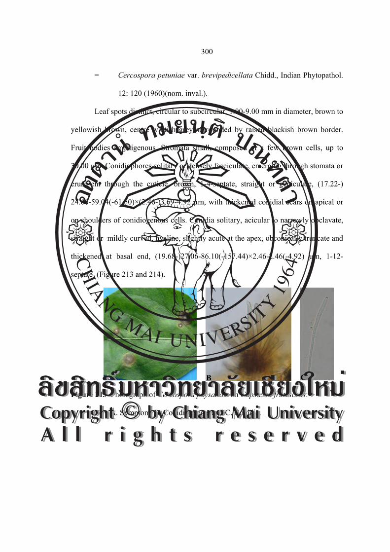
300
= Cercospora petuniae var. brevipedicellata Chidd., Indian Phytopathol.
12: 120 (1960)(nom. inval.).
Leaf spots distinct, circular to subcircular, 1.00-9.00 mm in diameter, brown to
yellowish brown, centre whitish grey surrounded by raised blackish brown border.
Fruit bodies amphigenous. Stromata small, composed of a few brown cells, up to
30.00 µm. Conidiophores solitary or densely fasciculate, emerging through stomata or
erumpent through the cuticle, brown, 1-4-septate, straight or geniculate, (17.22-)
24.60-59.04(-61.50)×(2.46-)3.69-4.92 µm, with thickened conidial scars on apical or
on shoulders of conidiogenous cells. Conidia solitary, acicular to narrowly obclavate,
straight or mildly curved, hyaline, slightly acute at the apex, obconically truncate and
thickened at basal end, (19.68-)27.06-86.10(-157.44)×2.46-2.46(-4.92) µm, 1-12-
septate (Figure 213 and 214).
Figure 213 Photograph of Cercospora physalidis on Capsicum frutescens:
A. Symptom, B. Conidiophores and C. Conidia.
A B C ÅÔ¢ÊÔ·¸Ô ìÁËÒÇÔ·ÂÒÅÑÂàªÕ§ãËÁèCopyright by Chiang Mai UniversityA l l r i g h t s r e s e r v e d
ÅÔ¢ÊÔ·¸Ô ìÁËÒÇÔ·ÂÒÅÑÂàªÕ§ãËÁèCopyright by Chiang Mai UniversityA l l r i g h t s r e s e r v e d
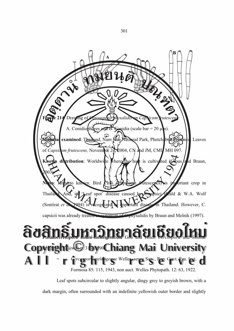
301
Figure 214 Drawing of Cercospora physalidis on Capsicum frutescens:
A. Conidiophores and B. Conidia (scale bar = 20 µm).
Material examined: Thailand, Nam Nao National Park, Phetchabun Province. Leaves
of Capsicum frutescens, November 24, 2004, CN and JM, CMU MH 097.
Known distribution: Worldwide where the host is cultivated (Crous and Braun,
2003).
Notes: Hitherto known. Bird Chili (Capsicum frutescencs) is important crop in
Thailand. And, its ‘Leaf spot’ disease caused by C.capsici Heald & W.A. Wolf
(Sontirat et al. 1994) is recognized as important disease in Thailand. However, C.
capsicii was already treated as synonym of C.physalidis by Braun and Melnik (1997).
Pseudocercospora solani-melongenicola Goh and Hsieh Cercospora and Similar
Fungi from Taiwan p. 318, 1990.
= Cercospora melongenae Welles, sensu Saw., Rep. Govt. Res. Inst.
Formosa 85: 115, 1943, non auct. Welles Phytopath. 12: 63, 1922.
Leaf spots subcircular to slightly angular, dingy grey to greyish brown, with a
dark margin, often surrounded with an indefinite yellowish outer border and slightly
AB
ÅÔ¢ÊÔ·¸Ô ìÁËÒÇÔ·ÂÒÅÑÂàªÕ§ãËÁèCopyright by Chiang Mai UniversityA l l r i g h t s r e s e r v e d
ÅÔ¢ÊÔ·¸Ô ìÁËÒÇÔ·ÂÒÅÑÂàªÕ§ãËÁèCopyright by Chiang Mai UniversityA l l r i g h t s r e s e r v e d

302
zonate in appearance, 2.00-4.00 mm in diameter. Fruit bodies chiefly epigenous.
Mycelium immersed. Stromata medium. Conidiophores pale olivaceous brown, paler
towards the apex, uniform in width, straight or curved, 1-2 geniculate, conically
truncate or bluntly rounded at the apex, not or rarely once branched, 1-3-septate, non-
constricted at the septa, 25.00-50.00×2.50-4.00 µm. Conidia hyaline, cylindric-
obclavate to slightly obclavate, straight to substraight, obtuse at the apex, 3-10-
septate, often mildly to moderately constricted at the septa, 25.00-90.00×3.50-5.50
µm, obconically truncate at the base (Figure 215 and 216).
Figure 215 Photograph of Pseudocercospora solani-melongenicola on
Solanum melongena: A. Symptom, B. Conidiophores and C. Conidia.
Figure 216 Drawing of Pseudocercospora solani-melongenicola on
Solanum melongena: A. Conidiophores and B. Conidia
(scale bar = 40 µm).
A B C
AB
ÅÔ¢ÊÔ·¸Ô ìÁËÒÇÔ·ÂÒÅÑÂàªÕ§ãËÁèCopyright by Chiang Mai UniversityA l l r i g h t s r e s e r v e d
ÅÔ¢ÊÔ·¸Ô ìÁËÒÇÔ·ÂÒÅÑÂàªÕ§ãËÁèCopyright by Chiang Mai UniversityA l l r i g h t s r e s e r v e d

303
Material examined: Thailand, Wiang Pa Pao, Chiang Mai Province. Leaves of
Solanum melongena, November 21, 2004, JM, CMU MH 098.
Known distribution: Worldwide where the host is cultivated (Crous and Braun,
2003).
Notes: This is the first record of Solanum melongena caused by Pseudocercospora
solani-melongenicola from Thailand.
Pseudocercospora sp.
Leaf spots amphigenous, scattered to confluent, circular to subcircular, 2.00-
8.00 mm in diameter or up to 15.00 mm when coalescing, initially appearing with
chlorotic discolourations, later becoming pale yellow to yellowish brown without
definite definite margins on the upper surface, finally brown to darkish brown on the
lower surface. Fruit bodies amphigenous, but mostly hypophyllous, effuse,
velutionous, greyish brown to dirty due to heavy fungal fructification. Primary
mycelium internal, hyphae septate, branched, hyaline, 2.00-3.00 µm in diameter.
Secondary mycelium external, superficial; hyphae septate, branched, subhyaline to
pale olivaceous brown, often forming ropes or climbing leaf hairs. Stromata lacking.
Conidiophores borne singly as lateral branches from the superficial secondary
mycelium, pale olivaceous to olivaceous brown, straight to slightly curved, 0-2 times
geniculate, not or rarely once branched, 1-7-septate, non-constricted at the septa,
(9.75-)21.95-39.02(-56.09)×(2.43-)4.87-7.31 µm, conidial scars minute, 1.00-1.50 µm
in diameter, conspicuous, apical or on small shoulders of conidiogenous cells caused
by geniculation. Conidia solitary to catenate, cylindric-obclavate to slightly obclavate,
straight to substraight, very pale olivaceous brown to pale olivaceous brown, slightly
ÅÔ¢ÊÔ·¸Ô ìÁËÒÇÔ·ÂÒÅÑÂàªÕ§ãËÁèCopyright by Chiang Mai UniversityA l l r i g h t s r e s e r v e d
ÅÔ¢ÊÔ·¸Ô ìÁËÒÇÔ·ÂÒÅÑÂàªÕ§ãËÁèCopyright by Chiang Mai UniversityA l l r i g h t s r e s e r v e d

304
attenuated towards the apex, 3-6-septate, often mildly to moderately constricted at the
septa, (31.70-)34.14-58.53(-65.85)×4.87-7.31 µm, hilum somewhat thickened,
darkened, somewhat refractive, and slightly protuberant (Figure 217 and 218).
Figure 217 Photograph of Pseudocercospora sp. on Solanum trilobatum:
A. Symptom, B. Conidiophores and C. Conidia.
Figure 218 Drawing of Pseudocercospora sp. on Solanum trilobatum:
A. Conidiophores and B. Conidia (scale bar = 40 µm).
Material examined: Thailand, Doi Suthep-Pui National Park, Chiang Mai Province.
Leaves of Solanum trilobatum, November 21, 2004, JM, CMU MH 099.
Known distribution: Known only Thailand.
A B C
A
B
ÅÔ¢ÊÔ·¸Ô ìÁËÒÇÔ·ÂÒÅÑÂàªÕ§ãËÁèCopyright by Chiang Mai UniversityA l l r i g h t s r e s e r v e d
ÅÔ¢ÊÔ·¸Ô ìÁËÒÇÔ·ÂÒÅÑÂàªÕ§ãËÁèCopyright by Chiang Mai UniversityA l l r i g h t s r e s e r v e d
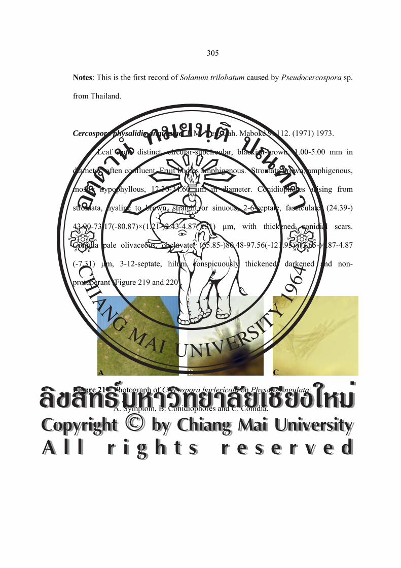
305
Notes: This is the first record of Solanum trilobatum caused by Pseudocercospora sp.
from Thailand.
Cercospora physalidis-angulatae J. M. Yen, Cah. Maboké 9: 112. (1971) 1973.
Leaf spots distinct, circular-subcircular, blackish-brown, 1.00-5.00 mm in
diameter, often confluent. Fruit bodies amphigenous. Stromata brown, amphigenous,
mostly hypophyllous, 12.30-24.60 µm in diameter. Conidiophores arising from
stromata, hyaline to brown, straight or sinuous, 2-6-septate, fasciculate, (24.39-)
43.90-73.17(-80.87)×(1.21-)2.43-4.87(7.31) µm, with thickened conidial scars.
Conidia pale olivaceous, obclavate, (65.85-)80.48-97.56(-121.95)×(3.65-)4.87-4.87
(-7.31) µm, 3-12-septate, hilum conspicuously thickened, darkened and non-
protuberant (Figure 219 and 220).
Figure 219 Photograph of Cercospora barlericola on Physalis angulata:
A. Symptom, B. Conidiophores and C. Conidia.
A B C
ÅÔ¢ÊÔ·¸Ô ìÁËÒÇÔ·ÂÒÅÑÂàªÕ§ãËÁèCopyright by Chiang Mai UniversityA l l r i g h t s r e s e r v e d
ÅÔ¢ÊÔ·¸Ô ìÁËÒÇÔ·ÂÒÅÑÂàªÕ§ãËÁèCopyright by Chiang Mai UniversityA l l r i g h t s r e s e r v e d

306
Figure 220 Drawing of Cercospora barlericola on Physalis angulata:
A. Conidiophores and B. Conidia (scale bar = 40 µm).
Material examined: Thailand, Doi Suthep-Pui National Park, Chiang Mai Province.
Leaves of Physalis angulata, October 2, 2005, JM, CMU MH 100.
Known distribution: Gabon (Crous and Braun, 2003).
Notes: This is the first record of Physalis angulata caused by Cercospora physalidis-
angulatae from Thailand.
Cercospora nicotianicola J. M. Yen, Rev. Mycol. 32: 188. 1967. [T: PC].
≡ Cercospora nicotianae Ellis and Everhart Proc. Acad. Sci. Phila.
45: 170 (1893).
= Cercospora raciborskii Sacc. and Sydow, Syll. Fung.
16: 1070 (1902).
Leaf spots orbicular, often zonate, 0.50-10.00 mm in diameter, pale brown to
dingy grey at center, with a darker margin. Fruit bodies amphigenous. Stromata
lacking or a few darker brown cells. Conidiophores medium brown, paler towards the
apex, uniform in width, straight, 3-13-septate, not branched, truncate at the apex,
AB
ÅÔ¢ÊÔ·¸Ô ìÁËÒÇÔ·ÂÒÅÑÂàªÕ§ãËÁèCopyright by Chiang Mai UniversityA l l r i g h t s r e s e r v e d
ÅÔ¢ÊÔ·¸Ô ìÁËÒÇÔ·ÂÒÅÑÂàªÕ§ãËÁèCopyright by Chiang Mai UniversityA l l r i g h t s r e s e r v e d

307
(39.00-) 52.00-140.40(-200.20)×(3.38-)3.90-7.80 μm. Conidia hyaline, acicular,
straight to curved, 3-18-septate, acute to subacute at the apex, truncate at the base,
(18.20-) 36.40-150.80(-202.80)×(3.64-)4.42-7.80 μm (Figure 221 and 222).
Figure 221 Photograph of Cercospora nicotianicola on Nicotiana tabacum:
A. Symptom, B. Conidiophores and C. Conidia.
Figure 222 Drawing of Cercospora nicotianicola on Nicotiana tabacum:
A. Conidiophores and B. Conidia (scale bar = 40 µm).
Material examined: Thailand, Mae Jo, Chiang Mai Province. Leaves of Nicotiana
tabacum, January 6, 2005, JM, CMU MH 101.
Known distribution: Malaysia, Singapore and Thailand (Crous and Braun, 2003).
Notes: The first record of Nicotiana tabacum caused by Cercospora nicotianae was
by Tongpun refer to Sontirat et al. (1980). A true Cercospora s.str. is distinct from
C. apii s. lat. (incl. C. nicotianae).
A B C
A B
ÅÔ¢ÊÔ·¸Ô ìÁËÒÇÔ·ÂÒÅÑÂàªÕ§ãËÁèCopyright by Chiang Mai UniversityA l l r i g h t s r e s e r v e d
ÅÔ¢ÊÔ·¸Ô ìÁËÒÇÔ·ÂÒÅÑÂàªÕ§ãËÁèCopyright by Chiang Mai UniversityA l l r i g h t s r e s e r v e d

308
Cercospora lycopersici Salam and P. N. Rao, J. Indian Bot. Soc. 36: 425. 1957.
[T: HY 50].
Leaf spots orbicular, often zonate, 0.50-10.00 mm in diameter, pale brown to
dingy grey at center, with a darker margin. Fruit bodies amphigenous. Stromata a few
darker brown cells, 24.39-36.58 µm in diameter. Conidiophores 2-14 in a divergent
fascicle, medium brown, paler towards the apex, uniform in width, straight, 2-6-
septate, not branched, truncate at the apex, (39.02-)43.90-48.78(-60.97)×(3.65-)4.87-
4.87(-7.31) μm. Conidia hyaline, obclavate, straight to curved, 3-8-septate, hilum
conspicuously thickened, darkened, (31.70-)65.85-73.17(-92.68)×3.65-4.87 μm
(Figure 223 and 224).
Figure 223 Photograph of Cercospora lycopersic on
Lycopersicon esculentum var. pyriforme: A. Symptom, B. Conidiophores
and C. Conidia.
A B C
ÅÔ¢ÊÔ·¸Ô ìÁËÒÇÔ·ÂÒÅÑÂàªÕ§ãËÁèCopyright by Chiang Mai UniversityA l l r i g h t s r e s e r v e d
ÅÔ¢ÊÔ·¸Ô ìÁËÒÇÔ·ÂÒÅÑÂàªÕ§ãËÁèCopyright by Chiang Mai UniversityA l l r i g h t s r e s e r v e d

309
Figure 224 Drawing of Cercospora barlericola on
Lycopersicon esculentum var. pyriforme: A. Conidiophores and
B. Conidia (scale bar = 40 µm).
Material examined: Thailand, Doi Suthep-Pui National Park, Chiang Mai Province.
Leaves of Lycopersicon esculentum var. pyriforme, Febuary 5, 2005, JM, CMU MH
102.
Known distribution: India (Crous and Braun, 2003).
Notes: This is the first record of Lycopersicon esculentum var. pyriforme caused by
Cercospora lycopersici from Thailand. Conidia pigmented, no Cercospora s.str.
Passalora sp.
Leaf spots orbicular, often zonate, 1.50-7.00 mm in diameter, pale brown to
dingy grey at center, with a darker margin. Fruit bodies amphigenous. Stromata 9.75-
43.90 µm in diameter. Conidiophores in a divergent fascicle, medium brown, paler
towards the apex, uniform in width, straight, 0-3-septate, not branched, truncate at the
apex, small conidial scars, 17.07-24.39(-41.46)×(2.43-)3.65-4.87 µm. Conidia
A
B
ÅÔ¢ÊÔ·¸Ô ìÁËÒÇÔ·ÂÒÅÑÂàªÕ§ãËÁèCopyright by Chiang Mai UniversityA l l r i g h t s r e s e r v e d
ÅÔ¢ÊÔ·¸Ô ìÁËÒÇÔ·ÂÒÅÑÂàªÕ§ãËÁèCopyright by Chiang Mai UniversityA l l r i g h t s r e s e r v e d

310
subhyaline, cylindric, straight to curved, 4-8-septate, subacute at the apex, (74.38-)
78.04-87.80(-102.43)×(2.43-)3.65-4.87 μm (Figure 225 and 226).
Figure 225 Photograph of Passalora sp. on Lycopersicon esculentum,: A. Symptom,
B. Conidiophores and C. Conidia.
Figure 226 Drawing of Passalora sp. on Lycopersicon esculentum:
A. Conidiophores and B. Conidia (scale bar = 40 µm).
Material examined: Thailand, A. Fang, Chiang Mai Province. Leaves of
Lycopersicon esculentum, November 21, 2005, JM, CMU MH 103.
Known distribution: Known only Thailand.
A B C
A
B
ÅÔ¢ÊÔ·¸Ô ìÁËÒÇÔ·ÂÒÅÑÂàªÕ§ãËÁèCopyright by Chiang Mai UniversityA l l r i g h t s r e s e r v e d
ÅÔ¢ÊÔ·¸Ô ìÁËÒÇÔ·ÂÒÅÑÂàªÕ§ãËÁèCopyright by Chiang Mai UniversityA l l r i g h t s r e s e r v e d

311
Notes: This is the first record of Lycopersicon esculentum caused by Passalora sp.
from Thailand.
Cercospora sp.
Leaf spots circular to irregular, 2.00-10.00 mm in diameter, pale brown to
dingy grey at center, with a darker margin. Fruit bodies amphigenous. Stromata small,
14.63-34.14 µm. Conidiophores 2-14 in a divergent fascicle, medium brown, paler
towards the apex, uniform in width, straight, 1-4-septate, not branched, truncate at the
apex, conidial scars conspicuously thickened, (29.26-)31.70-36.58(-48.78)×(3.65-)
4.87-4.87(-6.09) μm. Conidia hyaline, acicular, straight to curved, 3-9-septate, acute
to subacute at the apex, truncate at the base, hilum conspicuously thickened,
darkened, (29.26-)58.53-70.73(-85.36)×2.43-3.65(-4.87) μm (Figure 227 and 228).
Figure 227 Photograph of Cercospora sp. on Solanum pseudocapsicum:
A. Symptom, B. Conidiophores and C. Conidia.
A B C ÅÔ¢ÊÔ·¸Ô ìÁËÒÇÔ·ÂÒÅÑÂàªÕ§ãËÁèCopyright by Chiang Mai UniversityA l l r i g h t s r e s e r v e d
ÅÔ¢ÊÔ·¸Ô ìÁËÒÇÔ·ÂÒÅÑÂàªÕ§ãËÁèCopyright by Chiang Mai UniversityA l l r i g h t s r e s e r v e d
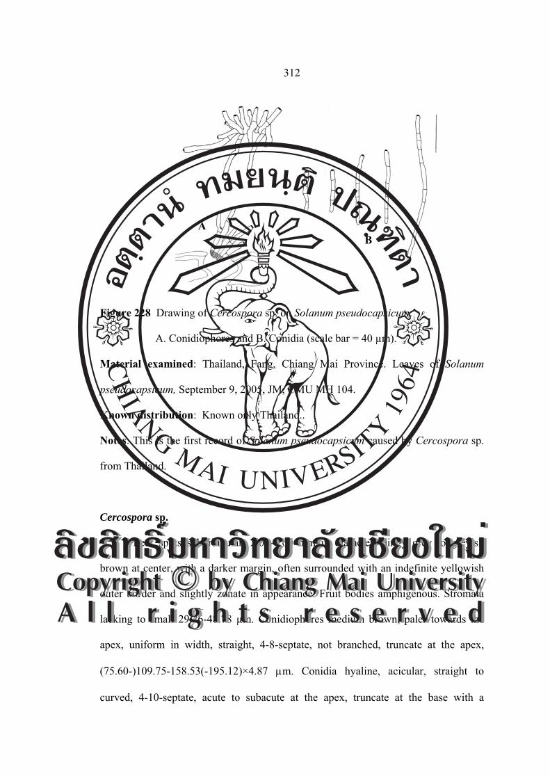
312
Figure 228 Drawing of Cercospora sp. on Solanum pseudocapsicum:
A. Conidiophores and B. Conidia (scale bar = 40 µm).
Material examined: Thailand, Fang, Chiang Mai Province. Leaves of Solanum
pseudocapsicum, September 9, 2005, JM, CMU MH 104.
Known distribution: Known only Thailand..
Notes: This is the first record of Solanum pseudocapsicum caused by Cercospora sp.
from Thailand.
Cercospora sp.
Leaf spots suborbicular, 2.00-8.00 mm in diameter, dingy grey to greyish
brown at center, with a darker margin, often surrounded with an indefinite yellowish
outer border and slightly zonate in appearance. Fruit bodies amphigenous. Stromata
lacking to small 29.26-48.78 µm. Conidiophores medium brown, paler towards the
apex, uniform in width, straight, 4-8-septate, not branched, truncate at the apex,
(75.60-)109.75-158.53(-195.12)×4.87 μm. Conidia hyaline, acicular, straight to
curved, 4-10-septate, acute to subacute at the apex, truncate at the base with a
A B
ÅÔ¢ÊÔ·¸Ô ìÁËÒÇÔ·ÂÒÅÑÂàªÕ§ãËÁèCopyright by Chiang Mai UniversityA l l r i g h t s r e s e r v e d
ÅÔ¢ÊÔ·¸Ô ìÁËÒÇÔ·ÂÒÅÑÂàªÕ§ãËÁèCopyright by Chiang Mai UniversityA l l r i g h t s r e s e r v e d

313
thickened hilum, very variable in length, (34.14-)48.78-131.70(-180.48)×2.43-2.43
(-4.87) μm (Figure 229 and 230).
Figure 229 Photograph of Cercospora sp. on Solanum wrightii: A. Symptom,
B. Conidiophores and C. Conidia.
Figure 230 Drawing of Cercospora sp. on Solanum wrightii: A. Conidiophores and
B. Conidia (scale bar = 40 µm).
Material examined: Thailand, Wiang Pa Pao, Chiang Mai Province. Leaves of
Solanum wrightii, December 18, 2005, JM, CMU MH 105.
Known distribution: Known only Thailand.
A B C
A
B
ÅÔ¢ÊÔ·¸Ô ìÁËÒÇÔ·ÂÒÅÑÂàªÕ§ãËÁèCopyright by Chiang Mai UniversityA l l r i g h t s r e s e r v e d
ÅÔ¢ÊÔ·¸Ô ìÁËÒÇÔ·ÂÒÅÑÂàªÕ§ãËÁèCopyright by Chiang Mai UniversityA l l r i g h t s r e s e r v e d
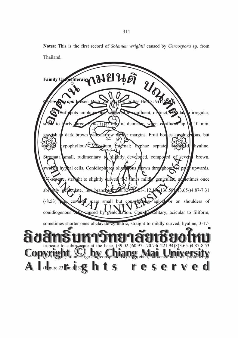
314
Notes: This is the first record of Solanum wrightii caused by Cercospora sp. from
Thailand.
Family Umbelliferae
Cercospora apii Fresen. Beitr. Zur Mykol. Drittes Heft 3: 91(1863)
Leaf spots amphigenous, scattered to confluent, distinct, circular to irregular,
small to fairly large, 2.00-10.00 mm in diameter, when confluent up to 10 mm,
greyish to dark brown with narrow darker margins. Fruit bodies amphigenous, but
mostly hypophyllous. Mycelium internal; hyphae septate, branched, hyaline.
Stromata small, rudimentary to slightly developed, composed of several brown,
swollen hyphal cells. Conidiophores olivaceous brown throughout, or paler upwards,
2-7-septate, straight to slightly curved, 1-3 times mildly geniculate, sometimes once
abruptly geniculate, not branched, (26.82-)51.21-112.19(-136.58)×(3.65-)4.87-7.31
(-8.53) µm, conidial scars small but conspicuous, apical or on shoulders of
conidiogenous cells caused by geniculation. Conidia solitary, acicular to filiform,
sometimes shorter ones obclavate-cylindric, straight to mildly curved, hyaline, 3-17-
septate, non-constricted at the septa, subacute to subobtuse at the apex, obconically
truncate to subtruncate at the base, (39.02-)60.97-170.73(-221.94)×(3.65-)4.87-8.53
(-9.75) µm, hilum large and conspicuously thickened, darkened and non-protuberant
(Figure 231 and 232).
ÅÔ¢ÊÔ·¸Ô ìÁËÒÇÔ·ÂÒÅÑÂàªÕ§ãËÁèCopyright by Chiang Mai UniversityA l l r i g h t s r e s e r v e d
ÅÔ¢ÊÔ·¸Ô ìÁËÒÇÔ·ÂÒÅÑÂàªÕ§ãËÁèCopyright by Chiang Mai UniversityA l l r i g h t s r e s e r v e d

315
Figure 231 Photograph of Cercospora apii on Apium graveolens: A. Symptom,
B. Conidiophores and C. Conidia.
Figure 232 Drawing of Cercospora apii on Apium graveolens: A. Conidiophores and
B. Conidia (scale bar = 40 µm).
Material examined: Thailand, Doi Suthep-Pui National Park, Chiang Mai Province.
Leaves of Apium graveolens, November 21, 2004, JM, CMU MH 106.
Known distribution: Worldwide, including Australia, Austria, Azerbaijan,
Bangladesh, Barbados, Brazil, Brunei, Bulgaria, Cambodia, Canada, Canary Islands,
China, Colombia, Congo, Cuba, Czech Republ., Denmark, Dominican Republ.,
Egypt, El Salvador, France, Germany, Greece, Guatemala, Hong Kong, India,
Indonesia, Iraq, Italy, Jamaica, Jordan, Kenya, Korea, Latvia, Lebanon, Libya,
A B
A B C
ÅÔ¢ÊÔ·¸Ô ìÁËÒÇÔ·ÂÒÅÑÂàªÕ§ãËÁèCopyright by Chiang Mai UniversityA l l r i g h t s r e s e r v e d
ÅÔ¢ÊÔ·¸Ô ìÁËÒÇÔ·ÂÒÅÑÂàªÕ§ãËÁèCopyright by Chiang Mai UniversityA l l r i g h t s r e s e r v e d

316
Lithuania, Mexico, Malaysia, Mauritius, Morocco, Myanmar, Nepal, Netherlands,
New Caledonia, New Zealand, Morocco, Nepal, Nigeria, New Zealand, Panama,
Papua New Guinea, Peru, Philippines, Poland, Puerto Rico, Romania, Russia, Sierra
Leone, Singapore, Slovakia, Slovenia, South Africa, Sudan, Suriname, Switzerland,
Taiwan, Tanzania, Thailand, Togo, Trinidad, Tobago, Uruguay, U.S.A (AK, CA, CT,
FL, HI, IL, KY, NC, NE, NJ, NY, MD, MN, MO, TX, WV), Vanuate, Venezuela and
Zimbabwe (Crous and Braun, 2003).
Notes: A new circumscription of C. apii emend. is given earlier and the synonyms are
cited and marked under the particular names in the present list. This item only
comprises data to C. apii s. str. (Distribution and literature to Apium graveolens and
some additional hosts of the Umbelliferae), supplemented with other host records
which have been directly referred to as C. apii
Nakata and Takimoto (1928) recorded this fungus from Korea, and Cho et al.
(1997) added a short morphological notes. Detailed description and illustration based
on Korean material of this species were provided by Kim and Shin (1999c). The
disease caused by the fungus was common at celery plantation in Korea.
Chupp (1954) published the following characters of Cercospora apii:
Conidiophores (25.00-300.00×4.00-6.50 µm) are long and rarely branched, conidia
(25.00-315.00×3.00-6.00 µm) with truncated base are long and acicular. The Korean
collection, however, agrees well with Chupp’s description, although the
conidiophores and conidia are somewhat shorter, since the differences are variable in
this species and other most taxonomic characters are fall within the Chupp’s concepts.
Chupp (1954) listed several varieties of C. apii, which he considered to be separate
species in their own right. These have been treated elsewhere, as Cercosporacarotae
ÅÔ¢ÊÔ·¸Ô ìÁËÒÇÔ·ÂÒÅÑÂàªÕ§ãËÁèCopyright by Chiang Mai UniversityA l l r i g h t s r e s e r v e d
ÅÔ¢ÊÔ·¸Ô ìÁËÒÇÔ·ÂÒÅÑÂàªÕ§ãËÁèCopyright by Chiang Mai UniversityA l l r i g h t s r e s e r v e d

317
(Pass.) Kazn. and Siemaszko and Passalora pastinacae (Sacc.) U. Braun. Pons and
Sutton (1988) mentioned that C. apii is not restricted to genera of the Umbelliferae.
They concluded that the species is very variable, and that the length of the
conidiophores and conidia can be strongly influenced by changes in the relative
humidity, temperature and light conditions. Crous and Braun (1996) are of the opinion
that it would be best treat C. apii as occurring on Apium only, as several other studies
have been found that cercosporoid fungi are highly host specific. Some Taiwanese
specimens on Apios taiwanianus is close to the present species, but differs from the
latter species by having comparatively narrower (4.00-5.00 µm) and paler
conidiophores. Therefore, Goh and Hsieh (1990) placed the Taiwanese species in a
separate species, Cercospora apios.
Family Verbenaceae
Passalora sp.nov.
≡ Mycovellosiella gmelinae-arboreae A. K. Sarbhoy, Hosag. And
N. Ahmad. J. Econ. Taxon. Bot. 7: 521.(1986).
Leaf spots scattered, distinct, irregular to angular, vein-limited, dark brown,
centre pale brown, 1.00-9.00 mm in diameter, later coalescing to large spot, grayish
white. Stromata epiphyllous, well-developed, composed of swollen brown to dark
brown hyphal cells, 25.00-57.50 µm in diameter. Conidiophores densely fascicule,
dark brown, pale toward apex, rough, percurrent proliferation, geniculate, conidial
scars small, slightly thickened, 1-5-septate, (15.00-)20.00-40.00(-52.50)×5.00 µm.
Conidia solitary, occasionally catenulate, cylindro-obclavate to obclavate, straight to
ÅÔ¢ÊÔ·¸Ô ìÁËÒÇÔ·ÂÒÅÑÂàªÕ§ãËÁèCopyright by Chiang Mai UniversityA l l r i g h t s r e s e r v e d
ÅÔ¢ÊÔ·¸Ô ìÁËÒÇÔ·ÂÒÅÑÂàªÕ§ãËÁèCopyright by Chiang Mai UniversityA l l r i g h t s r e s e r v e d

318
mildly curved, rough, (22.50-)27.50-72.50(-87.50)×3.75(-5.00)-5.00(-6.25) µm, 3-16-
septate, obtuse to subobtuse at the apex, obconically truncate and slightly thickened at
the base (Figure 233 and 234).
Figure 233 Photograph of Passalora gmelinae-arboreae on Gmelina arborea:
A. Symptom, B. Conidiophores and C. Conidia.
Figure 234 Drawing of Passalora gmelinae-arboreae on Gmelina arborea:
A. Conidiophores and B. Conidia (scale bar = 20 µm).
Material examined: Thailand, Doi Suthep-Pui National Park, Chiang Mai Province.
Leaves of Gmelina arborea, November 21, 2004, CN and JM, CMU MH 107.
Known distribution: India (Crous and Braun, 2003).
Notes: Passalora gmelinae-arboreae (A. K. Sarbhoy, Hosag. and N.Ahmad) U.Braun
and Crous (Sarbhoy et al. 1985; Crous and Braun 2003) is different from present
A
A B
A B C
ÅÔ¢ÊÔ·¸Ô ìÁËÒÇÔ·ÂÒÅÑÂàªÕ§ãËÁèCopyright by Chiang Mai UniversityA l l r i g h t s r e s e r v e d
ÅÔ¢ÊÔ·¸Ô ìÁËÒÇÔ·ÂÒÅÑÂàªÕ§ãËÁèCopyright by Chiang Mai UniversityA l l r i g h t s r e s e r v e d

319
species without stroma, with branched and wide conidiophore, narrow and short
conidia.
This is the first record of Gmelina arborea caused by Passalora gmelinae-arboreae
from Thailand.
Cercospora holmskiodiae Lall and Gill, Indian Phytopathol. 15: 244. (1962) 1963.
[T: HCIO 27197]. (= Cercospora apii s.lat.).
Leaf spots epiphyllous, circular to subcircular, 3.00-23.00 mm in diameter.
distinct on the upper surface, greyish-brown with blackish brown border line.
Stromata distinct, small to medium, moderately to well-developed, dark brown,
subglobular to angular, 27.06-36.90 µm in diameter. Conidiophores emerging from
the upper part of stromata, dark brown, densely fasciculate, simple, straight or slightly
curved, 2-8-septate, (17.22-)27.06-81.18(-113.16)×(3.69-)4.92-4.92(-5.16) µm, with
distinct and thickened conidial scars. Conidia acicular, straight, smooth, hyaline, with
thickened and truncate hilum, (9.84-)17.22-98.40(-184.50)×2.46-4.92 µm, 1-16-
septate (Figure 235 and 236).
Figure 235 Photograph of Cercospora holmskiodiae on Holmskioldia sanguinea:
A. Symptom, B. Conidiophores and C. Conidia.
A B C
ÅÔ¢ÊÔ·¸Ô ìÁËÒÇÔ·ÂÒÅÑÂàªÕ§ãËÁèCopyright by Chiang Mai UniversityA l l r i g h t s r e s e r v e d
ÅÔ¢ÊÔ·¸Ô ìÁËÒÇÔ·ÂÒÅÑÂàªÕ§ãËÁèCopyright by Chiang Mai UniversityA l l r i g h t s r e s e r v e d

320
Figure 236 Drawing of Cercospora holmskiodiae on Holmskioldia sanguinea:
A. Conidiophores and B. Conidia (scale bar = 20 µm).
Material examined: Thailand, Doi Suthep-Pui National Park, Chiang Mai Province.
Leaves of Holmskioldia sanguinea, November 21, 2004, JM, CMU MH 108.
Known distribution: Cuba, India and Venezuela (Crous and Braun, 2003).
Notes: This is the first record of Holmskioldia sanguinea caused by Cercospora
holmskiodiae from Thailand. Shorter conidia are obclavate-subcylindrical with
obconically truncate bases, but long conidia are acicular.
Pseudocercospora viticicola (J. M.Yen and Lim) J. M. Yen, Gard. Bull., Singapore
33: 190 (1980) (emend. Braun, 1998).
≡ Cercospora viticicola J. M. Yen and Lim, Cah. Pacifique 17: 104
(1973).
= Cercospora viticis Ellis and Everh., J. Mycol. 3: 18 (1887)
≡ Pseudocercosporella viticis (Ellis and Everh.) B. K.Gupta and Kamal,
Indian Phytopathol. 42: 388 (1989).
A
B
ÅÔ¢ÊÔ·¸Ô ìÁËÒÇÔ·ÂÒÅÑÂàªÕ§ãËÁèCopyright by Chiang Mai UniversityA l l r i g h t s r e s e r v e d
ÅÔ¢ÊÔ·¸Ô ìÁËÒÇÔ·ÂÒÅÑÂàªÕ§ãËÁèCopyright by Chiang Mai UniversityA l l r i g h t s r e s e r v e d

321
≡ Pseudocercospora viticicola U. Braun, Mycotaxon 48: 296 (1993).
= Cercospora viticis Sawada, Rep. Gov. Agric. Res. Inst. Taiwan 87: 90
(1944).
≡ Pseudocercospora viticis Goh and W. H. Hsieh, Trans. Mycol. Soc.
R.O.C. 4: 11 (1989)
= Cercospora viticis-quinatae J. M. Yen, Bull. Soc. Mycol. France 93:
158 (1979).
≡ Pseudocercospora viticis-quinatae (J. M. Yen) J. M. Yen, Bull. Soc.
Mycol. France 94: 388 (1979).
= Pseudocercospora viticigena J. M. Yen, A. K. Kar and B. K. Das,
Mycotaxon 16: 68 (1982).
Leaf spots circular to irregular, 1.00-20.00 mm in diameter, distinct on the
upper surface, grayish-brown with indefinite dark brown border, frequently confluent.
Stromata distinct, small on under surface, well-developed on upper surface, up to
55.00 µm in diameter, olivaceous brown, with external hyphae. Conidiophores,
olivaceous brown, loose to densely fasciculate, simple, straight or geniculate, 1-6-
septate, (19.68-)24.60-61.50(-83.64)×4.92 µm. Conidial scars distinct and denticulate.
Conidia cylindrical to obclavate, straight or mildly curved, smooth, pale olivaceous
brown, unthickened and obconically truncated basal end, obtuse at apex, (24.60-)
41.82-71.34(-86.10)×2.46-4.92 µm, 4-8 septate (Figure 237 and 238).
ÅÔ¢ÊÔ·¸Ô ìÁËÒÇÔ·ÂÒÅÑÂàªÕ§ãËÁèCopyright by Chiang Mai UniversityA l l r i g h t s r e s e r v e d
ÅÔ¢ÊÔ·¸Ô ìÁËÒÇÔ·ÂÒÅÑÂàªÕ§ãËÁèCopyright by Chiang Mai UniversityA l l r i g h t s r e s e r v e d

322
Figure 237 Photograph of Pseudocercospora viticicolaon Vitex quinata:
A. Symptom, B. Conidiophores and C. Conidia.
Figure 238 Drawing of Pseudocercospora viticicolaon Vitex quinata:
A. Conidiophores and B. Conidia (scale bar = 20 µm).
Material examined: Thailand, Doi Suthep-Pui National Park, Chiang Mai Province.
Leaves of Vitex quinata, November 21, 2004, JM, CMU MH 109.
Known distribution: Brazil, China, Cuba, India, Japan, Philippines, Puerto Rico,
Singapore, Taiwan, USA and Virgin Islands (Crous and Braun, 2003).
A B C
A B
ÅÔ¢ÊÔ·¸Ô ìÁËÒÇÔ·ÂÒÅÑÂàªÕ§ãËÁèCopyright by Chiang Mai UniversityA l l r i g h t s r e s e r v e d
ÅÔ¢ÊÔ·¸Ô ìÁËÒÇÔ·ÂÒÅÑÂàªÕ§ãËÁèCopyright by Chiang Mai UniversityA l l r i g h t s r e s e r v e d
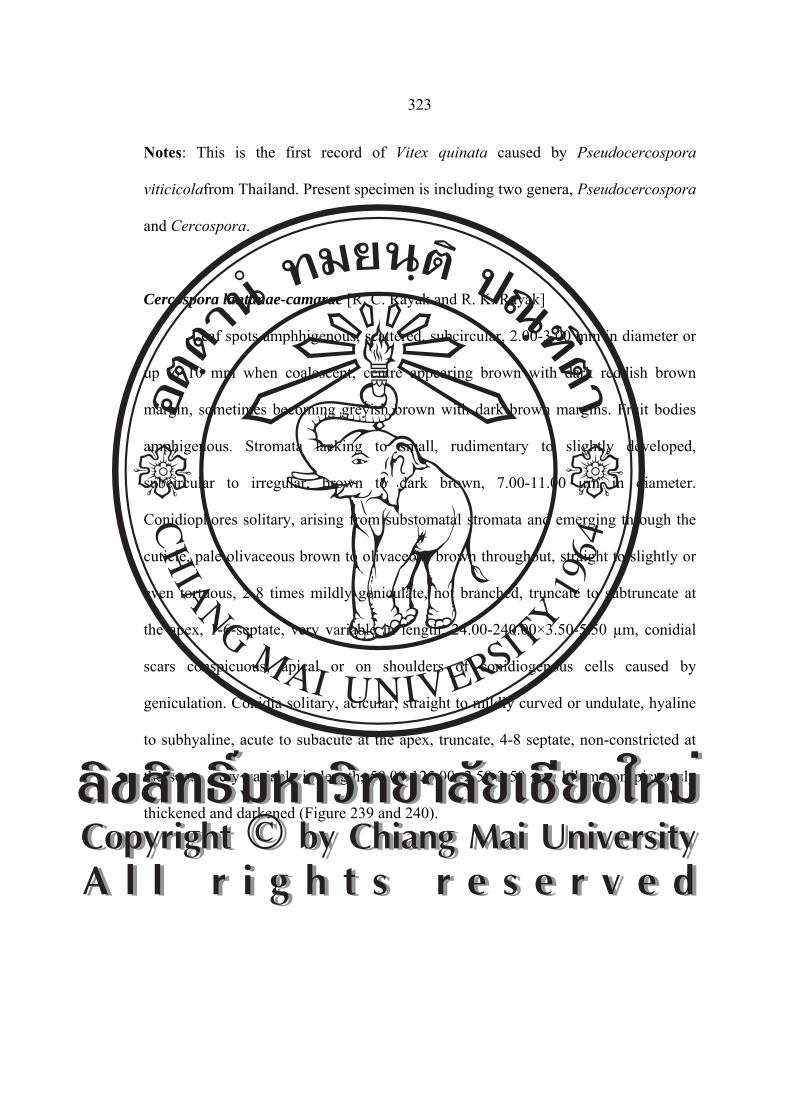
323
Notes: This is the first record of Vitex quinata caused by Pseudocercospora
viticicolafrom Thailand. Present specimen is including two genera, Pseudocercospora
and Cercospora.
Cercospora lantanae-camarae [R. C. Rayak and R. K. Rayak]
Leaf spots amphhigenous, scattered, subcircular, 2.00-3.00 mm in diameter or
up to 10 mm when coalescent, centre appearing brown with dark reddish brown
margin, sometimes becoming greyish brown with dark brown margins. Fruit bodies
amphigenous. Stromata lacking to small, rudimentary to slightly developed,
subcircular to irregular, brown to dark brown, 7.00-11.00 µm in diameter.
Conidiophores solitary, arising from substomatal stromata and emerging through the
cuticle, pale olivaceous brown to olivaceous brown throughout, straight to slightly or
even tortuous, 2-8 times mildly geniculate, not branched, truncate to subtruncate at
the apex, 1-6-septate, very variable in length, 24.00-240.00×3.50-5.50 µm, conidial
scars conspicuous, apical or on shoulders of conidiogenous cells caused by
geniculation. Conidia solitary, acicular, straight to mildly curved or undulate, hyaline
to subhyaline, acute to subacute at the apex, truncate, 4-8 septate, non-constricted at
the septa, very variable in length, 50.00-125.00×2.50-3.50 µm, hilum conspicuously
thickened and darkened (Figure 239 and 240). ÅÔ¢ÊÔ·¸Ô ìÁËÒÇÔ·ÂÒÅÑÂàªÕ§ãËÁèCopyright by Chiang Mai UniversityA l l r i g h t s r e s e r v e d
ÅÔ¢ÊÔ·¸Ô ìÁËÒÇÔ·ÂÒÅÑÂàªÕ§ãËÁèCopyright by Chiang Mai UniversityA l l r i g h t s r e s e r v e d
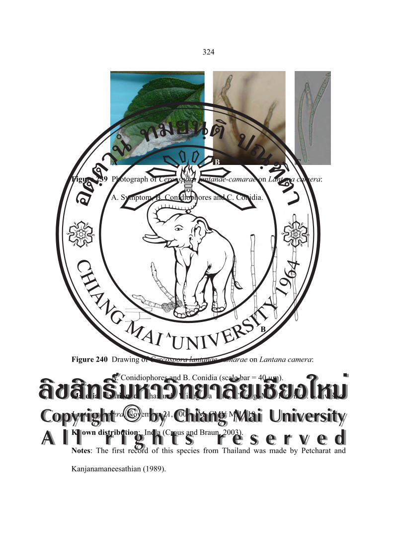
324
Figure 239 Photograph of Cercospora lantanae-camarae on Lantana camera:
A. Symptom, B. Conidiophores and C. Conidia.
Figure 240 Drawing of Cercospora lantanae-camarae on Lantana camera:
A. Conidiophores and B. Conidia (scale bar = 40 µm).
Material examined: Thailand, Wiang Pa Pao, Chiang Mai Province. Leaves of
Lantana camera, November 21, 2004, JM, CMU MH 110.
Known distribution: India (Crous and Braun, 2003).
Notes: The first record of this species from Thailand was made by Petcharat and
Kanjanamaneesathian (1989).
AB
A B C
ÅÔ¢ÊÔ·¸Ô ìÁËÒÇÔ·ÂÒÅÑÂàªÕ§ãËÁèCopyright by Chiang Mai UniversityA l l r i g h t s r e s e r v e d
ÅÔ¢ÊÔ·¸Ô ìÁËÒÇÔ·ÂÒÅÑÂàªÕ§ãËÁèCopyright by Chiang Mai UniversityA l l r i g h t s r e s e r v e d

325
Cercospora volkameriae Speg., Revista Mus. La Plata 15: 47. 1908. [T: LEP; LPS
955]. (= Cercospora apii s. lat.).
Leaf spots amphhigenous, scattered, subcircular, 2.00-4.00 mm in diameter,
centre appearing brown with dark reddish brown margin, sometimes becoming
greyish brown with dark brown margins. Fruit bodies amphigenous. Stromata lacking
to small, rudimentary to slightly developed, subcircular to irregular, brown to dark
brown, composed of a few brown hyphal cells. Conidiophores solitary, arising from
substomatal stromata and emerging through the cuticle, pale olivaceous brown to
olivaceous brown throughout, straight to slightly or even tortuous, not branched,
truncate to subtruncate at the apex, 5-11-septate, very variable in length, (158.53-)
241.46-256.09(-299.99)×4.87-4.87(-6.09) µm, conidial scars large, conspicuous,
apical or on shoulders of conidiogenous cells caused by geniculation. Conidia
solitary, cylindric, straight to mildly curved, hyaline to subhyaline, subacute at the
apex, truncate, 3-7-septate, non constricted at the septa, very variable in length,
(14.63-)28.04-31.70(-75.60)×(2.43-)\4.87-4.87(-6.09) µm, hilum conspicuously
thickened, darkened, and non-protuberant (Figure 241 and 242).
Figure 241 Photograph of Cercospora volkameriae on Clerodendrum paniculatum:
A. Symptom, B. Conidiophores and C. Conidia.
A B C
ÅÔ¢ÊÔ·¸Ô ìÁËÒÇÔ·ÂÒÅÑÂàªÕ§ãËÁèCopyright by Chiang Mai UniversityA l l r i g h t s r e s e r v e d
ÅÔ¢ÊÔ·¸Ô ìÁËÒÇÔ·ÂÒÅÑÂàªÕ§ãËÁèCopyright by Chiang Mai UniversityA l l r i g h t s r e s e r v e d
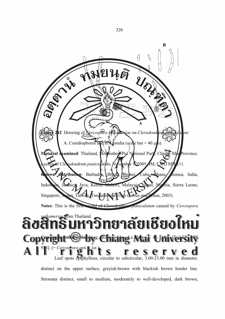
326
Figure 242 Drawing of Cercospora volkameriae on Clerodendrum paniculatum:
A. Conidiophores and B. Conidia (scale bar = 40 µm).
Material examined: Thailand, Doi Suthep-Pui National Park, Chiang Mai Province.
Leaves of Clerodendrum paniculatum, November 1, 2005, JM, CMU MH 111.
Known distribution: Barbados, Brazil, Brunei, Cuba, Ghana, Guinea, India,
Indonesia, Jamaica, Java, Korea, Malawi, Malaysia, Nepal, Nigeria, Sierra Leone,
Singapore, Sudan, Taiwan, Tanzania and Togo (Crous and Braun, 2003).
Notes: This is the first record of Clerodendrum paniculatum caused by Cercospora
volkameriae from Thailand.
Cercospora volkameriae Speg., Revista Mus. La Plata 15: 47. 1908. [T: LEP; LPS
955]. (= Cercospora apii s. lat.).
Leaf spots epiphyllous, circular to subcircular, 3.00-23.00 mm in diameter,
distinct on the upper surface, greyish-brown with blackish brown border line.
Stromata distinct, small to medium, moderately to well-developed, dark brown,
A
B
ÅÔ¢ÊÔ·¸Ô ìÁËÒÇÔ·ÂÒÅÑÂàªÕ§ãËÁèCopyright by Chiang Mai UniversityA l l r i g h t s r e s e r v e d
ÅÔ¢ÊÔ·¸Ô ìÁËÒÇÔ·ÂÒÅÑÂàªÕ§ãËÁèCopyright by Chiang Mai UniversityA l l r i g h t s r e s e r v e d
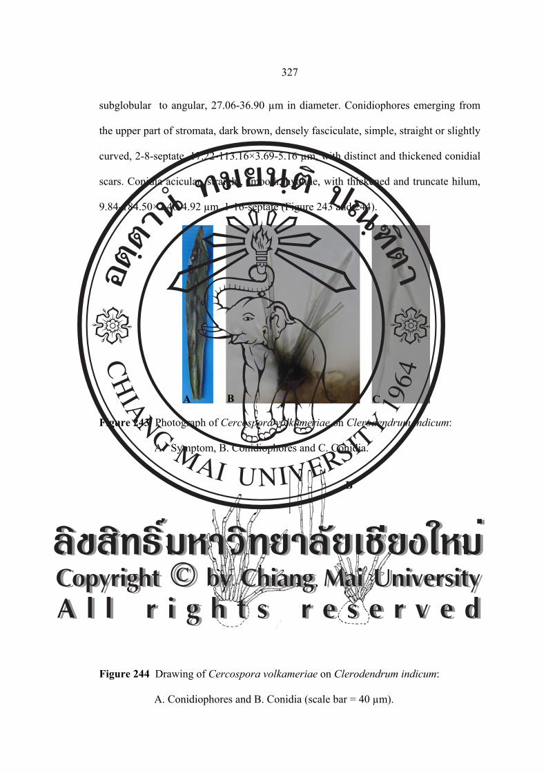
327
subglobular to angular, 27.06-36.90 µm in diameter. Conidiophores emerging from
the upper part of stromata, dark brown, densely fasciculate, simple, straight or slightly
curved, 2-8-septate, 17.22-113.16×3.69-5.16 µm, with distinct and thickened conidial
scars. Conidia acicular, straight, smooth, hyaline, with thickened and truncate hilum,
9.84-184.50×2.46-4.92 µm, 1-16-septate (Figure 243 and 244).
Figure 243 Photograph of Cercospora volkameriae on Clerodendrum indicum:
A. Symptom, B. Conidiophores and C. Conidia.
Figure 244 Drawing of Cercospora volkameriae on Clerodendrum indicum:
A. Conidiophores and B. Conidia (scale bar = 40 µm).
A
B
A B C
ÅÔ¢ÊÔ·¸Ô ìÁËÒÇÔ·ÂÒÅÑÂàªÕ§ãËÁèCopyright by Chiang Mai UniversityA l l r i g h t s r e s e r v e d
ÅÔ¢ÊÔ·¸Ô ìÁËÒÇÔ·ÂÒÅÑÂàªÕ§ãËÁèCopyright by Chiang Mai UniversityA l l r i g h t s r e s e r v e d

328
Material examined: Thailand, Doi Suthep-Pui National Park, Chiang Mai Province.
Leaves of Clerodendrum indicum, November 21, 2004, JM, CMU MH 112.
Known distribution: Barbados, Brazil, Brunei, Cuba, Ghana, Guinea, India,
Indonesia, Jamaica, Java, Korea, Malawi, Malaysia, Nepal, Nigeria, Sierra Leone,
Singapore, Sudan, Taiwan, Tanzania and Togo (Crous and Braun, 2003).
Notes: This is the first record of Clerodendrum indicum caused by Cercospora
volkameriae from Thailand.
Cercospora tectonae F. Stevens (tectoniae), Bernice P. Bishop Mus. Bull. 19: 155.
1925. [T: ILL 15736]. (=Cercospora apii s.lat).
Leaf spots angular or suborbicular, vein-limited, 1.00-14.00 mm in diameter,
confluent, dull brown to greyish brown or white, with a dark margin. Fruit bodies
amphigenous but chiefly epiphyllous. Stromata small to medium. Conidiophores
loosely fasciculate, pale olivaceous brown to medium brown, pale brown near the
base and almost hyaline at the tip, multiseptate, 0-5 geniculate or conic at the apex,
(48.78-)56.09-60.97(-75.60)×(3.65-)4.87-4.87 µm. Conidia hyaline, acicular to
obclavate-cylindric, straight to curved, 6-12-septate, subacute at the apex, truncate or
long obconically truncate at the base, (42.68-)53.65-80.48(-102.43)×(2.43-)3.65-3.65
µm (Figure 245 and 246). ÅÔ¢ÊÔ·¸Ô ìÁËÒÇÔ·ÂÒÅÑÂàªÕ§ãËÁèCopyright by Chiang Mai UniversityA l l r i g h t s r e s e r v e d
ÅÔ¢ÊÔ·¸Ô ìÁËÒÇÔ·ÂÒÅÑÂàªÕ§ãËÁèCopyright by Chiang Mai UniversityA l l r i g h t s r e s e r v e d

329
Figure 245 Photograph of Cercospora tectonae on Tectona grandis: A. Symptom,
B. Conidiophores and C. Conidia.
Figure 246 Drawing of Cercospora tectonae on Tectona grandis: A. Conidiophores
and B. Conidia (scale bar = 40 µm).
Material examined: Thailand, Multiple Cropping Centre, Chiang Mai University,
Chiang Mai Province. Leaves of Tectona grandis, December 1, 2005, JM, CMU MH
113.
Known distribution: Hawaii, India, Taiwan and Trinidad (Crous and Braun, 2003).
Notes: This is the first record of Tectona grandis caused by Cercospora tectonae
from Thailand.
A B C
A
B
ÅÔ¢ÊÔ·¸Ô ìÁËÒÇÔ·ÂÒÅÑÂàªÕ§ãËÁèCopyright by Chiang Mai UniversityA l l r i g h t s r e s e r v e d
ÅÔ¢ÊÔ·¸Ô ìÁËÒÇÔ·ÂÒÅÑÂàªÕ§ãËÁèCopyright by Chiang Mai UniversityA l l r i g h t s r e s e r v e d