sanja tunjic, bsc
TRANSCRIPT
Sanja Tunjic, BSc
Graphene and graphene derivatives for antibacterial application and biosensing
MASTER’S THESIS
to achieve the university degree of Master of Science
Master’s degree programme: Biotechnology
submitted to:
Graz University of Technology
Supervisor
Prof. Ivan Mijakovic Department of Biology and Biological Engineering, Chalmers University of Technology,
Gothenburg, Sweden
Assoc. Prof. Harald Pichler Institute of Molecular Biotechnology, Graz University of Technology, Austria
Graz, October 2017
2
AFFIDAVIT I declare that I have authored this thesis independently, that I have not used other than the
declared sources/resources, and that I have explicitly indicated all material which has been
quoted either literally or by content from the sources used. The text document uploaded to
TUGRAZonline is identical to the present master’s thesis.
___________________ _____________________
Date Signature
3
Table of content Acknowledgments ...................................................................................................................... 5
Zusammenfassung ...................................................................................................................... 6
Abstract ...................................................................................................................................... 7
1. Boron doped graphene for antibacterial applications ....................................................... 8
1.1. Introduction ................................................................................................................. 8
1.1.1. Graphene .............................................................................................................. 8
1.1.2. Chemical vapor deposition (CVD) graphene ........................................................ 9
1.1.3. Boron doped graphene ........................................................................................ 9
1.1.4. Cytotoxicity of graphene based nanostructures ................................................ 10
1.1.5. Biomaterials associated infections (BAI) ............................................................ 11
1.2. Materials and Methods ............................................................................................. 11
1.2.1. Materials ............................................................................................................. 11
1.2.2. Boron doped graphene: Experimental work ...................................................... 11
1.2.3. Preparation of boron doped graphene membranes .......................................... 12
1.2.4. Choosing a right solvent and boron doped graphene concentration ................ 12
1.2.5. Boron doped graphene membranes on different substrates ............................ 13
1.2.6. Bacterial strains and biofilm cultivation ............................................................. 13
1.2.7. Cultivation of NIH3T3 cells ................................................................................. 13
1.2.8. Colony Forming Units (CFUs) .............................................................................. 14
1.2.9. Raman Spectroscopy .......................................................................................... 14
1.2.10. Scanning Electron Microscopy (SEM) ............................................................. 14
1.2.11. Statistical analysis ........................................................................................... 14
1.3. Results and Discussion ............................................................................................... 15
1.3.1. RAMAN characterization .................................................................................... 15
1.3.2. Membrane integrity ........................................................................................... 15
1.3.3. Boron doped graphene on different substrates ................................................ 21
1.4. Conclusion ................................................................................................................. 22
2. Antibacterial activity of other 2D materials ..................................................................... 23
2.1. Introduction ............................................................................................................... 23
2.2. Materials and Methods ............................................................................................. 23
2.2.1. Materials ............................................................................................................. 23
2.2.2. Preparation of membranes and choosing the right solvent .............................. 23
2.3. Results and Discussion ............................................................................................... 24
3. Enhanced secretion of Exotoxin A by P. aeruginosa on boron doped graphene membranes .............................................................................................................................. 26
4
3.1. Materials and methods.............................................................................................. 26
3.1.1. RNA isolation ...................................................................................................... 26
3.1.2. Reverse transcription and semi-quantitative Real Time Polymerase Chain Reaction (RT-PCR) ............................................................................................................. 26
3.2. Results and discussion ............................................................................................... 27
4. Antibacterial activity of plant and bacteria derived nanoparticles.................................. 29
4.1. Introduction ............................................................................................................... 29
4.2. Materials and Methods ............................................................................................. 29
4.2.1. Synthesis of nanoparticles ................................................................................. 29
4.2.2. Determination of Minimum Inhibitory Concentration (MIC) and Minimum Bactericidal Concentration (MBC) .................................................................................... 30
4.2.3. Colony Forming Units (CFUs) .............................................................................. 30
4.2.4. Live Dead Staining .............................................................................................. 30
4.2.5. Scanning Electron Microscopy (SEM) ................................................................. 31
4.3. Results and discussion ............................................................................................... 31
4.4. Conclusion ................................................................................................................. 35
5. Bacterial biofilm sensor .................................................................................................... 36
5.1. Introduction ............................................................................................................... 36
5.2. Materials and Methods ............................................................................................. 37
5.3. Results and discussion ............................................................................................... 37
6. Literature .......................................................................................................................... 38
5
Acknowledgments
I would love to thank my supervisor Ivan Mijakovic and my mentors Dr. Raghu Mokkapati and
Dr. Santosh Pandit at the Division of Systems and Synthetic Biology of the Department of
Biology and Biological Engineering at Chalmers University of Technology. I have been
extremely lucky to work on my master’s thesis project with people who cared so much about
me and my work and made this experience valuable for my future career.
I am also very grateful to Prof. Harald Pichler from Graz University of Technology. Even though
we were far away, you were always available. Thank you for all the guidance and support.
I am thankful to Dr. Emanuele Celauro for the necessary support during the experimental work
and Ulf Sodervall for the training for the Clean Room.
6
Zusammenfassung
Der Transfer von Resistenzgenen zwischen pathogenen Bakterienpopulationen reduziert die
Wirksamkeit von Antibiotika und verursacht damit weltweit Millionen von Todesfällen. Es
besteht dringender Bedarf, neue Ansätze zur Behandlung von Infektionen mit pathogenen
Bakterien zu entwickeln. Seit seiner Entdeckung hat das Interesse an der Untersuchung der
antibakteriellen Aktivität von Graphen und Graphenderivaten zugenommen, obwohl
widersprüchliche Ergebnisse von verschiedenen Forschergruppen veröffentlicht wurden.
Diese wissenschaftlichen Widersprüche sind auf Variationen in den Parametern Graphen-
Flockengröße, -Konzentration, -Herstellungsverfahren, -Dicke und vor allem auf die Art der
untersuchten Bakterien zurückzuführen.
In der vorliegenden Arbeit wurde die antibakterielle Aktivität von mit Bor-Atomen dotiertem
Graphen (B-G) gegenüber drei Bakterienstämmen untersucht: grampositiver Staphylococcus
epidermidis, gramnegative Pseudomonas aeruginosa und Escherichia coli. Es wurde
festgestellt, dass B-G weder bakterizid noch bakteriostatisch für irgendeine Art der getesteten
Bakterienstämme ist. Der antibakterielle Effekt war auch nicht abhängig von der Art des
Substrats, auf dem B-G vorlag (Leiter, Halbleiter oder Isolator). Andererseits wurde
beobachtet, dass B-G die Sekretion von Exotoxin A durch P. aeruginosa verstärkt.
Auch für andere 2D-Materialien wie WS2 (Wolframdisulfid) und MoS2 (Molybdändisulfid)
wurde keinerlei antibakterielle Wirkung auf die oben erwähnten Bakterienstämme gefunden.
Silber (Ag) und Gold (Au) Nanopartikel von Cannabis sativa (Hemp) wurden auf ihre
bakteriostatische oder bakterizide Wirkung getestet. Silbernanopartikel zeigen eine
vielversprechende antibakterielle Wirkung gegen die gramnegativen Bakterien P. aeruginosa
und E. coli.
Neben dem Testen der antibakteriellen Aktivität von verschiedenen Graphenderivaten wurde
ein Graphen-basierter Biosensor hergestellt und getestet, um den Schwellenwert der
Biofilmbildung von Bakterien zu bestimmen. Dies soll zur Erkennung von Infektionen und zur
Reduktion der Infektionswahrscheinlichkeit von Patienten mit Implantaten führen.
7
Abstract The rapid transfer of resistance genes between pathogenic bacterial population reduces the
antibiotic efficacy, which causes millions of deaths worldwide. There is a need to develop
novel approaches for treatment of pathogenic bacteria. Since its discovery, the interest in
investigating the antibacterial activity of graphene and graphene derivatives has
predominantly increased where contradictory results have been published by various
researchers. These contradictions are due to various parameters like flake size, concentration,
method of preparation, thickness and most importantly the type of bacteria.
In the current work, antibacterial activity of boron doped graphene (B-G) was examined
against three bacterial strains: Gram-positive Staphylococcus epidermidis, Gram-negative
Pseudomonas aeruginosa and Escherichia coli. It was realized that B-G is neither bactericidal
nor bacteriostatic to any type of tested bacterial strains. Antibacterial effect was also not
dependent on the type of substrate B-G was present on (conductor, semiconductor or
insulator). On the other hand, an interesting observation was that B-G enhances the secretion
of exotoxin A by P. aeruginosa.
Other 2D materials like WS2 (Tungsten disulfide) and MoS2 (Molybdenum disulfide) were also
unsuccessfully tested for antibacterial effect on above mentioned bacterial strains.
Silver (Ag) and gold (Au) nanoparticles derived from Cannabis sativa (Hemp) were tested for
their bacteriostatic or bactericidal activity. Silver nanoparticles show promising antibacterial
effect against Gram-negative bacteria P. aeruginosa and E. coli.
Besides testing antibacterial activity of different graphene derivatives, graphene based
biosensor was fabricated and tested to detect the threshold of bacteria biofilm formation.
This helps in detection and reducing the infections for the patients with implants.
8
1. Boron doped graphene for antibacterial applications
1.1. Introduction
1.1.1. Graphene
Carbon is the basic element of organic chemistry and an essential element of life. Due to the
flexibility of its bonding, carbon-based systems can be found in different structures with
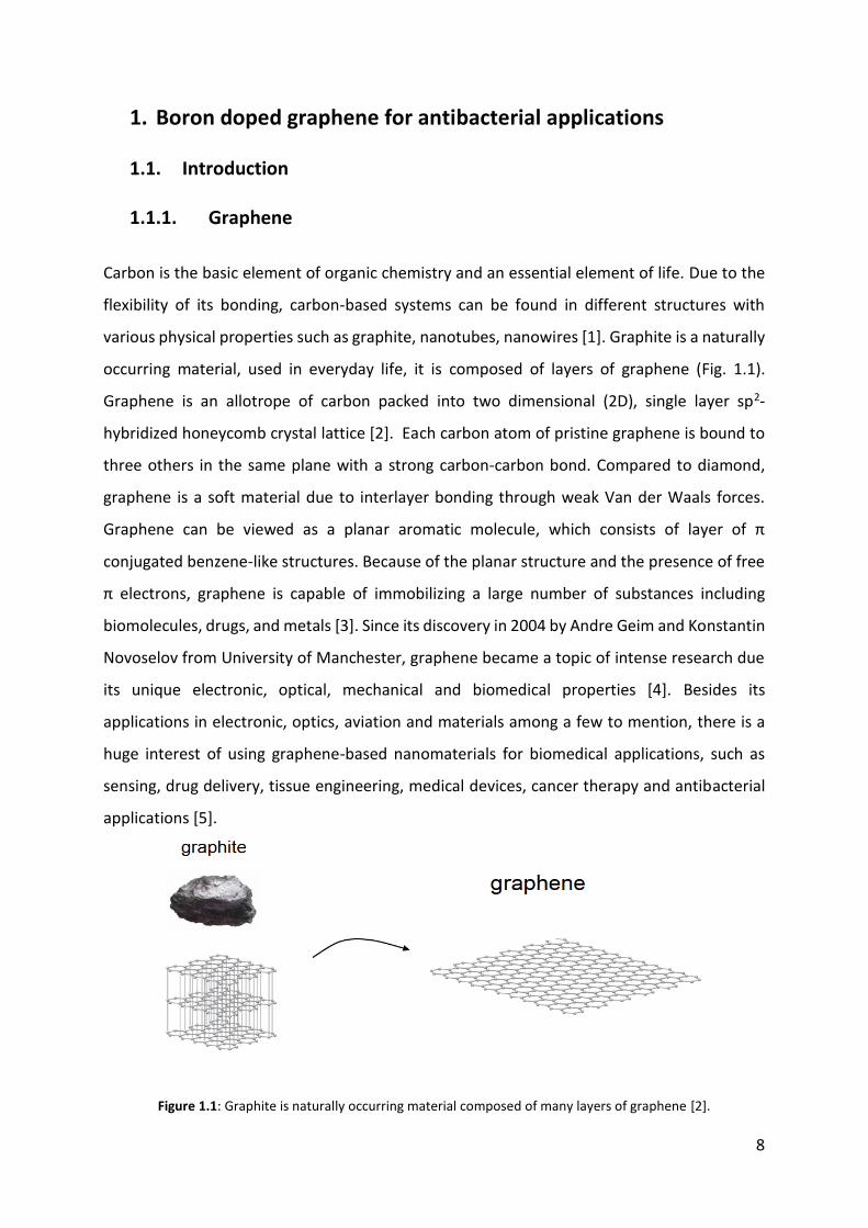
various physical properties such as graphite, nanotubes, nanowires [1]. Graphite is a naturally
occurring material, used in everyday life, it is composed of layers of graphene (Fig. 1.1).
Graphene is an allotrope of carbon packed into two dimensional (2D), single layer sp2-
hybridized honeycomb crystal lattice [2]. Each carbon atom of pristine graphene is bound to
three others in the same plane with a strong carbon-carbon bond. Compared to diamond,
graphene is a soft material due to interlayer bonding through weak Van der Waals forces.
Graphene can be viewed as a planar aromatic molecule, which consists of layer of π
conjugated benzene-like structures. Because of the planar structure and the presence of free
π electrons, graphene is capable of immobilizing a large number of substances including
biomolecules, drugs, and metals [3]. Since its discovery in 2004 by Andre Geim and Konstantin
Novoselov from University of Manchester, graphene became a topic of intense research due
its unique electronic, optical, mechanical and biomedical properties [4]. Besides its
applications in electronic, optics, aviation and materials among a few to mention, there is a
huge interest of using graphene-based nanomaterials for biomedical applications, such as
sensing, drug delivery, tissue engineering, medical devices, cancer therapy and antibacterial
applications [5].
Figure 1.1: Graphite is naturally occurring material composed of many layers of graphene [2].
9
1.1.2. Chemical vapor deposition (CVD) graphene Graphene can be prepared by different methods including chemical or mechanical exfoliation,
chemical vapor deposition (CVD), Epitaxial growth, Micro-wave assisted oxidation, Ion
implantation and by reduction [6]. Among all these methods, CVD graphene has gained
prominence as high quality large areas of graphene films can be grown on various metal
substrates [7]. CVD process is based on thermal decomposition of the hydrocarbon source on
a heated substrate at 1000°C. Due to its catalytic performance, the metal substrate lowers the
energy barrier of the reaction. The important parameter is carbon solubility on metal
substrate. Metals such as Nickel (Ni) have higher carbon solubility and carbon (C) dissolves
into heated substrate. On the other hand, metals with lower carbon solubility such as copper
(Cu) are widely used for preparation of large area monolayer graphene. Due to the low
solubility of C atoms in Cu, monolayer graphene can grow in a self-limiting process by which
graphene can be easily grown on commercially available Cu-foils [8, 9].
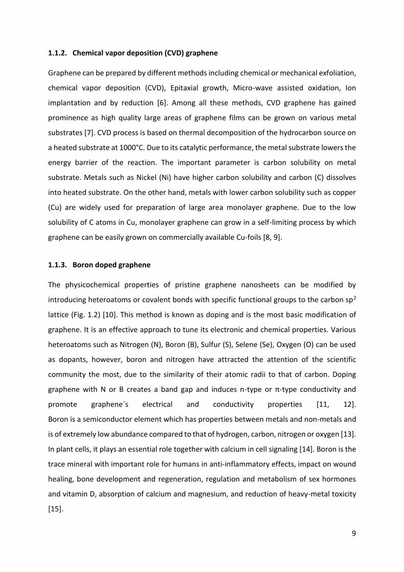
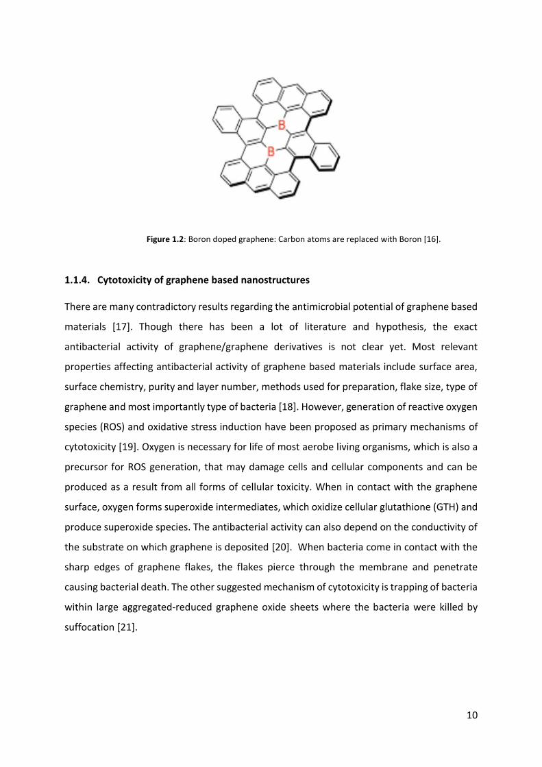
1.1.3. Boron doped graphene The physicochemical properties of pristine graphene nanosheets can be modified by
introducing heteroatoms or covalent bonds with specific functional groups to the carbon sp2
lattice (Fig. 1.2) [10]. This method is known as doping and is the most basic modification of
graphene. It is an effective approach to tune its electronic and chemical properties. Various
heteroatoms such as Nitrogen (N), Boron (B), Sulfur (S), Selene (Se), Oxygen (O) can be used
as dopants, however, boron and nitrogen have attracted the attention of the scientific
community the most, due to the similarity of their atomic radii to that of carbon. Doping
graphene with N or B creates a band gap and induces n-type or π-type conductivity and
promote graphene´s electrical and conductivity properties [11, 12].
Boron is a semiconductor element which has properties between metals and non-metals and
is of extremely low abundance compared to that of hydrogen, carbon, nitrogen or oxygen [13].
In plant cells, it plays an essential role together with calcium in cell signaling [14]. Boron is the
trace mineral with important role for humans in anti-inflammatory effects, impact on wound
healing, bone development and regeneration, regulation and metabolism of sex hormones
and vitamin D, absorption of calcium and magnesium, and reduction of heavy-metal toxicity
[15].
10
1.1.4. Cytotoxicity of graphene based nanostructures There are many contradictory results regarding the antimicrobial potential of graphene based
materials [17]. Though there has been a lot of literature and hypothesis, the exact
antibacterial activity of graphene/graphene derivatives is not clear yet. Most relevant
properties affecting antibacterial activity of graphene based materials include surface area,
surface chemistry, purity and layer number, methods used for preparation, flake size, type of
graphene and most importantly type of bacteria [18]. However, generation of reactive oxygen
species (ROS) and oxidative stress induction have been proposed as primary mechanisms of
cytotoxicity [19]. Oxygen is necessary for life of most aerobe living organisms, which is also a
precursor for ROS generation, that may damage cells and cellular components and can be
produced as a result from all forms of cellular toxicity. When in contact with the graphene
surface, oxygen forms superoxide intermediates, which oxidize cellular glutathione (GTH) and
produce superoxide species. The antibacterial activity can also depend on the conductivity of
the substrate on which graphene is deposited [20]. When bacteria come in contact with the
sharp edges of graphene flakes, the flakes pierce through the membrane and penetrate
causing bacterial death. The other suggested mechanism of cytotoxicity is trapping of bacteria
within large aggregated-reduced graphene oxide sheets where the bacteria were killed by
suffocation [21].
Figure 1.2: Boron doped graphene: Carbon atoms are replaced with Boron [16].
11
1.1.5. Biomaterials associated infections (BAI) Devices and implants inserted into the body provide favorable surfaces for bacterial
attachment. Infections of indwelling medical devices are mainly caused by Gram-positive
bacteria S. epidermidis and S. aureus and Gram-negative bacteria P. aeruginosa and E. coli. An
essential element in the pathogenesis of BAI is the formation of biofilms, consisting of
bacteria, bacterial products and host proteins on the biomaterial surface [22]. By attaching to
the surface, bacteria start producing extracellular polymeric matrix and make three-
dimensional structures called biofilms. The extracellular matrix slows down the penetration
of antibiotic and nutrients to the inner cell layer. Lack of nutrients results in higher resistance
of bacterial cells against antibiotics. Due to inappropriate use of antibiotics, pathogenic
bacteria develop resistance genes, which transfer rapidly between bacterial populations. At
this stage, the treatment of infections only by antibiotic therapy is not successful and the
replacement of implants via surgery cannot be avoided. This process is expensive and painful.
Thus, development of novel antibiotic materials is of high importance [23].
1.2. Materials and Methods
1.2.1. Materials Boron doped graphene (dry powder) was purchased from Graphitene (UK). If not stated
differently, all other chemicals used were purchased from VWR BDH Chemicals (UK), EMD
Millipore (USA) or Sigma-Aldrich (USA) with the highest purity available.
1.2.2. Boron doped graphene: Experimental work The antibacterial activity of boron doped graphene was evaluated by colony forming units
(CFUs) counting and scanning electron microscopy (SEM). CFUs counting was used for
quantitative analysis, while with SEM, morphological changes of bacterial cells in bacterial
biofilms were analyzed.
12
1.2.3. Preparation of boron doped graphene membranes Ethanol, sodium hypochlorite, acetone, methanol, DMF and DMSO were used as solvents in
order to realize the best solvent for uniform boron doped graphene membrane formation.
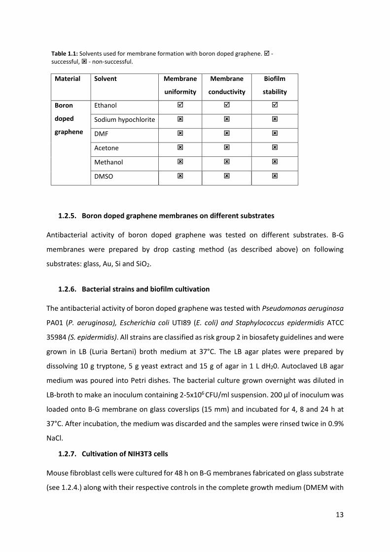
Used solvents and their characteristics are summarized in Table 1.1.
Solution of boron doped graphene flakes (B-G flakes) dispersed in solvent with different
concentrations (1, 2 and 4 mg/ml) were sonicated for 30 min (80 kHz, 100 W) using Elmasonic
P 30 H (Elma-Hans Schmidbauer GmbH, Singen, Germany) prior to every usage. B-G
membranes were initially fabricated by spray coating and standard spin coating methods (Karl
Suss, Model: Delta 20/BM, BLE) where it was realized that the membranes were not uniform.
In this case Drop casting method proved to be successful where ethanol and deionized water
(dH20) were used in a specific concentration to fabricate uniform B-G membranes on glass
coverslips (15 mm, VWR International GmbH, Germany). Just by using B-G solution (B-G flakes
dispersed in solvents) it was not possible to fabricate uniform membranes by drop casting due
to the stacking of B-G flakes. Addition of dH20 helps in better dispersibility and uniform
distribution of B-G flakes resulting in a more uniform B-G membrane.
1.2.4. Choosing a right solvent and boron doped graphene concentration The right solvent is the one that promotes uniform distribution of B-G flakes on the glass
substrate. Prior to drop casting, glass coverslips were cleaned with acetone and ethanol (70%).
Few drops of dH20 along with few drops of B-G solution were casted on separate coverslips
and left overnight for drying, followed by heat treatment (180°C, 2 h). The ratio of dH20 to one
of these B-G solutions was in the range of 1:1 to 1:4. The uniform spreading of B-G flakes
depends also on the external conditions such as cleanliness of the glass coverslip. After optical,
RAMAN and SEM characterization of the prepared membranes, samples with ethanol (EtOH)
provided the most uniform B-G membrane. Additionally, the membranes retained their
electrical conductivity and stability after the addition of bacterial suspension. For further
experiments, membranes were prepared with B-G in EtOH solution. Glass slides were casted
with B-G-EtOH (2.5 µg/ml) and dH20. First depositing 30 µl dH20 followed by the addition of
10 µl B-G-EtOH resulted in more homogenous deposition. These samples were used to analyze
the antibiotic effect against bacterial and mammalian cells.
Used solvents for B-G membranes and their parameters are summarized in Table 1.1.
13
Material Solvent Membrane
uniformity
Membrane
conductivity
Biofilm
stability
Boron
doped
graphene
Ethanol
Sodium hypochlorite
DMF
Acetone
Methanol
DMSO
1.2.5. Boron doped graphene membranes on different substrates Antibacterial activity of boron doped graphene was tested on different substrates. B-G
membranes were prepared by drop casting method (as described above) on following
substrates: glass, Au, Si and SiO2.
1.2.6. Bacterial strains and biofilm cultivation The antibacterial activity of boron doped graphene was tested with Pseudomonas aeruginosa
PA01 (P. aeruginosa), Escherichia coli UTI89 (E. coli) and Staphylococcus epidermidis ATCC
35984 (S. epidermidis). All strains are classified as risk group 2 in biosafety guidelines and were
grown in LB (Luria Bertani) broth medium at 37°C. The LB agar plates were prepared by
dissolving 10 g tryptone, 5 g yeast extract and 15 g of agar in 1 L dH20. Autoclaved LB agar
medium was poured into Petri dishes. The bacterial culture grown overnight was diluted in
LB-broth to make an inoculum containing 2-5x10⁶ CFU/ml suspension. 200 µl of inoculum was
loaded onto B-G membrane on glass coverslips (15 mm) and incubated for 4, 8 and 24 h at
37°C. After incubation, the medium was discarded and the samples were rinsed twice in 0.9%
NaCl.
1.2.7. Cultivation of NIH3T3 cells Mouse fibroblast cells were cultured for 48 h on B-G membranes fabricated on glass substrate
(see 1.2.4.) along with their respective controls in the complete growth medium (DMEM with
Table 1.1: Solvents used for membrane formation with boron doped graphene. - successful, - non-successful.
14
High Glucose, 4.0 mM L-Glu, sodium pyruvate and 10% iron-fortified Bovine Calf Serum, both
from ATCC, UK).
1.2.8. Colony Forming Units (CFUs) CFU counting was used to analyze the viability of bacteria in the biofilm. The rinsed biofilms
were collected in 5 ml of 0.89% NaCl, followed by sonication (20 s, 10 W, Branson 450 digital
sonifier, USA) to release the bacterial biofilm from glass coverslips. The homogenized
suspension (100 µL) was serially diluted in 0.89% NaCl and plated on LB agar respectively,
followed by incubation at 37°C overnight. The number of surviving bacteria in biofilm was
determined by counting the number of colonies and calculating the total number of CFU in 5
ml NaCl.
1.2.9. Raman Spectroscopy Raman spectroscopy is a tool to characterize material properties. The characteristic peaks for
carbon based materials in Raman spectra are clearly identified by this technique. The
membranes were casted by controlled drop casting method. The flake size of the used B-G is
in the range of 0.5 to 5 µm. The final thickness of the fabricated B-G membrane is
approximately 100 to 120 microns. These membranes were further used for RAMAN
characterization.
1.2.10. Scanning Electron Microscopy (SEM) For SEM, biofilms were fixed with glutaraldehyde (3%) for 2 h and dehydrated in graded series
of ethanol concentrations (40, 50, 60, 70, 80, 90, and 100%; v/v) for 10 min in each solution.
The dehydrated biofilms were dried at room temperature for 2 h. Prior to SEM, the samples
were coated with gold (5 nm, 5 s, 0.2 kW)) using MS 150 Sputter system (FHR, Anlagenbau
GmbH, Germany). SEM imaging was performed using JEOL JSM-6301F Scanning Electron
Microscope (USA).
1.2.11. Statistical analysis Experiments were performed in biological triplicates and data is presented as the mean ±
standard deviation. Statistical analysis of the results was estimated by the one-way analysis of

15
variance (ANOVA), followed by the multiple comparison (Tukey) test. Values at p<0.05 were
considered as statistically significant.
1.3. Results and Discussion
1.3.1. RAMAN characterization Raman spectroscopy (Wave length = 532 nm) was used for the characterization of the
fabricated B-G membranes. It can be observed that there is a slight shift and broadening of D
peak (to 1336 cm-1) in comparison to standard carbon materials (1330 cm-1). B-G shows more
intense D peak compared to 2D peak. As seen in the spectrum, the 2D peak is suppressed with
the addition of boron (Fig. 1.3) [24].
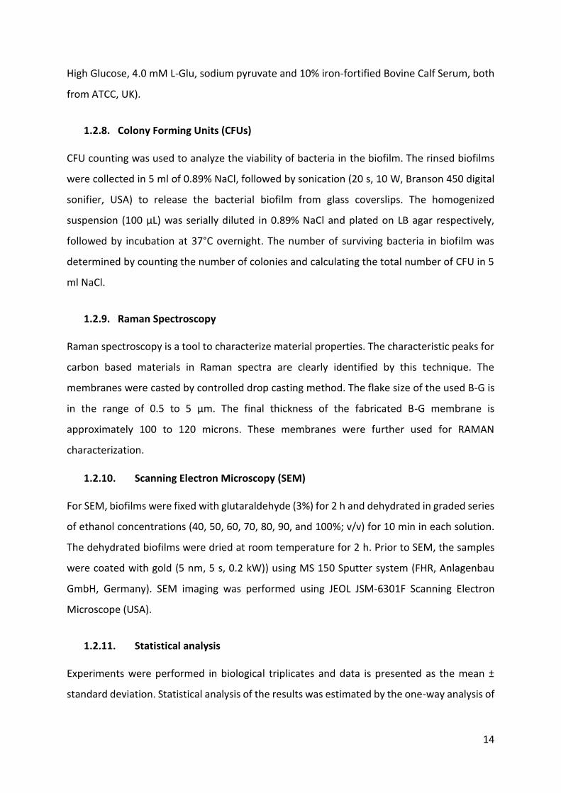
1.3.2. Membrane integrity Different solvents were used to fabricate B-G membranes on glass coverslips. Figure 1.4 shows
SEM images of B-G membranes prepared with sodium hypochlorite (Klorin, Danmark) and
acetone on glass coverslips.
Figure 1.3: RAMAN spectrum of Boron doped graphene membrane.
16
As seen in Figure 1.4, the membranes prepared with sodium hypochlorite seem to have
undergone a chemical reaction where a white layer was observed (this was later proven by
the conductivity test where the B-G membranes seemed to have lost their native properties).
Figure 1.4 also shows membranes prepared with acetone + B-G, which seem to have uniform
coverage on the glass coverslip, nevertheless, when these membranes were immersed in the
medium to form a bacterial biofilm, B-G flakes were detached from the membrane and were
seen floating within the medium. This is due to the problem of adhesion between acetone B-
G flakes and the cover glass surface. Inspite of heat treatment, this acetone based B-G
membranes showed similar dispersion within the medium.
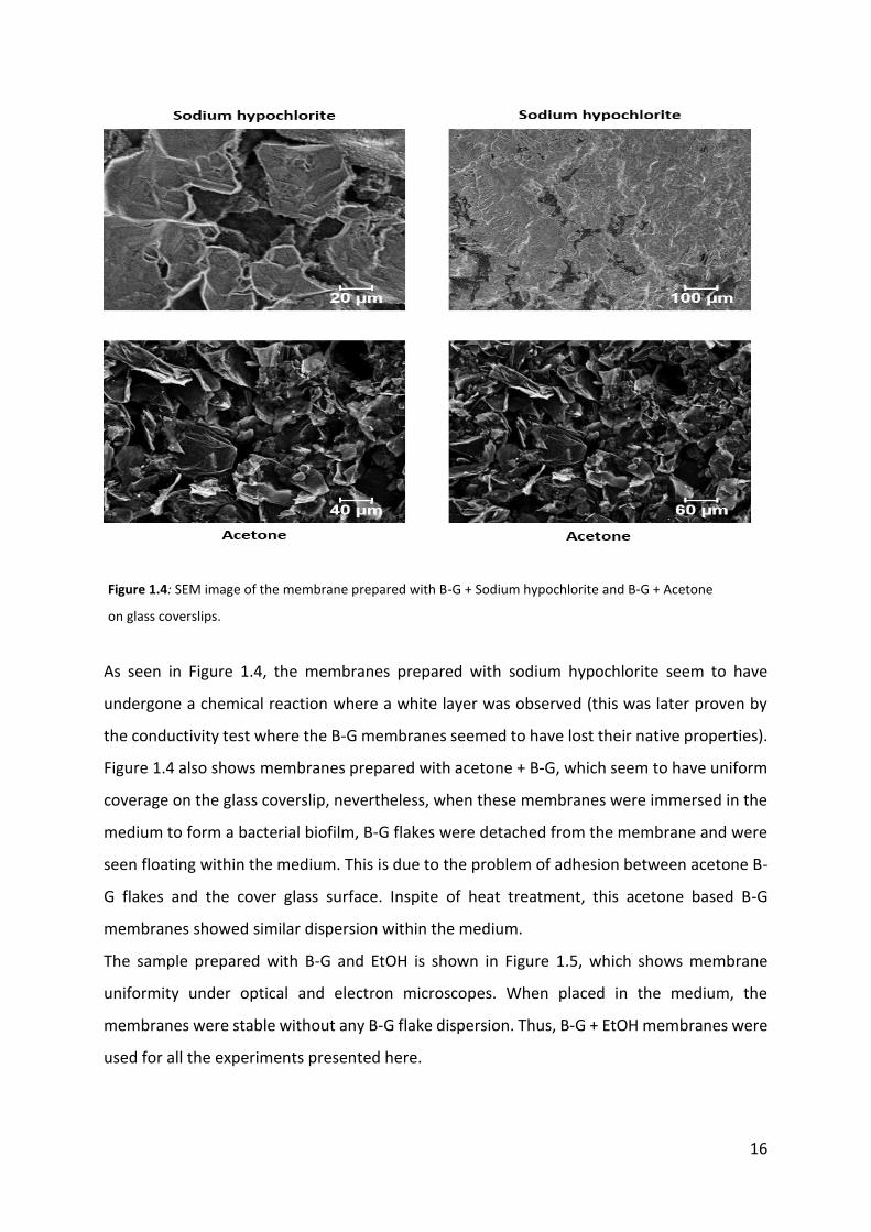
The sample prepared with B-G and EtOH is shown in Figure 1.5, which shows membrane
uniformity under optical and electron microscopes. When placed in the medium, the
membranes were stable without any B-G flake dispersion. Thus, B-G + EtOH membranes were
used for all the experiments presented here.
Figure 1.4: SEM image of the membrane prepared with B-G + Sodium hypochlorite and B-G + Acetone
on glass coverslips.
17
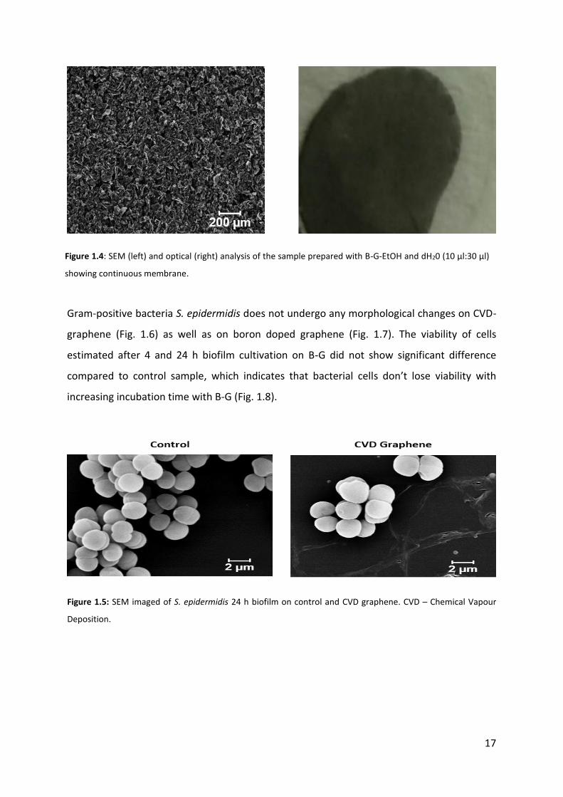
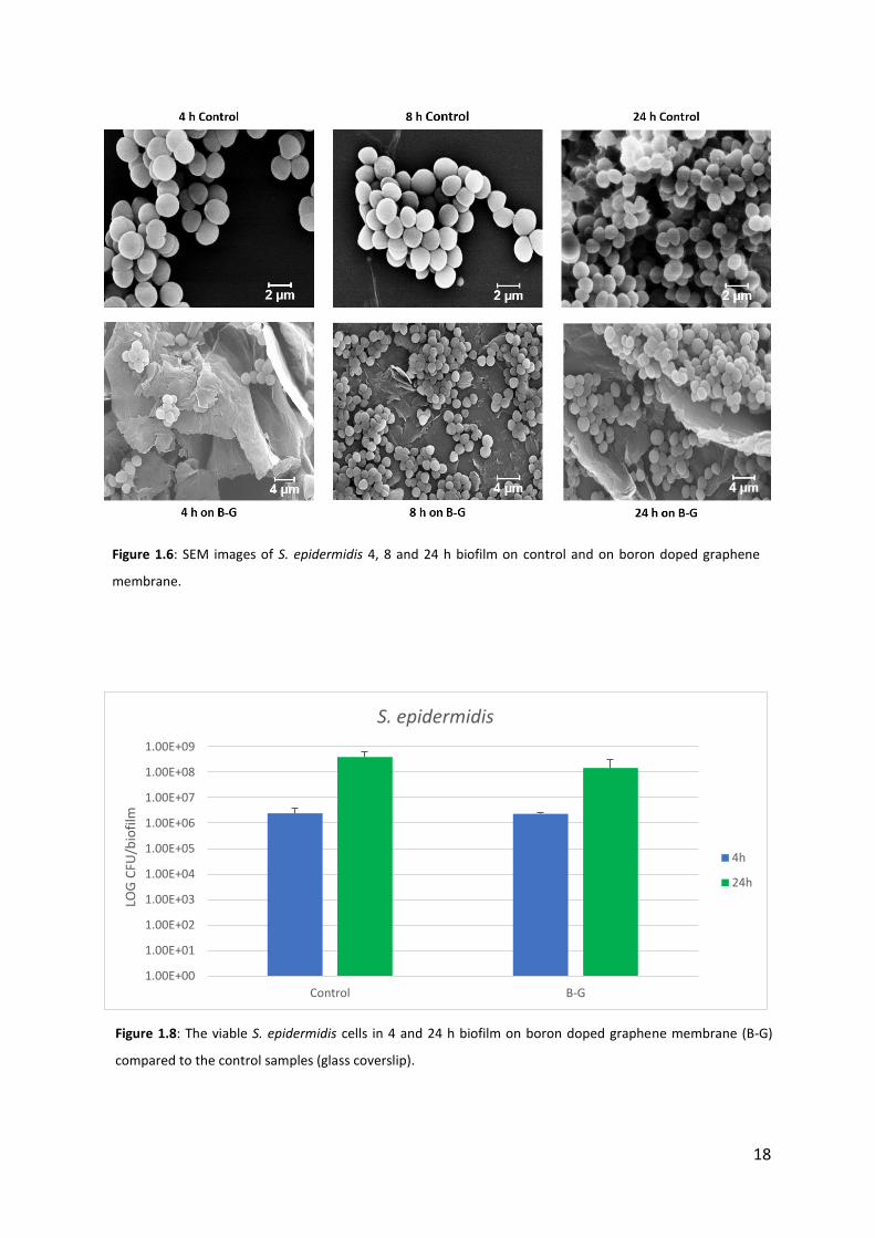
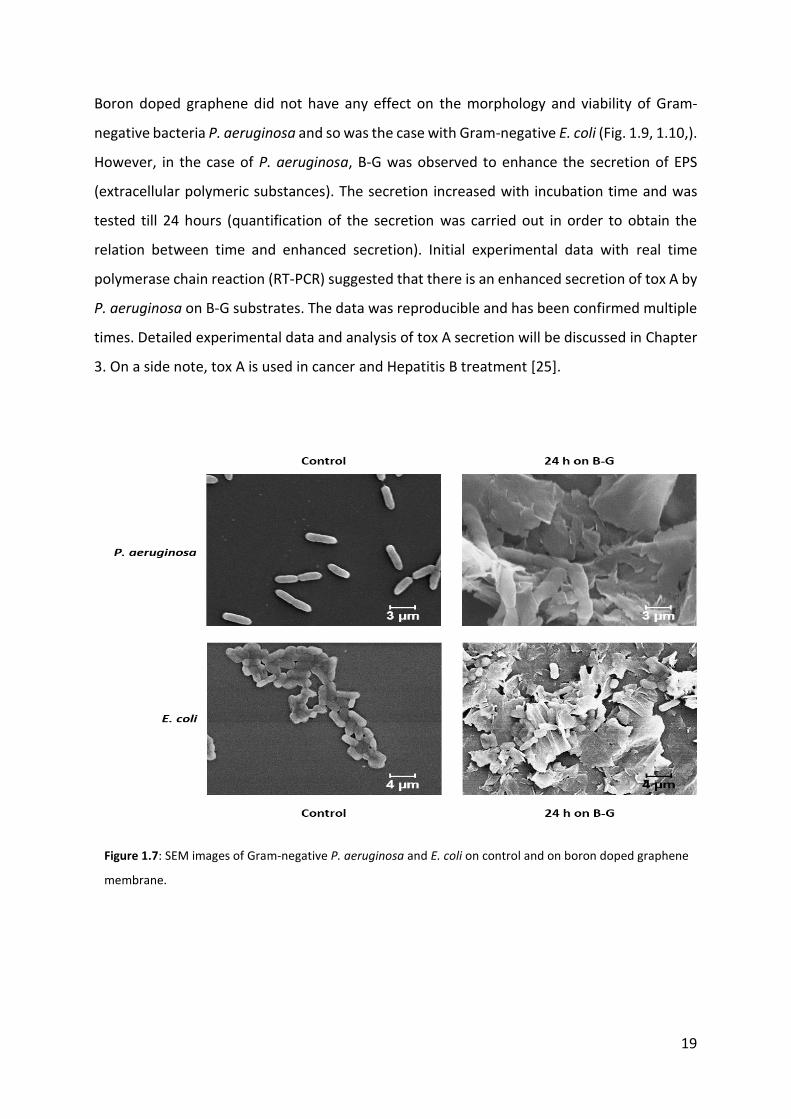
Gram-positive bacteria S. epidermidis does not undergo any morphological changes on CVD-
graphene (Fig. 1.6) as well as on boron doped graphene (Fig. 1.7). The viability of cells
estimated after 4 and 24 h biofilm cultivation on B-G did not show significant difference
compared to control sample, which indicates that bacterial cells don’t lose viability with
increasing incubation time with B-G (Fig. 1.8).
Figure 1.4: SEM (left) and optical (right) analysis of the sample prepared with B-G-EtOH and dH20 (10 µl:30 µl)
showing continuous membrane.
Figure 1.5: SEM imaged of S. epidermidis 24 h biofilm on control and CVD graphene. CVD – Chemical Vapour
Deposition.
18
1.00E+00
1.00E+01
1.00E+02
1.00E+03
1.00E+04
1.00E+05
1.00E+06
1.00E+07
1.00E+08
1.00E+09
Control B-G
LOG
CFU
/bio
film
S. epidermidis
4h
24h
Figure 1.8: The viable S. epidermidis cells in 4 and 24 h biofilm on boron doped graphene membrane (B-G)
compared to the control samples (glass coverslip).
Figure 1.6: SEM images of S. epidermidis 4, 8 and 24 h biofilm on control and on boron doped graphene
membrane.
19
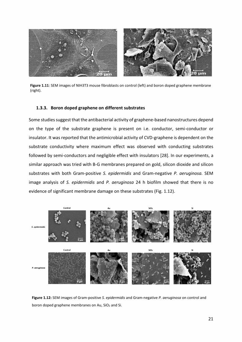
Boron doped graphene did not have any effect on the morphology and viability of Gram-
negative bacteria P. aeruginosa and so was the case with Gram-negative E. coli (Fig. 1.9, 1.10,).
However, in the case of P. aeruginosa, B-G was observed to enhance the secretion of EPS
(extracellular polymeric substances). The secretion increased with incubation time and was
tested till 24 hours (quantification of the secretion was carried out in order to obtain the
relation between time and enhanced secretion). Initial experimental data with real time
polymerase chain reaction (RT-PCR) suggested that there is an enhanced secretion of tox A by
P. aeruginosa on B-G substrates. The data was reproducible and has been confirmed multiple
times. Detailed experimental data and analysis of tox A secretion will be discussed in Chapter
3. On a side note, tox A is used in cancer and Hepatitis B treatment [25].
Figure 1.7: SEM images of Gram-negative P. aeruginosa and E. coli on control and on boron doped graphene
membrane.

20
The effect of B-G activity towards eukaryotic cells was also examined using NIH3T3 mouse
fibroblast cells. These mammalian cells are approximately 20 µm and are way larger than
bacterial cells (1-2 µm). Under SEM, no mechanical damage of these cells was observed (Fig.
1.11). In the literature, it was mentioned that sodium pentaborate pentahydrate, which is a
source of boron, when combined with graphene oxide (GO), enhances adherence and
proliferation of mammalian cells [26]. On the other hand, substrates with wrinkles and ripples
enhance cell proliferation and viability [27]. Inspite of our B-G membranes having a lot of
ripples and wrinkles, which is supposedly for interaction, enhanced proliferation was not
observed. It has to be noted that though boron can enhance the proliferation of mammalian
cells, the structural morphology, function and properties of mouse fibroblast cells used in this
experiment and mesenchymal stem cells used in above mentioned literature study are quite
different. The negative control (bacterial cells on glass coverslip) showed that incubation
conditions did not cause morphological damages.
1.00E+00
1.00E+01
1.00E+02
1.00E+03
1.00E+04
1.00E+05
1.00E+06
1.00E+07
1.00E+08
1.00E+09
1.00E+10
Control B-G
LOG
CFU
/bio
film
E. coli
P. aeruginosa
Figure 1.10: Comparison of cell viability between two Gram-negative bacteria: P. aeruginosa and E. coli in
24 h biofilm.
21
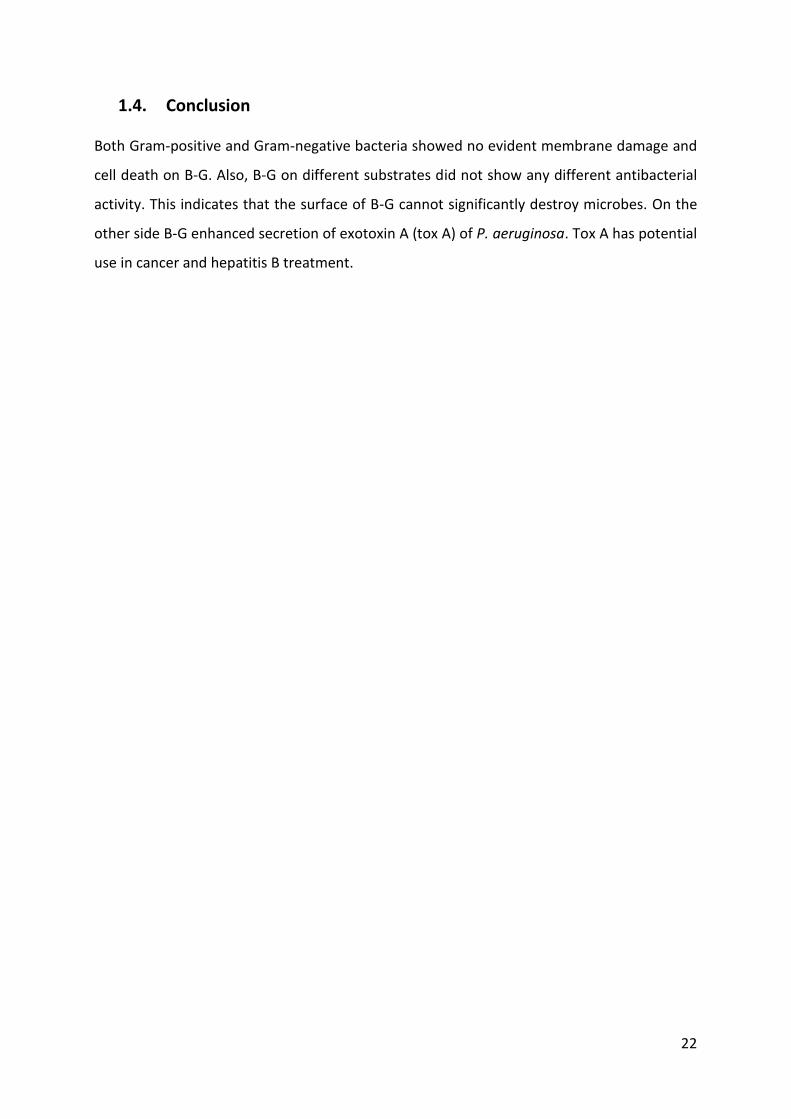
1.3.3. Boron doped graphene on different substrates Some studies suggest that the antibacterial activity of graphene-based nanostructures depend
on the type of the substrate graphene is present on i.e. conductor, semi-conductor or
insulator. It was reported that the antimicrobial activity of CVD-graphene is dependent on the
substrate conductivity where maximum effect was observed with conducting substrates
followed by semi-conductors and negligible effect with insulators [28]. In our experiments, a
similar approach was tried with B-G membranes prepared on gold, silicon dioxide and silicon
substrates with both Gram-positive S. epidermidis and Gram-negative P. aeruginosa. SEM
image analysis of S. epidermidis and P. aeruginosa 24 h biofilm showed that there is no
evidence of significant membrane damage on these substrates (Fig. 1.12).
Figure 1.11: SEM images of NIH3T3 mouse fibroblasts on control (left) and boron doped graphene membrane (right).
Figure 1.12: SEM images of Gram-positive S. epidermidis and Gram-negative P. aeruginosa on control and
boron doped graphene membranes on Au, SiO2 and Si.
22
1.4. Conclusion Both Gram-positive and Gram-negative bacteria showed no evident membrane damage and
cell death on B-G. Also, B-G on different substrates did not show any different antibacterial
activity. This indicates that the surface of B-G cannot significantly destroy microbes. On the
other side B-G enhanced secretion of exotoxin A (tox A) of P. aeruginosa. Tox A has potential
use in cancer and hepatitis B treatment.
23
2. Antibacterial activity of other 2D materials
2.1. Introduction Since the discovery of graphene and exploring its unique properties, 2D materials such as
chalcogenides, transition metal oxides have gained tremendous research interest [29]. A
single layer of transition metal dichalcogenides (TMDs) consist of two planes of hexagonal
arranged chalcogen atoms (X) linked to hexagonal plane of metal atom (M), with
stoichiometry MX2. The atoms are held together by weak Van der Waals forces. Depending
on the combination of chalcogen and metal atoms, various types of TMDs can occur. WS2 and
MoS2 are prototypical TMDs. Due to their semiconducting properties and tunable gap, WS2
and MoS2 have been identified as graphene analogue materials [30]. They are promising
materials with a wide range of potential applications including energy generation, catalysts,
sensing and biomedicine. MoS2 sheets can be used in biomedical applications for detection of
DNA molecules [31], however, their antibacterial properties are still unexplored.
2.2. Materials and Methods
2.2.1. Materials MoS2 and WS2 in ultrafine powder- and nanoflake solution-forms were obtained from
Graphene Supermarket (USA). If not stated differently, all other chemicals used were
purchased from VWR BDH Chemicals (UK), EMD Millipore (USA) or Sigma-Aldrich (USA) with
the highest purity available.
2.2.2. Preparation of membranes and choosing the right solvent MoS2 (Molybdenum disulfide) and WS2 (Tungsten disulfide) in liquid and powder forms were
tested for uniform membrane preparation. As suggested in the literature, acetone, ethanol
and isopropyl alcohol (IPA) were used as potential solvents that facilitate uniform distribution
of MoS2- and WS2-dry powders on glass coverslips [32]. Membranes were first fabricated by
using spray drying and spin coating methods only to realize that there was no uniformity in
MoS2- and WS2 dispersion. By using drop casting method, MoS2 and WS2 membranes were
successfully fabricated on glass coverslips. Dispersed solutions of MoS2 and WS2 flakes in

24
different concentrations (1, 2 and 4 mg/ml) were sonicated for 30 min prior to drop casting,
glass coverslips were cleaned with acetone and ethanol (70%). Few drops of MoS2 and WS2
were casted on separate glass slides and left overnight for drying, followed by heat treatment
(180°C, 2 h). MoS2 and WS2 in liquid form was casted as single and double layer. After optical
and SEM characterization of the fabricated membranes, it was realized that the samples with
MoS2 and WS2 dispersed in IPA provided the most uniform membranes, which were used for
further experiments. The antibacterial testing on these membranes was performed in the
same way as for B-G membranes (see page 13, 14). Used solvents for MoS2 and WS2
membranes and their parameters are summarized in Table 2.1.
Material Solvent Membrane
uniformity
Membrane
conductivity
Biofilm
stability
WS2, MoS2
(dry powder)
Ethanol
Acetone
IPA
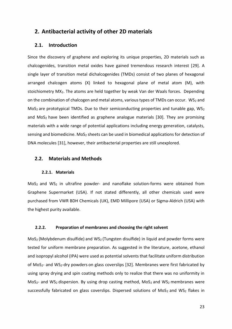
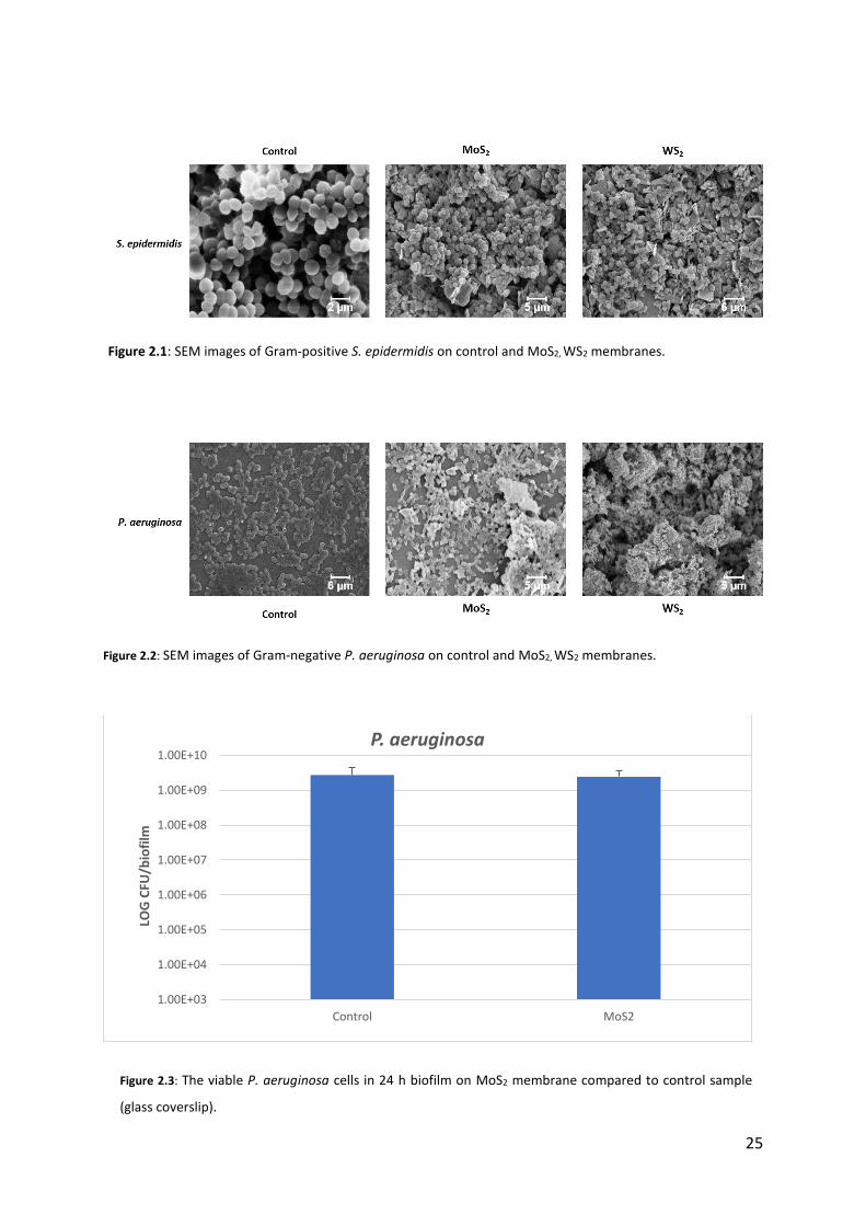
2.3. Results and Discussion The antibacterial activities of graphene analogue MoS2 and WS2 were tested. Some studies
claim that graphene oxide-MoS2 nanosheets show antibacterial effect towards Gram-negative
bacteria like E. coli, due to induction of oxidative stress [33]. However, membranes prepared
from both 2D materials did not have any antibacterial effect towards Gram-positive as well as
Gram-negative bacteria. Under SEM evaluation no morphological changes were observed (Fig.
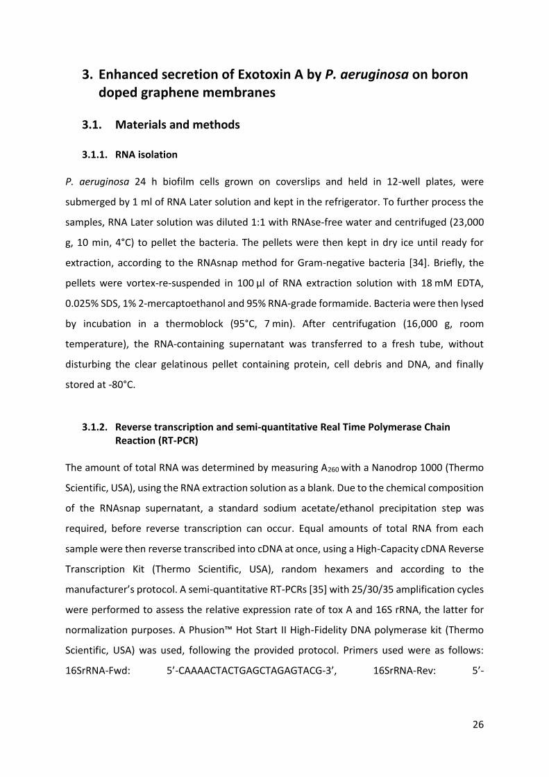
2.1, 2.2). Moreover, surfaces with MoS2 had no observable effect on P. aeruginosa viability
(Fig. 2.3).
Table 2.1: Solvents used for membrane formation with MoS2 (Molybdenum disulfide)
and WS2 (Tungsten disulfide). - successful, - non-successful.
25
1.00E+03
1.00E+04
1.00E+05
1.00E+06
1.00E+07
1.00E+08
1.00E+09
1.00E+10
Control MoS2
LOG
CFU
/bio
film
P. aeruginosa
Figure 2.1: SEM images of Gram-positive S. epidermidis on control and MoS2, WS2 membranes.
Figure 2.2: SEM images of Gram-negative P. aeruginosa on control and MoS2, WS2 membranes.
Figure 2.3: The viable P. aeruginosa cells in 24 h biofilm on MoS2 membrane compared to control sample
(glass coverslip).
26
3. Enhanced secretion of Exotoxin A by P. aeruginosa on boron doped graphene membranes
3.1. Materials and methods
3.1.1. RNA isolation
P. aeruginosa 24 h biofilm cells grown on coverslips and held in 12-well plates, were
submerged by 1 ml of RNA Later solution and kept in the refrigerator. To further process the
samples, RNA Later solution was diluted 1:1 with RNAse-free water and centrifuged (23,000
g, 10 min, 4°C) to pellet the bacteria. The pellets were then kept in dry ice until ready for
extraction, according to the RNAsnap method for Gram-negative bacteria [34]. Briefly, the
pellets were vortex-re-suspended in 100 µl of RNA extraction solution with 18 mM EDTA,
0.025% SDS, 1% 2-mercaptoethanol and 95% RNA-grade formamide. Bacteria were then lysed
by incubation in a thermoblock (95°C, 7 min). After centrifugation (16,000 g, room
temperature), the RNA-containing supernatant was transferred to a fresh tube, without
disturbing the clear gelatinous pellet containing protein, cell debris and DNA, and finally
stored at -80°C.
3.1.2. Reverse transcription and semi-quantitative Real Time Polymerase Chain Reaction (RT-PCR)
The amount of total RNA was determined by measuring A260 with a Nanodrop 1000 (Thermo
Scientific, USA), using the RNA extraction solution as a blank. Due to the chemical composition
of the RNAsnap supernatant, a standard sodium acetate/ethanol precipitation step was
required, before reverse transcription can occur. Equal amounts of total RNA from each
sample were then reverse transcribed into cDNA at once, using a High-Capacity cDNA Reverse
Transcription Kit (Thermo Scientific, USA), random hexamers and according to the
manufacturer’s protocol. A semi-quantitative RT-PCRs [35] with 25/30/35 amplification cycles
were performed to assess the relative expression rate of tox A and 16S rRNA, the latter for
normalization purposes. A Phusion™ Hot Start II High-Fidelity DNA polymerase kit (Thermo
Scientific, USA) was used, following the provided protocol. Primers used were as follows:
16SrRNA-Fwd: 5’-CAAAACTACTGAGCTAGAGTACG-3’, 16SrRNA-Rev: 5’-
27
TAAGATCTCAAGGATCCCAACGGCT-3’, ToxA-Fwd: 5’-ATGGTGTAGATCGGCGACAT-3’, ToxA-
Rev: 5’-AAGCCTTCGACCTCTGGAAC-3’.
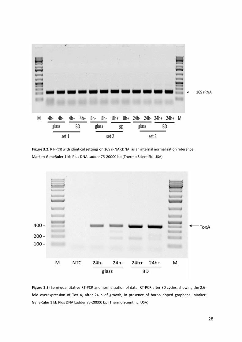
3.2. Results and discussion
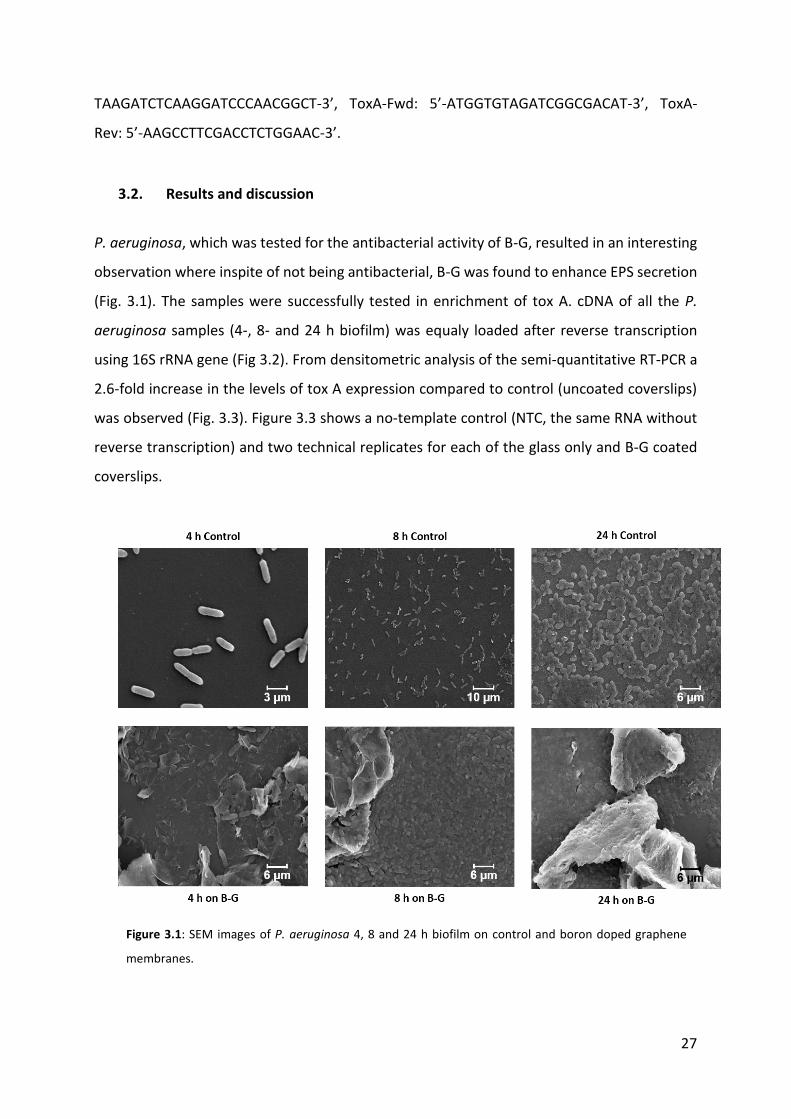
P. aeruginosa, which was tested for the antibacterial activity of B-G, resulted in an interesting
observation where inspite of not being antibacterial, B-G was found to enhance EPS secretion
(Fig. 3.1). The samples were successfully tested in enrichment of tox A. cDNA of all the P.
aeruginosa samples (4-, 8- and 24 h biofilm) was equaly loaded after reverse transcription
using 16S rRNA gene (Fig 3.2). From densitometric analysis of the semi-quantitative RT-PCR a
2.6-fold increase in the levels of tox A expression compared to control (uncoated coverslips)
was observed (Fig. 3.3). Figure 3.3 shows a no-template control (NTC, the same RNA without
reverse transcription) and two technical replicates for each of the glass only and B-G coated
coverslips.
Figure 3.1: SEM images of P. aeruginosa 4, 8 and 24 h biofilm on control and boron doped graphene
membranes.
28
Figure 3.3: Semi-quantitative RT-PCR and normalization of data: RT-PCR after 30 cycles, showing the 2.6-
fold overexpression of Tox A, after 24 h of growth, in presence of boron doped graphene. Marker:
GeneRuler 1 kb Plus DNA Ladder 75-20000 bp (Thermo Scientific, USA).
Figure 3.2: RT-PCR with identical settings on 16S rRNA cDNA, as an internal normalization reference.
Marker: GeneRuler 1 kb Plus DNA Ladder 75-20000 bp (Thermo Scientific, USA)-
29
4. Antibacterial activity of plant and bacteria derived nanoparticles
4.1. Introduction Metal nanoparticles have attracted interest of scientists, since implementation of
nanotechnology allows the synthesis of particles in nanometers [36]. Due to their high surface
area to volume ratio and unique physicochemical and biological properties, the use of NPS in
electrical, chemical or biological field has attracted global attention. Silver ions show high
toxicity against microorganisms. There are several chemical and physical methods for the
production of nanoparticles. These existing methods are usually expensive, labor extensive
and potentially hazardous to the surrounding environment as well as living organisms. Hence,
researchers are using alternative ways to produce metallic nanoparticles which are easy,
cheap and non hazardous. In this study silver and gold nanoparticles were produced by using
plant extracts and bacteria which have the ability to reduce silver and gold salt to respective
nanoparticles. Silver has been used in medical field for treatment of wounds, burns and
bacterial infections [37, 38]. Silver in nanoparticle shows enhanced antibacterial activity. The
antibacterial efficacy of gold nanoparticles (CS-Au-NPs) and silver nanoparticles (CS-Ag-NPS)
by Cannabis sativa (C. sativa) (Hemp) was evaluated by determining minimum growth
inhibitory concentration (MIC) and minimum bactericidal concentration (MBC) against Gram-
positive and Gram-negative bacteria.
4.2. Materials and Methods
4.2.1. Synthesis of nanoparticles Eco-friendly synthesis of nanoparticles has been gaining a lot of importance due to its
environment friendly approach. In this study, silver and gold nanoparticles were produced by
C. sativa which have the ability to reduce silver and gold salts to respective nanoparticles. The
antibacterial activity was evaluated by determining the MIC and MBC towards the following
bacterial strains: Pseudomonas aeruginosa PA01, E. coli (UTI89) and S. epidermidis (ATCC
35984). All strains are classified to risk group 2 in biosafety guidelines. Moreover, the
30
antibacterial activity was confirmed by colony forming units counting (CFUs), scanning
electron microscopy (SEM) and live /dead staining.
4.2.2. Determination of Minimum Inhibitory Concentration (MIC) and Minimum
Bactericidal Concentration (MBC)
MIC is the lowest concertation of material that inhibits visible growth of bacteria after
overnight incubation. Experiments were performed in 96-well plates. NPs were suspended in
dH20 (1 mg/ml) and sonicated for 15 min prior to use, followed by serial dilution in LB broth
media (total volume 180 µl) ranging from 6.25 µg/ml to 50 µg/ml. Bacterial culture in a
logarithmic phase (20 µl) was added to serial dilutions of NPs. The 96-well plates were
incubated with shaking (170 rpm, 37°C) on IKA KS 4000i Control Shaking Incubator (IKA-Werke
GmbH & Co. KG, Staufen, Germany) overnight. MIC was determined when no turbidity in the
wells was observed. After the MIC determination of NPs, the aliquots of cultures where no
growth was observed, were plated on LB-agar and incubated at 37°C for 24 h. MBC is defined
as the lowest concentration of material required to kill bacterial population
4.2.3. Colony Forming Units (CFUs) Biofilms were grown for 24 h without any disturbance (See 1.2.6.). After the 24 h old culture
medium was replaced with different concentration of AgNP containing fresh medium and
incubated for another 24 h. After 24 h of nanoparticle treatment, biofilms were homogenized
by sonication and plated on agar plates for CFU counting.
4.2.4. Live Dead Staining The toxicity of CS-Ag-NPs to the surfaces for P. aeruginosa and E. coli cells was tested by
staining the cells (24 h biofilm) with a membrane integrity evaluation kit (ReadyProbes® Cell
Viability Imaging Kit Blue/Red, Thermo Scientific, USA). Its active dyes show different
specificities within viable and damaged cells, with NucBlue® Live reagent staining the nuclei
of all the cells, while propidium iodide stains only the nuclei of cells with compromised
membrane integrity. After treating the biofilm with different concentration of AgNPs, as
described above, bacterial cells were stained according to the kit manufacturer’s instructions
and fixed with freshly made 4% paraformaldehyde in DPBS for 10 min at room temperature.
31
The samples were then rinsed in distilled water and mounted with a droplet of ProLong®
Diamond Antifade Mountant medium (Thermo Scientific, USA) against a glass coverslip and
the back of the coverslip attached, with a small amount of superglue, to a glass slide for
imaging. Imaging was performed with a confocal laser scanning microscope (LSM 700 NLO,
Carl Zeiss, Germany). For all samples three biological replicates were analyzed, with five
images analyzed per replicate. Representative images are shown.
4.2.5. Scanning Electron Microscopy (SEM) Biofilms were grown for 24 h without any disturbance. After the 24 h old culture medium was
replaced with different concentration of AgNP containing fresh medium and incubated for
another 24 h. After 24 h of nanoparticle treatment biofilms, were fixed with gluteraldehyde,
dehydrated with graded ethanol and imaging was performed by using SEM after gold coating
(see 1.2.10.).
4.3. Results and discussion The determined MIC and MBC values of NPs against 3 different bacterial strains are shown in
Table 4.1. S. epidermidis showed resistance against CS-Ag-NPs. CS-Ag-NPs showed strong
inhibitory as well as bactericidal effect against P. aeruginosa and E. coli. While P. aeruginosa
showed MIC value of 6.25 µg/ml, E. coli showed the MIC value of 12.5 µg/ml. Obtained MBC
value for E. coli was 25 µg/ml, while for P. aeruginosa 12.5 µg/ml.
Bacteria MIC [µg/ml] MBC [µg/ml]
P. aeruginosa PA01 6.25 12.5
E. coli UT189 12.5 25
S. epidermidis >50 >50
The MIC and MBC of gold nanoparticles was >50 µg/ml against above mentioned bacteria.
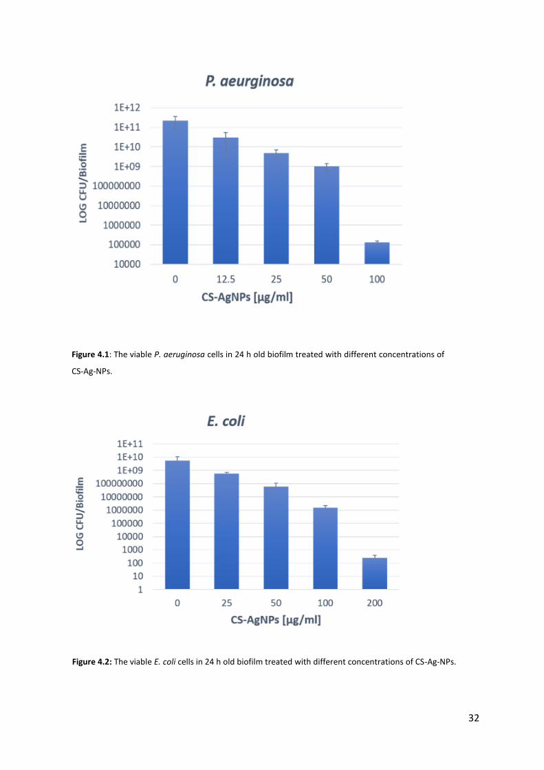
In Figures 4.1 and 4.2 the results from CFUs counting demonstrate loss of cell viability in P.
aeruginosa and E. coli biofilms treated with different concentrations of CS-Ag-NPs.
Table 4.1: Minimum inhibitory concentration (MIC) and minimum bactericidal concentration (MBC) of CS-Ag-NPs.
32
Figure 4.1: The viable P. aeruginosa cells in 24 h old biofilm treated with different concentrations of
CS-Ag-NPs.
Figure 4.2: The viable E. coli cells in 24 h old biofilm treated with different concentrations of CS-Ag-NPs.
33
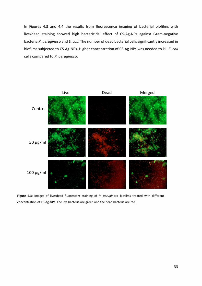
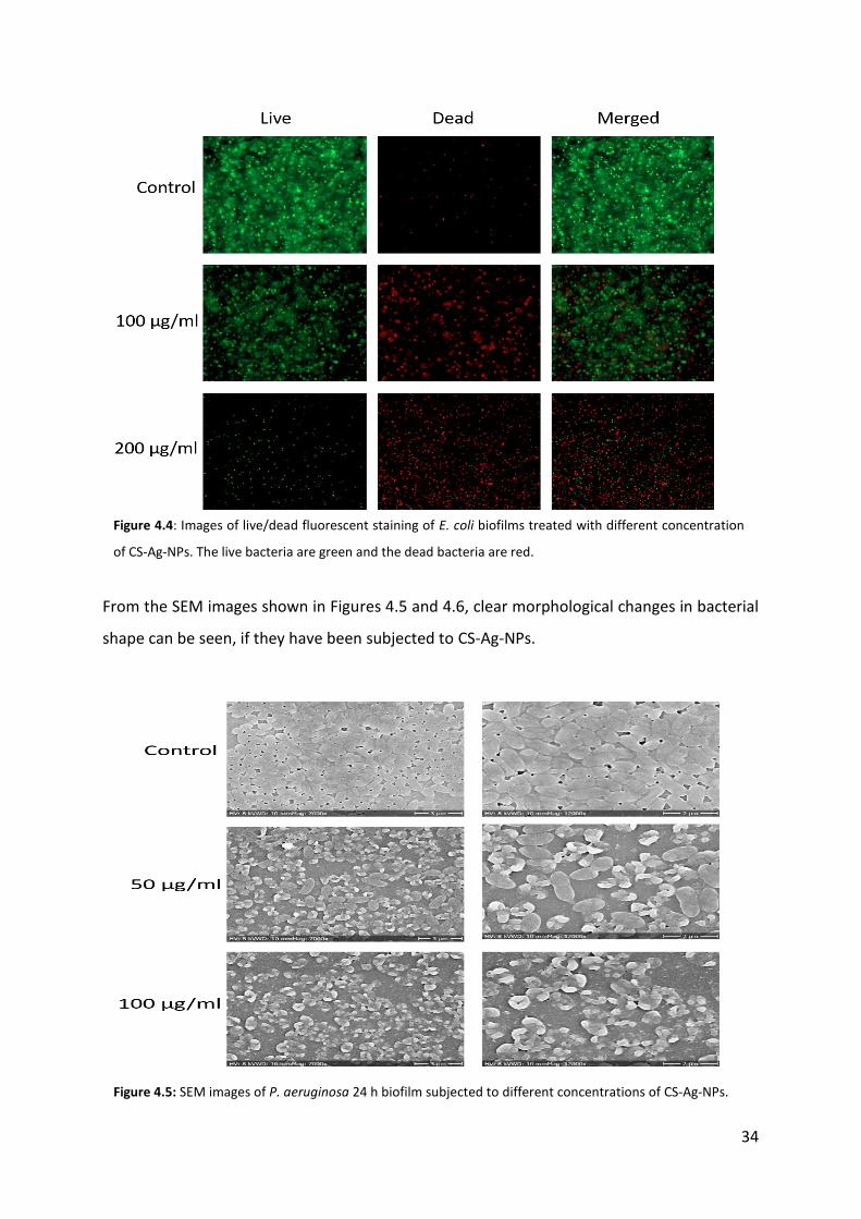
In Figures 4.3 and 4.4 the results from fluorescence imaging of bacterial biofilms with
live/dead staining showed high bactericidal effect of CS-Ag-NPs against Gram-negative
bacteria P. aeruginosa and E. coli. The number of dead bacterial cells significantly increased in
biofilms subjected to CS-Ag-NPs. Higher concentration of CS-Ag-NPs was needed to kill E. coli
cells compared to P. aeruginosa.
Figure 4.3: Images of live/dead fluorescent staining of P. aeruginosa biofilms treated with different
concentration of CS-Ag-NPs. The live bacteria are green and the dead bacteria are red.
34
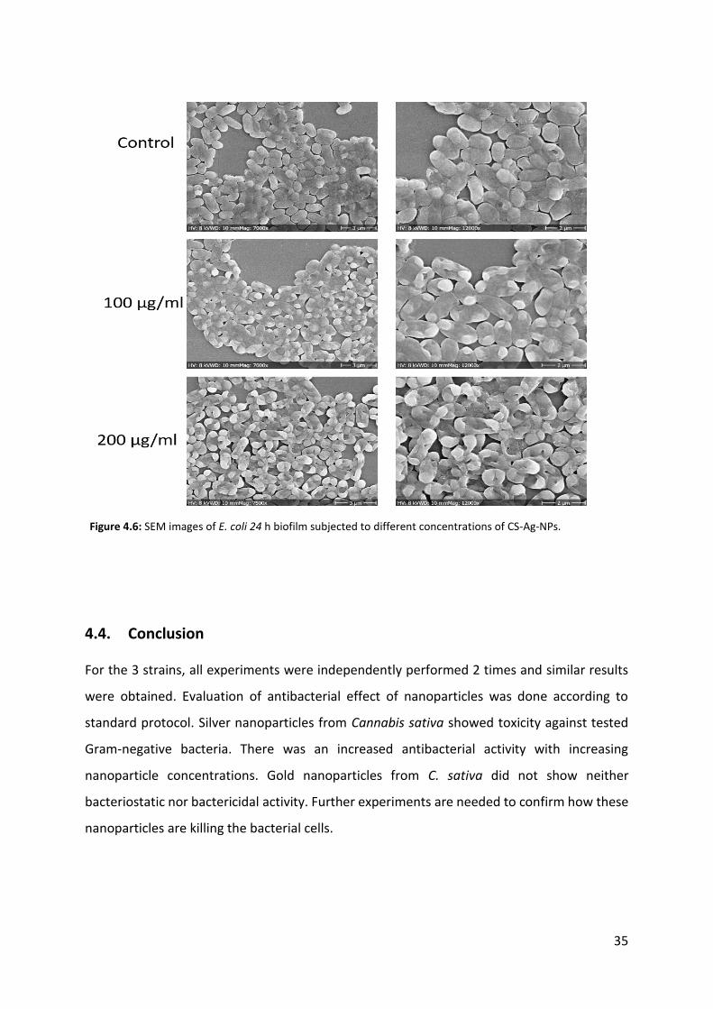
From the SEM images shown in Figures 4.5 and 4.6, clear morphological changes in bacterial
shape can be seen, if they have been subjected to CS-Ag-NPs.
Figure 4.5: SEM images of P. aeruginosa 24 h biofilm subjected to different concentrations of CS-Ag-NPs.
Figure 4.4: Images of live/dead fluorescent staining of E. coli biofilms treated with different concentration
of CS-Ag-NPs. The live bacteria are green and the dead bacteria are red.
35
4.4. Conclusion For the 3 strains, all experiments were independently performed 2 times and similar results
were obtained. Evaluation of antibacterial effect of nanoparticles was done according to
standard protocol. Silver nanoparticles from Cannabis sativa showed toxicity against tested
Gram-negative bacteria. There was an increased antibacterial activity with increasing
nanoparticle concentrations. Gold nanoparticles from C. sativa did not show neither
bacteriostatic nor bactericidal activity. Further experiments are needed to confirm how these
nanoparticles are killing the bacterial cells.
Figure 4.6: SEM images of E. coli 24 h biofilm subjected to different concentrations of CS-Ag-NPs.
36
5. Bacterial biofilm sensor
5.1. Introduction
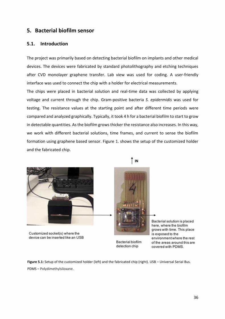
The project was primarily based on detecting bacterial biofilm on implants and other medical
devices. The devices were fabricated by standard photolithography and etching techniques
after CVD monolayer graphene transfer. Lab view was used for coding. A user-friendly
interface was used to connect the chip with a holder for electrical measurements.
The chips were placed in bacterial solution and real-time data was collected by applying
voltage and current through the chip. Gram-positive bacteria S. epidermidis was used for
testing. The resistance values at the starting point and after different time periods were
compared and analyzed graphically. Typically, it took 4 h for a bacterial biofilm to start to grow
in detectable quantities. As the biofilm grows thicker the resistance also increases. In this way,
we work with different bacterial solutions, time frames, and current to sense the biofilm
formation using graphene based sensor. Figure 1. shows the setup of the customized holder
and the fabricated chip.
Figure 5.1: Setup of the customized holder (left) and the fabricated chip (right). USB – Universal Serial Bus.
PDMS – Polydimethylsiloxane.
37
5.2. Materials and Methods After the fabrication and testing of the device, firstly the chip was placed in the holder and
calibrated using standard dH2O. Further, as a control, the chip was dipped into the control
medium (without bacteria) and run for 6 h. Real-time data was acquired using lab view
interface. In the next step, bacterial suspension with the medium was run for another 6 h and
the data was collected.
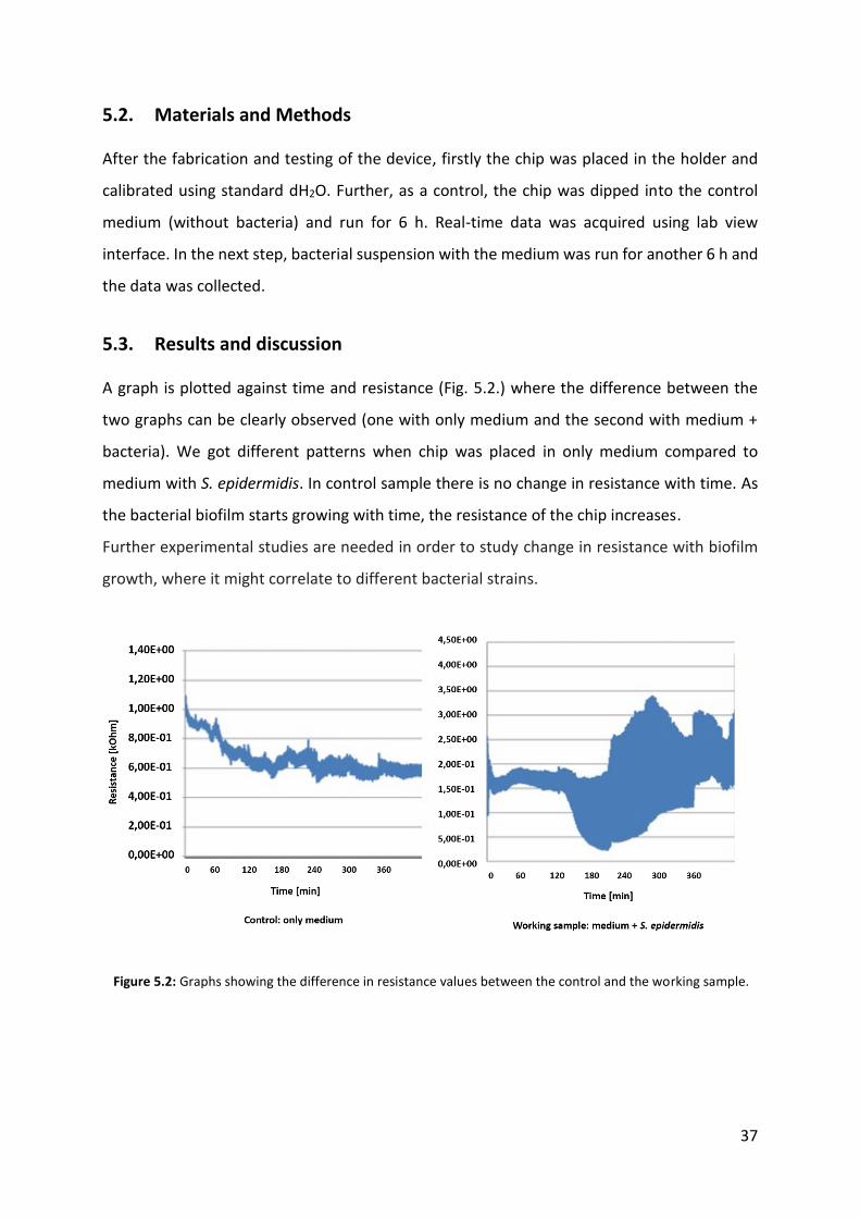
5.3. Results and discussion A graph is plotted against time and resistance (Fig. 5.2.) where the difference between the
two graphs can be clearly observed (one with only medium and the second with medium +
bacteria). We got different patterns when chip was placed in only medium compared to
medium with S. epidermidis. In control sample there is no change in resistance with time. As
the bacterial biofilm starts growing with time, the resistance of the chip increases.
Further experimental studies are needed in order to study change in resistance with biofilm
growth, where it might correlate to different bacterial strains.
Figure 5.2: Graphs showing the difference in resistance values between the control and the working sample.
38
6. Literature
1. Neto, A. C., Guinea, F., Peres, N. M., Novoselov, K. S., & Geim, A. K. (2009). The
electronic properties of graphene. Reviews of modern physics, 81(1), 109.
2. Geim, A. K., & Novoselov, K. S. (2007). The rise of graphene. Nature materials, 6(3),
183-191.
3. Liu, J., Cui, L., & Losic, D. (2013). Graphene and graphene oxide as new nanocarriers
for drug delivery applications. Acta biomaterialia, 9(12), 9243-9257.
4. Mao, H. Y., Laurent, S., Chen, W., Akhavan, O., Imani, M., Ashkarran, A. A., &
Mahmoudi, M. (2013). Graphene: promises, facts, opportunities, and challenges in
nanomedicine. Chemical reviews, 113(5), 3407-3424.
5. Goenka, S., Sant, V., & Sant, S. (2014). Graphene-based nanomaterials for drug
delivery and tissue engineering. Journal of Controlled Release, 173, 75-88.
6. Novoselov, K. S., Geim, A. K., Morozov, S. V., Jiang, D., Zhang, Y., Dubonos, S. V., Firsov,
A. A. (2004). Electric field effect in atomically thin carbon films. science, 306(5696),
666-669.
7. Chen, X., Zhang, L., & Chen, S. (2015). Large area CVD growth of graphene. Synthetic
Metals, 210, 95-108.
8. Zhang, Y. I., Zhang, L., & Zhou, C. (2013). Review of chemical vapor deposition of
graphene and related applications. Accounts of chemical research, 46(10), 2329-2339.
9. Chung, C., Kim, Y. K., Shin, D., Ryoo, S. R., Hong, B. H., & Min, D. H. (2013). Biomedical
applications of graphene and graphene oxide. Accounts of chemical research, 46(10),
2211-2224.
10. Agnoli, S., & Favaro, M. (2016). Doping graphene with boron: a review of synthesis
methods, physicochemical characterization, and emerging applications. Journal of
Materials Chemistry A, 4(14), 5002-5025.
11. Thirumal, V., Pandurangan, A., Jayavel, R., & Ilangovan, R. (2016). Synthesis and
characterization of boron doped graphene nanosheets for supercapacitor
applications. Synthetic Metals, 220, 524-532.
12. Rao, C. N. R., Gopalakrishnan, K., & Govindaraj, A. (2014). Synthesis, properties and
applications of graphene doped with boron, nitrogen and other elements. Nano
Today, 9(3), 324-343.
39
13. Bolaños, L., Lukaszewski, K., Bonilla, I., & Blevins, D. (2004). Why boron? Plant
Physiology and Biochemistry, 42(11), 907-912.
14. Nielsen, F. H., & Meacham, S. L. (2011). Growing evidence for human health benefits
of boron. Journal of Evidence-Based Complementary & Alternative Medicine, 16(3),
169-180.
15. Pizzorno, L. (2015). Nothing boring about boron. Integrative Medicine: A Clinician's
Journal, 14(4), 35.
16. Osumi, S., Saito, S., Dou, C., Matsuo, K., Kume, K., Yoshikawa, H., Yamaguchi, S. (2016).
Boron-doped nanographene: Lewis acidity, redox properties, and battery electrode
performance. Chemical Science, 7(1), 219-227.
17. Nanda, S. S., An, S. S. A., & Yi, D. K. (2015). Oxidative stress and antibacterial properties
of a graphene oxide-cystamine nanohybrid. International journal of nanomedicine, 10,
549.
18. Rojas-Andrade, M. D., Chata, G., Rouholiman, D., Liu, J., Saltikov, C., & Chen, S. (2017).
Antibacterial mechanisms of graphene-based composite nanomaterials. Nanoscale,
9(3), 994-1006.
19. Applerot, G., Lellouche, J., Lipovsky, A., Nitzan, Y., Lubart, R., Gedanken, A., & Banin,
E. (2012). Understanding the antibacterial mechanism of CuO nanoparticles: revealing
the route of induced oxidative stress. Small, 8(21), 3326-3337.
20. Li, J., Wang, G., Zhu, H., Zhang, M., Zheng, X., Di, Z., Wang, X. (2014). Antibacterial
activity of large-area monolayer graphene film manipulated by charge
transfer. Scientific reports, 4.
21. Akhavan, O., Ghaderi, E., & Esfandiar, A. (2011). Wrapping bacteria by graphene
nanosheets for isolation from environment, reactivation by sonication, and
inactivation by near-infrared irradiation. The Journal of Physical Chemistry B, 115(19),
6279-6288.
22. Donlan, R. M., & Costerton, J. W. (2002). Biofilms: survival mechanisms of clinically
relevant microorganisms. Clinical microbiology reviews, 15(2), 167-193.
23. Stewart, P. S., & Costerton, J. W. (2001). Antibiotic resistance of bacteria in biofilms.
The lancet, 358(9276), 135-138.
40
24. Tennyson, W. D., Tian, M., Papandrew, A. B., Rouleau, C. M., Puretzky, A. A., Sneed, B.
T., & Geohegan, D. B. (2017). Bottom up synthesis of boron-doped graphene for stable
intermediate temperature fuel cell electrodes. Carbon, 123, 605-615.
25. Hafkemeyer, P., Brinkmann, U., Brinkmann, E., Pastan, I., Blum, H. E., & Baumert, T. F.
(2008). Pseudomonas exotoxin antisense RNA selectively kills hepatitis B virus
infected cells. World Journal of Gastroenterology: WJG, 14(18), 2810.
26. Taşlı, P. N., Doğan, A., Demirci, S., & Şahin, F. (2016). Myogenic and neurogenic
differentiation of human tooth germ stem cells (hTGSCs) are regulated by pluronic
block copolymers. Cytotechnology, 68(2), 319-329.
27. Dulgar‐Tulloch, A. J., Bizios, R., & Siegel, R. W. (2009). Human mesenchymal stem cell
adhesion and proliferation in response to ceramic chemistry and nanoscale
topography. Journal of biomedical materials research Part A, 90(2), 586-594.
28. Li, J., Wang, G., Zhu, H., Zhang, M., Zheng, X., Di, Z., Wang, X. (2014). Antibacterial
activity of large-area monolayer graphene film manipulated by charge transfer.
Scientific reports, 4.
29. Radisavljevic, B., Radenovic, A., Brivio, J., Giacometti, I. V., & Kis, A. (2011). Single-layer
MoS2 transistors. Nature nanotechnology, 6(3), 147-150.
30. Xu, M., Liang, T., Shi, M., & Chen, H. (2013). Graphene-like two-dimensional materials.
Chemical reviews, 113(5), 3766-3798.
31. Zhu, C., Zeng, Z., Li, H., Li, F., Fan, C., & Zhang, H. (2013). Single-layer MoS2-based
nanoprobes for homogeneous detection of biomolecules. Journal of the American
Chemical Society, 135(16), 5998-6001.
32. Chua, X. J., & Pumera, M. (2017). The effect of varying solvents for MoS 2 treatment
on its catalytic efficiencies for HER and ORR. Physical Chemistry Chemical Physics,
19(9), 6610-6619.
33. Kim, T. I., Kwon, B., Yoon, J., Park, I. J., Bang, G. S., Park, Y., & Choi, S. Y. (2017).
Antibacterial Activities of Graphene Oxide–Molybdenum Disulfide Nanocomposite
Films. ACS Applied Materials & Interfaces, 9(9), 7908-7917.
34. Stead, M. B., Agrawal, A., Bowden, K. E., Nasir, R., Mohanty, B. K., Meagher, R. B., &
Kushner, S. R. (2012). RNA snap™: a rapid, quantitative and inexpensive, method for
isolating total RNA from bacteria. Nucleic acids research, 40(20), e156-e156.
41
35. Marone, M., Mozzetti, S., De Ritis, D., Pierelli, L., & Scambia, G. (2001).
Semiquantitative RT-PCR analysis to assess the expression levels of multiple
transcripts from the same sample. Biological procedures online, 3(1), 19-25.
36. Singh, P., Kim, Y. J., Wang, C., Mathiyalagan, R., & Yang, D. C. (2016). The development
of a green approach for the biosynthesis of silver and gold nanoparticles by using
Panax ginseng root extract, and their biological applications. Artificial cells,
nanomedicine, and biotechnology, 44(4), 1150-1157.
37. Rai, M., Yadav, A., & Gade, A. (2009). Silver nanoparticles as a new generation of
antimicrobials. Biotechnology advances, 27(1), 76-83.
38. Dar, M. A., Ingle, A., & Rai, M. (2013). Enhanced antimicrobial activity of silver
nanoparticles synthesized by Cryphonectria sp. evaluated singly and in combination
with antibiotics. Nanomedicine: Nanotechnology, Biology and Medicine, 9(1), 105-
110.
39. Chua, X. J., & Pumera, M. (2017). The effect of varying solvents for MoS 2 treatment
on its catalytic efficiencies for HER and ORR. Physical Chemistry Chemical Physics,
19(9), 6610-6619.