outer membrane protein x (ail) contributes to yersinia pestis
TRANSCRIPT
INFECTION AND IMMUNITY, Dec. 2010, p. 5233–5243 Vol. 78, No. 120019-9567/10/$12.00 doi:10.1128/IAI.00783-10Copyright © 2010, American Society for Microbiology. All Rights Reserved.
Outer Membrane Protein X (Ail) Contributes to Yersinia pestisVirulence in Pneumonic Plague and Its Activity Is Dependent
on the Lipopolysaccharide Core Length�
Anna M. Kolodziejek,1‡ Darren R. Schnider,2 Harold N. Rohde,2 Andrzej J. Wojtowicz,3Gregory A. Bohach,1† Scott A. Minnich,2# and Carolyn J. Hovde2#*
Department of Microbiology, Molecular Biology, and Biochemistry, University of Idaho, Moscow, Idaho 83844-30521; School ofFood Science, University of Idaho, Moscow, Idaho 83844-23122; and Department of Statistics, University of
Idaho, Moscow, Idaho 83844-11043
Received 20 July 2010/Returned for modification 23 August 2010/Accepted 1 September 2010
Yersinia pestis, the causative agent of plague, is one of the most virulent microorganisms known. The outermembrane protein X (OmpX) in Y. pestis KIM is required for efficient bacterial adherence to and internal-ization by cultured HEp-2 cells and confers resistance to human serum. Here, we tested the contribution ofOmpX to disease progression in the fully virulent Y. pestis CO92 strain by engineering a deletion mutant andcomparing its ability in mediating pneumonic plague to that of the wild type in two animal models. The deletionof OmpX delayed the time to death up to 48 h in a mouse model and completely attenuated virulence in a ratmodel of disease. All rats challenged with 1 � 108 CFU of the ompX mutant survived, compared to the 50%lethal dose (LD50) of 1.2 � 103 CFU for the wild-type strain. Because murine serum is not bactericidal for theompX mutant, the mechanism underlying the delay in time to death in mice was attributed to loss ofadhesion/internalization properties but not serum resistance. The rat model, which is most similar to humans,highlighted the critical role of serum resistance in disease. To resolve conflicting evidence for the role of Y.pestis lipopolysaccharide (LPS) and OmpX in serum resistance, ompX was cloned into Escherichia coli D21 andthree isogenic derivatives engineered to have progressively truncated LPS core saccharides. OmpX-mediatedserum resistance, adhesiveness, and invasiveness, although dependent on LPS core length, displayed thesefunctions in E. coli, independently of other Yersinia proteins and/or LPS. Also, autoaggregation was requiredfor efficient OmpX-mediated adhesiveness and internalization but not serum resistance.
Yersinia pestis, one of the most virulent microorganismsknown, has caused three major pandemics of plague. Thispathogen is transmitted by the bite of an infected flea (bubonicplague) or by inhalation of airborne bacteria (pneumonicplague) (31). Y. pestis carries a number of pathogenesis geneson the chromosome, but the pCD1 plasmid is essential forvirulence. pCD1 encodes a type III secretion system and ef-fector proteins with several functions, including circumventinghost innate immunity (8). One mechanism by which Y. pestisevades the immune system involves entry into epithelial cells(9, 32). Although it is not completely understood, Y. pestisadherence and internalization into epithelial cells involve thePsa fimbria (32), the yadBC operon (17), yapE (30), yapC (15),and the outer membrane protein X (OmpX) (26).
OmpX was first described in Enterobacter cloacae (54, 55),but homologues, including PagC, Lom, Rck, and Ail (the at-tachment-invasion locus protein of Yersinia enterocolitica),have been identified in other Gram-negative bacteria (13, 20–22, 35). Y. pestis KIM OmpX, encoded by the y1324 gene,
expressed at 28° and 37°C, is required for efficient bacterialadherence to and internalization by cultured HEp-2 cells andconfers resistance to the bactericidal effect of human serum (4,26). In vitro, deletion of OmpX reduces the autoaggregationphenotype, and pellicle formation is lost (26). OmpX is re-quired for efficient delivery of Yersinia outer proteins (Yops)into human epithelial and monocyte cell lines (14). Addition-ally, OmpX is one of the most abundant proteins found in theY. pestis outer membrane (OM) (37), and as such, is likely animportant part of that structure.
The OM of Gram-negative bacteria is comprised of an asym-metric lipid bilayer containing lipopolysaccharide (LPS)-phos-pholipid embedded with numerous proteins. LPS consists ofthree domains: lipid A (acylated glucosamine residues), core(hetero-oligosaccharide), and an O antigen (a strain-specific,highly variable polysaccharide) (48). The amphiphilic charac-ter of LPS is important for the transfer of Omps and theirpositioning in the bilayer (11). Close LPS-protein interactionsare required for protein support, proper folding, and biologicalactivity (16, 27). Additionally, the proper arrangement of cer-tain Omps in the bacterial cell wall is required for bacterialpathogenesis (27).
LPS of Y. pestis, in comparison to those of other Yersiniaspecies, does not contain O antigen and has characteristictetra-acylated lipid A at the mammalian host temperature (42,43, 47). This modified form of lipid A results in poor inductionof the host Toll-like receptor 4 (TLR4)-mediated innate im-mune response (36). In addition, a lack of O antigen and full
* Corresponding author. Mailing address: School of Food Science,University of Idaho, 606 Rayburn, Ag Science Bldg., Rm. 111, P.O.Box 442312, Moscow, ID 83844-2312. Phone: (208) 885-5906. Fax:(208) 885-6518. E-mail: [email protected].
‡ Present address: School of Food Science, University of Idaho,Moscow, ID 83844-2312.
† Present address: Mississippi State University, Starkville, MS.# These authors contributed equally.� Published ahead of print on 13 September 2010.
5233
on February 15, 2018 by guest
http://iai.asm.org/
Dow
nloaded from
LPS core length are essential for Pla protease activity (27, 56),which is responsible for dissemination of bacteria in the host(29, 50). Another component of Y. pestis LPS, the core frag-ment, was hypothesized to be responsible for bacterial serumresistance, an essential phenotype of pathogen survival in thebloodstream. It has been shown that Y. pestis strains that havea shortened LPS core, through directed mutagenesis or as aresult of spontaneous mutations, are much more sensitive tohuman serum (3, 24, 25). However, this effect may be indirect,because LPS is known to be required for the structural integ-rity of numerous Omps (11, 16, 27, 56).
Here we tested the contribution of OmpX to disease pro-gression with the fully virulent Y. pestis CO92 strain by engi-neering a deletion mutant and comparing its ability in medi-ating fatal disease to that of the wild type in a mouse model ofpneumonic plague. Because murine serum is not bactericidalfor Y. pestis �ompX, experiments with mice cannot fully assessthe role of serum resistance in plague pathogenesis, so rats,whose serum is bactericidal, were also used. To resolve theconflicting evidence for Y. pestis LPS and OmpX contributionsto serum resistance, OmpX was cloned into Escherichia coliD21 and its three isogenic derivatives engineered to have pro-
gressively truncated LPS core saccharides, and OmpX displayin the OM, cellular aggregation, attachment to and internal-ization into host cells, and serum resistance were assessed.
MATERIALS AND METHODS
Media, strains, and plasmids. The strains and plasmids used in the study arelisted in Table 1. E. coli strains were cultured in Luria-Bertani (LB) low-saltmedium (EMD). Y. pestis CO92 was cultured under biosafety level 3 (BSL-3)conditions in brain heart infusion (BHI) medium (EMD) in accordance with therequirements and procedures involving the use of select agents and toxins.Antibiotics were used at the following concentrations: ampicillin (Amp), 50 �gml�1; kanamycin (Kn), 50 �g ml�1. Cefsulodin-Irgasan-novobiocin (CIN) agar,also known as Yersinia selective agar (BD), was used to select for single-crossoverrecombinants, and LB agar with 5% sucrose and 50 �g ml�1 kanamycin andlacking NaCl was used to select for double-crossover recombinants, employingthe sacBR locus.
HEp-2 cells (ATCC CCL-23) were grown in 6% CO2 (37°C) in growth me-dium (GM) (low-glucose Dulbecco’s modified Eagle’s medium [Gibco] supple-mented with 10% [vol/vol] fetal bovine serum [FBS] [HighClone] and 1% [vol/vol] penicillin-streptomycin solution [Gibco]). For the cell association andinternalization assays, internalization medium (IM) (GM lacking FBS and anti-biotics) was used.
Generation of a �ompX mutant of Y. pestis CO92. The pRL250 plasmid was cutwith SalI and EcoRV, and the �2.6-kb band carrying the sacBR locus was gelpurified. The DNA fragment was cloned into pGP704, creating the pGP704.L
TABLE 1. Strains and plasmids used in the study
Strain or plasmid Genotype and/or relevant characteristics Reference or source
Y. pestis strainsCO92 pgm� pYV� pMT1� pPCP1� Centers for Disease Control, Fort
Collins, COCO92 ompX�/ompX::aph pgm� pYV� pMT1� pPCP1� ompX� with integrated pMHZ3;
merodiploid for ompXThis study
CO92 �ompX::aph pgm� pYV� pMT1� pPCP1� �ompX::aph This studyKIM6� pgm� pYV� pMT1� pPCP1� 26KIM6� �ompX::npt pgm� pYV� pMT1� pPCP1� �ompX::npt 26
E. coli K-12 strainsDH5� F� endA1 glnV44 thi-1 recA1 relA1 gyrA96 deoR nupG
�80dlacZ�M15 �(lacZYA-argF)U169 hsdR17(rK� mK
�)Invitrogen
TOP10 F� mcrA �(mrr-hsdRMS-mcrBC) �80lacZ�M15 �lacX74 nupGrecA1 araD139 �(ara-leu)7697 galE15 galK16 rpsL(Strr) endA1
Invitrogen
D21 F� proA23 lac-28 tsx-81 trp-30 his-51 rpsL173(Strr) ampCp-1 H. G. Boman, Coli Genetic StockCenter, Yale University
D21e7 F� proA23 lac-28 tsx-81 trp-30 his-51 rpsL173(Strr) rfa-1 ampCp-1 H. G. Boman, Coli Genetic StockCenter, Yale University
D21f1 F� proA23 lac-28 tsx-81 trp-30 his-51 rpsL173(Strr) rfa-21 rfa-1ampCp-1
H. G. Boman, Coli Genetic StockCenter, Yale University
D21f2 F� proA23 lac-28 tsx-81 trp-30 his-51 rpsL173(Strr) rfa-31 rfa-1ampCp-1
H. G. Boman, Coli Genetic StockCenter, Yale University
CC118 �pir r� m� �pir�; cloning strain 12SM10 �pir thi-1 thr leu tonA lacY supE recA::RP4-2-Tc::Mu Kmr �pir�;
conjugation strain51
PlasmidspPCR2.1-TOPO Ampr Kmr InvitrogenpOmpX pPCR2.1-TOPO carrying ompX gene with its native promoter
from Y. pestisThis study
pTA ompX� control plasmid derived from pOmpX by deletion of thefragment between EcoRI restriction enzyme-cut sites
This study
pMHZ1 mob�, pir-dependent oriR6K, sacBR Cmr containing y1324 ompXgene from Y. pestis
26
pMHZ3 pMHZ1 containing �ompX::aph This studypGP704 mob�, pir-dependent oriR6K, Ampr J. J. Mekalanos, PlasmID,
Harvard Medical SchoolpRL250 sacBR� 52pGP704.L pGP704 containing sacBR cloned from pRL250 This studypGP704.O pGP704.L containing �ompX::aph; allelic exchange plasmid This study
5234 KOLODZIEJEK ET AL. INFECT. IMMUN.
on February 15, 2018 by guest
http://iai.asm.org/
Dow
nloaded from
plasmid, which was transformed into E. coli CC118 �pir. The pMHZ1 plasmidharboring the ompX gene was cut with MfeI and NdeI (sites in ompX), gener-ating a 426-bp deletion. A gene conferring Knr (aminoglycoside 3-phospho-transferase; aph) was amplified by PCR (forward primer, 5ATGCCAATTGCGCAAAGAGAAAGCAGGTA3; reverse primer, 5CGCGCATATGTTTCAATTCAGAAGAACTCGTC3) using plasmid pCR2.1-TOPO (Invitrogen) as atemplate and cloned into pMHZ1 between the MfeI and NdeI sites. The result-ing construct, pMHZ3 with ompX disrupted by Knr, was used as a template forPCR with primers complementary to the cloned upstream (45-bp) and down-stream (99-bp) regions of the ompX gene and engineered SacI sites (forwardprimer, 5GCAGGAGCTCTCATGTCAGATATTTG3; reverse primer, 5ATACGAGCTCTAGCCTACCCCTATTAA3). The generated PCR product(1,351 bp) was cloned into the multiple-cloning site of plasmid pGP704.L, andthe resulting construct, pGP704.O, was transformed into Escherichia coli SM10�pir. A representative clone was mated with Y. pestis CO92 as described previ-ously (26) and counterselected on CIN agar. This merodiploid strain (ompX�
ompX::aph) was generated by a homologous single-crossover recombination. Itwas maintained and served as (i) a single-copy ompX complementation controland (ii) an isogenic precursor for selecting the ompX::aph disruption. The latterwas isolated on LB agar containing sucrose to select for a second crossover eventwhile maintaining selection for the ompX::aph disruption. Sucrose-resistant,Amp-sensitive colonies were tested by PCR (26) for the ompX::aph disruption.
Infection studies. Animal exposures with Y. pestis CO92 were performed underBSL-3 conditions in accordance with the requirements and procedures involvingthe use of select agents and toxins. The use of the animals was approved by theUniversity of Idaho Biosafety Committee, and the animals were handled inaccordance with the University of Idaho’s Animal Care and Use Committeeguidelines. Infection studies were performed as previously described (1). Briefly,Y. pestis CO92 strains were grown overnight at 28°C, washed, resuspended in25% glycerol, and frozen at �80°C. The bacterial numbers in the stock cultureswere verified by plate count on LB agar medium, and the pigmentation pheno-type of the ompX mutant was confirmed from the original frozen stocks used inthe animal challenge experiments by visualizing red colonies on Congo red agar.On the day of the experiment, the cultures were thawed, aliquoted, and dilutedin phosphate-buffered saline (PBS) to the desired concentration. Eight- to 10-week-old female BALB/c mice or 6- to 8-week-old Sprague-Dawley rats (Simon-sen Labs) were challenged intranasally (10 �l total [5 �l/naris] for mice and 50�l total [25 �l/naris] for rats). Challenges of 1 50% lethal dose (LD50), 10 LD50,and 100 LD50 established previously for the wild type (1 LD50 2 � 104 CFU[1]) were used for mouse infections. Serial 10-fold dilutions (102 to 108 CFU ofthe �ompX mutant and 102 to 104 CFU of the wild type) were used in the ratinfections. Bacterial numbers in the challenge inocula were confirmed by platecount on LB agar. The animals were monitored for morbidity (ruffled fur anddecreased mobility) and mortality through 10 days postchallenge.
Generation of pOmpX and pTA constructs. The DNA fragment including theY. pestis KIM6� ompX gene (y1324) with neighboring upstream and downstreamregions was amplified by PCR with the primers indicated above. The resulting750-bp PCR fragment was cloned into the pPCR2.1-TOPO plasmid, creating thepOmpX plasmid. The pTA plasmid (vector control) was made by deletion of the766-bp fragment between the EcoRI restriction enzyme cut sites in the pOmpXplasmid and ligation of the backbone. The resulting plasmids were transformedinto E. coli strains TOP10, D21, D21e7, D21f1, and D21f2. pOmpX-transformedcolonies with strong autoaggregation and pellicle formation were selected andused for further studies.
Proteomic analysis of pOmpX and pTA constructs. To verify expression ofOmpX from the pOmpX vector, whole-cell lysate proteins were separated bySDS-PAGE through 12.5% acrylamide as previously described (26). Proteinsextracted from bacteria grown at 37°C (200 rpm) in LB medium to mid-expo-nential phase (optical density at 600 nm [OD600] of �0.6 to 0.8) were used for theseparation.
Cell association and internalization assays. Cell association and internaliza-tion assays were performed as described previously with slight modifications (26).Briefly, E. coli cells from mid-exponential phase (as described above) werewashed in PBS (0.01 M sodium phosphate, 0.8% NaCl, pH 7.2) and resuspendedin IM. Dilutions were made in PBS to determine cell numbers by plate count.HEp-2 cells (1 � 105 per well) in GM were incubated in 24-well plates (6% CO2,37°C) for 42 h. The cell monolayers were washed thrice with IM, and approxi-mately 107 CFU E. coli was added to each well to produce cocultures with amultiplicity of infection (MOI) of 20 to 50. The plates were centrifuged (5 min,1,000 � g, 18°C) and incubated for 1 h as described above. Cocultures were eitherwashed nine times to remove unbound bacteria (cell association assay) or washedthree times with IM, incubated for another 1.5 h in IM with 500 �g gentamicinper well (Gibco) to kill extracellular bacteria without lysing host cells, and then
washed three times with PBS (internalization assay). Trypsin-like enzyme(Tryp-Le Express; Gibco) was added to each well and incubated for 7 min todetach the HEp-2 cells from the culture plates. Triton X-100 (0.025%) was addedto release intracellular bacteria, and bacterial numbers were determined by platecount.
Serum resistance assays. A serum resistance assay was performed as describedpreviously (26). Bacteria grown at 37°C (200 rpm) in LB medium were collectedat mid-exponential phase (OD600 of �0.6 to 0.8), washed twice in PBS (0.01 Msodium phosphate, 0.8% NaCl, pH 7.2), diluted 100-fold, mixed with an equalvolume of normal serum or heat-inactivated serum (HIS) (incubated at 56°C for30 min to inactivate complement), and incubated at 37°C. Viable bacteria werequantified by plate counts as described above following incubation in serumfor 1 h.
OMP purification. Bacteria were grown as described for the serum resistanceassay, collected by centrifugation (6,000 � g at 4°C), and frozen at �80°C. TritonX-100-insoluble outer membrane proteins (OMPs) were extracted as previouslydescribed by Biedzka-Sarek et al. (6). To confirm that the OmpX protein was inthe OM and not in cytosolic inclusion bodies, the cells were centrifuged andsonicated and the pellet was collected (1 h at 45,000 � g) as described previously(6). Potential protein aggregates in inclusion bodies were removed by a 1-h washwith 5 M urea in PBS at 4°C as described previously (33), followed by 2 h ofcentrifugation at 45,000 � g to collect the membrane fractions. Proteins wereseparated by 12.5% SDS-PAGE as described previously (26).
Field emission scanning electron microscopy (FESEM) imaging. The Hep-2cells were seeded (105 cells/chamber) in a Lab-Tek four-chamber slide system(Thermo Fisher Scientific) and grown for 48 h. Bacteria were grown, collected,and used for the adhesion assay as described above. Cocultures were incubatedfor 15 min, washed nine times, and fixed with 2% paraformaldehyde–2.5%glutaraldehyde in 0.1 M cacodylate buffer overnight at 4°C. The next day, sam-ples were rinsed three times (5 min each wash) with 0.1 M cacodylate buffer andpostfixed with 2% OsO4 in 0.1 M cacodylate buffer for 2 h room temperature(RT). After three rinses with water the samples were stained with 1% tannic acidfor 1 h at RT, rinsed twice with water, and dehydrated with ethanol and hexa-methyldisilizane (HMSDS) as the final dehydration step. Samples were sputtercoated with gold and analyzed by Quanta 200F (Field Emission Instruments)operating at 30 kV and at a magnification of 16,000.
Statistical analysis. Animal survival data were analyzed with the log rank test.Point estimates of the LD50 and 95% confidence intervals were determined usinga logistic regression model with the logit link function. Data from cell associa-tion, internalization, and serum resistance assays were analyzed using eitheranalysis of variance (ANOVA) with Tukey’s posttests, the Kruskal-Wallis testwith Dunn’s posttests, or repeated-measures ANOVA. These analyses wereconducted with R or SAS software.
RESULTS
Deletion of ompX extends the time to death in Y. pestis CO92pneumonic infections. To assess the biological significance ofOmpX in fully virulent Y. pestis in vivo, studies in a mousemodel of infection were performed. ompX was deleted andmarked by a kanamycin resistance cassette in Y. pestis CO92,and a merodiploid strain was used as a control. Mice werechallenged intranasally with 1, 10, and 100 LD50 and observedfor 10 days. Challenge with the �ompX strain at both 10 LD50
(Fig. 1A) and 100 LD50 (Fig. 1B) resulted in significant exten-sions of time to death. Pneumonic infection with the wild-typestrain resulted in death of the animals on day 3 (90% mortalityin animals infected with 10 LD50 and 100% in animals infectedwith 100 LD50), with no fatalities in the group infected with the�ompX mutant at that time. All mice challenged with the wildtype at both doses developed terminal plague by day 4, whilethe survival among animals challenged with the �ompX mutanton that day was 90% (10-LD50 group) and 50% (100-LD50
group). One hundred percent mortality of the animals infectedwith the �ompX mutant was delayed by 24 h (10-LD50 group)or 48 h (100-LD50 group). The merodiploid ompX�/�ompXstrain restored full virulence and all challenged mice died byday 4, similar to the case for mice challenged with the wild
VOL. 78, 2010 OmpX IS A PLAGUE VIRULENCE FACTOR 5235
on February 15, 2018 by guest
http://iai.asm.org/
Dow
nloaded from
type. These data and experiments using a dose of 1 LD50 (datanot shown) were used to calculate the LD50 value for the�ompX mutant as 2 � 104 CFU (95% confidence interval,(9.7 � 103 to 4.5 � 104 CFU), similar to that of the wild type.However, the pronounced delayed time to death demonstratedthat loss of OmpX altered the course of pneumonic plagueinfection.
Deletion of ompX decreases the virulence of Y. pestis CO92.Our previous work and that of others showed that humanserum is bactericidal to the �ompX (ail) mutant (4, 26). Also,work by Bartra et al. showed that mouse serum is not bacte-ricidal to the �ompX (ail) mutant (4), and for this reason themouse infection model cannot be used to fully assess the roleof OmpX and serum resistance in plague pathogenesis. Toselect a more appropriate animal model that would assessOmpX-conferred complement resistance, rat sera were tested.A resistance assay with Y. pestis KIM6� and its isogenic�ompX derivative was performed, comparing normal rat se-rum (NRS) and normal mouse serum (NMS) (Fig. 2). Onehour of incubation in NRS resulted in death of 2 orders ofmagnitude (98%) of the �ompX strain, and incubation for an
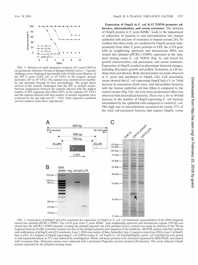
additional 1.5 h increased bacterial death by another order ofmagnitude (99.84%). In contrast, this strain survived incuba-tion in mouse serum for both of these durations, as expected.Similarly, the parental strain was not susceptible to comple-ment killing by mouse or rat serum. These serum assays indi-cated that rat infections with Y. pestis CO92 may better reflectthe role of OmpX in human disease; thus, a rat model ofpneumonic infections was employed. Rats were challengedintranasally with 102 to 108 CFU of the �ompX mutant and 102
to 104 CFU of the wild type and observed for 10 days (Fig. 3).Deletion of ompX caused complete attenuation of the Y. pestisCO92 strain in the rat pneumonic model of infection. Oneanimal infected with 107 CFU died on day 8, but no fatalitieswere recorded with the challenge dose of 108 CFU. Hence, theLD50 of the mutant was higher than 108 CFU, while the valueestimated for the wild type was 1.2 � 103 CFU (95% confi-dence interval, 4.2 � 102 to 3.2 � 103 CFU). These resultsindicate the essential role of serum resistance in pneumonicplague. They also underline the significant contribution ofOmpX-conferred serum resistance over bacterial adhesion andinvasion in the pathogenesis of Y. pestis.
FIG. 1. Deletion of ompX extends the time to death in Y. pestis CO92 pneumonic infections. Groups of BALB/c mice (n 8/group) werechallenged intranasally with 10 LD50 (A) or 100 LD50 (B), doses established previously for WT Y. pestis CO92 (LD50 2 � 104 CFU). The animalswere monitored for morbidity and mortality through 10 days postchallenge. The graph presents survival of the animals challenged with WT,�ompX, or ompX�/�ompX strains. A difference in survival times for animals infected with the �ompX strain was observed (log rank test, P �0.001). Data represent combined survival numbers from two experiments performed on separate days.
FIG. 2. Rat but not mouse serum is bactericidal to the �ompX strain. Y. pestis KIM6� and its isogenic �ompX derivative were grown tomid-exponential phase at 28°C; incubated in 50% normal mice serum (NMS), normal rat serum (NRS), or the respective heat-inactivated sera(HIS) for 0, 1, and 2.5 h at 37°C; and plated on LB agar. Graphs represent surviving bacteria after incubation with NMS and NRS as a percentageof the number of bacteria that survived treatment with HIS (100%). A significant effect of the type of serum for the �ompX strain was observed(repeated-measures ANOVA, P � 0.001). Results are the mean standard errors of the means (SEM) from data derived from two assaysperformed on separate days (n 4).
5236 KOLODZIEJEK ET AL. INFECT. IMMUN.
on February 15, 2018 by guest
http://iai.asm.org/
Dow
nloaded from
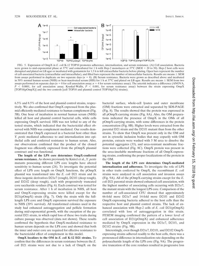
Expression of OmpX in E. coli K-12 TOP10 promotes ad-herence, internalization, and serum resistance. The deletionof OmpX protein in Y. pestis KIM6� leads to the impairmentof adherence of bacteria to and internalization into humanepithelial cells and loss of resistance to human serum (26). Toconfirm that these traits are mediated by OmpX protein inde-pendently from other Y. pestis proteins or LPS, the y1324 genewith its neighboring upstream and downstream DNA wascloned into plasmid pPCR2.1-TOPO, expressed in the stan-dard cloning strain E. coli TOP10 (Fig. 4), and tested forgrowth characteristics, cell association, and serum resistance.Expression of OmpX resulted in phenotypic bacterial changes,including flocculent growth and pellicle formation, in LB me-dium (data not shown). Both characteristics are traits observedin Y. pestis and attributed to OmpX (26). Cell associationassays showed that E. coli expressing OmpX had a 5- to 7-foldincrease in association (both extra- and intracellular bacteria)with the human epithelial cell line (Hep-2) compared to thecontrol strains (Fig. 5A). An even more pronounced effect wasobserved with internalized bacteria. There was a 16- to 30-foldincrease in the number of OmpX-expressing E. coli bacteriainternalized by the epithelial cells compared to control E. coli.This high rate of internalization accounted for nearly 27% ofthe total cell-associated bacteria that express OmpX, versus
FIG. 3. Deletion of ompX attenuates virulence of Y. pestis CO92 inrat pneumonic infection. Groups of Sprague-Dawley rats (n 3/group/challenge) were challenged intranasally with 10-fold serial dilutions ofthe WT Y. pestis CO92 (102 to 104 CFU) or the isogenic �ompXderivative (102 to 108 CFU). The animals were monitored for morbid-ity and mortality through 10 days postchallenge. The graph showssurvival of the animals challenged with the WT or �ompX strains.Survival comparisons between the animals infected with the highestnumber of WT organisms that killed 100% of the animals (104 CFU)and the animals infected with that number of �ompX organisms wereevaluated by the log rank test (P 0.01). Data represent combinedsurvival numbers from three experiments.
FIG. 4. Generation of pOmpX and pTA constructs for expression of OmpX in E. coli. (A) Schematic representation of the DNA fragmentcloned into plasmid pPCR2.1-TOPO. The y1324 gene from Y. pestis KIM6� with neighboring upstream and downstream regions (750 bp) wascloned into the pPCR2.1-TOPO plasmid, creating the pOmpX plasmid; the pTA plasmid (vector control) was made by deletion of the 766-bpfragment between EcoRI restriction enzyme-cut sites in the pOmpX plasmid and relegation of the backbone. (B) PCR analysis with M13 primersand confirmation of pOmpX and pTA constructs. Lane 1, DNA size marker (Fisher Scientific); lane 2, negative control (no DNA); lane 3, pOmpX;lane 4; pTA. (C) Analysis of OmpX-expressing E. coli TOP10 strains. E. coli Top10, E. coli Top10(pOmpX), and E. coli Top10(pTA) were grownto mid-exponential phase at 37°C and collected by centrifugation. Whole-cell lysate proteins were extracted, separated by SDS-PAGE, and stainedwith Coomassie blue. Molecular masses were estimated with a prestained Pageruler protein standard (Fermentas). The arrow indicates OmpXprotein expressed by the pOmpX-carrying strain.
VOL. 78, 2010 OmpX IS A PLAGUE VIRULENCE FACTOR 5237
on February 15, 2018 by guest
http://iai.asm.org/
Dow
nloaded from
6.5% and 8.5% of the host and plasmid control strains, respec-tively. We also confirmed that OmpX expressed from the plas-mid efficiently mediated resistance to human complement (Fig.5B). One hour of incubation in normal human serum (NHS)killed all host and plasmid control bacterial cells, while cellsexpressing OmpX survived. HIS was not lethal to any of thetested strains, which indicated that the bactericidal effect ob-served with NHS was complement mediated. Our results dem-onstrated that OmpX expressed in a bacterial host other thanY. pestis mediated adherence to and internalization into epi-thelial cells and conferred resistance to human serum. Also,our observations confirmed that the product of the clonedfragment was efficiently expressed from the pOmpX plasmidconstruct and was functional.
The length of the LPS core determines OmpX-conferredserum resistance. As shown previously by Knirel et al., Y. pestismutants possessing different LPS core lengths have alteredsensitivity to human serum (24). To investigate the potentialeffect of LPS core length on OmpX functions, the pOmpXplasmid was transformed into the E. coli D21 strain and itsthree isogenic derivatives D21e7 (rough), D21f1 (deep rough),and D21f2 (deep rough), each with progressively truncatedcore saccharide residues (Fig. 6). Each construct was tested forserum resistance. After 1 h of incubation in NHS, all hostand OmpX-expressing strains were killed except for theD21(pOmpX) strain (Fig. 7). Only the strain with the full-length LPS core and OmpX expression survived the exposureto NHS (20% survival). All transformed colonies used in thestudy had enhanced autoaggregation and pellicle formationduring mid-exponential growth, with the exception of the pa-rental D21 strain, in which rapid loss of these two traits duringculture passage was observed (data not shown). These resultsconfirmed the hypothesis that OmpX-mediated resistance tohuman serum depends on the LPS core and showed that boththe inner and outer core are required for effective resistance tothe bactericidal effect of complement in this model.
OmpX localizes to the OM in E. coli K-12 D21 strains. Toconfirm that the differences in serum resistance between the E.coli D21 strains were not due to a lack of OmpX on the
bacterial surface, whole-cell lysates and outer membrane(OM) fractions were extracted and separated by SDS-PAGE(Fig. 8). The results showed that the protein was expressed inall pOmpX-carrying strains (Fig. 8A). Also, the OM prepara-tions indicated the presence of OmpX in the OMs of allpOmpX-carrying strains, with some differences in the proteinconcentration (Fig. 8B). Higher levels were extracted from theparental D21 strain and the D21f1 mutant than from the otherstrains. To show that OmpX was present only in the OM andnot in cytosolic inclusion bodies that may copurify with OMproteins, extracts were washed with 5 M urea to dissolve thepotential aggregates (33), and urea-resistant membrane frac-tions were collected (Fig. 8C). OmpX protein was present inthe urea-insoluble membrane fractions of all OmpX-express-ing strains, confirming the proper localizations of the protein inthe OM.
The length of the LPS core determines OmpX-mediatedinternalization and adherence. To investigate the role of LPSin other traits conferred by OmpX, the recombinant E. colistrains were analyzed in cell association and invasion assays(Fig. 9A). All of the pOmpX-carrying strains except for the E.coli D21 parental strain showed enhanced cell association, withthe highest number of associating cells occurring with D21e7,the mutant strain with the longest LPS core. Comparison of thenumber of cell-associated CFU showed that approximately66-fold more D21e7 and 14-fold more D21f1 and D21f2OmpX-expressing bacteria adhered to the host cells than therespective host and plasmid control strains. The lack of en-hanced association with Hep-2 cells of E. coli D21(pOmpX)correlated with loss of autoaggregation in this strain.FESEM imaging confirmed the pattern of a lower level ofcell association of D21(pOmpX) and enhanced adherencemediated by OmpX expression in the D21e7, D21f1, andD21f2 strains (Fig. 9B).
Interestingly, even though D21e7, D21f1, and D21f2 OmpX-expressing strains adhered readily to the host cells, there was apronounced difference in internalization rate depending on thepolysaccharide length of the LPS core (Fig. 9A). The progres-sive truncation of the core residues resulted in progressive loss
FIG. 5. Expression of OmpX in E. coli K-12 TOP10 promotes adherence, internalization, and serum resistance. (A) Cell association. Bacteriawere grown to mid-exponential phase at 37°C and coincubated for 1 h with Hep-2 monolayers at 37°C (MOI 20 to 50). Hep-2 host cells weredisrupted and plated on LB agar or incubated with gentamicin for 1.5 h to kill extracellular bacteria before plating. Open bars represent the numberof cell-associated bacteria (extracellular and intracellular), and filled bars represent the number of intracellular bacteria. Results are means SEMfrom assays performed in duplicate on two separate days (n 8). (B) Serum resistance. Bacteria were grown as described above and incubatedin 50% normal human serum (NHS) or heat-inactivated serum (HIS) for 1 h at 37°C and plated on LB agar. Results are means SEM from twoassays performed on separate days (n 8 for cell association assay; n 4 for serum resistance assay). The asterisk indicates a difference (ANOVA,P � 0.0001, for cell association assay; Kruskal-Wallis, P � 0.001, for serum resistance assay) between the strain expressing OmpX[TOP10(pOmpX)] and the two controls [cell TOP10 and plasmid control TOP10(pTA) strains).
5238 KOLODZIEJEK ET AL. INFECT. IMMUN.
on February 15, 2018 by guest
http://iai.asm.org/
Dow
nloaded from

of OmpX-mediated internalization. Expression of OmpX inthe D21f2 strain with the shortest inner core, containing only2-keto-3-deoxyoctulosonic acid (KDO) residues, did not en-hance the bacterial invasion into Hep-2 cells [P � 0.05 for
D21f2(pOmpX) versus D21f2 and D21f2(pTA)]. The mutantstrain with three additional heptose residues in the core had ina small, yet significant (P � 0.0001), increase in internalization.Approximately 5-fold more OmpX-expressing D21f1 bacteriainvaded epithelial cells than the D21f1 or D21f1(pTA) controlstrains. The biggest difference in invasion between the OmpX-expressing strains and the controls was seen with the roughmutant D21e7, with an �20-fold increase versus the controls.As a consequence of the lack of enhanced adherence, therewas no change in internalization of the D21(pOmpX) straineven though the protein was present in the OM and this strainwas resistant to human serum (Fig. 7 and 8B and C).
These results confirmed that OmpX, independent fromother Y. pestis proteins, conferred bacterial adherence to andinternalization into host cells; however, the efficiency of adher-ence and invasion, similar to serum resistance, was affected bythe length of the heterosaccharide content in the LPS core.The results obtained with the D21 strain suggested that auto-aggregation of bacterial cells also depended on the display ofOmpX in a specific LPS core background and that flocculentgrowth was critical for effective bacterial adherence.
DISCUSSION
The most important findings of this study were that (i) theloss of a single gene (ompX) caused complete attenuation of Y.pestis by affecting serum resistance and adhesion to and inva-
FIG. 6. Schematic representation of E. coli and Y. pestis LPS structures and summary of the results. (A) Key to LPS moieties. (B) LPS structuresof E. coli K-12 D21 and its isogenic mutants (23, 40, 44, 46, 59). †, some sources indicate galactose (23); ‡, some sources indicate two or three KDOresidues (40). (C) LPS core structure of Y. pestis KM260 grown at ambient temperature; arrows indicate truncation of the core residues thatresulted in loss of serum resistance (24). The table shows presence (�) or lack (�) of OmpX-mediated phenotypes (autoaggregation and pellicleformation, adhesion to Hep-2 cells, internalization into Hep-2 cells, and human serum resistance) related to a particular strain. Data are a summaryof this study and previous work on OmpX of Y. pestis KIM6� (26).
FIG. 7. The length of the LPS core determines OmpX-conferredserum resistance. E. coli K-12 D21 (rough LPS) and its three iso-genic derivatives D21e7 (rough LPS), D21f1 (deep rough LPS), andD21f2 (deep rough LPS) with or without pOmpX were grown tomid-exponential phase at 37°C, incubated in 50% normal humanserum (NHS) or heat-inactivated serum (HIS) for 1 h at 37°C, andplated on LB agar. Results are means SEM from data derivedfrom two assays performed on separate days (n 4). The asterisk(*) indicates the difference (Kruskal-Wallis test, P � 0.001) be-tween E. coli carrying pOmpX and its respective cell control strainafter incubation with NHS.
VOL. 78, 2010 OmpX IS A PLAGUE VIRULENCE FACTOR 5239
on February 15, 2018 by guest
http://iai.asm.org/
Dow
nloaded from

sion of host cells, (ii) avirulence was dependent on an appro-priate animal model, (iii) OmpX conferred its characteristicsindependently from other Y. pestis proteins or LPS, and (iv)recombinant isogenic strains of E. coli showed that the OmpX-dependent phenotypes were influenced by the LPS core sac-charide length. For the first time, the role of OmpX in Y. pestisvirulence was comprehensively assessed using a fully virulentstrain, pneumonic route of infection, and two animal models toaccount for differences in serum resistance.
There are conflicting roles for ompX in an intravenousmouse model of plague with a �ompX mutant in the attenu-ated Y. pestis strain KIM5. Bartra et al. (4) showed that dele-tion of ompX does not affect virulence, while Felek et al. (15)showed that the deletion causes a �3,000-fold increase in theLD50 and that fewer bacteria colonize host organs in the earlydays of infection compared to the wild type. Our mouse modelstudies using the fully virulent Y. pestis CO92 strain with apneumonic route of challenge showed a delayed time to deathbut no decrease in virulence (no increased LD50) of an ompXmutant compared to the wild type. Thus, our studies are inagreement with the findings of Bartra et al. (4). Our assays alsoconfirmed that murine serum is not bactericidal for the ompXmutant (4). Thus, the mechanism underlying the delay in timeto death in mice challenged with Y. pestis �ompX may beattributed to loss of adhesion/internalization properties but notserum resistance. These studies also showed that the role ofserum resistance in Y. pestis pathogenesis cannot be tested inmice. To assess the full contribution of OmpX (adherence,internalization, and serum resistance) to virulence, we used arat model of pneumonic plague. In this model, the Y. pestis�ompX was completely attenuated (all rats challenged with1 � 108 CFU survived, compared to the LD50 of 1.2 � 103
CFU of the wild-type strain). Even though the lung is not anorgan known for high complement activity (7), the importanceof serum resistance in pneumonic plague was highlighted by
comparing differences in mortality between mice and rats. Se-rum resistance contributed to virulence more prominently thanbacterial association with host cells.
The bactericidal properties of complement vary by species,and activity often reflects the host’s response to infection andefficient bacterial transmission (28). For example, the patternof serum resistance of different Borrelia spp. correlates with thehost that they are able to infect (28). The mechanisms of thesedifferences are not clear, but they involve both bacterial andhost components (53). From the host side, the complementregulatory system, rather than components of the membraneattack complex, is suspected (45, 58). Some animal specieshave a more complex protease inhibitor system regulating thecomplement cascade; for example, analysis of mouse, rat, andhuman genomes reveals that the systems are encoded by 199,183, and 156 genes, respectively (45). Mouse complement isparticularly interesting because its bactericidal activity is lim-ited (18, 34). A number of common microorganisms are killedby sera of other species, e.g., human or rat, but remain resistantto murine sera (10, 19, 34, 57). Studies to distinguish comple-ment species specificity and activation on bacterial surfacesidentified two critical regulatory proteins: factor H (fH) andC4 binding protein (C4bp) (39). C4bp is involved in regulationof the classical pathway (5), and fH is an inhibitor of an alter-native complement pathway (53). Exclusive binding of humanfH and C4bp by N. gonorrhoeae ensures its resistance to humanserum and reflects restricted infection of the human species(38). Similarly, differential binding of fH obtained from sera ofvarious mammalian species and its correlation with the naturalhosts occurs for Borrelia burgdorferi (53). Although the under-lying differences between mouse and rat complement that af-fect Y. pestis are beyond the scope of this work, the findings ofBartra et al. (4) and our findings here confirm that speciesspecificity for the complement alternative pathway also appliesto Y. pestis �ompX.
FIG. 8. Analysis of OmpX localization in E. coli K-12 D21 strains. OmpX-expressing E. coli D21, D21e7, D21f1, and D21f2 and their parentalstrains were grown to mid-exponential phase at 37°C and collected by centrifugation. Whole-cell lysate proteins (A) and Triton X-100-insoluble(B) or 5 M urea-insoluble (C) outer membrane proteins were extracted, separated by SDS-PAGE, and stained with Coomassie blue. Molecularmass markers were estimated with a prestained Pageruler protein standard (Fermentas). Arrows indicate OmpX protein.
5240 KOLODZIEJEK ET AL. INFECT. IMMUN.
on February 15, 2018 by guest
http://iai.asm.org/
Dow
nloaded from
From the bacterial side, the mechanism of serum resistancein Y. pestis has been controversial. Previously we showed thatdeletion of ompX leads to a pronounced increased sensitivityto human serum (26). However, Knirel et al. (24) showed thatY. pestis loss of serum resistance was due to mutations affectingLPS core structure. To address these two observations, we firstdetermined that E. coli OmpX-expressing cells conferred theY. pestis phenotypes of serum resistance, autoaggregation, andinvasiveness. This indicated that these traits were independentof other Y. pestis factors. This is consistent with the observa-tions of Bartra et al. (4), who also showed that E. coli express-ing OmpX was serum resistant. We next examined OmpX-dependent serum resistance in three isogenic strains of E. colidiffering only in the length of the LPS core region. Deep rough
strains D21e7 (having the KDO moiety only) and D21f1 (hav-ing KDO with three heptose residues) and a rough strainD21e7 (with additional glucose and branched galactose) weresensitive to human serum. This is in contrast to the fully serum-resistant E. coli parental strain expressing OmpX. These ex-periments clearly show that serum resistance is OmpX depen-dent, but, significantly, this resistance is influenced by themolecular composition of the LPS core structure. This inter-action between OmpX and LPS composition explains the lossof serum resistance conferred by both by a mutation in ompX(26) and mutations affecting LPS (24).
The deletion of ompX in Y. pestis impairs adhesiveness andinvasiveness of the bacterium (26). A decrease in cell attach-ment after ompX deletion in Y. pestis and a gain of adhesive-
FIG. 9. The length of the LPS core determines OmpX-mediated internalization and adherence. (A) E. coli K-12 D21 (rough LPS) strain andits three isogenic derivatives D21e7 (rough LPS), D21f1 (deep rough LPS), and D21f2 (deep rough LPS) with or without pOmpX were grown tomid-exponential phase at 37°C and coincubated for 1 h with Hep-2 monolayers at 37°C (MOI 20 to 50). Hep-2 host cells were disrupted andplated on LB agar or incubated with gentamicin for 1.5 h to kill extracellular bacteria before plating. Open bars represent the number ofcell-associated bacteria (extracellular and intracellular), and filled bars represent the number of intracellular bacteria. Results are means SEMfrom assays performed in duplicate on two separate days (n 8). The asterisk (*) indicates differences (ANOVA, P � 0.0001) between the strainexpressing OmpX (pOmpX) and cell (D21, D21e7, D21f1, or D21f2) and plasmid (pTA) control strains. (B) Field emission scanning microscopyimaging of OmpX-expressing E. coli associated with Hep-2 cells. Bacteria and eukaryotic cells were grown as described above and cocultured for15 min at 37°C. After extensive washing, the cells were fixed, treated with tannic acid, OsO4, dehydrated, and gold coated. The samples wereanalyzed with Quanta 200F (Field Emission Instruments) at a magnification of �16,000. The bar represents 5 �m; white arrows indicate bacteriaassociated with Hep-2 cells.
VOL. 78, 2010 OmpX IS A PLAGUE VIRULENCE FACTOR 5241
on February 15, 2018 by guest
http://iai.asm.org/
Dow
nloaded from
ness in E. coli AAEC185 expressing OmpX were confirmed byFelek and Krukonis (14). Similar to the case for serum resis-tance, we showed that OmpX-conferred adhesiveness and in-vasion were dependent on the LPS background in which theprotein was expressed. While all of the E. coli OmpX-express-ing LPS mutant strains readily adhered to Hep-2 cells, theparental D21 strain did not, even though OmpX was present inthe OM and the strain was serum resistant. Lack of cell asso-ciation correlated with the fact that the OmpX-mediated au-toaggregation phenotype was not maintained in this strain.Also, progressive truncation of the LPS core resulted in aproportional decrease in internalization of OmpX-expressingE. coli cells, with the most pronounced effect in strain D21f2(possessing only KDO residues of the core inner portion),which had invasion levels similar to those of the OmpX-nega-tive control strain. LPS core mediates internalization of somebacteria (41), and alterations in its structure can lead to im-pairment of bacterial invasion (60). Our data indicated thatthis was also the case for the E. coli K-12 D21 mutant strains(lower invasion level of strains with shorter LPS cores). How-ever, introduction of OmpX significantly increased invasive-ness of strains having low levels of internalization (D21f1 andD21e7). If internalization was driven only by LPS, this wouldnot be observed. If LPS alone conferred these properties, onewould expect to see the levels of internalized bacteria increas-ing as the length of the LPS core increased, but with no dif-ference between OmpX-expressing and nonexpressing strains.Hence, we concluded that OmpX function as an invasin isinfluenced by the LPS core structure.
In summary, this work showed for the first time that the Y.pestis ompX gene is a novel essential virulence factor in asubset of mammalian hosts where OmpX-dependent serumresistance is required. The loss of ompX had little consequencein mice (where serum resistance is independent of OmpXexpression), with no change in LD50. This is in stark contrast tothe complete attenuation of a Y. pestis CO92 ompX deletionmutant in rats (where serum resistance is dependent on OmpXexpression), an animal model more similar to human disease(2, 49). Finally, OmpX-mediated serum resistance, as well ashost cell adhesion and internalization, is dependent on the LPScore structure, which contributes to the OM environment.
ACKNOWLEDGMENTS
This work was supported by the National Institutes of Health (grantsP20 RR15587, P20 RR016454, and U54AI57141) and the Idaho Ag-ricultural Experimental Station.
We appreciate advice and technical assistance with electron micros-copy provided by Valerie Roberts and technical assistance provided byDylan Sinclair.
REFERENCES
1. Airhart, C. L., H. N. Rohde, G. A. Bohach, C. J. Hovde, C. F. Deobald, S. S.Lee, and S. A. Minnich. 2008. Induction of innate immunity by lipid Amimetics increases survival from pneumonic plague. Microbiology 154:2131–2138.
2. Anderson, D. M., N. A. Ciletti, H. Lee-Lewis, D. Elli, J. Segal, K. L. DeBord,K. A. Overheim, M. Tretiakova, R. R. Brubaker, and O. Schneewind. 2009.Pneumonic plague pathogenesis and immunity in Brown Norway rats. Am. J.Pathol. 174:910–921.
3. Anisimov, A. P., S. V. Dentovskaya, G. M. Titareva, I. V. Bakhteeva, R. Z.Shaikhutdinova, S. V. Balakhonov, B. Lindner, N. A. Kocharova, S. N.Senchenkova, O. Holst, G. B. Pier, and Y. A. Knirel. 2005. Intraspecies andtemperature-dependent variations in susceptibility of Yersinia pestis to thebactericidal action of serum and to polymyxin B. Infect. Immun. 73:7324–7331.
4. Bartra, S. S., K. L. Styer, D. M. O’Bryant, M. L. Nilles, B. J. Hinnebusch, A.Aballay, and G. V. Plano. 2008. Resistance of Yersinia pestis to complement-dependent killing is mediated by the Ail outer membrane protein. Infect.Immun. 76:612–622.
5. Berggard, K., E. Johnsson, F. R. Mooi, and G. Lindahl. 1997. Bordetellapertussis binds the human complement regulator C4BP: role of filamentoushemagglutinin. Infect. Immun. 65:3638–3643.
6. Biedzka-Sarek, M., R. Venho, and M. Skurnik. 2005. Role of YadA, Ail, andlipopolysaccharide in serum resistance of Yersinia enterocolitica serotype O:3.Infect. Immun. 73:2232–2244.
7. Bolger, M. S., D. S. Ross, H. Jiang, M. M. Frank, A. J. Ghio, D. A. Schwartz,and J. R. Wright. 2007. Complement levels and activity in the normal andLPS-injured lung. Am. J. Physiol. 292:L748–L759.
8. Brubaker, R. R. 2003. Interleukin-10 and inhibition of innate immunity toYersiniae: roles of Yops and LcrV (V antigen). Infect. Immun. 71:3673–3681.
9. Cowan, C., H. A. Jones, Y. H. Kaya, R. D. Perry, and S. C. Straley. 2000.Invasion of epithelial cells by Yersinia pestis: evidence for a Y. pestis-specificinvasin. Infect. Immun. 68:4523–4530.
10. Cybulska, J., and J. Jeljaszewicz. 1966. Bacteriostatic activity of serumagainst staphylococci. J. Bacteriol. 91:953–962.
11. de Cock, H., K. Brandenburg, A. Wiese, O. Holst, and U. Seydel. 1999.Non-lamellar structure and negative charges of lipopolysaccharides requiredfor efficient folding of outer membrane protein PhoE of Escherichia coli.J. Biol. Chem. 274:5114–5119.
12. de Lorenzo, V., M. Herrero, U. Jakubzik, and K. N. Timmis. 1990. Mini-Tn5transposon derivatives for insertion mutagenesis, promoter probing, andchromosomal insertion of cloned DNA in gram-negative eubacteria. J. Bac-teriol. 172:6568–6572.
13. Dupont, M., E. De, R. Chollet, J. Chevalier, and J. M. Pages. 2004. Entero-bacter aerogenes OmpX, a cation-selective channel mar- and osmo-regulated.FEBS Lett. 569:27–30.
14. Felek, S., and E. S. Krukonis. 2009. The Yersinia pestis Ail protein mediatesbinding and Yop delivery to host cells required for plague virulence. Infect.Immun. 77:825–836.
15. Felek, S., M. B. Lawrenz, and E. S. Krukonis. 2008. The Yersinia pestisautotransporter YapC mediates host cell binding, autoaggregation and bio-film formation. Microbiology 154:1802–1812.
16. Ferguson, A. D., W. Welte, E. Hofmann, B. Lindner, O. Holst, J. W. Coulton,and K. Diederichs. 2000. A conserved structural motif for lipopolysacchariderecognition by procaryotic and eucaryotic proteins. Structure 8:585–592.
17. Forman, S., C. R. Wulff, T. Myers-Morales, C. Cowan, R. D. Perry, and S. C.Straley. 2008. yadBC of Yersinia pestis, a new virulence determinant forbubonic plague. Infect. Immun. 76:578–587.
18. Gondwe, E. N., M. E. Molyneux, M. Goodall, S. M. Graham, P. Mastroeni,M. T. Drayson, and C. A. MacLennan. 2010. Importance of antibody andcomplement for oxidative burst and killing of invasive nontyphoidal Salmo-nella by blood cells in Africans. Proc. Natl. Acad. Sci. U. S. A. 107:3070–3075.
19. Hanski, C., M. Naumann, A. Grutzkau, G. Pluschke, B. Friedrich, H. Hahn,and E. O. Riecken. 1991. Humoral and cellular defense against intestinalmurine infection with Yersinia enterocolitica. Infect. Immun. 59:1106–1111.
20. Heffernan, E. J., J. Harwood, J. Fierer, and D. Guiney. 1992. The Salmonellatyphimurium virulence plasmid complement resistance gene rck is homolo-gous to a family of virulence-related outer membrane protein genes, includ-ing pagC and ail. J. Bacteriol. 174:84–91.
21. Heffernan, E. J., S. Reed, J. Hackett, J. Fierer, C. Roudier, and D. Guiney.1992. Mechanism of resistance to complement-mediated killing of bacteriaencoded by the Salmonella typhimurium virulence plasmid gene rck. J. Clin.Invest. 90:953–964.
22. Heffernan, E. J., L. Wu, J. Louie, S. Okamoto, J. Fierer, and D. G. Guiney.1994. Specificity of the complement resistance and cell association pheno-types encoded by the outer membrane protein genes rck from Salmonellatyphimurium and ail from Yersinia enterocolitica. Infect. Immun. 62:5183–5186.
23. Junkes, C., A. Wessolowski, S. Farnaud, R. W. Evans, L. Good, M. Bienert,and M. Dathe. 2008. The interaction of arginine- and tryptophan-rich cyclichexapeptides with Escherichia coli membranes. J. Pept. Sci. 14:535–543.
24. Knirel, Y. A., S. V. Dentovskaya, O. V. Bystrova, N. A. Kocharova, S. N.Senchenkova, R. Z. Shaikhutdinova, G. M. Titareva, I. V. Bakhteeva, B.Lindner, G. B. Pier, and A. P. Anisimov. 2007. Relationship of the lipopoly-saccharide structure of Yersinia pestis to resistance to antimicrobial factors.Adv. Exp. Med. Biol. 603:88–96.
25. Knirel, Y. A., S. V. Dentovskaya, S. N. Senchenkova, R. Z. Shaikhutdinova,N. A. Kocharova, and A. P. Anisimov. 2006. Structural features and struc-tural variability of the lipopolysaccharide of Yersinia pestis, the cause ofplague. J. Endotoxin Res. 12:3–9.
26. Kolodziejek, A. M., D. J. Sinclair, K. S. Seo, D. R. Schnider, C. F. Deobald,H. N. Rohde, A. K. Viall, S. S. Minnich, C. J. Hovde, S. A. Minnich, and G. A.Bohach. 2007. Phenotypic characterization of OmpX, an Ail homologue ofYersinia pestis KIM. Microbiology 153:2941–2951.
27. Kukkonen, M., M. Suomalainen, P. Kyllonen, K. Lahteenmaki, H. Lang, R.Virkola, I. M. Helander, O. Holst, and T. K. Korhonen. 2004. Lack of
5242 KOLODZIEJEK ET AL. INFECT. IMMUN.
on February 15, 2018 by guest
http://iai.asm.org/
Dow
nloaded from
O-antigen is essential for plasminogen activation by Yersinia pestis and Sal-monella enterica. Mol. Microbiol. 51:215–225.
28. Kurtenbach, K., S. De Michelis, S. Etti, S. M. Schafer, H. S. Sewell, V.Brade, and P. Kraiczy. 2002. Host association of Borrelia burgdorferi sensulato—the key role of host complement. Trends Microbiol. 10:74–79.
29. Lathem, W. W., P. A. Price, V. L. Miller, and W. E. Goldman. 2007. Aplasminogen-activating protease specifically controls the development ofprimary pneumonic plague. Science 315:509–513.
30. Lawrenz, M. B., J. D. Lenz, and V. L. Miller. 2009. A novel autotransporteradhesin is required for efficient colonization during bubonic plague. Infect.Immun. 77:317–326.
31. Li, Y., Y. Cui, Y. Hauck, M. E. Platonov, E. Dai, Y. Song, Z. Guo, C. Pourcel,S. V. Dentovskaya, A. P. Anisimov, R. Yang, and G. Vergnaud. 2009. Geno-typing and phylogenetic analysis of Yersinia pestis by MLVA: insights into theworldwide expansion of Central Asia plague foci. PLoS One 4:e6000.
32. Liu, F., H. Chen, E. M. Galvan, M. A. Lasaro, and D. M. Schifferli. 2006.Effects of Psa and F1 on the adhesive and invasive interactions of Yersiniapestis with human respiratory tract epithelial cells. Infect. Immun. 74:5636–5644.
33. Marani, P., S. Wagner, L. Baars, P. Genevaux, J. W. de Gier, I. Nilsson, R.Casadio, and G. von Heijne. 2006. New Escherichia coli outer membraneproteins identified through prediction and experimental verification. ProteinSci. 15:884–889.
34. Marcus, S., D. W. Esplin, and D. M. Donaldson. 1954. Lack of bactericidaleffect of mouse serum on a number of common microorganisms. Science119:877.
35. Mecsas, J., R. Welch, J. W. Erickson, and C. A. Gross. 1995. Identificationand characterization of an outer membrane protein, OmpX, in Escherichiacoli that is homologous to a family of outer membrane proteins including Ailof Yersinia enterocolitica. J. Bacteriol. 177:799–804.
36. Montminy, S. W., N. Khan, S. McGrath, M. J. Walkowicz, F. Sharp, J. E.Conlon, K. Fukase, S. Kusumoto, C. Sweet, K. Miyake, S. Akira, R. J. Cotter,J. D. Goguen, and E. Lien. 2006. Virulence factors of Yersinia pestis areovercome by a strong lipopolysaccharide response. Nat. Immunol. 7:1066–1073.
37. Myers-Morales, T., C. Cowan, M. E. Gray, C. R. Wulff, C. E. Parker, C. H.Borchers, and S. C. Straley. 2007. A surface-focused biotinylation procedureidentifies the Yersinia pestis catalase KatY as a membrane-associated butnon-surface-located protein. Appl. Environ. Microbiol. 73:5750–5759.
38. Ngampasutadol, J., S. Ram, S. Gulati, S. Agarwal, C. Li, A. Visintin, B.Monks, G. Madico, and P. A. Rice. 2008. Human factor H interacts selec-tively with Neisseria gonorrhoeae and results in species-specific complementevasion. J. Immunol. 180:3426–3435.
39. Ngampasutadol, J., C. Tran, S. Gulati, A. M. Blom, E. A. Jerse, S. Ram, andP. A. Rice. 2008. Species-specificity of Neisseria gonorrhoeae infection: dohuman complement regulators contribute? Vaccine 26(Suppl. 8):I62–I66.
40. Nikaido, H., and M. Vaara. 1985. Molecular basis of bacterial outer mem-brane permeability. Microbiol. Rev. 49:1–32.
41. Pier, G. B., M. Grout, T. S. Zaidi, J. C. Olsen, L. G. Johnson, J. R. Yankas-kas, and J. B. Goldberg. 1996. Role of mutant CFTR in hypersusceptibilityof cystic fibrosis patients to lung infections. Science 271:64–67.
42. Prior, J. L., P. G. Hitchen, D. E. Williamson, A. J. Reason, H. R. Morris, A.Dell, B. W. Wren, and R. W. Titball. 2001. Characterization of the lipopoly-saccharide of Yersinia pestis. Microb. Pathog. 30:49–57.
43. Prior, J. L., J. Parkhill, P. G. Hitchen, K. L. Mungall, K. Stevens, H. R.Morris, A. J. Reason, P. C. Oyston, A. Dell, B. W. Wren, and R. W. Titball.2001. The failure of different strains of Yersinia pestis to produce lipopoly-saccharide O-antigen under different growth conditions is due to mutationsin the O-antigen gene cluster. FEMS Microbiol. Lett. 197:229–233.
44. Prokhorenko, I. R., S. V. Zubova, A. Y. Ivanov, and S. V. Grachev. 2009.Interaction of Gram-negative bacteria with cationic proteins: dependence onthe surface characteristics of the bacterial cell. Int. J. Gen. Med. 2:33–38.
45. Puente, X. S., L. M. Sanchez, A. Gutierrez-Fernandez, G. Velasco, and C.Lopez-Otin. 2005. A genomic view of the complexity of mammalian proteo-lytic systems. Biochem. Soc. Trans. 33:331–334.
46. Razatos, A., Y. L. Ong, M. M. Sharma, and G. Georgiou. 1998. Moleculardeterminants of bacterial adhesion monitored by atomic force microscopy.Proc. Natl. Acad. Sci. U. S. A. 95:11059–11064.
47. Rebeil, R., R. K. Ernst, B. B. Gowen, S. I. Miller, and B. J. Hinnebusch. 2004.Variation in lipid A structure in the pathogenic yersiniae. Mol. Microbiol.52:1363–1373.
48. Rietschel, E. T., H. Brade, O. Holst, L. Brade, S. Muller-Loennies, U.Mamat, U. Zahringer, F. Beckmann, U. Seydel, K. Brandenburg, A. J.Ulmer, T. Mattern, H. Heine, J. Schletter, H. Loppnow, U. Schonbeck, H. D.Flad, S. Hauschildt, U. F. Schade, F. Di Padova, S. Kusumoto, and R. R.Schumann. 1996. Bacterial endotoxin: chemical constitution, biological rec-ognition, host response, and immunological detoxification. Curr. Top. Mi-crobiol. Immunol. 216:39–81.
49. Sebbane, F., D. Gardner, D. Long, B. B. Gowen, and B. J. Hinnebusch. 2005.Kinetics of disease progression and host response in a rat model of bubonicplague. Am. J. Pathol. 166:1427–1439.
50. Sebbane, F., C. O. Jarrett, D. Gardner, D. Long, and B. J. Hinnebusch. 2006.Role of the Yersinia pestis plasminogen activator in the incidence of distinctsepticemic and bubonic forms of flea-borne plague. Proc. Natl. Acad. Sci.U. S. A. 103:5526–5530.
51. Simon, R., U. Priefer, and A. Puhler. 1983. A broad host range mobilizationsystem for in vivo genetic engineering: transposon mutagenesis in Gramnegative bacteria. Nat. Biotechnol. 1:784–791.
52. Smith, M. J. 2000. Genetic regulation of type III secretion systems in Yersiniaenterocolitica. PhD thesis. University of Idaho, Moscow, Idaho.
53. Stevenson, B., N. El-Hage, M. A. Hines, J. C. Miller, and K. Babb. 2002.Differential binding of host complement inhibitor factor H by Borrelia burg-dorferi Erp surface proteins: a possible mechanism underlying the expansivehost range of Lyme disease spirochetes. Infect. Immun. 70:491–497.
54. Stoorvogel, J., M. J. van Bussel, J. Tommassen, and J. A. van de Klundert.1991. Molecular characterization of an Enterobacter cloacae outer membraneprotein (OmpX). J. Bacteriol. 173:156–160.
55. Stoorvogel, J., M. J. van Bussel, and J. A. van de Klundert. 1991. Biologicalcharacterization of an Enterobacter cloacae outer membrane protein(OmpX). J. Bacteriol. 173:161–167.
56. Suomalainen, M., L. A. Lobo, K. Brandenburg, B. Lindner, R. Virkola, Y. A.Knirel, A. P. Anisimov, O. Holst, and T. K. Korhonen. 2010. Temperature-induced changes in the lipopolysaccharide of Yersinia pestis affect plasmin-ogen activation by the Pla surface protease. Infect. Immun. 78:2644–2652.
57. Wachtel, M. R., and V. L. Miller. 1995. In vitro and in vivo characterizationof an ail mutant of Yersinia enterocolitica. Infect. Immun. 63:2541–2548.
58. Younger, J. G., S. Shankar-Sinha, M. Mickiewicz, A. S. Brinkman, G. A.Valencia, J. V. Sarma, E. M. Younkin, T. J. Standiford, F. S. Zetoune, andP. A. Ward. 2003. Murine complement interactions with Pseudomonasaeruginosa and their consequences during pneumonia. Am. J. Respir. CellMol. Biol. 29:432–438.
59. Yu, F., and S. Mizushima. 1982. Roles of lipopolysaccharide and outermembrane protein OmpC of Escherichia coli K-12 in the receptor functionfor bacteriophage T4. J. Bacteriol. 151:718–722.
60. Zaidi, T. S., S. M. Fleiszig, M. J. Preston, J. B. Goldberg, and G. B. Pier.1996. Lipopolysaccharide outer core is a ligand for corneal cell binding andingestion of Pseudomonas aeruginosa. Invest. Ophthalmol. Vis. Sci. 37:976–986.
Editor: J. B. Bliska
VOL. 78, 2010 OmpX IS A PLAGUE VIRULENCE FACTOR 5243
on February 15, 2018 by guest
http://iai.asm.org/
Dow
nloaded from