visualization of intracellular transport of vesicular stomatitis virus nucleocapsids in living cells
TRANSCRIPT
2006, 80(13):6368. DOI: 10.1128/JVI.00211-06. J. Virol.
PattnaikSubash C. Das, Debasis Nayak, You Zhou and Asit K. Living Cells
inVesicular Stomatitis Virus Nucleocapsids Visualization of Intracellular Transport of
http://jvi.asm.org/content/80/13/6368Updated information and services can be found at:
These include:
REFERENCEShttp://jvi.asm.org/content/80/13/6368#ref-list-1at:
This article cites 53 articles, 44 of which can be accessed free
CONTENT ALERTS more»articles cite this article),
Receive: RSS Feeds, eTOCs, free email alerts (when new
http://journals.asm.org/site/misc/reprints.xhtmlInformation about commercial reprint orders: http://journals.asm.org/site/subscriptions/To subscribe to to another ASM Journal go to:
on August 2, 2014 by guest
http://jvi.asm.org/
Dow
nloaded from
on August 2, 2014 by guest
http://jvi.asm.org/
Dow
nloaded from
JOURNAL OF VIROLOGY, July 2006, p. 6368–6377 Vol. 80, No. 130022-538X/06/$08.00�0 doi:10.1128/JVI.00211-06Copyright © 2006, American Society for Microbiology. All Rights Reserved.
Visualization of Intracellular Transport of Vesicular StomatitisVirus Nucleocapsids in Living Cells
Subash C. Das, Debasis Nayak, You Zhou, and Asit K. Pattnaik*Department of Veterinary and Biomedical Sciences and Nebraska Center for Virology,
University of Nebraska—Lincoln, Lincoln, Nebraska 68588
Received 29 January 2006/Accepted 7 April 2006
The phosphoprotein (P) of vesicular stomatitis virus (VSV) is a subunit of the viral RNA polymerase. Inprevious studies, we demonstrated that insertion of 19 amino acids in the hinge region of the protein had nosignificant effect on P protein function. In the present study, we inserted full-length enhanced green fluorescentprotein (eGFP) in frame into the hinge region of P and show that the fusion protein (PeGFP) is functional inviral genome transcription and replication, albeit with reduced activity. A recombinant vesicular stomatitisvirus encoding PeGFP in place of the P protein (VSV-PeGFP), which possessed reduced growth kineticscompared to the wild-type VSV, was recovered. Using the recombinant VSV-PeGFP, we show that the viralreplication proteins and the de novo-synthesized RNA colocalize to sites throughout the cytoplasm, indicatingthat replication and transcription are not confined to any particular region of the cytoplasm. Real-timeimaging of the cells infected with the eGFP-tagged virus revealed that, following synthesis, the nucleocapsidsare transported toward the cell periphery via a microtubule (MT)-mediated process, and the nucleocapsidswere seen to be closely associated with mitochondria. Treatment of cells with nocodazole or Colcemid, drugsknown to inhibit MT polymerization, resulted in accumulation of the nucleocapsids around the nucleus andalso led to inhibition of infectious-virus production. These findings are compatible with a model in which theprogeny viral nucleocapsids are transported toward the cell periphery by MT and the transport may befacilitated by mitochondria.
Vesicular stomatitis virus (VSV) belongs to the familyRhabdoviridae in the order Mononegavirales. VSV is an envel-oped virus with a negative-stranded RNA genome of 11,161nucleotides. The genome encodes for five viral proteins,namely, nucleoprotein (N), phosphoprotein (P), matrix protein(M), glycoprotein (G), and the RNA-dependent RNA poly-merase (L) (1, 44). The viral genome is present within thevirion as a ribonucleoprotein (RNP) being tightly encapsidatedby the N protein and associated with the viral polymerasecomplex of L and P proteins (1, 44). The M protein is locatedunderneath the viral envelope, whereas the G protein formsspikes on the viral envelope. The G protein binds to the cellsurface receptor and is required for the entry of the virus intothe cells. The virus enters susceptible cells by receptor-medi-ated endocytosis. Fusion of the viral envelope with the endo-somal membrane in a pH-dependent process leads to the re-lease of the viral nucleocapsids into the cytoplasm for viralgene expression. During VSV assembly, the progeny nucleo-capsids are transported alone or in association with the viral Mprotein to the plasma membrane, where they are packaged intoan envelope containing the viral G protein and are releasedfrom the infected cells. Although some of the fundamentalsteps in the VSV infection pathway have been deciphered,many questions concerning the entry mechanisms and trans-port of viral nucleocapsids to synthesis sites and from synthesissites to assembly sites, as well as virus egress, remain poorlyunderstood. Studies to provide an understanding of these pro-
cesses have been hampered due to the inability to observethese events by live-cell imaging of infected cells.
In a recent report, it was demonstrated that the fusion of theviral envelope and the release of the nucleocapsid into the cyto-plasm are two independent but successive steps in the endocyticpathway of VSV infection (29). These studies revealed thatrelease of the viral nucleocapsid into the lumen of the endo-somal vesicle occurs by the fusion of the viral envelope with themembranes of the endosomes but that the nucleocapsid re-lease into the cytoplasm may require a back-fusion event inwhich the internal vesicles fuse with the membranes of the lateendosome (29). Following synthesis in the cytoplasm, the prog-eny nucleocapsids must then be transported to the plasmamembrane for viral assembly. The mechanism(s) by which thetransport of such large RNP complexes is accomplished isunclear. Because of the high viscosity of the cytoplasm,movement of the RNPs by diffusion is likely to be limited(32). Intracellular pathogens and their macromolecularcomponents overcome this obstacle by hijacking cytoplasmicmotors and utilizing the cellular cytoskeleton as a roadwayfor intracellular transport to reach their destination (23, 35,49, 50).
An understanding of some of the mechanistic details of virusentry by endocytic pathway and nucleocapsid release into thecytoplasm of infected cells has been possible only with the useof VSV chemically labeled with lipophilic fluorescent dyes(29). The use of such labeled viruses is limited to studiesinvolving tracking of the input virus (16, 26, 29, 47). Once theviral nucleocapsid is delivered into the cytoplasm, subsequenttracking of the viral nucleocapsids, particularly, the sites ofsynthesis and transport of the nucleocapsids to the sites of
* Corresponding author. Mailing address: E126 Beadle Center, 1901Vine Street, University of Nebraska—Lincoln, Lincoln, NE 68588. Phone:(402) 472-1067. Fax: (402) 472-8722. E-mail: [email protected].
6368
on August 2, 2014 by guest
http://jvi.asm.org/
Dow
nloaded from
virion assembly, would require genetic tagging of the viralnucleocapsid with fluorescent proteins.
VSV nucleocapsids are multiprotein-RNA complexes com-posed of viral RNA that is tightly wrapped with the N proteinand is associated with P and L proteins. P protein is a multi-functional protein that is an essential subunit of the viral RNA-dependent RNA polymerase. In addition to its role in poly-merase functions, it binds to the L protein and stabilizes itfrom proteolytic degradation (4, 13); it acts as a chaperone forthe N protein, which then specifically encapsidates the viralRNA (9, 34, 41); and it interacts with terminal sequences ofviral genome for viral RNA synthesis (21, 24). Our previousstudies showed that the protein is organized in a modularfashion relative to its function (Fig. 1A) (6, 8, 19, 39). Whilephosphorylation of specific amino acid residues at the amino-terminal domain I (amino acid residues 1 to 150) is responsiblefor transcription activity (39), phosphorylation of specificamino acid residues at the carboxy-terminal domain II (aminoacid residues 210 to 244) is important for optimal replicationactivity (19) of P protein. Phosphorylation of these residues atboth domain I and domain II is indispensable for virus growth(6). Domain III, which comprises 21 to 25 residues at theextreme carboxy-terminal region, is important for mediatingthe binding of P protein to the N RNA template (8). Theregion that links domain I and domain II is called the hyper-variable hinge region (approximately spanning amino acid res-
idues 150 to 210). We recently studied the role of this hingeregion and found that it plays an important role in VSV RNAsynthesis and assembly of infectious particles (7). In that study,we also demonstrated that insertion of 19 amino acids (aa)within the hinge region of the protein (Fig. 1A) has no signif-icant adverse effects on virus replication (7). In the presentstudy, we show that a fusion protein (PeGFP), in which full-length enhanced green fluorescent protein (eGFP) was in-serted in the hinge region of the P protein, is functional in viralgenome transcription and replication. A recombinant VSVencoding the PeGFP protein in place of P protein (VSV-PeGFP) was recovered. Using VSV-PeGFP, we have examinedthe intracellular sites of viral RNA synthesis. By live-cellimaging of VSV-PeGFP-infected cells, we have found that themovement of newly synthesized viral nucleocapsids toward thecell periphery is mediated by microtubules (MTs). In addition,our studies indicate that mitochondria may play a role in in-tracellular transport of the viral nucleocapsids.
MATERIALS AND METHODS
Cell culture, viruses, antibodies, and reagents. Baby hamster kidney (BHK-21) cells were maintained as described before (6, 7). Propagation and prepara-tion of stocks of VSV, recombinant vaccinia virus expressing T7 RNA polymer-ase (vTF7-3) (15), and defective-interfering (DI) particles were carried out inBHK-21 cells as described previously (40). Nocodazole (NOC), Colcemid, di-methyl sulfoxide (DMSO), 5-bromouridine 5�-triphosphate (BrUTP), and mono-clonal antibody (MAb) against antitubulin (clone DM 1A) were from Sigma-Aldrich. NOC and Colcemid were reconstituted in DMSO; BrUTP wasreconstituted in water. Rabbit polyclonal antibody against VSV P protein, mouseanti-VSV antibody, MAb against VSV N protein, and rabbit polyclonal antibodyagainst the NH2-terminal region of VSV L protein have been previously de-scribed (6, 46). Rabbit polyclonal antibody against eGFP was prepared in thelaboratory, and MAb against bromodeoxyuridine (BrdU) (clone BMC 9318) wasobtained from Roche Diagnostics. Secondary antibodies, goat anti-mouse Alexa-488, goat anti-mouse Alexa-594, goat anti-rabbit Alexa-594, and MitoTrackerRed were obtained from Molecular Probes; donkey anti-mouse Cy5 and donkeyanti-rabbit Cy2 were from Jackson ImmunoResearch Laboratories.
Plasmid construction and virus recovery. The construction of plasmids Pwtand PTn196 have been described in detail previously (7). Pwt is a plasmidencoding the wild-type (wt) VSV (VSVwt) (Indiana serotype) P protein inpGEM-3 vector (Promega) under the control of a T7 RNA polymerase pro-moter. The plasmid PTn196 is derived from Pwt by transposon-mediated mu-tagenesis and encodes the VSV P protein with a 19-aa insertion at residue 196.The eGFP sequence was amplified by PCR using primers eGFPNotF3, 5�ATATATGCGGCCGCAATGGTGAGCAAGGGC3�, and eGFPNotR3, 5�ATATATGCGGCCGCCTTGTACAGCTCGTC3�, each containing a NotI site (under-lined); pIRES2eGFP (Invitrogen) as a template; and Pfu polymerase (Stratagene).The PCR product was digested with NotI and cloned at the unique NotI site inPTn196. The resulting plasmid, PTneGFP, encodes a fusion protein (PeGFP) of 524amino acids, compared to wt P protein, which is 265 amino acids long.
The P protein coding region in the plasmid pVSVFL(�), which contains thefull-length VSV genome (28), was replaced with the coding region for PeGFPfrom PTneGFP by use of the EcoRV sites that flank the P gene, as describedpreviously (6). The resulting plasmid was designated pVSV-PeGFP, and therecombinant virus recovered from this plasmid was named VSV-PeGFP. For thegeneration of VSV encoding eGFP as an extra gene, an extra transcription unitwas incorporated in the pVSVFL(�) plasmid between the G and L noncodingregions. The pVSVFL(�) plasmid was linearized with NheI, which is locatedbetween the G and L noncoding regions. To generate the extra transcription unit,two complementary oligonucleotides (FLVSVBsiWI�, 5�TATGAAAAAAACTAACAGATATCCGTACG3�, and FLVSVBsiWI�, 5�CGTACGGATAT CTGTTAGTTTTTTTCATA3�), incorporating a VSV poly(A)/termination signal, anintergenic dinucleotide, and a transcription initiation signal followed by a uniquerestriction site (BsiWI site is underlined), were designed. These oligonucleotideswere annealed and kinased and then ligated with the linearized pVSVFL(�)plasmid, resulting in pVSVFLBsiWI. The eGFP coding region was amplified byPCR using the primers eGFPBsiWIF, 5�ATATATCGTACGGCCACCATGGTGAGCAAG3�, and eGFPBsiWIR, 5�ATATATCGTACGTTACTTGTACAGC
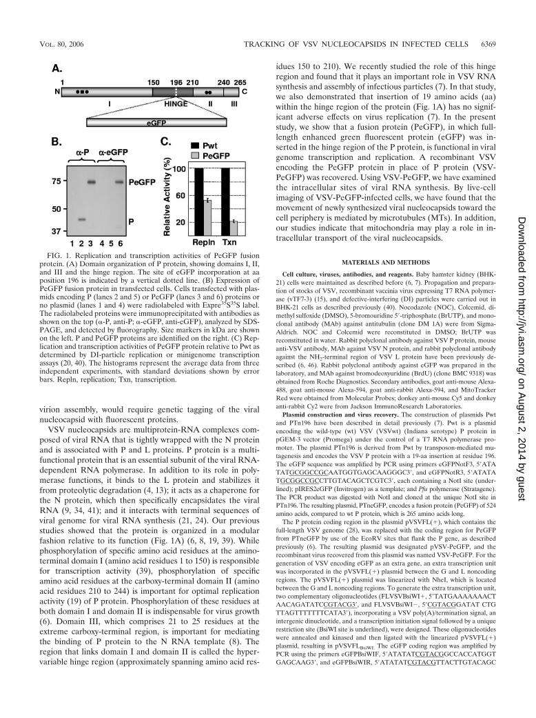
FIG. 1. Replication and transcription activities of PeGFP fusionprotein. (A) Domain organization of P protein, showing domains I, II,and III and the hinge region. The site of eGFP incorporation at aaposition 196 is indicated by a vertical dotted line. (B) Expression ofPeGFP fusion protein in transfected cells. Cells transfected with plas-mids encoding P (lanes 2 and 5) or PeGFP (lanes 3 and 6) proteins orno plasmid (lanes 1 and 4) were radiolabeled with Expre35S35S label.The radiolabeled proteins were immunoprecipitated with antibodies asshown on the top (�-P, anti-P; �-eGFP, anti-eGFP), analyzed by SDS-PAGE, and detected by fluorography. Size markers in kDa are shownon the left. P and PeGFP proteins are identified on the right. (C) Rep-lication and transcription activities of PeGFP protein relative to Pwt asdetermined by DI-particle replication or minigenome transcriptionassays (20, 40). The histograms represent the average data from threeindependent experiments, with standard deviations shown by errorbars. Repln, replication; Txn, transcription.
VOL. 80, 2006 TRACKING OF VSV NUCLEOCAPSIDS IN INFECTED CELLS 6369
on August 2, 2014 by guest
http://jvi.asm.org/
Dow
nloaded from
TCGTC3� (BsiWI site is underlined); digested with BsiWI; and cloned intopVSVFLBsiWI. Recombinant VSV recovered from the resulting plasmid, pVSV-eGFP, was named VSV-eGFP. The plasmids pN, pP, and pL (carrying the codingsequences of the N, P, and L proteins of VSV, respectively, and the plasmidp10BN), encoding a VSV minigenome, have been described previously (20, 38,40). Recombinant viruses were recovered as described previously (6, 7).
Metabolic labeling and analysis of RNA. Metabolic labeling and analysis ofRNA in plasmid-transfected and virus-infected cells were performed as de-scribed previously (7, 20, 40). To examine transcription activity of mutant Pproteins, BHK-21 cells were infected with vTF7-3 and subsequently transfectedwith p10BN, pN, and pL as well as Pwt or PTneGFP. RNAs were radiolabeledin the presence of actinomycin D and analyzed by electrophoresis in agarose-urea gel. To examine replication activity of mutant P proteins, cells infected withvTF7-3 and transfected with pN, pL, and Pwt or PTneGFP plasmids weresuperinfected with DI particles, and RNAs were radiolabeled and analyzed asdescribed above. To examine the viral RNAs in infected cells, BHK-21 cells wereinfected with either wt VSV or eGFP-tagged viruses and the RNAs were radio-labeled and analyzed as described above.
Metabolic labeling and analysis of viral proteins. Labeling of proteins intransfected or virus-infected cells, immunoprecipitation using anti-VSV antibody(1:200), anti-P antibody (1:200), and anti-eGFP antibody (1:200), sodium dode-cyl sulfate-polyacrylamide gel electrophoresis (SDS-PAGE) analysis, and detec-tion of proteins by fluorography were carried out as described previously (6, 7).To examine proteins in virions, BHK-21 cells were infected with VSV at 1PFU/cell and radiolabeled for 12 h at 4 h postinfection (hpi) with 100 �CiExpre35S35S per ml of 90% Dulbecco’s modified Eagle’s medium (DMEM)without methionine and cysteine and 10% regular DMEM. The virus from theclarified culture supernatant was pelleted through a 10% sucrose cushion at38,000 rpm in a Beckman SW41 rotor for 1 h at 4°C. The virus pellets wereresuspended, and viral proteins were analyzed by SDS-PAGE and detected asdescribed above.
Determination of single-step growth kinetics. Single-step growth kinetics ofmutant and wt viruses were determined in BHK-21 cells essentially as describedpreviously (6).
Drug treatment. Cells were pretreated with 10 �g/ml NOC or Colcemid for 3 hat 37°C and infected with viruses for 45 min at 4°C. The cells were washed andincubated at 37°C in medium containing 10 �M NOC or Colcemid. As controls,NOC-untreated cells were maintained in media containing similar concentra-tions of DMSO.
BrUTP labeling of viral RNA. BHK-21 cells were infected with VSV-PeGFP ata multiplicity of infection (MOI) of 10 PFU/cell and incubated for 2 h at 37°C.BrUTP labeling of de novo-synthesized viral RNA was performed as describedpreviously (17), with minor modifications. Briefly, the infected cells were treatedwith 15 �g/ml of actinomycin D for 1 h at 2 hpi and transfected with BrUTP ata final concentration of 10 mM by use of Lipofectamine 2000 in the presence ofactinomycin D for 2 h. The cells were fixed with 3.7% paraformaldehyde for 15min at room temperature (RT) and processed for immunofluorescence for de-tection of viral RNA.
Fluorescence microscopy. For epifluorescence and differential interferencecontrast (DIC) imaging, cells grown on glass coverslips or glass-bottomed cellculture dishes (MatTek) were either fixed as described above or visualizeddirectly under an Olympus FV500/IX81 inverted confocal microscope. For im-munofluorescence microscopy, the fixed cells were permeabilized with 0.1%Triton X-100 for 15 min at RT or with 100% ethanol at �20°C for 5 min. Thecells were blocked with phosphate-buffered saline with 0.05% Tween 20 contain-ing 3% bovine serum albumin for 30 min at RT. The primary and secondaryantibodies diluted in phosphate-buffered saline with 0.05% Tween 20 containing1% bovine serum albumin were added in succession after washing. The BrUTP-labeled RNAs were stained with an anti-BrdU MAb (1:10) followed by goatanti-mouse Alexa-594 (1:500) containing 1 U/�l of RNase inhibitor (RNase Out;Invitrogen). The cells were stained with DAPI (4�,6�-diamidino-2-phenylindole)and imaged using the inverted confocal microscope, and images were capturedwith a charge-coupled-device camera.
For real-time visualization of nucleocapsid movement, cells grown in glass-bottomed 35-mm dishes to 50% confluence were either treated with NOC or leftuntreated and then infected with VSV-PeGFP as described above and the dishwas placed in a closed chamber maintained at 37°C and 5% CO2. Live-cellimaging was performed using the inverted confocal microscope fitted with a100� lens. Images were captured at various times postinfection. For time-lapserecording of the same sets of cells, images were collected at intervals of 10 to 15 sfor 30 min.
For staining of mitochondria, infected cells were washed thoroughly withserum-free DMEM containing 25 mM HEPES buffer. MitoTracker Red diluted
in the above-described medium was added at 1 to 3 �M, and the cells wereincubated at RT for 30 min. After being washed, the cells were maintained inmedium containing serum and placed in the closed chamber.
RESULTS
PeGFP fusion protein supports transcription and replica-tion of VSV. Our recent observation (7) that insertion of 19 aaat position 196 in the hinge region of P protein had no signif-icant adverse effect on transcription and replication promptedus to insert full-length eGFP coding sequence at this position(Fig. 1A). To determine if the insertion resulted in a stableprotein, we examined the level of expression and the size of thePeGFP fusion protein in transfected cells. Accordingly, BHK-21cells were transiently transfected with plasmids encoding eitherthe wt or fusion proteins following vTF7-3 infection, and theproteins were radiolabeled and examined by immunoprecipita-tion with anti-P and/or anti-eGFP antibody and SDS-PAGE. Ascan be seen in Fig. 1B, the fusion protein PeGFP was stablyexpressed, could be immunoprecipitated with both anti-P andanti-eGFP antibodies, and possessed a predicted molecular massof �73 kDa.
In order to examine the effect of eGFP insertion on P pro-tein function in viral RNA synthesis, we used DI particles todetermine replication activity and a minigenome template(p10BN) (20) to assess the transcription activity of PeGFPfusion protein. The radiolabeled DI RNAs and minigenomeRNA were analyzed on urea-agarose gels and quantitated bydensitometry. Results from three independent experimentsshow that the PeGFP protein is �55% active in replication,whereas it is �22% active in transcription compared to the wtP protein (Fig. 1C). These results suggest that the PeGFPfusion protein is functional, albeit with reduced activity.
Recovery of infectious VSV encoding PeGFP fusion protein.To test whether infectious virus could be recovered from VSVgenome plasmids encoding P protein into which eGFP wasinserted in frame in the hinge region, a recombinant VSVgenome plasmid (pVSV-PeGFP) encoding PeGFP in place ofwt P protein was constructed (Fig. 2A). We also constructedanother recombinant VSV genome plasmid (pVSV-eGFP),containing eGFP coding sequence as an extra gene betweenthe G and L gene junctions (Fig. 2A). Both of these genomicconstructs led to recovery of recombinant VSV from trans-fected cells. In order to examine if the eGFP insertion into theP open reading frame has any effect on the growth of the virus,we examined single-cycle growth kinetics of the wt and mutantviruses. As can be seen from Fig. 2B, VSV-eGFP grew to titerssimilar to those of the wt VSV, whereas VSV-PeGFP grew totiters that were on average 8- to 10-fold less than those of thewt VSV. To determine if the viral growth correlated with theextent of viral macromolecular synthesis in infected cells, weexamined RNA and viral protein synthesis in cells infectedwith these viruses. Our results show that overall synthesis ofviral RNAs (Fig. 2C) and viral proteins (Fig. 2D) was notsignificantly different from results obtained with cells infectedwith wt VSV. These results indicate that in these mutant vi-ruses the extent of viral protein and RNA synthesis did notcorrelate with the viral growth rate. The sizes of the viralmRNAs were as predicted, with the PeGFP mRNA migratingmore slowly than mRNA for wt P protein or eGFP (Fig. 2C,
6370 DAS ET AL. J. VIROL.
on August 2, 2014 by guest
http://jvi.asm.org/
Dow
nloaded from
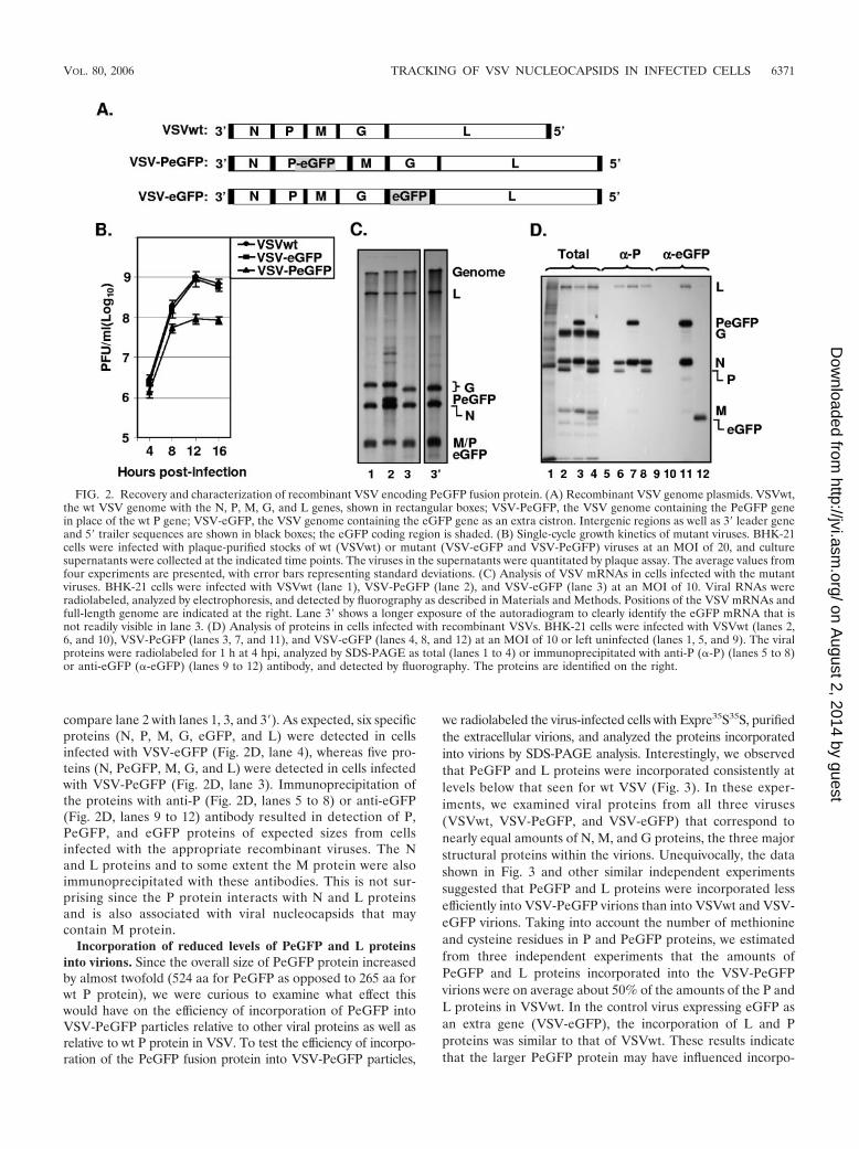
compare lane 2 with lanes 1, 3, and 3�). As expected, six specificproteins (N, P, M, G, eGFP, and L) were detected in cellsinfected with VSV-eGFP (Fig. 2D, lane 4), whereas five pro-teins (N, PeGFP, M, G, and L) were detected in cells infectedwith VSV-PeGFP (Fig. 2D, lane 3). Immunoprecipitation ofthe proteins with anti-P (Fig. 2D, lanes 5 to 8) or anti-eGFP(Fig. 2D, lanes 9 to 12) antibody resulted in detection of P,PeGFP, and eGFP proteins of expected sizes from cellsinfected with the appropriate recombinant viruses. The Nand L proteins and to some extent the M protein were alsoimmunoprecipitated with these antibodies. This is not sur-prising since the P protein interacts with N and L proteinsand is also associated with viral nucleocapsids that maycontain M protein.
Incorporation of reduced levels of PeGFP and L proteinsinto virions. Since the overall size of PeGFP protein increasedby almost twofold (524 aa for PeGFP as opposed to 265 aa forwt P protein), we were curious to examine what effect thiswould have on the efficiency of incorporation of PeGFP intoVSV-PeGFP particles relative to other viral proteins as well asrelative to wt P protein in VSV. To test the efficiency of incorpo-ration of the PeGFP fusion protein into VSV-PeGFP particles,
we radiolabeled the virus-infected cells with Expre35S35S, purifiedthe extracellular virions, and analyzed the proteins incorporatedinto virions by SDS-PAGE analysis. Interestingly, we observedthat PeGFP and L proteins were incorporated consistently atlevels below that seen for wt VSV (Fig. 3). In these exper-iments, we examined viral proteins from all three viruses(VSVwt, VSV-PeGFP, and VSV-eGFP) that correspond tonearly equal amounts of N, M, and G proteins, the three majorstructural proteins within the virions. Unequivocally, the datashown in Fig. 3 and other similar independent experimentssuggested that PeGFP and L proteins were incorporated lessefficiently into VSV-PeGFP virions than into VSVwt and VSV-eGFP virions. Taking into account the number of methionineand cysteine residues in P and PeGFP proteins, we estimatedfrom three independent experiments that the amounts ofPeGFP and L proteins incorporated into the VSV-PeGFPvirions were on average about 50% of the amounts of the P andL proteins in VSVwt. In the control virus expressing eGFP asan extra gene (VSV-eGFP), the incorporation of L and Pproteins was similar to that of VSVwt. These results indicatethat the larger PeGFP protein may have influenced incorpo-
FIG. 2. Recovery and characterization of recombinant VSV encoding PeGFP fusion protein. (A) Recombinant VSV genome plasmids. VSVwt,the wt VSV genome with the N, P, M, G, and L genes, shown in rectangular boxes; VSV-PeGFP, the VSV genome containing the PeGFP genein place of the wt P gene; VSV-eGFP, the VSV genome containing the eGFP gene as an extra cistron. Intergenic regions as well as 3� leader geneand 5� trailer sequences are shown in black boxes; the eGFP coding region is shaded. (B) Single-cycle growth kinetics of mutant viruses. BHK-21cells were infected with plaque-purified stocks of wt (VSVwt) or mutant (VSV-eGFP and VSV-PeGFP) viruses at an MOI of 20, and culturesupernatants were collected at the indicated time points. The viruses in the supernatants were quantitated by plaque assay. The average values fromfour experiments are presented, with error bars representing standard deviations. (C) Analysis of VSV mRNAs in cells infected with the mutantviruses. BHK-21 cells were infected with VSVwt (lane 1), VSV-PeGFP (lane 2), and VSV-eGFP (lane 3) at an MOI of 10. Viral RNAs wereradiolabeled, analyzed by electrophoresis, and detected by fluorography as described in Materials and Methods. Positions of the VSV mRNAs andfull-length genome are indicated at the right. Lane 3� shows a longer exposure of the autoradiogram to clearly identify the eGFP mRNA that isnot readily visible in lane 3. (D) Analysis of proteins in cells infected with recombinant VSVs. BHK-21 cells were infected with VSVwt (lanes 2,6, and 10), VSV-PeGFP (lanes 3, 7, and 11), and VSV-eGFP (lanes 4, 8, and 12) at an MOI of 10 or left uninfected (lanes 1, 5, and 9). The viralproteins were radiolabeled for 1 h at 4 hpi, analyzed by SDS-PAGE as total (lanes 1 to 4) or immunoprecipitated with anti-P (�-P) (lanes 5 to 8)or anti-eGFP (�-eGFP) (lanes 9 to 12) antibody, and detected by fluorography. The proteins are identified on the right.
VOL. 80, 2006 TRACKING OF VSV NUCLEOCAPSIDS IN INFECTED CELLS 6371
on August 2, 2014 by guest
http://jvi.asm.org/
Dow
nloaded from

ration of L-PeGFP protein complexes into the viral nucleocap-sids that are packaged in the virions.
Examination of intracellular sites of viral RNA synthesis.Subcellular localizations of eGFP were noticeably different incells infected with VSV-eGFP and VSV-PeGFP. While VSV-eGFP-infected cells showed typical eGFP distribution evenlythroughout the cytoplasm as well as in the nucleus (Fig. 4A),the fluorescence pattern in cells infected with VSV-PeGFPappeared granular and the granules were distributed through-out the cytoplasm but not in the nucleus (Fig. 4B). The differ-ential distribution patterns of PeGFP and eGFP likely reflectthe fact that PeGFP is associated with the viral nucleocapsidsand is intimately involved in viral genome replication and tran-scription, whereas eGFP is not.
To determine if the locations of the granular fluorescenceseen in cells infected with VSV-PeGFP represent the sites ofsynthesis of viral RNA, we examined the sites of de novosynthesis of viral RNA. Accordingly, cells infected with VSV-PeGFP were treated with actinomycin D followed by BrUTPto label de novo-synthesized RNA and examined by immuno-fluorescence using anti-BrdU antibody. Newly synthesizedRNA labeled with BrUTP was detected throughout the cyto-plasm (Fig. 4D), and PeGFP protein (Fig. 4C) colocalized tothese sites (Fig. 4E). The viral N and L proteins also colocal-ized with the PeGFP protein (Fig. 4F to K) in a manner similarto viral RNA. These data suggest that the de novo-synthesizedRNAs colocalize with the viral N, L, and PeGFP proteins,indicating that synthesis of viral RNA occurs at these sites. Thesites of viral RNA synthesis appear to be distributed through-out the cytoplasm. The staining observed near the cell periph-ery may represent the viral nucleocapsids in transit to the cellsurface for assembly.
Association of viral nucleocapsids with mitochondria. Live-cell imaging of VSV-PeGFP-infected cells revealed that manygreen fluorescent dots were detectable as early as 30 min afterinfection and became numerous by 1 to 2 hpi (data not shown).A size estimation of several individual green dots suggestedthat they represent individual viral nucleocapsids. Most ofthese fluorescent nucleocapsids were mobile within the cyto-plasm with time, moving toward the cell periphery away fromthe nucleus. High-magnification DIC images of infected cellsshowed that many of these nucleocapsids (Fig. 5A) appearedto be moving along with or on mitochondrion-like structures.Time-lapse imaging of one such nucleocapsid (Fig. 5A) indi-cated that the fluorescent nucleocapsids traversed along or inclose association with mitochondria or mitochondrion-likestructures in a nonlinear fashion toward the cell periphery (Fig.5A0 to A210). Tracking of several such nucleocapsids (n � 12)over time indicated that they moved with an average speed ofapproximately 30 nm/s.
To confirm that the viral nucleocapsids were moving alongand/or associated with mitochondria, we examined the infectedcells treated with MitoTracker Red, which specifically stainsonly mitochondria. By live-cell imaging, the green fluorescentnucleocapsids were seen closely associated with the red-stainedmitochondria (Fig. 5B) and were moving along mitochondria
FIG. 3. Incorporation of reduced levels of PeGFP and L proteinsinto VSV-PeGFP. Viral proteins in infected cells were radiolabeled,and the proteins incorporated into purified virions were analyzed bySDS-PAGE and detected as described in the text. The positions ofvarious proteins are shown on the right.
FIG. 4. Examination of sites of viral RNA synthesis in cells infectedwith VSV-PeGFP. (A and B) Distribution of fluorescence in cellsinfected with recombinant VSVs. BHK-21 cells infected with VSV-eGFP (A) or VSV-PeGFP (B) at an MOI of 10 were fixed at 4 hpi,stained with DAPI, and examined by fluorescence microscopy. (C toE) Cells infected with VSV-PeGFP were labeled with BrUTP, and thede novo-synthesized RNA was detected by MAb to BrdU and goatanti-mouse Alexa-594. Colocalization of PeGFP (C) with RNA (D) isshown in the merged image (E). (F to K) VSV-PeGFP-infected cellswere fixed at 4 hpi and stained with anti-N MAb (F to H) or anti-Lantibody (I to K) and the corresponding secondary antibodies conju-gated to Alexa-594. Colocalization of N (G) or L (J) with PeGFP (Fand I) is shown in the merged images (H and K).
6372 DAS ET AL. J. VIROL.
on August 2, 2014 by guest
http://jvi.asm.org/
Dow
nloaded from
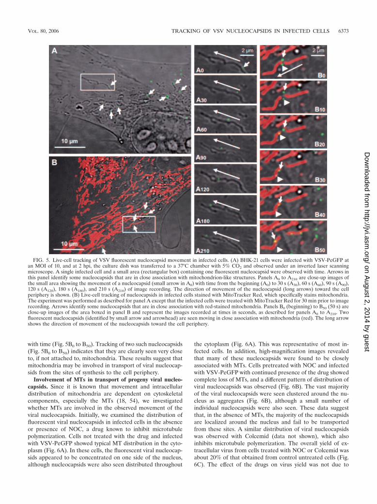
with time (Fig. 5B0 to B50). Tracking of two such nucleocapsids(Fig. 5B0 to B50) indicates that they are clearly seen very closeto, if not attached to, mitochondria. These results suggest thatmitochondria may be involved in transport of viral nucleocap-sids from the sites of synthesis to the cell periphery.
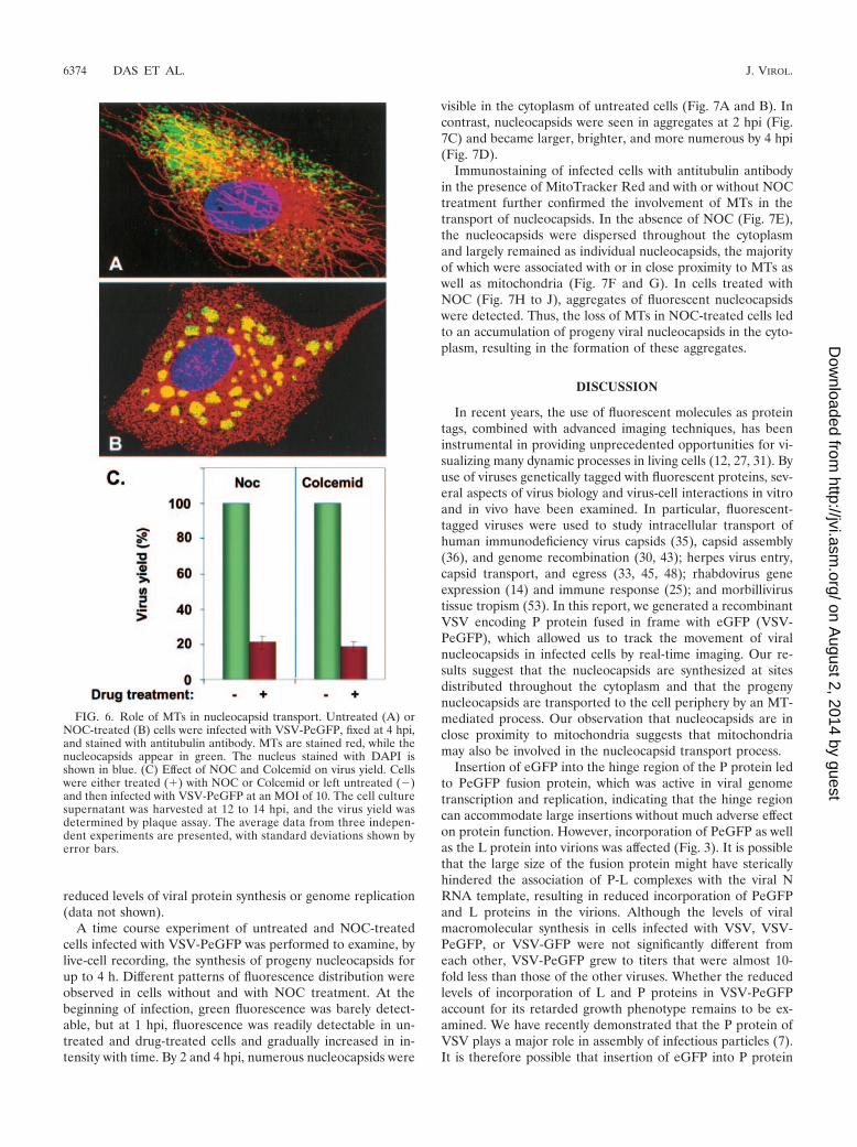
Involvement of MTs in transport of progeny viral nucleo-capsids. Since it is known that movement and intracellulardistribution of mitochondria are dependent on cytoskeletalcomponents, especially the MTs (18, 54), we investigatedwhether MTs are involved in the observed movement of theviral nucleocapsids. Initially, we examined the distribution offluorescent viral nucleocapsids in infected cells in the absenceor presence of NOC, a drug known to inhibit microtubulepolymerization. Cells not treated with the drug and infectedwith VSV-PeGFP showed typical MT distribution in the cyto-plasm (Fig. 6A). In these cells, the fluorescent viral nucleocap-sids appeared to be concentrated on one side of the nucleus,although nucleocapsids were also seen distributed throughout
the cytoplasm (Fig. 6A). This was representative of most in-fected cells. In addition, high-magnification images revealedthat many of these nucleocapsids were found to be closelyassociated with MTs. Cells pretreated with NOC and infectedwith VSV-PeGFP with continued presence of the drug showedcomplete loss of MTs, and a different pattern of distribution ofviral nucleocapsids was observed (Fig. 6B). The vast majorityof the viral nucleocapsids were seen clustered around the nu-cleus as aggregates (Fig. 6B), although a small number ofindividual nucleocapsids were also seen. These data suggestthat, in the absence of MTs, the majority of the nucleocapsidsare localized around the nucleus and fail to be transportedfrom these sites. A similar distribution of viral nucleocapsidswas observed with Colcemid (data not shown), which alsoinhibits microtubule polymerization. The overall yield of ex-tracellular virus from cells treated with NOC or Colcemid wasabout 20% of that obtained from control untreated cells (Fig.6C). The effect of the drugs on virus yield was not due to
FIG. 5. Live-cell tracking of VSV fluorescent nucleocapsid movement in infected cells. (A) BHK-21 cells were infected with VSV-PeGFP atan MOI of 10, and at 2 hpi, the culture dish was transferred to a 37°C chamber with 5% CO2 and observed under an inverted laser scanningmicroscope. A single infected cell and a small area (rectangular box) containing one fluorescent nucleocapsid were observed with time. Arrows inthis panel identify some nucleocapsids that are in close association with mitochondrion-like structures. Panels A0 to A210 are close-up images ofthe small area showing the movement of a nucleocapsid (small arrow in A0) with time from the beginning (A0) to 30 s (A30), 60 s (A60), 90 s (A90),120 s (A120), 180 s (A180), and 210 s (A210) of image recording. The direction of movement of the nucleocapsid (long arrows) toward the cellperiphery is shown. (B) Live-cell tracking of nucleocapsids in infected cells stained with MitoTracker Red, which specifically stains mitochondria.The experiment was performed as described for panel A except that the infected cells were treated with MitoTracker Red for 30 min prior to imagerecording. Arrows identify some nucleocapsids that are in close association with red-stained mitochondria. Panels B0 (beginning) to B50 (50 s) areclose-up images of the area boxed in panel B and represent the images recorded at times in seconds, as described for panels A0 to A210. Twofluorescent nucleocapsids (identified by small arrow and arrowhead) are seen moving in close association with mitochondria (red). The long arrowshows the direction of movement of the nucleocapsids toward the cell periphery.
VOL. 80, 2006 TRACKING OF VSV NUCLEOCAPSIDS IN INFECTED CELLS 6373
on August 2, 2014 by guest
http://jvi.asm.org/
Dow
nloaded from
reduced levels of viral protein synthesis or genome replication(data not shown).
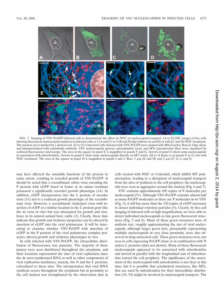
A time course experiment of untreated and NOC-treatedcells infected with VSV-PeGFP was performed to examine, bylive-cell recording, the synthesis of progeny nucleocapsids forup to 4 h. Different patterns of fluorescence distribution wereobserved in cells without and with NOC treatment. At thebeginning of infection, green fluorescence was barely detect-able, but at 1 hpi, fluorescence was readily detectable in un-treated and drug-treated cells and gradually increased in in-tensity with time. By 2 and 4 hpi, numerous nucleocapsids were
visible in the cytoplasm of untreated cells (Fig. 7A and B). Incontrast, nucleocapsids were seen in aggregates at 2 hpi (Fig.7C) and became larger, brighter, and more numerous by 4 hpi(Fig. 7D).
Immunostaining of infected cells with antitubulin antibodyin the presence of MitoTracker Red and with or without NOCtreatment further confirmed the involvement of MTs in thetransport of nucleocapsids. In the absence of NOC (Fig. 7E),the nucleocapsids were dispersed throughout the cytoplasmand largely remained as individual nucleocapsids, the majorityof which were associated with or in close proximity to MTs aswell as mitochondria (Fig. 7F and G). In cells treated withNOC (Fig. 7H to J), aggregates of fluorescent nucleocapsidswere detected. Thus, the loss of MTs in NOC-treated cells ledto an accumulation of progeny viral nucleocapsids in the cyto-plasm, resulting in the formation of these aggregates.
DISCUSSION
In recent years, the use of fluorescent molecules as proteintags, combined with advanced imaging techniques, has beeninstrumental in providing unprecedented opportunities for vi-sualizing many dynamic processes in living cells (12, 27, 31). Byuse of viruses genetically tagged with fluorescent proteins, sev-eral aspects of virus biology and virus-cell interactions in vitroand in vivo have been examined. In particular, fluorescent-tagged viruses were used to study intracellular transport ofhuman immunodeficiency virus capsids (35), capsid assembly(36), and genome recombination (30, 43); herpes virus entry,capsid transport, and egress (33, 45, 48); rhabdovirus geneexpression (14) and immune response (25); and morbillivirustissue tropism (53). In this report, we generated a recombinantVSV encoding P protein fused in frame with eGFP (VSV-PeGFP), which allowed us to track the movement of viralnucleocapsids in infected cells by real-time imaging. Our re-sults suggest that the nucleocapsids are synthesized at sitesdistributed throughout the cytoplasm and that the progenynucleocapsids are transported to the cell periphery by an MT-mediated process. Our observation that nucleocapsids are inclose proximity to mitochondria suggests that mitochondriamay also be involved in the nucleocapsid transport process.
Insertion of eGFP into the hinge region of the P protein ledto PeGFP fusion protein, which was active in viral genometranscription and replication, indicating that the hinge regioncan accommodate large insertions without much adverse effecton protein function. However, incorporation of PeGFP as wellas the L protein into virions was affected (Fig. 3). It is possiblethat the large size of the fusion protein might have stericallyhindered the association of P-L complexes with the viral NRNA template, resulting in reduced incorporation of PeGFPand L proteins in the virions. Although the levels of viralmacromolecular synthesis in cells infected with VSV, VSV-PeGFP, or VSV-GFP were not significantly different fromeach other, VSV-PeGFP grew to titers that were almost 10-fold less than those of the other viruses. Whether the reducedlevels of incorporation of L and P proteins in VSV-PeGFPaccount for its retarded growth phenotype remains to be ex-amined. We have recently demonstrated that the P protein ofVSV plays a major role in assembly of infectious particles (7).It is therefore possible that insertion of eGFP into P protein
FIG. 6. Role of MTs in nucleocapsid transport. Untreated (A) orNOC-treated (B) cells were infected with VSV-PeGFP, fixed at 4 hpi,and stained with antitubulin antibody. MTs are stained red, while thenucleocapsids appear in green. The nucleus stained with DAPI isshown in blue. (C) Effect of NOC and Colcemid on virus yield. Cellswere either treated (�) with NOC or Colcemid or left untreated (�)and then infected with VSV-PeGFP at an MOI of 10. The cell culturesupernatant was harvested at 12 to 14 hpi, and the virus yield wasdetermined by plaque assay. The average data from three indepen-dent experiments are presented, with standard deviations shown byerror bars.
6374 DAS ET AL. J. VIROL.
on August 2, 2014 by guest
http://jvi.asm.org/
Dow
nloaded from
may have affected the assembly functions of the protein tosome extent, resulting in retarded growth of VSV-PeGFP. Itshould be noted that a recombinant rabies virus encoding theP protein with eGFP fused in frame at its amino terminuspossessed a significantly retarded growth phenotype (14). Inaddition, eGFP incorporation into the L protein of measlesvirus (11) led to a reduced growth phenotype of the recombi-nant virus. However, a recombinant rinderpest virus with in-sertion of eGFP at a similar location in the L protein grew likethe wt virus in vitro but was attenuated for growth and viru-lence in its natural animal host, cattle (2). Clearly, these dataindicate that growth and virulence properties can be altered byinsertion of eGFP into the viral polymerase. It will be inter-esting to examine whether VSV-PeGFP with insertion ofeGFP in the P protein of the viral polymerase complex pos-sesses altered growth and virulence phenotypes in vivo.
In cells infected with VSV-PeGFP, the intracellular distri-bution of fluorescence was punctate. The majority of thesepuncta were seen distributed throughout the cytoplasm, andtheir locations represented the sites of viral replication sincethe de novo-synthesized RNA as well as other components ofviral replication machinery, namely, the N and the L proteins,colocalized to these sites. The interpretation that viral RNAsynthesis occurs throughout the cytoplasm but in proximity tothe cell nucleus was strengthened by the observation that in
cells treated with NOC or Colcemid, which inhibit MT poly-merization, leading to a disruption of nucleocapsid transportfrom the sites of synthesis to the cell periphery, the nucleocap-sids were seen as aggregates around the nucleus (Fig. 6 and 7).
VSV contains approximately 450 copies of P molecules pernucleocapsid (51). Although VSV-PeGFP contains almost halfas many PeGFP molecules as there are P molecules in wt VSV(Fig. 3), it still has more than the 120 copies of eGFP necessaryto detect individual rotavirus particles (5). Clearly, by live-cellimaging of infected cells at high magnification, we were able todetect individual nucleocapsids as tiny green fluorescent struc-tures (Fig. 5 and 6). Many of these green structures were ofuniform size, roughly approximating the size of viral nucleo-capsids, although larger green dots, presumably representingmultiple nucleocapsids in very close proximity, were also ob-served in drug-untreated cells. These green structures were notseen in cells expressing PeGFP alone or in combination with Nand/or L proteins (data not shown). Many of these fluorescentnucleocapsids appeared to be associated with mitochondriaand moved in parallel with the longitudinal axis of mitochon-dria toward the cell periphery. The significance of the associ-ation of the nucleocapsid with mitochondria is not clear at thistime, but it is possible that cytoskeletal structures, like MTs,that are used by mitochondria for their intracellular distribu-tion (18, 54) might be involved in nucleocapsid transport. The
FIG. 7. Imaging of VSV-PeGFP-infected cells to demonstrate the effect on NOC on nucleocapsid transport. (A to D) DIC images of live cellsshowing fluorescent nucleocapsid synthesis in infected cells at 2 (A and C) or 4 (B and D) hpi without (A and B) or with (C and D) NOC treatment.The nucleus (n) is marked by a dotted oval. (E to G) Untreated cells infected with VSV-PeGFP were stained with MitoTracker Red at 4 hpi, fixed,and immunostained with antitubulin antibody. VSV nucleocapsids (green), mitochondria (red), and MTs (pseudocolor blue) were visualized byconfocal fluorescence microscopy. The area in the square in panel E is magnified in panels F and G. Arrows in panel E show some nucleocapsidsin association with mitochondria. Arrows in panel F show some nucleocapsids directly on MT tracks. (H to J) Same as in panels E to G, but withNOC treatment. The area in the square in panel H is magnified in panels I and J. Bars, 5 �m (E and H) and 2 �m (F, G, I, and J).
VOL. 80, 2006 TRACKING OF VSV NUCLEOCAPSIDS IN INFECTED CELLS 6375
on August 2, 2014 by guest
http://jvi.asm.org/
Dow
nloaded from
association of nucleocapsids with mitochondria may thus betransient and possibly serve as bridges linking MTs duringtransport of the nucleocapsids. Alternatively, it is possible thatthe association of nucleocapsids with mitochondria may be justrandom. Further work will be necessary to provide any func-tional significance to the association of nucleocapsids with mi-tochondria.
Although the effect of NOC on nucleocapsid distributionwas dramatic (Fig. 6B), virus yield was reduced to about 20%of that from untreated control cells (Fig. 6C). It is possible thatnucleocapsid transport may occur by additional mechanismsthat are independent of MT. In this regard, it is of note that forDNA viruses whose capsids are specifically transported by MTstoward the cell periphery, a 20 to 25% reduction in virus yieldin the presence of NOC has been considered to be significant(3, 37). In the light of these observations, our data are consis-tent with the interpretation that MTs are involved in the trans-port of viral nucleocapsids by an anterograde movement to-ward the cell periphery. Although the nucleocapsid transporttoward the cell periphery was affected by NOC or Colcemidtreatment, virus entry and uncoating remained unaffected, asseen by synthesis and accumulation of viral nucleocapsids withtime in NOC-treated cells (Fig. 7C and D). This is consistentwith the recent findings that NOC has no significant adverseeffect on VSV entry, uncoating, or viral macromolecular syn-thesis (29).
How might the MTs be involved in transport of viral nucleo-capsids toward the cell periphery? Since the MTs form trackson which cellular cargos are transported by intracellular kine-sin and dynein motors (10, 52), it is possible to envision ascenario in which the nucleocapsids interact with kinesinand/or dynein motors and are transported by these motors onMT tracks. In this regard, it is interesting to note that the Pprotein of rabies virus (another rhabdovirus) has been shownto interact efficiently with the dynein light chain (14, 22, 42), acomponent of the dynein motor. Although the significance ofthis interaction in terms of intracellular transport of the viralnucleocapsids has not been established, it is tempting to spec-ulate that VSV nucleocapsids also interact with one or morecomponents of the intracellular motors that play a role in thetransport of the nucleocapsids mediated by MTs. With the avail-ability of fluorescently tagged nucleocapsids, it will be possible tostudy these interactions and examine intracellular events by real-time imaging of virus-infected cells for a better understandingof the mechanisms by which the viral nucleocapsids are trans-ported from the sites of synthesis to the assembly sites. Fur-thermore, VSV-PeGFP could be used to study entry and un-coating mechanisms that may provide significant insight intoreceptor-mediated endocytosis and uncoating of viral nucleo-capsids in infected cells.
ACKNOWLEDGMENTS
We thank A. Martinsen and T. Fangman for excellent technical help.This investigation was supported by a grant (AI 34956) from NIAID,
NIH, and also in part by P20RR15635 from NCRR, NIH.
REFERENCES
1. Banerjee, A. K. 1987. Transcription and replication of rhabdoviruses. Micro-biol. Rev. 51:66–87.
2. Brown, D. D., B. K. Rima, I. V. Allen, M. D. Baron, A. C. Banyard, T. Barrett,and W. P. Duprex. 2005. Rational attenuation of a morbillivirus by modu-
lating the activity of the RNA-dependent RNA polymerase. J. Virol. 79:14330–14338.
3. Bukrinskaya, A., B. Brichacek, A. Mann, and M. Stevenson. 1998. Estab-lishment of a functional human immunodeficiency virus type 1 (HIV-1)reverse transcription complex involves the cytoskeleton. J. Exp. Med. 188:2113–2125.
4. Canter, D. M., and J. Perrault. 1996. Stabilization of vesicular stomatitisvirus L polymerase protein by P protein binding: a small deletion in theC-terminal domain of L abrogates binding. Virology 219:376–386.
5. Charpilienne, A., M. Nejmeddine, M. Berois, N. Parez, E. Neumann, E.Hewat, G. Trugnan, and J. Cohen. 2001. Individual rotavirus-like particlescontaining 120 molecules of fluorescent protein are visible in living cells.J. Biol. Chem. 276:29361–29367.
6. Das, S. C., and A. K. Pattnaik. 2004. Phosphorylation of vesicular stomatitisvirus phosphoprotein P is indispensable for virus growth. J. Virol. 78:6420–6430.
7. Das, S. C., and A. K. Pattnaik. 2005. Role of the hypervariable hinge regionof phosphoprotein P of vesicular stomatitis virus in viral RNA synthesis andassembly of infectious virus particles. J. Virol. 79:8101–8112.
8. Das, T., A. K. Pattnaik, A. M. Takacs, T. Li, L. N. Hwang, and A. K.Banerjee. 1997. Basic amino acid residues at the carboxy-terminal elevenamino acid region of the phosphoprotein (P) are required for transcriptionbut not for replication of vesicular stomatitis virus genome RNA. Virology238:103–114.
9. Davis, N. L., H. Arnheiter, and G. W. Wertz. 1986. Vesicular stomatitis virusN and NS proteins form multiple complexes. J. Virol. 59:751–754.
10. Dohner, K., C. H. Nagel, and B. Sodeik. 2005. Viral stop-and-go alongmicrotubules: taking a ride with dynein and kinesins. Trends Microbiol.13:320–327.
11. Duprex, W. P., F. M. Collins, and B. K. Rima. 2002. Modulating the functionof the measles virus RNA-dependent RNA polymerase by insertion of greenfluorescent protein into the open reading frame. J. Virol. 76:7322–7328.
12. Ehrhardt, D. 2003. GFP technology for live cell imaging. Curr. Opin. PlantBiol. 6:622–628.
13. Emerson, S. U., and M. Schubert. 1987. Location of the binding domains forthe RNA polymerase L and the ribonucleocapsid template within differenthalves of the NS phosphoprotein of vesicular stomatitis virus. Proc. Natl.Acad. Sci. USA 84:5655–5659.
14. Finke, S., K. Brzozka, and K. K. Conzelmann. 2004. Tracking fluorescence-labeled rabies virus: enhanced green fluorescent protein-tagged phospho-protein P supports virus gene expression and formation of infectious parti-cles. J. Virol. 78:12333–12343.
15. Fuerst, T. R., E. G. Niles, F. W. Studier, and B. Moss. 1986. Eukaryotictransient-expression system based on recombinant vaccinia virus that syn-thesizes bacteriophage T7 RNA polymerase. Proc. Natl. Acad. Sci. USA83:8122–8126.
16. Georgi, A., C. Mottola-Hartshorn, A. Warner, B. Fields, and L. B. Chen.1990. Detection of individual fluorescently labeled reovirions in living cells.Proc. Natl. Acad. Sci. USA 87:6579–6583.
17. Gosert, R., A. Kanjanahaluethai, D. Egger, K. Bienz, and S. C. Baker. 2002.RNA replication of mouse hepatitis virus takes place at double-membranevesicles. J. Virol. 76:3697–3708.
18. Heggeness, M. H., M. Simon, and S. J. Singer. 1978. Association of mito-chondria with microtubules in cultured cells. Proc. Natl. Acad. Sci. USA75:3863–3866.
19. Hwang, L. N., N. Englund, T. Das, A. K. Banerjee, and A. K. Pattnaik. 1999.Optimal replication activity of vesicular stomatitis virus RNA polymeraserequires phosphorylation of a residue(s) at carboxy-terminal domain II of itsaccessory subunit, phosphoprotein P. J. Virol. 73:5613–5620.
20. Hwang, L. N., N. Englund, and A. K. Pattnaik. 1998. Polyadenylation ofvesicular stomatitis virus mRNA dictates efficient transcription terminationat the intercistronic gene junctions. J. Virol. 72:1805–1813.
21. Isaac, C. L., and J. D. Keene. 1982. RNA polymerase-associated interactionsnear template promoter sequences of defective interfering particles of ve-sicular stomatitis virus. J. Virol. 43:241–249.
22. Jacob, Y., H. Badrane, P. E. Ceccaldi, and N. Tordo. 2000. Cytoplasmicdynein LC8 interacts with lyssavirus phosphoprotein. J. Virol. 74:10217–10222.
23. Jouvenet, N., P. Monaghan, M. Way, and T. Wileman. 2004. Transport ofAfrican swine fever virus from assembly sites to the plasma membrane isdependent on microtubules and conventional kinesin. J. Virol. 78:7990–8001.
24. Keene, J. D., B. J. Thornton, and S. U. Emerson. 1981. Sequence-specificcontacts between the RNA polymerase of vesicular stomatitis virus and theleader RNA gene. Proc. Natl. Acad. Sci. USA 78:6191–6195.
25. Koser, M. L., J. P. McGettigan, G. S. Tan, M. E. Smith, H. Koprowski, B.Dietzschold, and M. J. Schnell. 2004. Rabies virus nucleoprotein as a carrierfor foreign antigens. Proc. Natl. Acad. Sci. USA 101:9405–9410.
26. Lakadamyali, M., M. J. Rust, H. P. Babcock, and X. Zhuang. 2003. Visual-izing infection of individual influenza viruses. Proc. Natl. Acad. Sci. USA100:9280–9285.
27. Lalonde, S., D. W. Ehrhardt, and W. B. Frommer. 2005. Shining light on
6376 DAS ET AL. J. VIROL.
on August 2, 2014 by guest
http://jvi.asm.org/
Dow
nloaded from
signaling and metabolic networks by genetically encoded biosensors. Curr.Opin. Plant Biol. 8:574–581.
28. Lawson, N. D., E. A. Stillman, M. A. Whitt, and J. K. Rose. 1995. Recom-binant vesicular stomatitis viruses from DNA. Proc. Natl. Acad. Sci. USA92:4477–4481.
29. Le Blanc, I., P. P. Luyet, V. Pons, C. Ferguson, N. Emans, A. Petiot, N.Mayran, N. Demaurex, J. Faure, R. Sadoul, R. G. Parton, and J. Gruenberg.2005. Endosome-to-cytosol transport of viral nucleocapsids. Nat. Cell Biol.7:653–664.
30. Levy, D. N., G. M. Aldrovandi, O. Kutsch, and G. M. Shaw. 2004. Dynamicsof HIV-1 recombination in its natural target cells. Proc. Natl. Acad. Sci. USA101:4204–4209.
31. Lippincott-Schwartz, J., and G. H. Patterson. 2003. Development and use offluorescent protein markers in living cells. Science 300:87–91.
32. Luby-Phelps, K. 2000. Cytoarchitecture and physical properties of cyto-plasm: volume, viscosity, diffusion, intracellular surface area. Int. Rev. Cytol.192:189–221.
33. Luxton, G. W., S. Haverlock, K. E. Coller, S. E. Antinone, A. Pincetic, andG. A. Smith. 2005. Targeting of herpesvirus capsid transport in axons iscoupled to association with specific sets of tegument proteins. Proc. Natl.Acad. Sci. USA 102:5832–5837.
34. Masters, P. S., and A. K. Banerjee. 1988. Resolution of multiple complexesof phosphoprotein NS with nucleocapsid protein N of vesicular stomatitisvirus. J. Virol. 62:2651–2657.
35. McDonald, D., M. A. Vodicka, G. Lucero, T. M. Svitkina, G. G. Borisy, M.Emerman, and T. J. Hope. 2002. Visualization of the intracellular behaviorof HIV in living cells. J. Cell Biol. 159:441–452.
36. Muller, B., J. Daecke, O. T. Fackler, M. T. Dittmar, H. Zentgraf, and H. G.Krausslich. 2004. Construction and characterization of a fluorescently la-beled infectious human immunodeficiency virus type 1 derivative. J. Virol.78:10803–10813.
37. Naranatt, P. P., H. H. Krishnan, M. S. Smith, and B. Chandran. 2005.Kaposi’s sarcoma-associated herpesvirus modulates microtubule dynamicsvia RhoA-GTP-diaphanous 2 signaling and utilizes the dynein motors todeliver its DNA to the nucleus. J. Virol. 79:1191–1206.
38. Pattnaik, A. K., L. A. Ball, A. W. LeGrone, and G. W. Wertz. 1992. Infectiousdefective interfering particles of VSV from transcripts of a cDNA clone. Cell69:1011–1020.
39. Pattnaik, A. K., L. Hwang, T. Li, N. Englund, M. Mathur, T. Das, and A. K.Banerjee. 1997. Phosphorylation within the amino-terminal acidic domain Iof the phosphoprotein of vesicular stomatitis virus is required for transcrip-tion but not for replication. J. Virol. 71:8167–8175.
40. Pattnaik, A. K., and G. W. Wertz. 1990. Replication and amplification ofdefective interfering particle RNAs of vesicular stomatitis virus in cellsexpressing viral proteins from vectors containing cloned cDNAs. J. Virol.64:2948–2957.
41. Peluso, R. W. 1988. Kinetic, quantitative, and functional analysis of multipleforms of the vesicular stomatitis virus nucleocapsid protein in infected cells.J. Virol. 62:2799–2807.
42. Raux, H., A. Flamand, and D. Blondel. 2000. Interaction of the rabies virusP protein with the LC8 dynein light chain. J. Virol. 74:10212–10216.
43. Rhodes, T. D., O. Nikolaitchik, J. Chen, D. Powell, and W. S. Hu. 2005.Genetic recombination of human immunodeficiency virus type 1 in oneround of viral replication: effects of genetic distance, target cells, accessorygenes, and lack of high negative interference in crossover events. J. Virol.79:1666–1677.
44. Rose, J. K., and M. A. Whitt. 2001. Rhabdoviridae: the viruses and theirreplication, p. 1221–1244. In B. N. Fields and D. M. Knipe (ed.), Fieldsvirology, 4th ed. Lippincott Williams & Wilkins, Philadelphia, Pa.
45. Sampaio, K. L., Y. Cavignac, Y. D. Stierhof, and C. Sinzger. 2005. Humancytomegalovirus labeled with green fluorescent protein for live analysis ofintracellular particle movements. J. Virol. 79:2754–2767.
46. Schubert, M., G. G. Harmison, C. D. Richardson, and E. Meier. 1985.Expression of a cDNA encoding a functional 241-kilodalton vesicular sto-matitis virus RNA polymerase. Proc. Natl. Acad. Sci. USA 82:7984–7988.
47. Seisenberger, G., M. U. Ried, T. Endress, H. Buning, M. Hallek, and C.Brauchle. 2001. Real-time single-molecule imaging of the infection pathwayof an adeno-associated virus. Science 294:1929–1932.
48. Smith, G. A., L. Pomeranz, S. P. Gross, and L. W. Enquist. 2004. Localmodulation of plus-end transport targets herpesvirus entry and egress insensory axons. Proc. Natl. Acad. Sci. USA 101:16034–16039.
49. Sodeik, B., M. W. Ebersold, and A. Helenius. 1997. Microtubule-mediatedtransport of incoming herpes simplex virus 1 capsids to the nucleus. J. CellBiol. 136:1007–1021.
50. Suomalainen, M., M. Y. Nakano, S. Keller, K. Boucke, R. P. Stidwill, andU. F. Greber. 1999. Microtubule-dependent plus- and minus end-directedmotilities are competing processes for nuclear targeting of adenovirus.J. Cell Biol. 144:657–672.
51. Thomas, D., W. W. Newcomb, J. C. Brown, J. S. Wall, J. F. Hainfeld, B. L.Trus, and A. C. Steven. 1985. Mass and molecular composition of vesicularstomatitis virus: a scanning transmission electron microscopy analysis. J. Vi-rol. 54:598–607.
52. Vale, R. D. 2003. The molecular motor toolbox for intracellular transport.Cell 112:467–480.
53. von Messling, V., D. Milosevic, and R. Cattaneo. 2004. Tropism illuminated:lymphocyte-based pathways blazed by lethal morbillivirus through the hostimmune system. Proc. Natl. Acad. Sci. USA 101:14216–14221.
54. Yaffe, M. P., N. Stuurman, and R. D. Vale. 2003. Mitochondrial positioningin fission yeast is driven by association with dynamic microtubules andmitotic spindle poles. Proc. Natl. Acad. Sci. USA 100:11424–11428.
VOL. 80, 2006 TRACKING OF VSV NUCLEOCAPSIDS IN INFECTED CELLS 6377
on August 2, 2014 by guest
http://jvi.asm.org/
Dow
nloaded from