probing the interaction between vesicular stomatitis virus and phosphatidylserine
TRANSCRIPT

ARTICLE
Fabiana A. Carneiro Æ Pedro A. Lapido-Loureiro
Sandra M. Cordo Æ Fausto Stauffer
Gilberto Weissmuller Æ M. Lucia. Bianconi
Maria A. Juliano Æ Luiz Juliano Æ Paulo M. Bisch
Andrea T. Da. Poian
Probing the interaction between vesicular stomatitisvirus and phosphatidylserine
Received: 20 May 2005 / Revised: 30 June 2005 / Accepted: 20 July 2005 / Published online: 24 September 2005� EBSA 2005
Abstract The entry of enveloped animal viruses intotheir host cells always depends on membrane fusiontriggered by conformational changes in viral envelopeglycoproteins. Vesicular stomatitis virus (VSV) infectionis mediated by virus spike glycoprotein G, which inducesmembrane fusion between the viral envelope and theendosomal membrane at the acidic environment of thiscompartment. In this work, we evaluated VSV interac-tions with membranes of different phospholipid com-positions, at neutral and acidic pH, using atomic forcemicroscopy (AFM) operating in the force spectroscopymode, isothermal calorimetry (ITC) and moleculardynamics simulation. We found that the binding forcesdiffered dramatically depending on the membranephospholipid composition, revealing a high specificity of
G protein binding to membranes containing phospha-tidylserine (PS). In a previous work, we showed that thesequence corresponding amino acid 145–164 of VSV Gprotein was as efficient as the virus in catalyzing mem-brane fusion at pH 6.0. Here, we used this sequence toexplore VSV–PS interaction using ITC. We found thatpeptide binding to membranes was exothermic, sug-gesting the participation of electrostatic interactions.Peptide–membrane interaction at pH 7.5 was shown tobe specific to PS and dependent on the presence of Hisresidues in the fusion peptide. The application of thesimplified continuum Gouy–Chapman theory to oursystem predicted a pH of 5.0 at membrane surface,suggesting that the His residues should be protonatedwhen located close to the membrane. Moleculardynamics simulations suggested that the peptide inter-acts with the lipid bilayer through its N-terminal resi-dues, especially Val145 and His148.
Introduction
The plasma membrane of eukaryotic cells serves as abarrier against invading parasites and viruses. To infecta cell, viruses must be capable of transporting theirgenome and accessory proteins into the cytosol or, insome cases, into the nucleus of the host cell, thusbypassing or modifying the barrier properties imposedby the plasma membrane. Enveloped viruses always gainentry to the cytoplasm by fusion of their lipid envelopewith the plasma or endosomal membranes (Hernandezet al. 1996; Skehel and Wiley 2000; Eckert and Kim2001), whereas nonenveloped viruses must use alterna-tive strategies to cross the membrane. Membrane fusioninduced by viruses is mediated by viral fusion glyco-proteins, which have already been identified for anumber of different viruses (Hernandez et al. 1996). Thefusion reaction depends on conformational changes inthe fusion glycoproteins that can be triggered either by
Fabiana A. Carneiro and Pedro A. Lapido-Loureiro contributedequally to this work
F. A. Carneiro Æ S. M. Cordo Æ F. Stauffer Æ M. L. BianconiA. T. Da. Poian (&)Instituto de Bioquımica Medica,Universidade Federal do Rio de Janeiro,Rio de Janeiro, 21941-590 BrazilE-mail: [email protected].: +55-21-22706264Fax: +55-21-22708647
P. A. Lapido-Loureiro Æ G. Weissmuller Æ P. M. BischF. A. CarneiroLaboratorio de Fısica Biologica,Instituto de Biofısica Carlos Chagas Filho,Universidade Federal do Rio de Janeiro,Rio de Janeiro, RJ, 21949-900 Brazil
S. M. CordoLaboratorio de Virologıa, Departamento de Quimica Biologica,Facultad de Ciencias Exactas y Naturales,Universidad de Buenos Aires, Buenos Aires, Argentina
M. A. Juliano Æ L. JulianoDepartamento de Biofısica, Escola Paulista de Medicina,UNIFESP, Rua Tresde Maio, 100,SaoPaulo, 04044-020 Brazil
Eur Biophys J (2006) 35: 145–154DOI 10.1007/s00249-005-0012-z

the interaction with a specific virus receptor on cellsurface, or by the acidic pH of the endosomal environ-ment.
Vesicular stomatitis virus (VSV) belongs to theRhabdoviridae family, a group of enveloped negativesingle strand RNA viruses. The VSV envelope containsapproximately 1,200 molecules of a single transmem-brane glycoprotein, the G protein, that form about 400trimeric spikes on the virus surface. VSV G protein isinvolved in both virus attachment to the host cell surfaceand in the membrane fusion mediated by the virus. VSV-induced membrane fusion occurs at the endosomalcompartment where the acidic pH induces conforma-tional changes on G protein, leading to the exposure ofhydrophobic domains (Crimmins et al. 1983; Durreret al. 1995; Pak et al. 1987), followed by dramaticstructural reorganization (Carneiro et al. 2001).
The cellular receptor for VSV is still to be deter-mined. There are several evidences in the literaturesuggesting an important role for phospholipids in therhabdovirus entry into the host cell (Schlegel et al.1982, 1983; Superti et al. 1984; Mastromarino et al.1987). A high affinity, saturable binding site has beendescribed for VSV on Vero cells, indicating that thebinding occurs through a specific receptor (Schlegelet al. 1982). The binding could be inhibited by mem-brane extracts, which were resistant to protease, neur-aminidase and heating, but inactivated by treatmentwith phospholipase C (Schlegel et al. 1983). Thesefindings, together with the observation that onlyphosphatidylserine (PS) among various purified lipidswas able to inhibit VSV binding to membranes, led theauthors to suggest that PS could participate in thecellular binding site for VSV (Schlegel et al. 1983). Onthe other hand, a recent work from Coil and Miller(2004) provided consistent evidences supporting thatthe VSV binding to PS is not a determinant event inthe context of the cellular plasma membrane. For theauthors, virus recognition at cell surface must occurthrough an unknown cellular receptor, and the bindingto PS should be important in a subsequent step of theentry process. Despite this controversy regarding theparticipation of PS in VSV binding site at the host cellsurface, several findings indicate that PS is essential forVSV–membrane interactions. Membrane fusion medi-ated by VSV G protein reconstituted in lipid vesiclesshowed a large preference for target membranes con-taining phosphatidylserine or phosphatidic acid (Eid-elman et al. 1984). In a previous work, we showed thatthe extent of pH-induced G protein conformationalchanges and the membrane fusion mediated by thisprotein could be correlated to the PS content in thevesicles (Carneiro et al. 2002). A PS-binding segmentwas mapped in G protein from several rhabdoviruses(Coll 1997). This sequence was first identified in viralhemorrhagic septicemia virus (VHSV), a fish rhabdo-virus that infects salmonids (Estepa and Coll 1996),and then found in rabies, VSV and infectious hae-matopoietic necrosis virus (IHNV), another fish
rhabdovirus (Coll 1997). We have demonstrated that asynthetic peptide corresponding to the VSV PS bindingsite (amino acids 145 to 164 of G protein) was asefficient as the whole virus to mediate fusion (Carneiroet al. 2003). This segment contains two His residues,whose substitution by Ala or modification withdiethylpyrocarbonate (DEPC) inhibits the fusogenicproperties of the peptide.
In the present work, we compared the interactionforces between VSV and membranes of different phos-pholipid composition and we found a high specificity forPS on VSV binding to vesicles. We evaluated the role ofthis His residues on membrane recognition at neutralbulk pH, a condition in which no fusion occurs. Weshow that the high specificity of VSV binding to mem-branes containing PS was due to His–PS interaction. Inaddition, a model for this interaction is proposed.
Materials and methods
Chemicals
Phosphatidylserine (PS), phosphatidylcholine (PC) andphosphatidylethanolamine (PE) from bovine brain,phosphatidylglycerol (PG) from egg yolk lecithin,phosphatidylinositol (PI) from bovine liver, anddiethylpyrocarbonate (DEPC) were purchased fromSigma Chemical Co., St. Louis, MO, USA. All otherreagents were of analytical grade.
Virus propagation and purification
VSV Indiana was propagated in monolayer cultures ofBHK21 cells. The cells were grown at 37�C in petridishes containing Dulbecco’s modified Eagle mediumsupplemented with 10% fetal bovine serum, 100 lg/mlpenicillin, 0.0085% streptomycin sulfate. When thecells reached confluence, the medium was removed, andthe cell monolayer was infected with VSV at a multi-plicity of infection (MOI) of 0.1. The cultures werekept at 37�C for 16–20 h and the virus was harvestedand purified by differential centrifugation followed byequilibrium sedimentation in a sucrose gradient as de-scribed elsewhere (Da Poian et al. 1996). For all theexperiments the purified virus was dialyzed against Tris10 mM buffer, pH 7.4 for 4 h. Purified virions werestored at �70�C.
Peptides synthesis
The VSV G protein peptide corresponding to the se-quence between amino acids 145 and 164,VTPHHVLVDEYTGEWVDSQF, and the same pep-tide except for the substitution of then two His for Alaresidues, VTPAAVLVDEYTGEWVDSQF, were syn-thesized by solid phase using the Fmoc methodology
146

and all protected amino acids were purchased fromCalbiochem–Novabiochem (San Diego, USA) or fromNeosystem (Strasbourg, France). The syntheses weredone in an automated bench-top simultaneous multiplesolid-phase peptide synthesizer (PSSM 8 system fromShimadzu). The final deprotected peptides were purifiedby semipreparative HPLC using an Econosil C-18 col-umn (10 lm, 22.5 · 250 mm) and a two-solvent system:(a) trifluoroacetic acid/H2O (1:1000, v/v) and (b) triflu-oroacetic acid/acetonitrile/H2O (1:900:100, v/v/v). Thecolumn was eluted at a flow rate of 5 ml min�1 with a 10or 30 to 50 or 60% gradient of solvent B over 30 or45 min. Analytical HPLC was performed using a binaryHPLC system from Shimadzu with a SPD-10AV Shi-madzu UV/vis detector, coupled to an Ultrasphere C-18column (5 lm, 4.6 · 150 mm), which was eluted withsolvent systems A1 (H3PO4/H2O, 1:1000, v/v) and B1(acetonitrile/H2O/H3PO4, 900:100:1, v/v/v) at a flow rateof 1.7 ml min�1 and a 10–80% gradient of B1 over15 min. The HPLC column eluted materials were mon-itored by their absorbance at 220 nm. The molecularmass and purity of synthesized peptides were checked byMALDI-TOF mass spectrometry (TofSpec-E, Micro-mass) and/or peptide sequencing using a protein se-quencer PPSQ-23 (Shimadzu Tokyo, Japan).
Sample modification with DEPC
DEPC solutions were freshly prepared by dilution of thereagent in cold ethanol. The concentration of stockDEPC solution was determined by reaction with 10 mMimidazole (Miles 1977). For modification with DEPC,VSV (0.6 mg/ml) was incubated with DEPC at finalconcentration of 0.5 mM for 15 min at room tempera-ture. Then VSV was diluted in 20 mM MES, 30 mMTris buffer, pH 7.5 to a final protein concentration of60 lg/ml. For modification of peptides, the process wascarried out under the same conditions except that theinitial and final protein concentrations were 10 mg/mland 400 lg/ml, respectively.
Preparation of liposomes
Phospholipids were dissolved in chloroform and evap-orated under nitrogen. The lipid film was resuspended in20 mM MES, 30 mM Tris buffer (pH 7.5 or 6.0) in afinal lipid concentration of 1 mM. The suspension wasvortexed vigorously for 5 min. Small unilamellar vesicleswere obtained by sonicating the turbid suspension usinga Branson Sonifier (Sonic Power Company, Danbury,CT, USA) equipped with a titanium microtip probe.Sonication was performed in an ice bath, alternatingcycles of 30 s at 20% full power, with 60 s restingintervals until a transparent solution was obtained(approximately ten cycles). The phospholipids used in
this study were composed of PC only, PC:PE, PC:PS,PC:PI and PC:PG at the proportions indicated in thefigure legends.
Atomic force microscopy
The AFM used in this work was built in collaborationwith the Ludwig-Maximilians-Universitat, Lehrstuhl furAngewandte Physik in Munchen, Germany. For all theexperiments, the AFM was used in force-spectroscopymode (Florin et al. 1994; Gergely et al. 2000; Zlatanovaet al. 2000). Mica coverslips were glued to magneticstainless steel punches, and mounted in a fluid cellwithout using the O-ring. The mica surfaces were pre-incubated with vesicles before transferring to the fluidcell (Jass et al. 2000; Puu et al. 2000). Since the presenceof calcium ions appears to facilitate as well as to increasethe rate of planar membrane formation from vesicles(Puu et al. 1997; Reviakine et al. 2000), mica surfaceswere incubated with 20 ll of the vesicle suspensioncontaining 1 mM phospholipids, plus 10 ll of 20 mMMES, 30 mM Tris buffer, pH 7.4 containing 1 mMCaCl2, for approximately half an hour at room tem-perature (25±0.5�C). After incubation, the slips werewashed repeatedly with the same buffer used to preparevesicles. All experiments were performed at room tem-perature using standard V-shaped cantilevers, contain-ing a silicon nitride tip with a 4 lm2 pyramidal base(Digital Instruments Inc.). The cantilevers have a springconstant of 0.06 N/m (manufacturer’s data) and werepre-incubated with VSV as follows. The cantilevers wereimmersed in a virus suspension (total protein concen-tration of 0.28 mg/ml) for 24 h at 4–6�C. The instrumentallows the performance of ‘‘approach-retraction’’ cycles,in which the maximal contact force, interaction time andthe approach-retracting rates can be controlled inde-pendently. The maximal force was limited to approxi-mately 3 nN, the interaction time was set to zero and theapproach-retracting rate was set to 7,500 nm/s. Tips canbe reused but they should be cleaned soon and shouldnot dry out before the cleaning procedure.
Calorimetric studies
Binding to lipid vesicles and membrane fusion inducedby VSV or wild type and mutant peptide [145–164] werestudied by isothermal titration calorimetry (ITC) using aMCS-ITC from MicroCal, Llc. (Northampton, MA,USA). Membrane fusion was studied by following theheat effect of four injections of 5 ll of a VSV suspension(60 lg/ml) or peptide solution (400 lg/ml) into thesample cell containing 1 mM PC:PS (1:3) vesicles in20 mM MES, 30 mM Tris buffer (pH 6.0), after equili-bration at 37�C. For the binding experiments, the sam-ples were prepared at pH 7.5, and four injections (5 lleach) of the virus suspension or peptide solution weredone into the sample cell (V= 1.38 ml) containing
147

1 mM PC:PS (1:3) or PC:PG (1:3) vesicles in 20 mMMES, 30 mM Tris buffer (pH 7.5), after equilibration at37 � C. The heat of dilution of the peptides was mea-sured by injecting the same solutions into buffer only.
Molecular dynamics simulations
Initially, we decided to simulate the peptide [145–164] insolution by molecular dynamics (MD) in order to eval-uate the consistency of the force field and compare itwith experimental NMR data in aqueous environment(C.S. Lima et al., unpublished results). The startingatomic coordinates were parameterized with the GRO-MOS96 united atom force field implemented in theGROMACS MD program (Lindahl et al. 2001). Thepeptide [145–164] with protonated His148 and His149 wassolvated in a SPC water box (Berendsen et al. 1987) with4,737 water molecules and two sodium ions, to neu-tralize the �2 e net charge. After energy minimizationwith constraints of 1,000 kJ/mol applied on C, N and Oatoms, we performed an unconstrained MD simulationin the NPT ensemble (fixed number of particles andconstant pressure/temperature) (Berendsen et al. 1984)for 10 ns, at 298 K and 1 bar.
To gain insight into the interaction between thepeptide [145–164] and charged membranes at anatomistic resolution, we chose to simulate a dimyri-stoylphosphosphatidylserine (DMPS) bilayer in the li-quid-crystalline phase. The last configuration of thepeptide [145–164] in the previous simulation was in-serted in the aqueous phase of a pre-equilibrated 126DMPS bilayer, with 7,347 SPC water molecules and 126sodium counterions. The system was energy minimizedwith constraints (see above) applied on the protein. Thelipids were simulated with the parameters taken fromPandit et al. (2002) and Chandrasekhar et al. (2003) inthe NPT ensemble, with anisotropic pressure scaling(i.e., six box dimensions xx, yy, zz, xy/yx, xz/zx and yz/zy were independently coupled to pressure ‘‘baths’’) for10 ns. In both systems, bonds were constrained with theSHAKE algorithm (Ryckaert et al. 1977) allowing a 2 fsintegration step. Electrostatic forces were calculatedusing the particle mesh Ewald method (Darden et al.1993) with 1.2 A grid spacing and a fourth-order splinefor interpolation. Van der Waals forces were computedwith a cut-off radius of 14 A .
Continuum electrostatics models
Assuming that the electrostatics of model lipid mem-branes is well described by the Gouy–Chapmanapproximation for interfaces of the generalized Poisson–Boltzmann equation (Cevc 1990), we can calculate thesurface potential as:
w0 ¼2kBT
Ze
� �sinh�1
Zerelk2ee0kBT
� �
where, Y 0 is the surface potential, Z is the co- andcounter-ion valency, e, e0 and kB are the dielectric con-stant of water, the permittivity of free space and theBoltzmann constant, respectively. rel is the surfacecharge density and k is the Debye screening length (orthe width of the double layer), defined as:
k ¼ ee0kBT103NAe2RZ2
i ci
� �12
ð2Þ
According to the Boltzmann distribution, we have:
½ion�0 ¼ ½ion�bulk exp �ZeW0
kBT
� �ð3Þ
where, [ion]0 and [ ion]bulk are the ionic species concen-tration near the surface and in the bulk, respectively.
Results
Probing the VSV–membrane interaction using atomicforce microscopy
Force spectroscopy was used to measure the interactionforces between VSV and membranes of different phos-pholipid composition at pH 7.5, a condition that simu-lates binding to membrane but not fusion; and atpH 6.0, the optimum pH for VSV fusion. A set of fivelipid film compositions was used: three negativelycharged, composed of PC:PS (3:1), PC:PI (3:1), andPC:PG (3:1), and two neutral, composed of PC only andPC:PE (3:1). Sets of scans were acquired with delaytimes increasing from 0 to 800 ms between the approachand the retraction of the scanner (Figs. 1, 2).
At pH 7.5, the binding between the virus and lipidfilms containing PS was remarkably stronger whencompared to that observed for other lipid compositions(Fig. 1). Moreover, as shown in Table 1, the retractioncurves obtained for the interaction between VSV andPC:PS showed several rupture events that extended forhundreds of nanometers, while for other lipid films theinteraction peaks appear much closer to the contactpoint. The interaction between the virus and films ofPC:PS and PC:PG increased with contact time (Fig. 1).Although PC:PG films exhibited force curves with sev-eral rupture events after longer contact times, the forcepeaks were considerably smaller than that observed forPC:PS films. For PC:PI films, no significant interactionwas observed even after increasing the contact time,indicating that not only the charge is important forvirus–membrane interaction, but also the specific lipidhead group. Films containing PC:PE showed a very smallinteraction peak, close to the contact point, exhibitingonly a small variation with contact time. Furthermore,films containing PC only did not show any detectableinteraction even after 800 ms of contact (not shown).
In order to analyze the interactions under conditionsreflecting the protein–lipid interaction events that take
148

place during membrane fusion reaction, similar experi-ments were performed at pH 6.0 (Fig. 2). As observed atpH 7.5, for PS and PG containing lipid films, theinteraction forces increased with the increase in thecontact time and no interaction was detected betweenthe virus and PC:PI films. A small interaction close tothe contact point was observed with PC:PE films and asmall increase in the force peak was observed withlonger contact times. The films containing PC only didnot show any response to the pH change (not shown).
All experiments were repeated with different lipid andtip preparations where a set of at least ten scans wasacquired for each pH and delay time. With short contacttimes, we did not observe a significant variation in theadhesion peaks even after a large set of scans (20–30scans). However, as the contact time increased, thenumber of reproducible scans obtained with PC:PS films
was reduced when the interaction peaks become as largeas 3–4 nN. One explanation for this could be a stronginteraction between the virus and PS at pH 6.0 thatprobably involves insertion of VSV G protein into thelipid bilayer, resulting in the removal of VSV from thetip during retraction.
Calorimetric studies of VSV and peptide [145–164]interaction with vesicles
The role of PS in the interaction between membranesand VSV or G protein peptide [145–164] was alsostudied using microcalorimetry at the pHs of fusion andbinding. Membrane fusion was studied by ITC at 37� C,by following the heat flow after injection of VSV or thepeptide [145–164] into PC:PS (1:3) vesicles at pH 6.0.
Fig. 1 Interaction forces between VSV and membranes of differentphospholipid composition probed by atomic force microscopy atpH 7.5. Force–distance curves were recorded on lipid-coveredmica substrates. Retracting curves were obtained with VSV
adsorbed on the tip and mica substrates covered with PC:PS(3:1), PC:PG (3:1), PC:PI (3:1) or PC:PE (3:1) after a delay time of0 (a), 200 (b), 400 (c), 600 (d) and 800 (e) ms. Data were collected in20 mM MES, 30 mM Tris, pH 7.5, at room temperature
Fig. 2 Interaction forces between VSV and membranes of differentphospholipid composition probed by atomic force microscopy atpH 6.0. Force–distance curves were recorded on lipid-coveredmica substrates. Retracting curves were obtained with VSV
adsorbed on the tip and mica substrates covered with PC:PS(3:1), PC:PG (3:1), PC:PI (3:1) or PC:PE (3:1) after a delay time of0 (a), 200 (b), 400 (c), 600 (d) and 800 (e) ms. Data were collected in20 mM MES, 30 mM Tris, pH 6.0, at room temperature
149
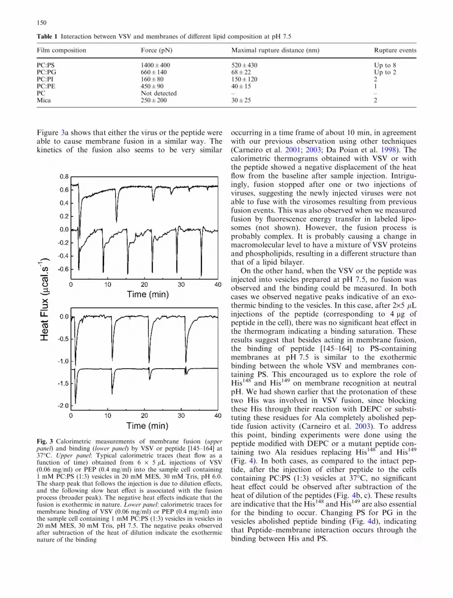
Figure 3a shows that either the virus or the peptide wereable to cause membrane fusion in a similar way. Thekinetics of the fusion also seems to be very similar
occurring in a time frame of about 10 min, in agreementwith our previous observation using other techniques(Carneiro et al. 2001; 2003; Da Poian et al. 1998). Thecalorimetric thermograms obtained with VSV or withthe peptide showed a negative displacement of the heatflow from the baseline after sample injection. Intrigu-ingly, fusion stopped after one or two injections ofviruses, suggesting the newly injected viruses were notable to fuse with the virosomes resulting from previousfusion events. This was also observed when we measuredfusion by fluorescence energy transfer in labeled lipo-somes (not shown). However, the fusion process isprobably complex. It is probably causing a change inmacromolecular level to have a mixture of VSV proteinsand phospholipids, resulting in a different structure thanthat of a lipid bilayer.
On the other hand, when the VSV or the peptide wasinjected into vesicles prepared at pH 7.5, no fusion wasobserved and the binding could be measured. In bothcases we observed negative peaks indicative of an exo-thermic binding to the vesicles. In this case, after 2·5 lLinjections of the peptide (corresponding to 4 lg ofpeptide in the cell), there was no significant heat effect inthe thermogram indicating a binding saturation. Theseresults suggest that besides acting in membrane fusion,the binding of peptide [145–164] to PS-containingmembranes at pH 7.5 is similar to the exothermicbinding between the whole VSV and membranes con-taining PS. This encouraged us to explore the role ofHis148 and His149 on membrane recognition at neutralpH. We had shown earlier that the protonation of thesetwo His was involved in VSV fusion, since blockingthese His through their reaction with DEPC or substi-tuting these residues for Ala completely abolished pep-tide fusion activity (Carneiro et al. 2003). To addressthis point, binding experiments were done using thepeptide modified with DEPC or a mutant peptide con-taining two Ala residues replacing His148 and His149
(Fig. 4). In both cases, as compared to the intact pep-tide, after the injection of either peptide to the cellscontaining PC:PS (1:3) vesicles at 37�C, no significantheat effect could be observed after subtraction of theheat of dilution of the peptides (Fig. 4b, c). These resultsare indicative that the His148 and His149 are also essentialfor the binding to occur. Changing PS for PG in thevesicles abolished peptide binding (Fig. 4d), indicatingthat Peptide–membrane interaction occurs through thebinding between His and PS.
Table 1 Interaction between VSV and membranes of different lipid composition at pH 7.5
Film composition Force (pN) Maximal rupture distance (nm) Rupture events
PC:PS 1400±400 520±430 Up to 8PC:PG 660±140 68±22 Up to 2PC:PI 160±80 150±120 2PC:PE 450±90 40±15 1PC Not detected – –Mica 250±200 30±25 2
Fig. 3 Calorimetric measurements of membrane fusion (upperpanel) and binding (lower panel) by VSV or peptide [145–164] at37�C. Upper panel: Typical calorimetric traces (heat flow as afunction of time) obtained from 6 · 5 lL injections of VSV(0.06 mg/ml) or PEP (0.4 mg/ml) into the sample cell containing1 mM PC:PS (1:3) vesicles in 20 mM MES, 30 mM Tris, pH 6.0.The sharp peak that follows the injection is due to dilution effects,and the following slow heat effect is associated with the fusionprocess (broader peak). The negative heat effects indicate that thefusion is exothermic in nature. Lower panel: calorimetric traces formembrane binding of VSV (0.06 mg/ml) or PEP (0.4 mg/ml) intothe sample cell containing 1 mM PC:PS (1:3) vesicles in vesicles in20 mM MES, 30 mM Tris, pH 7.5. The negative peaks observedafter subtraction of the heat of dilution indicate the exothermicnature of the binding
150

Simulation of peptide [145–164] interactionwith PS-containing membranes
Since classical MD techniques are carried out with fixedpartial atomic charges, it was necessary to make a choiceregarding the protonation state of the His148 and His149
residues. By using the simplified continuum Gouy-Chapman theory (see Material and methods) an inter-facial H3O
+ concentration was calculated and the Hisimidazol ring protonation state was inferred, assuming apKa of 6.0. Applying Eqs. 1 and 2, a Debye screeninglength of approximately 10 A and a surface potential inthe range of �120 mV were obtained. The parametersused in the calculations were: surface charge density, rel,of 0.2 Cm�2, and ionic strength, S Zi
2 ci, of 100 mM.The surface charge density was calculated assuming anideal mixture of the PS and PC molecules and an areaper lipid of 55 and 64 A2, respectively. Inserting thesevalues into the Boltzmann distribution (Eq. 3) of H3O
+
ion and neglecting changes in H3O+ ion activity coeffi-
cient, a 10�5 mol l�1 hydronium concentration wasobtained, which corresponds to a local surface pH of 5.0(Bostrom et al. 2004). At this pH, the His imidazol ringis mostly protonated, displaying a net charge of +1e.
Although the starting structure of the DMPS +peptide [145–164] system was somewhat arbitrary, a10 ns-long MD simulation was probably sufficient tominimize any major artifacts arising from this initialchoice. Moreover, using the last 2 ns of simulation, anarea per lipid of 55±0.7 A2 was calculated (data notshown), in accordance with other simulations (Panditand Berkowitz 2002). This value, together with the factthat we used fully anisotropic pressure coupling (i.e., thebilayer was free to adjust its area), underlines the con-sistency of the lipid model.
As can be observed in Fig. 5, the peptide seems tointeract with the simulated membrane patch mainlythrough its N-terminal residues. The rest of the peptidechain displays larger fluctuations, with the negativelycharged residues (Asp and Glu) initially repelled fromand afterwards approximating the water-DMPS inter-face. In fact, by analyzing the distance between thecenters of mass of titratable aminoacids and the bilayercenter (see Fig. 6), it can be seen that Val145 and His148
are stabilized in their positions. On the contrary, at leastin the 10 ns time frame, the other charged residues(His149, Asp153,161 and Glu154,158) have not reached astable position along the normal to the membrane.
Discussion
The early events of envelope virus infection comprise atleast three distinct steps: (a) the cell recognition, whichoccurs generally through the interaction between thevirus and a specific receptor on cell surface; (b) theinteraction between a viral surface protein and a cellularmembrane; and (c) membrane fusion reaction inducedby the viral fusion proteins. In this work, we focused ondissecting the interaction between VSV and the mem-brane at neutral pH, which might take place afterbinding to the receptor but before the events involved inthe membrane fusion reaction. We have taken advantageof our previous demonstration that atomic forcemicroscopy (AFM) operating in the force spectroscopymode could efficiently measure the interaction forcesbetween a virus particle and a lipid bilayer (Carneiroet al. 2002). Using this technique, we showed that VSVinteracts very strongly with membranes containing PS,while no interaction was observed with membranescomposed of PC only. A question not completely an-swered was whether VSV–membrane interaction de-pends only on electrostatic interaction or it was specificfor PS. Although VSV fusion has already been testedvarying the phospholipid composition (Eidelman et al.1984; Hermann et al. 1990), the binding events at neu-tral pH were not explored so far. To address this pointwe measured the interaction forces between virus parti-cles and lipid films supported on mica surfaces atpH 7.5, a condition in which binding but not fusioncould occur. These experiments revealed a high speci-ficity for membrane-containing PS, suggesting that PS is
Fig. 4 Specificity of the peptide interaction with phospholipidvesicles. The heat flow as a function of temperature is shown aftersubtraction of the heat of dilution of the peptides. The calorimetrictraces were obtained at 37�C where the peptide [145–164] wasinjected in the sample cell containing 1 mM PC:PS 1:3 (a) orPC:PG 1:3 (d) vesicles. Traces b and c show the importance of theHis residues for the interaction as the mutant peptide (b) and theDEPC-modified peptide (c) since no significant heat effect isobserved
151

also important for VSV–membrane interactions at neu-tral pH. This does not mean that the receptor for thevirus is PS, but suggests that although other components
in the cell surface might act as the VSV receptors, asindicated by the results from Coil and Miller (2004), adirect interaction between G protein and PS in themembrane could take place before the acidification in-side the endosome.
The identification of G protein amino acid residuesdirectly involved in VSV binding to membranes is animportant point in the understanding of VSV–mem-brane interaction. Photolabeling studies of VSV Gprotein showed that its interaction with membranesstrongly increases when the pH is lowered from 7.0 to6.0 (Durrer et al. 1995). At the pH of fusion, the labeledsite was located in the ectodomain comprising the aminoacids 59 to 221. Based on several mutagenesis studies,the sequence between the residues 117 and 136 has beenproposed as the putative fusion peptide of VSV G pro-tein (Li et al. 1993; Zhang and Ghosh 1994; Frederick-sen and Whitt 1995). However, direct evidence that thisparticular region interacts with the target membrane isstill lacking and further investigation will be necessary toprovide unambiguous evidence whether the segmentbetween amino acids 117 and 136 of the G protein di-rectly participates in VSV fusion or whether the substi-tution of its conserved amino acids affects theconformation or the exposure of other membrane-
Fig. 5 Snapshots of the system configuration during 10 ns MDsimulation. The protein backbone is represented as a cyan tube,except that the titratable residues are depicted as van der Waalsspheres (cyan for the His and red for the Asp/Glu residues). Water
is represented as red lines. The peptide is seen in detail along thesimulation time. A representation of the water–DMPS interface isdepicted as a green line. The DMPS bilayer is shown with the lipidtails in green, after 10 ns of simulation
Fig. 6 Distance between titratable aminoacids and the bilayercenter during the 10 ns simulation. It can be seen that Val145 andHis148 display stable center of mass distances to the DMPS bilayer.The negatively charged aminoacids, on the contrary, displayunstable positions in relation to the membrane as well as beingmore distant to the bilayer center of mass
152
interacting sequence in the G protein. Another region ofthe G protein, encompassing residues 395–418 for VSVhas been identified as a segment that affects the fuso-genic activity of the protein by influencing the low-pH-induced conformational changes (Shokralla et al. 1998).In addition, it has also been shown that not only theectodomain segment but also the membrane anchoringdomain is required for VSV fusion activity (Odell et al.1997; Cleverly and Lenard 1998).
Recently, we identified a specific sequence in theVSV G protein directly involved in membrane inter-action and fusion (Carneiro et al. 2003). This segmenthas been previously characterized as the PS-binding sitein the VSV G protein together with similar regions ofG proteins from other rhabdoviruses (Coll et al. 1997).We showed that this segment, which corresponds to thesequence between amino acids 145 to 164 of the VSV Gprotein, is very efficient in catalyzing membrane fusion(Carneiro et al. 2003). Here we used the peptide 145–164 to explore G protein–membrane interaction. Thestudy of the interaction between membrane and syn-thetic peptides corresponding to the fusogenic domainof the fusion protein is sometimes an important strat-egy to adopt. Although the results obtained with thepeptides have to be considered cautiously since manyfeatures of the complex viral full length protein are notpresent in the isolated peptide–vesicle model system,analysis of the molecular mechanisms underlying fusionpeptide activity in the whole protein would be notviable in most cases. As revised by Nieva and Agirre(2003), several findings support the view that syntheticpeptides are useful models to study viral cell fusion. Inour case, we were encouraged to use this peptide by thesimilarity between peptide- and whole virus-inducedfusion. Besides showing the same kinetics of whole
virus fusion, the peptide-induced fusion was dependenton pH and on the presence of PS in the target mem-brane.
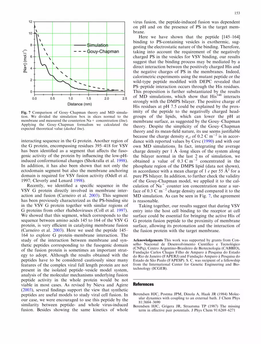
Here we have shown that the peptide [145–164]binding to PS-containing vesicles is exothermic, sug-gesting the electrostatic nature of the binding. Therefore,taking into account the requirement of the negativelycharged PS in the vesicles for VSV binding, our resultssuggest that the binding process may be mediated by adirect interaction between the positively charged His andthe negative charges of PS in the membranes. Indeed,calorimetric experiments using the mutant peptide or thewild-type peptide modified with DEPC revealed thatPS–peptide interaction occurs through the His residues.This proposition is further substantiated by the resultsof MD simulations, which show that His148 interactsstrongly with the DMPS bilayer. The positive charge ofHis residues at pH 7.5 could be explained by the prox-imity of the peptide to the negativelly charged head-groups of the lipids, which can lower the pH atmembrane surface, as suggested by the Gouy–Chapmantheory. Despite the simplicity of the Gouy–Chapmantheory and its mean-field nature, its use seems justifiablebecause the charge density rel of 0.2 C m�2 is in accor-dance with reported values by Cevc (1990) and with ourown MD simulations. In fact, integrating the averagecharge density per 1 A -long slices of the system alongthe bilayer normal in the last 2 ns of simulation, weobtained a value of 0.3 C m�2 concentrated in thephosphate region of the DMPS lipid (data not shown),in accordance with a mean charge of 1 e per 55 A2 for apure PS bilayer. In addition, to further check the validityof the Gouy-Chapman model, we applied it to the cal-culation of Na+ counter ion concentration near a sur-face of 0.3 C m�2 charge density and compared it to theMD simulation. As can be seen in Fig. 7, the agreementis reasonable.
Taking together, our results suggest that during VSVentry into the host cell binding to the receptor at cellsurface could be essential for bringing the active His ofG protein fusion peptide to the proximity of membranesurface, allowing its protonation and the interaction ofthe fusion protein with the target membrane.
Acknowledgements This work was supported by grants from Con-selho Nacional de Desenvolvimento Cientıfico e Tecnologico(CNPq), Centro Argentino-Brasileiro de Biotecnologia (CABBIO),Fundacao Carlos Chagas Filho de Amparo a Pesquisa do Estadodo Rio de Janeiro (FAPERJ) and Fundacao Amparo a Pesquisa doEstado de Sao Paulo (FAPESP). S. C. was recipient of a fellowshipfrom the International Center for Genetic Engineering and Bio-technology (ICGEB).
References
Berendsen HJC, Postma JPM, Dinola A, Haak JR (1984) Molec-ular dynamics with coupling to an external bath. J Chem Phys81:3684–3690
Berendsen HJC, Grigera JR, Straatsma TP (1987) The missingterm in effective pair potentials. J Phys Chem 91:6269–6271
Fig. 7 Comparison of Gouy–Chapman theory and MD simula-tion. We divided the simulation box in slices normal to themembrane and measured the counterion Na+ concentration (line).Applying the Gouy–Chapman formalism we calculated theexpected theoretical value (dashed line).
153

Bostrom M, Williams DRM, Ninham BW (2004) Specific ion ef-fects: Role of salt and buffer in protonation of cytochrome c.Eur Phys J 13:239–245
Carneiro FA, Ferradosa AS, Da Poian AT (2001) Low pH-inducedconformational changes in vesicular stomatitis virus glycopro-tein involve dramatic structure reorganization. J Biol Chem276:62–67
Carneiro FA, Bianconi ML, Weissmuller G, Stauffer F, Da PoianAT (2002) Membrane recognition by vesicular stomatitis virusinvolves enthalpy-driven protein-lipid interactions. J Virol76:3756–3764
Carneiro FA, Stauffer F, Lima CS, Juliano MA, Juliano L, DaPoian AT (2003) Membrane fusion induced by vesicular sto-matitis virus depends on histidine protonation. J Biol Chem278:13789–13794
Cevc G (1990) Membrane electrostatics. Biochim Biophys Acta1031(3):311–382
Chandrasekhar I, Kastenholz M, Lins RD, Oostenbrink C, SchulerLD, Tieleman DP, van Gunsteren WF (2003) A consistentpotential energy parameter set for lipids: dipal-mitoylphosphatidylcholine as a benchmark of the GROMOS9645 A3 force field. Eur Biophys J 32:62–67
Cleverley PZ, Lenard J (1998) The transmembrane domain in viralfusion: essential role for a conserved glycine residue in vesicularstomatitis virus G protein. Proc Natl Acad Sci USA 95:3425–3430
Coil DA, Miller AD (2004) Phosphatidylserine is not the cell sur-face receptor for vesicular stomatitis virus. J Virol 78:10920–10926
Coll JM (1997) Synthetic peptids from the heptad repeats of theglicoproteins of rabies, vesicular stomatitis and fish rhabdovi-ruses bind phosphatidylserine. Arch Virol 142:2089–2097
Crimmins DL, Mehard WB, Schlesinger S (1983) Physical prop-erties of a soluble form of the glycoprotein of vesicularstomatitis virus at neutral and acidic pH. Biochemistry22:5790–5796
Da Poian AT, Gomes AMO, Oliveira RJN, Silva JL (1996)Migration of vesicular stomatitis virus glycoprotein to the nu-cleus of infected cells. Proc Natl Acad Sci USA 93:8268–8273
Da Poian AT, Gomes AMO and Coelho-Sampaio T (1998)Kinetics of intracellular viral disassembly probed by bodipyfluorescence dequenching. J Virol Meth 70:45–58
Darden T, York D, Pedersen L (1993) Particle Mesh Ewald: anNlog (N) method for Ewald sums in large systems. J Chem Phys98:10089–10092
Durrer P, Gaudin Y, Ruigrok RW, Graf R and Brunner J (1995)Photolabeling identifies a putative fusion domain in the enve-lope glycoprotein of rabies and vesicular stomatitis viruses. JBiol Chem 270:17575–17581
Eckert DM, Kim PS (2001) Mechanisms of viral membrane fusionand its inhibition. Annu Rev Biochem 70:777–810
Eidelman O, Schlegel R, Tralka TS, Blumenthal R (1984) pH-dependent fusion induced by vesicular stomatitis virus glyco-protein reconstituted into phospholipid vesicles. J Biol Chem259:4622–4628
Estepa A, Coll JM (1996) Pepscan mapping and fusion relatedproperties of the major phosphatidylserine-binding domain ofthe glycoprotein of viral hemorragic septicemia virus, a sal-monid rhabdovirus. Virology 216:60–70
Florin EL, Moy VT, Gaub HE (1994) Adhesion forces betweenindividual ligand-receptor pairs. Science 264:415–417
Fredericksen B, Whitt MA (1995) Vesicular stomatitis virus gly-coprotein mutations that affect membrane fusion activity andabolish virus infectivity. J Virol 69:1435–1443
Gergely C, Voegel J-C, Schaaf P, Senger B, Maaloum M, HorberJKH, Hermmerle J (2000) Unbinding process of adsorbedproteins under external stress studied by atomic force micros-copy spectroscopy. Proc Natl Acad Sci USA 97:10802–10807
Hernandez LD, Hoffman LR, Wolfsberg TG, White JM (1996)Virus-cell and cell-cell fusion. Annu Rev Cell Dev Biol 12:627–661
Herrmann A, Clague MJ, Puri A, Morris SJ, Blumenthal R andGrimaldi S (1990) Effect of erythrocyte transbilayer phospho-lipid distribution on fusion with vesicular stomatitis virus.Biochemistry 29:4054–4058
Jass J, Tjarnhage T, Puu G (2000) From liposomes to supported,planar bilayer structures on hydrophilic and hydrophobic sur-faces: an atomic force microscopy study. Biophys J 79:3153–3163
Li Y, Drone C, Sat E, Ghosh HP (1993) Mutational analysis of thevesicular stomatitis virus glycoprotein G for membrane fusiondomains. J Virol 67:4070–4077
Lindahl E, Hess B, van der Spoel D (2001) GROMACS 3.0: Apackage for molecular simulation and trajectory analysis. J MolMod 7:306–317
Mastromarino P, Conti C, Goldoni P, Hauttecoeur B, Orsi N(1987) Characterization of membrane components of theerythrocyte involved in vesicular stomatitis virus attachmentand fusion at acidic pH. J Gen Virol 68:2359–2369
Miles EW (1977) Modification of histidyl residues in proteins bydiethyl pyrocarbonate. Meth Enzymol 47:431–442
Nieva JL, Agirre A (2003) Are fusion peptides a good model tostudy viral cell fusion? Biochim Biophys Acta 1614(1):104–115
Odell D, Wanas E, Yan J, Ghosh HP (1997) Influence of mem-brane anchoring and cytoplasmic domains on the fusogenicactivity of vesicular stomatitis virus glycoprotein G. J Virol71:7996–8000
Pak CC, Puri A, Blumenthal R (1987) Conformational changes andfusion activity of vesicular stomatitis virus glycoprotein: [125
I]iodonaphtyl azide photolabeling studies in biological mem-branes. Biochemistry 36:8890–8896
Pandit SA, Berkowitz ML (2002) Molecular dynamics simulationof dipalmitoylphosphatidylserine bilayer with Na+ counteri-ons. Biophys J 82:1818–1827
Puu G, Gustafson I (1997) Planar lipid bilayers on solid supportsfrom liposomes-factors of importance for kinetics and stability.Biochim Biophys Acta 1327:149–161
Puu G, Artursson E, Gustafson I, Lundstro M, Jass J (2000)Distribution and stability of membrane proteins in lipid mem-branes on solid supports. Biosensors Bioelectronics 15:31–41
Reviakine I, Brisson A (2000) Formation of supported phospho-lipid bilayers from unilamellar vesicles investigated by atomicforce microscopy. Langmuir 16:1806–1815
Ryckaert JP, Ciccott GI, Berendsen HJC (1977) Numerical inte-gration of the cartesian equations of motion of a system withconstraints: molecular dynamics of n-alkanes. J Comp Phys23:327–341
Schlegel R, Willingham MC, Pastan IH (1982) Saturable bindingsites for vesicular stomatitis virus on the surface of Vero cells. JVirol 43:871–875
Schlegel R, Tralka TS, Willingham MC, Pastan I (1983) Inhibitionof VSV binding and infectivity by phosphatidylserine: is phos-phatidylserine a VSV-binding site? Cell 32:639–646
Shokralla S, He Y, Wanas E, Ghosh HP (1998) Mutations in acarboxy-terminal region of vesicular stomatitis virus glycopro-tein G that affect membrane fusion activity. Virology 256:119–129
Skehel JJ, Wiley DC (2000) Receptor binding and membrane fu-sion in virus entry: the influenza hemagglutinin. Annu RevBiochem 69:531–569
Superti F, Seganti L, Tsiang H, Orsi N (1984) Role of phospho-lipids in rhabdovirus attachment to CER cells. Arch Virol81:321–328
Zhang L, Ghosh HP (1994) Characterization of the putative fus-ogenic domain in vesicular stomatitis virus glycoprotein G. JVirol 68:2186–2193
Zlatanova J, Lindsay SM, Leuba SH (2000) Single molecule forcespectroscopy in biology using the atomic force microscope.Prog Biophys Mol Biol 74:37–61
154