preferential translation of vesicular stomatitis virus mrnas is conferred by transcription from the...
TRANSCRIPT

JOURNAL OF VIROLOGY, Dec. 2006, p. 11733–11742 Vol. 80, No. 230022-538X/06/$08.00�0 doi:10.1128/JVI.00971-06Copyright © 2006, American Society for Microbiology. All Rights Reserved.
Preferential Translation of Vesicular Stomatitis Virus mRNAs IsConferred by Transcription from the Viral Genome�
Zackary W. Whitlow,* John H. Connor,§ and Douglas S. LylesDepartment of Biochemistry, Wake Forest University School of Medicine, Winston-Salem, North Carolina 27157
Received 11 May 2006/Accepted 11 September 2006
Host protein synthesis is inhibited in cells infected with vesicular stomatitis virus (VSV). It has beenproposed that viral mRNAs are subjected to the same inhibition but are predominantly translated because oftheir abundance. To compare translation efficiencies of viral and host mRNAs during infection, we used anenhanced green fluorescent protein (EGFP) reporter expressed from a recombinant virus or from the hostnucleus in stably transfected cells. Translation efficiency of host-derived EGFP mRNA was reduced more thanthreefold at eight hours postinfection, while viral-derived mRNA was translated around sevenfold moreefficiently than host-derived EGFP mRNA in VSV-infected cells. To test whether mRNAs transcribed in thecytoplasm are resistant to shutoff of translation during VSV infection, HeLa cells were infected with arecombinant simian virus 5 (rSV5) that expressed GFP. Cells were then superinfected with VSV or mocksuperinfected. GFP mRNA transcribed by rSV5 was not resistant to translation inhibition during superinfec-tion with VSV, indicating that transcription in the cytoplasm is not sufficient for preventing translationinhibition. To determine if cis-acting sequences in untranslated regions (UTRs) were involved in preferentialtranslation of VSV mRNAs, we constructed EGFP reporters with VSV or control UTRs and measured thetranslation efficiency in mock-infected and VSV-infected cells. The presence of VSV UTRs did not affect mRNAtranslation efficiency in mock- or VSV-infected cells, indicating that VSV mRNAs do not contain cis-actingsequences that influence translation. However, we found that when EGFP mRNAs transcribed by VSV or by thehost were translated in vitro, VSV-derived EGFP mRNA was translated 22 times more efficiently thanhost-derived EGFP mRNA. This indicated that VSV mRNAs do contain cis-acting structural elements (that arenot sequence based), which enhance translation efficiency of viral mRNAs.
All viruses rely on host translation machinery for synthesis ofviral proteins. In addition, many viruses inhibit translation ofhost mRNA to suppress host antiviral responses (21). Theseviruses have developed a variety of mechanisms to inhibit hostprotein synthesis while viral mRNAs are preferentially trans-lated. Understanding how viral mRNAs are translated at thesame time that host mRNA translation is inhibited is criticalfor understanding viral replication. Furthermore, studyingthese viral mechanisms of translation has also increased ourknowledge of cellular mechanisms for translational control.The goal of the experiments presented here was to addressthese questions for vesicular stomatitis virus (VSV).
VSV is widely studied as a model for other negative-sense,single-stranded RNA viruses. Following virus penetration anduncoating, viral mRNAs are synthesized by the viral RNA-dependent RNA polymerase. When viral proteins begin toaccumulate, progeny viral genomes are replicated and are usedfor secondary transcription. mRNAs from both primary andsecondary transcription are similar in structure to hostmRNAs. They have a 5� end containing 2�-O-methylated aden-osine capped by 7mG linked by 5�-5� triphosphate (18, 24, 25,32, 33, 38). VSV mRNAs also have a 3� poly(A) tail that is
similar in length to that of cellular mRNAs (11, 13, 14). Thesynthesis of VSV mRNAs, including the 5� and 3� end modi-fications, is accomplished entirely in the cytoplasm of the in-fected cell (9).
During VSV infection, host translation is rapidly inhibited.This is likely a result of modification to the eukaryotic trans-lation initiation factor 4F (eIF4F) (7, 8). However, it seemsparadoxical that this modification would affect translation ofhost mRNAs but not of VSV mRNAs, since VSV mRNAs arestructurally similar to host mRNAs. Yet, in cells infected withVSV, viral protein synthesis becomes predominant as hostprotein synthesis is inhibited (7, 8, 22, 26, 37, 40). The issueaddressed here is whether VSV mRNAs are subject to thesame inhibition of translation as host mRNAs. It has beensuggested that the inhibition of translation in VSV-infectedcells affects viral mRNAs as much as host mRNAs duringinfection but that the abundance of viral mRNAs leads to thepredominance of viral protein synthesis (20). In the experi-ments described here, we compared the translation efficienciesof a viral-derived and host-derived reporter mRNA. We havedetermined that in VSV-infected cells, mRNAs derived fromthe viral genome are translated seven times more efficientlythan host-derived mRNAs.
A common way for viral mRNAs to resist the inhibition oftranslation imposed on host mRNAs is through cis-acting se-quences that recruit cellular or viral factors which promotetranslation. For example, picornavirus positive-strand genomescontain internal ribosome entry sites, in their 5� untranslatedregions (UTRs), that direct initiation of translation efficientlywhen cap-dependent translation is inhibited (16, 29, 30). Sim-
* Corresponding author. Mailing address: Department of Biochem-istry, Wake Forest University School of Medicine, Winston-Salem, NC27157. Phone: (336) 716-2270. Fax: (336) 716-7671. E-mail: [email protected].
§ Present address: Department of Microbiology, Boston UniversitySchool of Medicine, Boston, MA 02218.
� Published ahead of print on 27 September 2006.
11733
on Decem
ber 9, 2015 by guesthttp://jvi.asm
.org/D
ownloaded from
ilarly, rotavirus mRNAs contain a cis-acting sequence in their3� UTRs that recruits a viral protein NSP3 which binds toeIF4F, competing with polyadenosine binding protein for acommon binding site (6, 23, 28, 31, 36, 39). Another exampleis adenovirus late mRNAs, which contain cis-acting sequencesin their 5� UTRs that, along with adenovirus 100k protein,direct ribosome shunting (42–45). However, we have foundthat cis-acting sequences are not involved in preferential trans-lation of VSV mRNAs. In contrast to these examples wherecis-acting elements that function in resisting translation shutoffare embedded in mRNA nucleotide sequences, here wepresent a case where a negative-strand RNA virus producesmRNA that contains a cis-acting element that is not a nucle-otide sequence. The cis-acting element acquired by VSVmRNAs allows VSV protein synthesis to predominate, in in-fected cells, by conferring high translation efficiencies to over-come translation inhibition.
MATERIALS AND METHODS
Cells and Viruses. HeLa cells were cultured in Dulbecco’s modified Eagle’smedium (DMEM) containing 7.5% fetal bovine serum (FBS). Recombinantwild-type VSV (rwt virus) and recombinant VSV-expressing enhanced greenfluorescent protein (EGFP) as a foreign gene (rVSV-EGFP) were grown in BHKcells as described previously (19). Recombinant simian virus 5 expressing GFP(rSV5-GFP) was isolated and grown in MDBK cells as described previously (12).The following cloning steps were required to produce the infectious cDNA clonecorresponding to the rVSV-EGFP virus. The cassette vector pGEM.XK3.1 (19)was modified to include a VSV transcription control site closely followed by NotIand PacI restriction sites. Oligonucleotides of positive sense (5� CGCGCTATGAAAAAAACTAACAGGCGGCCGCTTAATTAAGG TAC 3�) and negativesense (5� CTTAATTCCGC GGCCGCCTGTTAGTTTTTTTCATAG 3�) wereannealed and ligated into pGEM.XK3.1 prepared by digestion with BssHII andKpnI restriction endonucleases. The resulting plasmid was called pGEM-BNPK3.1. The DNA segment containing the EGFP coding region and 5� mul-tiple cloning site (MCS) was removed from the EGFP-N1 plasmid (Clontech) bydigestion using BssHII and PacI and was ligated into pGEM-BNPK3.1. A DNAfragment containing the VSV-M gene, the new transcription stop-start site, andthe EGFP gene was cleaved with SpeI and PacI and cloned into the recombinantVSV cDNA infectious clone pVSV.XK4.1 (41) to make the new infectious cloneused to recover rVSV-EGFP, as described previously (19). Virus stocks wereprepared as described previously (19). Infections with VSV were done at amultiplicity of infection of 10 PFU/cell in DMEM with 2% FBS. To infect HeLacells with SV5-GFP, SV5 was added in DMEM without serum at 10 PFU/cell.After adding virus, cells were incubated for 1 h before replacing infection mediawith DMEM containing 2% FBS. The rwt virus was then added 11 h later inDMEM with 2% FBS at a multiplicity of infection of 10 PFU/cell.
EGFP plasmids. The VSV-P gene 5� UTR was cloned into pEGFP-N1 (Clon-tech) using complementary oligonucleotides of positive sense (5� TTAAGCATAGGGATAGAAAAGACAGGATATTAGTTGTTCTTTATTCGCGCCTTAATTAATTAACTT 3�) and negative sense (5� GTACAAGTAATTAATTAAGGCGCGAATAAAGAACAACTAATATCCTGTCTTTTCTATCCCTATGC 3�)that were annealed and ligated into pEGFP-N1 cleaved with AflII and BsrGI.The EGFP-N1 plasmid 3� UTR was then replaced with a sequence of the VSVG gene 3� UTR modified to include a eukaryotic poly(A) signal. Oligonucleo-tides of positive sense (5� GTACAAGTAACCAACCAAGGCGCGAATAAAGAACAACTAATATCCTGTCTTTTCTATCCCTATGCTTAATAC 3�) andnegative sense (5� TTAAGCATAGGGATAGAAAAGACAGGATATTAGTTGTTCTTTATTCGCGCCTTAATTAATTACTT 3�) were annealed and ligatedinto the modified EGFP-N1 plasmid that was prepared by digestion with BsrGIand DraIII.
Metabolic labeling. Approximately 7 � 105 cells grown in six-well dishes werewashed twice with DMEM without methionine and then incubated in DMEMwithout methionine for 0.5 h. Cells were pulse-labeled for one hour in DMEMcontaining 200 �Ci/ml [35S]methionine. Cells were then harvested for immuno-precipitation using 500 �l radioimmunoprecipitation assay (RIPA) buffer (0.15M NaCl, 1% deoxycholic acid, 1% Triton X-100, 10 mM Tris-Cl, pH 7.4, and0.1% sodium dodecyl sulfate [SDS]) with 1 mg/ml bovine serum albumin (BSA),10 mM benzamidine, and 10 mM phenylmethylsulfonyl fluoride. Plates contain-
ing RIPA buffer were rocked gently until cells were visibly lifted from the dish.Lysates were then centrifuged at 20,000 � g for 15 min at 4°C. For analysis oftotal protein synthesis, cells were harvested following pulse-labeling using 500 �lRIPA buffer without BSA, and 360 �l of cell extract was added to 40 �l of 10�
SDS-polyacrylamide gel electrophoresis (PAGE) sample loading buffer.Immunoprecipitation. Immunoprecipitation of EGFP was performed by add-
ing 3.8 �g goat anti-GFP (RDI; code RDI-GRNFP3abg) to 100 �l of cell lysate.Samples were incubated overnight at 4°C. Protein G-Sepharose (Sigma; 20 �l) inNETN buffer (20 mM Tris-Cl, pH 8.0, 1 mM EDTA, 150 mM NaCl, 0.5% NP-40,and 4% BSA) was added and incubated for 1 h. Samples were centrifuged at500 � g at 4°C, and pellets were washed five times with 400 �l of RIPA bufferwith high SDS (1% SDS). SDS loading buffer (5 �l) was added to final pellets,and samples were heated to �95°C and run on 12% SDS-PAGE gels. Gels weredried and analyzed by phosphor imaging (Molecular Dynamics). Quantitationwas performed using ImageQuant 5.2 (Molecular Dynamics).
Northern blotting. RNA was harvested from 6 � 106 HeLa cells using 3 ml ofTRIzol (Invitrogen), according to the manufacturer’s specifications. RNA (5 �g)harvested from stably transfected cells or 0.125 �g of RNA harvested from HeLacells infected with rVSV-EGFP was run on a 1.2% glyoxal agarose gel. The gelwas then incubated in 800 ml of transfer buffer (0.01 M NaOH, 3 M NaCl) for20 min and then transferred to a GeneScreen Plus (PerkinElmer) hybridizationtransfer membrane by upward capillary transfer (35). [�-32P]dCTP-labeledEGFP probe was prepared using a prime-a-gene kit (Promega). Membraneswere probed using ExpressHyb hybridization solution (BD Biosciences Clon-tech) according to the manufacturer’s specifications. Membranes were analyzedby phosphorimaging.
Polysome profiles. Cells were treated with puromycin (puromycin added tomedia to 360 �M) or mock treated 1 h prior to harvesting. Several minutes priorto harvesting cells, cycloheximide (CHX) was added to the media to a concen-tration of 0.1 mg/ml. HeLa cells (1.5 � 107 to 1.8 � 107) were prepared byscraping off the culture dish in ice-cold PBS containing 0.1 mg/ml CHX. Cellswere pelleted and resuspended in ice-cold PBS containing 0.1 mg/ml CHX. Cellswere pelleted again and resuspended in 0.2 ml RSB buffer (10 mM NaCl, 3 mMMgCl2, and 10 mM Tris-Cl, pH 7.4) containing 20% vanadyl adenosine ribonu-cleoside complex and 0.1 mg/ml CHX. Cells were incubated on ice for 5 minfollowed by addition of 0.2 ml RSB buffer containing 1% deoxycholic acid and2% Tween 40; Cells were briefly vortexed before and after addition of RSBcontaining detergents. Cells were again incubated on ice for 5 min followed bybrief vortexing and centrifugation at 2000 � g for 15 min at 4°C to pellet nuclei.Cytoplasmic fractions were transferred to a new tube, and 0.1 ml 5� HSB (5�
HSB contains 2.5 M NaCl, 250 mM MgCl2, and 50 mM Tris-Cl, pH 7.4) wasadded and solutions were quickly mixed. Solutions were then carefully overlaidonto �10% to 50% sucrose gradients in 1� HSB. Gradients were centrifuged at37,000 rpm for 1.75 h at 4°C. Polysome profiles were analyzed by pumping off thetop of the gradient using an AUTO DENSI-FLOW (LABCONCO) gradientpump, through an EM-1 Econo UV monitor (Bio-Rad). Absorbance at 254 nmwas recorded using a Rec-111 (GE Healthcare) recorder. Sixteen fractions werecollected from each gradient using a Frac-100 (Pharmacia) fraction collector.RNA was precipitated by adding 20 �l glycogen and 0.5 ml isopropanol followedby overnight incubation at �20°C. RNA was pelleted by centrifugation for 20min at 12,000 � g at 4°C. Pellets were washed with 70% ethanol and brieflycentrifuged. Pellets were suspended in 300 �l 1% N-leuroyl sarcosine (N-LS) 10�l proteinase K (20 mg/ml) was added, followed by incubation for 30 min at 37°C.GTC (300 �l; 4 M guanidine thiocyanate, 25 mM sodium citrate, pH 7.0, and0.5% N-LS, with �-mercaptoethanol added to 0.7% immediately prior to use)and 600 �l isopropanol were added and mixed, and solutions were incubated at�20°C for longer than 30 min. RNA was pelleted by centrifugation at 12,000 � gfor 15 min at 4°C, washed with 70% ethanol, and resuspended in 10 �l distilledwater and 10 �l NorthernMax-Gly sample loading dye (Ambion). EGFP mRNAwas analyzed by Northern blotting as described above.
In vitro translation. RNA harvested eight hours postinfection with TRIzol(Invitrogen) was used to direct translation in rabbit reticulocyte lysates (Pro-mega). Reactions were 0.1 ml in total volume with 27 �g total RNA or 1.6 �g ofpoly(A) RNA added. Poly(A) RNA was isolated from total RNA using oli-go(dT)-cellulose columns (Amersham Biosciences) with two rounds of purifica-tion. Translation reactions were carried out in a water bath at 30°C for 2 h andstopped by incubating on ice. Three microliters of each reaction was removed foranalysis of the all products of protein synthesis. Remaining volumes were dilutedin 1.2 ml RIPA buffer plus 1 mg/ml BSA, 10 mM benzamidine, and 10 mMphenylmethylsulfonyl fluoride, and EGFP was immunoprecipitated as describedabove.
11734 WHITLOW ET AL. J. VIROL.
on Decem
ber 9, 2015 by guesthttp://jvi.asm
.org/D
ownloaded from
RESULTS
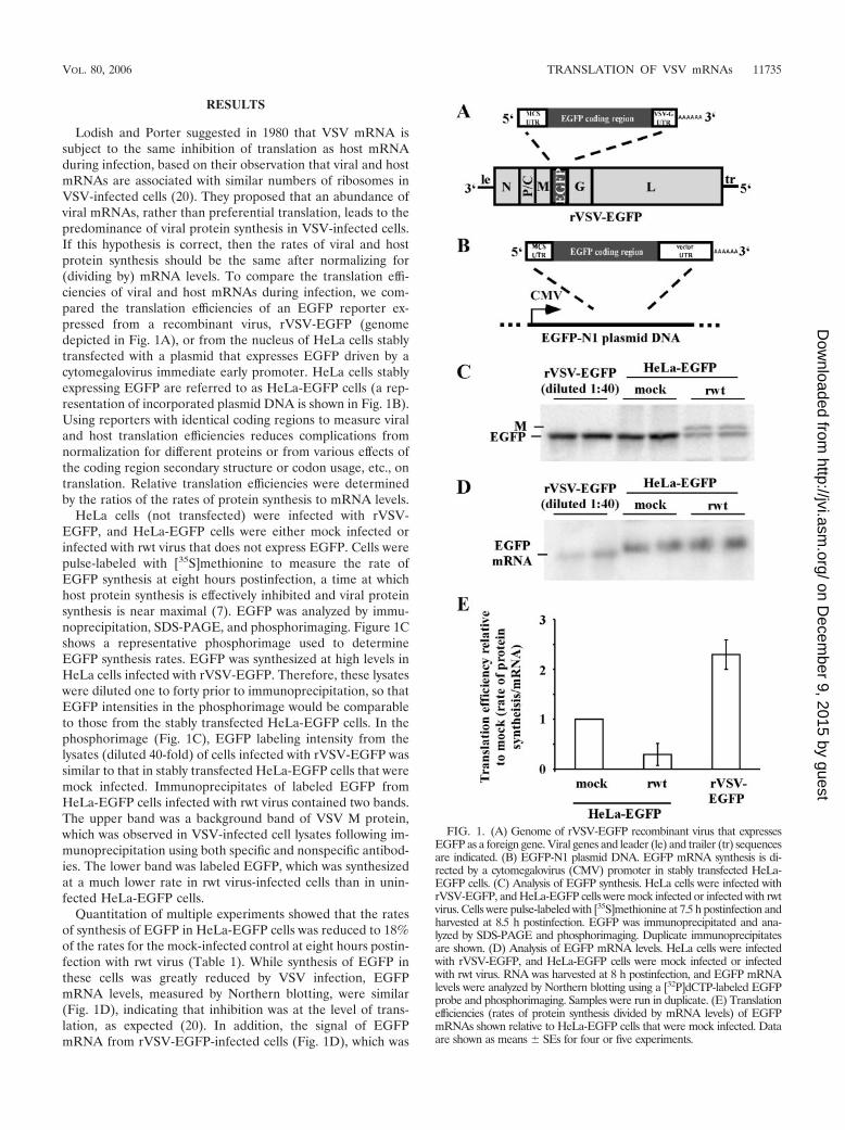
Lodish and Porter suggested in 1980 that VSV mRNA issubject to the same inhibition of translation as host mRNAduring infection, based on their observation that viral and hostmRNAs are associated with similar numbers of ribosomes inVSV-infected cells (20). They proposed that an abundance ofviral mRNAs, rather than preferential translation, leads to thepredominance of viral protein synthesis in VSV-infected cells.If this hypothesis is correct, then the rates of viral and hostprotein synthesis should be the same after normalizing for(dividing by) mRNA levels. To compare the translation effi-ciencies of viral and host mRNAs during infection, we com-pared the translation efficiencies of an EGFP reporter ex-pressed from a recombinant virus, rVSV-EGFP (genomedepicted in Fig. 1A), or from the nucleus of HeLa cells stablytransfected with a plasmid that expresses EGFP driven by acytomegalovirus immediate early promoter. HeLa cells stablyexpressing EGFP are referred to as HeLa-EGFP cells (a rep-resentation of incorporated plasmid DNA is shown in Fig. 1B).Using reporters with identical coding regions to measure viraland host translation efficiencies reduces complications fromnormalization for different proteins or from various effects ofthe coding region secondary structure or codon usage, etc., ontranslation. Relative translation efficiencies were determinedby the ratios of the rates of protein synthesis to mRNA levels.
HeLa cells (not transfected) were infected with rVSV-EGFP, and HeLa-EGFP cells were either mock infected orinfected with rwt virus that does not express EGFP. Cells werepulse-labeled with [35S]methionine to measure the rate ofEGFP synthesis at eight hours postinfection, a time at whichhost protein synthesis is effectively inhibited and viral proteinsynthesis is near maximal (7). EGFP was analyzed by immu-noprecipitation, SDS-PAGE, and phosphorimaging. Figure 1Cshows a representative phosphorimage used to determineEGFP synthesis rates. EGFP was synthesized at high levels inHeLa cells infected with rVSV-EGFP. Therefore, these lysateswere diluted one to forty prior to immunoprecipitation, so thatEGFP intensities in the phosphorimage would be comparableto those from the stably transfected HeLa-EGFP cells. In thephosphorimage (Fig. 1C), EGFP labeling intensity from thelysates (diluted 40-fold) of cells infected with rVSV-EGFP wassimilar to that in stably transfected HeLa-EGFP cells that weremock infected. Immunoprecipitates of labeled EGFP fromHeLa-EGFP cells infected with rwt virus contained two bands.The upper band was a background band of VSV M protein,which was observed in VSV-infected cell lysates following im-munoprecipitation using both specific and nonspecific antibod-ies. The lower band was labeled EGFP, which was synthesizedat a much lower rate in rwt virus-infected cells than in unin-fected HeLa-EGFP cells.
Quantitation of multiple experiments showed that the ratesof synthesis of EGFP in HeLa-EGFP cells was reduced to 18%of the rates for the mock-infected control at eight hours postin-fection with rwt virus (Table 1). While synthesis of EGFP inthese cells was greatly reduced by VSV infection, EGFPmRNA levels, measured by Northern blotting, were similar(Fig. 1D), indicating that inhibition was at the level of trans-lation, as expected (20). In addition, the signal of EGFPmRNA from rVSV-EGFP-infected cells (Fig. 1D), which was
FIG. 1. (A) Genome of rVSV-EGFP recombinant virus that expressesEGFP as a foreign gene. Viral genes and leader (le) and trailer (tr) sequencesare indicated. (B) EGFP-N1 plasmid DNA. EGFP mRNA synthesis is di-rected by a cytomegalovirus (CMV) promoter in stably transfected HeLa-EGFP cells. (C) Analysis of EGFP synthesis. HeLa cells were infected withrVSV-EGFP, and HeLa-EGFP cells were mock infected or infected with rwtvirus. Cells were pulse-labeled with [35S]methionine at 7.5 h postinfection andharvested at 8.5 h postinfection. EGFP was immunoprecipitated and ana-lyzed by SDS-PAGE and phosphorimaging. Duplicate immunoprecipitatesare shown. (D) Analysis of EGFP mRNA levels. HeLa cells were infectedwith rVSV-EGFP, and HeLa-EGFP cells were mock infected or infectedwith rwt virus. RNA was harvested at 8 h postinfection, and EGFP mRNAlevels were analyzed by Northern blotting using a [32P]dCTP-labeled EGFPprobe and phosphorimaging. Samples were run in duplicate. (E) Translationefficiencies (rates of protein synthesis divided by mRNA levels) of EGFPmRNAs shown relative to HeLa-EGFP cells that were mock infected. Dataare shown as means SEs for four or five experiments.
VOL. 80, 2006 TRANSLATION OF VSV mRNAs 11735
on Decem
ber 9, 2015 by guesthttp://jvi.asm
.org/D
ownloaded from
also diluted one to forty, was lower than EGFP signal intensityfrom HeLa-EGFP cells. This result, combined with the resultshown in Fig. 1C, indicates that EGFP mRNA expressed fromrVSV-EGFP was translated more efficiently than EGFPmRNA expressed from stably transfected plasmid DNA inboth mock-infected and rwt virus-infected HeLa-EGFP cells.
The relative translation efficiencies of EGFP mRNAs weredetermined by quantitating data from multiple experiments;similar to the quantitations in Fig. 1C and 1D. These data areshown relative to HeLa-EGFP mock-infected cells in Table 1,after correction for the dilution factors in Fig. 1. To determinethe relative translation efficiencies of EGFP mRNA, relativerates of EGFP protein synthesis, determined by pulse-labeling,were divided by the relative levels of EGFP mRNA from theNorthern blots (Fig. 1E and Table 1). Comparison of relativetranslation efficiencies shows that infection with rwt virus re-duced EGFP translation efficiency in HeLa-EGFP cells morethan threefold. In contrast, EGFP mRNA encoded by rVSV-EGFP was translated over twofold more efficiently than EGFPmRNA in mock-infected HeLa-EGFP cells and around seven-fold more efficiently than EGFP mRNA in rwt virus-infectedcells (Fig. 1E and Table 1). These results show that EGFPmRNA expressed from the viral genome is translated muchmore efficiently than host-derived EGFP mRNA during VSVinfection.
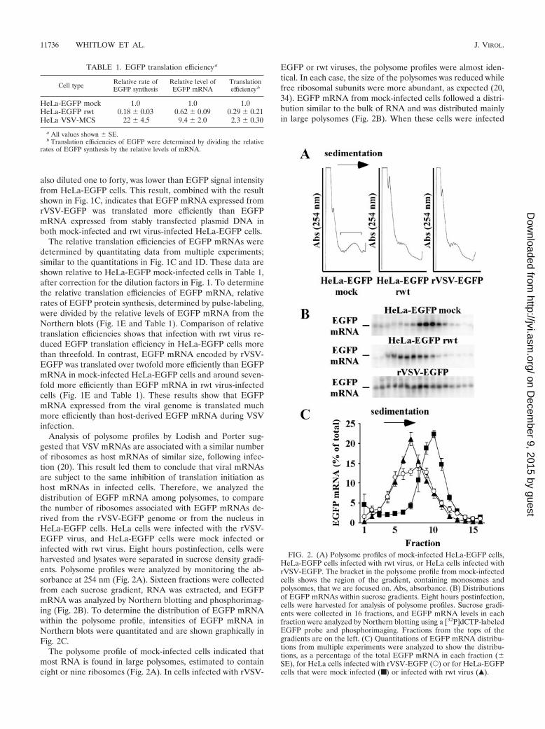
Analysis of polysome profiles by Lodish and Porter sug-gested that VSV mRNAs are associated with a similar numberof ribosomes as host mRNAs of similar size, following infec-tion (20). This result led them to conclude that viral mRNAsare subject to the same inhibition of translation initiation ashost mRNAs in infected cells. Therefore, we analyzed thedistribution of EGFP mRNA among polysomes, to comparethe number of ribosomes associated with EGFP mRNAs de-rived from the rVSV-EGFP genome or from the nucleus inHeLa-EGFP cells. HeLa cells were infected with the rVSV-EGFP virus, and HeLa-EGFP cells were mock infected orinfected with rwt virus. Eight hours postinfection, cells wereharvested and lysates were separated in sucrose density gradi-ents. Polysome profiles were analyzed by monitoring the ab-sorbance at 254 nm (Fig. 2A). Sixteen fractions were collectedfrom each sucrose gradient, RNA was extracted, and EGFPmRNA was analyzed by Northern blotting and phosphorimag-ing (Fig. 2B). To determine the distribution of EGFP mRNAwithin the polysome profile, intensities of EGFP mRNA inNorthern blots were quantitated and are shown graphically inFig. 2C.
The polysome profile of mock-infected cells indicated thatmost RNA is found in large polysomes, estimated to containeight or nine ribosomes (Fig. 2A). In cells infected with rVSV-
EGFP or rwt viruses, the polysome profiles were almost iden-tical. In each case, the size of the polysomes was reduced whilefree ribosomal subunits were more abundant, as expected (20,34). EGFP mRNA from mock-infected cells followed a distri-bution similar to the bulk of RNA and was distributed mainlyin large polysomes (Fig. 2B). When these cells were infected
FIG. 2. (A) Polysome profiles of mock-infected HeLa-EGFP cells,HeLa-EGFP cells infected with rwt virus, or HeLa cells infected withrVSV-EGFP. The bracket in the polysome profile from mock-infectedcells shows the region of the gradient, containing monosomes andpolysomes, that we are focused on. Abs, absorbance. (B) Distributionsof EGFP mRNAs within sucrose gradients. Eight hours postinfection,cells were harvested for analysis of polysome profiles. Sucrose gradi-ents were collected in 16 fractions, and EGFP mRNA levels in eachfraction were analyzed by Northern blotting using a [32P]dCTP-labeledEGFP probe and phosphorimaging. Fractions from the tops of thegradients are on the left. (C) Quantitations of EGFP mRNA distribu-tions from multiple experiments were analyzed to show the distribu-tions, as a percentage of the total EGFP mRNA in each fraction (SE), for HeLa cells infected with rVSV-EGFP (E) or for HeLa-EGFPcells that were mock infected (■) or infected with rwt virus (Œ).
TABLE 1. EGFP translation efficiency a
Cell type Relative rate ofEGFP synthesis
Relative level ofEGFP mRNA
Translationefficiency b
HeLa-EGFP mock 1.0 1.0 1.0HeLa-EGFP rwt 0.18 0.03 0.62 0.09 0.29 0.21HeLa VSV-MCS 22 4.5 9.4 2.0 2.3 0.30
a All values shown SE.b Translation efficiencies of EGFP were determined by dividing the relative
rates of EGFP synthesis by the relative levels of mRNA.
11736 WHITLOW ET AL. J. VIROL.
on Decem
ber 9, 2015 by guesthttp://jvi.asm
.org/D
ownloaded from
with rwt virus, EGFP mRNA shifted to the lighter region of thesucrose gradient, suggesting that fewer ribosomes were asso-ciated with each mRNA. In HeLa cells infected with the rVSV-EGFP, EGFP mRNA transcribed from the viral genome ap-peared intermediate in its distribution, in that more mRNAwas distributed throughout the gradient, although it wasmainly distributed in the light region of the gradient, indicatingthat in infected cells, viral-derived and host-derived EGFPmRNAs were all found in complexes with similar sedimenta-tion velocities. The similarity in distribution of EGFP mRNAsin infected cells is apparent in Fig. 2C, which shows quantita-tion of Northern blots from multiple experiments.
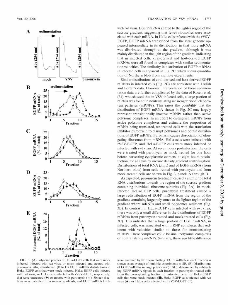
Similar distributions of viral-derived and host-derived EGFPmRNAs in infected cells (Fig. 2C) are consistent with Lodishand Porter’s data. However, interpretation of these sedimen-tation data are further complicated by the data of Rosen et al.(34), who showed that in VSV-infected cells, a large portion ofmRNA was found in nontranslating messenger ribonucleopro-tein particles (mRNPs). This raises the possibility that thedistribution of EGFP mRNA shown in Fig. 2C may largelyrepresent translationally inactive mRNPs rather than activepolysome complexes. In an effort to distinguish mRNPs fromactive polysome complexes and estimate the proportion ofmRNA being translated, we treated cells with the translationinhibitor puromycin to disrupt polysomes and obtain distribu-tions of EGFP mRNPs. Puromycin causes dissociation of elon-gating ribosomes from mRNA. HeLa cells were infected withrVSV-EGFP, and HeLa-EGFP cells were mock infected orinfected with rwt virus. At seven hours postinfection, the cellswere treated with puromycin or mock treated for one hourbefore harvesting cytoplasmic extracts, at eight hours postin-fection, for analysis by sucrose density gradient centrifugation.Distributions of total RNA (A254) and of EGFP mRNA (fromNorthern blots) from cells treated with puromycin and frommock-treated cells are shown in Fig. 3, panels A through D.
As expected, puromycin treatment caused a shift in the totalRNA distributions towards the region of the sucrose gradientcontaining individual ribosome subunits (Fig. 3A). In mock-infected HeLa-EGFP cells, puromycin treatment caused alarge redistribution of EGFP mRNA from the region of thegradient containing large polysomes to the lighter region of thegradient where mRNPs and small polysomes sediment (Fig.3B). In contrast, in HeLa-EGFP cells infected with rwt virus,there was only a small difference in the distributions of EGFPmRNAs from puromycin-treated and mock-treated cells (Fig.3C). This indicates that a large portion of EGFP mRNA, ininfected cells, was associated with mRNP complexes that sed-iment with velocities similar to those for nontranslatingmRNPs. These complexes could be small polysomal complexesor nontranslating mRNPs. Similarly, there was little difference
FIG. 3. (A) Polysome profiles of HeLa-EGFP cells that were mockinfected, infected with rwt virus, or mock infected and treated withpuromycin. Abs, absorbance. (B to D) EGFP mRNA distributions inHeLa-EGFP cells that were mock infected, HeLa-EGFP cells infectedwith rwt virus, or HeLa cells infected with rVSV-EGFP, respectively,that were untreated (}) or treated with puromycin (�). Sixteen frac-tions were collected from sucrose gradients, and EGFP mRNA levels
were analyzed by Northern blotting. EGFP mRNA in each fraction isshown as an average of multiple experiments SE. (E) Distributionsof EGFP mRNAs in large polysomes ( SE); determined by subtract-ing EGFP mRNA signals in each fraction in puromycin-treated cellsfrom the corresponding fraction in untreated cells, for HeLa-EGFPcells that were mock infected (■), HeLa-EGFP cells infected with rwtvirus (Œ), or HeLa cells infected with rVSV-EGFP (E).
VOL. 80, 2006 TRANSLATION OF VSV mRNAs 11737
on Decem
ber 9, 2015 by guesthttp://jvi.asm
.org/D
ownloaded from
in sedimentations of EGFP mRNAs from cells infected withrVSV-EGFP that were either treated with puromycin or mocktreated (Fig. 3D). To estimate the percentage of mRNA inpolysomes, EGFP mRNP distributions from puromycin-treated cells were subtracted from EGFP mRNA distributionsfrom untreated cells to eliminate the contribution from inac-tive mRNPs (Fig. 3E). Negative values where EGFP mRNPswere more abundant than polysomes were plotted as zero toreflect that most EGFP mRNA was not in polysomes in thesefractions. The resulting distributions represent an estimate ofthe minimum proportion of EGFP mRNA in active polysomes(Fig. 3E). Since small polysomes and mRNPs sediment withsimilar velocities, small polysomes may be underrepresentedby this method. In mock-infected HeLa-EGFP cells, largepolysomes (polysomes that sediment faster than nontranslatingmRNPs) containing EGFP mRNA were more abundant thanin rwt virus-infected cells. The total amount of EGFP mRNAin large polysomes was calculated as the sum of EGFP mRNAin all fractions in Fig. 3E. In mock-infected cells, 48% of EGFPmRNA was found in large polysomes while in infected cells,approximately 18% of EGFP mRNA was associated with largepolysomal complexes, regardless of the mRNA source. Thesedata indicate that viral mRNAs are no more likely to be inlarge polysomes than host mRNAs, which is consistent withLodish and Porter’s findings (20). However, the higher trans-lation efficiency of EGFP mRNA expressed from rVSV-EGFP(Fig. 1) suggests that viral mRNAs in these polysomes aremore efficient in directing protein synthesis than host poly-somal mRNAs.
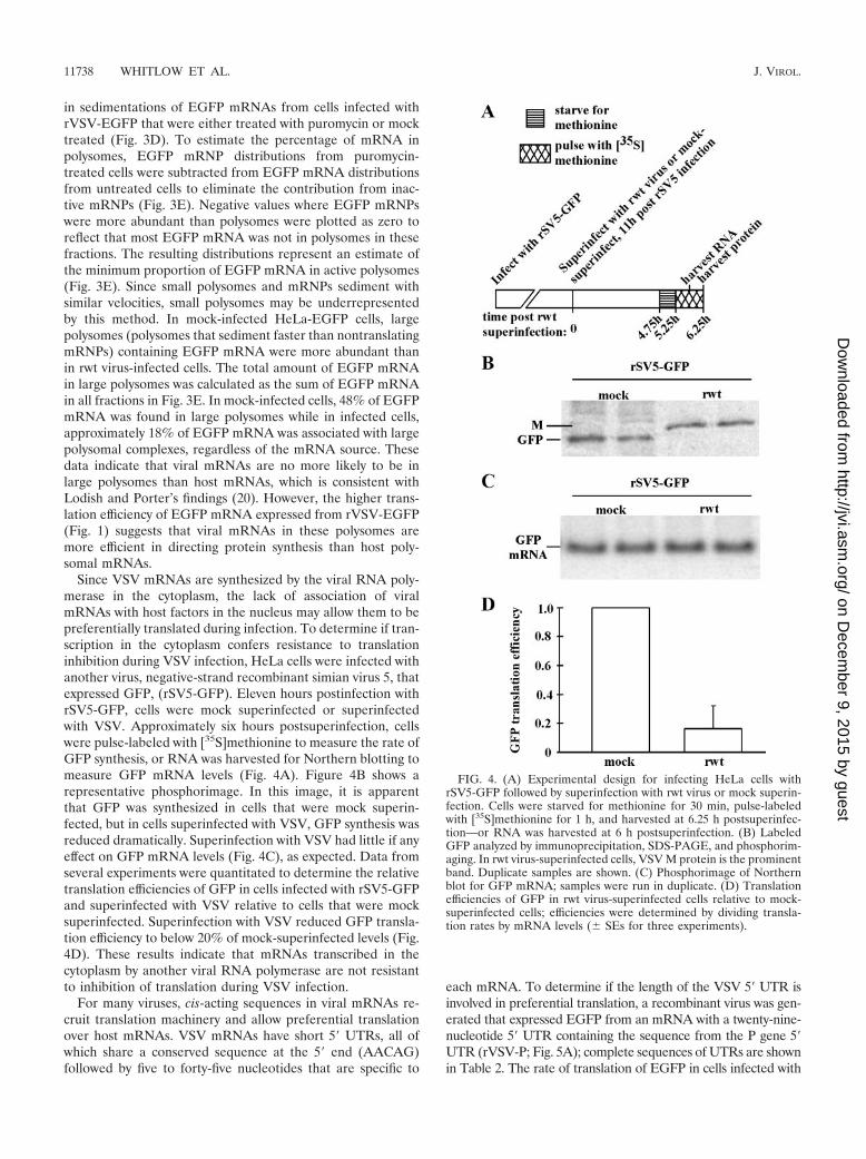
Since VSV mRNAs are synthesized by the viral RNA poly-merase in the cytoplasm, the lack of association of viralmRNAs with host factors in the nucleus may allow them to bepreferentially translated during infection. To determine if tran-scription in the cytoplasm confers resistance to translationinhibition during VSV infection, HeLa cells were infected withanother virus, negative-strand recombinant simian virus 5, thatexpressed GFP, (rSV5-GFP). Eleven hours postinfection withrSV5-GFP, cells were mock superinfected or superinfectedwith VSV. Approximately six hours postsuperinfection, cellswere pulse-labeled with [35S]methionine to measure the rate ofGFP synthesis, or RNA was harvested for Northern blotting tomeasure GFP mRNA levels (Fig. 4A). Figure 4B shows arepresentative phosphorimage. In this image, it is apparentthat GFP was synthesized in cells that were mock superin-fected, but in cells superinfected with VSV, GFP synthesis wasreduced dramatically. Superinfection with VSV had little if anyeffect on GFP mRNA levels (Fig. 4C), as expected. Data fromseveral experiments were quantitated to determine the relativetranslation efficiencies of GFP in cells infected with rSV5-GFPand superinfected with VSV relative to cells that were mocksuperinfected. Superinfection with VSV reduced GFP transla-tion efficiency to below 20% of mock-superinfected levels (Fig.4D). These results indicate that mRNAs transcribed in thecytoplasm by another viral RNA polymerase are not resistantto inhibition of translation during VSV infection.
For many viruses, cis-acting sequences in viral mRNAs re-cruit translation machinery and allow preferential translationover host mRNAs. VSV mRNAs have short 5� UTRs, all ofwhich share a conserved sequence at the 5� end (AACAG)followed by five to forty-five nucleotides that are specific to
each mRNA. To determine if the length of the VSV 5� UTR isinvolved in preferential translation, a recombinant virus was gen-erated that expressed EGFP from an mRNA with a twenty-nine-nucleotide 5� UTR containing the sequence from the P gene 5�UTR (rVSV-P; Fig. 5A); complete sequences of UTRs are shownin Table 2. The rate of translation of EGFP in cells infected with
FIG. 4. (A) Experimental design for infecting HeLa cells withrSV5-GFP followed by superinfection with rwt virus or mock superin-fection. Cells were starved for methionine for 30 min, pulse-labeledwith [35S]methionine for 1 h, and harvested at 6.25 h postsuperinfec-tion—or RNA was harvested at 6 h postsuperinfection. (B) LabeledGFP analyzed by immunoprecipitation, SDS-PAGE, and phosphorim-aging. In rwt virus-superinfected cells, VSV M protein is the prominentband. Duplicate samples are shown. (C) Phosphorimage of Northernblot for GFP mRNA; samples were run in duplicate. (D) Translationefficiencies of GFP in rwt virus-superinfected cells relative to mock-superinfected cells; efficiencies were determined by dividing transla-tion rates by mRNA levels ( SEs for three experiments).
11738 WHITLOW ET AL. J. VIROL.
on Decem
ber 9, 2015 by guesthttp://jvi.asm
.org/D
ownloaded from
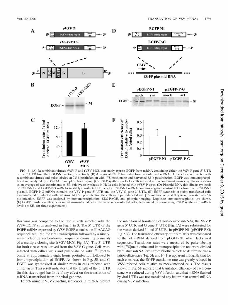
this virus was compared to the rate in cells infected with therVSV-EGFP virus analyzed in Fig. 1 to 3. The 5� UTR of theEGFP mRNA expressed by rVSV-EGFP contains the 5� AACAGsequence required for viral transcription followed by a ninety-nine-nucleotide vector-derived sequence consisting primarilyof a multiple cloning site (rVSV-MCS; Fig. 5A). The 3� UTRfor both viruses was derived from the VSV G gene. Cells wereinfected with either virus and pulse-labeled with [35S]methi-onine at approximately eight hours postinfection followed byimmunoprecipitation of EGFP. As shown in Fig. 5B and C,EGFP was synthesized at similar rates in cells infected witheither virus. This result indicates that the length of the 5� UTR(in this size range) has little if any effect on the translation ofmRNA transcribed from the viral genome.
To determine if VSV cis-acting sequences in mRNA prevent
the inhibition of translation of host-derived mRNAs, the VSV Pgene 5� UTR and G gene 3� UTR (Fig. 5A) were substituted forthe vector-derived 5� and 3� UTRs in pEGFP-N1 (pEGFP-P-G;Fig. 5D). The translation efficiency of this mRNA was comparedto that of mRNA derived from pEGFP-N1, which lacks viralsequences. Translation rates were measured by pulse-labelingwith [35S]methionine and immunoprecipitation and were dividedby relative mRNA levels from Northern blots to determine trans-lation efficiencies (Fig. 5E and F). It is apparent in Fig. 5E that foreach construct, the EGFP translation rate was greatly reduced inVSV-infected cells relative to mock-infected cells. The resultsshown in Fig. 5F indicate that translation efficiency of each con-struct was reduced during VSV infection and that mRNA flankedby viral UTRs was not translated any better than control mRNAduring VSV infection.
FIG. 5. (A) Recombinant viruses rVSV-P and rVSV-MCS that stably express EGFP from mRNA containing either the VSV P gene 5� UTRor the 5� UTR from the EGFP-N1 vector, respectively. (B) Analysis of EGFP translated from viral-derived mRNA. HeLa cells were infected withrecombinant viruses and pulse-labeled at 7.5 h postinfection with [35S]methionine and harvested 8.5 h postinfection. EGFP was immunoprecipi-tated and analyzed by SDS-PAGE and phosphorimaging. (C) EGFP synthesis in HeLa cells infected with recombinant viruses. Synthesis is shownas an average of two experiments SE, relative to synthesis in HeLa cells infected with rVSV-P virus. (D) Plasmid DNA that directs synthesisof EGFP-N1 and EGFP-P-G mRNAs in stably transfected HeLa cells. EGFP-N1 mRNA contains negative control UTRs from the pEGFP-N1plasmid. EGFP-P-G mRNA contains the VSV P gene 5� UTR and the VSV G gene 3� UTR. (E) EGFP synthesis in stably transfected cellsmock-infected or infected with rwt virus. At 7.5 h postinfection the cells were pulse-labeled with [35S]methionine, and they were harvested at 8.5 hpostinfection. EGFP was analyzed by immunoprecipitation, SDS-PAGE, and phosphorimaging. Duplicate immunoprecipitates are shown.(F) EGFP translation efficiencies in rwt virus-infected cells relative to mock-infected cells, determined by normalizing EGFP synthesis to mRNAlevels ( SEs for three experiments).
VOL. 80, 2006 TRANSLATION OF VSV mRNAs 11739
on Decem
ber 9, 2015 by guesthttp://jvi.asm
.org/D
ownloaded from
If VSV UTRs contained cis-acting sequences involved intranslation, then we would expect a reporter flanked by viralUTRs to be more resistant to inhibition of translation duringVSV infection than a control mRNA. However, translation ofmRNA with VSV UTRs was inhibited as much as translationof control mRNA (Fig. 5F). Therefore, we concluded that viralUTRs do not contain cis-acting sequences involved in prefer-ential translation.
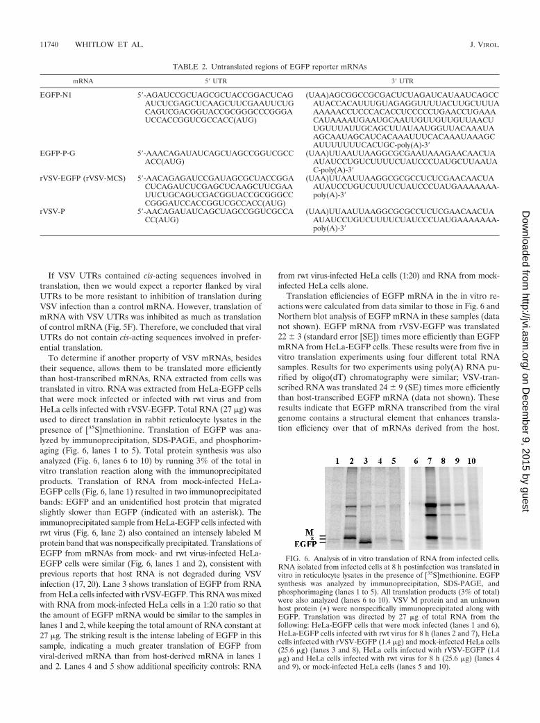
To determine if another property of VSV mRNAs, besidestheir sequence, allows them to be translated more efficientlythan host-transcribed mRNAs, RNA extracted from cells wastranslated in vitro. RNA was extracted from HeLa-EGFP cellsthat were mock infected or infected with rwt virus and fromHeLa cells infected with rVSV-EGFP. Total RNA (27 �g) wasused to direct translation in rabbit reticulocyte lysates in thepresence of [35S]methionine. Translation of EGFP was ana-lyzed by immunoprecipitation, SDS-PAGE, and phosphorim-aging (Fig. 6, lanes 1 to 5). Total protein synthesis was alsoanalyzed (Fig. 6, lanes 6 to 10) by running 3% of the total invitro translation reaction along with the immunoprecipitatedproducts. Translation of RNA from mock-infected HeLa-EGFP cells (Fig. 6, lane 1) resulted in two immunoprecipitatedbands: EGFP and an unidentified host protein that migratedslightly slower than EGFP (indicated with an asterisk). Theimmunoprecipitated sample from HeLa-EGFP cells infected withrwt virus (Fig. 6, lane 2) also contained an intensely labeled Mprotein band that was nonspecifically precipitated. Translations ofEGFP from mRNAs from mock- and rwt virus-infected HeLa-EGFP cells were similar (Fig. 6, lanes 1 and 2), consistent withprevious reports that host RNA is not degraded during VSVinfection (17, 20). Lane 3 shows translation of EGFP from RNAfrom HeLa cells infected with rVSV-EGFP. This RNA was mixedwith RNA from mock-infected HeLa cells in a 1:20 ratio so thatthe amount of EGFP mRNA would be similar to the samples inlanes 1 and 2, while keeping the total amount of RNA constant at27 �g. The striking result is the intense labeling of EGFP in thissample, indicating a much greater translation of EGFP fromviral-derived mRNA than from host-derived mRNA in lanes 1and 2. Lanes 4 and 5 show additional specificity controls: RNA
from rwt virus-infected HeLa cells (1:20) and RNA from mock-infected HeLa cells alone.
Translation efficiencies of EGFP mRNA in the in vitro re-actions were calculated from data similar to those in Fig. 6 andNorthern blot analysis of EGFP mRNA in these samples (datanot shown). EGFP mRNA from rVSV-EGFP was translated22 3 (standard error [SE]) times more efficiently than EGFPmRNA from HeLa-EGFP cells. These results were from five invitro translation experiments using four different total RNAsamples. Results for two experiments using poly(A) RNA pu-rified by oligo(dT) chromatography were similar; VSV-tran-scribed RNA was translated 24 9 (SE) times more efficientlythan host-transcribed EGFP mRNA (data not shown). Theseresults indicate that EGFP mRNA transcribed from the viralgenome contains a structural element that enhances transla-tion efficiency over that of mRNAs derived from the host.
TABLE 2. Untranslated regions of EGFP reporter mRNAs
mRNA 5� UTR 3� UTR
EGFP-N1 5�-AGAUCCGCUAGCGCUACCGGACUCAGAUCUCGAGCUCAAGCUUCGAAUUCUGCAGUCGACGGUACCGCGGGCCCGGGAUCCACCGGUCGCCACC(AUG)
(UAA)AGCGGCCGCGACUCUAGAUCAUAAUCAGCCAUACCACAUUUGUAGAGGUUUUACUUGCUUUAAAAAACCUCCCACACCUCCCCCUGAACCUGAAACAUAAAAUGAAUGCAAUUGUUGUUGUUAACUUGUUUAUUGCAGCUUAUAAUGGUUACAAAUAAGCAAUAGCAUCACAAAUUUCACAAAUAAAGCAUUUUUUUCACUGC-poly(A)-3�
EGFP-P-G 5�-AAACAGAUAUCAGCUAGCCGGUCGCCACC(AUG)
(UAA)UUAAUUAAGGCGCGAAUAAAGAACAACUAAUAUCCUGUCUUUUCUAUCCCUAUGCUUAAUAC-poly(A)-3�
rVSV-EGFP (rVSV-MCS) 5�-AACAGAGAUCCGAUAGCGCUACCGGACUCAGAUCUCGAGCUCAAGCUUCGAAUUCUGCAGUCGACGGUACCGCGGGCCCGGGAUCCACCGGUCGCCACC(AUG)
(UAA)UUAAUUAAGGCGCGCCUCUCGAACAACUAAUAUCCUGUCUUUUCUAUCCCUAUGAAAAAAA-poly(A)-3�
rVSV-P 5�-AACAGAUAUCAGCUAGCCGGUCGCCACC(AUG)
(UAA)UUAAUUAAGGCGCGCCUCUCGAACAACUAAUAUCCUGUCUUUUCUAUCCCUAUGAAAAAAA-poly(A)-3�
FIG. 6. Analysis of in vitro translation of RNA from infected cells.RNA isolated from infected cells at 8 h postinfection was translated invitro in reticulocyte lysates in the presence of [35S]methionine. EGFPsynthesis was analyzed by immunoprecipitation, SDS-PAGE, andphosphorimaging (lanes 1 to 5). All translation products (3% of total)were also analyzed (lanes 6 to 10). VSV M protein and an unknownhost protein (*) were nonspecifically immunoprecipitated along withEGFP. Translation was directed by 27 �g of total RNA from thefollowing: HeLa-EGFP cells that were mock infected (lanes 1 and 6),HeLa-EGFP cells infected with rwt virus for 8 h (lanes 2 and 7), HeLacells infected with rVSV-EGFP (1.4 �g) and mock-infected HeLa cells(25.6 �g) (lanes 3 and 8), HeLa cells infected with rVSV-EGFP (1.4�g) and HeLa cells infected with rwt virus for 8 h (25.6 �g) (lanes 4and 9), or mock-infected HeLa cells (lanes 5 and 10).
11740 WHITLOW ET AL. J. VIROL.
on Decem
ber 9, 2015 by guesthttp://jvi.asm
.org/D
ownloaded from
However, this element must be an mRNA feature other thansequence, since EGFP with VSV UTRs is translated with anefficiency similar to that of EGFP mRNA with control UTRs.
DISCUSSION
A striking feature of VSV infection is the rapid shutoff ofhost translation and the abundant synthesis of viral proteins.The inhibition of host translation is at the level of translationinitiation (20) and is likely due to virus-induced modification ofthe cap-binding complex eIF4F (7, 8). The conclusion that hosttranslation is inhibited at the level of translation initiation wasbased on analysis of polysome profiles, by Lodish and Porter,in which host mRNAs were found to be in smaller polysomesin VSV-infected cells compared to mock-infected cells (20).They also observed that viral mRNAs were present in the samesize polysomes as host mRNAs of similar size, suggesting thatviral mRNAs are subject to the same inhibition of translationas host mRNAs (20). In this model, the predominant synthesisof viral proteins is due to the high abundance of viral mRNAs.The question of how VSV protein synthesis predominates ininfected cells, in the face of inhibition of host protein synthesis,has remained unresolved (3, 10).
It has been proposed that VSV has developed mechanismsto avoid the inhibition of translation (that limits host proteinsynthesis) of viral mRNAs (3). However, the idea that predom-inant synthesis of viral proteins is due simply to the abundanceof viral mRNAs has not been ruled out previously. We ad-dressed this question by comparing translation efficiencies ofEGFP reporter mRNAs expressed from the host nucleus orfrom the viral genome as a foreign gene. We found that thetranslation efficiency of host-derived EGFP mRNA was de-creased dramatically in VSV-infected cells compared to mock-infected cells, as expected. We also found that VSV-derivedEGFP mRNA was translated seven times more efficiently thanhost-derived EGFP mRNA in infected cells. This indicates thatthe predominance of VSV protein synthesis is not a simpleresult of viral mRNA abundance.
Even though EGFP mRNAs derived from the VSV genomewere translated more efficiently than those transcribed in thenucleus, we found that in infected cells, VSV-derived mRNAsfollowed a similar pattern of polysomal association as EGFPmRNAs from the nucleus (Fig. 2). These results were consistentwith Lodish and Porter’s observation that viral mRNAs wereassociated with similar numbers of ribosomes as host mRNAs ofsimilar length, in infected cells (20). Although Lodish and Porterfound that during infection, host and viral mRNAs were in poly-somes of similar sizes, Rosen et al. found later, by treating cellswith puromycin, that most mRNAs from VSV-infected cells werein nontranslating mRNPs (34). mRNPs containing VSV mRNAswere isolated by Rosen et al. and found to contain VSV N proteinas a well as a host protein with an apparent molecular weight of90,000 (34). It is likely that these mRNPs also contain other RNAbinding proteins that were not labeled by [35S]methionine inVSV-infected cells. This suggested that the small polysomes de-scribed by Lodish and Porter may instead be nontranslatingmRNPs (34). Our experiments with puromycin (Fig. 3) wereconsistent with those of Rosen et al. and showed that a largeportion of mRNAs transcribed either from the viral genome or inthe host nucleus were in nontranslating mRNPs. Therefore, we
have concluded that only a small portion, around eighteen per-cent, of viral mRNAs is translated efficiently and that thosemRNAs are in large polysomes.
Many viruses have been shown to use cis-acting sequences toenhance translation of viral mRNAs under conditions in whichtranslation of normal host mRNAs is compromised. However,we have found that when EGFP reporter mRNAs contain viralUTRs, they are no more resistant to the shutoff of translationduring VSV infection than an EGFP reporter mRNA withcontrol UTRs (Fig. 5). We have also found that VSV UTRs donot enhance translation efficiency of EGFP mRNAs over con-trol UTRs in uninfected cells (data not shown). Therefore, wehave concluded that VSV UTRs do not contain cis-actingsequences that affect translation. We have also concluded,based on the levels of expression of EGFP as a foreign gene,that the coding regions of VSV mRNAs do not contain cis-acting sequences that affect translation. After normalizing forthirty percent transcription attenuation (15) and normalizingfor methionine content, EGFP protein was found to be trans-lated at levels similar to those for VSV proteins in cells in-fected with rVSV-EGFP (data not shown).
Many viruses have developed mechanisms to evade the in-hibition of translation that often occurs during infections.These mechanisms involve recruitment of translation initiationfactors such as eIF4F to viral cis-acting sequences in mRNAs,often through intermediate trans-acting factors. For example,the rotavirus NSP3 protein binds to a cis-acting sequence inrotavirus 3� UTRs and to the eIF4G scaffolding subunit ofeIF4F. Similarly, adenovirus 100k protein binds to the 5� UTRof adenovirus late mRNAs and to eIF4G, enhancing transla-tion of adenovirus mRNAs that contain the cis-acting sequencerecognized by 100k protein (42–45). However, VSV mRNAsdo not contain cis-acting sequences that promote translation,and the translation advantage of VSV mRNAs over host mRNAs, ininfected cells, is not a result of VSV mRNAs being resistant toinhibition of translation. This is evident by the failure of rVSV-EGFP-derived mRNAs to exceed or even maintain, in infectedcells, their 22-fold advantage in translation efficiency over host-derived EGFP mRNAs that is observed in vitro. Instead ofusing cis-acting sequences to evade translation inhibition, VSVachieves predominance in gene expression by transcribingmRNAs containing a cis-acting element (not sequence) thatenhances translation efficiency over normal host mRNAs.While translation of VSV mRNAs appears to be inhibited incells, the cis-acting element functions so well as to allow highlevels of translation even when inhibited.
Several groups have studied features of VSV mRNA structurethat are known to influence translation efficiency. These featuresare the 5� guanosine cap, methylation at the 5� end, and the 3�poly(A) tail. So far, these features have been found to be similarfor VSV mRNAs and host mRNAs. Efficient translation ofmRNAs in eukaryotic cells requires a 5� guanosine nucleotide capmethylated at position seven of the guanine base linked to the 5�end of the mRNA by a 5�-5� pyrophosphate bond (1, 4, 5, 27, 38,46). In addition to the methyl group of the cap, eukaryoticmRNAs are also methylated to various extents at the first twonucleotides on the 2�-O of the ribose ring and at various internalpositions. Some mRNAs are methylated at the first two codednucleotides (cap 2), some at only the first (cap 1), while for othermRNAs the only methyl group is that of the 7-methylguanosine
VOL. 80, 2006 TRANSLATION OF VSV mRNAs 11741
on Decem
ber 9, 2015 by guesthttp://jvi.asm
.org/D
ownloaded from
cap (cap 0) (1). VSV polymerase catalyzes capping and methyl-ation of viral mRNAs to form 7mGppp2�OmApA (cap 1) (24, 25,33). In some cell types, mRNAs may contain additional methylgroups. However, in these situations, it appears that host mRNAsand viral mRNAs are methylated similarly (1, 24). VSV mRNAshave also been shown to have 3� poly(A) tails similar in length tothose of host mRNAs (2, 13). Based on our data that VSVmRNAs do not contain cis-acting sequences and on previousreports of VSV mRNA structure, we believe this cis-acting ele-ment is a structural element other than methylation or poly(A)tail. Additional studies on the chemical structure of VSV mRNAswill address the specific nature of the translation-stimulating ele-ment.
ACKNOWLEDGMENTS
We thank Maryam Ahmed and Elizabeth Pettit-Kneller for helpfuladvice and comments on the manuscript. We thank David Ornelles forhelp and advice regarding polysome profile experiments. We thankDavid Ornelles and Felicia Goodrum for their work developing themethod used for extracting RNA from sucrose gradient fractions. Wealso thank the reviewers for their insightful comments and suggestionsthat helped in developing this story.
This work was supported by NIH grant RO1AI052304.
REFERENCES
1. Banerjee, A. K. 1980. 5�-Terminal cap structure in eucaryotic messengerribonucleic acids. Microbiol. Rev. 44:175–205.
2. Banerjee, A. K., S. A. Moyer, and D. P. Rhodes. 1974. Studies on the in vitroadenylation of RNA by vesicular stomatitis virus. Virology 61:547–558.
3. Barber, G. N. 2005. VSV-tumor selective replication and protein translation.Oncogene 24:7710–7719.
4. Both, G. W., A. K. Banerjee, and A. J. Shatkin. 1975. Methylation-dependenttranslation of viral messenger RNAs in vitro. Proc. Natl. Acad. Sci. USA72:1189–1193.
5. Both, G. W., Y. Furuichi, S. Muthukrishnan, and A. J. Shatkin. 1975.Ribosome binding to reovirus mRNA in protein synthesis requires 5� termi-nal 7-methylguanosine. Cell 6:185–195.
6. Bushell, M., and P. Sarnow. 2002. Hijacking the translation apparatus byRNA viruses. J. Cell Biol. 158:395–399.
7. Connor, J. H., and D. S. Lyles. 2005. Inhibition of host and viral translationduring vesicular stomatitis virus infection. eIF2 is responsible for the inhi-bition of viral but not host translation. J. Biol. Chem. 280:13512–13519.
8. Connor, J. H., and D. S. Lyles. 2002. Vesicular stomatitis virus infectionalters the eIF4F translation initiation complex and causes dephosphorylationof the eIF4E binding protein 4E-BP1. J. Virol. 76:10177–10187.
9. Fields, B. N., D. M. Knipe, and P. M. Howley. 1996. Fundamental virology,3rd ed. Lippincott-Raven, Philadelphia, Pa.
10. Gale, M., Jr., S. L. Tan, and M. G. Katze. 2000. Translational control of viralgene expression in eukaryotes. Microbiol. Mol. Biol. Rev. 64:239–280.
11. Gupta, A. K., M. Mathur, and A. K. Banerjee. 2002. Unique capping activityof the recombinant RNA polymerase (L) of vesicular stomatitis virus: asso-ciation of cellular capping enzyme with the L protein. Biochem. Biophys.Res. Commun. 293:264–268.
12. He, B., R. G. Paterson, C. D. Ward, and R. A. Lamb. 1997. Recovery ofinfectious SV5 from cloned DNA and expression of a foreign gene. Virology237:249–260.
13. Hunt, D. M. 1983. Vesicular stomatitis virus mutant with altered polyade-nylic acid polymerase activity in vitro. J. Virol. 46:788–799.
14. Hunt, D. M., E. F. Smith, and D. W. Buckley. 1984. Aberrant polyadenyla-tion by a vesicular stomatitis virus mutant is due to an altered L protein.J. Virol. 52:515–521.
15. Iverson, L. E., and J. K. Rose. 1981. Localized attenuation and discontinuoussynthesis during vesicular stomatitis virus transcription. Cell 23:477–484.
16. Jang, S. K., H. G. Krausslich, M. J. Nicklin, G. M. Duke, A. C. Palmenberg,and E. Wimmer. 1988. A segment of the 5� nontranslated region of encepha-lomyocarditis virus RNA directs internal entry of ribosomes during in vitrotranslation. J. Virol. 62:2636–2643.
17. Jaye, M. C., W. Godchaux III, and J. Lucas-Lenard. 1982. Further studies onthe inhibition of cellular protein synthesis by vesicular stomatitis virus. Vi-rology 116:148–162.
18. Keene, J. D., and R. A. Lazzarini. 1976. A comparison of the extents ofmethylation of vesicular stomatitis virus messenger RNA. Virology 69:364–367.
19. Kopecky, S. A., M. C. Willingham, and D. S. Lyles. 2001. Matrix protein and
another viral component contribute to induction of apoptosis in cells in-fected with vesicular stomatitis virus. J. Virol. 75:12169–12181.
20. Lodish, H. F., and M. Porter. 1980. Translational control of protein synthesisafter infection by vesicular stomatitis virus. J. Virol. 36:719–733.
21. Lyles, D. S. 2000. Cytopathogenesis and inhibition of host gene expression byRNA viruses. Microbiol. Mol. Biol. Rev. 64:709–724.
22. McAllister, P. E., and R. R. Wagner. 1976. Differential inhibition of hostprotein synthesis in L cells infected with RNA� temperature-sensitive mu-tants of vesicular stomatitis virus. J. Virol. 18:550–558.
23. Michel, Y. M., D. Poncet, M. Piron, K. M. Kean, and A. M. Borman. 2000.Cap-Poly(A) synergy in mammalian cell-free extracts. Investigation of therequirements for poly(A)-mediated stimulation of translation initiation.J. Biol. Chem. 275:32268–32276.
24. Moyer, S. A., G. Abraham, R. Adler, and A. K. Banerjee. 1975. Methylatedand blocked 5� termini in vesicular stomatitis virus in vivo mRNAs. Cell5:59–67.
25. Moyer, S. A., and A. K. Banerjee. 1976. In vivo methylation of vesicularstomatitis virus and its host-cell messenger RNA species. Virology 70:339–351.
26. Mudd, J. A., and D. F. Summers. 1970. Protein synthesis in vesicular sto-matitis virus-infected HeLa cells. Virology 42:328–340.
27. Muthukrishnan, S., G. W. Both, Y. Furuichi, and A. J. Shatkin. 1975.5�-Terminal 7-methylguanosine in eukaryotic mRNA is required for trans-lation. Nature 255:33–37.
28. Patton, J. T., and E. Spencer. 2000. Genome replication and packaging ofsegmented double-stranded RNA viruses. Virology 277:217–225.
29. Pelletier, J., G. Kaplan, V. R. Racaniello, and N. Sonenberg. 1988. Cap-independent translation of poliovirus mRNA is conferred by sequence ele-ments within the 5� noncoding region. Mol. Cell. Biol. 8:1103–1112.
30. Pelletier, J., and N. Sonenberg. 1988. Internal initiation of translation ofeukaryotic mRNA directed by a sequence derived from poliovirus RNA.Nature 334:320–325.
31. Piron, M., P. Vende, J. Cohen, and D. Poncet. 1998. Rotavirus RNA-bindingprotein NSP3 interacts with eIF4GI and evicts the poly(A) binding proteinfrom eIF4F. EMBO J. 17:5811–5821.
32. Rhodes, D. P., and A. K. Banerjee. 1975. 5�-Terminal sequence of vesicularstomatitis virus mRNA’s synthesized in vitro. J. Virol. 17:33–42.
33. Rhodes, D. P., S. A. Moyer, and A. K. Banerjee. 1974. In vitro synthesisof methylated messenger RNA by the virion-associated RNA polymerase ofvesicular stomatitis virus. Cell 3:327–333.
34. Rosen, C. A., H. L. Ennis, and P. S. Cohen. 1982. Translational control ofvesicular stomatitis virus protein synthesis: isolation of an mRNA-seques-tering particle. J. Virol. 44:932–938.
35. Sambrook, J., and D. W. Russell. 2001. Molecular cloning: a laboratorymanual, 3rd ed. Cold Spring Harbor Laboratory Press, Cold Spring Harbor,N.Y.
36. Sarnow, P., P. Hearing, C. W. Anderson, D. N. Halbert, T. Shenk, and A. J.Levine. 1984. Adenovirus early region 1B 58,000-dalton tumor antigen isphysically associated with an early region 4 25,000-dalton protein in produc-tively infected cells. J. Virol. 49:692–700.
37. Stanners, C. P., A. M. Francoeur, and T. Lam. 1977. Analysis of VSV mutantwith attenuated cytopathogenicity: mutation in viral function, P, for inhibi-tion of protein synthesis. Cell 11:273–281.
38. Toneguzzo, F., and H. P. Ghosh. 1976. Characterization and translation ofmethylated and unmethylated vesicular stomatitis virus mRNA synthesizedin vitro by ribonucleoprotein particles from vesicular stomatitis virus-in-fected L cells. J. Virol. 17:477–491.
39. Vende, P., M. Piron, N. Castagne, and D. Poncet. 2000. Efficient translationof rotavirus mRNA requires simultaneous interaction of NSP3 with theeukaryotic translation initiation factor eIF4G and the mRNA 3� end. J. Vi-rol. 74:7064–7071.
40. Wertz, G. W., and J. S. Youngner. 1972. Inhibition of protein synthesis in Lcells infected with vesicular stomatitis virus. J. Virol. 9:85–89.
41. Whelan, S. P., L. A. Ball, J. N. Barr, and G. T. Wertz. 1995. Efficient recoveryof infectious vesicular stomatitis virus entirely from cDNA clones. Proc. Natl.Acad. Sci. USA 92:8388–8392.
42. Xi, Q., R. Cuesta, and R. J. Schneider. 2005. Regulation of translation byribosome shunting through phosphotyrosine-dependent coupling of adeno-virus protein 100k to viral mRNAs. J. Virol. 79:5676–5683.
43. Xi, Q., R. Cuesta, and R. J. Schneider. 2004. Tethering of eIF4G to adeno-viral mRNAs by viral 100k protein drives ribosome shunting. Genes Dev.18:1997–2009.
44. Yueh, A., and R. J. Schneider. 1996. Selective translation initiation by ribo-some jumping in adenovirus-infected and heat-shocked cells. Genes Dev.10:1557–1567.
45. Yueh, A., and R. J. Schneider. 2000. Translation by ribosome shunting onadenovirus and hsp70 mRNAs facilitated by complementarity to 18S rRNA.Genes Dev. 14:414–421.
46. Zan-Kowalczewska, M., M. Bretner, H. Sierakowska, E. Szczesna, W.Filipowicz, and A. J. Shatkin. 1977. Removal of 5�-terminal m7G fromeukaryotic mRNAs by potato nucleotide pyrophosphatase and its effect ontranslation. Nucleic Acids Res. 4:3065–3081.
11742 WHITLOW ET AL. J. VIROL.
on Decem
ber 9, 2015 by guesthttp://jvi.asm
.org/D
ownloaded from