recombinant vesicular stomatitis virus-based west nile vaccine elicits strong humoral and cellular...
TRANSCRIPT

Rhc
AVPa
b
c
d
a
ARRAA
KWVMVH
1
1
a[voa
Sf
0d
Vaccine 27 (2009) 893–903
Contents lists available at ScienceDirect
Vaccine
journa l homepage: www.e lsev ier .com/ locate /vacc ine
ecombinant vesicular stomatitis virus-based west Nile vaccine elicits strongumoral and cellular immune responses and protects mice against lethalhallenge with the virulent west Nile virus strain LSU-AR01
run V. Iyera,b, Bapi Paharc, Marc J. Boudreauxa, Nobuko Wakamatsub, Alma F. Royb,d,ladimir N. Chouljenkoa,b, Abolghasem Baghiana,b, Cristian Apetrei c,reston A. Marxc, Konstantin G. Kousoulasa,b,∗
Division of Biotechnology and Molecular Medicine (BIOMMED), Louisiana State University School of Veterinary Medicine (LSU SVM), Baton Rouge, LA 70803, United StatesDepartment of Pathobiological Sciences, Louisiana State University School of Veterinary Medicine, Baton Rouge, LA 70803, United StatesTulane National Primate Research Center, Covington, LA 70433, United StatesLouisiana Animal Disease Diagnostic Laboratory (LADDL), Louisiana State University School of Veterinary Medicine, Baton Rouge, LA 70803, United States
r t i c l e i n f o
rticle history:eceived 18 August 2008eceived in revised form 11 November 2008ccepted 20 November 2008vailable online 12 December 2008
eywords:est Nile virus
esicular stomatitis virusucosal immunization
iral vector
a b s t r a c t
Vesicular stomatitis virus (VSV) has been extensively utilized as a viral vector system for the inductionof protective immune responses against a variety of pathogens. We constructed recombinant VSVs spec-ifying either the Indiana or Chandipura virus G glycoprotein and expressing the West Nile virus (WNV)envelope (E) glycoprotein. Mice were intranasally vaccinated using a prime (Indiana)-boost (Chandipura)immunization approach and challenged with the virulent WNV-LSU-AR01. Ninety-percent (9 of 10) ofthe vaccinated mice survived as compared to 10% of the mock-vaccinated mice after WNV lethal chal-lenge. Histopathological examination of brain tissues revealed neuronal necrosis in mock-vaccinatedmice but not in vaccinated mice, and vaccinated, but not mock-vaccinated mice developed a strong neu-tralizing antibody response against WNV. Extensive immunological analysis using polychromatic flowcytometry staining revealed that vaccinated, but not mock-vaccinated mice developed robust cellularimmune responses as evidenced by up-regulation of CD4+ CD154+ IFN�+ T cells in vaccinated, but not
umoral and cellular immunitymock-vaccinated mice. Similarly, vaccinated mice developed robust E-glycoprotein-specific CD8+ T cellimmune responses as evidenced by the presence of a high percentage of CD8+ CD62Llow IFN�+ cells.In addition, a sizeable population of CD8+ CD69+ cells was detected indicating E-specific activation ofmature T cells and CD4+ CD25+ CD127low T regulatory (T reg) cells were down-regulated. These resultssuggest that VSV-vectored vaccines administered intranasally can efficiently induce protective humoralonse
and cellular immune resp. Introduction
.1. West Nile virus (WNV)
West Nile virus (WNV) was first isolated more than 70 yearsgo from a febrile patient in the West Nile province of Uganda
1]. WNV is a positive-sense RNA virus belonging to genus Fla-ivirus in the Falviviridae family [2]. The lipid-bilayer membranef the nascent virus contains 180 molecules of the envelope (E)nd premembrane (preM) proteins organized into 60 asymmetric∗ Corresponding author at: BIOMMED, School of Veterinary Medicine, Louisianatate University, Baton Rouge, LA 70803, USA. Tel.: +1 225 578 9683;ax: +1 225 578 9655.
E-mail address: [email protected] (K.G. Kousoulas).
264-410X/$ – see front matter © 2008 Published by Elsevier Ltd.oi:10.1016/j.vaccine.2008.11.087
s against WNV infections.© 2008 Published by Elsevier Ltd.
trimeric spikes of preM-E heterodimers [3,4]. The E glycoproteinis the major antigenic determinant and is involved in virus bindingand fusion [5]. WNV spread rapidly in North America after its initialintroduction in New York [6]. WNV was transmitted via mosquitovectors and caused substantial morbidity and mortality in birds,horses and other animals including humans. Humans constitute adead-end host because the virus does not efficiently replicate inhumans. WNV can be transmitted by the intrauterine route [7],through breast milk [8,9], blood transfusion [10–12], bone-marrowtransplant [13], organ transplantation [14,15] and through kidneydialysis [16,17].
The human incubation period for West Nile is 2–14 days[18]. WNV-infected persons may exhibit a variety of clinicalsymptoms including fever, headache, muscle weakness, fatigue,nausea, vomiting, gastrointestinal manifestations, lymphadenopa-thy and non-pruritic maculopapular skin rash [19–21]. Additional

8 cine 2
n[ct(a[haW
1
ehtvpRbacEcvi[ypt(mt
1
tatamVfvsavpaiVier
cdCtpWvVb
94 A.V. Iyer et al. / Vac
on-neurological clinical manifestations include rhabdomyolysis22,23], pancreatitis [24], hepatitis [25], myositis, orchitis [26],horioretinitis [27] and cardiac dysrhythmias [28]. Typically, lesshan 1% of patients suffer from West Nile neuroinvasive diseaseWND) including West Nile meningitis (WNM), encephalitis (WNE)nd acute flaccid paralysis (poliomyelitis-like syndrome, WNP)29]. Among WND cases, an estimated 55–60% of the patientsad WNE resulting in an estimated 20% case fatality. Addition-lly, 10–50% of mortalities in humans could be attributed toNP [29].
.2. WNV vaccines
The absence of effective treatment against WNV infection hasncouraged vaccine development. A variety of different approachesave been employed to produce WNV vaccines including inac-ivated virus, subunit and DNA-based vaccines. Most of theseaccines appeared to be highly immunogenic, and in some casesrotected against WNV-infection in experimental animals [30].ecently, recombinant viruses expressing WNV antigens haveeen shown to induce strong immune responses and protectiongainst WNV challenge in animals. Specifically, a recombinant liveanarypox-vectored vaccine expressing the preM protein and theglycoprotein induced strong immune responses in horses and
ats [31–34], that appeared to be partially protective [35]. Otheriral-vectored vaccines that elicited protective immune responsesn mice include a lentivirus vector based vaccine (TRIP/sEWNV)36], and a measles virus-vectored vaccine [37]. Recombinantellow fever virus (YFV) has also been used to express WNVreM and E proteins based on the extensive safety record ofhe YFV attenuated vaccine [38,39]. A YFV recombinant vaccineChimeriVaxTM) has shown good immune responses in hamster,
ice, non-human primates and humans [40–42]. A Phase II clinicalrial with ChimeriVaxTM-WNV is currently underway [43].
.3. Vesicular stomatitis-vectored vaccines
VSV is an enveloped, negative strand RNA virus belongingo the Rhabdoviridae family. Natural VSV infections of humansre rare causing at most mild flu-like illness [44]. VSV infec-ious viruses can be efficiently recovered by a reverse geneticpproach that utilizes multiple plasmids expressing VSV genes. Thisethodology has enabled the rapid construction of recombinantSV viruses expressing a variety of viral and bacterial antigens
or vaccine purposes including influenza virus, bovine diarrheairus, cotton-tail papillomavirus, human immunodeficiency virus,imian immunodeficiency virus, respiratory syncytial virus, hep-titis C, measles virus, Ebola virus, Lassa fever virus, Marburgirus, severe acute respiratory syndrome virus (SARS), and her-es simplex type-2 virus [45–63]. Recombinant VSVs have beenlso constructed and tested as vaccines for bacterial pathogensncluding Mycobacterium tuberculosis and Yersinia pestis [64,65].SV-vectored vaccines have been administered via intranasal,
ntramuscular and subcutaneous routes and have been shown tolicit robust mucosal and systemic humoral and cellular immuneesponses [45–63,66–68].
We constructed recombinant VSVs expressing the WNV E gly-oprotein. A prime-boost approach was employed utilizing twoifferent recombinant VSVs expressing either the Indiana or thehandipura G glycoproteins for priming and boosting immuniza-ions, respectively. Intranasal immunization of mice conferred high
rotection against lethal challenge with the virulent WNV strainNV-LSU-AR01 [69]. Neuronal necrosis was observed in mock-accinated but not in vaccinated mice. These results suggest thatSV recombinant vaccines expressing the WNV E glycoprotein maye efficacious intranasal vaccines for animal and human use.
7 (2009) 893–903
2. Materials and methods
2.1. Cells and plasmids
Baby hamster kidney cells (BHK-21) were obtained from theAmerican Tissue Culture Collection (ATCC). These cells were grownusing Dulbecco’s modified minimal essential media (DMEM) sup-plemented with 10% fetal bovine serum (FBS) and appropriateamounts of antibiotics. The West Nile virus envelope (E) genewas obtained by first producing a cDNA of the E gene fromthe WNV-LSU-AR01 strain, and subsequently cloning this geneinto the pcDNA3.1 plasmid (Invitrogen, Inc.) after PCR amplifica-tion. The E gene was further amplified by PCR from this plasmidusing primers that introduced unique NotI and BamHI sites atthe 5′ and 3′ using 5′ WNE-FLAG-Not-I (5′-GACGACGCGGCCGC-ATGTTTAACTGCCTTGGAA TGAGC-3′) and 3′ WNE-FLAG-BamHI (5′-GCAGCAGGATCCAGCGTGCACGTTCACGG AGAGG-3′) primers. NotIand BamHI sites are italicized. The fragment was then cloned intoplasmid p3XFLAG-CMV-14 (Sigma) placing the FLAG epitope cod-ing sequence downstream and in-frame with the E glycoproteinsequence. All recombinant plasmids were confirmed by restrictionendonuclease digestion and DNA sequencing.
2.2. Transient expression of the WNV E gene
BHK-21 cells were transfected with the WNV E-3XFLAG plasmidusing Lipofectamine 2000 (Invitrogen) as suggested by the manu-facturer. E glycoprotein was detected at 48 h post-transfection usinganti-FLAG (Sigma) and anti-West Nile rabbit polyclonal antibody(Abcam). For immunofluorescence assay (IFA), cells were washedtwice with phosphate buffered saline (PBS) and fixed with ice-coldmethanol. Cells were then washed with PBS and wells were blockedwith 2% BSA and 5% goat serum in TBS (Tris-buffered saline) for1 h. Mouse anti-FLAG antibodies (Sigma) in blocking buffer andrabbit anti-WNV antibodies were added to respective wells at a1:500 dilution and incubated for 1 h at room temperature. Cellswere then washed six times with TBS and the secondary anti-body Alexa Fluor® 488 goat anti-mouse IgG and goat anti-rabbitIgG (Invitrogen) were added to the respective wells at the samedilution. Cells were incubated in dark for 1 h. Finally, cells werewashed six times with TBS and observed under a fluorescencemicroscope.
2.3. Construction of recombinant VSVs expressing the WNV E gene
Plasmid clones that efficiently expressed the WNV E gene wereused as the template for PCR amplification of the gene, whileat the same time introducing unique XhoI and NheI sites at the5′ and 3′ ends of the gene fragment using 5′-XN2-Xho-I (5′-CCGCGGCTCGAGATGTTT AACTGCCTTGGAATGAGC-3′) and 3′-XN2-Nhe-I (5′-GACGACGCTAGCGGATCACTAC TTGTCATCGTC-3′) primers.XhoI and NheI restriction sites are italicized. This DNA fragment wascloned into the pVSV-XN2-IN and pVSV-XN2-CH transfer vectors.Cells were infected with recombinant vaccinia virus expressing T7polymerase (vTF7-3) at a multiplicity of infection (MOI) of 10 for1 h. Subsequently, BHK 21 cells were co-transfected with pBS-N,pBS-P, pBS-L and pVSV-XN2 containing the WNV E gene and recom-binant virus was recovered as described in detail previously [70,71].Control viruses having no exogenous inserted genes were also pro-duced using pBS-N, pBS-P, pBS-L and the pVSV-XN2 (empty vector).Anti-FLAG and anti-WNV-E antibodies were used to detect expres-
sion of the E glycoprotein by IFA in VSV-infected BHK cells. Viralisolates expressing high amounts of the WNV E glycoprotein wereselected through multiple rounds of plaque purification. Viral titerswere determined and stocks were stored at −80 ◦C for vaccinationstudies.
cine 2
2
aaguecot1COapatcpbdtcroudiD
2(
Imfbwfadap
2
3evpheffiwtr
2
ma
A.V. Iyer et al. / Vac
.4. Vaccination study
All animal studies were carried out after the appropriatepprovals were obtained from the LSU Institutional Animal Carend Use Committee (IACUC) and BSL3 Biosafety Committee. Fourroups of ten 4-week-old female Balb/c mice (Harlan, IN, USA) weresed in this study. Each individual mouse was identified with anar tag (National Band and Tag Company, KY, USA). Group I (vac-ine group): These animals were mildly anesthetized by inhalationf 2–3% isoflurane and 10 �l dose of vaccine containing 105 PFU ofhe vaccine (rVSV-IN-WNV E) was administered intranasally using a0 �l pipette (5 �l per nostril). Animals were boosted with the rVSV-H-WNV E at 21 days post-vaccination using the same technique.ne mouse from this group was not included in the fluorescence-ctivated cell sorting (FACS) analysis due to sample preparationroblems (n = 9). Group II (control for vaccine group): Control groupnimals were vaccinated in the same way as described above withhe exception that they were inoculated with 10 �l of uninfectedell culture supernatant. These animals were boosted at 21 daysost-vaccination with uninfected cell culture supernatant. Animalselonging to Groups I and II were humanely euthanized at 14ays post-boost. Spleens were collected in Eppendorf tubes con-aining RPMI and processed by flow cytometry for intracellularytokines and cell-surface markers associate with memory T cells,egulatory T cells and cytotoxic T cells among others. For serol-gy, animals were bled by the sub-mandibular route (cheek bleed)sing Golden-Rod lancets (Medipoint, NY). Animals were bled on 21ays post-vaccination and 14 days post-boost. Blood was collected
n Becton Dickinson microtainers with serum separators (Bectonickinson).
.5. Challenge studies: Group III (challenge group) and Group IVchallenge group control)
These 20 animals were treated exactly in the same way as Groupsand II until the boost stage. At 10 days post-boost, these ani-als were transported to the animal biosafety level-3 (ABSL-3)
acility for acclimatization. Blood was collected at 14 days post-oost (before challenge). Animals were challenged intraperitoniallyith 105 PFU of WNV-LSU-AR01 and observed 2–3 times a day
or 18 days. Animals showing severe neurological symptoms (liketaxia and hunching posture) were humanely euthanized andead animals were surgically processed immediately (thoracic andbdominal cavities opened up and placed in 10% formalin jars) forathological studies.
.6. Plaque reduction neutralization test (PRNT90)
Serum samples were inactivated by incubation at 65 ◦C for0 min. Serial two-fold dilutions of the serum were incubated withqual volumes of 50 PFU of WNV-LSU-AR01 at 37 ◦C for 1 h. Serum-irus mixtures were then added to Vero cell monolayers in 12-welllates in triplicates and the plates were incubated for anotherour. Plates were then overlaid with Dulbecco’s modified minimumssential media (DMEM) containing 1% methyl cellulose and 2%etal bovine serum. Plates were incubated at 37 ◦C for 72 h and thenxed with 10% formalin in phosphate buffered saline (PBS). Platesere washed three times with PBS and stained with 0.01% crys-
al violet. Plaques were counted and the highest dilution of serumesulting in reduction of 90% of the plaques was noted.
.7. Polychromatic flow cytometric staining and analysis
Mouse splenocytes were adjusted to 107 cells/ml. One-hundredicrolitre aliquots of splenocyte suspension was incubated with
ppropriately diluted concentrations of antibodies for 30 min at
7 (2009) 893–903 895
room temperature. Cells were washed once with PBS and fixed with1X BD stabilizing fixative buffer (BD Biosciences) in distilled water.Cells were kept protected from light at 4 ◦C and flow cytometricacquisition was completed within 24 h of staining. Polychromatic(7 parameters) flow cytometric acquisition was performed on a LSRII Becton Dickinson instrument having three lasers (488 nm bluelaser, 633 nm red laser and 407 violet laser) by using FITC, PE-Texasred, APC, APC-Cy7 and Pacific Blue as the available fluorochromeparameters. Single-stained controls for each fluorochrome wereused for setting flow cytometry compensation. Monoclonal anti-bodies including CD127 FITC (A7R34, eBioscience), CD62L PE-TexasRed (MEL-14, Invitrogen), CD25 APC (3C7, BD Biosciences), CD4APC-Cy7 (GK1.5, BD Biosciences) and CD8a Pacific Blue (53-6.7, BDBiosciences) were used. At least 50,000 events were collected bygating on CD4+ T cells and those data were analyzed using FlowJosoftware (TreeStar Inc.) version 8.7.1.
To test CD4+ or CD8+ T lymphocytes subsets for IFN� production,intracellular cytokine flow cytometry (CFC) assay was employedin response to each WNV peptide pool stimulation as describedpreviously [72]. Briefly, processed splenocytes were resuspendedat 1 × 10⁶ cells/ml in complete RPMI-10 with 10% FCS, and stimu-lated with 2 different WNV peptide pools at a final concentrationof 1 �g/ml of each peptide pool. Peptide pools (15–19mers with10–11 amino acids overlap) derived from the WNV E glycopro-tein were based on the WNV-NY99 E amino acid sequence (NIHBiodefense and Emerging Infections Research Resources Reposi-tory, NIAID, NIH). The 67 peptide array was divided to generate twopeptide pools. Peptide pool 1 (pp1) was made of peptides 1–34and peptide pool 2 (pp2) was composed of peptides 35–67. Forpositive controls, PMA (50 ng/ml, Sigma) and ionomycin (1 �g/ml,Sigma) were used. Negative controls had no antigen or mito-gen stimulation. Brefeldin A (10 �g/ml, Sigma) was added tocultures after the first hour, in a 6-h incubation period. Follow-ing stimulation, cells were stained for cell-surface markers withdirectly conjugated mAbs to CD69 FITC (H1.2F3, BD Biosciences),CD62L PE-TR, CD4 APC-Cy7 and CD8a pacific blue for 30 min atroom temperature and washed with dPBS/BSA wash buffer. Cellswere then fixed and permeabilized by using Cytofix/Cytoperm(BD Biosciences), washed twice in Perm Buffer (BD Biosciences),and stained with intracellular mAbs. IFN� PE (XMG1.2, BD Bio-sciences) and/or CD154 APC (MR1, eBiosciences) were added tocells and incubated at room temperature for 30 min. Single colorand isotype-matched control antibodies were used to confirmstaining specificity. After washing, cells were resuspended in 1%paraformaldehyde in PBS and stored in the dark at 4 ◦C. Data wereacquired within 24 h of staining using a LSR II instrument (BDImmunocytometry System) and FACSDiva software (BD Immuno-cytometry System). For each sample, 50,000 events were collectedby gating on CD4+ T cells. Data analysis was performed using FlowJosoftware. Gated CD4+ and CD8+ T cells were further analyzed forits cytokine production. Positive cytokine responses were deter-mined based on the percentage of cytokine responses obtainedabove background responses (unstimulated medium control) ineach experiment.
2.8. Histopathology
Tissues (brain, lung, liver, bilateral kidneys, heart, spleen, skull,and vertebra) were collected from the mice, euthanized or afterdead, and fixed by immersion in 10% neutral buffered formalin.The skull and vertebra were decalcified in 10% formic acid for 3
days. All sampled tissues were routinely processed into paraffin,and 3–4 �m sections were cut for hematoxylin and eosin staining(H&E). H&E sections of the nasal olfactory epithelium and bulb inthe skull and four sections of the spinal cord including two consec-utive anterior cervico-thoracic and two consecutive lumbar-sacral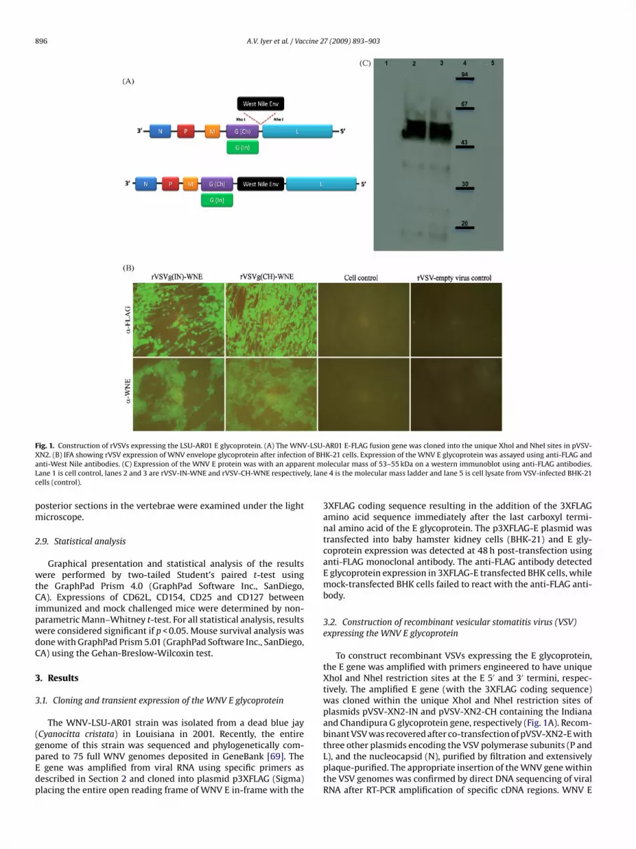
896 A.V. Iyer et al. / Vaccine 27 (2009) 893–903
Fig. 1. Construction of rVSVs expressing the LSU-AR01 E glycoprotein. (A) The WNV-LSU-AR01 E-FLAG fusion gene was cloned into the unique XhoI and NheI sites in pVSV-X of BHa ent mL ly, lanc
pm
2
wtCipwdC
3
3
(gpEdp
N2. (B) IFA showing rVSV expression of WNV envelope glycoprotein after infectionnti-West Nile antibodies. (C) Expression of the WNV E protein was with an apparane 1 is cell control, lanes 2 and 3 are rVSV-IN-WNE and rVSV-CH-WNE respectiveells (control).
osterior sections in the vertebrae were examined under the lighticroscope.
.9. Statistical analysis
Graphical presentation and statistical analysis of the resultsere performed by two-tailed Student’s paired t-test using
he GraphPad Prism 4.0 (GraphPad Software Inc., SanDiego,A). Expressions of CD62L, CD154, CD25 and CD127 between
mmunized and mock challenged mice were determined by non-arametric Mann–Whitney t-test. For all statistical analysis, resultsere considered significant if p < 0.05. Mouse survival analysis wasone with GraphPad Prism 5.01 (GraphPad Software Inc., SanDiego,A) using the Gehan-Breslow-Wilcoxin test.
. Results
.1. Cloning and transient expression of the WNV E glycoprotein
The WNV-LSU-AR01 strain was isolated from a dead blue jayCyanocitta cristata) in Louisiana in 2001. Recently, the entire
enome of this strain was sequenced and phylogenetically com-ared to 75 full WNV genomes deposited in GeneBank [69]. Thegene was amplified from viral RNA using specific primers asescribed in Section 2 and cloned into plasmid p3XFLAG (Sigma)lacing the entire open reading frame of WNV E in-frame with the
K-21 cells. Expression of the WNV E glycoprotein was assayed using anti-FLAG andolecular mass of 53–55 kDa on a western immunoblot using anti-FLAG antibodies.e 4 is the molecular mass ladder and lane 5 is cell lysate from VSV-infected BHK-21
3XFLAG coding sequence resulting in the addition of the 3XFLAGamino acid sequence immediately after the last carboxyl termi-nal amino acid of the E glycoprotein. The p3XFLAG-E plasmid wastransfected into baby hamster kidney cells (BHK-21) and E gly-coprotein expression was detected at 48 h post-transfection usinganti-FLAG monoclonal antibody. The anti-FLAG antibody detectedE glycoprotein expression in 3XFLAG-E transfected BHK cells, whilemock-transfected BHK cells failed to react with the anti-FLAG anti-body.
3.2. Construction of recombinant vesicular stomatitis virus (VSV)expressing the WNV E glycoprotein
To construct recombinant VSVs expressing the E glycoprotein,the E gene was amplified with primers engineered to have uniqueXhoI and NheI restriction sites at the E 5′ and 3′ termini, respec-tively. The amplified E gene (with the 3XFLAG coding sequence)was cloned within the unique XhoI and NheI restriction sites ofplasmids pVSV-XN2-IN and pVSV-XN2-CH containing the Indianaand Chandipura G glycoprotein gene, respectively (Fig. 1A). Recom-binant VSV was recovered after co-transfection of pVSV-XN2-E with
three other plasmids encoding the VSV polymerase subunits (P andL), and the nucleocapsid (N), purified by filtration and extensivelyplaque-purified. The appropriate insertion of the WNV gene withinthe VSV genomes was confirmed by direct DNA sequencing of viralRNA after RT-PCR amplification of specific cDNA regions. WNV E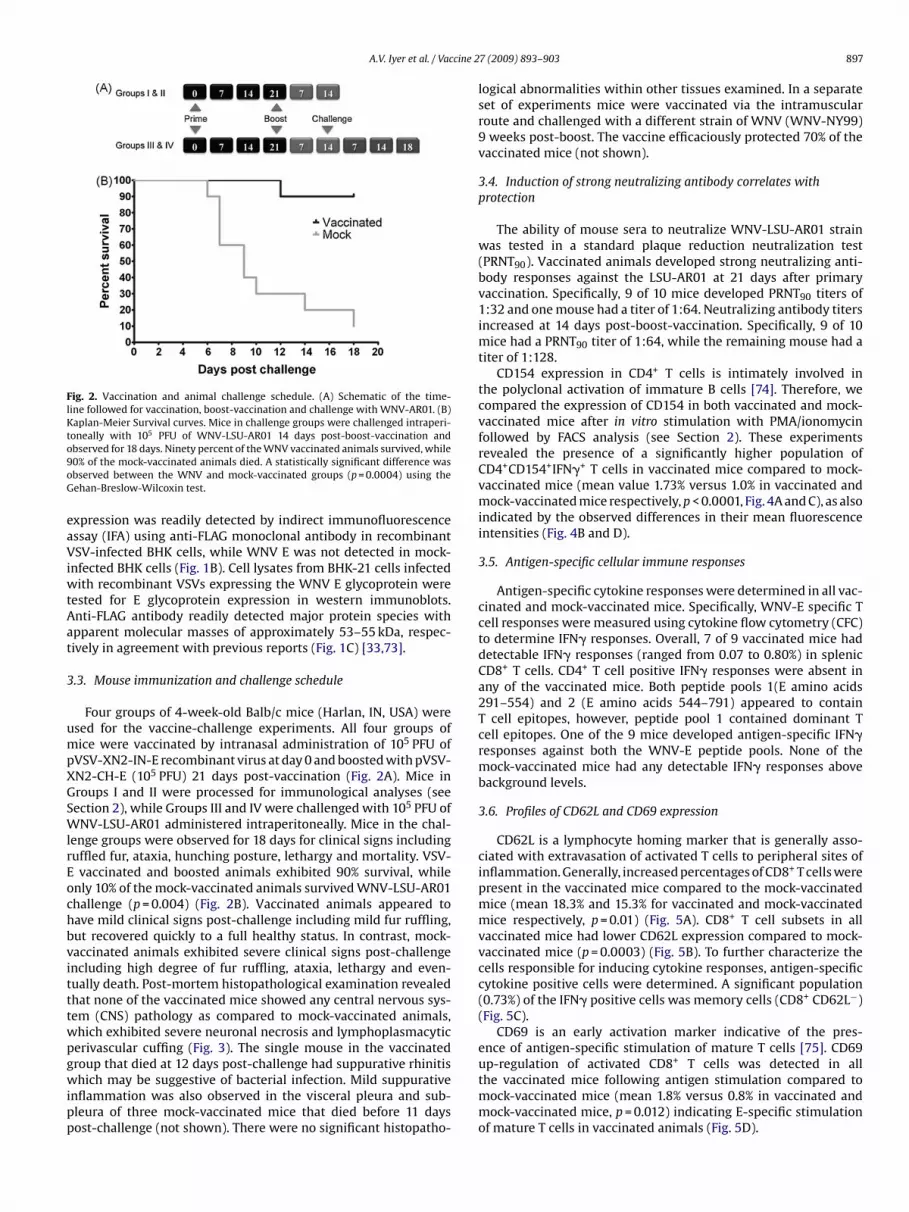
A.V. Iyer et al. / Vaccine 2
Fig. 2. Vaccination and animal challenge schedule. (A) Schematic of the time-line followed for vaccination, boost-vaccination and challenge with WNV-AR01. (B)Kaplan-Meier Survival curves. Mice in challenge groups were challenged intraperi-toneally with 105 PFU of WNV-LSU-AR01 14 days post-boost-vaccination ando9oG
eaViwtAat
3
umpXGSWlrEochbvitttwpgwipp
bserved for 18 days. Ninety percent of the WNV vaccinated animals survived, while0% of the mock-vaccinated animals died. A statistically significant difference wasbserved between the WNV and mock-vaccinated groups (p = 0.0004) using theehan-Breslow-Wilcoxin test.
xpression was readily detected by indirect immunofluorescencessay (IFA) using anti-FLAG monoclonal antibody in recombinantSV-infected BHK cells, while WNV E was not detected in mock-
nfected BHK cells (Fig. 1B). Cell lysates from BHK-21 cells infectedith recombinant VSVs expressing the WNV E glycoprotein were
ested for E glycoprotein expression in western immunoblots.nti-FLAG antibody readily detected major protein species withpparent molecular masses of approximately 53–55 kDa, respec-ively in agreement with previous reports (Fig. 1C) [33,73].
.3. Mouse immunization and challenge schedule
Four groups of 4-week-old Balb/c mice (Harlan, IN, USA) weresed for the vaccine-challenge experiments. All four groups ofice were vaccinated by intranasal administration of 105 PFU of
VSV-XN2-IN-E recombinant virus at day 0 and boosted with pVSV-N2-CH-E (105 PFU) 21 days post-vaccination (Fig. 2A). Mice inroups I and II were processed for immunological analyses (seeection 2), while Groups III and IV were challenged with 105 PFU ofNV-LSU-AR01 administered intraperitoneally. Mice in the chal-
enge groups were observed for 18 days for clinical signs includinguffled fur, ataxia, hunching posture, lethargy and mortality. VSV-
vaccinated and boosted animals exhibited 90% survival, whilenly 10% of the mock-vaccinated animals survived WNV-LSU-AR01hallenge (p = 0.004) (Fig. 2B). Vaccinated animals appeared toave mild clinical signs post-challenge including mild fur ruffling,ut recovered quickly to a full healthy status. In contrast, mock-accinated animals exhibited severe clinical signs post-challengencluding high degree of fur ruffling, ataxia, lethargy and even-ually death. Post-mortem histopathological examination revealedhat none of the vaccinated mice showed any central nervous sys-em (CNS) pathology as compared to mock-vaccinated animals,hich exhibited severe neuronal necrosis and lymphoplasmacyticerivascular cuffing (Fig. 3). The single mouse in the vaccinated
roup that died at 12 days post-challenge had suppurative rhinitishich may be suggestive of bacterial infection. Mild suppurativenflammation was also observed in the visceral pleura and sub-leura of three mock-vaccinated mice that died before 11 daysost-challenge (not shown). There were no significant histopatho-
7 (2009) 893–903 897
logical abnormalities within other tissues examined. In a separateset of experiments mice were vaccinated via the intramuscularroute and challenged with a different strain of WNV (WNV-NY99)9 weeks post-boost. The vaccine efficaciously protected 70% of thevaccinated mice (not shown).
3.4. Induction of strong neutralizing antibody correlates withprotection
The ability of mouse sera to neutralize WNV-LSU-AR01 strainwas tested in a standard plaque reduction neutralization test(PRNT90). Vaccinated animals developed strong neutralizing anti-body responses against the LSU-AR01 at 21 days after primaryvaccination. Specifically, 9 of 10 mice developed PRNT90 titers of1:32 and one mouse had a titer of 1:64. Neutralizing antibody titersincreased at 14 days post-boost-vaccination. Specifically, 9 of 10mice had a PRNT90 titer of 1:64, while the remaining mouse had atiter of 1:128.
CD154 expression in CD4+ T cells is intimately involved inthe polyclonal activation of immature B cells [74]. Therefore, wecompared the expression of CD154 in both vaccinated and mock-vaccinated mice after in vitro stimulation with PMA/ionomycinfollowed by FACS analysis (see Section 2). These experimentsrevealed the presence of a significantly higher population ofCD4+CD154+IFN�+ T cells in vaccinated mice compared to mock-vaccinated mice (mean value 1.73% versus 1.0% in vaccinated andmock-vaccinated mice respectively, p < 0.0001, Fig. 4A and C), as alsoindicated by the observed differences in their mean fluorescenceintensities (Fig. 4B and D).
3.5. Antigen-specific cellular immune responses
Antigen-specific cytokine responses were determined in all vac-cinated and mock-vaccinated mice. Specifically, WNV-E specific Tcell responses were measured using cytokine flow cytometry (CFC)to determine IFN� responses. Overall, 7 of 9 vaccinated mice haddetectable IFN� responses (ranged from 0.07 to 0.80%) in splenicCD8+ T cells. CD4+ T cell positive IFN� responses were absent inany of the vaccinated mice. Both peptide pools 1(E amino acids291–554) and 2 (E amino acids 544–791) appeared to containT cell epitopes, however, peptide pool 1 contained dominant Tcell epitopes. One of the 9 mice developed antigen-specific IFN�responses against both the WNV-E peptide pools. None of themock-vaccinated mice had any detectable IFN� responses abovebackground levels.
3.6. Profiles of CD62L and CD69 expression
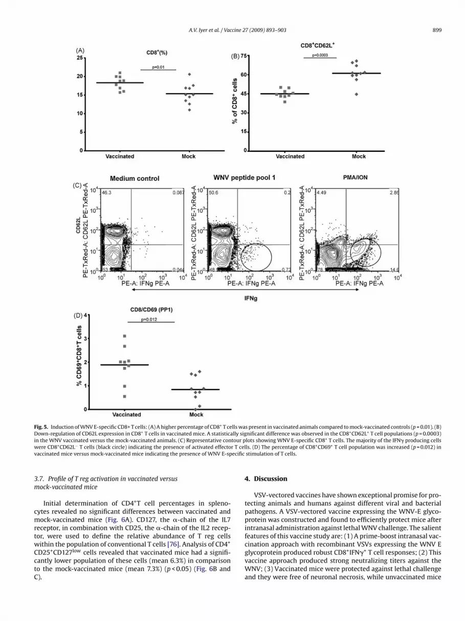
CD62L is a lymphocyte homing marker that is generally asso-ciated with extravasation of activated T cells to peripheral sites ofinflammation. Generally, increased percentages of CD8+ T cells werepresent in the vaccinated mice compared to the mock-vaccinatedmice (mean 18.3% and 15.3% for vaccinated and mock-vaccinatedmice respectively, p = 0.01) (Fig. 5A). CD8+ T cell subsets in allvaccinated mice had lower CD62L expression compared to mock-vaccinated mice (p = 0.0003) (Fig. 5B). To further characterize thecells responsible for inducing cytokine responses, antigen-specificcytokine positive cells were determined. A significant population(0.73%) of the IFN� positive cells was memory cells (CD8+ CD62L−)(Fig. 5C).
CD69 is an early activation marker indicative of the pres-ence of antigen-specific stimulation of mature T cells [75]. CD69
up-regulation of activated CD8+ T cells was detected in allthe vaccinated mice following antigen stimulation compared tomock-vaccinated mice (mean 1.8% versus 0.8% in vaccinated andmock-vaccinated mice, p = 0.012) indicating E-specific stimulationof mature T cells in vaccinated animals (Fig. 5D).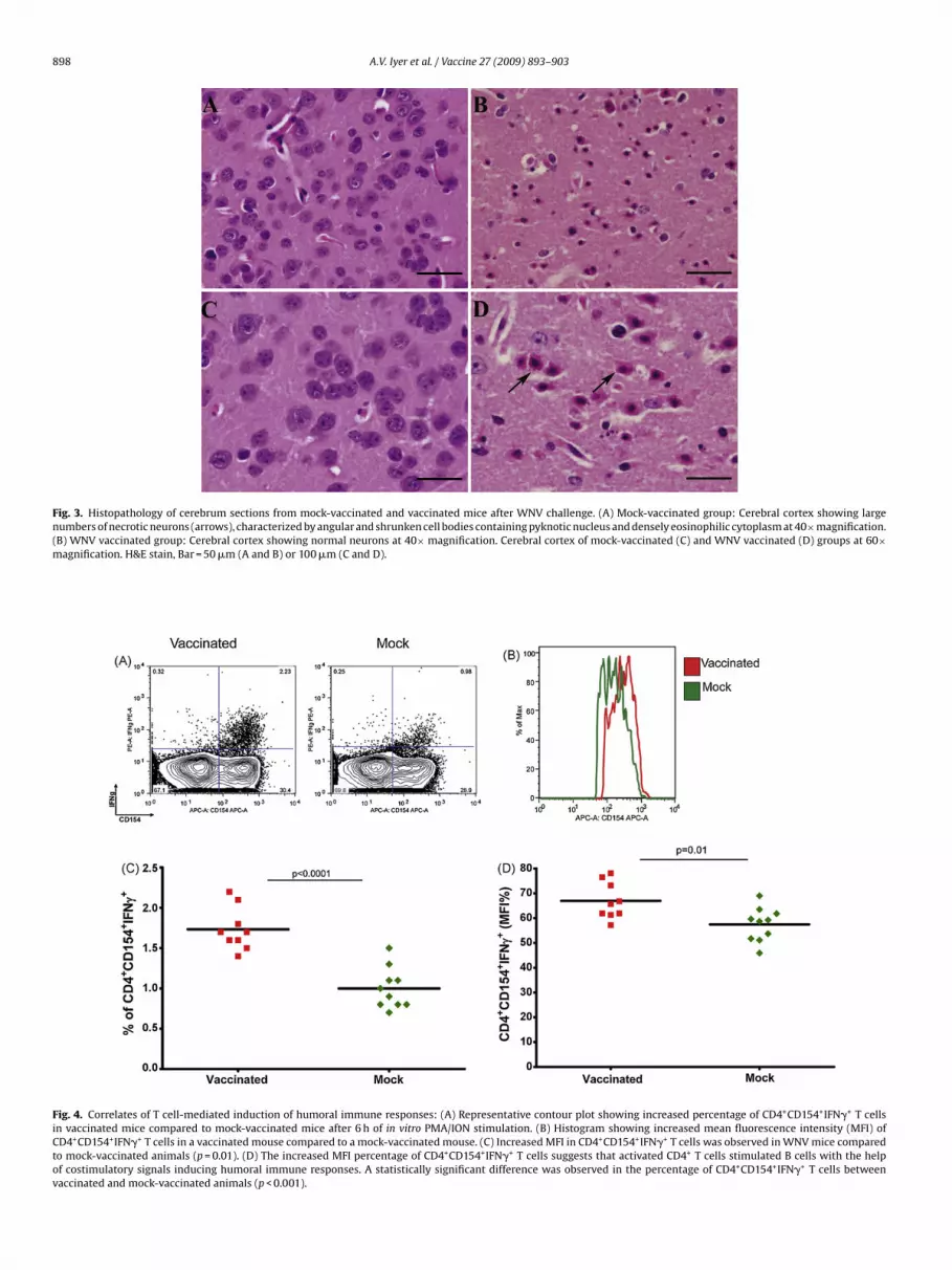
898 A.V. Iyer et al. / Vaccine 27 (2009) 893–903
Fig. 3. Histopathology of cerebrum sections from mock-vaccinated and vaccinated mice after WNV challenge. (A) Mock-vaccinated group: Cerebral cortex showing largenumbers of necrotic neurons (arrows), characterized by angular and shrunken cell bodies containing pyknotic nucleus and densely eosinophilic cytoplasm at 40×magnification.(m
FiCtov
B) WNV vaccinated group: Cerebral cortex showing normal neurons at 40× magnificatioagnification. H&E stain, Bar = 50 �m (A and B) or 100 �m (C and D).
ig. 4. Correlates of T cell-mediated induction of humoral immune responses: (A) Repren vaccinated mice compared to mock-vaccinated mice after 6 h of in vitro PMA/ION stiD4+CD154+IFN�+ T cells in a vaccinated mouse compared to a mock-vaccinated mouse. (o mock-vaccinated animals (p = 0.01). (D) The increased MFI percentage of CD4+CD154+
f costimulatory signals inducing humoral immune responses. A statistically significantaccinated and mock-vaccinated animals (p < 0.001).
n. Cerebral cortex of mock-vaccinated (C) and WNV vaccinated (D) groups at 60×
sentative contour plot showing increased percentage of CD4+CD154+IFN�+ T cellsmulation. (B) Histogram showing increased mean fluorescence intensity (MFI) ofC) Increased MFI in CD4+CD154+IFN�+ T cells was observed in WNV mice comparedIFN�+ T cells suggests that activated CD4+ T cells stimulated B cells with the helpdifference was observed in the percentage of CD4+CD154+IFN�+ T cells between
A.V. Iyer et al. / Vaccine 27 (2009) 893–903 899
Fig. 5. Induction of WNV E-specific CD8+ T cells: (A) A higher percentage of CD8+ T cells was present in vaccinated animals compared to mock-vaccinated controls (p = 0.01). (B)Down-regulation of CD62L expression in CD8+ T cells in vaccinated mice. A statistically significant difference was observed in the CD8+CD62L+ T cell populations (p = 0.0003)i tour pw r T celv ecific
3m
cmrtwCctC
n the WNV vaccinated versus the mock-vaccinated animals. (C) Representative conere CD8+CD62L− T cells (black circle) indicating the presence of activated effecto
accinated mice versus mock-vaccinated mice indicating the presence of WNV E-sp
.7. Profile of T reg activation in vaccinated versusock-vaccinated mice
Initial determination of CD4+T cell percentages in spleno-ytes revealed no significant differences between vaccinated andock-vaccinated mice (Fig. 6A). CD127, the �-chain of the IL7
eceptor, in combination with CD25, the �-chain of the IL2 recep-or, were used to define the relative abundance of T reg cells
ithin the population of conventional T cells [76]. Analysis of CD4+D25+CD127low cells revealed that vaccinated mice had a signifi-antly lower population of these cells (mean 6.3%) in comparisono the mock-vaccinated mice (mean 7.3%) (p < 0.05) (Fig. 6B and).
lots showing WNV E-specific CD8+ T cells. The majority of the IFN� producing cellsls. (D) The percentage of CD8+CD69+ T cell population was increased (p = 0.012) instimulation of T cells.
4. Discussion
VSV-vectored vaccines have shown exceptional promise for pro-tecting animals and humans against different viral and bacterialpathogens. A VSV-vectored vaccine expressing the WNV-E glyco-protein was constructed and found to efficiently protect mice afterintranasal administration against lethal WNV challenge. The salientfeatures of this vaccine study are: (1) A prime-boost intranasal vac-
cination approach with recombinant VSVs expressing the WNV Eglycoprotein produced robust CD8+IFN�+ T cell responses; (2) Thisvaccine approach produced strong neutralizing titers against theWNV; (3) Vaccinated mice were protected against lethal challengeand they were free of neuronal necrosis, while unvaccinated mice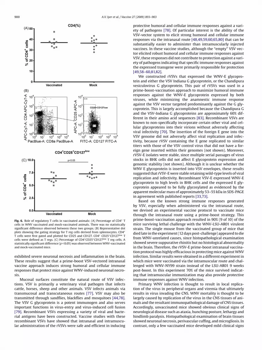
900 A.V. Iyer et al. / Vaccine 2
Fig. 6. Role of regulatory T cells in vaccinated animals: (A) Percentage of CD4+ Tcells in WNV vaccinated and mock-vaccinated animals. There was no statisticallysignificant difference observed between these two groups. (B) Representative dotplots showing the gating strategy for T reg cells derived from spleenocytes. CD4+
Tcsa
eTvrs
tcttTi[rrl
cells were first gated and plotted for CD25 and CD127. CD4+ CD25+CD127low Tells were defined as T regs. (C) Percentage of CD4+CD25+CD127low T reg cells. Atatistically significant difference (p < 0.05) was observed between WNV-vaccinatednd mock-vaccinated mice.
xhibited severe neuronal necrosis and inflammation in the brain.hese results suggest that a prime-boost VSV-vectored intranasalaccine approach induces strong humoral and cellular immuneesponses that protect mice against WNV-induced neuronal necro-is.
Mucosal surfaces constitute the natural route of VSV infec-ions. VSV is primarily a veterinary viral pathogen that infectsattle, horses, sheep and other animals. VSV infects animals viaransmucosal and transcutaneous routes [77]. VSV may also beransmitted through sandflies, blackflies and mosquitoes [44,78].he VSV G glycoprotein is a potent immunogen and also serves
mportant functions in virus-entry and virus-induced cell fusion79]. Recombinant VSVs expressing a variety of viral and bacte-ial antigens have been constructed. Vaccine studies with theseecombinant VSVs have showed that intranasal and intramuscu-ar administration of the rVSVs were safe and efficient in inducing7 (2009) 893–903
protective humoral and cellular immune responses against a vari-ety of pathogens [78]. Of particular interest is the ability of theVSV-vector system to elicit strong humoral and cellular immuneresponses via the intranasal route [48,49,59,60,65,80] that can besubstantially easier to administer than intramuscularly injectedvaccines. In these vaccine studies, although the “empty” VSV vec-tor elicited robust humoral and cellular immune responses againstVSV, these responses did not contribute to protection against a vari-ety of pathogens indicating that specific immune responses againstthe expressed transgene were primarily responsible for protection[49,58–60,81,82].
We constructed rVSVs that expressed the WNV-E glycopro-tein and either the VSV Indiana G glycoprotein, or the Chandipuravesiculovirus G glycoprotein. This pair of rVSVs was used in aprime-boost-vaccination approach to maximize humoral immuneresponses against the WNV-E glycoprotein expressed by bothviruses, while minimizing the anamnestic immune responseagainst the VSV vector targeted predominantly against the G gly-coprotein. This is largely accomplished because the Chandipura Gand the VSV-Indiana G glycoproteins are approximately 60% dif-ferent in their amino acid sequences [83]. Recombinant VSVs areknown to non-specifically incorporate certain other viral and cel-lular glycoproteins into their virions without adversely affectingviral infectivity [70]. The insertion of the foreign E gene into theVSV genome did not adversely affect viral replication and infec-tivity, because rVSV containing the E gene replicated to similartiters with those of the VSV control virus that did not have a for-eign gene inserted within their genomes (not shown). Moreover,rVSV-E isolates were stable, since multiple serial passages of virusstocks in BHK cells did not affect E glycoprotein expression andgenomic stability (not shown). Although it is unclear whether theWNV E glycoprotein is inserted into VSV envelopes, these resultssuggested that rVSV-E were stable retaining wild-type levels of viralreplication and infectivity. Recombinant VSV-E expressed WNV-Eglycoprotein to high levels in BHK cells and the expressed E gly-coprotein appeared to be fully glycosylated as evidenced by theapparent molecular mass of approximately 53–55 kDa in SDS-PAGEin agreement with published reports [33,73].
Based on the known strong immune responses generatedby VSV, especially when administered via the intranasal route,we devised an experimental vaccine protocol to vaccinate micethrough the intranasal route using a prime-boost strategy. Thisprime-boost-vaccination approach resulted in 90% (9 of 10) of themice surviving lethal challenge with the WNV-LSU-AR01 virulentstrain. The single mouse from the vaccinated group of mice thatdied late in the experiment (12 days post-challenge) appeared to diefrom WNV-unrelated causes, since histopathological examinationshowed severe suppurative rhinitis but no histological abnormalityin the brain. Therefore, the rVSV-E prime-boost intranasal vaccina-tion protocol was highly efficacious in protecting mice against WNVinfection. Similar results were obtained in a different experiment inwhich mice were vaccinated via the intramuscular route and chal-lenged with WNV-NY99 strain instead of the LSU-AR01 9 weekspost-boost. In this experiment 70% of the mice survived indicat-ing that intramuscular immunization may also provide protectiveimmune responses against WNV infection.
Primary WNV infection is thought to result in local replica-tion of the virus in peripheral organs and viremia that ultimatelyresults in virus invading the CNS. WNV mortality is thought to belargely caused by replication of the virus in the CNS tissues of ani-mals and the resultant immunopathological damage of CNS tissues.
Accordingly, unvaccinated mice showed obvious clinical signs ofneurological disease such as ataxia, hunching posture, lethargy andhindlimb paralysis. Histopathological examination of brain tissuesshowed neuronal necrosis, perivascular cuffing, and microgliosis. Incontrast, only a few vaccinated mice developed mild clinical signs
cine 2
st
Tiluerawswieeevpr
iaWc3cittwbWfIsle(iteretttmlciuftrdudFsCCTtat
W
A.V. Iyer et al. / Vac
uch as mild ruffled fur, but recovered quickly. Importantly, none ofhe vaccinated mice exhibited any neuronal necrosis.
The interaction of CD40 on B cells with CD154 (CD40L) on CD4+
cells results in T cell mediated activation of B cells resulting inmmunoglobulin class switching, somatic hypermutation and pro-iferation [84–86]. Accordingly, CD4+ CD154+ IFN�+ T cells werep-regulated in vaccinated but not control mice indicating gen-ration of T cell mediated B cell activation. The specificity of thisesponse is not discernable, since it may be due to either or both VSVnd WNV antigens. However, strong neutralizing antibody titersere also produced against WNV indicating the induction of E-
pecific humoral immune responses. This result is in agreementith previous reports showing that other VSV-vectored vaccines
nduced strong humoral immune responses against different VSV-xpressed antigens. Specifically, recombinant VSVs expressingither the respiratory syncytial virus F glycoprotein [49], or rVSVxpressing the severe acute respiratory syndrome (SARS) coronairus (SARS-CoV) produced high antibody titers against the F glyco-rotein and SARC-CoV spike (S) glycoprotein, while strong immuneesponses against the VSV virus was noted [59].
The WNV E glycoprotein contains multiple predicted and exper-mentally verified cytotoxic T cell (CTL) epitopes [87–90]. Thevailability of a library of overlapping peptides derived from the
NV E glycoprotein allowed the elucidation of antigen-specificellular immune responses. Peptide pool 1 composed of the first4 peptides averaging 12–18 amino acids each generated strongerellular CD8+IFN�+ T cell responses in in vitro proliferation assays,n comparison to peptide pool 2, which represented the carboxylerminus-half of the WNV E glycoprotein. Peptide pool 1 containshe experimentally verified CTL epitope RSYCYLAT (E 347–354)hile peptide pool 2 contains the CTL epitope IALTFLAV (E771–778),
oth of which have been shown to confer protection against lethalNV-challenge in mice [87,89]. In vitro stimulation of lymphocytes
rom vaccinated mice revealed the presence of antigen-specificFN� responses specifically in CD8+CD62Llow T cells. CD62L (L-electin) mediates adhesion of resting lymphocytes to peripheralymph nodes. Typically, high expression of CD62L (CD62Lhi) revealsntrapment of lymphocytes within lymph nodes, while low CD62LCD62Llow) cell-surface expression (the result of T cell activation)s indicative of lymphocyte extravasation to sites of inflamma-ion [91]. Splenocytes from vaccinated mice had significantly lowerxpression of the CD62L marker on E-specific IFN�+ CD8+T cellsevealing activation and extravasation of these cells to periph-ral sites, potentially involved in killing virus-infected cells prioro transmission to the CNS. CD69 is an early activation markerhat is absent in resting lymphocytes [75]. The up-regulation ofhe CD8+CD69+ E-specific T cell responses in vaccinated versus
ock-vaccinated mice provides additional evidence for the stimu-ation of T cells. Accordingly, CD8+CD69+ E-specific population of Tells was up-regulated in vaccinated versus mock-vaccinated micendicating the generation of activated memory CD8+ T cells. It isnclear whether the observed CD8+T cell memory responses con-
er long-term immunity against WNV infection. T regs are knowno play important roles in down-regulation of anti-self immuneesponses [92], and to suppress proliferation and cytokine pro-uction of effector T cells [93]. Typically, during viral infections,p-regulation of humoral and cellular immune responses causesown-regulation of T reg activation. Typically, T regs express theoxP3 and CD25 markers. The IL-7 receptor CD127 marker expres-ion is inversely correlated to FoxP3 expression and CD127low
D25+ cells have been shown to be positive for FoxP3 [93,94].
onsequently, the CD25+CD127low population was used to defineregs. As expected, there was a negative correlation betweenhe relative population of T reg cells (CD4+CD25+CD127low) andntigen-specific CTL responses in the vaccinated mice. However,he specificity of this immune response cannot be discerned, since
7 (2009) 893–903 901
it most likely is caused by both VSV and E glycoprotein anti-gens.
A variety of experimental vaccine approaches have beenreported to generate protective humoral and cellular immuneresponses against flaviviruses and specifically WNV. The relativerole of humoral versus cellular immune responses has been exten-sively debated in the literature. Certain studies have suggested thata strong humoral immune response evidenced by the productionof high titer anti-WNV titers is necessary and sufficient to pro-tect mice from CNS infection, while other reports have arguedthat a cellular immune response characterized by a robust anti-
NV CD8+ T cell responses is necessary for protecting and clearingbrain tissues from WNV [89,95,96]. One report has argued thatCTL-immune responses may result in exacerbated immunopathol-ogy in brain and CNS tissues at infections with low WNV titers(103 PFU) [95]. In our experiments, 105 WNV PFU were inoculatedintraperitoneally. Vaccinated mice had no evidence of neuronalnecrosis suggesting the CD8+T cell responses conferred protectionand virus clearance. It is probable that both humoral and cellularimmune responses generated against the WNV E glycoprotein pre-vented the virus from entering CNS, potentially arresting the virusat peripheral sites. Alternatively, if some virus escaped peripheralimmune surveillance, it is possible that CTLs cleared the virus frombrain tissues before it could cause significant damage and resultantimmunopathological manifestations.
In summary, the VSV-E-vectored vaccine appeared to elicitrobust humoral and cellular immune responses that efficientlyprotected mice from WNV lethal challenge. Intranasal vaccina-tion is second only to oral vaccination with regard to the relativeease of administration and patient compliance issues renderingthis approach attractive for human use. Recently, single-cycle VSV-vectored vaccines have been shown to generate robust immuneresponses against a number of viral pathogens including HIV, Ebola,Marburg, Lassa, influenza, avian influenza, hepatitis C and RSVviruses [46,47,49,54–58,81,97]. Based on these results, it is expectedthat single cycle VSV-WNV vaccines would be also efficacious. Addi-tional improvements in attenuating VSV can be made by providingmore than one viral protein in trans through complementing cells,as well as engineering additional mutations that are known toattenuate VSV.
Acknowledgements
This work was supported by NIH:NCRR 2P51RR000164 project.KGK and AI were supported by the Division of Biotechnologyand Molecular Medicine (BIOMMED), LSU School of VeterinaryMedicine. BP was supported by the FACS Immunology Core Lab-oratory of the Tulane National Primate Research Center (NIH:NCRR2P51RR000164). The following reagent was obtained through theNIH Biodefense and Emerging Infections Research Resources Repos-itory, NIAID, NIH: Peptide Array West Nile Virus Gene E, NR-435. Wegratefully acknowledge Dr. John K. Rose (Yale University, School ofMedicine) for providing us with the VSV reverse genetics systemsand Ms. Li Huang and other BIOMMED staff for technical assistance.
References
[1] Smithburn KC, Hughes TP, Burke AW, Paul JH. A neurotropic virus isolated fromthe blood of a native of Uganda. Am J Trop Med Hyg 1940;20:471–92.
[2] Lindenbach BD, Thiel H-J, Rice CM. Flaviviridae: the viruses and their replica-tion. In: Fields BN, Knipe DM, Howley PM, editors. Fields’ virology. 5th EditionPhiladelphia: Wolters Kluwer Health/Lippincott Williams & Wilkins; 2007. p.
1101–52.[3] Zhang Y, Corver J, Chipman PR, Zhang W, Pletnev SV, Sedlak D, et al. Structuresof immature flavivirus particles. Embo J 2003;22(June (11)):2604–13.
[4] Zhang Y, Zhang W, Ogata S, Clements D, Strauss JH, Baker TS, et al. Conforma-tional changes of the flavivirus E glycoprotein. Structure 2004;12(September(9)):1607–18.

9 cine 2
[
[
[
[
[
[
[
[
[
[
[
[
[
[
[
[
[
[
[
[
[
[
[
[
[
[
[
[
[[
[
[
[
[
[
[
[
[
[
[
[
[
[
[
[
[
[
[
[
[
[
02 A.V. Iyer et al. / Vac
[5] Ledizet M, Kar K, Foellmer HG, Bonafe N, Anthony KG, Gould LH, et al. Antibodiestargeting linear determinants of the envelope protein protect mice against WestNile virus. J Infect Dis 2007;196(December (12)):1741–8.
[6] Lanciotti RS, Roehrig JT, Deubel V, Smith J, Parker M, Steele K, et al. Origin of theWest Nile virus responsible for an outbreak of encephalitis in the northeasternUnited States. Science 1999;286(December (5448)):2333–7.
[7] CDC. Intrauterine West Nile virus infection–New York, 2002. MMWR MorbMortal Wkly Rep 2002;51(December (50)):1135–6.
[8] CDC. Possible West Nile virus transmission to an infant through breast-feeding–Michigan, 2002. MMWR Morb Mortal Wkly Rep 2002;51(October (39)):877–8.
[9] Hayes EB, O’Leary DR. West Nile virus infection: a pediatric perspective. Pedi-atrics 2004;113(May (5)):1375–81.
10] CDC. Transfusion-associated transmission of West Nile virus–Arizona, 2004.MMWR Morb Mortal Wkly Rep 2004;53(September (36)):842–4.
11] CDC. Investigations of West Nile virus infections in recipients of blood transfu-sions. MMWR Morb Mortal Wkly Rep 2002;51(November (43)):973–4.
12] CDC. Detection of West Nile virus in blood donations–United States, 2003.MMWR Morb Mortal Wkly Rep 2003;52(August (32)):769–72.
13] Hiatt B, DesJardin L, Carter T, Gingrich R, Thompson C, de Magalhaes-SilvermanM. A fatal case of West Nile virus infection in a bone marrow transplant recipi-ent. Clin Infect Dis 2003;37(November (9)):e129–31.
14] CDC. West Nile virus infection in organ donor and transplant recipients–Georgia and Florida, 2002. MMWR Morb Mortal Wkly Rep 2002;51(September(35)):790.
15] Iwamoto M, Jernigan DB, Guasch A, Trepka MJ, Blackmore CG, Hellinger WC,et al. Transmission of West Nile virus from an organ donor to four transplantrecipients. N Engl J Med 2003;348(May (22)):2196–203.
16] CDC. Possible dialysis-related west nile virus transmission–Georgia, 2003.MMWR Morb Mortal Wkly Rep 2004;53(August (32)):738–9.
17] Cairoli O. The West Nile Virus and the dialysis/transplant patient. Nephrol NewsIssues 2005;19(November (12)):73–5.
18] Gea-Banacloche J, Johnson RT, Bagic A, Butman JA, Murray PR, Agrawal AG.West Nile virus: pathogenesis and therapeutic options. Ann Intern Med2004;140(April (7)):545–53.
19] Del Giudice P, Schuffenecker I, Zeller H, Grelier M, Vandenbos F, Dellamon-ica P, et al. Skin manifestations of West Nile virus infection. Dermatology2005;211(4):348–50.
20] Ferguson DD, Gershman K, LeBailly A, Petersen LR. Characteristics of therash associated with West Nile virus fever. Clin Infect Dis 2005;41(October(8)):1204–7.
21] Davis LE, DeBiasi R, Goade DE, Haaland KY, Harrington JA, Harnar JB, etal. West Nile virus neuroinvasive disease. Ann Neurol 2006;60(September(3)):286–300.
22] Jeha LE, Sila CA, Lederman RJ, Prayson RA, Isada CM, Gordon SM. West Nile virusinfection: a new acute paralytic illness. Neurology 2003;61(July (1)):55–9.
23] Kulstad EB, Wichter MD. West Nile encephalitis presenting as a stroke. AnnEmerg Med 2003;41(February (2)):283.
24] Perelman A, Stern J. Acute pancreatitis in West Nile fever. Am J Trop Med Hyg1974;23(November (6)):1150–2.
25] Sampson BA, Ambrosi C, Charlot A, Reiber K, Veress JF, Armbrustmacher V.The pathology of human West Nile virus infection. Hum Pathol 2000;31(May(5)):527–31.
26] Smith RD, Konoplev S, DeCourten-Myers G, Brown T. West Nile virus encephali-tis with myositis and orchitis. Hum Pathol 2004;35(February (2)):254–8.
27] Khairallah M, Ben Yahia S, Ladjimi A, Zeghidi H, Ben Romdhane F, Besbes L,et al. Chorioretinal involvement in patients with West Nile virus infection.Ophthalmology 2004;111(November (11)):2065–70.
28] Hayes EB, Sejvar JJ, Zaki SR, Lanciotti RS, Bode AV, Campbell GL. Virology, pathol-ogy, and clinical manifestations of West Nile virus disease. Emerg Infect Dis2005;11(August (8)):1174–9.
29] Sejvar JJ. The long-term outcomes of human West Nile virus infection. Clin InfectDis 2007;44(June (12)):1617–24.
30] Dauphin G, Zientara S. West Nile virus: recent trends in diagnosis and vaccinedevelopment. Vaccine 2007;25(July (30)):5563–76.
31] Grosenbaugh DA, Backus CS, Karaca K, Minke JM, Nordgren RM. The anamnesticserologic response to vaccination with a canarypox virus-vectored recombi-nant West Nile virus (WNV) vaccine in horses previously vaccinated with aninactivated WNV vaccine. Vet Ther 2004;5(Winter (4)):251–7.
32] Siger L, Bowen RA, Karaca K, Murray MJ, Gordy PW, Loosmore SM, et al. Assess-ment of the efficacy of a single dose of a recombinant vaccine against West Nilevirus in response to natural challenge with West Nile virus-infected mosquitoesin horses. Am J Vet Res 2004;65(November (11)):1459–62.
33] Minke JM, Siger L, Karaca K, Austgen L, Gordy P, Bowen R, et al. Recom-binant canarypoxvirus vaccine carrying the prM/E genes of West Nile virusprotects horses against a West Nile virus-mosquito challenge. Arch Virol Suppl2004;(18):221–30.
34] Karaca K, Bowen R, Austgen LE, Teehee M, Siger L, Grosenbaugh D, et al.Recombinant canarypox vectored West Nile virus (WNV) vaccine protectsdogs and cats against a mosquito WNV challenge. Vaccine 2005;23(May (29)):3808–13.
35] Siger L, Bowen R, Karaca K, Murray M, Jagannatha S, Echols B, et al. Evaluation ofthe efficacy provided by a Recombinant Canarypox-Vectored Equine West NileVirus vaccine against an experimental West Nile Virus intrathecal challenge inhorses. Vet Ther 2006;7(Fall (3)):249–56.
36] Iglesias MC, Frenkiel MP, Mollier K, Souque P, Despres P, Charneau P. A singleimmunization with a minute dose of a lentiviral vector-based vaccine is highly
[
7 (2009) 893–903
effective at eliciting protective humoral immunity against West Nile virus. JGene Med 2006;8(March (3)):265–74.
37] Despres P, Combredet C, Frenkiel MP, Lorin C, Brahic M, Tangy F. Live measlesvaccine expressing the secreted form of the West Nile virus envelope glycopro-tein protects against West Nile virus encephalitis. J Infect Dis 2005;191(January(2)):207–14.
38] Monath TP. Yellow fever: an update. Lancet Infect Dis 2001;1(August (1)):11–20.39] Monath TP. Prospects for development of a vaccine against the West Nile virus.
Ann N Y Acad Sci 2001;951(December):1–12.40] Tesh RB, Arroyo J, Travassos Da Rosa AP, Guzman H, Xiao SY, Monath TP. Effi-
cacy of killed virus vaccine, live attenuated chimeric virus vaccine, and passiveimmunization for prevention of West Nile virus encephalitis in hamster model.Emerg Infect Dis 2002;8(December (12)):1392–7.
41] Arroyo J, Miller C, Catalan J, Myers GA, Ratterree MS, Trent DW, etal. ChimeriVax-West Nile virus live-attenuated vaccine: preclinical evalu-ation of safety, immunogenicity, and efficacy. J Virol 2004;78(November(22)):12497–507.
42] Monath TP, Liu J, Kanesa-Thasan N, Myers GA, Nichols R, Deary A, et al. Alive, attenuated recombinant West Nile virus vaccine. Proc Natl Acad Sci USA2006;103(April (17)):6694–9.
43] Hall RA, Khromykh AA. ChimeriVax-West Nile vaccine. Curr Opin Mol Ther2007;9(October (5)):498–504.
44] Lichty BD, Power AT, Stojdl DF, Bell JC. Vesicular stomatitis virus: re-inventingthe bullet. Trends Mol Med 2004;10(May (5)):210–6.
45] Roberts A, Kretzschmar E, Perkins AS, Forman J, Price R, Buonocore L, et al. Vac-cination with a recombinant vesicular stomatitis virus expressing an influenzavirus hemagglutinin provides complete protection from influenza virus chal-lenge. J Virol 1998;72(June (6)):4704–11.
46] Roberts A, Buonocore L, Price R, Forman J, Rose JK. Attenuated vesicular stom-atitis viruses as vaccine vectors. J Virol 1999;73(May (5)):3723–32.
47] Schwartz JA, Buonocore L, Roberts A, Suguitan Jr A, Kobasa D, KobingerG, et al. Vesicular stomatitis virus vectors expressing avian influenza H5HA induce cross-neutralizing antibodies and long-term protection. Virology2007;366(September (1)):166–73.
48] Grigera PR, Marzocca MP, Capozzo AV, Buonocore L, Donis RO, Rose JK. Pres-ence of bovine viral diarrhea virus (BVDV) E2 glycoprotein in VSV recombinantparticles and induction of neutralizing BVDV antibodies in mice. Virus Res2000;69(August (1)):3–15.
49] Kahn JS, Roberts A, Weibel C, Buonocore L, Rose JK. Replication-competentor attenuated, nonpropagating vesicular stomatitis viruses expressing respi-ratory syncytial virus (RSV) antigens protect mice against RSV challenge. J Virol2001;75(November (22)):11079–87.
50] Brandsma JL, Shlyankevich M, Buonocore L, Roberts A, Becker SM, Rose JK. Ther-apeutic efficacy of vesicular stomatitis virus-based E6 vaccination in rabbits.Vaccine 2007;25(January (4)):751–62.
51] Brandsma JL, Shylankevich M, Su Y, Roberts A, Rose JK, Zelterman D, et al.Vesicular stomatitis virus-based therapeutic vaccination targeted to the E1, E2,E6, and E7 proteins of cottontail rabbit papillomavirus. J Virol 2007;81(June(11)):5749–58.
52] Ezelle HJ, Markovic D, Barber GN. Generation of hepatitis C virus-like par-ticles by use of a recombinant vesicular stomatitis virus vector. J Virol2002;76(December (23)):12325–34.
53] Schlereth B, Buonocore L, Tietz A, Meulen Vt V, Rose JK, Niewiesk S. Successfulmucosal immunization of cotton rats in the presence of measles virus-specificantibodies depends on degree of attenuation of vaccine vector and virus dose.J Gen Virol 2003;84(August (Pt 8)):2145–51.
54] Garbutt M, Liebscher R, Wahl-Jensen V, Jones S, Moller P, Wagner R, etal. Properties of replication-competent vesicular stomatitis virus vectorsexpressing glycoproteins of filoviruses and arenaviruses. J Virol 2004;78(May(10)):5458–65.
55] Jones SM, Feldmann H, Stroher U, Geisbert JB, Fernando L, Grolla A, et al. Liveattenuated recombinant vaccine protects nonhuman primates against Ebolaand Marburg viruses. Nat Med 2005;11(July (7)):786–90.
56] Daddario-DiCaprio KM, Geisbert TW, Geisbert JB, Stroher U, Hensley LE, GrollaA, et al. Cross-protection against Marburg virus strains by using a live, attenu-ated recombinant vaccine. J Virol 2006;80(October (19)):9659–66.
57] Daddario-DiCaprio KM, Geisbert TW, Stroher U, Geisbert JB, Grolla A, Fritz EA, etal. Postexposure protection against Marburg haemorrhagic fever with recom-binant vesicular stomatitis virus vectors in non-human primates: an efficacyassessment. Lancet 2006;367(April (9520)):1399–404.
58] Geisbert TW, Jones S, Fritz EA, Shurtleff AC, Geisbert JB, Liebscher R, et al.Development of a new vaccine for the prevention of Lassa fever. PLoS Med2005;2(June (6)):e183.
59] Kapadia SU, Rose JK, Lamirande E, Vogel L, Subbarao K, Roberts A. Long-termprotection from SARS coronavirus infection conferred by a single immuniza-tion with an attenuated VSV-based vaccine. Virology 2005;340(September(2)):174–82.
60] Natuk RJ, Cooper D, Guo M, Calderon P, Wright KJ, Nasar F, et al. Recom-binant vesicular stomatitis virus vectors expressing herpes simplex virustype 2 gD elicit robust CD4+ Th1 immune responses and are protective in
mouse and guinea pig models of vaginal challenge. J Virol 2006;80(May (9)):4447–57.61] Haglund K, Forman J, Krausslich HG, Rose JK. Expression of human immun-odeficiency virus type 1 Gag protein precursor and envelope proteins froma vesicular stomatitis virus recombinant: high-level production of virus-likeparticles containing HIV envelope. Virology 2000;268(March (1)):112–21.
cine 2
[
[
[
[
[
[
[
[
[
[
[
[
[
[
[
[
[
[
[
[
[
[
[
[
[
[
[
[
[
[
[
[
[
[
(24)):13323–34.
A.V. Iyer et al. / Vac
62] Johnson JE, Rodgers W, Rose JK. A plasma membrane localization signal in theHIV-1 envelope cytoplasmic domain prevents localization at sites of vesic-ular stomatitis virus budding and incorporation into VSV virions. Virology1998;251(November (2)):244–52.
63] Johnson JE, Schnell MJ, Buonocore L, Rose JK. Specific targeting to CD4+ cells ofrecombinant vesicular stomatitis viruses encoding human immunodeficiencyvirus envelope proteins. J Virol 1997;71(July (7)):5060–8.
64] Xing Z, Lichty BD. Use of recombinant virus-vectored tuberculosis vaccinesfor respiratory mucosal immunization. Tuberculosis (Edinb) 2006;86(May–July(3–4)):211–7.
65] Palin A, Chattopadhyay A, Park S, Delmas G, Suresh R, Senina S, et al. Anoptimized vaccine vector based on recombinant vesicular stomatitis virusgives high-level, long-term protection against Yersinia pestis challenge. Vaccine2007;25(January (4)):741–50.
66] Egan MA, Chong SY, Rose NF, Megati S, Lopez KJ, Schadeck EB, et al. Immuno-genicity of attenuated vesicular stomatitis virus vectors expressing HIV type 1Env and SIV Gag proteins: comparison of intranasal and intramuscular vacci-nation routes. AIDS Res Hum Retroviruses 2004;20(September (9)):989–1004.
67] Ramsburg E, Rose NF, Marx PA, Mefford M, Nixon DF, Moretto WJ, et al. Highlyeffective control of an AIDS virus challenge in macaques by using vesicularstomatitis virus and modified vaccinia virus Ankara vaccine vectors in a single-boost protocol. J Virol 2004;78(April (8)):3930–40.
68] Rose NF, Marx PA, Luckay A, Nixon DF, Moretto WJ, Donahoe SM, et al. Aneffective AIDS vaccine based on live attenuated vesicular stomatitis virusrecombinants. Cell 2001;106(September (5)):539–49.
69] Iyer AV, Boudreaux MJ, Wakamatsu N, Roy AF, Baghian A, Chouljenko VN, etal. The Louisiana West Nile virus strain LSU-AR01 isolated from a blue jay(Cyanocitta cristata) exhibits increased mouse neurovirulence in comparison tothe prototypic New York-99 strain and is closely related to the Connecticut-99mosquito isolate, Virus Genes, in press.
70] Schnell MJ, Buonocore L, Kretzschmar E, Johnson E, Rose JK. Foreign glycopro-teins expressed from recombinant vesicular stomatitis viruses are incorporatedefficiently into virus particles. Proc Natl Acad Sci USA 1996;93(October(21)):11359–65.
71] Schnell MJ, Buonocore L, Whitt MA, Rose JK. The minimal conserved transcrip-tion stop-start signal promotes stable expression of a foreign gene in vesicularstomatitis virus. J Virol 1996;70(April (4)):2318–23.
72] Pahar B, Wang X, Dufour J, Lackner AA, Veazey RS, Virus-specific. T cell responsesin macaques acutely infected with SHIV(sf162p3). Virology 2007;363(June(1)):36–47.
73] Davis BS, Chang GJ, Cropp B, Roehrig JT, Martin DA, Mitchell CJ, et al. West Nilevirus recombinant DNA vaccine protects mouse and horse from virus challengeand expresses in vitro a noninfectious recombinant antigen that can be used inenzyme-linked immunosorbent assays. J Virol 2001;75(May (9)):4040–7.
74] Brines RD, Klaus GG. Polyclonal activation of immature B cells by preacti-vated T cells: the role of IL-4 and CD40 ligand. Int Immunol 1993;5(November(11)):1445–50.
75] Sancho D, Gomez M, Sanchez-Madrid F. CD69 is an immunoregulatory moleculeinduced following activation. Trends Immunol 2005;26(March (3)):136–40.
76] Seddiki N, Santner-Nanan B, Martinson J, Zaunders J, Sasson S, LandayA, et al. Expression of interleukin (IL)-2 and IL-7 receptors discriminatesbetween human regulatory and activated T cells. J Exp Med 2006;203(July(7)):1693–700.
77] Office international des épizooties (Paris) OIE. Vesicular stomatitis. In: Manualof diagnostic tests and vaccines for terrestrial animals 2004. fifth edition Paris:OIE. Office international des épizooties; 2004. pp. 129–35.
78] Clarke DK, Cooper D, Egan MA, Hendry RM, Parks CL, Udem SA. Recombinantvesicular stomatitis virus as an HIV-1 vaccine vector. Springer seminars inimmunopathology 2006;28(November (3)):239–53.
79] Roche S, Rey FA, Gaudin Y, Bressanelli S. Structure of the prefusion formof the vesicular stomatitis virus glycoprotein G. Science 2007;315(February(5813)):843–8.
[
[
7 (2009) 893–903 903
80] Reuter JD, Vivas-Gonzalez BE, Gomez D, Wilson JH, Brandsma JL, GreenstoneHL, et al. Intranasal vaccination with a recombinant vesicular stomatitis virusexpressing cottontail rabbit papillomavirus L1 protein provides complete pro-tection against papillomavirus-induced disease. J Virol 2002;76(September(17)):8900–9.
81] Publicover J, Ramsburg E, Rose JK. A single-cycle vaccine vector basedon vesicular stomatitis virus can induce immune responses comparable tothose generated by a replication-competent vector. J Virol 2005;79(November(21)):13231–8.
82] Cooper D, Wright KJ, Calderon PC, Guo M, Nasar F, Johnson JE, et al. Attenuationof recombinant vesicular stomatitis virus-human immunodeficiency virus type1 vaccine vectors by gene translocations and g gene truncation reduces neu-rovirulence and enhances immunogenicity in mice. J Virol 2008;82(January(1)):207–19.
83] Rose NF, Roberts A, Buonocore L, Rose JK. Glycoprotein exchange vectors basedon vesicular stomatitis virus allow effective boosting and generation of neutral-izing antibodies to a primary isolate of human immunodeficiency virus type 1.J Virol 2000;74(December (23)):10903–10.
84] Grewal IS, Foellmer HG, Grewal KD, Xu J, Hardardottir F, Baron JL, et al.Requirement for CD40 ligand in costimulation induction, T cell activation,and experimental allergic encephalomyelitis. Science 1996;273(September(5283)):1864–7.
85] Kawabe T, Naka T, Yoshida K, Tanaka T, Fujiwara H, Suematsu S, et al. Theimmune responses in CD40-deficient mice: impaired immunoglobulin classswitching and germinal center formation. Immunity 1994;1(June (3)):167–78.
86] O’Keefe GM, Nguyen VT, Benveniste EN. Regulation and function of class II majorhistocompatibility complex, CD40, and B7 expression in macrophages andmicroglia: Implications in neurological diseases. J Neurovirol 2002;8(December(6)):496–512.
87] Brien JD, Uhrlaub JL, Nikolich-Zugich J. Protective capacity and epitope speci-ficity of CD8(+) T cells responding to lethal West Nile virus infection. Eur JImmunol 2007;37(July (7)):1855–63.
88] McMurtrey CP, Lelic A, Piazza P, Chakrabarti AK, Yablonsky EJ, Wahl A, et al.Epitope discovery in West Nile virus infection: identification and immunerecognition of viral epitopes. Proc Natl Acad Sci USA 2008;105(February(8)):2981–6.
89] Purtha WE, Myers N, Mitaksov V, Sitati E, Connolly J, Fremont DH, et al.Antigen-specific cytotoxic T lymphocytes protect against lethal West Nile virusencephalitis. Eur J Immunol 2007;37(July (7)):1845–54.
90] De Groot AS, Saint-Aubin C, Bosma A, Sbai H, Rayner J, Martin W. Rapid deter-mination of HLA B*07 ligands from the West Nile virus NY99 genome. EmergInfect Dis 2001;7(July–August (4)):706–13.
91] Waters WR, Rahner TE, Palmer MV, Cheng D, Nonnecke BJ, Whipple DL. Expres-sion of L-Selectin (CD62L), CD44, and CD25 on activated bovine T cells. InfectImmun 2003;71(January (1)):317–26.
92] Zhang L, Zhao Y. The regulation of Foxp3 expression in regulatoryCD4(+)CD25(+)T cells: multiple pathways on the road. J Cell Physiol2007;211(June (3)):590–7.
93] Banham AH. Cell-surface IL-7 receptor expression facilitates the purification ofFOXP3(+) regulatory T cells. Trends Immunol 2006;27(December (12)):541–4.
94] Liu W, Putnam AL, Xu-Yu Z, Szot GL, Lee MR, Zhu S, et al. CD127 expressioninversely correlates with FoxP3 and suppressive function of human CD4+ T regcells. J Exp Med 2006;203(July (7)):1701–11.
95] Wang Y, Lobigs M, Lee E, Mullbacher A. CD8+ T cells mediate recovery andimmunopathology in West Nile virus encephalitis. J Virol 2003;77(December
96] Shrestha B, Diamond MS. Role of CD8+ T cells in control of West Nile virusinfection. J Virol 2004;78(August (15)):8312–21.
97] Buonocore L, Blight KJ, Rice CM, Rose JK. Characterization of vesicular stomatitisvirus recombinants that express and incorporate high levels of hepatitis C virusglycoproteins. J Virol 2002;76(July (14)):6865–72.