regulation of cholinergic activity by the vesicular acetylcholine transporter
TRANSCRIPT
Biochem. J. (2013) 450, 265–274 (Printed in Great Britain) doi:10.1042/BJ20121662 265
REVIEW ARTICLERegulation of cholinergic activity by the vesicular acetylcholine transporterVania F. PRADO*†‡1, Ashbeel ROY*‡, Benjamin KOLISNYK*, Robert GROS*‡§ and Marco A. M. PRADO*†‡1
*Robarts Research Institute, The University of Western Ontario, 100 Perth Drive, London, ON, Canada, N6A 5K8, †Department of Anatomy and Cell Biology, The University of WesternOntario, 100 Perth Drive, London, ON, Canada, N6A 5K8, ‡Department of Physiology and Pharmacology, The University of Western Ontario, 100 Perth Drive, London, ON, Canada, N6A5K8, and §Department of Medicine, The University of Western Ontario, 100 Perth Drive, London, ON, Canada, N6A 5K8
Acetylcholine, the first chemical to be identified as aneurotransmitter, is packed in synaptic vesicles by the activityof VAChT (vesicular acetylcholine transporter). A decrease inVAChT expression has been reported in a number of diseases,and this has consequences for the amount of acetylcholine loadedin synaptic vesicles as well as for neurotransmitter release. Severalgenetically modified mice targeting the VAChT gene have beengenerated, providing novel models to understand how changes inVAChT affect transmitter release. A surprising finding is that mostcholinergic neurons in the brain also can express a second type ofvesicular neurotransmitter transporter that allows these neuronsto secrete two distinct neurotransmitters. Thus a given neuron
can use two neurotransmitters to regulate different physiologicalfunctions. In addition, recent data indicate that non-neuronal cellscan also express the machinery used to synthesize and releaseacetylcholine. Some of these cells rely on VAChT to secreteacetylcholine with potential physiological consequences in theperiphery. Hence novel functions for the oldest neurotransmitterknown are emerging with the potential to provide new targets forthe treatment of several pathological conditions.
Key words: Alzheimer’s disease, heart failure, Parkinson’sdisease, sepsis, synaptic vesicle, vascular dementia.
INTRODUCTION
Cholinergic neurons in the CNS (central nervous system) and inthe periphery secrete the neurotransmitter ACh (acetylcholine)to regulate a plethora of physiological functions. In addition toACh, many cholinergic neurons in the brain can also secretethe neurotransmitter glutamate [1], whereas cholinergic neuronsin the periphery can also secrete a number of peptides andATP, suggesting the potential for sophisticated modulation ofphysiological functions by these neurons. Moreover, ACh is alsopresent in a number of non-neuronal cells, where it may haveparacrine or autocrine functions [2,3]. Given its cationic nature,ACh does not diffuse effectively through membranes; therefore,a transport mechanism is required for this neurotransmitter to besecreted. Although certain organic cation transporters can carryACh [2], both in neurons, as well as certain non-neuronal tissues,this chemical messenger is first stored in vesicles prior to beingreleased by exocytosis [4–6].
ACh synthesis (Figure 1) depends on the uptake ofthe ACh precursor choline by CHT1 [high-affinity cholinetransporter/SLC5A7 (solute carrier family 5 member 7)] that ismainly expressed in cholinergic neurons [7], although it can alsobe found in certain non-neuronal cells [2,5]. In the cytoplasmof nerve endings, ACh is synthesized by the enzyme ChAT(choline acetyltransferase) (EC 2.3.1.6) and is then loaded intosynaptic vesicles by VAChT [vesicular ACh transporter/SLC18A3(solute carrier family 18 member 3)] [4,8]. VAChT is a 12transmembrane domain protein that is part of a Major FacilitatorSuperfamily of transporters [9]. This superfamily also includesthe neurotransmitter transporters VMAT (vesicular monoaminetransporter) 1 and VMAT2, which share a high degree of
homology with VAChT in their transmembrane segments. Thesetransporters use the electrochemical gradient generated by a V-type proton ATPase to transport and accumulate neurotransmittersin vesicles [8].
Synaptic vesicles accumulate thousands of ACh moleculesto form a quantum. Interestingly, in vitro analysis suggeststhat VAChT is a very slow transporter [10], thus serving as alimiting factor in the recycling of functional cholinergic synapticvesicles (loaded with ACh) to maintain neurotransmitter release.Indeed, recent experiments in neurons using glutamate uncagingindicate that the VGLUT (vesicular glutamate transporter) is alsoa very slow transporter [11]. Therefore expression of vesiculartransporters and their activity may have major influences on therelease of ACh. In the present review we will evaluate novelgenetic insights regarding the role of VAChT for transmitterrelease in neuronal and non-neuronal cells as well as the functionalconsequences of alterations in VAChT expression. Excellentreviews on the use of genetically modified mice to probe for thespecific roles of subtypes of muscarinic and nicotinic receptorshave been published previously [12–18], so we will not discussthese results.
VAChT BIOCHEMISTRY AND CELL BIOLOGY
The structure of VAChT has not been resolved experimentally,but a three-dimensional model for this transporter has beenproposed based on structural information from two membersof the Major Facilitator Superfamily (lactose permease andglycerol 3-phosphate) [9]. The 12 transmembrane domains ofVAChT are proposed to fold into two main bundles comprising
Abbreviations used: ACh, acetylcholine; ChAT, choline acetyltransferase; CHT1, high-affinity choline transporter 1; CNS, central nervous system;DA, dopamine; GABA, γ-aminobutyric acid; GI, gastrointestinal; hnRNP, heterogeneous nuclear ribonucleoprotein; IL, interleukin; KD, knockdown; KDHET,heterozygous KD; KDHOM, homozygous KD; KO, knockout; LDCV, large-dense core vesicle; LTP, long-term potentiation; MEPP, miniature-endplate potential;MSN, medium spiny neuron; MWM, Morris water maze; NMJ, neuromuscular junction; OCT, organic cation transporter; PKC, protein kinase C; VAChT,vesicular ACh transporter; VGLUT, vesicular glutamate transporter; VMAT, vesicular monoamine transporter.
1 Correspondence may be addressed to either of these authors (email [email protected] or [email protected]).
c© The Authors Journal compilation c© 2013 Biochemical Society
Bio
chem
ical
Jo
urn
al
ww
w.b
ioch
emj.o
rg

266 V. F. Prado and others
Figure 1 Schematic drawing of ACh storage and release
(1) Uptake of the ACh precursor choline by CHT1 that is mainly expressed in cholinergic neurons. (2) In the cytoplasm of nerve endings, ACh is synthesized by the enzyme ChAT, and then it isloaded into synaptic vesicles (3) by VAChT. (4) Upon arrival of the nerve impulse, vesicles fuse to the plasma membrane and release the neurotransmitter that can then signal through nicotinic (N)and muscarinic (M) receptors (5). ACh is rapidly degraded into acetate and choline (6) by the enzyme AChE (acetylcholinesterase). The number of transmembrane domains for CHT1 is 13 [160] andfor VAChT is 12. These are not shown faithfully in the Figure due to space limitations. Ch, choline.
transmembrane helices I–VI and VII–XII respectively [19], withN- and C-terminal regions directed to the cytoplasm. Accordingto this model, a central transport path is formed by these twobundles and a rocker motion of the bundles allows for exposureof the substrate-binding site to the cytoplasm or to the interior ofthe synaptic vesicle. VAChT exchanges two luminal protons foreach cytoplasmic ACh molecule [20]. Site-directed mutagenesisstudies suggest that the ACh-binding site is located close to Trp331
at the beginning of transmembrane helix VIII, in the luminal partof the transport channel [21]. Asp398 is suggested to be involvedin translocation of one of the protons [22,23].
Studies in PC12 cells overexpressing human VAChT [10]indicate that transport of [3H]ACh by VAChT is saturable, with anapparent Km value of 1 mM and a Vmax value of 580 pmol/min/mg.In vivo, VAChT concentrates ACh inside synaptic vesicles by100-fold. This gradient is around 30-fold smaller than thatpredicted from the available free energy from the exchangeof two protons [8], suggesting that ACh storage is regulated[4,24]. Although the mechanisms for regulation are unknown,it has been demonstrated that the amount of ACh stored pervesicle depends on the amount of VAChT that is expressed[25–27]. Thus VAChT is likely to be rate limiting for AChrelease. Indeed, early pharmacological experiments using the drugvesamicol [( − )-trans-2-(4-phenylpiperidino) cyclohexanol], theprototype VAChT inhibitor, provided evidence that ACh storagein synaptic vesicles is critical for release; although considerationsof pharmacological specificity in vivo need to be considered(for a review see [24]). For example, early work used highconcentrations of vesamicol and binding to other unrelatedtargets was observed in the peripheral nervous system [28].Also, interaction with sigma receptors in the brain has beendescribed. However, novel vesamicol analogues have been pro-duced showing higher specificity for VAChT [29,30].
In the striatum, cholinergic neurons were shown to expressVGLUT3, and this transporter activity influences ACh loading in
synaptic vesicles by a process named vesicular synergy [31]. Theexact mechanism is not yet clear, but it is likely that negativelycharged glutamate may affect the �pH value to increase transportactivity [1,32]. Hence, in addition to accumulating glutamateinside cholinergic vesicles, VGLUT3 may also influence AChstorage. Dopaminergic neurons, on the other hand, expressVGLUT2, and DA (dopamine)/glutamate co-transmission hasbeen suggested to play important roles during development andto regulate DA-dependent functions [33,34]. Interestingly, recentexperiments suggest that the closely related transporter, VMAT2,mediates release of GABA (γ -aminobutyric acid) in the striatum[35]. This adds to the remarkable lack of specificity for thesetransporters. Indeed, choline can also be taken up by VAChT,although the affinity for ACh is 7-fold higher than that of choline[36,37].
Vesamicol, a tertiary amine that spontaneously passes throughmembranes, binds to VAChT and inhibits the transport of ACh[8,22,23,38]. Vesamicol is a non-competitive inhibitor ofACh transport, exhibiting a dissociation constant of 20 nM.Phosphorylation of VAChT at a PKC (protein kinase C) site inthe C-terminal domain of VAChT blocks inhibition of transportby vesamicol and a high-affinity analogue of vesamicol [39–41]. However, it has not been determined whether VAChTphosphorylation is important for the modulation of ACh storagein vivo. Vesamicol and related compounds bind to VAChT withhigh affinity and readily cross the blood–brain barrier, thereforeintensive efforts have been dedicated to develop analogues ofvesamicol that emit either a positron or a gamma photon, suitablefor imaging by PET (positron emission tomography) or SPECT(single-photon emission computed tomography) respectively[29,30,42–44]. These compounds have potential applications inthe diagnosis of a number of diseases characterized by cholinergicdysfunction including Alzheimer’s disease, Down’s syndrome,Parkinson’s disease, autonomic dysfunction in cardiovasculardiseases and schizophrenia.
c© The Authors Journal compilation c© 2013 Biochemical Society

VAChT expression and ACh storage 267
VAChT LOCALIZATION AND TRAFFICKING
The molecular basis for localization of vesicular transporters insynaptic vesicles has been reviewed elsewhere [45,46]. VAChTis found predominantly in synaptic vesicles in nerve terminals[47]. Early work on the trafficking of VMATs and VAChTindicated that, in cultured cells, these transporters differ intheir localization; VAChT was predominantly present in synapticvesicles, whereas VMATs were found mostly in LDCVs (large-dense core vesicles) [48-50]. Synaptic vesicles and LDCVs arepresent in most neurons and may regulate the secretion ofclassical neurotransmitters and peptides or other neuromodulatorsrespectively. Additional studies demonstrated that the C-terminalregion of VAChT contains a di-leucine motif required for clathrin-mediated endocytosis [51–54]. Interestingly, this di-leucine motifis regulated by a phosphorylation site that can alter the proportionof VAChT present in small synaptic vesicles or LDCVs [49],suggesting that ACh storage in distinct types of vesicles mightoccur and be subject to regulation. The C-terminal region ofVAChT is also important for its localization in small synapticvesicles [50,53,55]. Extensive mutational analysis failed touncover other motifs in the C-terminal tail, other than the di-leucine motif, that can influence VAChT trafficking [53,55].Interestingly, endocytosis of CHT1, which is also located insynaptic vesicles, is dependent on clathrin and on a di-leucine-likemotif present in its C-terminal tail [7,56–58].
A small number of proteins have been described to interactwith VAChT. Notably, clathrin adaptor proteins interact with theC-terminal tail of VAChT [52]. In addition, a functional interactionbetween VAChT and synaptobrevin has also been described inCaenorhabditis elegans [59]. These interactions are believed toparticipate in VAChT trafficking. SEC14, a phosphatidylinositoltransfer protein, has also been shown to interact with VAChT[60]; however, the functional consequences of this interaction areunknown.
VAChT expression levels have been shown to change inresponse to drug treatments [61], as well as in diseases includingAlzheimer’s disease [62,63], sepsis [64], hypertension [65] andHuntington’s disease [66]. Small changes in the expression ofVAChT in vesicles may have the potential to change synaptictransmission, as the amount of transmitter released by a singlevesicle does not seem to be enough to saturate post-synapticreceptors [67]. This appears to be the case at the neuromuscularjunction, as suggested by the extensive variability of quantumsize [24]. In central cholinergic terminals, especially wherecholinergic terminals do not have opposed post-synaptic cellsforming classical synapses, volume transmission may not beenough to saturate ACh receptors [68]. However, classical formsof neurotransmission may also be relevant for ACh in the brain.For example, VAChT-expressing terminals are found close to α7nicotinic ACh receptors thus suggesting that classical synapsesare involved in fast transmission of information by ACh in thebrain, in addition to volume transmission [69].
REGULATION OF ACh RELEASE BY VAChT
The relationship between ACh storage and release is complex[24]. Experiments using vesamicol and vesamicol analogueshave demonstrated that inhibition of ACh transport into synapticvesicles decreases ACh release from nerve terminals [8,24].Overexpression of VAChT in immature neurons [25] has alsobeen used to investigate the in vitro relationship between AChstorage and release. These elegant experiments demonstrated thatincreased VAChT expression augmented the amplitude (quantalsize) and frequency of miniature excitatory currents, presumably
by increasing the number of vesicles capable of storing ACh[25]. In addition, mice with increased VAChT expression showincreased ACh release [70]. Other experiments demonstrated thatVAChT activity is required for physiological storage of ACh,as VAChT-KO (knockout) mice do not survive following birthowing to compromised respiratory activity [71]. Decreased levelsof VAChT affect both the peripheral nervous system and the CNS[26,27,71–76], suggesting that, in contrast with the VMAT familywhich has two members [77], VAChT is the unique transporterfor ACh. Neuromuscular phenotypes in VAChT-KO mice weresimilar, if not identical, to phenotypes described for ChAT-KOmice [78,79]. The normal apoptotic process that prunes thenumber of motoneurons during development is compromisedby lack of VAChT and motoneuron numbers are increased.At the NMJ (neuromuscular junction), nerve endings showincreased size and number. Moreover, skeletal muscle presentsdegeneration and atrophy indicating that, during development,secretion of ACh required for neuromuscular developmentdepends mainly on VAChT activity. Remarkably, the levels ofACh in terminals lacking VAChT were increased, suggestingthat feedback inhibition of ACh synthesis is not operational incholinergic nerve terminals [71].
Surprisingly, electrophysiological analysis of VAChT-KO micedetected small-amplitude MEPPs (miniature-endplate potentials)of very low frequency in the NMJ preparations of null embryos,suggesting that some diffusion and accumulation of ACh insynaptic vesicles occurs in the absence of VAChT. However,this process is insufficient to sustain minimal levels of AChrelease at the NMJ. In agreement with these data, release ofnewly synthesized ACh from brain synaptosomes is completelyabolished in VAChT-KO mice [71].
Interestingly, a 50 % decrease in VAChT expression inheterozygous VAChT-KO mice did not affect muscular function[72]. Conversely, these mutant mice present behavioural deficitsin object recognition memory [72,80]. These results suggest thatcentral cholinergic synapses are much more dependent on VAChTthan NMJ terminals to sustain neurotransmitter release. This isprobably because of reduced numbers of synaptic vesicles, ortheir frequent reuse, in central nerve terminals compared withthe NMJ. Further insight into the mechanisms through whichaltered VAChT expression regulates ACh release came fromstudies using VAChT-KD (knockdown) mice [26,27]. VAChT-KDHET (heterozygous KD) mice show a 40% decrease in VAChTexpression. Similar to heterozygous VAChT-KO mice, VAChT-KDHET mice do not present muscular dysfunction. Microdialysisanalysis in freely moving mice showed that VAChT-KDHET micehave decreased levels of ACh release in the brain. These mutantsshow deficits in object recognition memory and social recognitionmemory [26]. These results suggest that the NMJ has a muchhigher safety factor than central synapses to maintain ACh release.VAChT-KDHOM (homozygous KD) mice have a 70% decreasein VAChT expression and provide a model for understandingthe consequences of profound decrease in VAChT expression forACh release. Quantal analysis demonstrated that VAChT-KDHET
mice have normal MEPP frequency and slightly reduced MEPPamplitude, indicative of the amount of ACh within vesicles.In contrast, VAChT-KDHOM mice show reduced amplitude ofMEPPs, consistent with decreased ACh storage, but also a 50 %decrease in the frequency of MEPPs [26,27]. As a consequenceof these molecular changes, VAChT-KDHOM mice are myasthenic.Moreover, endplate potentials were also reduced in these mutants[27]. Independent analysis of exocytosis and endocytosis using thefluorescent dye FM1-43 indicated that synaptic vesicle fusion andrecycling were not affected in these mutant mice. Furthermore,post-tetanic potentiation is compromised in the NMJ of these
c© The Authors Journal compilation c© 2013 Biochemical Society

268 V. F. Prado and others
Figure 2 Cholinergic nerve terminal in mice with decreased VAChT expression
The left-hand side shows a nerve terminal in wild-type (WT) mice. The right-hand side shows a nerve terminal in VAChT-KDHOM mice. Some synaptic vesicles will have no transporter and will beunable to store significant levels of ACh. These vesicles will compete for release sites and decrease fusion of vesicles fully loaded with ACh. An animated version of this Figure can be found athttp://www.biochemj.org/bj/450/0265/bj4500265add.htm. Ch, choline; M, muscarinic receptor; N, nicotinic receptor.
VAChT mutant mice, suggesting that ACh storage can regulatesynaptic plasticity [27]. Together, these experiments suggest thatreduced levels of VAChT affect ACh storage in synaptic vesicles(Figure 2).
The number of copies of VAChT in a synaptic vesicleis unknown. Studies considering other vesicular transportersestimated that one to three transporters are present in one synapticvesicle [24]. However, central synapses may contain up to tencopies of neurotransmitter transporters [81]. It is tempting tohypothesize that, in conditions of reduced VAChT expression,some synaptic vesicles will have no transporter and will be unableto store significant levels of ACh (Figure 2). These vesicleswould not be able to sustain ACh release; however, becauseexocytosis and endocytosis in these ACh-empty vesicles isnormal, they would compete for releasing sites at the NMJ. The neteffect would be a reduction in the frequency of fusion of vesiclescontaining ACh, which can be detected using electrophysiology(MEPP frequency). These results suggest that even small changesin the levels of VAChT found in Alzheimer’s disease and otherpathological conditions would have drastic effects on ACh releasein the brain, with a reduction in the amount of transmitter releasedby vesicles, but also a decrease in the number of synaptic vesiclescapable of sustaining ACh release.
VAChT AND CHOLINERGIC TONE IN THE CNS
Cholinergic tone has been proposed to modulate a number ofbrain functions including learning, memory, attention, arousal,sleep, food intake and drug abuse [82–88]. Owing to spacelimitations we will not discuss these effects in the present review;rather, we will focus on novel aspects of cholinergic neurotrans-mission uncovered in recent years.
It has been shown that a large number of cholinergic neuronsin the CNS co-express a member of the vesicular glutamate-transport protein family and therefore have the potential to co-release glutamate. To note, cholinergic neurons in the habenulaco-express VGLUT1 [89], whereas basal forebrain cholinergicneurons projecting to the amygdala [90] as well as tonically active
cholinergic interneurons in the striatum co-express VGLUT3 [91].In the retina, GABA has been shown to be co-released with ACh[92]. In cultured cholinergic neurons from the basal forebrain,glutamate release has been shown to occur [90]. More recently,evidence from optogenetic studies indicates that cholinergicneurons can co-release glutamate in brain slices [89,93]. Briefphotostimulation of cholinergic axonal terminals was shownto induce fast excitatory post-synaptic currents mediated byglutamate receptors [89,93], whereas tetanic photostimulationgenerated slow post-synaptic currents mediated by nicotinicreceptors in the habenula [89].
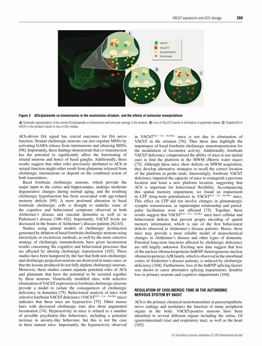
Genetic approaches have been used to investigate thephysiological significance of ACh/glutamate co-transmission inthe striatum (Figure 3). Studies using mice with null expressionof VGLUT3 (VGLUT3-KO) show evidence that VGLUT3 is co-expressed with VAChT in synaptic vesicles and facilitates AChfilling of these vesicles [31]. VGLUT3-KO mice are hyperactive,more responsive to cocaine and less prone to haloperidol-induced catalepsy than their wild-type littermates [31]. Giventhat mice with ablated cholinergic neurons in the striatum showa similar phenotype [94,95], it was initially suggested that thesebehavioural changes resulted from the decreased striatal AChrelease observed in VGLUT3-KO mice [31]. Recently it wasdemonstrated that mice with selective elimination of VAChTfrom striatal cholinergic interneurons (VAChTD2 − Cre − flox/flox mice)are not hyperactive and show minimal alteration in behaviouralresponses to cocaine. These results strongly suggest that glutamatereleased from cholinergic neurons, rather than ACh, is criticalfor cocaine-induced behavioural manifestations [76]. Conversely,it was shown that elimination of striatal ACh release affectsDA metabolism. It also appeared to affect the response ofMSNs (medium spiny neurons) to DA where up-regulationof DA receptors and a change in behavioural responses todopaminergic agonists was observed [76]. These data indicatethat cholinergic interneurons use two distinct neurotransmittersto differentially regulate behaviour. Moreover, synchronizedactivity of cholinergic interneurons was shown to depolarize DAnerve terminals directly and evoke DA release, independentlyof the action potentials in DA soma [96,97], indicating that the
c© The Authors Journal compilation c© 2013 Biochemical Society
VAChT expression and ACh storage 269
Figure 3 ACh/glutamate co-transmission in the mammalian striatum, and the effects of molecular manipulations
(A) Schematic representation of the normal ACh/glutamate co-transmission and vesicular synergy in the striatum. (B) Loss of VGLUT3 results in elimination of glutamate release. (C) Targeted KO ofVAChT in the striatum results in loss of ACh release.
ACh-driven DA signal has crucial outcomes for DA nervefunction. Striatal cholinergic neurons can also regulate MSNs byactivating GABA release from interneurons and silencing MSNs[98]. Importantly, these findings demonstrate that co-transmissionhas the potential to significantly affect the functioning ofstriatal neurons and hence of basal ganglia. Additionally, theseresults suggest that other roles previously attributed to ACh instriatal function might either result from glutamate released fromcholinergic interneurons or depend on the combined action ofboth transmitters.
Basal forebrain cholinergic neurons, which provide themajor input to the cortex and hippocampus, undergo moderatedegenerative changes during normal aging, and the resultingcholinergic hypofunction has been associated with age-relatedmemory deficits [99]. A more profound alteration in basalforebrain cholinergic cells is thought to underlie some ofthe cognitive and behavioural symptoms observed in bothAlzheimer’s disease and vascular dementia as well as inParkinson’s disease [100–102]. Importantly, VAChT levels aredecreased in the brains of Alzheimer’s disease patients [62,63].
Studies using animal models of cholinergic dysfunctiongenerated by ablation of basal forebrain cholinergic neurons usingelectrolytic or excitotoxic methods, as well by the more selectivestrategy of cholinergic immunolesion, have given inconsistentresults concerning the cognitive and behavioural processes thatare affected by altering cholinergic transmission [103]. Thesestudies have been hampered by the fact that both non-cholinergicand cholinergic projection neurons are destroyed in many cases, orthat the lesions produced do not fully deplete cholinergic neurons.Moreover, these studies cannot separate potential roles of AChand glutamate that have the potential to be secreted togetherby these neurons. Genetically modified mice with selectiveelimination of VAChT expression in forebrain cholinergic neuronsprovide a model to isolate the consequences of cholinergicdeficiency in dementia [75]. Behavioural analysis of mice withselective forebrain VAChT deficiency (VAChTSix3 − Cre − flox/flox mice)indicates that these mice are hyperactive [75]. Other mouselines with decreased cholinergic tone also show augmentedlocomotion [74]. Hyperactivity in mice is related to a numberof possible psychiatric-like behaviours, including a potentialincrease in anxiety-like behaviour, but this is not the casein these mutant mice. Importantly, the hyperactivity observed
in VAChTSix3 − Cre − flox/flox mice is not due to elimination ofVAChT in the striatum [76]. Thus these data highlight theimportance of basal forebrain cholinergic neurotransmission forthe modulation of locomotor activity. Additionally, forebrainVAChT deficiency compromised the ability of mice to use spatialcues to find the platform in the MWM (Morris water maze)[75]. Although these mice show deficits on MWM acquisition,they develop alternative strategies to recall the correct locationof the platform in probe trials. Interestingly, forebrain VAChTdeficiency impaired the capacity of mice to extinguish a previouslocation and learn a new platform location, suggesting thatACh is important for behavioural flexibility. Accompanyingthis spatial memory impairment, we found an impairmentin LTP (long-term potentiation) in VAChTSix3 − Cre − flox/flox mice.This effect on LTP did not involve changes in glutamatergicsynaptic transmission, as input/output relationship and paired-pulse facilitation were not affected [75]. Together, theseresults suggest that VAChTSix3 − Cre − flox/flox mice have cellular andbehavioural deficits that prevent proper encoding of spatialmemory information, which is one of the first behaviouraldeficits observed in Alzheimer’s disease patients. Hence, thesemice may provide a more reliable model of neurochemicalchanges in Alzheimer’s disease and other types of dementia.Potential long-term functions affected by cholinergic deficiencyare still largely unknown. Exciting new data suggest that lossof the nuclear ribonucleoproteins hnRNP (heterogeneous nuclearribonucleoprotein) A/B family, which is observed in the entorhinalcortex of Alzheimer’s disease patients, is induced by cholinergicdeficiency [104]. Furthermore, loss of the hnRNP splicing factorswas shown to cause alternative splicing impairments, dendriteloss in primary neurons and cognitive impairments [104].
REGULATION OF CHOLINERGIC TONE IN THE AUTONOMICNERVOUS SYSTEM BY VAChT
ACh is the primary chemical neurotransmitter at parasympatheticnerve endings and modulates the function of many peripheralorgans in the body. VAChT-positive neurons have beenidentified in several different organs including the retina, GI(gastrointestinal) tract and respiratory tract, as well as the heart[105].
c© The Authors Journal compilation c© 2013 Biochemical Society

270 V. F. Prado and others
In the heart, co-ordinated interplay between the two branchesof the ANS (autonomic nervous system) is important inmaintaining proper function. Activation of the sympathetic branchincreases heart rate and contractile force, whereas activation ofthe parasympathetic branch reduces heart rate by altering theconduction velocity of both the sinoatrial and atrioventricularnodes [106]. It has long been recognized that overactivationof the sympathetic tone contributes to cardiac dysfunction[107–109]; in contrast, much less is known about the roleof failing cholinergic neurotransmission in cardiac disease.Vagal stimulation has been shown to improve the outcomesin experimental heart failure [110–118] and it is an approachcurrently being explored to ameliorate a number of diseasesin humans. Recent studies using VAChT-KDHOM mice, whichshow a systemic reduction in VAChT, provided direct evidencethat decreased cholinergic neurotransmission also causes plasticalterations that contribute to heart dysfunction [73,119]. Thehearts of VAChT-KDHOM mice have altered calcium handling,show changes in myocyte contractility and express severalmarkers of cardiac stress, which are activated during cardiacremodelling and heart failure [73]. Importantly, all of thesechanges can be reversed through treatment with pyridostigmine,a peripheral cholinesterase inhibitor, suggesting that cardiacdysfunction in these mice results from a deficiency in cholinergictone [73]. Mice lacking M2 muscarinic receptors show increasedcardiac stress [120], and experiments using mice lacking oneof the high-affinity choline transporter alleles confirmed thatdecreased cholinergic tone affects heart function [121].
The cholinergic system is important in regulating the innateimmune response. In fact, several studies have provided novelinsight into the specific mechanisms through which neuronalACh can act peripherally to control the immune response. Thisinflammatory reflex, termed the cholinergic anti-inflammatorypathway, is dependent on the peripheral actions of ACh releasedfrom the vagus nerve [110,122,123]. ACh binds to α7 nAChRs(nicotinic acetylcholine receptors) on macrophages and therebyinhibits the release of pro-inflammatory cytokines includingTNFα (tumour necrosis factor α), IL (interleukin)-1β and IL-6 [122,123]. Inhibition of cytokine release has been shown tobe beneficial in several disease states, including endotoxaemia,sepsis and heart failure [124–127]. Consistent with these results,VAChT-KDHOM mice have been shown to develop an increasedinflammatory immune response when infected with parasites[128], indicating disturbance of the cholinergic anti-inflammatoryreflex in this mutant.
In the retina, ACh is released in response to light stimulationand leads to direct and rapid excitation of the retinal ganglioncells [129]. Release of ACh in the enteric nervous systeminduces smooth muscle contractions in the GI tract [130,131].Cholinergic signalling also leads to smooth muscle contractionin the respiratory tract [132]. Whether decreased cholinergic tonehas pathological implications related to eye function, the entericsystem or the respiratory tract has not yet been systematicallyinvestigated.
NON-NEURONAL CHOLINERGIC MACHINERY
ACh is also produced in many different cell types, leadingto the idea that a non-neuronal cholinergic system plays asignificant role in regulating various physiological functions[133]. Indeed, the machinery necessary to produce ACh as wellas the neurotransmitter itself has been identified in a range ofcells including lymphocytes [134,135], epithelial cells [136–138], vascular endothelial cells [139] and the α-cells of the
pancreas [6]. Furthermore, this machinery has been identifiedin cardiomyocytes [5,140,141].
Immune cells, including lymphocytes, possess the machinerynecessary to synthesize ACh [134,135,142,143]. It has beenrecently demonstrated that a small population of T-lymphocytescan synthesize and release ACh in response to autonomic nervoussystem activity in the spleen as part of the cholinergic anti-inflammatory pathway [144]. It is important to note that T-cellsas well as macrophages express both muscarinic and nicotinicACh receptors and are also able to produce ACh [145]. The exactmechanism by which ACh is released from these cells to regulatethe immune system has not yet been uncovered.
The ability of epithelial cells to synthesize ACh is wellcharacterized; in fact, it has previously been reported that thismolecule can be secreted from cultured bronchial epithelial cells[137]. However, ACh release from epithelial cells does notappear to be dependent on VAChT, but rather on the organiccation transporters OCT (organic cation transporter) 1 and OCT2[146,147]. ACh released by epithelial cells acts through bothnicotinic as well as M1 muscarinic receptors and increases theirproliferation rate [148]. Moreover, in a number of cancer cells,particularly in lung cancer, ACh has been shown to play anautocrine role [149–151].
ACh is important in regulating insulin release from β-cellsof the pancreas. It has long been suggested that the source ofthis ACh is the parasympathetic nerve endings in the endocrinepancreas, which can trigger insulin release following bindingto the M3 muscarinic receptor [152,153]. Interestingly, recentwork has revealed that, although insulin secretion from β-cellsis regulated by neuronal parasympathetic signalling in mice,humans possess the machinery to synthesize and release AChfrom α-cells. VAChT plays an important role in the release ofACh from α-cells as positive immunostaining was observed forthe transporter in these cells and pharmacological manipulationblocked ACh release. This suggests that ACh is maintained inexocytotic vesicles within α-cells and is released quantally in amanner similar to that observed in neurons [6]. Owing to the factthat β-cells are mostly localized close to α-cells in the pancreaticislets, the non-neuronal ACh released by α-cells can act as aparacrine molecule on neighbouring β-cells and initiate the insulinsecretion cascade [6].
Vascular endothelial cells play a critical role in the regulationof blood pressure by inducing the relaxation of vascular smoothmuscle cells. This pathway is well characterized and ACh isknown to mediate vasodilation through binding to muscarinicACh receptors on endothelial cells. This interaction leads to theproduction of the EDRF (endothelium-derived relaxing factor),now known to be nitric oxide, which leads to vasodilation [154].Although ACh appears to have a profound effect on vascularfunction, it is important to note that parasympathetic innervationof endothelial cells is virtually non-existent and there are highlevels of acetylcholinesterases in circulation. It is now evidentthat endothelial cells have the ability to synthesize and secreteACh via a PKC-independent mechanism [139,155]. Importantly,previous work has confirmed the presence of VAChT in twodifferent endothelial cell culture models, suggesting that ACh maybe released from these cells via a VAChT-dependent mechanism[156,157].
It has previously been proposed that cardiomyocytes areable to synthesize and release ACh at the cellular level asthey possess the machinery for production of ACh [140,141].Owing to limited innervation of ventricular cardiomyocytesby parasympathetic neurons [158,159], it has been suggestedthat ventricular cardiomyocytes may synthesize ACh. VAChTwas shown to be present in vesicles in cardiomyocytes [141]
c© The Authors Journal compilation c© 2013 Biochemical Society

VAChT expression and ACh storage 271
suggesting a quantal release of ACh from these cells [5]. Incell culture models, isoproterenol and other adrenergic activatorscan induce cellular hypertrophy and remodelling. ACh releasedby cardiomyocytes plays an important role in protecting thesecells against isoproterenol-induced cardiomyocyte remodelling[5]. Furthermore, expression of both VAChT and ChAT wasincreased in cultured cardiomyocytes treated with adrenergicdrugs [5], suggesting a potential mechanism for regulationof this machinery. This non-neuronal ACh may then act inan autocrine/paracrine fashion to enhance neuronal cholinergicsignalling. Although demonstrated only in vitro, these resultssuggest a novel mechanism to protect the heart under stressfulconditions. These data provide for an unanticipated mechanism bywhich non-neuronal ACh, secreted from VAChT-positive vesicles,may play an important role in cardiac function.
CONCLUSION
The potential to uncover novel physiological functions of AChusing genetically modified mice, in which the cholinergicmachinery can be spatially and temporally targeted, has alreadychanged our understanding of functions by this neurotransmitter.Otto Loewi’s [161] findings of ACh as a neurotransmitter cannow be expanded to a role in paracrine/autocrine communicationin a number of non-neuronal cells. By using the Cre/lox system,BAC (bacterial artificial chromosomes) transgenic mice andoptogenetics, we now have the ability to activate or inactivatethe cholinergic machinery or cholinergic neurons at will. Thesenovel approaches will lead to increased knowledge of howACh contributes to different bodily functions. Additionally,these approaches will be fundamental to unravelling howdifferent populations of cholinergic neurons in the brain canregulate distinct biochemical and physiological processes. Itis tempting to speculate that optogenetics, used to activateor inactivate cholinergic neurons in the peripheral nervoussystem, may be effectively used in the future as treatment fora number of pathologies involving dysregulated sympatheticor parasympathetic activity. Finally, inactivation of cholinergicmachinery in non-neuronal tissues using genetically modifiedmice will provide the ultimate proof for the physiologicalsignificance of non-neuronal release of ACh.
REFERENCES
1 El Mestikawy, S., Wallen-Mackenzie, A., Fortin, G. M., Descarries, L. and Trudeau, L. E.(2011) From glutamate co-release to vesicular synergy: vesicular glutamatetransporters. Nat. Rev. Neurosci. 12, 204–216
2 Wessler, I. and Kirkpatrick, C. J. (2008) Acetylcholine beyond neurons: the non-neuronalcholinergic system in humans. Br. J. Pharmacol. 154, 1558–1571
3 Wessler, I. K. and Kirkpatrick, C. J. (2012) Activation of muscarinic receptors bynon-neuronal acetylcholine. Handb. Exp. Pharmacol. 208, 469–491
4 Prado, M. A., Reis, R. A., Prado, V. F., de Mello, M. C., Gomez, M. V. and de Mello, F. G.(2002) Regulation of acetylcholine synthesis and storage. Neurochem. Int. 41, 291–299
5 Rocha-Resende, C., Roy, A., Resende, R., Ladeira, M. S., Lara, A., de Morais Gomes,E. R., Prado, V. F., Gros, R., Guatimosim, C., Prado, M. A. and Guatimosim, S. (2012)Non-neuronal cholinergic machinery present in cardiomyocytes offsets hypertrophicsignals. J. Mol. Cell. Cardiol. 53, 206–216
6 Rodriguez-Diaz, R., Dando, R., Jacques-Silva, M. C., Fachado, A., Molina, J.,Abdulreda, M. H., Ricordi, C., Roper, S. D., Berggren, P. O. and Caicedo, A. (2011) Alphacells secrete acetylcholine as a non-neuronal paracrine signal priming beta cell functionin humans. Nat. Med. 17, 888–892
7 Ribeiro, F. M., Black, S. A., Prado, V. F., Rylett, R. J., Ferguson, S. S. and Prado, M. A.(2006) The ‘ins’ and ‘outs’ of the high-affinity choline transporter CHT1. J. Neurochem.97, 1–12
8 Parsons, S. M. (2000) Transport mechanisms in acetylcholine and monoamine storage.FASEB J. 14, 2423–2434
9 Vardy, E., Arkin, I. T., Gottschalk, K. E., Kaback, H. R. and Schuldiner, S. (2004)Structural conservation in the major facilitator superfamily as revealed by comparativemodeling. Protein Sci. 13, 1832–1840
10 Varoqui, H. and Erickson, J. D. (1996) Active transport of acetylcholine by the humanvesicular acetylcholine transporter. J. Biol. Chem. 271, 27229–27232
11 Hori, T. and Takahashi, T. (2012) Kinetics of synaptic vesicle refilling withneurotransmitter glutamate. Neuron 76, 511–517
12 Wess, J. (2012) Novel muscarinic receptor mutant mouse models. Handb. Exp.Pharmacol. 208, 95–117
13 Wess, J., Eglen, R. M. and Gautam, D. (2007) Muscarinic acetylcholine receptors: mutantmice provide new insights for drug development. Nat. Rev. Drug Discovery 6, 721–733
14 Wess, J. (2004) Muscarinic acetylcholine receptor knockout mice: novel phenotypes andclinical implications. Annu. Rev. Pharmacol. Toxicol. 44, 423–450
15 Bymaster, F. P., McKinzie, D. L., Felder, C. C. and Wess, J. (2003) Use of M1–M5
muscarinic receptor knockout mice as novel tools to delineate the physiological roles ofthe muscarinic cholinergic system. Neurochem. Res. 28, 437–442
16 Changeux, J. P. (2010) Nicotine addiction and nicotinic receptors: lessons fromgenetically modified mice. Nat. Rev. Neurosci. 11, 389–401
17 Champtiaux, N. and Changeux, J. P. (2004) Knockout and knockin mice to investigate therole of nicotinic receptors in the central nervous system. Prog. Brain Res. 145, 235–251
18 Cordero-Erausquin, M., Marubio, L. M., Klink, R. and Changeux, J. P. (2000) Nicotinicreceptor function: new perspectives from knockout mice. Trends Pharmacol. Sci. 21,211–217
19 Khare, P., Ojeda, A. M., Chandrasekaran, A. and Parsons, S. M. (2010) Possibleimportant pair of acidic residues in vesicular acetylcholine transporter. Biochemistry 49,3049–3059
20 Nguyen, M. L., Cox, G. D. and Parsons, S. M. (1998) Kinetic parameters for thevesicular acetylcholine transporter: two protons are exchanged for one acetylcholine.Biochemistry 37, 13400–13410
21 Ojeda, A. M., Kolmakova, N. G. and Parsons, S. M. (2004) Acetylcholine binding site inthe vesicular acetylcholine transporter. Biochemistry 43, 11163–11174
22 Kim, M. H., Lu, M., Lim, E. J., Chai, Y. G. and Hersh, L. B. (1999) Mutational analysis ofaspartate residues in the transmembrane regions and cytoplasmic loops of rat vesicularacetylcholine transporter. J. Biol. Chem. 274, 673–680
23 Bravo, D. T., Kolmakova, N. G. and Parsons, S. M. (2005) Mutational and pH analysis ofionic residues in transmembrane domains of vesicular acetylcholine transporter.Biochemistry 44, 7955–7966
24 Van der Kloot, W. (2003) Loading and recycling of synaptic vesicles in the Torpedoelectric organ and the vertebrate neuromuscular junction. Prog. Neurobiol. 71, 269–303
25 Song, H., Ming, G., Fon, E., Bellocchio, E., Edwards, R. H. and Poo, M. (1997)Expression of a putative vesicular acetylcholine transporter facilitates quantal transmitterpackaging. Neuron 18, 815–826
26 Prado, V. F., Martins-Silva, C., de Castro, B. M., Lima, R. F., Barros, D. M., Amaral, E.,Ramsey, A. J., Sotnikova, T. D., Ramirez, M. R., Kim, H. G. et al. (2006) Mice deficientfor the vesicular acetylcholine transporter are myasthenic and have deficits in object andsocial recognition. Neuron 51, 601–612
27 Lima, R. F., Prado, V. F., Prado, M. A. and Kushmerick, C. (2010) Quantal release ofacetylcholine in mice with reduced levels of the vesicular acetylcholine transporter.J. Neurochem. 113, 943–951
28 Prior, C., Marshall, I. G. and Parsons, S. M. (1992) The pharmacology of vesamicol: aninhibitor of the vesicular acetylcholine transporter. Gen. Pharmacol. 23, 1017–1022
29 Tu, Z., Wang, W., Cui, J., Zhang, X., Lu, X., Xu, J. and Parsons, S. M. (2012) Synthesisand evaluation of in vitro bioactivity for vesicular acetylcholine transporter inhibitorscontaining two carbonyl groups. Bioorg. Med. Chem. 20, 4422–4429
30 Efange, S. M., Khare, A. B., von, H. K., Mach, R. H., Parsons, S. M. and Tu, Z. (2010)Synthesis and in vitro biological evaluation of carbonyl group-containing inhibitors ofvesicular acetylcholine transporter. J. Med. Chem. 53, 2825–2835
31 Gras, C., Amilhon, B., Lepicard, E. M., Poirel, O., Vinatier, J., Herbin, M., Dumas, S.,Tzavara, E. T., Wade, M. R., Nomikos, G. G. et al. (2008) The vesicular glutamatetransporter VGLUT3 synergizes striatal acetylcholine tone. Nat. Neurosci. 11, 292–300
32 Hnasko, T. S. and Edwards, R. H. (2012) Neurotransmitter corelease: mechanism andphysiological role. Annu. Rev. Physiol. 74, 225–243
33 Trudeau, L. E. and Gutierrez, R. (2007) On cotransmission and neurotransmitterphenotype plasticity. Mol. Interv. 7, 138–146
34 Mendez, J. A., Bourque, M. J., Dal Bo, G., Bourdeau, M. L., Danik, M., Williams, S.,Lacaille, J. C. and Trudeau, L. E. (2008) Developmental and target-dependent regulationof vesicular glutamate transporter expression by dopamine neurons. J. Neurosci. 28,6309–6318
35 Tritsch, N. X., Ding, J. B. and Sabatini, B. L. (2012) Dopaminergic neurons inhibitstriatal output through non-canonical release of GABA. Nature 490, 262–266
36 Bravo, D. T., Kolmakova, N. G. and Parsons, S. M. (2004) Choline is transported byvesicular acetylcholine transporter. J. Neurochem. 91, 766–768
c© The Authors Journal compilation c© 2013 Biochemical Society

272 V. F. Prado and others
37 Bravo, D. T., Kolmakova, N. G. and Parsons, S. M. (2005) New transport assaydemonstrates vesicular acetylcholine transporter has many alternative substrates.Neurochem. Int. 47, 243–247
38 Khare, P., Mulakaluri, A. and Parsons, S. M. (2010) Search for the acetylcholine andvesamicol binding sites in vesicular acetylcholine transporter: the region around thelumenal end of the transport channel. J. Neurochem. 115, 984–993
39 Barbosa, Jr, J., Clarizia, A. D., Gomez, M. V., RomanoSilva, M. A., Prado, V. F. andPrado, M. A. M. (1997) Effect of protein kinase C activation on the release of[3H]Acetylcholine in the presence of vesamicol. J. Neurochem. 69, 2608–2611
40 Clarizia, A. D., Gomez, M. V., Romano-Silva, M. A., Parsons, S. M., Prado, V. F. andPrado, M. A. (1999) Control of the binding of a vesamicol analog to the vesicularacetylcholine transporter. NeuroReport 10, 2783–2787
41 Clarizia, A. D., Romano-Silva, M. A., Prado, V. F., Gomez, M. V. and Prado, M. A. M.(1998) Role of protein kinase C in the release of [3H]acetylcholine from myenteric plexustreated with vesamicol. Neurosci. Lett. 244, 115–117
42 Efange, S. M., von Hohenberg, K., Khare, A. B., Tu, Z., Mach, R. H. and Parsons, S. M.(2000) Synthesis and biological characterization of stable and radioiodinated(+−)-trans-2-hydroxy-3-{4-(3-iodophenyl)piperidyl}-1,2,3,4-tetrahydronaphthalene(3′-IBVM). Nucl. Med. Biol. 27, 749–755
43 Tu, Z., Efange, S. M., Xu, J., Li, S., Jones, L. A., Parsons, S. M. and Mach, R. H. (2009)Synthesis and in vitro and in vivo evaluation of 18F-labeled positron emissiontomography (PET) ligands for imaging the vesicular acetylcholine transporter. J. Med.Chem. 52, 1358–1369
44 Kozaka, T., Uno, I., Kitamura, Y., Miwa, D., Ogawa, K. and Shiba, K. (2012) Synthesesand in vitro evaluation of decalinvesamicol analogues as potential imaging probes forvesicular acetylcholine transporter (VAChT). Bioorg. Med. Chem. 20, 4936–4941
45 Grygoruk, A., Fei, H., Daniels, R. W., Miller, B. R., Chen, A., DiAntonio, A. and Krantz,D. E. (2010) Vesicular neurotransmitter transporter trafficking in vivo: moving from cellsto flies. Fly 4, 302–305
46 Prado, V. F. and Prado, M. A. (2002) Signals involved in targeting membrane proteins tosynaptic vesicles. Cell. Mol. Neurobiol. 22, 565–577
47 Gilmor, M. L., Nash, N. R., Roghani, A., Edwards, R. H., Yi, H., Hersch, S. M. and Levey,A. I. (1996) Expression of the putative vesicular acetylcholine transporter in rat brain andlocalization in cholinergic synaptic vesicles. J. Neurosci. 16, 2179–2190
48 Liu, Y. and Edwards, R. H. (1997) The role of vesicular transport proteins in synaptictransmission and neural degeneration. Annu. Rev. Neurosci. 20, 125–156
49 Krantz, D. E., Waites, C., Oorschot, V., Liu, Y., Wilson, R. I., Tan, P. K., Klumperman, J.and Edwards, R. H. (2000) A phosphorylation site regulates sorting of the vesicularacetylcholine transporter to dense core vesicles. J. Cell Biol. 149, 379–396
50 Varoqui, H. and Erickson, J. D. (1998) The cytoplasmic tail of the vesicular acetylcholinetransporter contains a synaptic vesicle targeting signal. J. Biol. Chem. 273, 9094–9098
51 Tan, P. K., Waites, C., Liu, Y., Krantz, D. E. and Edwards, R. H. (1998) A leucine-basedmotif mediates the endocytosis of vesicular monoamine and acetylcholine transporters.J. Biol. Chem. 273, 17351–17360
52 Barbosa, Jr, J., Ferreira, L. T., Martins-Silva, C., Santos, M. S., Torres, G. E., Caron,M. G., Gomez, M. V., Ferguson, S. S., Prado, M. A. and Prado, V. F. (2002) Trafficking ofthe vesicular acetylcholine transporter in SN56 cells: a dynamin-sensitive step andinteraction with the AP-2 adaptor complex. J. Neurochem. 82, 1221–1228
53 Ferreira, L. T., Santos, M. S., Kolmakova, N. G., Koenen, J., Barbosa, Jr, J., Gomez,M. V., Guatimosim, C., Zhang, X., Parsons, S. M., Prado, V. F. and Prado, M. A. (2005)Structural requirements for steady-state localization of the vesicular acetylcholinetransporter. J. Neurochem. 94, 957–969
54 Santos, M. S., Barbosa, J., Kushmerick, C., Gomez, M. V., Ferguson, S. S. G., Prado, V.F. and Prado, M. A. M. (2001) Visualization and trafficking of the vesicular acetylcholinetransporter in living cholinergic cells. J. Neurochem. 78, 37–37
55 Colgan, L., Liu, H., Huang, S. Y. and Liu, Y. J. (2007) Dileucine motif is sufficient forinternalization and synaptic vesicle targeting of vesicular acetylcholine transporter.Traffic 8, 512–522
56 Ribeiro, F. M., Alves-Silva, J., Volknandt, W., Martins-Silva, C., Mahmud, H., Wilhelm,A., Gomez, M. V., Rylett, R. J., Ferguson, S. S., Prado, V. F. and Prado, M. A. (2003) Thehemicholinium-3 sensitive high affinity choline transporter is internalized byclathrin-mediated endocytosis and is present in endosomes and synaptic vesicles.J. Neurochem. 87, 136–146
57 Ribeiro, F. M., Black, S. A., Cregan, S. P., Prado, V. F., Prado, M. A., Rylett, R. J. andFerguson, S. S. (2005) Constitutive high-affinity choline transporter endocytosis isdetermined by a carboxyl-terminal tail dileucine motif. J. Neurochem. 94, 86–96
58 Ribeiro, F. M., Pinthong, M., Black, S. A., Gordon, A. C., Prado, V. F., Prado, M. A.,Rylett, R. J. and Ferguson, S. S. (2007) Regulated recycling and plasma membranerecruitment of the high-affinity choline transporter. Eur. J. Neurosci. 26, 3437–3448
59 Sandoval, G. M., Duerr, J. S., Hodgkin, J., Rand, J. B. and Ruvkun, G. (2006) A geneticinteraction between the vesicular acetylcholine transporter VAChT/UNC-17 andsynaptobrevin/SNB-1 in C. elegans. Nat. Neurosci. 9, 599–601
60 Ribeiro, F. M., Ferreira, L. T., Marion, S., Fontes, S., Gomez, M., Ferguson, S. S., Prado,M. A. and Prado, V. F. (2007) SEC14-like protein 1 interacts with cholinergictransporters. Neurochem. Int. 50, 356–364
61 Terry, Jr, A., Gearhart, D. A., Warner, S. E., Zhang, G., Bartlett, M. G., Middlemore,M. L., Beck, Jr, W. D., Mahadik, S. P. and Waller, J. L. (2007) Oral haloperidol orrisperidone treatment in rats: temporal effects on nerve growth factor receptors,cholinergic neurons, and memory performance. Neuroscience 146, 1316–1332
62 Chen, K. H., Reese, E. A., Kim, H. W., Rapoport, S. I. and Rao, J. S. (2011) Disturbedneurotransmitter transporter expression in Alzheimer’s disease brain. J. Alzheimers Dis.26, 755–766
63 Efange, S. M., Garland, E. M., Staley, J. K., Khare, A. B. and Mash, D. C. (1997)Vesicular acetylcholine transporter density and Alzheimer’s disease. Neurobiol. Aging18, 407–413
64 Semmler, A., Frisch, C., Debeir, T., Ramanathan, M., Okulla, T., Klockgether, T. andHeneka, M. T. (2007) Long-term cognitive impairment, neuronal loss and reducedcortical cholinergic innervation after recovery from sepsis in a rodent model. Exp.Neurol. 204, 733–740
65 Li, D. J., Evans, R. G., Yang, Z. W., Song, S. W., Wang, P., Ma, X. J., Liu, C., Xi, T., Su,D. F. and Shen, F. M. (2011) Dysfunction of the cholinergic anti-inflammatory pathwaymediates organ damage in hypertension. Hypertension 57, 298–307
66 Smith, R., Chung, H., Rundquist, S., Maat-Schieman, M. L., Colgan, L., Englund, E.,Liu, Y. J., Roos, R. A., Faull, R. L., Brundin, P. and Li, J. Y. (2006) Cholinergic neuronaldefect without cell loss in Huntington’s disease. Hum. Mol. Genet. 15, 3119–3131
67 Edwards, R. H. (2007) The neurotransmitter cycle and quantal size. Neuron 55, 835–85868 Contant, C., Umbriaco, D., Garcia, S., Watkins, K. C. and Descarries, L. (1996)
Ultrastructural characterization of the acetylcholine innervation in adult rat neostriatum.Neuroscience 71, 937–947
69 Duffy, A. M., Zhou, P., Milner, T. A. and Pickel, V. M. (2009) Spatial and intracellularrelationships between the alpha7 nicotinic acetylcholine receptor and the vesicularacetylcholine transporter in the prefrontal cortex of rat and mouse. Neuroscience 161,1091–1103
70 Nagy, P. M. and Aubert, I. (2012) Overexpression of the vesicular acetylcholinetransporter increased acetylcholine release in the hippocampus. Neuroscience 218,1–11
71 de Castro, B. M., De Jaeger, X., Martins-Silva, C., Lima, R. F., Amaral, E., Menezes, C.,Lima, P., Neves, C. M. L., Pires, R. G., Gould, T. W. et al. (2009) The vesicularacetylcholine transporter is required for neuromuscular development and function. Mol.Cell. Biol. 29, 5238–5250
72 de Castro, B. M., Pereira, G. S., Magalhaes, V., Rossato, J. I., De Jaeger, X.,Martins-Silva, C., Leles, B., Lima, P., Gomez, M. V., Gainetdinov, R. R. et al. (2009)Reduced expression of the vesicular acetylcholine transporter causes learning deficits inmice. Genes Brain Behav. 8, 23–35
73 Lara, A., Damasceno, D. D., Pires, R., Gros, R., Gomes, E. R., Gavioli, M., Lima, R. F.,Guimaraes, D., Lima, P., Bueno, Jr, C. R. et al. (2010) Dysautonomia due to reducedcholinergic neurotransmission causes cardiac remodeling and heart failure. Mol. Cell.Biol. 30, 1746–1756
74 Martins-Silva, C., De Jaeger, X., Guzman, M. S., Lima, R. D., Santos, M. S., Kushmerick,C., Gomez, M. V., Caron, M. G., Prado, M. A. and Prado, V. F. (2011) Novel strains ofmice deficient for the vesicular acetylcholine transporter: insights on transcriptionalregulation and control of locomotor behavior. PLoS ONE 6, e17611
75 Martyn, A. C., De Jaeger, X., Magalhaes, A. C., Kesarwani, R., Goncalves, D. F., Raulic,S., Guzman, M. S., Jackson, M. F., Izquierdo, I., Macdonald, J. F. et al. (2012)Elimination of the vesicular acetylcholine transporter in the forebrain causeshyperactivity and deficits in spatial memory and long-term potentiation. Proc. Natl.Acad. Sci. U.S.A. 109, 17651–17656
76 Guzman, M. S., De Jaeger, X., Raulic, S., Souza, I. A., Li, A. X., Schmid, S., Menon, R.S., Gainetdinov, R. R., Caron, M. G., Bartha, R. et al. (2011) Elimination of the vesicularacetylcholine transporter in the striatum reveals regulation of behaviour bycholinergic-glutamatergic co-transmission. PLoS Biol. 9, e1001194
77 Liu, Y., Peter, D., Roghani, A., Schuldiner, S., Prive, G. G., Eisenberg, D., Brecha, N. andEdwards, R. H. (1992) A cDNA that suppresses MPP+ toxicity encodes a vesicularamine transporter. Cell 70, 539–551
78 Misgeld, T., Burgess, R. W., Lewis, R. M., Cunningham, J. M., Lichtman, J. W. andSanes, J. R. (2002) Roles of neurotransmitter in synapse formation: development ofneuromuscular junctions lacking choline acetyltransferase. Neuron 36, 635–648
79 Brandon, E. P., Lin, W., D’Amour, K. A., Pizzo, D. P., Dominguez, B., Sugiura, Y., Thode,S., Ko, C. P., Thal, L. J., Gage, F. H. and Lee, K. F. (2003) Aberrant patterning ofneuromuscular synapses in choline acetyltransferase-deficient mice. J. Neurosci. 23,539–549
80 De Jaeger, X., Cammarota, M., Prado, M. A., Izquierdo, I., Prado, V. F. and Pereira, G. S.(2012) Decreased acetylcholine release delays the consolidation of object recognitionmemory. Behav. Brain Res. 238C, 62–68
c© The Authors Journal compilation c© 2013 Biochemical Society

VAChT expression and ACh storage 273
81 Takamori, S., Holt, M., Stenius, K., Lemke, E. A., Gronborg, M., Riedel, D., Urlaub, H.,Schenck, S., Brugger, B., Ringler, P. et al. (2006) Molecular anatomy of a traffickingorganelle. Cell 127, 831–846
82 Hasselmo, M. E. and Sarter, M. (2011) Modes and models of forebrain cholinergicneuromodulation of cognition. Neuropsychopharmacology 36, 52–73
83 Sarter, M. and Paolone, G. (2011) Deficits in attentional control: cholinergicmechanisms and circuitry-based treatment approaches. Behav. Neurosci. 125, 825–835
84 Howe, W. M., Ji, J., Parikh, V., Williams, S., Mocaer, E., Trocme-Thibierge, C. and Sarter,M. (2010) Enhancement of attentional performance by selective stimulation ofalpha4beta2(*) nAChRs: underlying cholinergic mechanisms.Neuropsychopharmacology 35, 1391–1401
85 Parikh, V. and Sarter, M. (2008) Cholinergic mediation of attention: contributions ofphasic and tonic increases in prefrontal cholinergic activity. Ann. N.Y. Acad. Sci. 1129,225–235
86 Hasselmo, M. E. and Giocomo, L. M. (2006) Cholinergic modulation of corticalfunction. J. Mol. Neurosci. 30, 133–135
87 Hasselmo, M. E. and Stern, C. E. (2006) Mechanisms underlying working memory fornovel information. Trends Cogn. Sci. 10, 487–493
88 Hasselmo, M. E. (2006) The role of acetylcholine in learning and memory. Curr. Opin.Neurobiol. 16, 710–715
89 Ren, J., Qin, C., Hu, F., Tan, J., Qiu, L., Zhao, S., Feng, G. and Luo, M. (2011) Habenula‘cholinergic’ neurons corelease glutamate and acetylcholine and activate postsynapticneurons via distinct transmission modes. Neuron 69, 445–452
90 Nickerson Poulin, A., Guerci, A., El Mestikawy, S. and Semba, K. (2006) Vesicularglutamate transporter 3 immunoreactivity is present in cholinergic basal forebrainneurons projecting to the basolateral amygdala in rat. J. Comp. Neurol. 498, 690–711
91 Schafer, M. K., Varoqui, H., Defamie, N., Weihe, E. and Erickson, J. D. (2002) Molecularcloning and functional identification of mouse vesicular glutamate transporter 3 and itsexpression in subsets of novel excitatory neurons. J. Biol. Chem. 277, 50734–50748
92 Lee, S., Kim, K. and Zhou, Z. J. (2010) Role of ACh-GABA cotransmission in detectingimage motion and motion direction. Neuron 68, 1159–1172
93 Higley, M. J., Gittis, A. H., Oldenburg, I. A., Balthasar, N., Seal, R. P., Edwards, R. H.,Lowell, B. B., Kreitzer, A. C. and Sabatini, B. L. (2011) Cholinergic interneurons mediatefast VGluT3-dependent glutamatergic transmission in the striatum. PLoS ONE 6, e19155
94 Hikida, T., Kaneko, S., Isobe, T., Kitabatake, Y., Watanabe, D., Pastan, I. and Nakanishi, S.(2001) Increased sensitivity to cocaine by cholinergic cell ablation in nucleusaccumbens. Proc. Natl. Acad. Sci. U.S.A. 98, 13351–13354
95 Kitabatake, Y., Hikida, T., Watanabe, D., Pastan, I. and Nakanishi, S. (2003) Impairmentof reward-related learning by cholinergic cell ablation in the striatum. Proc. Natl. Acad.Sci. U.S.A. 100, 7965–7970
96 Threlfell, S., Lalic, T., Platt, N. J., Jennings, K. A., Deisseroth, K. and Cragg, S. J. (2012)Striatal dopamine release is triggered by synchronized activity in cholinergicinterneurons. Neuron 75, 58–64
97 Cachope, R., Mateo, Y., Mathur, B. N., Irving, J., Wang, H. L., Morales, M., Lovinger,D. M. and Cheer, J. F. (2012) Selective activation of cholinergic interneurons enhancesaccumbal phasic dopamine release: setting the tone for reward processing. Cell Rep. 2,33–41
98 English, D. F., Ibanez-Sandoval, O., Stark, E., Tecuapetla, F., Buzsaki, G., Deisseroth, K.,Tepper, J. M. and Koos, T. (2012) GABAergic circuits mediate the reinforcement-relatedsignals of striatal cholinergic interneurons. Nat. Neurosci. 15, 123–130
99 Gallagher, M. and Colombo, P. J. (1995) Ageing: the cholinergic hypothesis of cognitivedecline. Curr. Opin. Neurobiol. 5, 161–168
100 Mesulam, M. (2004) The cholinergic lesion of Alzheimer’s disease: pivotal factor or sideshow? Learn. Mem. 11, 43–49
101 Mesulam, M., Shaw, P., Mash, D. and Weintraub, S. (2004) Cholinergic nucleus basalistauopathy emerges early in the aging-MCI-AD continuum. Ann. Neurol. 55, 815–828
102 Schliebs, R. and Arendt, T. (2006) The significance of the cholinergic system in the brainduring aging and in Alzheimer’s disease. J. Neural Transm. 113, 1625–1644
103 Schliebs, R., Rossner, S. and Bigl, V. (1996) Immunolesion by 192IgG-saporin of ratbasal forebrain cholinergic system: a useful tool to produce cortical cholinergicdysfunction. Prog. Brain Res. 109, 253–264
104 Berson, A., Barbash, S., Shaltiel, G., Goll, Y., Hanin, G., Greenberg, D. S., Ketzef, M.,Becker, A. J., Friedman, A. and Soreq, H. (2012) Cholinergic-associated loss ofhnRNP-A/B in Alzheimer’s disease impairs cortical splicing and cognitive function inmice. EMBO Mol. Med. 4, 730–742
105 Arvidsson, U., Riedl, M., Elde, R. and Meister, B. (1997) Vesicular acetylcholinetransporter (VAChT) protein: a novel and unique marker for cholinergic neurons in thecentral and peripheral nervous systems. J. Comp. Neurol. 378, 454–467
106 Olshansky, B., Sabbah, H. N., Hauptman, P. J. and Colucci, W. S. (2008)Parasympathetic nervous system and heart failure: pathophysiology and potentialimplications for therapy. Circulation 118, 863–871
107 Hasking, G. J., Esler, M. D., Jennings, G. L., Burton, D., Johns, J. A. and Korner, P. I.(1986) Norepinephrine spillover to plasma in patients with congestive heart failure:evidence of increased overall and cardiorenal sympathetic nervous activity. Circulation73, 615–621
108 Nolan, J., Flapan, A. D., Capewell, S., MacDonald, T. M., Neilson, J. M. and Ewing, D. J.(1992) Decreased cardiac parasympathetic activity in chronic heart failure and itsrelation to left ventricular function. Br. Heart J. 67, 482–485
109 Floras, J. S. (1993) Clinical aspects of sympathetic activation and parasympatheticwithdrawal in heart failure. J. Am. Coll. Cardiol. 22, 72A–84A
110 Borovikova, L. V., Ivanova, S., Zhang, M., Yang, H., Botchkina, G. I., Watkins, L. R.,Wang, H., Abumrad, N., Eaton, J. W. and Tracey, K. J. (2000) Vagus nervestimulation attenuates the systemic inflammatory response to endotoxin. Nature 405,458–462
111 Kawada, T., Yamazaki, T., Akiyama, T., Li, M., Ariumi, H., Mori, H., Sunagawa, K. andSugimachi, M. (2006) Vagal stimulation suppresses ischemia-induced myocardialinterstitial norepinephrine release. Life Sci. 78, 882–887
112 Kawada, T., Yamazaki, T., Akiyama, T., Shishido, T., Inagaki, M., Uemura, K., Miyamoto,T., Sugimachi, M., Takaki, H. and Sunagawa, K. (2001) In vivo assessment ofacetylcholine-releasing function at cardiac vagal nerve terminals. Am. J. Physiol. HeartCirc. Physiol. 281, H139–H145
113 Kessler, W., Traeger, T., Westerholt, A., Neher, F., Mikulcak, M., Muller, A., Maier, S. andHeidecke, C. D. (2006) The vagal nerve as a link between the nervous and immunesystem in the instance of polymicrobial sepsis. Langenbecks Arch. Surg. 391,83–87
114 Li, M., Zheng, C., Sato, T., Kawada, T., Sugimachi, M. and Sunagawa, K. (2004) Vagalnerve stimulation markedly improves long-term survival after chronic heart failure inrats. Circulation 109, 120–124
115 Lopez, N. E., Krzyzaniak, M., Costantini, T. W., De, M. A., Baird, A., Eliceiri, B. P. andCoimbra, R. (2012) Vagal nerve stimulation blocks peritoneal macrophage inflammatoryresponsiveness after severe burn injury. Shock 38, 294–300
116 Tsutsumi, T., Ide, T., Yamato, M., Kudou, W., Andou, M., Hirooka, Y., Utsumi, H., Tsutsui,H. and Sunagawa, K. (2008) Modulation of the myocardial redox state by vagal nervestimulation after experimental myocardial infarction. Cardiovasc. Res. 77, 713–721
117 Uemura, K., Li, M., Tsutsumi, T., Yamazaki, T., Kawada, T., Kamiya, A., Inagaki, M.,Sunagawa, K. and Sugimachi, M. (2007) Efferent vagal nerve stimulation induces tissueinhibitor of metalloproteinase-1 in myocardial ischemia-reperfusion injury in rabbit. Am.J. Physiol. Heart Circ. Physiol. 293, H2254–H2261
118 Zheng, C., Li, M., Inagaki, M., Kawada, T., Sunagawa, K. and Sugimachi, M. (2005)Vagal stimulation markedly suppresses arrhythmias in conscious rats with chronic heartfailure after myocardial infarction. Conf. Proc. IEEE Eng. Med. Biol. Soc. 7, 7072–7075
119 Roy, A., Lara, A., Guimaraes, D., Pires, R., Gomes, E. R., Carter, D. E., Gomez, M. V.,Guatimosim, S., Prado, V. F., Prado, M. A. and Gros, R. (2012) An analysis of themyocardial transcriptome in a mouse model of cardiac dysfunction with decreasedcholinergic neurotransmission. PLoS ONE 7, e39997
120 LaCroix, C., Freeling, J., Giles, A., Wess, J. and Li, Y. F. (2008) Deficiency of M2
muscarinic acetylcholine receptors increases susceptibility of ventricular function tochronic adrenergic stress. Am. J. Physiol. Heart Circ. Physiol. 294, H810–H820
121 English, B. A., Appalsamy, M., Diedrich, A., Ruggiero, A. M., Lund, D., Wright, J., Keller,N. R., Louderback, K. M., Robertson, D. and Blakely, R. D. (2010) Tachycardia, reducedvagal capacity, and age-dependent ventricular dysfunction arising from diminishedexpression of the presynaptic choline transporter. Am. J. Physiol. Heart Circ. Physiol.299, H799–H810
122 Tracey, K. J. (2002) The inflammatory reflex. Nature 420, 853–859123 Wang, H., Yu, M., Ochani, M., Amella, C. A., Tanovic, M., Susarla, S., Li, J. H., Wang,
H., Yang, H., Ulloa, L. et al. (2003) Nicotinic acetylcholine receptor alpha7 subunit is anessential regulator of inflammation. Nature 421, 384–388
124 Mann, D. L. (2002) Inflammatory mediators and the failing heart: past, present, and theforeseeable future. Circ. Res. 91, 988–998
125 Wang, H., Liao, H., Ochani, M., Justiniani, M., Lin, X., Yang, L., Al Abed, Y., Wang, H.,Metz, C., Miller, E. J. et al. (2004) Cholinergic agonists inhibit HMGB1 release andimprove survival in experimental sepsis. Nat. Med. 10, 1216–1221
126 Pavlov, V. A., Ochani, M., Yang, L. H., Gallowitsch-Puerta, M., Ochani, K., Lin, X., Levi,J., Parrish, W. R., Rosas-Ballina, M., Czura, C. J. et al. (2007) Selective alpha7-nicotinicacetylcholine receptor agonist GTS-21 improves survival in murine endotoxemia andsevere sepsis. Crit. Care Med. 35, 1139–1144
127 van Westerloo, D. J., Giebelen, I. A., Florquin, S., Daalhuisen, J., Bruno, M. J., de Vos,A. F., Tracey, K. J. and van der Poll, T. (2005) The cholinergic anti-inflammatory pathwayregulates the host response during septic peritonitis. J. Infect. Dis. 191, 2138–2148
128 Ribeiro Machado, M. P., Rocha, A. M., Oliveira, L. F., Cuba, M. B., Loss, I. O.,Castellano, L. C., Silva, M. V., Machado, J. R., Nascentes, G. A., Paiva, L. H. et al.(2012) Autonomic nervous system modulation affects the inflammatory immuneresponse in mice with acute Chagas disease. Exp. Physiol. 97, 1186–1202
c© The Authors Journal compilation c© 2013 Biochemical Society

274 V. F. Prado and others
129 Masland, R. H., Mills, J. W. and Cassidy, C. (1984) The functions of acetylcholine in therabbit retina. Proc. R. Soc. London Ser. B 223, 121–139
130 Furness, J. B. (2000) Types of neurons in the enteric nervous system. J. Auton. Nerv.Syst. 81, 87–96
131 Bornstein, J. C., Costa, M. and Grider, J. R. (2004) Enteric motor and interneuronalcircuits controlling motility. Neurogastroenterol. Motil. 16, 34–38
132 Barnes, P. J. (2004) Distribution of receptor targets in the lung. Proc. Am. Thorac. Soc.1, 345–351
133 Wessler, I., Kirkpatrick, C. J. and Racke, K. (1998) Non-neuronal acetylcholine, a locallyacting molecule, widely distributed in biological systems: expression and function inhumans. Pharmacol. Ther. 77, 59–79
134 Kawashima, K. and Fujii, T. (2000) Extraneuronal cholinergic system in lymphocytes.Pharmacol. Ther. 86, 29–48
135 Fujii, T. and Kawashima, K. (2001) An independent non-neuronal cholinergic system inlymphocytes. Jpn. J. Pharmacol. 85, 11–15
136 Reinheimer, T., Bernedo, P., Klapproth, H., Oelert, H., Zeiske, B., Racke, K. and Wessler, I.(1996) Acetylcholine in isolated airways of rat, guinea pig, and human: speciesdifferences in role of airway mucosa. Am. J. Physiol. 270, L722–L728
137 Proskocil, B. J., Sekhon, H. S., Jia, Y., Savchenko, V., Blakely, R. D., Lindstrom, J. andSpindel, E. R. (2004) Acetylcholine is an autocrine or paracrine hormone synthesizedand secreted by airway bronchial epithelial cells. Endocrinology 145, 2498–2506
138 Klapproth, H., Reinheimer, T., Metzen, J., Munch, M., Bittinger, F., Kirkpatrick, C. J.,Hohle, K. D., Schemann, M., Racke, K. and Wessler, I. (1997) Non-neuronalacetylcholine, a signalling molecule synthezised by surface cells of rat and man. NaunynSchmiedebergs Arch. Pharmacol. 355, 515–523
139 Kawashima, K., Watanabe, N., Oohata, H., Fujimoto, K., Suzuki, T., Ishizaki, Y., Morita, I.and Murota, S. (1990) Synthesis and release of acetylcholine by cultured bovine arterialendothelial cells. Neurosci. Lett. 119, 156–158
140 Kakinuma, Y., Akiyama, T. and Sato, T. (2009) Cholinoceptive and cholinergic propertiesof cardiomyocytes involving an amplification mechanism for vagal efferent effects insparsely innervated ventricular myocardium. FEBS J. 276, 5111–5125
141 Rana, O. R., Schauerte, P., Kluttig, R., Schroder, J. W., Koenen, R. R., Weber, C., Nolte,K. W., Weis, J., Hoffmann, R., Marx, N. and Saygili, E. (2010) Acetylcholine as anage-dependent non-neuronal source in the heart. Auton. Neurosci. 156, 82–89
142 Kawashima, K. and Fujii, T. (2003) The lymphocytic cholinergic system and itscontribution to the regulation of immune activity. Life Sci. 74, 675–696
143 Kawashima, K. and Fujii, T. (2004) Expression of non-neuronal acetylcholine inlymphocytes and its contribution to the regulation of immune function. Front. Biosci. 9,2063–2085
144 Rosas-Ballina, M., Olofsson, P. S., Ochani, M., Valdes-Ferrer, S. I., Levine, Y. A.,Reardon, C., Tusche, M. W., Pavlov, V. A., Andersson, U., Chavan, S. et al. (2011)Acetylcholine-synthesizing T cells relay neural signals in a vagus nerve circuit. Science334, 98–101
145 Kawashima, K., Yoshikawa, K., Fujii, Y. X., Moriwaki, Y. and Misawa, H. (2007)Expression and function of genes encoding cholinergic components in murine immunecells. Life Sci. 80, 2314–2319
146 Lips, K. S., Volk, C., Schmitt, B. M., Pfeil, U., Arndt, P., Miska, D., Ermert, L., Kummer,W. and Koepsell, H. (2005) Polyspecific cation transporters mediate luminal release ofacetylcholine from bronchial epithelium. Am. J. Respir. Cell Mol. Biol. 33, 79–88
147 Kummer, W., Wiegand, S., Akinci, S., Wessler, I., Schinkel, A. H., Wess, J., Koepsell, H.,Haberberger, R. V. and Lips, K. S. (2006) Role of acetylcholine and polyspecific cationtransporters in serotonin-induced bronchoconstriction in the mouse. Respir. Res. 7, 65
148 Metzen, J., Bittinger, F., Kirkpatrick, C. J., Kilbinger, H. and Wessler, I. (2003)Proliferative effect of acetylcholine on rat trachea epithelial cells is mediated by nicotinicreceptors and muscarinic receptors of the M1-subtype. Life Sci. 72, 2075–2080
149 Improgo, M. R., Tapper, A. R. and Gardner, P. D. (2011) Nicotinic acetylcholinereceptor-mediated mechanisms in lung cancer. Biochem. Pharmacol. 82, 1015–1021
150 Pettersson, A., Nilsson, L., Nylund, G., Khorram-Manesh, A., Nordgren, S. and Delbro,D. S. (2009) Is acetylcholine an autocrine/paracrine growth factor via the nicotinicalpha7-receptor subtype in the human colon cancer cell line HT-29? Eur. J. Pharmacol.609, 27–33
151 Cheng, K., Samimi, R., Xie, G., Shant, J., Drachenberg, C., Wade, M., Davis, R. J.,Nomikos, G. and Raufman, J. P. (2008) Acetylcholine release by human colon cancercells mediates autocrine stimulation of cell proliferation. Am. J. Physiol. Gastrointest.Liver Physiol. 295, G591–G597
152 Gilon, P. and Henquin, J. C. (2001) Mechanisms and physiological significance of thecholinergic control of pancreatic beta-cell function. Endocr. Rev. 22, 565–604
153 Gautam, D., Gavrilova, O., Jeon, J., Pack, S., Jou, W., Cui, Y., Li, J. H. and Wess, J.(2006) Beneficial metabolic effects of M3 muscarinic acetylcholine receptor deficiency.Cell Metab. 4, 363–375
154 Furchgott, R. F. and Zawadzki, J. V. (1980) The obligatory role of endothelial cells in therelaxation of arterial smooth muscle by acetylcholine. Nature 288, 373–376
155 Ikeda, C., Morita, I., Mori, A., Fujimoto, K., Suzuki, T., Kawashima, K. and Murota, S.(1994) Phorbol ester stimulates acetylcholine synthesis in cultured endothelial cellsisolated from porcine cerebral microvessels. Brain Res. 655, 147–152
156 Kirkpatrick, C. J., Bittinger, F., Unger, R. E., Kriegsmann, J., Kilbinger, H. and Wessler, I.(2001) The non-neuronal cholinergic system in the endothelium: evidence and possiblepathobiological significance. Jpn. J. Pharmacol. 85, 24–28
157 Haberberger, R. V., Bodenbenner, M. and Kummer, W. (2000) Expression of thecholinergic gene locus in pulmonary arterial endothelial cells. Histochem. Cell Biol.113, 379–387
158 Hoover, D. B., Ganote, C. E., Ferguson, S. M., Blakely, R. D. and Parsons, R. L. (2004)Localization of cholinergic innervation in guinea pig heart by immunohistochemistry forhigh-affinity choline transporters. Cardiovasc. Res. 62, 112–121
159 Crick, S. J., Wharton, J., Sheppard, M. N., Royston, D., Yacoub, M. H., Anderson, R. H.and Polak, J. M. (1994) Innervation of the human cardiac conduction system. Aquantitative immunohistochemical and histochemical study. Circulation 89, 1697–1708
160 Okuda, T., Osawa, C., Yamada, H., Hayashi, K., Nishikawa, S., Ushio, T., Kubo, Y., Satou,M., Ogawa, H. and Haga, T. (2012) Transmembrane topology and oligomeric structure ofthe high-affinity choline transporter. J. Biol. Chem., doi:10.1074/jbc.M112.405027
161 Loewi, O. (1921) Uber humorale Ubertragbarkeit der Herznervenwirkung. I. PflugersArchiv. 189, 239–242
Received 1 November 2012/15 November 2012; accepted 22 November 2012Published on the Internet 15 February 2013, doi:10.1042/BJ20121662
c© The Authors Journal compilation c© 2013 Biochemical Society