vip enhances synaptic transmission to hippocampal ca1 pyramidal cells through activation of both...
TRANSCRIPT
www.elsevier.com/locate/brainres
Brain Research 1049
Research report
VIP enhances synaptic transmission to hippocampal CA1 pyramidal
cells through activation of both VPAC1 and VPAC2 receptors
Diana Cunha-Reis*, Joaquim Alexandre Ribeiro, Ana M. Sebastiao
Institute of Pharmacology and Neurosciences, Faculty of Medicine and Institute of Molecular Medicine,
University of Lisbon, Av. Prof. Egas Moniz, 1649-028 Lisbon, Portugal
Accepted 26 April 2005
Available online 3 June 2005
Abstract
We previously described that vasoactive intestinal peptide (VIP) increases synaptic transmission to hippocampal CA1 pyramidal cells at
concentrations known to activate VIP-selective receptors (VPAC1 and VPAC2) but not the PACAP-selective PAC1 receptor. We now
investigated the involvement of VPAC1 and VPAC2 receptors in the effects elicited by VIP as well as the transduction pathways activated by
VIP to cause enhancement of synaptic transmission. Blockade of either VPAC1 or VPAC2 receptors with PG 97–269 (100 nM) or PG 99–
465 (100 nM) inhibited VIP-induced enhancement of synaptic transmission. Selective activation of VPAC1 receptors with [K15, R16, L27]
VIP(1–7)/GRF(8–27) (10 nM) or of VPAC2 receptors with RO 25–1553 (10 nM) increased synaptic transmission to CA1 pyramidal cells,
and this increase was larger when both agonists were applied together. Inhibition of either PKAwith H-89 (1 AM) or PKC with GF109203X
(1 AM) attenuated the effect of VIP (1 nM). GF109203X (1 AM) abolished the effect of the VPAC1 agonist [K15, R16, L27] VIP(1–7)/
GRF(8–27) (10 nM) on hippocampal synaptic transmission but that effect was not changed by H-89 (1 AM). The effect of RO 25–1553 (100
nM) obtained in the presence of both the PAC1 and VPAC1 antagonists, M65 (30 nM) and PG 97–269 (100 nM), was strongly inhibited by
H-89 (1 AM) but not GF109203X (1 AM). It is concluded that VIP enhances synaptic transmission to CA1 pyramidal cell dendrites through
VPAC1 and VPAC2 receptor activation. VPAC1-mediated actions are dependent on PKC activity, and VPAC2-mediated actions are
responsible for the PKA-dependent actions of VIP on CA1 hippocampal transmission.
D 2005 Published by Elsevier B.V.
Theme: Neurotransmitters, modulators, transporters, and receptors
Topic: Peptides: anatomy and physiology
Keywords: VPAC1; VPAC2; PKA; PKC; Hippocampal slices; fEPSPs
1. Introduction
Vasoactive intestinal peptide (VIP) increases synaptic
transmission to hippocampal CA1 pyramidal cells [15]. We
recently showed that this excitatory action of VIP involves
enhanced inhibition of GABAergic interneurones that
control pyramidal cells, thereby promoting their disinhibi-
tion [8]. This action occurs both through presynaptic
enhancement of GABA release [8,30] and postsynaptic
0006-8993/$ - see front matter D 2005 Published by Elsevier B.V.
doi:10.1016/j.brainres.2005.04.077
* Corresponding author. Fax: +351 217999454.
E-mail address: [email protected] (D. Cunha-Reis).
facilitation of GABAergic currents in interneurones [8].
The nature of VIP receptors involved was, so far, not
investigated.
VIP acts through activation of two selective high
affinity VIP receptors: VPAC1 and VPAC2 receptors that
belong to the class II family of G-protein-coupled
receptors. These receptors are encoded by two different
genes sharing only 55% similarity, have similar affinities
for VIP and are positively coupled to adenylate cyclase by
Gs activation (see [18] and [27] for review). The VIP
receptor family also includes a third receptor, PAC1, which
binds VIP with low affinity [18,27]. The three receptors
also recognize with high affinity pituitary adenylate
(2005) 52 – 60
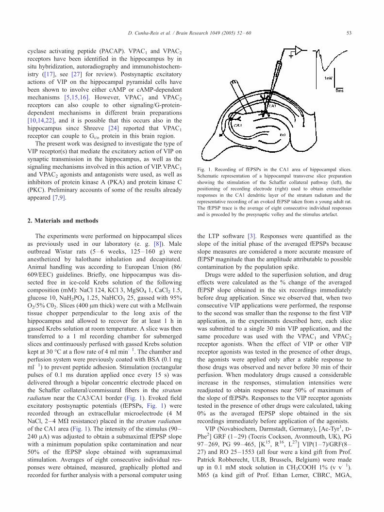
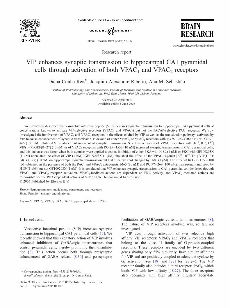
Fig. 1. Recording of fEPSPs in the CA1 area of hippocampal slices.
Schematic representation of a hippocampal transverse slice preparation
showing the stimulation of the Schaffer collateral pathway (left), the
positioning of recording electrode (right) used to obtain extracellular
responses in the CA1 dendritic layer of the stratum radiatum and the
D. Cunha-Reis et al. / Brain Research 1049 (2005) 52–60 53
cyclase activating peptide (PACAP). VPAC1 and VPAC2
receptors have been identified in the hippocampus by in
situ hybridization, autoradiography and immunohistochem-
istry ([17], see [27] for review). Postsynaptic excitatory
actions of VIP on the hippocampal pyramidal cells have
been shown to involve either cAMP or cAMP-dependent
mechanisms [5,15,16]. However, VPAC1 and VPAC2
receptors can also couple to other signaling/G-protein-
dependent mechanisms in different brain preparations
[10,14,22], and it is possible that this occurs also in the
hippocampus since Shreeve [24] reported that VPAC1
receptor can couple to Gi/o protein in this brain region.
The present work was designed to investigate the type of
VIP receptor(s) that mediate the excitatory action of VIP on
synaptic transmission in the hippocampus, as well as the
signaling mechanisms involved in this action of VIP.VPAC1
and VPAC2 agonists and antagonists were used, as well as
inhibitors of protein kinase A (PKA) and protein kinase C
(PKC). Preliminary accounts of some of the results already
appeared [7,9].
representative recording of an evoked fEPSP taken from a young adult rat.The fEPSP trace is the average of eight consecutive individual responses
and is preceded by the presynaptic volley and the stimulus artefact.
2. Materials and methods
The experiments were performed on hippocampal slices
as previously used in our laboratory (e. g. [8]). Male
outbread Wistar rats (5–6 weeks, 125–160 g) were
anesthetized by halothane inhalation and decapitated.
Animal handling was according to European Union (86/
609/EEC) guidelines. Briefly, one hippocampus was dis-
sected free in ice-cold Krebs solution of the following
composition (mM): NaCl 124, KCl 3, MgSO4 1, CaCl2 1.5,
glucose 10, NaH2PO4 1.25, NaHCO3 25, gassed with 95%
O2/5% C02. Slices (400 Am thick) were cut with a McIlwain
tissue chopper perpendicular to the long axis of the
hippocampus and allowed to recover for at least 1 h in
gassed Krebs solution at room temperature. A slice was then
transferred to a 1 ml recording chamber for submerged
slices and continuously perfused with gassed Krebs solution
kept at 30 -C at a flow rate of 4 ml min�1. The chamber and
perfusion system were previously coated with BSA (0.1 mg
ml�1) to prevent peptide adhesion. Stimulation (rectangular
pulses of 0.1 ms duration applied once every 15 s) was
delivered through a bipolar concentric electrode placed on
the Schaffer collateral/commissural fibers in the stratum
radiatum near the CA3/CA1 border (Fig. 1). Evoked field
excitatory postsynaptic potentials (fEPSPs, Fig. 1) were
recorded through an extracellular microelectrode (4 M
NaCl, 2–4 MV resistance) placed in the stratum radiatum
of the CA1 area (Fig. 1). The intensity of the stimulus (90–
240 AA) was adjusted to obtain a submaximal fEPSP slope
with a minimum population spike contamination and near
50% of the fEPSP slope obtained with supramaximal
stimulation. Averages of eight consecutive individual res-
ponses were obtained, measured, graphically plotted and
recorded for further analysis with a personal computer using
the LTP software [3]. Responses were quantified as the
slope of the initial phase of the averaged fEPSPs because
slope measures are considered a more accurate measure of
fEPSP magnitude than the amplitude attributable to possible
contamination by the population spike.
Drugs were added to the superfusion solution, and drug
effects were calculated as the % change of the averaged
fEPSP slope obtained in the six recordings immediately
before drug application. Since we observed that, when two
consecutive VIP applications were performed, the response
to the second was smaller than the response to the first VIP
application, in the experiments described here, each slice
was submitted to a single 30 min VIP application, and the
same procedure was used with the VPAC1 and VPAC2
receptor agonists. When the effect of VIP or other VIP
receptor agonists was tested in the presence of other drugs,
the agonists were applied only after a stable response to
those drugs was observed and never before 30 min of their
perfusion. When modulatory drugs caused a considerable
increase in the responses, stimulation intensities were
readjusted to obtain responses near 50% of maximum of
the slope of fEPSPs. Responses to the VIP receptor agonists
tested in the presence of other drugs were calculated, taking
0% as the averaged fEPSP slope obtained in the six
recordings immediately before application of the agonists.
VIP (Novabiochem, Darmstadt, Germany), [Ac-Tyr1, d-
Phe2] GRF (1–29) (Tocris Cockson, Avonmouth, UK), PG
97–269, PG 99–465, [K15, R16, L27] VIP(1–7)/GRF(8–
27) and RO 25–1553 (all four were a kind gift from Prof.
Patrick Robberecht, ULB, Brussels, Belgium) were made
up in 0.1 mM stock solution in CH3COOH 1% (v v�1).
M65 (a kind gift of Prof. Ethan Lerner, CBRC, MGA,
D. Cunha-Reis et al. / Brain Research 1049 (2005) 52–6054
MA, USA) was made in 0.1 mM stock solution in PBS.
HA1004, H-89, chelerythrine and GF109203X (Sigma-
Aldrich, Sintra, Portugal) were made up in 5 mM stock
solution in DMSO. The maximal DMSO and CH3COOH
concentrations added to the slices, 0.02 and 0.001% (v
v�1), respectively, were devoid of effects on fEPSP slope
(n = 3). Aliquots of the stock solutions were kept frozen at
�20 -C until use. In each experiment, one aliquot was
thawed and diluted in Krebs solution.
Values are presented as mean T SEM from n observa-
tions, and the significance of the means was calculated by
the Student’s t test. When comparing the effect of VIP in
more than two experimental conditions, one way analysis of
variance (ANOVA) was used followed by Dunnett’s multi-
ple comparison test. P values of 0.05 or less were
considered to represent significant differences.
3. Results
3.1. VIP activates both VPAC1 and VPAC2 receptors to
increase synaptic transmission to CA1 pyramidal cells
We have previously described that VIP causes a biphasic
increase in synaptic transmission to pyramidal cell den-
drites, generating a bell-shaped concentration–response
curve for the facilitation of fEPSP slope with a maximum
increase for 1 nM VIP [8]. In this new set of experiments,
we used 1 nM VIP which caused a consistent and reversible
increase in the slope (21.2 T 0.7%, n = 7, P < 0.05, Fig. 2)
of the fEPSPs recorded in the CA1 area of the hippocampus.
The involvement of each of the VIP-selective receptors
in the effect of VIP on hippocampal synaptic transmission
was tested using the VPAC1-selective antagonist PG 97–
269 [11] and the VPAC2-selective antagonist PG 99–465
Fig. 2. VIP enhancement of synaptic transmission to CA1 pyramidal cell
dendrites is dependent on both VPAC1 and VPAC2 receptor activation. (A)
Recordings of field excitatory postsynaptic potentials (fEPSPs, left panels)
and time-course of changes in fEPSP slope (right panels) obtained in
individual experiments to evaluate the action of VIP either (from top to
bottom) applied alone, in the presence of the VPAC1 receptor antagonist PG
97–269 (100 nM), in the presence of VPAC2 receptor antagonist PG 99–
465 (100 nM) or in the presence of VIP receptor antagonist [Ac-Tyr1, d-
Phe2] GRF(1–29) (300 nM, selective for VPAC1 and VPAC2 vs. PAC1
receptors). fEPSPs obtained in the presence and in the absence of 1 nM VIP
are superimposed and were recorded in the stratum radiatum as described in
Materials and methods. Each trace is composed of the stimulus artefact
followed by the presynaptic volley and the fEPSP and is the average of
eight consecutive responses obtained in one typical experiment. VIP (1 nM)
was added to the slices at the time indicated by the horizontal bars. (B) The
ability of the selective VPAC1 antagonist PG97–269, of the selective
VPAC2 antagonist PG 99–465 and of the VIP receptor antagonist [Ac-Tyr1,
d-Phe2] GRF (1–29) to inhibit the enhancement of fEPSP slope caused by
1 nM VIP is shown. Each column represents the mean T SEM of results
obtained in 4–7 experiments. jP < 0.05 (Student’s t test) as compared
with 0%. .P < 0.05 (ANOVA, followed by Dunnett’s multiple comparison
test) as compared with the effect of 1 nM VIP in the absence of other drugs
(first column).
D. Cunha-Reis et al. / Brain Research 1049 (2005) 52–60 55
[21]. In the presence of the VPAC1 receptor antagonist, PG
97–269 (100 nM), the excitatory effect of VIP (1 nM) was
nearly abolished (% increase in fEPSP slope caused by VIP
was 6.0 T 1.6, P < 0.05, n = 4, Fig. 2). When the VPAC2
receptor antagonist PG 99–465 (100 nM) was present, the
effect of 1 nM VIP was attenuated, now increasing the
fEPSP slope by only 12.3 T 2.5% (P < 0.05, n = 4, Fig. 2).
To exclude the involvement of pathways other than VPAC1
and VPAC2 receptor activation in VIP enhancement of
synaptic transmission, we used [Ac-Tyr1, d-Phe2] GRF (1–
29) (300 nM), a peptide that can selectively block VIP
receptors [29] but not other receptors of the family present
in the hippocampus. The effect of VIP (1 nM) on fEPSP
slope was abolished (P > 0.05, n = 5, Fig. 2) in the presence
of a supramaximal concentration (300 nM) [20] of this
antagonist. In most experiments, and for reasons of time of
the protocol, the VIP receptor antagonists were added to the
slices in an early stage of slice setup which did not allow the
quantification of the effects on fEPSP slope. In experiments
where the effects were quantified [Ac-Tyr1, d-Phe2], GRF
(1–29) (300 nM, n = 3) caused no appreciable change on
fEPSP slope, PG 97–269 (100 nM) enhanced fEPSP slope
by 8.6 T 3.4%, (n = 4) and PG 99–465 (100 nM) increased
fEPSP slope by 13.8 T 2.6%, (n = 2). An enhancement of
synaptic transmission by the VPAC2 antagonist was not
surprising since this compound is a partial agonist of
VPAC1 receptors [21].
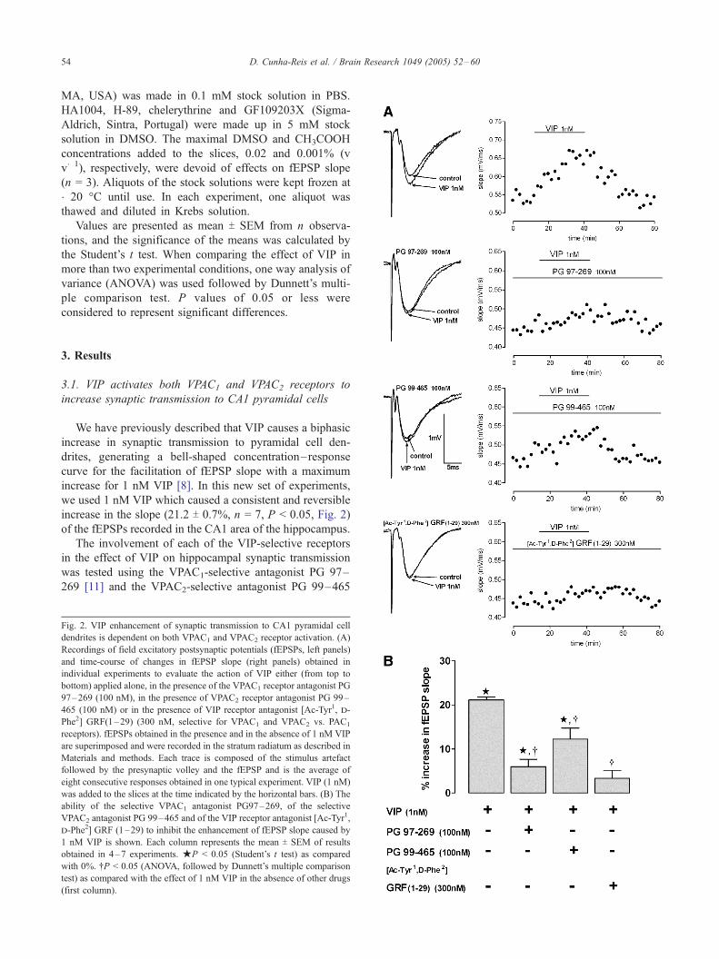
To confirm the involvement of both VIP receptors in the
facilitation of synaptic transmission to pyramidal cell
dendrites in the CA1 area of the hippocampus, we tested
the action of the VPAC1-selective agonist [K15, R16, L27]
VIP(1–7)/GRF(8–27) [12] and the VPAC2-selective agonist
RO 25–1553 [13] on fEPSPs. [K15, R16, L27] VIP(1–7)/
GRF(8–27) was used in a concentration (10 nM) that is ten
times the EC50 for stimulation of cAMP production through
rat VPAC1 receptors expressed in CHO cells [12] and, as
shown in Fig. 3, it increased the fEPSP slope by 14.1 T 0.4%(P < 0.05, n = 6). RO 25–1553 was used in a concentration
(10 nM) that is ten times its IC50 for VIP binding to rat VPAC2
receptors expressed in CHO cells [28] and that binds
negligibly to rat VPAC1 [28] and rat PAC1 receptors [13].
RO 25–1553 (10 nM) increased the fEPSP slope by 5.8 T0.6% (P < 0.05, n = 4, Fig. 3). As shown also in Fig. 3,
Fig. 3. VPAC1 and VPAC2 receptor activation enhances synaptic trans-
mission to CA1 pyramidal cell dendrites. (A) Recordings of field excitatory
postsynaptic potentials (fEPSPs, left panels) and time-course of changes in
fEPSP slope (right panels) recorded in individual experiments to evaluate
the action of the VPAC1 receptor agonist [K15, R16, L27] VIP(1–7)/
GRF(8–27) (10 nM, top), of the VPAC2 receptor agonist RO 25–1553 (10
nM, middle) or both applied together (bottom). fEPSPs obtained in the
presence and in the absence of the agonists are superimposed. Selective
agonists were added to the slices at the time indicated by the horizontal
bars. (B) Averaged effects of the selective VIP receptor agonists upon
fEPSP slope. Each column represents the mean T SEM of results obtained
in 3–7 experiments.jP < 0.05 (Student’s t test) as compared with 0%. .P< 0.05 (ANOVA, followed by Dunnett’s multiple comparison test) as
compared with the effect of 1 nM VIP (first column).
the agonists of VPAC1 and VPAC2 receptors at the
concentrations tested had nearly additive effects, increasing
fEPSP slope by 19.9 T 1.2% (P < 0.05, n = 4), an
enhancement similar to that obtained with 1 nM VIP. A
higher concentration of the VPAC2-selective agonist (100
nM, causing nearly maximal activation of rat VPAC2
receptors in the rat gastric fundus [23]) was tested in the
presence of the VPAC1 receptor antagonist (PG 97–269,
100 nM) [11] and the PAC1 receptor antagonist (M65, 30
nM) [26] to avoid loss of selectivity at VPAC2 receptors.
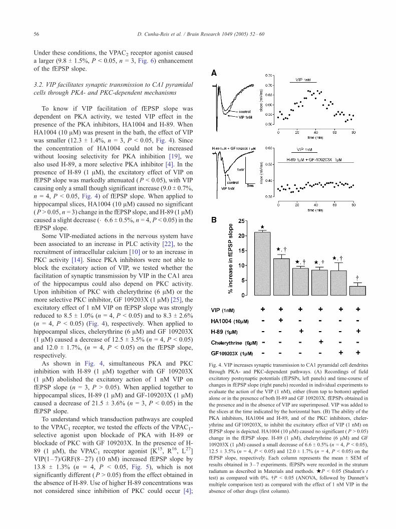
Fig. 4. VIP increases synaptic transmission to CA1 pyramidal cell dendrites
through PKA- and PKC-dependent pathways. (A) Recordings of field
excitatory postsynaptic potentials (fEPSPs, left panels) and time-course of
changes in fEPSP slope (right panels) recorded in individual experiments to
evaluate the action of the VIP (1 nM), either (from top to bottom) applied
alone or in the presence of both H-89 and GF 109203X. fEPSPs obtained in
the presence and in the absence of VIP are superimposed. VIP was added to
the slices at the time indicated by the horizontal bars. (B) The ability of the
PKA inhibitors, HA1004 and H-89, and of the PKC inhibitors, cheler-
ythrine and GF109203X, to inhibit the excitatory effect of VIP (1 nM) on
fEPSP slope is depicted. HA1004 (10 AM) caused no significant ( P > 0.05)
change in the fEPSP slope. H-89 (1 AM), chelerythrine (6 AM) and GF
109203X (1 AM) caused a small decrease of 6.6 T 0.5% (n = 4, P < 0.05),
12.5 T 3.5% (n = 4, P < 0.05) and 12.0 T 1.7% (n = 4, P < 0.05) on the
fEPSP slope, respectively. Each column represents the mean T SEM of
results obtained in 3–7 experiments. fEPSPs were recorded in the stratum
radiatum as described in Materials and methods. jP < 0.05 (Student’s t
test) as compared with 0%. .P < 0.05 (ANOVA, followed by Dunnett’s
multiple comparison test) as compared with the effect of 1 nM VIP in the
absence of other drugs (first column).
D. Cunha-Reis et al. / Brain Research 1049 (2005) 52–6056
Under these conditions, the VPAC2 receptor agonist caused
a larger (9.8 T 1.5%, P < 0.05, n = 3, Fig. 6) enhancement
of the fEPSP slope.
3.2. VIP facilitates synaptic transmission to CA1 pyramidal
cells through PKA- and PKC-dependent mechanisms
To know if VIP facilitation of fEPSP slope was
dependent on PKA activity, we tested VIP effect in the
presence of the PKA inhibitors, HA1004 and H-89. When
HA1004 (10 AM) was present in the bath, the effect of VIP
was smaller (12.3 T 1.4%, n = 3, P < 0.05, Fig. 4). Since
the concentration of HA1004 could not be increased
without loosing selectivity for PKA inhibition [19], we
also used H-89, a more selective PKA inhibitor [4]. In the
presence of H-89 (1 AM), the excitatory effect of VIP on
fEPSP slope was markedly attenuated (P < 0.05), with VIP
causing only a small though significant increase (9.0 T 0.7%,
n = 4, P < 0.05, Fig. 4) of fEPSP slope. When applied to
hippocampal slices, HA1004 (10 AM) caused no significant
(P > 0.05, n = 3) change in the fEPSP slope, and H-89 (1 AM)
caused a slight decrease (�6.6 T 0.5%, n = 4, P < 0.05) in the
fEPSP slope.
Some VIP-mediated actions in the nervous system have
been associated to an increase in PLC activity [22], to the
recruitment of intracellular calcium [10] or to an increase in
PKC activity [14]. Since PKA inhibitors were not able to
block the excitatory action of VIP, we tested whether the
facilitation of synaptic transmission by VIP in the CA1 area
of the hippocampus could also depend on PKC activity.
Upon inhibition of PKC with chelerythrine (6 AM) or the
more selective PKC inhibitor, GF 109203X (1 AM) [25], the
excitatory effect of 1 nM VIP on fEPSP slope was strongly
reduced to 8.5 T 1.0% (n = 4, P < 0.05) and to 8.3 T 2.6%
(n = 4, P < 0.05) (Fig. 4), respectively. When applied to
hippocampal slices, chelerythrine (6 AM) and GF 109203X
(1 AM) caused a decrease of 12.5 T 3.5% (n = 4, P < 0.05)
and 12.0 T 1.7%, (n = 4, P < 0.05) on the fEPSP slope,
respectively.
As shown in Fig. 4, simultaneous PKA and PKC
inhibition with H-89 (1 AM) together with GF 109203X
(1 AM) abolished the excitatory action of 1 nM VIP on
fEPSP slope (n = 3, P > 0.05). When applied together to
hippocampal slices, H-89 (1 AM) and GF-109203X (1 AM)
caused a decrease of 21.5 T 3.6% (n = 3, P < 0.05) in the
fEPSP slope.
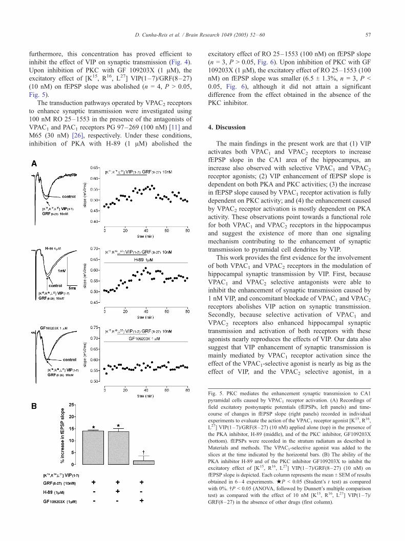
To understand which transduction pathways are coupled
to the VPAC1 receptor, we tested the effects of the VPAC1-
selective agonist upon blockade of PKA with H-89 or
blockade of PKC with GF 109203X. In the presence of H-
89 (1 AM), the VPAC1 receptor agonist [K15, R16, L27]
VIP(1–7)/GRF(8–27) (10 nM) increased fEPSP slope by
13.8 T 1.3% (n = 4, P < 0.05, Fig. 5), which is not
significantly different (P > 0.05) from the effect obtained in
the absence of H-89. Use of higher H-89 concentrations was
not considered since inhibition of PKC could occur [4];
D. Cunha-Reis et al. / Brain Research 1049 (2005) 52–60 57
furthermore, this concentration has proved efficient to
inhibit the effect of VIP on synaptic transmission (Fig. 4).
Upon inhibition of PKC with GF 109203X (1 AM), the
excitatory effect of [K15, R16, L27] VIP(1–7)/GRF(8–27)
(10 nM) on fEPSP slope was abolished (n = 4, P > 0.05,
Fig. 5).
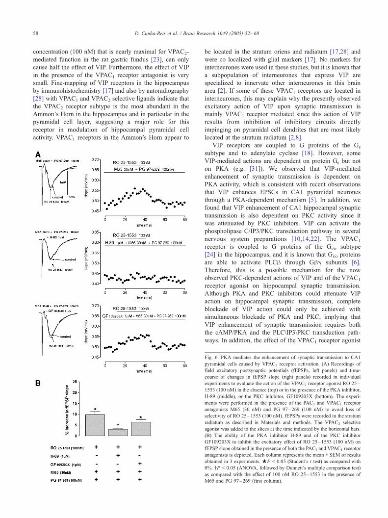
The transduction pathways operated by VPAC2 receptors
to enhance synaptic transmission were investigated using
100 nM RO 25–1553 in the presence of the antagonists of
VPAC1 and PAC1 receptors PG 97–269 (100 nM) [11] and
M65 (30 nM) [26], respectively. Under these conditions,
inhibition of PKA with H-89 (1 AM) abolished the
excitatory effect of RO 25–1553 (100 nM) on fEPSP slope
(n = 3, P > 0.05, Fig. 6). Upon inhibition of PKC with GF
109203X (1 AM), the excitatory effect of RO 25–1553 (100
nM) on fEPSP slope was smaller (6.5 T 1.3%, n = 3, P <
0.05, Fig. 6), although it did not attain a significant
difference from the effect obtained in the absence of the
PKC inhibitor.
4. Discussion
The main findings in the present work are that (1) VIP
activates both VPAC1 and VPAC2 receptors to increase
fEPSP slope in the CA1 area of the hippocampus, an
increase also observed with selective VPAC1 and VPAC2
receptor agonists; (2) VIP enhancement of fEPSP slope is
dependent on both PKA and PKC activities; (3) the increase
in fEPSP slope caused by VPAC1 receptor activation is fully
dependent on PKC activity; and (4) the enhancement caused
by VPAC2 receptor activation is mostly dependent on PKA
activity. These observations point towards a functional role
for both VPAC1 and VPAC2 receptors in the hippocampus
and suggest the existence of more than one signaling
mechanism contributing to the enhancement of synaptic
transmission to pyramidal cell dendrites by VIP.
This work provides the first evidence for the involvement
of both VPAC1 and VPAC2 receptors in the modulation of
hippocampal synaptic transmission by VIP. First, because
VPAC1 and VPAC2 selective antagonists were able to
inhibit the enhancement of synaptic transmission caused by
1 nM VIP, and concomitant blockade of VPAC1 and VPAC2
receptors abolishes VIP action on synaptic transmission.
Secondly, because selective activation of VPAC1 and
VPAC2 receptors also enhanced hippocampal synaptic
transmission and activation of both receptors with these
agonists nearly reproduces the effects of VIP. Our data also
suggest that VIP enhancement of synaptic transmission is
mainly mediated by VPAC1 receptor activation since the
effect of the VPAC1-selective agonist is nearly as big as the
effect of VIP, and the VPAC2 selective agonist, in a
Fig. 5. PKC mediates the enhancement synaptic transmission to CA1
pyramidal cells caused by VPAC1 receptor activation. (A) Recordings of
field excitatory postsynaptic potentials (fEPSPs, left panels) and time-
course of changes in fEPSP slope (right panels) recorded in individual
experiments to evaluate the action of the VPAC1 receptor agonist [K15, R16,
L27] VIP(1–7)/GRF(8–27) (10 nM) applied alone (top) in the presence of
the PKA inhibitor, H-89 (middle), and of the PKC inhibitor, GF109203X
(bottom). fEPSPs were recorded in the stratum radiatum as described in
Materials and methods. The VPAC1-selective agonist was added to the
slices at the time indicated by the horizontal bars. (B) The ability of the
PKA inhibitor H-89 and of the PKC inhibitor GF109203X to inhibit the
excitatory effect of [K15, R16, L27] VIP(1–7)/GRF(8–27) (10 nM) on
fEPSP slope is depicted. Each column represents the mean T SEM of results
obtained in 6–4 experiments. jP < 0.05 (Student’s t test) as compared
with 0%. .P < 0.05 (ANOVA, followed by Dunnett’s multiple comparison
test) as compared with the effect of 10 nM [K15, R16, L27] VIP(1–7)/
GRF(8–27) in the absence of other drugs (first column).
D. Cunha-Reis et al. / Brain Research 1049 (2005) 52–6058
concentration (100 nM) that is nearly maximal for VPAC2-
mediated function in the rat gastric fundus [23], can only
cause half the effect of VIP. Furthermore, the effect of VIP
in the presence of the VPAC1 receptor antagonist is very
small. Fine-mapping of VIP receptors in the hippocampus
by immunohistochemistry [17] and also by autoradiography
[28] with VPAC1 and VPAC2 selective ligands indicate that
the VPAC2 receptor subtype is the most abundant in the
Ammon’s Horn in the hippocampus and in particular in the
pyramidal cell layer, suggesting a major role for this
receptor in modulation of hippocampal pyramidal cell
activity. VPAC1 receptors in the Ammon’s Horn appear to
be located in the stratum oriens and radiatum [17,28] and
were co localized with glial markers [17]. No markers for
interneurones were used in these studies, but it is known that
a subpopulation of interneurones that express VIP are
specialized to innervate other interneurones in this brain
area [2]. If some of these VPAC1 receptors are located in
interneurones, this may explain why the presently observed
excitatory action of VIP upon synaptic transmission is
mainly VPAC1 receptor mediated since this action of VIP
results from inhibition of inhibitory circuits directly
impinging on pyramidal cell dendrites that are most likely
located at the stratum radiatum [2,8].
VIP receptors are coupled to G proteins of the Gs
subtype and to adenylate cyclase [18]. However, some
VIP-mediated actions are dependent on protein Gs but not
on PKA (e.g. [31]). We observed that VIP-mediated
enhancement of synaptic transmission is dependent on
PKA activity, which is consistent with recent observations
that VIP enhances EPSCs in CA1 pyramidal neurones
through a PKA-dependent mechanism [5]. In addition, we
found that VIP enhancement of CA1 hippocampal synaptic
transmission is also dependent on PKC activity since it
was attenuated by PKC inhibitors. VIP can activate the
phospholipase C/IP3/PKC transduction pathway in several
nervous system preparations [10,14,22]. The VPAC1
receptor is coupled to G proteins of the Gi/o subtype
[24] in the hippocampus, and it is known that Gi/o proteins
are able to activate PLCh through Ghg subunits [6].
Therefore, this is a possible mechanism for the now
observed PKC-dependent actions of VIP and of the VPAC1
receptor agonist on hippocampal synaptic transmission.
Although PKA and PKC inhibitors could attenuate VIP
action on hippocampal synaptic transmission, complete
blockade of VIP action could only be achieved with
simultaneous blockade of PKA and PKC, implying that
VIP enhancement of synaptic transmission requires both
the cAMP/PKA and the PLC\IP3\PKC transduction path-
ways. In addition, the effect of the VPAC1 receptor agonist
Fig. 6. PKA mediates the enhancement of synaptic transmission to CA1
pyramidal cells caused by VPAC2 receptor activation. (A) Recordings of
field excitatory postsynaptic potentials (fEPSPs, left panels) and time-
course of changes in fEPSP slope (right panels) recorded in individual
experiments to evaluate the action of the VPAC2 receptor agonist RO 25–
1553 (100 nM) in the absence (top) or in the presence of the PKA inhibitor,
H-89 (middle), or the PKC inhibitor, GF109203X (bottom). The experi-
ments were performed in the presence of the PAC1 and VPAC1 receptor
antagonists M65 (30 nM) and PG 97–269 (100 nM) to avoid loss of
selectivity of RO 25–1553 (100 nM). fEPSPs were recorded in the stratum
radiatum as described in Materials and methods. The VPAC2 selective
agonist was added to the slices at the time indicated by the horizontal bars.
(B) The ability of the PKA inhibitor H-89 and of the PKC inhibitor
GF109203X to inhibit the excitatory effect of RO 25–1553 (100 nM) on
fEPSP slope obtained in the presence of both the PAC1 and VPAC1 receptor
antagonists is depicted. Each column represents the mean T SEM of results
obtained in 3 experiments. jP < 0.05 (Student’s t test) as compared with
0%. .P < 0.05 (ANOVA, followed by Dunnett’s multiple comparison test)
as compared with the effect of 100 nM RO 25–1553 in the presence of
M65 and PG 97–269 (first column).
D. Cunha-Reis et al. / Brain Research 1049 (2005) 52–60 59
was not modified by the PKA inhibitor, suggesting that
this receptor operates mainly through a PKA-independent
mechanism and that VPAC2 receptor activation is the
source for the PKA-dependent actions of VIP.
The fact that two different VIP receptors both lead to an
enhancement of synaptic transmission in hippocampal
slices may result from the measurement of a final outcome
of multiple cellular responses to VIP. VIP is expressed in
the hippocampus in three distinct subtypes of interneurones
[1,2], selectively targeting either pyramidal cells or
interneurones in different hippocampal layers. VIP
enhancement of synaptic transmission in the CA1 area of
the hippocampus depends on both pre- and postsynaptic
modulation of GABAergic transmission [8]. Yet, VIP
enhancement of EPSCs in pyramidal cells is observed in
the absence of GABAergic transmission [5]. Thus, VIP
appears to modulate hippocampal synaptic transmission in
a complex manner, involving multiple mechanisms that
can influence the response at the integrated level. The
uneven cellular and layer distribution of VPAC1 and
VPAC2 receptors in the hippocampus may also add to
this complexity, and receptors located at different sites in
the hippocampus may well be activated by different
stimulus occurring in vivo [1,2,17,28]. Further elucidation
of the cellular location (dendritic, somatic or nerve
terminal) of the different VIP receptors might prove useful
to understand the role of each receptor in the control of
hippocampal synaptic transmission.
Acknowledgments
We greatly acknowledge Prof. P. Robberecht, SM, ULB,
Belgium for the kind gift of VIP selective agonists and
antagonists, Prof. Ethan Lerner, CBRC, MGA, MA, USA
for the PAC1 selective ligands and the Institute of
Physiology, FML for animal housing facilities. Diana
Cunha-Reis was in receipt of an FCT PhD fellowship. This
work was supported by FCT.
References
[1] L. Acsady, D. Arabadzisz, T.F. Freund, Correlated morphological and
neurochemical features identify different subsets of vasoactive
intestinal polypeptide-immunoreactive interneurons in the rat hippo-
campus, Neuroscience 73 (1996) 299–315.
[2] L. Acsady, T.J. Gorcs, T.F. Freund, Different populations of vasoactive
intestinal polypeptide-immunoreactive interneurons are specialized to
control pyramidal cells or interneurons in the hippocampus, Neuro-
science 73 (1996) 317–334.
[3] W.W. Anderson, G.L. Collingridge, The LTP Program: a data
acquisition program for on-line analysis of long-term potentiation
and other synaptic events, J. Neurosci. Methods 108 (2001) 71–83.
[4] T. Chijiwa, A. Mishima, M. Hagiwara, M. Sano, K. Hayashi, T. Inoue,
K. Naito, et al., Inhibition of forskolin-induced neurite outgrowth and
protein phosphorylation by a newly synthesized selective inhibitor of
cyclic AMP-dependent protein kinase, N-[2-( p-Bromocinnamylami-
no)ethyl]-5-isoquinolinesulfonamide (H-89), of PC12 pheochromocy-
toma cells, J. Biol. Chem. 265 (1990) 5267–5272.
[5] L. Ciranna, S. Cavallaro, Opposing effects by pituitary adenylate
cyclase-activating polypeptide and vasoactive intestinal peptide on
hippocampal synaptic transmission, Exp. Neurol. 184 (2003)
778–784.
[6] D.E. Clapham, E.J. Neer, G protein hg subunits, Annu. Rev.
Pharmacol. Toxicol. 37 (1997) 167–203.
[7] D. Cunha-Reis, A.M. Sebastiao, J.A. Ribeiro, Transduction mecha-
nisms involved in the action of VIP in the rat hippocampus, Abstr.-
Eur. J. Neurosci. 12 (Suppl. 11) (2000) 49, (017.23).
[8] D. Cunha-Reis, A.M. Sebastiao, K. Wirkner, P. Illes, J.A. Ribeiro, VIP
enhances both pre- and post-synaptic GABAergic transmission to
hippocampal interneurones leading to increased excitatory synaptic
transmission to CA1 pyramidal cells, Br. J. Pharmacol. 143 (2004)
733–744.
[9] D. Cunha-Reis, A.M. Sebastiao, J.A. Ribeiro, VIP modulates K+-
evoked[3H]-GABA release from hippocampal synaptosomes through
activation of both VPAC1 and VPAC2 receptors, Abstr.-Fundam. Clin.
Pharmacol. 18 (Suppl. 1) (2004) 28, (P02.02).
[10] A. Fatatis, L.A. Holtzclaw, R. Avidor, D.E. Brenneman, J.T. Russel,
Vasoactive intestinal peptide increases intracellular calcium in astro-
glia: synergism with a-adrenergic receptors, Proc. Natl. Acad. Sci. 91
(1994) 2036–2040.
[11] P. Gourlet, P. De Neef, J. Cnudde, M. Waelbroeck, P. Robberecht, In
vitro properties of a high affinity selective antagonist of the VIP1receptor, Peptides 18 (1997) 1555–1560.
[12] P. Gourlet, A. Vandermeers, P. Vertongen, J. Rathe, P. De Neef, J.
Cnudde, M. Waelbroeck, P. Robberecht, Development of high affinity
selective VIP1 receptor agonists, Peptides 18 (1997) 1539–1545.
[13] P. Gourlet, P. Vertongen, A. Vandermeers, M.-C. Vandermeers-Piret, J.
Rathe, P. de Neef, M. Waelbroeck, P. Robberecht, The long-acting
vasoactive intestinal polypeptide agonist RO 25–1553 is highly
selective of the VIP2 receptor subclass, Peptides 18 (1997) 403–408.
[14] P. Gressens, S. Marret, J.-L. Martin, A. Laquerriere, A. Lombet, P.
Evrard, Regulation of neuroprotective action of vasoactive intestinal
peptide in the murine developing brain by protein kinase C and
mitogen-activated protein kinase cascades: in vivo and in vitro studies,
J. Neurochem. 70 (1998) 2574–2584.
[15] H.L. Haas, B.H. Gahwiller, Vasoactive intestinal polypeptide modu-
lates neuronal excitability in hippocampal slices of the rat, Neuro-
science 47 (1992) 273–277.
[16] T. Haug, J.F. Storm, Protein kinase A mediates the modulation of the
slow Ca2+-dependent K+ Current, ISahp, by the neuropeptides CRF,
VIP, and CGRP in hippocampal pyramidal neurons, J. Neurophysiol.
83 (2000) 2071–2079.
[17] K.M. Joo, Y.H. Chung, M.K. Kim, R.H. Nam, B.L. Lee, K.H.
Lee, C.I. Cha, Distribution of vasoactive intestinal peptide and
pituitary adenylate cyclase-activating peptide receptors (VPAC1,
VPAC2, and PAC1 receptor) in the rat brain, J. Comp. Neurol. 476
(2004) 388–413.
[18] M. Laburthe, A. Couvineau, J.-C. Marie, VPAC receptors for VIP and
PACAP, Recept. Channels 8 (2002) 137–153.
[19] J.C. Leahy, M.L. Vallano, Differential effects of isoquinolinesulfona-
mide protein kinase inhibitors on CA1 responses in hippocampal
slices, Neuroscience 44 (1991) 361–370.
[20] D.-L. Liu, J. Cuevas, D.J. Adams, VIP and PACAP potentiation of
nicotinic ACh-evoked currents in rat parasympathetic neurons is
mediated by G-protein activation, Eur. J. Neurosci. 12 (2000)
2243–2251.
[21] D. Moreno, P. Gourlet, P. De Neef, J. Cnudde, M. Waelbroeck, P.
Robberecht, Development of selective agonists and antagonists for the
human vasoactive intestinal polypeptide VPAC2 receptor, Peptides 21
(2000) 1543–1549.
[22] H.S. Nielsen, J. Hannibal, J. Fahrenkrug, Vasoactive intestinal peptide
induces per1 and per2 gene expression in the rat suprachiasmatic
nucleus late at night, Eur. J. Neurosci. 15 (2002) 570–574.
D. Cunha-Reis et al. / Brain Research 1049 (2005) 52–6060
[23] P. Robberecht, P. De Neef, R.A. Lefebvre, Influence of selective VIP
receptor agonists in the rat gastric fundus, Eur. J. Pharmacol. 359
(1998) 77–80.
[24] M.S. Shreeve, Identification of G-proteins coupling to the vasoactive
intestinal peptide receptor VPAC1 using immunoaffinity chromatog-
raphy: evidence for precoupling, Biochem. Biophys. Res. Commun.
290 (2002) 1300–1307.
[25] D. Toullec, P. Pianetti, H. Coste, P. Bellevergue, T. Grand-Perret, M.
Ajakane, V. Baudet, et al., The bisindolylmaleimide GF109203X is a
potent and selective inhibitor of protein kinase C, J. Biol. Chem. 266
(1991) 15771–15781.
[26] D. Uchida, I. Tatsuno, T. Tanaka, A. Hirai, Y. Saito, O. Moro, M.
Tajima, Maxadilan is a specific agonist and its deleted peptide (M65)
is a specific antagonist for PACAP type I receptor, Ann. N. Y. Acad.
Sci. 865 (1998) 253–258.
[27] D. Vaudry, B.J. Gonzalez, M. Basille, L. Yon, A. Fournier, H.
Vaudry, Pituitary adenylate cyclase-activating polypeptide and its
receptors: from structure to functions, Pharmacol. Rev. 52 (2000)
269–324.
[28] P. Vertongen, S.N. Schiffmann, P. Gourlet, P. Robberecht, Auto-
radiographic visualization of the receptor subclasses for vasoactive
intestinal polypeptide (VIP) in rat brain, Peptides 18 (1997)
1547–1554.
[29] M. Waelbroeck, P. Robberecht, D.H. Coy, J.C. Camus, P. de Neef, J.
Christophe, Interaction of growth hormone-releasing factor (GRF) and
14 GRF analogs with vasoactive intestinal peptide (VIP) receptors of
rat pancreas. Discovery of (N-Ac-Tyr1, d-Phe2)-GRF(1–29)-NH2 as a
VIP antagonist, Endocrinology 116 (1985) 2643–2649.
[30] H.-L. Wang, A. Li, T. Wu, Vasoactive intestinal polypeptide enhances
the GABAergic synaptic transmission in cultured hippocampal
neurons, Brain Res. 746 (1997) 294–300.
[31] Y. Zhu, S.R. Ikeda, VIP inhibits N-type Ca2+ channels of sympathetic
neurons via a pertussis toxin-insensitive but cholera toxin-sensitive
pathway, Neuron 13 (1994) 657–669.