enhanced sensitivity to ethanol-induced inhibition of ltp in ca1 pyramidal neurons of socially...
TRANSCRIPT

ORIGINAL RESEARCH ARTICLEpublished: 21 October 2011
doi: 10.3389/fendo.2011.00056
Enhanced sensitivity to ethanol-induced inhibition of LTPin CA1 pyramidal neurons of socially isolated C57BL/6Jmice: role of neurosteroidsGiuseppeTalani 1*, Giovanni Biggio1,2 and Enrico Sanna1,2
1 Section of Neuroscience, Department of Experimental Biology, Center of Excellence for the Neurobiology of Dependence, University of Cagliari, Monserrato,Cagliari, Italy
2 Institute of Neuroscience, National Research Council of Italy, Monserrato, Cagliari, Italy
Edited by:
Hubert Vaudry, University of Rouen,France
Reviewed by:
Gustavo M. Somoza, Instituto deInvestigacionesBiotecnologicas – InstitutoTecnologico de Chascomus, ArgentinaCharles Zorumski, WashingtonUniversity, USA
*Correspondence:
Giuseppe Talani , Department ofExperimental Biology, University ofCagliari, Cittadella Universitaria diMonserrato, Cagliari 09042, Italy.e-mail: [email protected]
Ethanol (EtOH) induced impairment of long-term potentiation (LTP) in the rat hippocampusis prevented by the 5α-reductase inhibitor finasteride, suggesting that this effect of EtOHis dependent on the increased local release of neurosteroids such as 3α,5α-THP that pro-mote GABA-mediated transmission. Given that social isolation (SI) in rodents is associatedwith altered plasma and brain levels of such neurosteroids as well as with an enhancedneurosteroidogenic action of EtOH, we examined whether the inhibitory effect of EtOH onLTP at CA3–CA1 hippocampal excitatory synapses is altered in C57BL/6J mice subjectedto SI for 6 weeks in comparison with group-housed (GH) animals. Extracellular recordingof field excitatory postsynaptic potentials (fEPSPs) as well as patch-clamp analysis wereperformed in hippocampal slices prepared from both SI and GH mice. Consistent withprevious observations, recording of fEPSPs revealed that the extent of LTP induced in theCA1 region of SI mice was significantly reduced compared with that in GH animals. EtOH(40 mM) inhibited LTP in slices from SI mice but not in those from GH mice, and this effectof EtOH was abolished by co-application of 1 μM finasteride. Current-clamp analysis of CA1pyramidal neurons revealed a decrease in action potential (AP) frequency and an increasein the intensity of injected current required to evoke the first AP in SI mice compared withGH mice, indicative of a decrease in neuronal excitability associated with SI. Together, ourdata suggest that SI results in reduced levels of neuronal excitability and synaptic plastic-ity in the hippocampus. Furthermore, the increased sensitivity to the neurosteroidogeniceffect of EtOH associated with SI likely accounts for the greater inhibitory effect of EtOHon LTP in SI mice.The increase in EtOH sensitivity induced by SI may be important for thechanges in the effects of EtOH on anxiety and on learning and memory associated withthe prolonged stress attributable to SI.
Keywords: social isolation, ethanol, hippocampus, neurosteroids, neuronal excitability, stress, LTP
INTRODUCTIONEthanol (EtOH) influences the function of inhibitory γ-aminobutyric acid (GABA) type A receptors (GABAARs) by var-ious direct and indirect mechanisms (Criswell and Breese, 2005;Breese et al., 2006; Weiner and Valenzuela, 2006; Kumar et al.,2009; Kelm et al., 2011). EtOH thus selectively enhances thefunction of recombinant GABAARs containing α4 or α6 as wellas δ subunits expressed in Xenopus laevis oocytes (Sundstrom-Poromaa et al., 2002; Wallner et al., 2003) as well as tonic currentsmediated by GABAARs containing α4 and δ subunits in hip-pocampal and thalamic slices in vitro (Hanchar et al., 2004; Weiet al., 2004; Glykys et al., 2007; Jia et al., 2007, 2008; Santhaku-mar et al., 2007). Indirect modulation of GABAARs by EtOHhas been shown to be mediated both presynaptically, throughan increase in the probability of GABA release (Roberto et al.,2003; Carta et al., 2004; Sanna et al., 2004; Zhu and Lovinger,2006), and postsynaptically, through stimulation of the biosynthe-sis of neuroactive steroids (Barbaccia et al., 1999; Morrow et al.,
1999, 2001; VanDoren et al., 2000; Sanna et al., 2004; Kumar et al.,2009).
Neuroactive steroids such as 3α,5α-tetrahydroprogesterone(3α,5α-THP, also known as allopregnanolone) and 3α,5α-tetrahydrodeoxycorticosterone (3α,5α-THDOC) are reducedmetabolites of progesterone that rapidly alter neuronal excitabilityboth by acting as potent positive modulators of GABAARs, espe-cially those that contain the α4 and δ subunits and are locatedat extrasynaptic sites, as well as by influencing other neurotrans-mission systems (Rupprecht and Holsboer, 1999; Lambert et al.,2009). These steroids are produced by peripheral organs includingthe adrenal glands and gonads (Paul and Purdy, 1992), but they arealso synthesized de novo in brain cells from cholesterol, in whichcase they are also referred to as neurosteroids (Hu et al., 1987;Mathur et al., 1993). 3α,5α-THP potentiates GABAAR-mediatedresponses in vitro at nanomolar concentrations, a potency greaterthan that of benzodiazepines or barbiturates (Majewska et al.,1986; Morrow et al., 1987, 1990; Reddy et al., 2004), and it exerts
www.frontiersin.org October 2011 | Volume 2 | Article 56 | 1

Talani et al. Ethanol sensitivity, neurosteroids, and stress
anxiolytic and sedative effects in vivo (Bitran et al., 1993, 1995;Freeman et al., 1993; Picazo and Fernandez-Guasti, 1995; Kokateet al., 1999; Reddy et al., 2004). Plasma and brain levels of GABAer-gic neuroactive steroids are affected by acute or chronic stress aswell as by the acute administration of EtOH in rodents (Purdyet al., 1991; Concas et al., 1996; Barbaccia et al., 1999; Morrowet al., 1999; Serra et al., 2006).
The steroidogenic action of EtOH is thought to be mediatedby stimulation of the hypothalamic–pituitary–adrenal (HPA) axisand results in an increase in the circulating and brain levels ofneuroactive steroids. However, EtOH directly stimulates neuros-teroidogenesis in rat brain slices and thus independently of periph-eral organs (Sanna et al., 2004; Criswell and Breese, 2005; Izumiet al., 2007). EtOH induces the local release of neurosteroids at thesynaptic level as well as positive modulation of GABAARs in CA1pyramidal neurons, with both effects being prevented by the 5α-reductase inhibitor finasteride, suggesting that local production ofneurosteroids such as 3α,5α-THP is necessary for modulation ofGABAAR function by EtOH. In addition, this neurosteroidogeniceffect of EtOH was shown to result in inhibition of long-termpotentiation (LTP) in the CA1 hippocampal region (Izumi et al.,2007). LTP in the hippocampus is a form of synaptic plasticitythat provides a consolidated cellular mechanism of memory for-mation (Mayford, 2007). Membrane excitability in CA1 pyramidalneurons has been found to be related both to the performance oflearning tasks (Moyer et al., 2000; Tombaugh et al., 2005) as wellas to successful learning (Moyer et al., 1996; Thompson et al.,1996).
Postweaning social isolation (SI) is a well-characterized par-adigm of mild prolonged stress that is associated with markedbehavioral, neuroendocrine, and neurochemical changes (Hallet al., 1998; Serra et al., 2000, 2005, 2006; Ferdman et al., 2007) andthat is dependent on gender (Pietropaolo et al., 2008). A decreasein the brain concentrations of GABAergic neuroactive steroidssuch as 3α,5α-THP in animals subjected to SI is accompanied byan increased efficacy of EtOH in the stimulation of steroidogen-esis and GABAAR function (Serra et al., 2003, 2006). Moreover,SI induces increased voluntary consumption of EtOH in adultC57BL/6J mice (Sanna et al., 2011).
We have now evaluated the effects of SI on neuronal excitabilityand on the inhibition by EtOH of LTP induction in the CA1 regionof the mouse hippocampus as well as the role of neurosteroids insuch effects. The decrease in neurosteroid levels associated withSI was found to result in a reduced level of neuronal excitabilityand an enhanced inhibitory effect of EtOH on LTP in the CA1hippocampal region.
MATERIALS AND METHODSANIMALSC57BL/6J mice (Charles River, Como, Italy) were bred in ouranimal facility and maintained under an artificial 12-h-light, 12-h-dark cycle (lights on from 08:00 to 20:00 h), a constant tem-perature of 22 ± 2˚C, and a relative humidity of 65%. They hadfree access to water and standard laboratory food at all time. Ani-mal care and handling throughout the experimental procedureswere in accordance with the European Communities CouncilDirective of November 24, 1986 (86/609/EEC). The experimental
protocols were also approved by the Animal Ethics Committee ofthe University of Cagliari.
SI STRESS PARADIGMNewborn mouse pups were left undisturbed with their mothersuntil weaning (21 days). After weaning, male mice were randomlyassigned to be housed six per cage (group-housed, GH) or oneper cage (SI) for 6 weeks. In a separate set of experiments, 12SI mice were injected once a day subcutaneously with proges-terone (5 mg/kg, dissolved in 20% β-cyclodextrin), and six ofwhich were co-treated with finasteride (25 mg/kg, dissolved in 20%β-cyclodextrin, s.c.) throughout the entire 6-week period of isola-tion; control SI mice received an equivalent injection of the vehiclesolution according to the same schedule.
PREPARATION OF MOUSE HIPPOCAMPAL SLICESHippocampal slices were prepared from GH and SI mice as pre-viously described (Sanna et al., 2011). In brief, the animals weresubjected to deep anesthesia with chloroform and decapitated,and the brain was rapidly removed from the skull. For extracel-lular recordings, the brain was transferred to a standard artificialcerebrospinal fluid (ACSF) containing (in mM): 126 NaCl, 3 KCl,2 CaCl2, 1 MgCl2, 26 NaHCO3, 1.25 NaH2PO4, and 10 d-glucose(pH 7.4, set by aeration with 95% O2 and 5% CO2). Alternatively,for patch-clamp experiments, the brain was transferred to a mod-ified ACSF containing (in mM): 220 sucrose, 2 KCl, 0.2 CaCl2, 6MgSO4, 26 NaHCO3, 1.3 NaH2PO4, and 10 d-glucose (pH 7.4,set by aeration with 95% O2 and 5% CO2). Coronal brain slices(thickness of 300 or 400 μm) containing the hippocampus werecut in ice-cold standard or modified ACSF with the use of a LeicaVT1200S vibratome (Leica, Heidelberg, Germany). The slices werethen transferred immediately to a nylon net submerged in stan-dard ACSF for at least 40 min at a controlled temperature of 35˚C(for patch-clamp experiments) or at room temperature (for extra-cellular recordings). After subsequent incubation for at least 1 h atroom temperature, hemi-slices were transferred to the recordingchamber, which was perfused with standard ACSF at a constantflow rate of ∼2 ml/min. For all recordings, the temperature of thebath was maintained at 33˚C.
EXTRACELLULAR RECORDING OF fEPSPsRecordings of field excitatory postsynaptic potentials (fEPSPs)were obtained from the stratum radiatum of the CA1 region of thehippocampus after stimulation of the Schaffer collateral afferents.Extracellular recording electrodes were prepared from borosili-cate capillaries with an internal filament and an outer diameterof 1.5 μm (Sutter Instruments, Novato, CA, USA) and were filledwith 4 M NaCl (resistance, 1–2 MΩ). For stimulation of afferents, aconcentric bipolar electrode (FHC, Bowdoin, ME, USA) was posi-tioned ∼300 μm from the recording site. Responses were triggereddigitally every 20 s with the use of an interval generator (Master8, FHC) and a stimulus isolator by application of a constant cur-rent pulse of 0.2–0.4 mA with a duration of 60 μs, which yieldeda half-maximal response. For determination of the input–output(I–O) relation, the stimulation current was adjusted from 0 to1 mA in steps of 0.1 mA. The fEPSPs were amplified with the useof an Axoclamp 2B amplifier (Axon Instruments, Union City, CA,
Frontiers in Endocrinology | Neuroendocrine Science October 2011 | Volume 2 | Article 56 | 2

Talani et al. Ethanol sensitivity, neurosteroids, and stress
USA), digitized, and then analyzed with Clampfit 9.02 software(Axon Instruments). Several kinetic parameters of fEPSPs wereanalyzed, but the slope values were considered for quantitation ofthe responses. For elicitation of LTP, after 10 min of stable baselinerecording of fEPSPs evoked every 20 s at the current intensity thattriggered 50% of the maximal fEPSP response, high-frequencystimulation (HFS) consisting of a single train of 100 stimuli at250 Hz was delivered and recording was then continued for 60 minwith stimulation of fEPSPs every 20 s.
WHOLE-CELL PATCH-CLAMP RECORDINGSWhole-cell recordings from hippocampal CA1 pyramidal neu-rons were performed as previously described (Sanna et al., 2011).Recording pipettes were prepared from borosilicate glass withthe use of a Flaming Brown micropipette puller (MolecularDevices, Novato, CA, USA). Resistance of the pipettes rangedfrom 2.5 to 4.5 MΩ when they were filled with an internal solu-tion containing 135 mM potassium gluconate, 10 mM MgCl2,0.1 mM CaCl2, 1 mM EGTA, 10 mM Hepes–KOH (pH 7.3), and2 mM ATP (disodium salt). We analyzed only recordings withaccess resistance of <25 MΩ (values usually ranged from 9 to20 MΩ). Series resistance was not compensated, and cells wereexcluded from further analysis if access resistance changed by>20% during the course of the recording. Membrane poten-tials were recorded with the use of an Axopatch 200-B amplifier(Axon Instruments), filtered at 2 kHz, and digitized at 5 kHz. Forpatch-clamp experiments, we used pClamp 9.2 software (Molec-ular Devices, Union City, CA, USA), which allowed us to measurevarious characteristics of the neuronal membrane and actionpotentials (APs). For current-clamp experiments, we applied aprotocol with injected current steps of 400-ms duration and rang-ing in intensity from −80 to 200 pA in intervals of 20 pA inorder to hyperpolarize or depolarize the membrane potential. Theparameters analyzed included resting membrane potential, inputresistance, AP threshold, minimum injected current capable ofevoking the first AP, spike latency (time required for the first AP tooccur in response to depolarization), and spike frequency. Inputresistance was calculated from only the hyperpolarizing currentsteps.
RECORDING OF SPONTANEOUS LOCOMOTOR ACTIVITYGroup-housed and SI mice were tested at the end of the 6-weekhousing period. To determine general locomotor activity levelsand exploration habits, we used a motility meter (Omnitech Elec-tronics Inc.). Animals were left in the same room in which theapparatus was placed for at least 2 h before the beginning of exper-iments in order to allow their habituation to the environment.Individual mice were placed in the center of a square arena (20.3by 20.3 cm) and allowed to move freely for 60 min while beingtracked by the automated tracking system. Parameters were mon-itored every 5 min (total of 12 acquisitions). Data obtained duringthe first 10 min were used for comparison among groups. Thearena was assembled with specially designed sound-attenuatingshells made of polypropylene and an expanded PVC sheet. Theanimals were isolated from noise of the recorder and printer usedto acquire the data by placing these devices in a different room.The parameters measured included horizontal activity (number of
photobeam interruptions), total distance traveled (centimeters),locomotion time (seconds), and rest time (seconds).
STATISTICAL ANALYSISData are presented as means ± SEM and were compared by one-way analysis of variance (ANOVA) or Student’s t -test with the useof Prism software (version 5, GraphPad). A p value of <0.05 wasconsidered statistically significant.
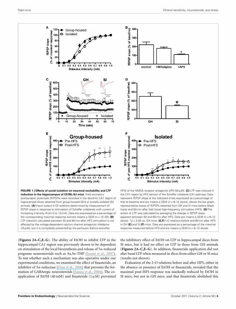
RESULTSEFFECTS OF SI ON NEURONAL EXCITABILITY AND LTP INDUCTION INTHE HIPPOCAMPAL CA1 REGION OF C57BL/6J MICETo examine the effect of SI on neuronal excitability in the CA1region of the mouse hippocampus, we generated I–O curves bystimulating the Schaffer collateral glutamatergic afferents withincreasing (0–1.0 mA) current intensity and recorded fEPSPsfrom the dendritic region within the CA1 stratum radiatum.Consistent with previous observations (Bartesaghi, 2004; Sannaet al., 2011), the intensity of the stimulatory current that evokeda half-maximal response (quantified by analysis of the fEPSPslope) was significantly (p < 0.05) increased in hippocampal slicesfrom SI mice (0.45 ± 0.02 mA) compared with that for GH mice(0.37 ± 0.01 mA; Figure 1A).
We then examined the effect of SI on LTP induction in theCA1 region. After a 10-min baseline recording was obtained bystimulation at the current intensity that elicited a half-maximalresponse and at a frequency of 0.05 Hz, LTP was induced by HFSof the Schaffer collateral afferents with a single train of 100 stim-uli of the same intensity and at 250 Hz. Separate experimentsin hippocampal slices obtained from GH mice revealed that theLTP induced with such stimulus frequency was dependent on N -methyl-d-aspartate (NMDA) receptors, as bath perfusion of AP5(50 μM) completely prevented its induction, and it was not influ-enced by the L-type voltage-dependent Ca2+ channel antagonistnifedipine (10 μM; Figure 1B). As expected (Sanna et al., 2011),the extent of LTP, calculated by averaging the slope values of fEP-SPs recorded between 50 and 60 min after HFS, was significantly(p < 0.05) lower in slices from SI mice than in those from GHanimals (Figures 1C,D). The effect of HFS on the I–O curves alsodiffered between the two groups of animals. Indeed, at 60 minafter HFS, the maximal response was enhanced to a greater extentin GH mice (+167%) than in SI animals (+60%; Figures 1E,F).The current intensity that evoked a half-maximal response afterHFS did not differ significantly between GH and SI animals (datanot shown).
ENHANCED SENSITIVITY OF SI ANIMALS TO THE INHIBITORY EFFECTOF EtOH ON LTP IN THE CA1 REGIONGiven that SI was previously shown to increase the sensitivity ofrats to the steroidogenic effect of acute systemic administrationof EtOH (Serra et al., 2006), we examined the effect of EtOH onLTP in hippocampal slices from both GH and SI mice. The con-centration of EtOH studied (40 mM) was selected as the highestlevel that had no effect on LTP in GH mice. EtOH (40 mM) wasapplied to hippocampal slices of both GH and SI mice 30 minbefore HFS and was found to significantly (p < 0.05) reduce(by 67%) the extent of LTP in SI mice but not in GH animals
www.frontiersin.org October 2011 | Volume 2 | Article 56 | 3
Talani et al. Ethanol sensitivity, neurosteroids, and stress
FIGURE 1 | Effects of social isolation on neuronal excitability and LTP
induction in the hippocampus of C57BL/6J mice. Field excitatorypostsynaptic potentials (fEPSPs) were recorded in the dendritic CA1 region ofhippocampal slices obtained from group-housed (GH) or socially isolated (SI)animals. (A) Input–output (I–O) relations determined by measurement offEPSP slope in response to stimulation of Schaffer collaterals with current ofincreasing intensity (from 0 to 1.0 mA). Data are expressed as a percentage ofthe corresponding maximal response and are means ± SEM (n = 12–20). (B)
LTP induction calculated between 50 and 60 min after HFS stimulation is notaffected by the voltage-dependent calcium channel antagonist nifedipine(10 μM), but it is completely prevented by the perfusion (before and after
HFS) of the NMDA receptor antagonist AP5 (50 μM). (C) LTP was induced inthe CA1 region by HFS (arrow) of the Schaffer collateral–CA1 pathway. Datarepresent fEPSP slope at the indicated times expressed as a percentage ofthat at baseline and are means ± SEM (n = 8–12 slices). Above the bar graph,representative traces of fEPSPs obtained from GH and SI mice before (blacktrace) and 50 min after (red trace) high-frequency stimulation (HFS). (D) Theextent of LTP was calculated by averaging the change in fEPSP slopeapparent between 50 and 60 min after HFS. Data are means ± SEM (n = 8–12slices). *p < 0.05 vs. GH mice. (E,F) I–O relations before and 60 min after HFSin GH (E) and SI (F) mice. Data are expressed as a percentage of the maximalresponse measured before HFS and are means ± SEM (n = 8–12 slices).
(Figures 2A–C,E–G). The ability of EtOH to inhibit LTP in thehippocampal CA1 region was previously shown to be dependenton stimulation of the local biosynthesis and release of 5α-reducedpregnane neurosteroids such as 3α,5α-THP (Izumi et al., 2007).To test whether such a mechanism was also operative under ourexperimental conditions, we examined the effect of finasteride, aninhibitor of 5α-reductase (Finn et al., 2006) that prevents the for-mation of GABAergic neurosteroids (Sanna et al., 2004). The co-application of EtOH (40 mM) and finasteride (1 μM) prevented
the inhibitory effect of EtOH on LTP in hippocampal slices fromSI mice, but it had no effect on LTP in those from GH animals(Figures 2A–C,E–G). In addition, finasteride application did notalter basal LTP when measured in slices from either GH or SI mice(results not shown).
Evaluation of the I–O relations before and after HFS, either inthe absence or presence of EtOH or finasteride, revealed that themaximal post-HFS response was markedly reduced by EtOH inSI mice, but not in GH mice, and that finasteride abolished this
Frontiers in Endocrinology | Neuroendocrine Science October 2011 | Volume 2 | Article 56 | 4

Talani et al. Ethanol sensitivity, neurosteroids, and stress
FIGURE 2 | Differential effects of EtOH and finasteride on LTP
induction in the CA1 region of SI and GH mice. (A,E)
Representative traces of fEPSPs obtained from hippocampal slices ofGH (A) and SI (E) mice before (black trace) and 60 min after (red trace)HFS in the absence or presence of EtOH or finasteride (Fin). EtOH (40 mM)was applied to hippocampal slices 30 min before HFS, whereas finasteride(1 μM) was applied 10 min before EtOH. (B,F) LTP was induced in the CA1region of hippocampal slices from GH (B) and SI (F) mice by HFS (arrow) ofthe Schaffer collateral–CA1 pathway in the absence or presence of EtOH or
finasteride, as indicated. Data represent fEPSP slope at the indicated timesexpressed as a percentage of that at baseline and are means ± SEM (n = 5 or6 slices). (C,G) The extent of LTP was calculated by averaging the change infEPSP slope apparent between 50 and 60 min after HFS in hippocampal slicesfrom GH (C) and SI (G) mice. Data are means ± SEM (n = 5 or 6 slices).*p < 0.05 vs. control. (D,H) I–O relations determined before and 60 min afterHFS in GH (D) and SI (H) mice. Data are expressed as a percentage of themaximal response measured before HFS and are means ± SEM(n = 8–12 slices).
inhibitory effect of EtOH in slices from SI mice without having aneffect in slices from GH animals (Figures 2D,H).
EFFECTS OF SI ON MEMBRANE EXCITABILITY OF CA1 PYRAMIDALNEURONSTo evaluate further the effects of SI on CA1 neuronal excitabil-ity, we performed patch-clamp experiments in the current-clampmode with single pyramidal neurons present in hippocam-pal slices of both GH and SI animals. The resting membrane
potential was similar in SI (−65.7 ± 1.2 mV, n = 25 cells) andGH (−69.6 ± 1.3 mV, n = 37) mice (Figure 3A). We measuredthe input resistance by injecting hyperpolarizing current pulsesat various intensities and recording the relative negative deflec-tions in membrane potential. Analysis of the voltage–currentcurves indicated that input resistance did not differ significantlybetween GH and SI mice (GH, 0.221 ± 0.01 GΩ, n = 37; SI,0.199 ± 0.02 GΩ, n = 25; Figures 3B,C). Firing of APs was alsoanalyzed by injection of depolarizing current pulses (Figure 3D).
www.frontiersin.org October 2011 | Volume 2 | Article 56 | 5

Talani et al. Ethanol sensitivity, neurosteroids, and stress
FIGURE 3 | Effects of SI on hippocampal CA1 pyramidal
neuron excitability and their reversal by progesterone treatment.
(A–C) Lack of effect of SI or progesterone (Prog) treatment onmembrane resting potential (A) the membrane potential–currentrelation (B) or membrane input resistance (C) determined by whole-cellpatch-clamp analysis in the current-clamp mode. SI mice were treated dailywith progesterone or vehicle before analysis. Data are for 37, 25, and 9neurons in (A) and for 37, 25, and 9 neurons in (B,C) for GH, SI, andprogesterone-treated SI mice, respectively; those in (B,C) are means ± SEM.
(D) Representative membrane voltage responses to negative (−20 pA) andpositive (+60 pA) current pulses applied to CA1 pyramidal neurons inhippocampal slices from GH or SI (with or without progesterone treatment)mice. (E–H) Effects of SI and progesterone treatment on the mean actionpotential (AP) threshold (E), minimum current intensity required for inductionof the first AP (F), AP frequency at each depolarizing current step (G), and APlatency (H). Data are means ± SEM for 37 neurons of GH mice, 25 neurons ofSI mice, and 9 neurons of SI mice treated with progesterone. *p < 0.01vs. GH.
The threshold membrane potential for AP firing did not differbetween the two groups of mice (Figure 3E). However, the min-imum current intensity required for generation of an AP wassignificantly greater for CA1 pyramidal neurons of SI mice thanfor those of GH mice (Figure 3F). In addition, pyramidal neu-rons from SI mice were characterized by a reduced AP frequency(Figure 3G) and an increased AP latency (Figure 3H) comparedwith GH animals.
EFFECTS OF PROGESTERONE TREATMENT ON SI-INDUCED CHANGESIN NEURONAL EXCITABILITY AND LTPSocial isolation in rodents is associated with reduced plasma andbrain levels of neuroactive steroids (Serra et al., 2000). We testedwhether such an SI-induced decrease in neuroactive steroid levels
might affect hippocampal neuronal excitability and synaptic plas-ticity. We thus treated a separate group of mice with progesterone(5 mg/kg, subcutaneous, once a day) during the entire 6-weekperiod of SI (Figure 3). Progesterone treatment resulted in a par-tial reversal of the decrease in CA1 pyramidal neuron excitabilityapparent in SI mice, as reflected by the change in spike frequency(Figure 3G, n = 9), spike latency (Figure 3H, n = 9), and the min-imum current required to evoke the first AP (Figure 3F, n = 9).Progesterone treatment also partially reversed the reduction in theextent of LTP induced by SI (Figure 4); this effect is particularlyevident for the late phase of LTP, while the early phase appearsto be less influenced by such treatment. Furthermore, given thatwe recently showed that SI results in a decrease in spontaneouslocomotor activity in mice subjected to the open field test (Sanna
Frontiers in Endocrinology | Neuroendocrine Science October 2011 | Volume 2 | Article 56 | 6

Talani et al. Ethanol sensitivity, neurosteroids, and stress
et al., 2011), we also evaluated the effect of progesterone treatmentin this behavioral paradigm. Progesterone treatment reversed theSI-induced decreases in both horizontal activity and total distancetraveled (Table 1). However, the changes in locomotion time andrest time induced by SI were not significantly affected by proges-terone treatment. Finally, the effects of progesterone treatment inSI mice on LTP and spontaneous locomotor activity were abol-ished when measured in SI animals that were co-administeredwith progesterone and finasteride (25 mg/kg, s.c.; Figure 4 andTable 1).
DISCUSSIONWe have found that rearing of mice in isolation for 6 weeks afterweaning resulted in a decrease in the excitability of CA1 pyrami-dal neurons that was accompanied by a reduction in the extent ofLTP induction as well as an enhanced inhibitory effect of EtOHon LTP as compared with control mice housed in groups. These
FIGURE 4 | Reversal of the effect of SI on LTP in the CA1 region by
progesterone treatment: effect of the co-treatment with progesterone
and finasteride. (A) LTP was induced in the CA1 region by HFS (arrow) ofthe Schaffer collateral–CA1 pathway in hippocampal slices obtained fromGH mice as well as from SI mice that had been treated or not withprogesterone. Data represent fEPSP slope at the indicated timesexpressed as a percentage of that at baseline and are means ± SEM(n = 6–10 slices). (B) The extent of LTP was calculated by averaging thechange in fEPSP slope apparent between 50 and 60 min after HFS. Data aremeans ± SEM (n = 6–10 slices). *p < 0.05 vs. GH mice.
effects of SI on neuronal excitability and LTP appear to be relatedto the decrease in neuroactive steroid levels induced by this con-dition, given that they were reversed by treatment of mice withprogesterone during the SI period.
Postweaning SI in C57BL/6J mice is studied as an animal modelof prolonged mild stress and has been shown to be associatedwith marked changes in the activity of the HPA axis, increasedsensitivity to the steroidogenic effect of EtOH, and increased vol-untary consumption of EtOH (Matsumoto et al., 1999; Serraet al., 2000, 2006; Sanna et al., 2011). We have now shown thatSI-induced a marked decrease in neuronal excitability in CA1pyramidal neurons of C57BL/6J mice. Current-clamp record-ings thus revealed different responses of neurons from GH orSI mice to depolarizing current pulses injected to trigger thefiring of APs. CA1 neurons of SI mice needed higher currentpulses to evoke the first AP, and they showed a lower AP fre-quency and increased spike latency. Neither resting membranepotential nor input resistance differed significantly between CA1neurons from the two types of mice. These data are consis-tent with those of previous studies showing that SI is associ-ated with pronounced changes in neuronal membrane excitability(Moyer et al., 1996, 2000; Thompson et al., 1996; Tombaughet al., 2005). Extracellular recordings of fEPSPs in the hippocam-pal CA1 region of SI mice also revealed a reduced excitability atthe Schaffer collateral–CA1 glutamatergic synapses, as revealedby a rightward shift in the I–O curve compared with that ofGH mice. These results are also in agreement with those ofa previous study of the guinea pig hippocampus (Bartesaghi,2004).
As we suggested previously (Sanna et al., 2011), the reducedexcitability of CA1 pyramidal neurons of SI mice might be respon-sible for the reduced level of LTP induced at Schaffer collateral–CA1 synapses in these animals. Our present data are thus con-sistent with previous studies suggesting that long-term synapticplasticity in several brain areas is markedly modified by the stressassociated with SI (Roberts and Greene, 2003; Bianchi et al., 2006;Conrad et al., 2011).
We found that EtOH at 40 mM inhibited the induction ofLTP in the CA1 region of SI mice but not in that of GH mice,consistent with the enhanced sensitivity to this drug previouslyshown to be induced by SI. The inhibitory effect of EtOH onLTP was previously shown to be dependent on its stimulation ofneurosteroidogenesis (Izumi et al., 2007). Co-application of the5α-reductase inhibitor finasteride was also previously shown toabolish the inhibitory effect of EtOH on GABAAR function aswell as neurosteroid synthesis in the brain (Sanna et al., 2004).Ethanol impairs LTP or other forms of synaptic plasticity in sev-eral brain regions including the hippocampus (Morrisett andSwartzwelder, 1993; Izumi et al., 2005), striatum (Yin et al., 2007),and cerebellum (Belmeguenai et al., 2008), but a role for neu-rosteroids in the EtOH induced impairment of LTP in the rathippocampus was only recently proposed (Izumi et al., 2007).The reduced levels of hippocampal excitability and LTP observedin SI mice might explain the deficits in learning and memoryinduced by isolation in C57BL/6J mice (Voikar et al., 2005) aswell as in rats (Lu et al., 2003; Bianchi et al., 2006; Quan et al.,2010).
www.frontiersin.org October 2011 | Volume 2 | Article 56 | 7

Talani et al. Ethanol sensitivity, neurosteroids, and stress
Table 1 | Changes in spontaneous locomotor activity in SI mice: effect of treatment with progesterone and co-treatment with progesterone and
finasteride.
Parameter Group-housed Isolated Isolated + progesterone Isolated + progesterone + finasteride
Horizontal activity 1607 ± 88.9 1266 ± 83.5* 1479 ± 89.7 1190 ± 45.3*
Total distance (cm) 399.1 ± 38.76 256.9 ± 27.2* 332.1 ± 24.4 231 ± 20.5*
Locomotion time (s) 53.7 ± 7.52 34.3 ± 4.6* 37.8 ± 2.7* 35 ± 5.9*
Rest time (s) 242.2 ± 7.7 265.5 ± 7.3* 260.8 ± 2.8* 261 ± 6.0*
A group of SI mice were treated with progesterone (5 mg/kg, s.c.) or progesterone together with finasteride (25 mg/kg, s.c.) once a day for the 6-week isolation
period. Data are means ± SEM for six mice and were obtained during the first 10 min (of a total of 60 min) of the experiment. *p < 0.05 vs. GH mice.
To examine whether the reduced brain levels of neurosteroidssuch as 3α,5α-THP and 3α,5α-THDOC associated with SI mightbe important for the changes in hippocampal excitability and insensitivity to the steroidogenic effect of EtOH induced by isolationin mice, we administered progesterone daily during the 6-weekperiod of isolation in an attempt to restore the normal levelsof neuroactive steroids in the hippocampus (Costa et al., 1995;Moran et al., 1998). We found that such progesterone treatmentduring SI partially reversed the decreases in the levels of neuronalmembrane excitability and LTP induced by SI. It should be notedthat the protective effect of progesterone treatment on SI-induceddecreased in LTP appears to be mostly involving the late phaseof this phenomenon, whereas the early phase was less influenced(Figure 4); however, the reason for such differential action is atpresent unknown.
Progesterone treatment also attenuated the decrease in sponta-neous locomotor activity shown to be induced by SI in previousstudies (Valzelli et al., 1974; Voikar et al., 2005; Fone and Porkess,2008; Pietropaolo et al., 2008; Arndt et al., 2009; Sanna et al.,2011). Furthermore, the effects of progesterone on LTP and spon-taneous locomotor activity induced by SI were prevented by theconcomitant administration of finasteride.
Social isolation is associated with increased expression of sev-eral subunits of GABAARs, in particular δ and α4 subunits, in bothrats (Serra et al., 2006) and mice (Sanna et al., 2011). Tonic inhi-bition is thought to play a key role in regulation of membraneexcitability under both physiological and pathological conditionsthat have been shown to be associated with marked modula-tion of extrasynaptic GABAAR function and subunit expression(Maguire et al., 2005; Serra et al., 2006; Maguire and Mody, 2008;
Sanna et al., 2009, 2011). We propose that the effects of SI onCA1 excitability and LTP are mediated in part by a decrease inexcitability of granule cells in the dentate gyrus that results froman increase in GABAergic tonic current (Sanna et al., 2011) andwhich may lead to suppression of the activity of the entire den-tate gyrus–CA3–CA1 circuitry. In line with this idea, Bartesaghi(2004) demonstrated that early isolation in guinea pigs resultsin a reduction in the synaptic function of the DG–CA3–CA1neuronal circuitry. However, the impact of prolonged stress onhippocampal function may be more complex. In fact, Airan et al.(2007) working in the ventral hippocampal found that follow-ing chronic mild stress exposure in adult female rats there wasdiminished inflow through dentate, consistent with our results,but output from area CA1 was actually increased, resulting in anI–O mismatch.
Our present results provide new insight into changes in hip-pocampal function induced by SI in mice, including impairmentof neuronal excitability and LTP associated with an increased sen-sitivity to the steroidogenic effect of moderate doses of EtOH.Furthermore, the increased sensitivity to the neurosteroidogeniceffect of EtOH associated with SI likely accounts for the greaterinhibitory effect of EtOH on LTP in SI mice. These effects of SI maybe related to the deficits in learning and memory and changes inresponses to EtOH that are associated with isolation, and they war-rant further investigation with regard to the control of GABAergictransmission by neurosteroids at the level of the dentate gyrus.
ACKNOWLEDGMENTSFunding for this study was provided by grant L.R. 7/2007 no.CRP3_63 from RAS (Regione Autonoma della Sardegna).
REFERENCESAiran, R. D., Meltzer, L. A., Roy,
M., Gong, Y., Chen, H., and Deis-seroth, K. (2007). High-speed imag-ing reveals neurophysiological linksto behavior in an animal model ofdepression. Science 317, 819–823.
Arndt, S. S., Laarakker, M. C., van Lith,H. A., van der Staay, F. J., Gieling,E., Salomons, A. R., van’t Klooster, J.,and Ohl, F. (2009). Individual hous-ing of mice – impact on behaviourand stress responses. Physiol. Behav.97, 385–393.
Barbaccia, M. L., Affricano, D., Tra-bucchi, M., Purdy, R. H., Colombo,
G., Agabio, R., and Gessa, G.L. (1999). Ethanol markedlyincreases “GABAergic” neuros-teroids in alcohol-preferringrats. Eur. J. Pharmacol. 384,R1–R2.
Bartesaghi, R. (2004). Effect of earlyisolation on the synaptic functionin the dentate gyrus and field CAof the guinea pig. Hippocampus 14,482–498.
Belmeguenai, A., Botta, P., Weber, J. T.,Carta, M., De Ruiter, M., De Zeeuw,C. I.,Valenzuela, C. F., and Hansel, C.(2008). Alcohol impairs long-termdepression at the cerebellar parallel
fiber–Purkinje cell synapse. J. Neu-rophysiol. 100, 3167–3174.
Bianchi, M., Fone, K. F., Azmi, N., Heid-breder, C. A., Hagan, J. J., and Mars-den, C. A. (2006). Isolation rearinginduces recognition memory deficitsaccompanied by cytoskeletal alter-ations in rat hippocampus. Eur. J.Neurosci. 24, 2894–2902.
Bitran, D., Purdy, R. H., and Kellogg, C.K. (1993). Anxiolytic effect of prog-esterone is associated with increasesin cortical allopregnanoloneand GABAA receptor function.Pharmacol. Biochem. Behav. 45,423–428.
Bitran, D., Shiekh, M., and McLeod,M. (1995). Anxiolytic effect ofprogesterone is mediated by theneurosteroid allopregnanolone atbrain GABAA receptors. J. Neuroen-docrinol. 7, 171–177.
Breese, G. R., Criswell, H. E., Carta,M., Dodson, P. D., Hanchar, H. L.,Khisti, R. T., Mameli, M., Ming, Z.,Morrow, A. L., Olsen, R. L., Otis, T.S., Parsons, L. H., Penland, S. N.,Roberto, M., Siggins, G. R., Valen-zuela, C. F., and Wallner, M. (2006).Basis of the GABAmimetic profile ofethanol. Alcohol. Clin. Exp. Res. 30,731–744.
Frontiers in Endocrinology | Neuroendocrine Science October 2011 | Volume 2 | Article 56 | 8
Talani et al. Ethanol sensitivity, neurosteroids, and stress
Carta, M., Mameli, M., and Valen-zuela, C. F. (2004). Alcohol enhancesGABAergic transmission to cerebel-lar granule cells via an increase inGolgi cell excitability. J. Neurosci. 24,3746–3751.
Concas, A., Mostallino, M. C., Perra,C., Lener, R., Roscetti, G., Barbac-cia, M. L., Purdy, R. H., and Big-gio, G. (1996). Functional correla-tion between allopregnanolone and[35S]-TBPS binding in the rat brainexposed to isoniazide, pentylenete-trazol or stress. Br. J. Pharmacol. 118,839–846.
Conrad, K. L., Louderback, K. M.,Gessner, C. P., and Winder, D. G.(2011). Stress-induced alterations inanxiety-like behavior and adapta-tions in plasticity in the bed nucleusof the stria terminalis. Physiol. Behav.104, 248–256.
Costa, A. M., Spence, K. T., Smith, S.S., and ffrench-Mullen, J. M. (1995).Withdrawal from the endogenoussteroid progesterone results inGABAA currents insensitive tobenzodiazepine modulation in ratCA1 hippocampus. J. Neurophysiol.74, 464–469.
Criswell, H. E., and Breese, G. R. (2005).A conceptualization of integratedactions of ethanol contributing to itsGABAmimetic profile: a commen-tary. Neuropsychopharmacology 30,1407–1425.
Ferdman, N., Murmu, R. P., Bock, J.,Braun, K., and Leshem, M. (2007).Weaning age, social isolation, andgender interact to determine adultexplorative and social behavior, anddendritic and spine morphology inprefrontal cortex of rats. Behav.Brain Res. 180, 174–182.
Finn, D. A., Beadles-Bohling, A. S.,Beckley, E. H., Ford, M. M., Kather-ine, R., Gililland, K. R., Gorin-Meyer,R. E., and Wiren, K. M. (2006).A new look at the 5α-reductaseinhibitor finasteride. CNS Drug Rev.12, 53–76.
Fone, K. C., and Porkess, M. V.(2008). Behavioural and neuro-chemical effects of post-weaningsocial isolation in rodents – rele-vance to developmental neuropsy-chiatric disorders. Neurosci. Biobe-hav. Rev. 32, 1087–1102.
Freeman, E. W., Purdy, R. H., Couti-faris, C., Rickels, K., and Paul,S. M. (1993). Anxiolytic metabo-lites of progesterone: correlationwith mood and performance mea-sures following oral progesteroneadministration to healthy femalevolunteers. Neuroendocrinology 58,478–484.
Glykys, J., Peng, Z., Chandra, D.,Homanics, G. E., Houser, C. R., and
Mody, I. (2007). A new naturallyoccurring GABAA receptor subunitpartnership with high sensitivity toethanol. Nat. Neurosci. 10, 40–48.
Hall, F. S., Huang, S., Fong, G. W., Pert,A., and Linnoia, M. (1998). Effect ofisolation-rearing on voluntary con-sumption of ethanol, sucrose andsaccharin in Fawn Hooded Wistarrats. Psychopharmacolgy (Berl.) 139,210–216.
Hanchar, H. J., Wallner, M., and Olsen,R. W. (2004). Alcohol effects ongamma-aminobutyric acid type Areceptors: are extrasynaptic recep-tors the answer? Life Sci. 76, 1–8.
Hu, Z. Y., Bourreau, E., Jung-Testas,I., Robel, P., and Baulieu, E. E.(1987). Neurosteroids: oligodendro-cyte mitochondria convert choles-terol to pregnenolone. Proc. Natl.Acad. Sci. U.S.A. 84, 8215–8219.
Izumi, Y., Murayama, K., Tokuda, K.,Covey, D. F., and Zorumski, C.F. (2007). GABAergic neurosteroidsmediate the effects of ethanol onlong-term potentiation in rat hip-pocampal slices. Eur. J. Neurosci. 26,1881–1888.
Izumi,Y., Nagashima, K., Murayama, K.,and Zorumski, C. F. (2005). Acuteeffects of ethanol on hippocampallong-term potentiation and long-term depression are mediated bydifferent mechanisms. Neuroscience136, 509–517.
Jia, F., Chandra, D., Homanics, G. E.,and Harrison, N. L. (2008). Ethanolmodulates synaptic and extrasynap-tic GABAA receptors in the thala-mus. J. Pharmacol. Exp. Ther. 326,475–482.
Jia, F., Pignataro, L., and Harrison,N. L. (2007). GABAA receptors inthe thalamus: α4 subunit expressionand alcohol sensitivity. Alcohol 41,177–185.
Kelm, M. K., Criswell, H. E., and Breese,G. R. (2011). Ethanol-enhancedGABA release: a focus on G protein-coupled receptors. Brain Res. Rev. 65,113–123.
Kokate, T. G., Banks, M. K., Magee, T.,Yamaguchi, S., and Rogawski, M. A.(1999). Finasteride, a 5α-reductaseinhibitor, blocks the anticonvulsantactivity of progesterone in mice. J.Pharmacol. Exp. Ther. 288, 679–684.
Kumar, S., Porcu, P., Werner, D. F.,Matthews, D. B., Diaz-Granados,J. L., Helfand, R. S., and Mor-row, A. L. (2009). The role ofGABAA receptors in the acuteand chronic effects of ethanol: adecade of progress. Psychopharma-cology (Berl.) 205, 529–564.
Lambert, J. J., Cooper, M. A., Simmons,R. D., Weir, C. J., and Belelli, D.(2009). Neurosteroids: endogenous
allosteric modulators of GABAAreceptors. Psychoneuroendocrinology34(Suppl. 1), S48–S58.
Lu, L., Bao, G., Chen, H., Xia, P., Fan,X., Zhang, J., Pei, G., and Ma, L.(2003). Modification of hippocam-pal neurogenesis and neuroplasticityby social isolation. Exp. Neurol. 183,600–609.
Maguire, J., and Mody, I. (2008).GABAAR plasticity duringpregnancy: relevance to post-partum depression. Neuron 59,207–213.
Maguire, J. L., Stell, B. M., Rafizadeh, M.,and Mody, I. (2005). Ovarian cycle-linked changes in GABAA recep-tors mediating tonic inhibition alterseizure susceptibility and anxiety.Nat. Neurosci. 8, 797–804.
Majewska, M. D., Harrison, N. L.,Schwartz, R. D., Barker, J. L., andPaul, S. M. (1986). Steroid hor-mone metabolites are barbiturate-like modulators of the GABA recep-tor. Science 232, 1004–1007.
Mathur, C., Prasad, V. V., Raju, V. S.,Welch, M., and Lieberman, S. (1993).Steroids and their conjugates in themammalian brain. Proc. Natl. Acad.Sci. U.S.A. 90, 85–88.
Matsumoto, K., Uzunova, V., Pinna, G.,Taki, K., Uzunov, D. P., Watanabe,H., Mienville, J. M., Guidotti, A.,and Costa, E. (1999). Permissive roleof brain allopregnanolone contentin the regulation of pentobarbital-induced righting reflex loss. Neu-ropharmacology 38, 955–963.
Mayford, M. (2007). Protein kinasesignaling in synaptic plasticity andmemory. Curr. Opin. Neurobiol. 17,313–317.
Moran, M. H., Goldberg, M., and Smith,S. S. (1998). Progesterone with-drawal II: insensitivity to the sedativeeffect of a benzodiazepine. Brain Res.807, 91–100.
Morrisett, R. A., and Swartzwelder, H. S.(1993). Attenuation of hippocampallong-term potentiation by ethanol: apatch-clamp analysis of glutamater-gic and GABAergic mechanisms. J.Neurosci. 13, 2264–2272.
Morrow, A. L., Janis, G. C., VanDoren,M. J., Matthews, D. B., Samson, H.H., Janak, P. H., and Grant, K. A.(1999). Neurosteroids mediate phar-macological effects of ethanol: a newmechanism of ethanol action? Alco-hol. Clin. Exp. Res. 23, 1933–1940.
Morrow, A. L., Pace, J. R., Purdy,R. H., and Paul, S. M. (1990).Characterization of steroid inter-actions with gamma-aminobutyricacid receptor-gated chloride ionchannels: evidence for multiplesteroid recognition sites. Mol. Phar-macol. 37, 263–270.
Morrow, A. L., Suzdak, P. D., andPaul, S. M. (1987). Steroid hor-mone metabolites potentiate GABAreceptor-mediated chloride ion fluxwith nanomolar potency. Eur. J.Pharmacol. 142, 483–485.
Morrow, A. L., VanDoren, M. J., Pen-land, S. N., and Matthews, D.B. (2001). The role of GABAer-gic neuroactive steroids in ethanolaction, tolerance and dependence.Brain Res. Brain Res. Rev. 37,98–109.
Moyer, J. R. Jr., Power, J. M., Thomp-son, L. T., and Disterhoft, J. F. (2000).Increased excitability of aged rab-bit CA1 neurons after trace eye-blink conditioning. J. Neurosci. 20,5476–5482.
Moyer, J. R. Jr., Thompson, L. T., andDisterhoft, J. F. (1996). Trace eye-blink conditioning increases CA1excitability in a transient andlearning-specific manner. J. Neu-rosci. 16, 5536–5546.
Paul, S. M., and Purdy, R. H. (1992).Neuroactive steroids. FASEB J. 6,2311–2322.
Picazo, O., and Fernandez-Guasti, A.(1995). Anti-anxiety effects of prog-esterone and some of its reducedmetabolites: an evaluation using theburying behavior test. Brain Res. 680,135–141.
Pietropaolo, S., Singer, P., Feldon, J., andYee, B. K. (2008). The postwean-ing social isolation in C57BL/6 mice:preferential vulnerability in the malesex. Psychopharmacology (Berl.) 197,613–628.
Purdy, R. H., Morrow, A. L., Moore,P. H. Jr., and Paul, S. M. (1991).Stress-induced elevations of γ-aminobutyric acid type A receptor-active steroids in the rat brain.Proc. Natl. Acad. Sci. U.S.A. 88,4553–4557.
Quan, M. N., Tian, Y. T., Xu, K. H.,Zhang, T., and Yang, Z. (2010).Post weaning social isolation influ-ences spatial cognition, prefrontalcortical synaptic plasticity and hip-pocampal potassium ion channelsin Wistar rats. Neuroscience 169,214–222.
Reddy, D. S., Castaneda, D. C., O’Malley,B. W., and Rogawski, M. A. (2004).Anticonvulsant activity of proges-terone and neurosteroids in prog-esterone receptor knockout mice. J.Pharmacol. Exp. Ther. 310, 230–239.
Roberto, M., Madamba, S. G., Moore,S. D., Tallent, M. K., and Siggins,G. R. (2003). Ethanol increasesGABAergic transmission at bothpre- and postsynaptic sites inrat central amygdala neurons.Proc. Natl. Acad. Sci. U.S.A. 100,2053–2058.
www.frontiersin.org October 2011 | Volume 2 | Article 56 | 9

Talani et al. Ethanol sensitivity, neurosteroids, and stress
Roberts, L., and Greene, J. R. (2003).Post-weaning social isolation of ratsleads to a diminution of LTP in theCA1 to subiculum pathway. BrainRes. 991, 271–273.
Rupprecht, R., and Holsboer, F. (1999).Neuroactive steroids: mechanisms ofaction and neuropsychopharmaco-logical perspectives. Trends Neurosci.22, 410–416.
Sanna, E., Mostallino, M. C., Murru, L.,Carta, M., Talani, G., Zucca, S., Mura,M. L., Maciocco, E., and Biggio, G.(2009). Changes in expression andfunction of extrasynaptic GABAAreceptors in the rat hippocampusduring pregnancy and after delivery.J. Neurosci. 29, 1755–1765.
Sanna, E., Talani, G., Busonero, F., Pisu,M. G., Purdy, R. H., Serra, M., andBiggio, G. (2004). Brain steroido-genesis mediates ethanol modula-tion of GABAA receptor activity inrat hippocampus. J. Neurosci. 24,6521–6530.
Sanna, E., Talani, G., Obili, N., Mas-cia, M. P., Mostallino, M. C.,Secci, P. P., Pisu, M. G., Biggio,F., Utzeri, C., Olla, P., Biggio, G.,and Follesa, P. (2011). Voluntaryethanol consumption induced bysocial isolation reverses the increaseof GABAA receptor gene expressionand function in the hippocampusof C57BL/6J mice. Front. Neurosci.5:15. doi:10.3389/fnins.2011.00015
Santhakumar, V., Wallner, M., and Otis,T. S. (2007). Ethanol acts directly onextrasynaptic subtypes of GABAAreceptors to increase tonic inhibi-tion. Alcohol 41, 211–221.
Serra, M., Mostallino, M. C., Talani, G.,Pisu, M. G., Carta, M., Mura, M. L.,
Floris, I., Maciocco, E., Sanna, E., andBiggio, G. (2006). Social isolation-induced increase in alpha and deltasubunit gene expression is associatedwith a greater efficacy of ethanol onsteroidogenesis and GABA receptorfunction. J. Neurochem. 98, 122–133.
Serra, M., Pisu, M. G., Floris, I.,and Biggio, G. (2005). Socialisolation-induced changes inthe hypothalamic-pituitary-adrenal axis in the rat. Stress 8,259–264.
Serra, M., Pisu, M. G., Floris, I., Cara, V.,Purdy, R. H., and Biggio, G. (2003).Social isolation-induced increase inthe sensitivity of rats to the steroido-genic effect of ethanol. J. Neurochem.85, 257–263.
Serra, M., Pisu, M. G., Littera, M., Papi,G., Sanna, E., Tuveri, F., Usala, L.,Purdy, R. H., and Biggio, G. (2000).Social isolation-induced decreases inboth the abundance of neuroactivesteroids and GABAA receptor func-tion in rat brain. J. Neurochem. 75,732–740.
Sundstrom-Poromaa, I., Smith, D. H.,Gong, Q. H., Sabado, T. N., Li, X.,Light, A., Wiedmann, M., Williams,K., and Smith, S. S. (2002). Hormon-ally regulated α4β2δ GABAA recep-tors are a target for alcohol. Nat.Neurosci. 5, 721–722.
Thompson, L. T., Moyer, J. R., andDisterhoft, J. F. (1996). Transientchanges in excitability of rabbitCA3 neurons with a time courseappropriate to support memoryconsolidation. J. Neurophysiol. 76,1836–1849.
Tombaugh, G. C., Rowe, W. B.,and Rose, G. M. (2005). The
slow afterhyperpolarization in hip-pocampal CA1 neurons covarieswith spatial learning ability in agedFisher 344 rats. J. Neurosci. 25,2609–2616.
Valzelli, L., Bernasconi, S., and Gomba,P. (1974). Effect of isolation on somebehavioral characteristics in threestrains of mice. Biol. Psychiatry 9,329–334.
VanDoren, M. J., Matthews, D. B.,Janis, G. C., Grobin, A. C., Devaud,L. L., and Morrow, A. L. (2000).Neuroactive steroid 3α-hydroxy-5α-pregnan-20-one modulateselectrophysiological and behavioralactions of ethanol. J. Neurosci. 20,1982–1989.
Voikar, V., Polus, A., Vasar, E., and Rau-vala, H. (2005). Long-term individ-ual housing in C57BL/6J and DBA/2mice: assessment of behavioral con-sequences. Genes Brain Behav. 4,240–252.
Wallner, M., Hanchar, H. J., and Olsen,R. W. (2003). Ethanol enhancesα4β3δ and α6β3δ γ-aminobutyricacid type A receptors at low con-centrations known to affect humans.Proc. Natl. Acad. Sci. U.S.A. 100,15218–15223.
Wei, W. Z., Faria, L. C., and Mody, I.(2004). Low ethanol concentrationsselectively augment the tonic inhi-bition mediated by delta subunit-containing GABAA receptors in hip-pocampal neurons. J. Neurosci. 24,8379–8382.
Weiner, J. L., and Valenzuela, C.F. (2006). Ethanol modulation ofGABAergic transmission: the viewfrom the slice. Pharmacol. Ther. 111,533–554.
Yin, H. H., Park, B. S., Adermark, L.,and Lovinger, D. M. (2007). Ethanolreverses the direction of long-termsynaptic plasticity in the dorsome-dial striatum. Eur. J. Neurosci. 25,3226–3232.
Zhu, P. J., and Lovinger, D. M. (2006).Ethanol potentiates GABAergicsynaptic transmission in a post-synaptic neuron/synaptic boutonpreparation from basolateralamygdala. J. Neurophysiol. 96,433–441.
Conflict of Interest Statement: Theauthors declare that the research wasconducted in the absence of anycommercial or financial relationshipsthat could be construed as a potentialconflict of interest.
Received: 04 July 2011; paper pendingpublished: 01 August 2011; accepted: 03October 2011; published online: 21 Octo-ber 2011.Citation: Talani G, Biggio G andSanna E (2011) Enhanced sensitivityto ethanol-induced inhibition of LTPin CA1 pyramidal neurons of sociallyisolated C57BL/6J mice: role of neu-rosteroids. Front. Endocrin. 2:56. doi:10.3389/fendo.2011.00056This article was submitted to Frontiersin Neuroendocrine Science, a specialty ofFrontiers in Endocrinology.Copyright © 2011 Talani, Biggio andSanna. This is an open-access article sub-ject to a non-exclusive license between theauthors and Frontiers Media SA, whichpermits use, distribution and reproduc-tion in other forums, provided the originalauthors and source are credited and otherFrontiers conditions are complied with.
Frontiers in Endocrinology | Neuroendocrine Science October 2011 | Volume 2 | Article 56 | 10