learning-dependent plasticity of hippocampal ca1 pyramidal neuron postburst afterhyperpolarizations...
TRANSCRIPT

Learning-Dependent Plasticity of Hippocampal CA1 PyramidalNeuron Postburst Afterhyperpolarizations and Increased Excitability
After Inhibitory Avoidance Learning Depend Upon BasolateralAmygdala Inputs
George E. Farmer and Lucien T. Thompson*
ABSTRACT: Hippocampal pyramidal neurons in vitro exhibit transientlearning-dependent reductions in the amplitude and duration of cal-cium-dependent postburst afterhyperpolarizations (AHPs), accompaniedby other increases in excitability (i.e., increased firing rate, or reducedspike-frequency accommodation) after trace eyeblink conditioning orspatial learning, with a time-course appropriate to support consolidationof the learned tasks. Both these tasks require multiple days of trainingfor acquisition. The hippocampus also plays a role in acquisition of sin-gle trial inhibitory avoidance learning. The current study assessed AHPplasticity in this single-trial learning task using in vitro tissue slices pre-pared at varying intervals posttrial using intracellular current-clamprecordings. Reduced AHPs and reduced accommodation were seen inventral CA1 pyramidal neurons within 1 h posttraining, plasticity whichpersisted 24 h but was extinguished >72 h posttrial. There was also areduction in ventral CA1 AHPs and accommodation 1 h following sim-ple exposure to the IA apparatus (a novel context) but this change wasextinguished by 24 h postexposure. Reductions in AHPs and accommo-dation were also seen in dorsal CA1 pyramidal neurons, but weredelayed until 24 h posttrial and extinguished at >72 h posttrial. Finally,transient inactivation of the basolateral complex of the amygdala (withthe local anesthetics lidocaine or bupivacaine) either immediatelybefore or immediately posttrial blocked both learning and learning-dependent changes in excitability in the hippocampus assessed 24 hposttrial. CA3 pyramidal neurons showed no reductions in AHP peakamplitude or accommodation following IA training or context exposure.VVC 2012 Wiley Periodicals, Inc.
KEY WORDS: postburst AHP; single trial learning; acquisition;consolidation; inactivation
INTRODUCTION
Hippocampal CA1 and CA3 pyramidal neurons show prolonged (sev-eral hundred millisecond) postburst afterhyperpolarizations (AHPs),(Schwartzkroin, 1975; Hotson and Prince, 1980; Schwartzkroin andStafstrom, 1980; Matthews et al., 2009). These AHPs clamp the mem-brane potential below the threshold for firing, enhancing spike-frequencyadaptation (accommodation), and reducing neuronal excitability in
response to afferent stimulation (Madison and Nicoll,1982, 1984; Haas and Greene, 1984).
Learning-dependent AHP plasticity has been repeat-edly reported in multiple species following acquisitionof several different multitrial tasks. In the earliest exam-ple, decreases in peak AHP amplitude of rabbit CA1pyramidal neurons from trained animals after delayeyeblink conditioning (EBC) were reported (Disterhoftet al., 1986). After trace EBC, a hippocampal-depend-ent task requiring hundreds of trials for acquisition,both CA1 (Moyer et al., 1996) and CA3 pyramidalneurons (Thompson et al., 1996) showed reductions inpeak AHP amplitude, duration, and integrated area,along with reductions in accommodation. This hippo-campal AHP plasticity occurred within 1 h postacquisi-tion and persisted for up to 5 days after learning, aninterval during which blockade or lesions of hippocam-pus ablate memory consolidation of the conditionedresponse (Kim et al., 1995). AHP plasticity was learn-ing-specific, and did not occur in nonlearners.
Learning-dependent AHP plasticity occurs in othermultitrial learning tasks, including learning the loca-tion of a hidden platform in a water maze (Oh et al.,2003) or acquisition of a delayed spatial win-shift task(Farmer et al., submitted for review). This plasticitywas not expressed in neurons from nonlearners (Gantand Thompson, 2000; Thompson et al., 2000; Ohet al., 2003). Multitrial odor discrimination learningis dependent on both hippocampus and piriform cor-tex (Staubli et al., 1984). Piriform pyramidal neuronsfrom rats with extensive discrimination trainingshowed reduced AHPs compared to briefly trainedrats, and both exhibited reduced AHPs compared tocontrols (Saar et al., 1998). Following odor discrimi-nation rule learning, AHP reductions persisted for4-day postacquisition in piriform pyramidal neurons(Saar et al., 1998) but for only 24 h in CA1 pyrami-dal neurons (Zelcer et al., 2006).
The amygdala, adjacent to hippocampus, is crit-ically involved in acquisition of many learning tasks(McKernan and Shinnick-Gallagher, 1997; Hennevinet al., 1998). The basolateral nucleus (BLA) ofthe amygdala projects to hippocampal CA1 and tosubiculum (Ottersen, 1982; Petrovich et al., 1996;Pikkarainen et al., 1999) and influences hippocampal
Cognition and Neuroscience Program, School of Behavioral and BrainSciences, the University of Texas at Dallas, Richardson, TexasGrant sponsor: Clark Foundation.*Correspondence to: L.T. Thompson, GR4.1, School of Behavioral andBrain Sciences, University of Texas at Dallas, 800 W. Campbell Rd.,Richardson, TX 75080, USA. E-mail: [email protected] for publication 4 January 2012DOI 10.1002/hipo.22005Published online 27 February 2012 in Wiley Online Library(wileyonlinelibrary.com).
HIPPOCAMPUS 22:1703–1719 (2012)
VVC 2012 WILEY PERIODICALS, INC.

function (McGaugh et al., 2002; McIntyre et al., 2003b).Amygdala activation can facilitate or attenuate acquisition ofhippocampal-dependent tasks (Pare, 2003).
One single-trial task often used to assess learning and mem-ory is inhibitory avoidance learning. Lesions of hippocampusor amygdala impair acquisition (Ammassari-Teule et al., 1991;Liang et al., 1982). While multitrial tasks have variable learningrates, single-trial tasks are well suited to determine the timecourse of plasticity mechanisms and their propagation betweenregions or neuronal phenotypes, and of the necessity for func-tional connectivity between the BLA and hippocampus forboth behavioral plasticity (learning) and for learning-dependentAHP plasticity.
The study which follows addresses the following questions:(1) Is hippocampal AHP plasticity (i.e., reduction of postsynap-tic intrinsic excitability) expressed after single-trial learning, ina task with both contextual and emotionally arousing compo-nents? (2) Is the plasticity expressed differentially in differenthippocampal pyramidal neuron populations (i.e., dorsal vs.ventral CA1 or CA3)? (3) Are all later components of the AHPinvolved, or is plasticity specific only to mAHPs or to sAHPs?(4) What is the time course of the plasticity (i.e., is it consist-ent with reports of a relatively short memory consolidation pe-riod)? (5) If immediate pre- or posttrial temporary inactivationof the basolateral amygdala (BLA) blocks behavioral retentionof the single-trial task, is AHP plasticity also dependent oninputs from the BLA, or independent of BLA inputs?
MATERIALS AND METHODS
Subjects
One hundred and sixty one experimentally naive 2- to 4-month old male Long Evans rats from commercial vendors(Harlan or Charles River) served as subjects for this study. Ratswere housed in pairs on a 12 h light/dark cycle with ad libitumaccess to food and water in our animal facility for 1 week priorto study, and handled for 5 min daily for 2 successive days im-mediately prior to training in the IA task. All procedures wereconducted in accordance with the Animal Care and Use Com-mittee regulations of the University of Texas at Dallas andUSDA guidelines. All training and testing groups were counter-balanced in the order in which they were run.
Temporary BLA Inactivation
A subgroup of 30 rats that remained behaviorally naive andof 31 rats that later underwent behavioral training were pre-pared as follows. Rats were anesthetized with isoflurane, andbody temperature and respiration monitored and maintained atphysiological levels. Rats were stereotaxically implanted withbilateral guide cannulae into the BLA under aseptic conditions,as follows. Two skull screw anchors were fixed to the skull, andcannulae (23 ga. stainless steel) were implanted (coordinates—from bregma P 23.0 mm, L 4.9 mm; from cortical surface
27 mm) and secured in place with dental acrylic. Rats weregiven a single injection of tobramycin (1 mg kg21) and allowed72 h to recover, with ad libitum access to food and water, thenhandled and trained as were all other subjects.
For temporary inactivation of the BLA, bilateral injection nee-dles extending 0.3 mm beyond the tip of the guide cannulaewere placed in the basolateral nucleus of the amygdala. In aholding cage, rats received infusions of 2% lidocaine or of 1%bupivacaine in 0.1 M phosphate buffered saline (PBS) or of thePBS vehicle alone either immediately prior to (i.e., within 5 minbefore) training trials or immediately posttrial (i.e., within 5 minafter). All infusions were 0.35 lL per hemisphere (0.7 lL total)administered over a period of 1 min. Injection cannulae wereleft in place for an additional 1 min following the completion ofthe infusions to allow intracranial pressure to equilibrate. Injec-tion cannulae were then withdrawn and capped.
Inhibitory Avoidance Learning
Training
The apparatus used to train 61 rats consisted of a troughshaped alley 90-cm long by 15-cm high by 22-cm wide at thetop by 5-cm wide at the bottom, divided into two compart-ments. A lamp was placed over a plastic-walled lit compartment(30-cm long) separated from a dark steel walled compartment(60-cm long) by a guillotine door. With the door open, a ratwas placed in the lit compartment and its latency to escape fullyinto the dark compartment measured. Once the rat escaped intothe dark compartment, the door was closed. After reaching theend of the compartment and turning around, the rat received amoderate foot shock (0.5 mA, 1-s duration). Moderate footshock intensities have been shown in previous studies to produceretention latencies that can be reduced by treatments impairingamygdalar function and enhanced by treatments enhancing it(Roozendaal et al., 2009). Rats remained in the dark compart-ment for an additional 15 s after the shock-dark compartmentpairing before removal to a transport box. Rats that underwenttemporary BLA inactivation were also given behavioral retentiontesting 24 h following IA training.
Context-only exposure
The same apparatus used for inhibitory avoidance learningwas used for contextual exposure. 41 rats were placed in the litcompartment and escape latency to the dark compartmentmeasured. However, these rats received no foot shock in thedark compartment, instead simply remained in the compart-ment for 15 s after turning around.
Naive controls
Thirty naive control rats were handled for 5 min daily for 2days prior to use. Rats in each group were then removed to atransport box.
1704 FARMER AND THOMPSON
Hippocampus

Slice Preparation
At intervals posttrial rats were anesthetized with isofluraneand rapidly decapitated. The brains were hemisected, removed,and placed in ice cold (0–18C) oxygenated (95% O2–5%CO2) sucrose artificial cerebrospinal fluid (aCSF), with equi-molar concentration of sucrose to replace NaCl, containing (inmM): 3.0 KCl, 1.3 MgSO4, 1.24 NaH2PO4�H2O, 24.0CaCl2�2H2O, 124 sucrose, 26.0 NaHCO3, and 10.0 D-glucose;pH 7.4. Sections (400 lM) of hippocampus were cut using avibratome with one hemisphere used for coronal sections (fordorsal CA1) and the other hemisphere used for horizontal sec-tions (for all other regions). Sections were then transferred to aholding chamber containing room temperature (�238C) oxy-genated (95% O2–5% CO2) normal aCSF containing (inmM): 3.0 KCl, 1.3 MgSO4, 1.24 NaH2PO4�H2O, 24.0CaCl2�2H2O, 124 NaCl, 26.0 NaHCO3, and 10.0 D-glucose;pH 7.4. Sections were incubated in the holding chamber for at
least 1 h then transferred, one at a time, to a chamber for re-cording where submerged slices were perfused at a rate of 1.5mL min21 with normal aCSF at 318C 6 18C.
Neurophysiological Recording
Recording electrodes were pulled from thick wall (O.D. 1.5mm, I.D. 0.89 mm) borosilicate capillary tubing (Sutter Instru-ment). Electrodes were filled with 3 M KCl. Only electrodeswith a series resistance of 30–80 MX were used for intracellularrecording. Voltage traces from individual CA1 or CA3 pyramidalneurons identified using an IRDC microscope (Leica Microsys-tems) were collected at 10 KHz using an Axoclamp 2-B ampli-fier (Axon Instruments) and National Instruments interface andsoftware for analysis. A series of 400 ms current pulses rangingfrom 21.0 nA to 0.2 nA were injected into the cell and the sub-sequent voltage responses were recorded. Sag was calculated asthe difference between the maximum negative membrane poten-
FIGURE 1. (A) Rats exhibited significantly enhanced escapelatencies 1 h and 24 h following inhibitory avoidance learning (P< 0.0001). Rats that received context-only exposure did not ex-hibit enhanced escape latencies 1 h or 24 h posttrial. (B) Controlrats exhibited significantly longer escape latencies 24 h posttrialfollowing inhibitory avoidance learning (P < 0.0001), while tem-porary BLA inactivation (immediately pre- or posttrial with the
local anesthetics lidocaine or bupivacaine) reduced escape latenciesat the same interval to levels similar to those observed prior toconditioning (initial escape latency). Values reported are means 6SEM; **P < 0.0001. (C) Representative example of a BLA localanesthetic infusion site, stained with 0.5 lL of 0.01% fast greenw/v in 0.1 M PBS. Scale bar 5 500 lm. [Color figure can be viewedin the online issue, which is available at wileyonlinelibrary.com.]
AHP PLASTICITY IN CA1 AFTER INHIBITORY AVOIDANCE LEARNING 1705
Hippocampus

tial during the first 100 ms of the stimulus pulse and the averagemembrane potential during the last 75 ms of 21.0 nA stimuluspulses. Input resistance was calculated as the slope of the linearregression line produced from the current–voltage curve for20.6 nA to 20.05 nA stimulus pulses. A 100 ms positive cur-rent pulse, sufficient to elicit four action potentials during thecurrent pulse, was used to evoke postburst AHPs. The peakAHP was calculated as the difference between the resting mem-brane and the peak negative membrane potential after the termi-nation of the stimulus pulse. AHP durations were calculated asthe time required for AHPs to return to the resting membranepotential for at least 10 ms after the termination of stimuluspulses. AHP amplitude was also assessed at specific intervals fol-lowing the stimulus pulse (250, 500, 750, 1,000, and 2,000 ms)to assess both medium (mAHP) and slow (sAHP) componentsof the AHP (Matthews et al., 2009). Accommodation measureswere calculated as the number of spikes elicited during 800 ms
stimulus pulses of the same amplitude used to evoke AHPs.Resting membrane potential was calculated as the difference inpotential before and after withdrawal of the recording electrodefrom the cell. Rigorous inclusion criterion were applied uni-formly, so that all neurons with a resting membrane potential of268 6 3 mV, action potential amplitude �80 mV, and inputresistance �30 MX were included for analysis.
Data Analysis
All data were analyzed with the experimenter blind to the ex-perimental conditions. Raw data collected using LabView(National Instruments) were processed using Igor (Wavemet-rics), with all raw data run in a batch analysis so all traces weretreated in an identical manner. Statistical analyses were runusing StatView (SAS Institute). Comparisons between differentconditions were made using ANOVA and Scheffe’s posttests.
FIGURE 2. Ventral CA1 pyramidal neurons showed learning-dependent AHP plasticity following inhibitory avoidance learning.(A) Postburst afterhyperpolarizations were significantly reduced1 h following training, and further reduced 24 h posttraining,with significant reductions in both (B) fast (mAHP), (1 h P <0.05, 24 h P < 0.01) and more persistent slow (sAHP) (24 hP < 0.01) components. Significant reductions in AHP (C) peakamplitude (1 h P < 0.05, 24 h P < 0.01), (D) duration (1 h P <
0.05, 24 h P < 0.05), and (E) integrated area (1 h P < 0.05, 24 hP < 0.05) were observed both 1 h and 24 h posttrial but extin-guished by 72 h posttrial. Context-only exposure to the IA appara-tus reduced AHPs 1 h postexposure (C&E P < 0.01) but this plas-ticity was extinguished by 24 h posttrial. Values reported aremeans 6 SEM; *P < 0.05, **P < 0.01.
1706 FARMER AND THOMPSON
Hippocampus

RESULTS
Behavioral Measures
Rats initially exposed to the lit compartment of the IA appa-ratus took an average of 12.9 6 1.5 s to escape to the darkcompartment, with no significant difference in escape latenciesbetween rats that underwent inhibitory avoidance training (i.e.,received a foot shock in the dark compartment) and those thatwere not (P 5 0.37). All conditioned rats reacted to this mod-erate intensity foot shock by jumping and/or squeaking (Vaz-darjanova and McGaugh, 1999; Schenberg et al., 2005). Con-text-only exposed rats did not show any fear-related responseswhile in the apparatus (i.e., no freezing, defecating/urinating,vocalizations, etc.). A separate group of rats (n 5 6) testedwith this moderate intensity shock prolonged escape latency(see Fig. 1A) when retested 1 h or 24 h later, while thoseexposed to the context-only (n 5 8) without foot shock pairingdid not (ANOVA, F(4, 130) 5 70.5, P < 0.0001). Rats thatreceived pretraining infusions of lidocaine (n 5 10) or post-training infusions of bupivacaine (n 5 10) exhibited statisti-cally similar retention latencies when retested 24 h later andwhere condensed into one group (BLA inactivated). Temporary
pre- or posttrial BLA inactivation blocked the enhancement ofescape latency observed in rats with intact BLA 24 h followingtraining (see Fig. 1B).
Ventral CA1 Plasticity
Intracellular current clamp recordings from 116 pyramidalneurons in the ventral CA1 were used to assess plasticity after in-hibitory avoidance learning. CA1v pyramidal neurons exhibitedpostburst AHPs similar to those reported in earlier studies (Gus-tafsson and Wigstrom, 1981; Dutar and Nicoll, 1989; Moyeret al., 1996). As seen in Figure 2A, AHPs from trained animalswere reduced 1 h and 24 h posttraining compared to naive con-trols. An analysis of AHP amplitude at various postburst intervals(250, 500, 750, 1,000, and 2,000 ms) revealed reductions inboth fast (mAHP; measured at 250 ms postburst) and slow(sAHP; measured from 500 to 1,000 ms postburst) components(see Fig. 2B). mAHP would effect peak AHP amplitude whilesAHP would effect duration and either could affect integratedarea of the AHP. As predicted from this analysis, conditioned ratsexhibited reductions in peak AHP amplitude (see Fig. 2C), dura-tion (see Fig. 2D), and area (see Fig. 2E) 1 h and 24 h posttrain-
FIGURE 3. Ventral CA1 pyramidal neurons showed learning-dependent AHP plasticity following contextual exposure to the IAapparatus. (A) Postburst afterhyperpolarizations were significantlyreduced 1 h following context-only exposure to the IA apparatus.(B) Both mAHP (250 ms postburst, P < 0.01) and sAHP (750 ms,P < 0.01 and 1,000 ms postburst, P < 0.05) components of thepostburst afterhyperpolarization were significantly reduced 1 h fol-
lowing context-only exposure. (C) Postburst afterhyperpolariza-tions were indistinguishable from controls when tested 24 h aftercontext-only exposure to the IA apparatus. (D) Both mAHP andsAHP plasticity were extinguished when tested 24 h post context-only exposure. Values reported are means 6 SEM; *P < 0.05, **P< 0.01.
AHP PLASTICITY IN CA1 AFTER INHIBITORY AVOIDANCE LEARNING 1707
Hippocampus

FIGURE 4. Training in the IA task transiently also reducedspike-frequency accommodation in ventral CA1 pyramidal neu-rons. (A) Ventral CA1 pyramidal neurons fired more action poten-tials in response to a prolonged depolarizing pulse following train-ing. (B) Transient reductions in accommodation were apparent 1 h
(P < 0.05) and 24 h (P < 0.05) posttrial but extinguished by 72 hfollowing training. Similar to the AHP plasticity seen in Figure 1,context-only exposure reduced accommodation 1 h postexposure(P < 0.05), an effect that was extinguished by 24 h posttrial. Val-ues reported are means 6 SEM; *P < 0.05.
TABLE 1.
Membrane Properties of Ventral CA1, Dorsal CA1, and CA3 Pyramidal Neurons From Controls and After Contextual Fear Conditioning or
Context-Only Exposure to the IA Apparatus
Rats Cells Input resistance Sag Resting potential Spike width AP amplitude
(n) (n) (MX) (mV) (mV) (mV) (mV)
CA1v
Control 19 29 35.5 6 1.9 6.2 6 0.4 266.8 6 0.9 1.3 6 0.02 86.0 6 1.2
Context 1 h 18 25 37.5 6 2.1 5.6 6 0.5 266.9 6 1.3 1.3 6 0.02 85.2 6 0.8
Context 24 h 4 11 31.6 6 2.3 6.4 6 0.7 266.9 6 1.3 1.4 6 0.01 88.1 6 0.6
Trained 1 h 22 32 41.0 6 1.9 6.2 6 0.4 265.4 6 0.9 1.3 6 0.02 84.5 6 0.8
Trained 24 h 5 14 41.8 6 1.6 5.7 6 0.6 266.7 6 1.2 1.4 6 0.02 84.4 6 .08
Trained > 72 h 2 5 44.3 6 3.4 5.7 6 1.2 265.6 6 3.5 1.3 6 0.06 84.8 6 1.4
CA1d
Control 8 12 40.9 6 1.5 8.3 6 0.5 265.6 6 1.6 1.2 6 0.02 83.0 6 2.5
Context 1 h 8 19 35.3 6 1.9 7.3 6 0.4 267.0 6 0.6 1.2 6 0.02 83.6 6 0.7
Context 24 h 4 9 41.8 6 2.4 9.0 6 0.6 268.5 6 1.3 1.2 6 0.01 88.0 6 1.0
Trained 1 h 9 15 37.5 6 2.2 8.3 6 0.6 265.9 6 1.2 1.3 6 0.02 86.0 6 1.0
Trained 24 h 6 14 36.7 6 2.8 9.4 6 0.7 266.7 6 1.0 1.3 6 0.01 88.2 6 0.8
Trained > 72 h 3 6 41.2 6 3.1 9.8 6 0.8 265.5 6 2.6 1.3 6 0.02 87.3 6 1.0
CA3
Control 15 21 46.6 6 2.5 4.9 6 0.5 267.0 6 0.8 1.2 6 0.02 81.7 6 1.1
Context 1 h 18 20 47.5 6 2.9 3.4 6 0.5 268.8 6 1.1 1.2 6 0.02 85.8 6 0.5
Context 24 h 7 9 66.0 6 5.8 2.5 6 1.2 267.1 6 1.8 1.3 6 0.03 87.7 6 0.8
Trained 1 h 17 24 53.5 6 2.4 4.4 6 0.5 268.1 6 1.4 1.1 6 0.03 82.2 6 1.6
Trained 24 h 12 19 43.9 6 4.3 5.7 6 0.9 268.5 6 1.3 1.3 6 0.01 85.1 6 0.9
No differences in membrane properties of ventral CA1, dorsal CA1, and CA3 pyramidal neurons were observed between controls and after inhibitory avoidancelearning or context-only exposure to the IA apparatus. Input resistance, sag, resting potential, spike width, and action potential amplitude values represent means6 SEM.
1708 FARMER AND THOMPSON
Hippocampus

ing. This learning-dependent AHP plasticity was transient andextinguished by 72 h posttraining. Context-only exposure alsoreduced fast and slow components of AHPs at 1 h (see Figs.3A,B) compared to controls. This contextual effect had extin-guished by 24 h later (see Figs. 3C,D).
Analyses of variance indicated a significant effect of training onthe AHP amplitude of CA1v pyramidal neurons at 250 ms(F(5,115) 5 12.7, P < 0.0001), 500 ms (F(5,115) 5 11.2, P <0.0001), 750 ms (F(5,115) 5 10.2, P < 0.0001), and 1,000 ms(F(5,115) 5 7.7, P < 0.0001) postburst. Similarly, significantlearning-dependent reductions in peak AHP amplitude wereobserved 1 h and 24 h posttrial but for only 1 h following context-only exposure (F(5,115)5 13.37, P< 0.0001). Significant reduc-tions in AHP duration were also observed 1 h and 24 h posttrial(F(5,115) 5 5.37, P < 0.0001). Significant reductions in AHParea were also observed 1 h and 24 h posttrial but for only 1 h fol-lowing context-only exposure (F(5,115)5 5.22, P< 0.001).
Figure 4 shows another learning-dependent increase in excit-ability, a reduction in accommodation. Neurons from trainedand context-only exposed animals fired more action potentials inresponse to a sustained depolarizing pulse (see Fig. 4A), but withdifferent time courses for extinction of this plasticity. Condi-tioned and context-only exposed rats exhibited reductions inaccommodation (see Fig. 4B) compared to controls. Significantreductions in accommodation were observed 1 h and 24 h post-trial but for only 1 h following context-only exposure (F(5,115)5 9.11, P < 0.0001). Inhibitory avoidance learning did not al-ter other membrane properties measured (see Table 1).
Because learning-dependent plasticity was observed in CA1vpyramidal neurons with a transient time course that persistedfor only 24 h posttrial in this single-trial aversive learning task,the next experiment investigated similarities and differences inAHP plasticity in dorsal CA1 pyramidal neurons from control,conditioned, or context-only exposed rats.
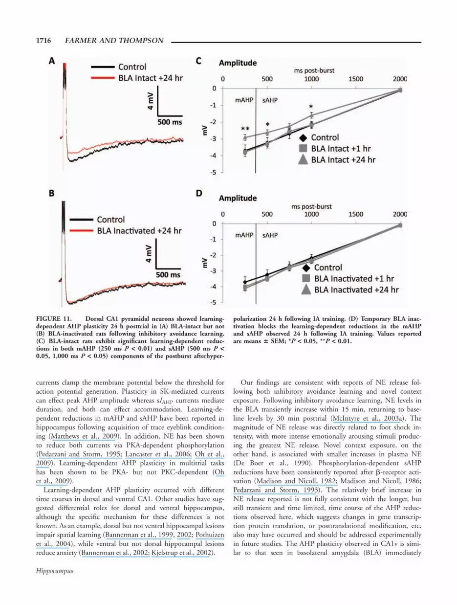
FIGURE 5. Dorsal CA1 pyramidal neurons exhibited transientlearning-dependent AHP plasticity that developed more slowly butextinguished at the same rate seen in ventral CA1 following inhibi-tory avoidance learning, i.e., AHP reductions were not observed1 h posttrial but were significant 24 h posttrial and extinguishedby 72 h posttrial. (A) Postburst afterhyperpolarizations were signif-icantly reduced 24 h following training with significant reductionsin both (B) mAHP (P < 0.05) and sAHP (P < 0.05) components.
Significant reductions in AHP (C) peak amplitude (P < 0.01), (D)duration (P < 0.01), and (E) integrated area (P < 0.05) wereobserved 24 h posttrial but extinguished by 72 h posttrial. Unlikein ventral CA1, context-only exposure to the IA apparatus did notresult AHP reductions in dorsal CA1 neurons at any intervaltested. Values reported are means 6 SEM; *P < 0.05, **P < 0.01.
AHP PLASTICITY IN CA1 AFTER INHIBITORY AVOIDANCE LEARNING 1709
Hippocampus

Dorsal CA1 Plasticity
Intracellular current clamp recordings from 75 pyramidalneurons in the dorsal CA1 (CA1d) were used to assess plastic-ity after inhibitory avoidance learning. CA1d pyramidal neu-rons exhibited postburst AHPs similar to those reported inearlier studies (Oh et al., 2003). However, while AHPs fromconditioned animals were reduced in CA1d neurons 24 hposttrial compared to naive controls (Fig. 5A), this effect wasnot seen 1 h posttrial unlike in CA1v neurons. An analysis ofAHP amplitudes at various postburst intervals (from 250 to1,000 ms) 24 h posttrial revealed reductions in both mAHPand sAHP components in neurons from conditioned ratscompared to controls (see Fig. 5B). Conditioned rats exhib-ited reductions in peak AHP amplitude (see Fig. 5C), dura-tion (see Fig. 5D), and area (see Fig. 5E) 24 h posttrial butnot 1 h posttrial in neurons from conditioned rats. Figures5C–E also shows that neurons from context-only exposed ratsexhibited no AHP plasticity. The learning-dependent AHPplasticity in CA1d was transient, beginning later than inCA1v, but similarly extinguished by 72 h followingconditioning.
Analyses of variance showed a significant effect of condition-ing on fast and slow components of the AHP in CA1d pyrami-dal neurons at 250 ms (F(5,74) 5 6.5, P < 0.0001), 500 ms(F(5,74) 5 9.4, P < 0.01), 750 ms (F(5,74) 5 11.6, P <0.0001), and 1,000 ms (F(5,74) 5 5.9, P < 0.0001) postburstwhen assessed 24 h posttrial. Similarly, significant reductionswere observed 24 h posttrial, in conditioned but not context-only exposed rats in peak AHP amplitude (F(5,74) 5 5.60, P< 0.0001), in AHP duration (F(5,74) 5 4.72, P < 0.001),and in AHP area (F(5,74) 5 1.81, P < 0.01).
Figure 6 shows learning-dependent plasticity in accommoda-tion was observed in conditioned but not in context-onlyexposed rats. Significant reductions in accommodation wereobserved 24 h posttrial, in conditioned but not context-onlyexposed rats (F(5,74) 5 4.31, P < 0.001). Inhibitory avoid-ance learning did not alter other membrane properties meas-ured (see Table 1).
Because learning-dependent AHP and accommodation plas-ticity was observed in ventral and dorsal CA1 pyramidal neu-rons, the next experiment investigated similarities and differen-ces in learning-dependent plasticity in CA3 pyramidal neurons.
CA3 Plasticity
Intracellular current clamp recordings from 93 pyramidalneurons in ventral CA3 were used to assess plasticity after in-hibitory avoidance learning. CA3 pyramidal neurons exhibitedpostburst AHPs similar to those reported in earlier studies(Schwartzkroin and Stafstrom, 1980; Aicardi and Schwartz-kroin, 1990; Thompson et al., 1996). Similar to CA1d pyrami-dal neurons (Fig. 7A), AHPs in CA3 neurons from conditionedrats were reduced 24 h posttrial compared to naive controls.An analysis of AHP amplitude at various postburst intervals(250, 500, 750, 1,000, 2,000, 3,000, and 4,000 ms) revealedreductions in sAHP (from 500 to 1,000 ms postburst) but notin mAHP components (see Fig. 7B). This analysis revealedlearning-dependent AHP plasticity, while more traditional anal-yses of peak AHP amplitude (Fig. 7C), duration (Fig. 7D), andarea (Fig. 7E) did not. None of the analyses showed any AHPplasticity related to context-only exposure.
Analyses of variance indicated a significant effect of condi-tioning but not context-only exposure on the AHP amplitude
FIGURE 6. Training in the IA task transiently reduced spike-frequency accommodation in dorsal CA1 pyramidal neurons, aneffect that again developed more slowly but extinguished at thesame rate as in ventral CA1. (A) Dorsal CA1 pyramidal neuronsfired more action potentials in response to a prolonged depolariz-ing pulse 24 h following training. (B) Transient reductions in
accommodation were observed 24 h (P < 0.05) posttrial but extin-guished by 72 h following training. Also different from ventralCA1, context-only exposure to the IA apparatus did not reduceaccommodation in dorsal CA1 neurons at any interval tested. Val-ues reported are means 6 SEM; *P < 0.05.
1710 FARMER AND THOMPSON
Hippocampus

of CA3 pyramidal neurons at 500 ms (F(4,92) 5 6.2, P <0.05), 750 ms (F(4,92) 5 8.9, P < 0.01), and 1,000 ms(F(4,92) 5 5.8, P < 0.05) postburst when assessed 24 h post-trial. However, no significant reductions were observed posttrialin conditioned or context-only exposed rats in measures ofpeak AHP amplitude (F(4,92) 5 1.23, P 5 0.3), AHP dura-tion (F(4,92) 5 0.92, P 5 0.45), or AHP area (F(4,92) 50.10, P 5 0.98).
Figure 8 shows that no learning-dependent plasticity inaccommodation was observed posttrial in CA3 neurons fromconditioned or context-only exposed rats (F(4,92) 5 0.43, P 50.78). Inhibitory avoidance learning also did not alter othermembrane properties measured in CA3 pyramidal neurons (seeTable 1).
Because learning-dependent AHP plasticity was observed inventral and dorsal CA1 pyramidal neurons in this single-trialaversive learning task, the next experiments investigated the
necessity of BLA inputs for learning-dependent plasticity inboth dorsal and ventral CA1 pyramidal neurons.
BLA Inactivation
Block of ventral CA1 plasticity
Intracellular current clamp recordings from 135 pyramidalneurons in ventral CA1 were used to assess effects of BLA inac-tivation on hippocampal plasticity after inhibitory avoidancelearning.
While the literature suggests that immediate posttrial BLAtreatments would be most disruptive of consolidation (Vazdar-janova and McGaugh, 1999; Miranda et al., 2003; Hui et al.,2004), both immediate pretrial and immediate posttrial BLAinactivation produced the same behavioral and neurophysiologi-cal effects in the current study. Rats without cannula implants(intact) and vehicle-treated rats both exhibited AHP and
FIGURE 7. AHP plasticity in CA3 pyramidal neurons was dif-ferent from that seen in CA1, i.e., peak AHP amplitudes wereunaffected, so reductions were restricted to the sAHP. (A) WhileCA3 pyramidal neurons exhibited reductions concentrated in thelate (sAHP) components of the postburst afterhyperpolarization,these effects developed sooner after training than the AHP reduc-tions seen in CA1 pyramidal neurons. Reductions were observed
1 h posttrial (B) with significant reductions only in the sAHP (P< 0.05); these effects extinguished within 24 h posttrial. No reduc-tions in AHP (C) peak amplitude, (D) duration or (E) integratedarea were observed following IA training or context-only exposure.Values reported are means 6 SEM; *P < 0.05.
AHP PLASTICITY IN CA1 AFTER INHIBITORY AVOIDANCE LEARNING 1711
Hippocampus

accommodation measures when assessed 1 h and 24 h posttrialthat were statistically identical between groups and comparableto that reported above in Figures 2–4. Because intact and vehi-cle-treated groups exhibited no significant differences betweengroups (using unpaired t tests) in peak AHP amplitude (1 h, P5 0.9; 24 h, P 5 0.9), duration (1 h, P 5 0.9; 24 h, P 50.9), area (1 h, P 5 0.3; 24 h, P 5 0.9), or accommodation(1 h, P 5 0.2; 24 h, P 5 0.9) data from these groups werecondensed for graphic purposes (Figs. 9 and 10) and arereferred to as BLA intact. In a similar manner, rats thatreceived immediate pretrial lidocaine or immediate posttrial bu-pivacaine infusions into the BLA had AHP and accommoda-tion measures when assessed 1 h and 24 h posttrial that werestatistically identical between these treatment groups. Becausepre- and posttrial BLA inactivation exhibited no significant dif-ferences (using unpaired t tests) between groups in measures ofpeak AHP amplitude (1 h, P 5 0.7; 24 h, P 5 0.7), duration(1 h, P 5 0.9; 24 h, P 5 0.8), area (1 h, P 5 0.9; 24 h, P 50.3), or accommodation (1 h, P 5 0.9; 24 h, P 5 0.9) datafrom these groups were also condensed for graphic purposesand are referred to as BLA inactivated.
Learning-dependent AHP plasticity occurred only in CA1vneurons from BLA-intact (Fig. 9A) but not BLA-inactivatedrats (Fig. 9C). An analysis of AHP amplitude at various post-burst intervals (from 250 to 1,000 ms) revealed reductions inboth mAHP and sAHP components in neurons from the BLA-intact (Fig. 9B) but not the BLA-inactivated rats (Fig. 9D).Analyses of variance indicated a significant effect of BLA inacti-vation on the AHP amplitude of CA1v pyramidal neurons at250 ms (F(4,125) 5 13.0, P < 0.0001), 500 ms (F(4,125) 510.5, P < 0.0001), 750 ms (F(4,125) 5 11.4, P < 0.0001),and 1,000 ms (F(4,125) 5 8.3, P < 0.0001) postburst inBLA-intact but not BLA-inactivated rats.
Similarly, increases in excitability were seen in CA1v neuronsfrom BLA-intact but not BLA-inactivated rats, with plasticityin peak AHP amplitude, duration, area, and in accommodation1 h and 24 h posttraining (Figs. 10A–D). Significant reduc-tions were observed 1 h and 24 h posttrial in BLA-intact butnot BLA-inactivated rats in peak AHP amplitude (F(4,125) 518.6, P < 0.0001), duration (F(4,125) 5 15.5, P < 0.0001),area (F(4,125) 5 6.9, P < 0.0001), and in accommodation(F(4,125) 5 12.9, P < 0.0001). Inhibitory avoidance learningdid not alter other membrane properties measured in BLA-intact or BLA-inactivated ventral CA1 pyramidal neurons (seeTable 2).
BLA inactivation also blocked the context-dependent tran-sient AHP plasticity seen in ventral CA1 neurons 1 h followingexposure to the IA apparatus. An analysis of AHP amplitudesat various postburst intervals (from 250 to 1,000 ms) revealedtransient reductions in both mAHP and sAHP components inneurons from BLA-intact but not from BLA-inactivated ratsthat underwent context-only exposure to the IA apparatus(Figs. 9E,F). Significant differences in AHP amplitude weredetected at 250 ms (F(2,62) 5 23.9, P < 0.0001), 500 ms(F(2,62) 5 14.2, P < 0.0001), 750 ms (F(2,62) 5 25.0, P <0.0001), and 1,000 ms (F(2,62) 5 20.5, P < 0.0001) post-burst. BLA-inactivated rats exhibited AHP and accommodationmeasures indistinguishable from controls (peak AHP amplitude,25.5 6 0.3 mV; duration, 1,443 6 158 ms; area, 21,008 644 mVms; and accommodation, 9.8 6 0.5 spikes). AHPs weresignificantly reduced 1 h following context-exposure in BLA-intact but not BLA-inactivated rats, with effects on peak AHPamplitude (F(2,62) 5 13.3, P < 0.0001), area (F(2,62) 511.9, P < 0.0001), and accommodation (F(2,62) 5 15.9, P <0.0001), but not duration (F(2,62) 5 2.1, P 5 0.12) whencompared to naive controls. Context-only exposure did not
FIGURE 8. Training in the IA task or context-only exposuredid not reduce spike-frequency accommodation in CA3 pyramidalneurons. (A) CA3 pyramidal neurons fired similar numbers ofaction potentials as controls following IA training in response to a
prolonged depolarizing pulse. (B) No reductions in accommoda-tion were observed 1 h or 24 h following IA training or after con-text-only exposure. Values reported are means 6 SEM.
1712 FARMER AND THOMPSON
Hippocampus

FIGURE 9. Ventral CA1 pyramidal neurons showed learning-de-pendent AHP plasticity in (A) BLA-intact but not (C) BLA-inactivatedrats following inhibitory avoidance learning. (E) BLA inactivation alsoblocked the context-dependent AHP plasticity seen 1 h postcontext-only exposure that is shown in Figure 3A. Learning-dependent reduc-tions in mAHP and sAHP components of the postburst afterhyperpo-larization of ventral CA1 pyramidal neurons were blocked by tempo-rary BLA inactivation following inhibitory avoidance learning. (B)BLA-intact rats exhibited significant learning-dependent reductions in
both mAHP (1 h P < 0.05, 24 h P < 0.01) and sAHP (1 h P < 0.05,24 h P < 0.01) components of the postburst afterhyperpolarization.(D) Temporary BLA inactivation blocked the learning-dependentreductions in the mAHP and sAHP both 1 h and 24 h following IAtraining. (E) Temporary BLA inactivation blocked the context-depend-ent reductions in the mAHP and sAHP 1 h following exposure to theIA apparatus. Values reported are means 6 SEM; *P < 0.05, **P <0.01.
AHP PLASTICITY IN CA1 AFTER INHIBITORY AVOIDANCE LEARNING 1713
Hippocampus

alter other membrane properties measured in BLA-inactivatedventral CA1 pyramidal neurons (see Table 2).
Block of dorsal CA1 plasticity
Intracellular current clamp recordings from 107 pyramidalneurons in dorsal CA1 were used to assess effects of BLA inac-tivation on hippocampal plasticity after inhibitory avoidancelearning.
Dorsal CA1 neurons from intact and vehicle-treated rats hadAHP and accommodation measures when assessed 1 h and24 h posttrial that were statistically identical between thesegroups and comparable to that reported above in Figures 5 and6. Because intact and vehicle-treated groups exhibited no signif-icant differences between groups (using unpaired t tests) inpeak AHP amplitude (1 h, P 5 0.8; 24 h, P 5 0.9), duration(1 h, P 5 0.9; 24 h, P 5 0.9), area (1 h, P 5 0.9; 24 h, P 50.9), or in accommodation (1 h, P 5 0.5; 24 h, P 5 0.1) datafrom these groups were condensed for graphical purposes (Figs.11 and 12) and are referred to as BLA intact. Similar to obser-vations in CA1v, CA1d neurons from rats that received lido-
caine or bupivacaine infusions into the BLA also had AHP andaccommodation measures when assessed 1 h and 24 h posttrialthat were statistically identical between these treatment groups.Because pre- and posttrial BLA inactivation exhibited no signif-icant differences (using unpaired t tests) between groups inmeasures of peak AHP amplitude (1 h, P 5 0.4; 24 h, P 50.4), duration (1 h, P 5 0.9; 24 h, P 5 0.9), area (1 h, P 50.9; 24 h, P 5 0.9), or accommodation (1 h, P 5 0.6; 24 h,P 5 0.5) data from these groups were condensed and arereferred to as BLA inactivated.
Learning-dependent AHP plasticity occurred only in CA1dneurons from BLA-intact (Fig. 11A) but not BLA-inactivatedrats (Fig. 11B). An analysis of AHP amplitude at various post-burst intervals (from 250 to 1,000 ms) revealed reductions inboth mAHP and sAHP components in neurons from the BLA-intact (Fig. 11C) but not the BLA-inactivated rats (Fig. 11D).Analyses of variance indicated a significant effect of BLA inacti-vation on the AHP amplitude of CA1d pyramidal neurons at250 ms (F(4,106) 5 7.6, P < 0.0001), 500 ms (F(4,106) 515.7, P < 0.001), and 1,000 ms (F(4, 106) 5 6.3, P <0.0001) postburst 24 h posttrial in the BLA-intact animals.
FIGURE 10. Learning-dependent changes in ventral CA1 py-ramidal neuron excitability observed following inhibitory avoid-ance learning was blocked by temporary BLA inactivation with thelocal anesthetics lidocaine or bupivacaine. BLA inactivation
blocked the reductions in AHP (A) peak amplitude, (B) duration,(C) integrated area and (D) accommodation observed in BLAintact treated rats both 1 h and 24 h following IA training. Valuesreported are means 6 SEM; *P < 0.05, **P < 0.01.
1714 FARMER AND THOMPSON
Hippocampus

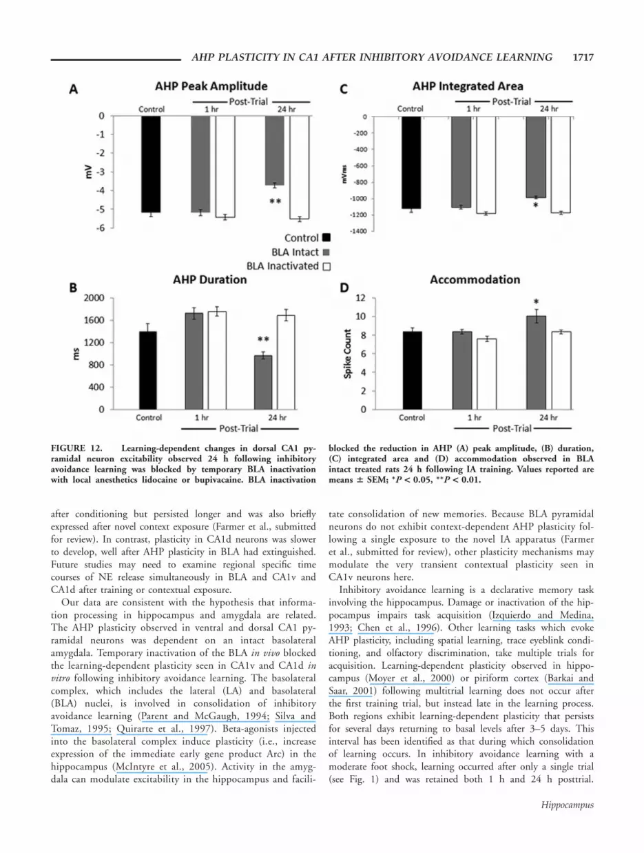
Increases in excitability were seen in CA1d neurons fromBLA-intact but not BLA-inactivated rats, with plasticity inpeak AHP amplitude, duration, area, and in accommodation24 h posttrial (Fig. 12A–D). Significant reductions wereobserved 24 h posttrial in BLA-intact but not BLA-inactivatedrats in peak AHP amplitude (F(4,106) 5 18.2, P < 0.0001),duration (F(4,106) 5 11.3, P < 0.0001), area (F(4,106) 56.9, P < 0.001), and in accommodation (F(4,106) 5 4.4, P <0.01). Inhibitory avoidance learning did not alter other mem-brane properties measured in BLA-intact or BLA-inactivateddorsal CA1 pyramidal neurons (see Table 2).
DISCUSSION
Hippocampal pyramidal neurons in ventral and dorsal CA1exhibited enhanced excitability following single-trial inhibitoryavoidance learning, exhibiting learning-dependent plasticity inboth AHP and accommodation measures. Reductions in bothmAHP and sAHP components were accompanied by reduc-tions in peak AHP amplitude, duration, and area. These learn-ing-dependent reductions in AHPs were associated with anotherincrease in excitability, i.e., a reduction in accommodation, fir-ing more action potentials during a prolonged depolarizing
pulse than neurons from naive animals. The AHP and accom-modation plasticity observed in CA1v neurons following condi-tioning were transient (i.e., seen 1 h and 24 h posttrial) andextinguished within 72 h posttrial. In addition to learning-spe-cific plasticity, context-dependent plasticity was observed inCA1v neurons 1 h after a single exposure to the IA apparatus.This very transient context-dependent plasticity was unique toCA1v neurons and extinguished by 24 h posttrial. Learning-de-pendent plasticity observed in CA1d neurons followed a timecourse different from that observed in CA1v neurons. Learn-ing-dependent increases in excitability were slower to developin CA1d neurons (i.e., observed 24 h posttrial) than in CA1vneurons, but still extinguished 72 h posttrial as in CA1v.Learning-dependent reductions in the sAHP from CA3 pyrami-dal neurons were also observed 24 h posttrial and extinguishedby 72 h, but this plasticity was not associated with learning-de-pendent changes in more traditional measures of AHP plastic-ity and was also insufficient to affect accommodation.
The calcium-dependent potassium currents underlyingmAHPs and sAHPs are mediated largely by apamin-sensitiveSK and apamin-insensitive sIAHP channels, respectively.Although calcium-dependence of the mAHP via SK channelactivation is a dominant viewpoint in the literature, there isevidence that the mAHP may also be generated by voltage-de-pendent K1 channels (Storm, 1989; Gu et al., 2005). Both
TABLE 2.
Membrane Properties of Ventral CA1 and Dorsal CA1 Pyramidal Neurons From Controls and After BLA Inactivation Following Contextual
Fear Conditioning
Rats Cells Input resistance Sag Resting potential Spike width AP amplitude
(n) (n) (MX) (mV) (mV) (mV) (mV)
CA1v
Control 19 29 35.5 6 1.9 6.2 6 0.4 266.8 6 0.9 1.3 6 0.02 86.0 6 1.2
Trained 1 h 22 37 41.0 6 1.9 6.2 6 0.4 265.4 6 0.9 1.3 6 0.02 84.5 6 0.8
Saline 1 h 2 6 53.4 6 4.0 8.0 6 1.4 269.6 6 1.2 1.4 6 0.04 89.3 6 0.8
Lidocaine 1 h 2 5 38.9 6 4.7 8.5 6 0.2 267.2 6 3.5 1.3 6 0.03 86.7 6 1.5
Bupivacaine 1 h 1 3 46.6 6 13.4 8.2 6 1.9 269.6 6 1.9 1.3 6 0.03 88.1 6 1.0
Context Inactivation 1 h 2 9 42.3 6 2.6 7.9 6 0.7 268.1 6 0.3 1.3 6 0.02 87.2 6 0.9
Trained 24 h 5 14 41.8 6 1.6 5.7 6 0.6 266.7 6 1.2 1.4 6 0.02 84.4 6 .08
Saline 24 h 3 8 52.5 6 2.5 9.5 6 0.4 267.8 6 1.3 1.5 6 0.05 83.5 6 0.9
Lidocaine 24 h 4 7 41.8 6 2.4 9.0 6 0.6 267.4 6 3.5 1.2 6 0.01 86.5 6 0.7
Bupivacaine 24 h 7 17 40.4 6 2.7 9.0 6 0.4 267.5 6 1.3 1.3 6 0.02 88.0 6 0.8
CA1d
Control 8 12 40.9 6 1.5 8.3 6 0.5 265.6 6 1.6 1.2 6 0.02 83.0 6 2.5
Trained 1 h 9 15 37.5 6 2.2 8.3 6 0.6 265.9 6 1.2 1.3 6 0.02 86.0 6 1.0
Saline 1 h 3 6 42.6 6 0.0 8.0 6 0.7 267.7 6 0.7 1.3 6 0.03 87.3 6 1.3
Lidocaine 1 h 2 6 36.3 6 5.1 6.4 6 0.7 267.6 6 3.1 1.3 6 0.02 86.7 6 1.5
Bupivacaine 1 h 1 3 42.4 6 0.0 6.6 6 0.6 267.4 6 2.9 1.3 6 0.01 85.1 6 1.3
Trained 24 h 6 14 36.7 6 2.8 9.4 6 0.7 266.7 6 1.0 1.3 6 0.01 88.2 6 0.8
Saline 24 h 5 15 42.3 6 2.7 7.9 6 0.7 268.9 6 1.0 1.3 6 0.01 85.3 6 0.8
Lidocaine 24 h 5 18 43.5 6 3.8 7.1 6 0.4 264.2 6 2.5 1.4 6 0.02 88.6 6 0.6
Bupivacaine 24 h 7 18 39.1 6 2.9 8.2 6 0.4 269.2 6 1.6 1.2 6 0.02 87.6 6 0.5
No differences in membrane properties of ventral CA1 and dorsal CA1 pyramidal neurons were observed between controls and after BLA inactivation following in-hibitory avoidance learning. Input resistance, sag, resting potential, spike width, and action potential amplitude values represent means 6 SEM.
AHP PLASTICITY IN CA1 AFTER INHIBITORY AVOIDANCE LEARNING 1715
Hippocampus
currents clamp the membrane potential below the threshold foraction potential generation. Plasticity in SK-mediated currentscan effect peak AHP amplitude whereas sIAHP currents mediateduration, and both can effect accommodation. Learning-de-pendent reductions in mAHP and sAHP have been reported inhippocampus following acquisition of trace eyeblink condition-ing (Matthews et al., 2009). In addition, NE has been shownto reduce both currents via PKA-dependent phosphorylation(Pedarzani and Storm, 1995; Lancaster et al., 2006; Oh et al.,2009). Learning-dependent AHP plasticity in multitrial taskshas been shown to be PKA- but not PKC-dependent (Ohet al., 2009).
Learning-dependent AHP plasticity occurred with differenttime courses in dorsal and ventral CA1. Other studies have sug-gested differential roles for dorsal and ventral hippocampus,although the specific mechanism for these differences is notknown. As an example, dorsal but not ventral hippocampal lesionsimpair spatial learning (Bannerman et al., 1999, 2002; Pothuizenet al., 2004), while ventral but not dorsal hippocampal lesionsreduce anxiety (Bannerman et al., 2002; Kjelstrup et al., 2002).
Our findings are consistent with reports of NE release fol-lowing both inhibitory avoidance learning and novel contextexposure. Following inhibitory avoidance learning, NE levels inthe BLA transiently increase within 15 min, returning to base-line levels by 30 min posttrial (McIntyre et al., 2003a). Themagnitude of NE release was directly related to foot shock in-tensity, with more intense emotionally arousing stimuli produc-ing the greatest NE release. Novel context exposure, on theother hand, is associated with smaller increases in plasma NE(De Boer et al., 1990). Phosphorylation-dependent sAHPreductions have been consistently reported after b-receptor acti-vation (Madison and Nicoll, 1982; Madison and Nicoll, 1986;Pedarzani and Storm, 1993). The relatively brief increase inNE release reported is not fully consistent with the longer, butstill transient and time limited, time course of the AHP reduc-tions observed here, which suggests changes in gene transcrip-tion protein translation, or posttranslational modification, etc.also may have occurred and should be addressed experimentallyin future studies. The AHP plasticity observed in CA1v is simi-lar to that seen in basolateral amygdala (BLA) immediately
FIGURE 11. Dorsal CA1 pyramidal neurons showed learning-dependent AHP plasticity 24 h posttrial in (A) BLA-intact but not(B) BLA-inactivated rats following inhibitory avoidance learning.(C) BLA-intact rats exhibit significant learning-dependent reduc-tions in both mAHP (250 ms P < 0.01) and sAHP (500 ms P <0.05, 1,000 ms P < 0.05) components of the postburst afterhyper-
polarization 24 h following IA training. (D) Temporary BLA inac-tivation blocks the learning-dependent reductions in the mAHPand sAHP observed 24 h following IA training. Values reportedare means 6 SEM; *P < 0.05, **P < 0.01.
1716 FARMER AND THOMPSON
Hippocampus
after conditioning but persisted longer and was also brieflyexpressed after novel context exposure (Farmer et al., submittedfor review). In contrast, plasticity in CA1d neurons was slowerto develop, well after AHP plasticity in BLA had extinguished.Future studies may need to examine regional specific timecourses of NE release simultaneously in BLA and CA1v andCA1d after training or contextual exposure.
Our data are consistent with the hypothesis that informa-tion processing in hippocampus and amygdala are related.The AHP plasticity observed in ventral and dorsal CA1 py-ramidal neurons was dependent on an intact basolateralamygdala. Temporary inactivation of the BLA in vivo blockedthe learning-dependent plasticity seen in CA1v and CA1d invitro following inhibitory avoidance learning. The basolateralcomplex, which includes the lateral (LA) and basolateral(BLA) nuclei, is involved in consolidation of inhibitoryavoidance learning (Parent and McGaugh, 1994; Silva andTomaz, 1995; Quirarte et al., 1997). Beta-agonists injectedinto the basolateral complex induce plasticity (i.e., increaseexpression of the immediate early gene product Arc) in thehippocampus (McIntyre et al., 2005). Activity in the amyg-dala can modulate excitability in the hippocampus and facili-
tate consolidation of new memories. Because BLA pyramidalneurons do not exhibit context-dependent AHP plasticity fol-lowing a single exposure to the novel IA apparatus (Farmeret al., submitted for review), other plasticity mechanisms maymodulate the very transient contextual plasticity seen inCA1v neurons here.
Inhibitory avoidance learning is a declarative memory taskinvolving the hippocampus. Damage or inactivation of the hip-pocampus impairs task acquisition (Izquierdo and Medina,1993; Chen et al., 1996). Other learning tasks which evokeAHP plasticity, including spatial learning, trace eyeblink condi-tioning, and olfactory discrimination, take multiple trials foracquisition. Learning-dependent plasticity observed in hippo-campus (Moyer et al., 2000) or piriform cortex (Barkai andSaar, 2001) following multitrial learning does not occur afterthe first training trial, but instead late in the learning process.Both regions exhibit learning-dependent plasticity that persistsfor several days returning to basal levels after 3–5 days. Thisinterval has been identified as that during which consolidationof learning occurs. In inhibitory avoidance learning with amoderate foot shock, learning occurred after only a single trial(see Fig. 1) and was retained both 1 h and 24 h posttrial.
FIGURE 12. Learning-dependent changes in dorsal CA1 py-ramidal neuron excitability observed 24 h following inhibitoryavoidance learning was blocked by temporary BLA inactivationwith local anesthetics lidocaine or bupivacaine. BLA inactivation
blocked the reduction in AHP (A) peak amplitude, (B) duration,(C) integrated area and (D) accommodation observed in BLAintact treated rats 24 h following IA training. Values reported aremeans 6 SEM; *P < 0.05, **P < 0.01.
AHP PLASTICITY IN CA1 AFTER INHIBITORY AVOIDANCE LEARNING 1717
Hippocampus

Learning-dependent plasticity was observed 1 h (CA1v) and 24h (both CA1v and CA1d) following this single trial, and extin-guished within 72 h in both regions. This is consistent withother findings that the consolidation period for this single-trialtask is much shorter than that for multitrial tasks (Hui et al.,2004). The single-trial learning-dependent plasticity in CA1pyramidal neurons not only showed a more rapid developmentbut was also more transient than that observed in multitriallearning paradigms. Taken together, the transient nature of theexcitability changes reported here suggest that persistence oflearning-dependent plasticity in CA1 and in other brain regionsmay be related to the time required for acquisition and consoli-dation of a task.
Acknowledgments
The authors thank A. Taylor, K. Bruckmann, A. Lovitz, C.Hovitz, and F. Elhorr for their technical assistance, and C.K.McIntyre for suggestions and use of the behavioral apparatus.
REFERENCES
Aicardi G, Schwartzkroin PA. 1990. Suppression of epileptiform burstdischarges in CA3 neurons of rat hippocampal slices by the organiccalcium channel blocker, verapamil. Exp Brain Res 81:288–296.
Ammassari-Teule M, Pavone F, Castellano C, McGaugh JL. 1991.Amygdala and dorsal hippocampus lesions block the effects ofGABAergic drugs on memory storage. Brain Res 551:104–109.
Bannerman DM, Yee BK, Good MA, Heupel MJ, Iversen SD, Raw-lins JN. 1999. Double dissociation of function within the hippo-campus: A comparison of dorsal, ventral, and complete hippocam-pal cytotoxic lesions. Behav Neurosci 113:1170–1188.
Bannerman DM, Deacon RM, Offen S, Friswell J, Grubb M, RawlinsJN. 2002. Double dissociation of function within the hippocampus:Spatial memory and hyponeophagia. Behav Neurosci 116:884–901.
Barkai E, Saar D. 2001. Cellular correlates of olfactory learning in therat piriform cortex. Rev Neurosci 12:111–120.
Chen C, Kim JJ, Thompson RF, Tonegawa S. 1996. Hippocampallesions impair contextual fear conditioning in two strains of mice.Behav Neurosci 110:1177–1180.
De Boer SF, Slangen JL, Van der Gugten J. 1990. Plasma catechol-amine and corticosterone levels during active and passive shock-prod avoidance behavior in rats: Effects of chlordiazepoxide. Phys-iol Behav 47:1089–1098.
Disterhoft JF, Coulter DA, Alkon DL. 1986. Conditioning-specificmembrane changes of rabbit hippocampal neurons measured invitro. Proc Natl Acad Sci USA 83:2733–2737.
Dutar P, Nicoll RA. 1989. Pharmacological characterization of musca-rinic responses in rat hippocampal pyramidal cells. Exs 57:68–76.
Gant JC, Thompson LT. 2000. Calmodulin mediated plasticity in rat CA1neurons following spatial learning. Soc Neurosci Abstr 30, 73.11.
Gu N, Vervaeke K, Hu H, Storm JF. 2005. Kv7/KCNQ/M andHCN/h, but not KCa2/SK channels, contribute to the somatic me-dium after-hyperpolarization and excitability control in CA1 hip-pocampal pyramidal cells. J Physiol 566 (Part 3):689–715.
Gustafsson B, Wigstrom H. 1981. Evidence for two types of afterhy-perpolarization in CA1 pyramidal cells in the hippocampus. BrainRes 206:462–468.
Haas HL, Greene RW. 1984. Adenosine enhances afterhyperpolariza-tion and accommodation in hippocampal pyramidal cells. PflugersArch 402:244–247.
Hennevin E, Maho C, Hars B. 1998. Neuronal plasticity induced byfear conditioning is expressed during paradoxical sleep: Evidencefrom simultaneous recordings in the lateral amygdala and themedial geniculate in rats. Behav Neurosci 112:839–862.
Hotson JR, Prince DA. 1980. A calcium-activated hyperpolarizationfollows repetitive firing in hippocampal neurons. J Neurophysiol43:409–419.
Hui GK, Figueroa IR, Poytress BS, Roozendaal B, McGaugh JL,Weinberger NM. 2004. Memory enhancement of classical fear con-ditioning by posttraining injections of corticosterone in rats. Neu-robiol Learn Mem 81:67–74.
Izquierdo I, Medina JH. 1993. Role of the amygdala, hippocampusand entorhinal cortex in memory consolidation and expression.Braz J Med Biol Res 26:573–589.
Kim JJ, Clark RE, Thompson RF. 1995. Hippocampectomy impairsthe memory of recently, but not remotely, acquired trace eyeblinkconditioned responses. Behav Neurosci 109:195–203.
Kjelstrup KG, Tuvnes FA, Steffenach HA, Murison R, Moser EI,Moser MB. 2002. Reduced fear expression after lesions of the ven-tral hippocampus. Proc Natl Acad Sci USA 99:10825–10830.
Lancaster B, Hu H, Gibb B, Storm JF. 2006. Kinetics of ion channelmodulation by cAMP in rat hippocampal neurones. J Physiol 576(Part 2):403–417.
Liang KC, McGaugh JL, Martinez JL Jr, Jensen RA, Vasquez BJ,Messing RB. 1982. Posttraining amygdaloid lesions impair reten-tion of an inhibitory avoidance response. Behav Brain Res 4:237–249.
Madison DV, Nicoll RA. 1982. Noradrenaline blocks accommodationof pyramidal cell discharge in the hippocampus. Nature 299:636–638.
Madison DV, Nicoll RA. 1984. Control of the repetitive discharge ofrat CA 1 pyramidal neurones in vitro. J Physiol 354:319–331.
Madison DV, Nicoll RA. 1986. Actions of noradrenaline recordedintracellularly in rat hippocampal CA1 pyramidal neurones, invitro. J Physiol 372:221–244.
Matthews EA, Linardakis JM, Disterhoft JF. 2009. The fast and slowafterhyperpolarizations are differentially modulated in hippocampalneurons by aging and learning. J Neurosci 29:4750–4755.
McGaugh JL, McIntyre CK, Power AE. 2002. Amygdala modulationof memory consolidation: Interaction with other brain systems.Neurobiol Learn Mem 78:539–552.
McIntyre CK, Power AE, Roozendaal B, McGaugh JL. 2003a. Role ofthe basolateral amygdala in memory consolidation. Ann N Y AcadSci 985:273–293.
McIntyre CK, Power AE, Roozendaal B, McGaugh JL. 2003b. Role ofthe basolateral amygdala in memory consolidation. Ann N Y AcadSci 985:273–293.
McIntyre CK, Miyashita T, Setlow B, Marjon KD, Steward O,Guzowski JF, McGaugh JL. 2005. Memory-influencing intra-baso-lateral amygdala drug infusions modulate expression of Arc proteinin the hippocampus. Proc Natl Acad Sci USA 102:10718–10723.
McKernan MG, Shinnick-Gallagher P. 1997. Fear conditioning indu-ces a lasting potentiation of synaptic currents in vitro. Nature390:607–611.
Miranda MI, LaLumiere RT, Buen TV, Bermudez-Rattoni F,McGaugh JL. 2003. Blockade of noradrenergic receptors in the ba-solateral amygdala impairs taste memory. Eur J Neurosci 18:2605–2610.
Moyer JR Jr, Thompson LT, Disterhoft JF. 1996. Trace eyeblink con-ditioning increases CA1 excitability in a transient and learning-spe-cific manner. J Neurosci 16:5536–5546.
Moyer JR Jr, Power JM, Thompson LT, Disterhoft JF. 2000. Increasedexcitability of aged rabbit CA1 neurons after trace eyeblink condi-tioning. J Neurosci 20:5476–5482.
Oh MM, Kuo AG, Wu WW, Sametsky EA, Disterhoft JF. 2003.Watermaze learning enhances excitability of CA1 pyramidal neu-rons. J Neurophysiol 90:2171–2179.
1718 FARMER AND THOMPSON
Hippocampus

Oh MM, McKay BM, Power JM, Disterhoft JF. 2009. Learning-related postburst afterhyperpolarization reduction in CA1 pyrami-dal neurons is mediated by protein kinase A. Proc Natl Acad SciUSA 106:1620–1625.
Ottersen OP. 1982. Connections of the amygdala of the rat. IV: Corti-coamygdaloid and intraamygdaloid connections as studied withaxonal transport of horseradish peroxidase. J Comp Neurol205:30–48.
Pare D. 2003. Role of the basolateral amygdala in memory consolida-tion. Prog Neurobiol 70:409–420.
Parent MB, McGaugh JL. 1994. Posttraining infusion of lidocaineinto the amygdala basolateral complex impairs retention of inhibi-tory avoidance training. Brain Res 661:97–103.
Pedarzani P, Storm JF. 1993. PKA mediates the effects of monoaminetransmitters on the K1 current underlying the slow spike frequencyadaptation in hippocampal neurons. Neuron 11:1023–1035.
Pedarzani P, Storm JF. 1995. Dopamine modulates the slow Ca(21)-activated K1 current IAHP via cyclic AMP-dependent protein ki-nase in hippocampal neurons. J Neurophysiol 74:2749–2753.
Petrovich GD, Risold PY, Swanson LW. 1996. Organization of projec-tions from the basomedial nucleus of the amygdala: A PHAL studyin the rat. J Comp Neurol 374:387–420.
Pikkarainen M, Ronkko S, Savander V, Insausti R, Pitkanen A. 1999.Projections from the lateral, basal, and accessory basal nuclei of theamygdala to the hippocampal formation in rat. J Comp Neurol403:229–260.
Pothuizen HH, Zhang WN, Jongen-Relo AL, Feldon J, Yee BK.2004. Dissociation of function between the dorsal and the ventralhippocampus in spatial learning abilities of the rat: A within-sub-ject, within-task comparison of reference and working spatial mem-ory. Eur J Neurosci 19:705–712.
Quirarte GL, Roozendaal B, McGaugh JL. 1997. Glucocorticoidenhancement of memory storage involves noradrenergic activationin the basolateral amygdala. Proc Natl Acad Sci USA 94:14048–14053.
Roozendaal B, McReynolds JR, Van der Zee EA, Lee S, McGaugh JL,McIntyre CK. 2009. Glucocorticoid effects on memory
consolidation depend on functional interactions between themedial prefrontal cortex and basolateral amygdala. J Neurosci29:14299–14308.
Saar D, Grossman Y, Barkai E. 1998. Reduced after-hyperpolarizationin rat piriform cortex pyramidal neurons is associated withincreased learning capability during operant conditioning. Eur JNeurosci 10:1518–1523.
Schenberg EE, Soares JC, Oliveira MG. 2005. Effects of pre- or post-training entorhinal cortex AP5 injection on fear conditioning.Physiol Behav 86:508–515.
Schwartzkroin PA. 1975. Characteristics of CA1 neurons recordedintracellularly in the hippocampal in vitro slice preparation. BrainRes 85:423–436.
Schwartzkroin PA, Stafstrom CE. 1980. Effects of EGTA on the cal-cium-activated afterhyperpolarization in hippocampal CA3 pyrami-dal cells. Science 210:1125–1126.
Silva MA, Tomaz C. 1995. Amnesia after diazepam infusion into ba-solateral but not central amygdala of Rattus norvegicus. Neuropsy-chobiology 32:31–36.
Staubli U, Ivy G, Lynch G. 1984. Hippocampal denervation causesrapid forgetting of olfactory information in rats. Proc Natl AcadSci USA 81:5885–5887.
Storm JF. 1989. An after-hyperpolarization of medium duration in rathippocampal pyramidal cells. J Physiol 409:171–190.
Thompson LT, Gant JC, Lea P. 2000. Postsynaptic plasticity in youngand aging rat CA1 neurons and spatial learning: Further emphasison accommodation. Soc Neurosci Abstr 30, 73.12.
Thompson LT, Moyer JR Jr, Disterhoft JF. 1996. Transient changes inexcitability of rabbit CA3 neurons with a time course appropriateto support memory consolidation. J Neurophysiol 76:1836–1849.
Vazdarjanova A, McGaugh JL. 1999. Basolateral amygdala is involvedin modulating consolidation of memory for classical fear condition-ing. J Neurosci 19:6615–6622.
Zelcer I, Cohen H, Richter-Levin G, Lebiosn T, Grossberger T, BarkaiE. 2006. A cellular correlate of learning-induced metaplasticity inthe hippocampus. Cereb Cortex 16:460–468.
AHP PLASTICITY IN CA1 AFTER INHIBITORY AVOIDANCE LEARNING 1719
Hippocampus