aging changes in voltage-gated calcium currents in hippocampal ca1 neurons
TRANSCRIPT
Aging Changes in Voltage-Gated Calcium Currents in HippocampalCA1 Neurons
Lee W. Campbell, Su-Yang Hao, Olivier Thibault, Eric M. Blalock, and Philip W. Landfield
Department of Pharmacology, College of Medicine, University of Kentucky, Lexington, Kentucky 40536
Previous current-clamp studies in rat hippocampal slice CA1neurons have found aging-related increases in long-lastingcalcium (Ca)-dependent and Ca-mediated potentials. Thesechanges could reflect an increase in Ca influx through voltage-gated Ca channels but also could reflect a change in potassiumcurrents. Moreover, if altered Ca influx is involved, it is unclearwhether it arises from generally increased Ca channel activity,lower threshold, or reduced inactivation. To analyze the basisfor altered Ca potentials, whole-cell voltage-clamp studies ofCA1 hippocampal neurons were performed in nondissociatedhippocampal slices of adult (3- to 5-month-old) and aged (25-to 26-month-old) rats. An aging-related increase was found inhigh-threshold Ca and barium (Ba) currents, particularly in theless variable, slowly inactivating (late) current at the end of adepolarization step. Input resistance of neurons did not differ
between age groups. In steady-state inactivation andrepetitive-pulse protocols, inactivation of Ca and Ba currentswas not reduced and, in some cases, was slightly greater inaged neurons, apparently because of larger inward current. Thecurrent blocked by nimodipine was greater in aged neurons,indicating that some of the aging increase was in L-type cur-rents. These results indicate that whole-cell Ca currents areincreased with aging in CA1 neurons, apparently attributable togreater channel activity rather than to reduced inactivation. Theelevated Ca influx seems likely to play a role in impaired func-tion and enhanced susceptibility to neurotoxic influences.
Key words: hippocampus; aging; calcium currents; inactiva-tion; afterhyperpolarization; neurotoxicity; barium currents; cal-cium homeostasis; Alzheimer’s disease
Increasing evidence gathered over more than a decade haspointed to altered neuronal calcium (Ca) homeostasis as a corre-late of brain aging (for review, see Khachaturian, 1984, 1989;Gibson and Peterson, 1987; Landfield, 1987, 1995; Landfield etal., 1992; Michaelis et al., 1992; Disterhoft et al., 1993, 1994;Michealis, 1994). Several Ca regulatory processes have been im-plicated in this Ca dysregulation, including those involved in thebuffering and extrusion of cytosolic Ca (Michaelis et al., 1984,1989; Peterson and Gibson, 1984; Gibson and Peterson, 1987;Martinez-Serrano et al., 1992).In addition, it appears that voltage-gated Ca influx may be
elevated in aged hippocampal neurons. An aging-related increasein voltage-activated potentials that are Ca-mediated or Ca-dependent [e.g., the Ca action potential and the Ca-dependentafterhyperpolarization (AHP)] has been found consistently inCA1 neurons of rats (Landfield and Pitler, 1984; Kerr et al., 1989;Pitler and Landfield, 1990) and rabbits (Moyer et al., 1992; Dis-terhoft et al., 1993). Furthermore, hippocampal frequency poten-tiation (facilitation), a form of short-term synaptic plasticity that isimpaired in aging rats, also is Ca-dependent. This type of facili-tation can be strengthened in aging rat hippocampal neurons byelevating external magnesium (Mg) (Landfield et al., 1986), whichblocks Ca influx through both voltage- and receptor-operated
Ca channels (Lansman et al., 1986; Mayer and Westbrook,1987).Several factors could account for an aging-related increase in
long-lasting Ca-mediated potentials. Not all potassium (K) cur-rents are blocked under the current-clamp protocols measuringvoltage, and therefore reduced K currents could underlie theprolongation and/or increased amplitude of Ca-mediated poten-tials. Alternatively, if the aging changes do arise from altered Cachannel activity, several possible factors could underlie thesechanges, including an increase in available Ca channels (Thibaultand Landfield, 1996), a decrease in threshold for Ca currentactivation, or reduced sensitivity of Ca currents to inactivation.However, these factors have not yet been evaluated in detail in theCA1 neurons in which increased Ca-mediated potentials havebeen found.High-threshold voltage-gated Ca currents in neurons are sub-
ject to Ca current-dependent inactivation as well as to voltage-dependent inactivation in a wide range of excitable cells (Eckertand Chad, 1984; Armstrong and Eckert, 1987; Obejero-Paz et al.,1991; Imredy and Yue, 1992), including hippocampal pyramidalneurons (Pitler and Landfield, 1987; Nistri and Cherubini, 1990;Kay, 1991). Moreover, independent voltage- and Ca-dependentforms of inactivation processes coexist in some of the same celltypes (Hadley and Lederer, 1991; Kay, 1991; Obejero-Paz et al.,1991). Clearly, a reduction of Ca- (or voltage)-dependent inacti-vation processes in aged neurons could be a factor in the increasein long-lasting Ca-mediated potentials in CA1 neurons. Ratherthan a reduction, several investigators instead have observedaging-related increases or no age differences in the inactivation ofCa currents, but this has been in other neuron types and in thecontext of different overall results (Reynolds and Carlen, 1989;
Received May 15, 1996; revised July 10, 1996; accepted July 15, 1996.This work was supported in part by grants from the National Institute on Aging
(AG04542 and AG10836) and Bayer, Inc. We thank Lisa Lowery for excellentassistance with this manuscript.Correspondence should be addressed to Dr. Philip W. Landfield, Department of
Pharmacology, College of Medicine, University of Kentucky Medical Center, MS-305, Lexington, KY 40536-0084.Dr. Campbell’s present address: The Salk Institute, La Jolla, CA 92037.Dr Hao’s present address: Department of Microbiology and Molecular Genetics,
University of Kansas Medical Center, Kansas City, KS 66160.Copyright q 1996 Society for Neuroscience 0270-6474/96/166286-10$05.00/0
The Journal of Neuroscience, October 1, 1996, 16(19):6286–6295
Kostyuk et al., 1993; Murchison and Griffith, 1995) (seeDiscussion).Thus, the mechanistic basis of the aging-related increase in Ca
influx in CA1 neurons or, in fact, whether such an increase inhigh-threshold current even is observed in CA1 neurons underwhole-cell voltage clamp remains uncertain. To address thesequestions, voltage-clamp analyses were carried out in the presentstudy, using the intracellular sharp electrode voltage-clamp(SEVC) method (Johnston et al., 1980) in nondissociated hip-pocampal slices to minimize alteration of the internal milieu ofthe neurons.
MATERIALS AND METHODSAnimals used in these experiments were healthy, male Fischer 344 (F344)rats obtained from the National Institute on Aging-sponsored HarlanIndustries specific-pathogen-free colony. Rats were 3–5 months (youngadult) or 25–26 months (aged) of age when used and were housed in anair-barrier protected system before use. Slices were prepared and main-tained using techniques generally similar to those described elsewhere(for review, see Dingledine, 1984) (see also Pitler and Landfield, 1990;Thibault et al., 1994). After decapitation, the brains were removed rapidlyand chilled to 08C in artificial CSF (ACSF). The hippocampi werecarefully dissected free and placed on the tissue chopper. Approximatelyten 450-mM-thick slices were cut from the middle of each hippocampustransverse to its longitudinal axis. The slices were maintained in aninterface type recording chamber at a temperature of 32.58C.A nylon mesh net supported slices at the interface of an atmosphere of
moistened 95%O2/5% CO2 and the ACSF bath containing (in mM): NaCl128, KH2PO4 1.25, glucose 10, NaHCO3 26, KCl 3, CaCl2 2, MgCl2 2.Before recording, slices were allowed to equilibrate with the medium andrecover for 1 hr.Borosilicate glass micropipettes (World Precision Instruments, Gaith-
erburg, MD), pulled on a Sutter Instruments P-80/PC puller (Novato,CA) (70–100 MV, filled with 2 M cesium (Cs)Cl2, pH 7.15), were used toimpale CA1 neurons. Data were recorded in both current-clamp anddiscontinuous SEVC modes (Axoclamp 2A, Axon Instruments, FosterCity, CA) and displayed on a digital storage oscilloscope (Nicolet model3091). Cells generally were voltage-clamped with a 2–3 kHz samplingfrequency using the continuous output of the headstage to ensure fulldecay of voltage across the electrode before each sampling point. Thelong time constants of these neurons (15–25 msec) met the requirementsfor effective discontinuous voltage clamp (e.g., membrane time constant.. electrode time constant) even with these high resistance micropipettes(cf. Johnston et al., 1980; Finkel and Redman, 1985). Leak subtractionwas performed digitally on-line by the method of fractional hyperpolar-izing pulses. The data were stored and analyzed off-line with cursors ona computer equipped with TECMAR analog/digital converter and mathcoprocessor using programs developed in the laboratory based on ASYSTTechnologies software (L. Campbell, unpublished programs). Repeated-measures ANOVA and post hoc Bonferroni group comparisons wereused to analyze responses of adult and aged rat neurons.Although differences in the passive, electrotonic properties of the cell
could result in altered control of regenerative voltages (Johnston andBrown, 1983; Spruston et al., 1994), this does not appear to be a factor inthe present studies. Most intracellular studies have found that there areno major aging-related changes in the passive membrane properties[input resistance (IR) and time constants]) of several types of hippocam-pal neurons (cf. Barnes and McNaughton, 1980; Landfield and Pitler,1984; Kerr et al., 1989; Reynolds and Carlen, 1989; Pitler and Landfield,1990; Potier et al., 1993; Moyer and Disterhoft, 1994) (for review, seeBarnes, 1994). In addition, although estimates in CA1 pyramids indicatethat the electrotonic length is ;0.9 l (Brown et al., 1981; Johnston andBrown, 1983; Turner and Schwartzkroin, 1984), the large dendritic shafts,which with the soma contain the vast majority of L-type channels (Hell etal., 1993), are electrotonically close to the soma (e.g., 0.1–0.2 spaceconstants). Cells in this study also were Cs-loaded, which presumablyreduced electrotonic distance significantly (Johnston et al., 1980), andmany of the currents were studied at the end of the pulse (late current)from a holding potential of 240 mV, which enhanced clamp efficacy (seeResults).Inconsistent viability of slices and neurons can affect substantially the
variance of Ca current recordings, making it extremely difficult to detectsubtle aging changes. Therefore, all slices and cells included for analysis
had to meet several rigorous criteria. A slice was considered healthy andusable in the present study if a population spike of 4–8 mV amplitudecould be recorded by an extracellular pipette (3–7 MV) located in theCA1 pyramidal layer in response to a 100 msec, 300–400 mA pulseapplied through a bipolar stimulating electrode to the Schaffer-commissural fibers and if other slices in the same well did not exhibitseizure-like activity during strong repetitive stimulation (;1000 mA) at 2Hz. IR measurements were obtained from each cell (during a 40 msec, 0.2nA constant current hyperpolarizing pulse) from a holding potential of270 mV. To reduce variability attributable to either cell size or poorhealth, only cells from healthy slices that exhibited sodium (Na) spikes ofat least 75 mV, near-complete Cs blockade of the Ca-dependent AHP,and an IR between 35 and 65 MV were used in these studies. No agedifferences were found in the proportions of neurons meeting thesecriteria.For cells meeting the criteria above, tetrodotoxin (TTX) (1 3 1026 M)
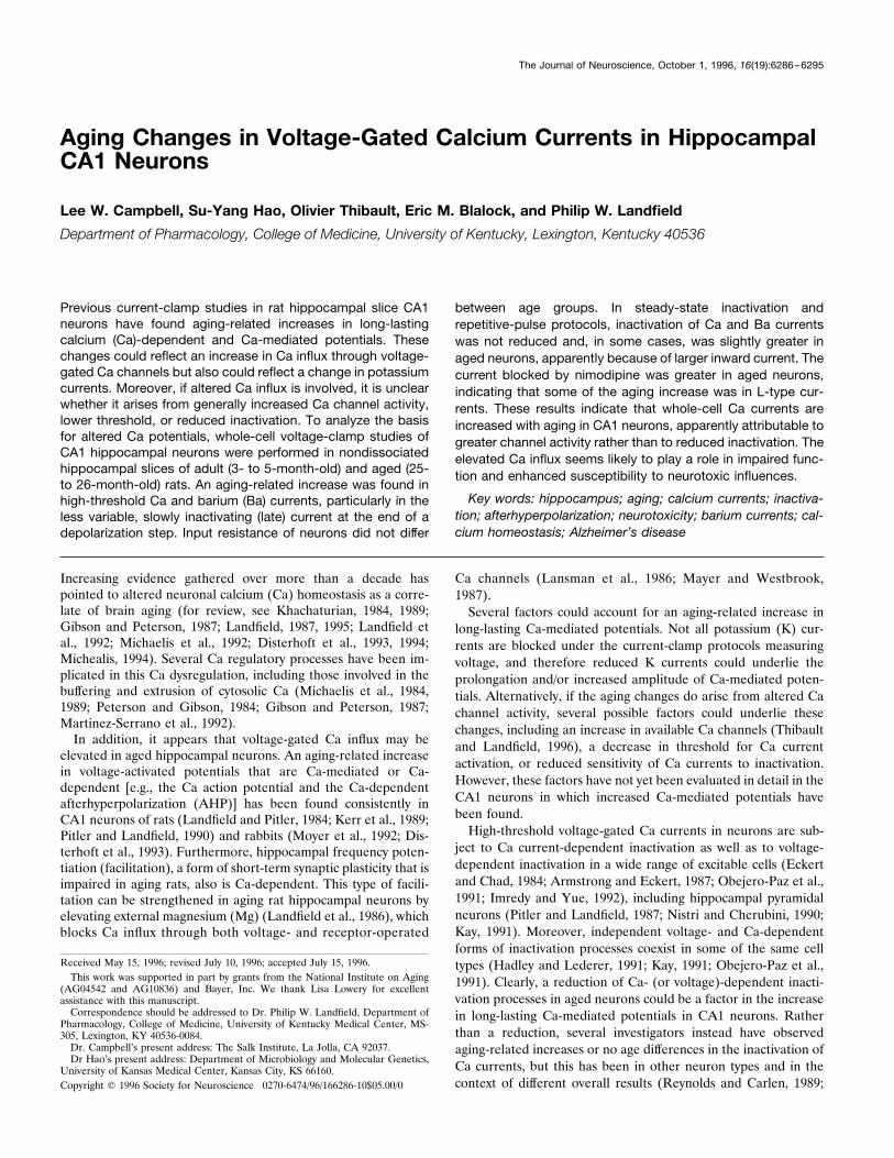
was applied to the bath to block Na spikes (Fig. 1). After Cs loading andapplication of TTX, a Ca action potential (spike) was elicited in current-clamp mode (Fig. 1B) by holding the cell at 270 mV and applying anintracellular 40 msec constant current depolarizing pulse (at 150% ofthreshold for the Ca spike). For subsequent study in voltage-clamp mode,slices then were treated with tetraethylammonium chloride (TEA) (5
Figure 1. Voltage-response records of a Cs-loaded CA1 hippocampalneuron from a young animal. A, Intracellular current injection induces aburst of Na action potentials. Note that 2 M CsCl in the pipette blocks theAHP. B, Blocking Na action potentials with TTX unmasks a sharp Caspike followed by a slow lower amplitude (“hump”) phase lasting .200msec. C, Additional block of repolarizing K conductances with TEAprevents repolarization of the sharp spike component resulting in a longCa action potential plateau at near-maximum amplitude (;2 sec). Allrecords are from the same cell and were recorded at a holding potential of270 mV; 400 msec horizontal scale bar applies to C only.
Campbell et al. • Ca Currents in Neurons from Adult and Aged Rats J. Neurosci., October 1, 1996, 16(19):6286–6295 6287
mM) to block most remaining voltage-activated K conductances (Storm,1990). Efficacy of TEA block was assessed for each neuron used in thesestudies by the occurrence of a substantial prolongation of the plateau ofthe initial fast component of the Ca spike (from ;30 msec without TEAto about ;3000 msec with TEA) and was checked repeatedly throughoutthe experiment and strengthened by adding TEA if necessary (Fig. 1C).In some studies, the dihydropyridine (DHP) L-type Ca channel
antagonist nimodipine (Bayer, West Haven, CT) was applied to thebath at a saturating final concentration of 10 mM. This concentration issubstantially higher than necessary to saturate L channels (McCarthyand TanPiengco, 1992) but was used in light of the variable drugavailability and diffusion that occurs in a thick slice preparation.Nimodipine was mixed in stock solutions with 100% ethanol andprotected from light during storage and throughout the experiments.The stock solutions were mixed with ACSF to achieve a final alcoholdilution of no more than 0.05%.
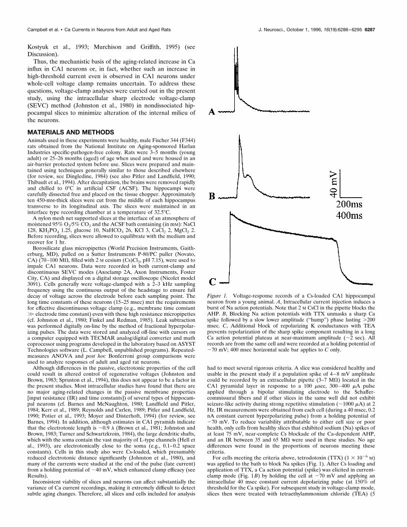
RESULTSCalcium action potentials (spikes)Before performing the voltage-clamp studies, it was important toensure that the particular cells being analyzed in this study alsoshowed the aging-related increases in Ca spikes that had beenseen previously. Therefore, before establishing voltage clamp,neurons were run through a Ca spike measurement and inactiva-tion protocol similar to those used in previous studies. Under ourconditions (Pitler and Landfield, 1987, 1990), Ca spikes exhibittwo distinct phases: (1) a fast spike component with a sharp onsetand peak amplitude of ;80 mV, lasting 20–30 msec, and (2) asubsequent lower amplitude plateau phase, or “hump,” compo-nent lasting ;200-250 msec, followed by a gradual return toresting membrane potential (Fig. 1B) (see also Disterhoft et al.,1993). After stabilization, Ca spike duration, amplitude, and in-activation during a 2 Hz train of depolarizing current pulses(150% threshold) were measured. As shown in Figure 2A, theaged rat CA1 neurons in the present study exhibited longer Caspike durations than did neurons from young-adult rats (F(1,39) 514.9, p , 0.001; adult cells, n 5 28; aged cells, n 5 13), replicatingearlier findings (Pitler and Landfield, 1990; Disterhoft et al.,1993). Again, no differences in peak spike amplitude were ob-served. Inactivation of the Ca spike during repetitive activationhas been shown previously to be Ca-dependent, because it is muchreduced in barium (Ba) (Pitler and Landfield, 1987). The presentstudies also replicated the observation that Ca-dependent inacti-vation of the Ca spike is not reduced with aging (Pitler andLandfield, 1990) and, in fact, may have been somewhat increasedin the present study, as indicated by a significant interactionbetween age and the train of five consecutive depolarizationselicited at 2 Hz (F(4,156) 5 7.80; p , 0.001) (Fig. 2B). Thisinteraction appears to reflect somewhat steeper inactivation be-tween the first and second pulses in the aged neurons (Fig. 2B).Thus, this population of neurons showed aging changes in long-lasting Ca-mediated potentials similar to those seen previouslyand, therefore, provided an appropriate population in which toinvestigate the underlying currents in voltage-clamp mode. Inaddition, as in earlier studies in rat brain neurons (Barnes andMcNaughton, 1980; Landfield and Pitler, 1984; Kerr et al., 1989;Potier et al., 1993; Barnes, 1994), IR of neurons did not differ withaging (adult, 47 6 3.1 MV, aged 48.3 6 2.7 MV), indicating thatneurons from the two age groups generally were similar in sizeand passive electrotonic structure.
Ca currentsAfter voltage measures were obtained, neurons were treated withTEA to block most of the remaining K conductances (Fig. 1C),and the recording mode was changed from current clamp to
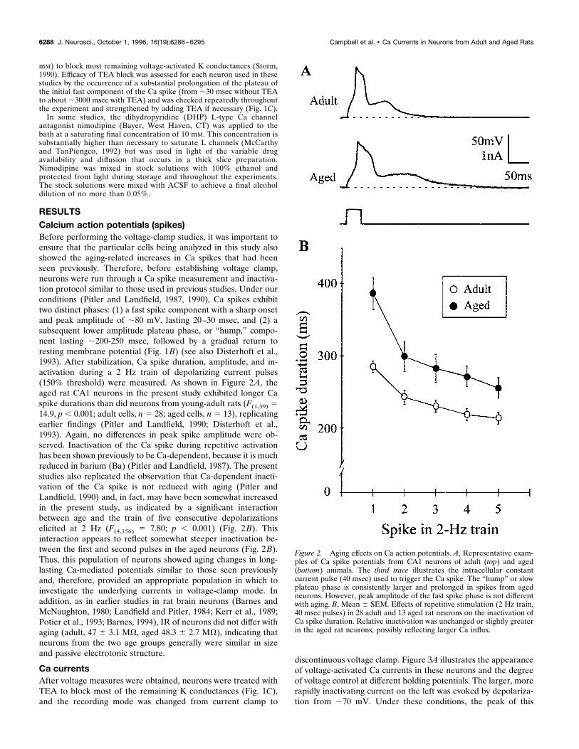
discontinuous voltage clamp. Figure 3A illustrates the appearanceof voltage-activated Ca currents in these neurons and the degreeof voltage control at different holding potentials. The larger, morerapidly inactivating current on the left was evoked by depolariza-tion from 270 mV. Under these conditions, the peak of this
Figure 2. Aging effects on Ca action potentials. A, Representative exam-ples of Ca spike potentials from CA1 neurons of adult (top) and aged(bottom) animals. The third trace illustrates the intracellular constantcurrent pulse (40 msec) used to trigger the Ca spike. The “hump” or slowplateau phase is consistently larger and prolonged in spikes from agedneurons. However, peak amplitude of the fast spike phase is not differentwith aging. B, Mean 6 SEM. Effects of repetitive stimulation (2 Hz train,40 msec pulses) in 28 adult and 13 aged rat neurons on the inactivation ofCa spike duration. Relative inactivation was unchanged or slightly greaterin the aged rat neurons, possibly reflecting larger Ca influx.
6288 J. Neurosci., October 1, 1996, 16(19):6286–6295 Campbell et al. • Ca Currents in Neurons from Adult and Aged Rats
current was difficult to clamp completely with the SEVC, as shownby the error in the clamp voltage (Fig. 3A, arrow, bottom left) (seealso Johnston et al., 1980; Finkel and Redman, 1985). However,much of this initial large peak current could be inactivated at a
holding potential of 240 mV (right) allowing a substantially moreeffective clamp (Fig. 3A, bottom right) (see also Gahwiler andBrown, 1987; Nistri and Cherubini, 1990). Therefore, the moreslowly inactivating Ca currents, which include the L-type andsome N- and P/Q-type currents (Fox et al., 1987; Llinas et al.,1989; Plummer et al., 1989; Swartz and Bean, 1992; Eliot andJohnston, 1994; Randall and Tsien, 1995), appear to be studiedmore accurately by holding at 240 mV. In addition, because ofinactivation during the pulse, measurement accuracy was en-hanced further by measuring current at the end of the depolariz-ing command step (late current) rather than at the peak. Conse-quently, many of the aging comparisons were performed from aholding potential of 240 mV, and late current generally wasmeasured along with peak current.Because of this difficulty in accurately clamping the large,
rapidly inactivating peak currents, a current–voltage (I–V) analy-sis of activation patterns was not performed for each neuron inthis study. However, I–V relations were studied in subsets of adultand aged neurons in which the clamp appeared to be mosteffective (as determined by a gradual activation curve and minimalloss of control in the voltage trace). In these subsets (n 5 6 adultand 5 aged neurons), the voltage dependence of the Ca currentsappeared similar in adult and aged neurons with maximum cur-rent elicited during steps to the 220 to 210 mV range (Fig. 3B).Consequently, activation protocols for aging comparisons in thepresent studies ensured full activation by employing voltage com-mand steps to 0 mV, well above maximum. A prominent long-lasting tail current generally follows each depolarization pulse(Fig. 3A). As shown in Figure 3A (right), the long tail oftenexhibits a delayed activation at lower holding potentials. Theselong tail currents are observed consistently in adult slice hip-pocampal neurons (Pitler and Landfield, 1987; Nistri and Cheru-bini, 1990; Kerr et al., 1992) and resemble a space-clamp artifactthat could arise from unclamped distal dendrites. However, singleCa channel openings during the repolarization period that followsa depolarization pulse also are observed on the somata of hip-pocampal neurons (Fisher et al., 1990; Thibault et al., 1993,Kavalali and Plummer, 1996). In addition, several lines of evi-dence indicate that the long tail current is a Ca current and thatit does not arise in the large apical dendrite (Thibault et al., 1995).On the other hand, the single-channel openings on the somaduring the repolarization phase do not seem sufficient to accountfor these large tails under relatively physiological conditions.Therefore, the tail currents may arise from a combination ofrepolarization openings of Ca channels and unclamped smalldendrites. However, this is a complex and unresolved issue, andthe tail current was not investigated systematically in the presentstudy.
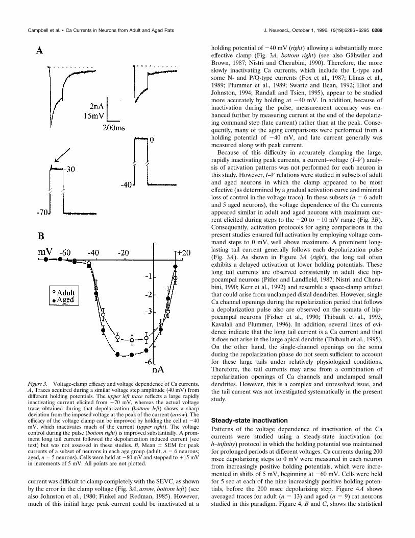
Steady-state inactivationPatterns of the voltage dependence of inactivation of the Cacurrents were studied using a steady-state inactivation (orh–infinity) protocol in which the holding potential was maintainedfor prolonged periods at different voltages. Ca currents during 200msec depolarizing steps to 0 mV were measured in each neuronfrom increasingly positive holding potentials, which were incre-mented in shifts of 5 mV, beginning at 260 mV. Cells were heldfor 5 sec at each of the nine increasingly positive holding poten-tials, before the 200 msec depolarizing step. Figure 4A showsaveraged traces for adult (n 5 13) and aged (n 5 9) rat neuronsstudied in this paradigm. Figure 4, B and C, shows the statistical
Figure 3. Voltage-clamp efficacy and voltage dependence of Ca currents.A, Traces acquired during a similar voltage step amplitude (40 mV) fromdifferent holding potentials. The upper left trace reflects a large rapidlyinactivating current elicited from 270 mV, whereas the actual voltagetrace obtained during that depolarization (bottom left) shows a sharpdeviation from the imposed voltage at the peak of the current (arrow). Theefficacy of the voltage clamp can be improved by holding the cell at 240mV, which inactivates much of the current (upper right). The voltagecontrol during the pulse (bottom right) is improved substantially. A prom-inent long tail current followed the depolarization induced current (seetext) but was not assessed in these studies. B, Mean 6 SEM for peakcurrents of a subset of neurons in each age group (adult, n 5 6 neurons;aged, n 5 5 neurons). Cells were held at 280 mV and stepped to 115 mVin increments of 5 mV. All points are not plotted.
Campbell et al. • Ca Currents in Neurons from Adult and Aged Rats J. Neurosci., October 1, 1996, 16(19):6286–6295 6289
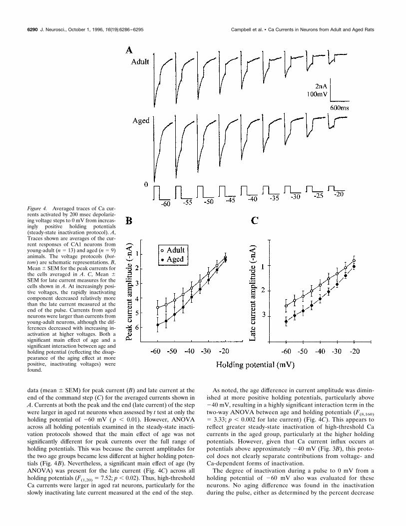
data (mean 6 SEM) for peak current (B) and late current at theend of the command step (C) for the averaged currents shown inA. Currents at both the peak and the end (late current) of the stepwere larger in aged rat neurons when assessed by t test at only theholding potential of 260 mV ( p , 0.01). However, ANOVAacross all holding potentials examined in the steady-state inacti-vation protocols showed that the main effect of age was notsignificantly different for peak currents over the full range ofholding potentials. This was because the current amplitudes forthe two age groups became less different at higher holding poten-tials (Fig. 4B). Nevertheless, a significant main effect of age (byANOVA) was present for the late current (Fig. 4C) across allholding potentials (F(1,20) 5 7.52; p , 0.02). Thus, high-thresholdCa currents were larger in aged rat neurons, particularly for theslowly inactivating late current measured at the end of the step.
As noted, the age difference in current amplitude was dimin-ished at more positive holding potentials, particularly above240 mV, resulting in a highly significant interaction term in thetwo-way ANOVA between age and holding potentials (F(8,160)5 3.33; p , 0.002 for late current) (Fig. 4C). This appears toreflect greater steady-state inactivation of high-threshold Cacurrents in the aged group, particularly at the higher holdingpotentials. However, given that Ca current influx occurs atpotentials above approximately 240 mV (Fig. 3B), this proto-col does not clearly separate contributions from voltage- andCa-dependent forms of inactivation.The degree of inactivation during a pulse to 0 mV from a
holding potential of 260 mV also was evaluated for theseneurons. No aging difference was found in the inactivationduring the pulse, either as determined by the percent decrease
Figure 4. Averaged traces of Ca cur-rents activated by 200 msec depolariz-ing voltage steps to 0 mV from increas-ingly positive holding potentials(steady-state inactivation protocol). A,Traces shown are averages of the cur-rent responses of CA1 neurons fromyoung-adult (n 5 13) and aged (n 5 9)animals. The voltage protocols (bot-tom) are schematic representations. B,Mean6 SEM for the peak currents forthe cells averaged in A. C, Mean 6SEM for late current measures for thecells shown in A. At increasingly posi-tive voltages, the rapidly inactivatingcomponent decreased relatively morethan the late current measured at theend of the pulse. Currents from agedneurons were larger than currents fromyoung-adult neurons, although the dif-ferences decreased with increasing in-activation at higher voltages. Both asignificant main effect of age and asignificant interaction between age andholding potential (reflecting the disap-pearance of the aging effect at morepositive, inactivating voltages) werefound.
6290 J. Neurosci., October 1, 1996, 16(19):6286–6295 Campbell et al. • Ca Currents in Neurons from Adult and Aged Rats
in current from beginning to end of the pulse or by the timeconstant of the decay. In young-adult rat neurons, the percentdecrease during the pulse was 42.8 6 3.0%, whereas in aged ratneurons, the decrease was 46.2 6 1.9%. The average timeconstant of decay over the 200 msec pulse was 51.3 msec for theadult rat neurons and 60.1 msec for the aged rat neurons.
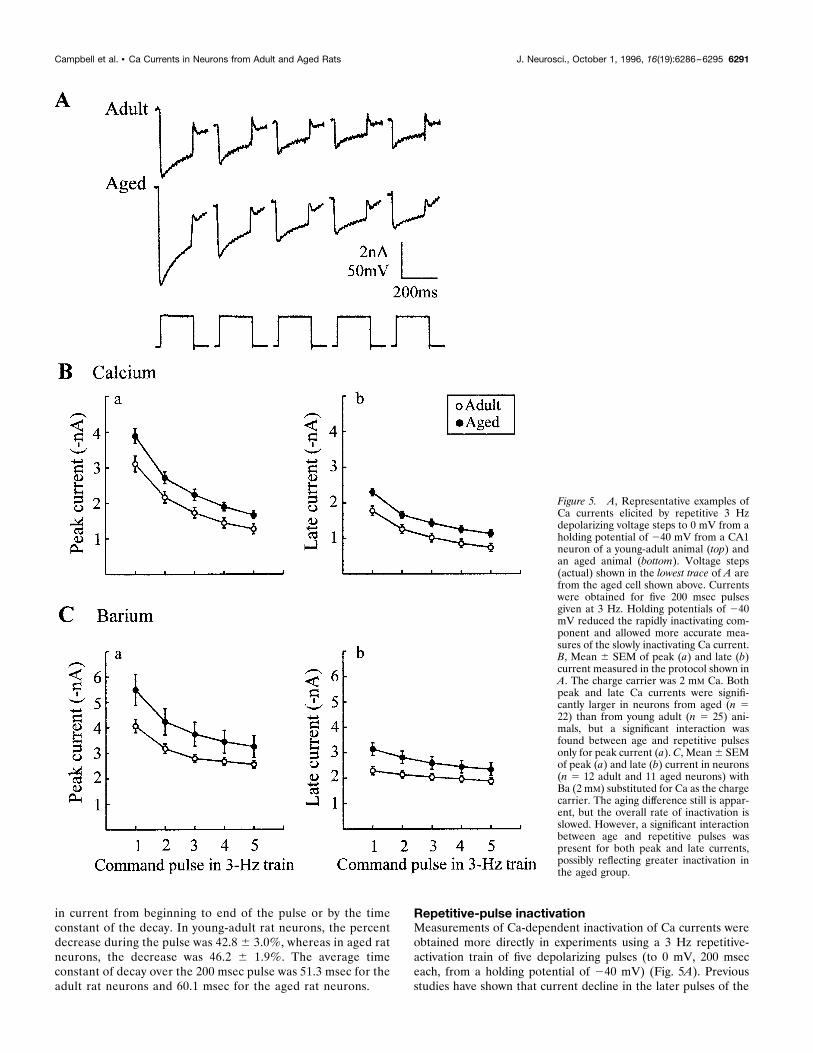
Repetitive-pulse inactivationMeasurements of Ca-dependent inactivation of Ca currents wereobtained more directly in experiments using a 3 Hz repetitive-activation train of five depolarizing pulses (to 0 mV, 200 mseceach, from a holding potential of 240 mV) (Fig. 5A). Previousstudies have shown that current decline in the later pulses of the
Figure 5. A, Representative examples ofCa currents elicited by repetitive 3 Hzdepolarizing voltage steps to 0 mV from aholding potential of 240 mV from a CA1neuron of a young-adult animal (top) andan aged animal (bottom). Voltage steps(actual) shown in the lowest trace of A arefrom the aged cell shown above. Currentswere obtained for five 200 msec pulsesgiven at 3 Hz. Holding potentials of 240mV reduced the rapidly inactivating com-ponent and allowed more accurate mea-sures of the slowly inactivating Ca current.B, Mean 6 SEM of peak (a) and late (b)current measured in the protocol shown inA. The charge carrier was 2 mM Ca. Bothpeak and late Ca currents were signifi-cantly larger in neurons from aged (n 522) than from young adult (n 5 25) ani-mals, but a significant interaction wasfound between age and repetitive pulsesonly for peak current (a). C, Mean6 SEMof peak (a) and late (b) current in neurons(n 5 12 adult and 11 aged neurons) withBa (2 mM) substituted for Ca as the chargecarrier. The aging difference still is appar-ent, but the overall rate of inactivation isslowed. However, a significant interactionbetween age and repetitive pulses waspresent for both peak and late currents,possibly reflecting greater inactivation inthe aged group.
Campbell et al. • Ca Currents in Neurons from Adult and Aged Rats J. Neurosci., October 1, 1996, 16(19):6286–6295 6291
train in this protocol directly reflects Ca-dependent inactivation inthese neurons, because the degree of inactivation is reduced whenBa is the charge carrier and because no detectable outwardcurrents are present between pulses (Pitler and Landfield, 1987).In 25 adult and 22 aged neurons, a significant main effect of agewas found on peak current (Fig. 5B, a) across the 3 Hz train(F(1,45) 5 6.07; p, 0.02). In addition, the main effect of repetitivestimulation was highly significant ( p , 0.001). The interactionbetween age and repeated pulses also was significant (F(4,180) 52.96; p , 0.03), again reflecting a difference in the inactivationpattern between the young and aged groups. As with the Ca spikedata (Fig. 2B), this interaction appeared to result primarily fromrelatively steeper inactivation between the first and the secondpulses in the aged group. However, it is difficult to conclude thatinactivation generally was greater in the aged group, becausewhen currents were normalized to the first pulse in each neuron,no significant age differences were observed in the decline offractional current across pulses (data not shown). Late current(Fig. 5B, b) also showed a significant main effect of age (F(1,45) 57.21; p , 0.01), although a significant interaction with repetitivepulses was not seen. As with peak current, analyses of normalizedfractional late current did not show an age difference (data notshown).A similar experiment was performed on a different set of cells
(adult, n 5 12; aged, n 5 11) in which Ba (2 mM) was substitutedfor Ca as the primary external divalent cation charge carrier (Fig.5C, a,b). In Ba, overall currents were larger, and the generaldegree of inactivation was reduced compared with Ca-bathedcells, as would be anticipated from the relatively greater perme-ability of Ba through Ca channels (Tsien et al., 1988) and itsrelatively weaker effect in inducing current-dependent inactiva-tion of Ca channels (Eckert and Chad, 1984; Pitler and Landfield,1987; Kay, 1991; Obejero-Paz et al., 1991). Nevertheless, agingeffects were analogous to those in Ca in that the main effect ofaging was significant across the five-pulse train for the late current(F(1,21) 5 4.8, p , 0.04). Some differences from Ca were noted,however, in that main effects of age on peak current were notsignificant (apparently reflecting greater variability attributable tolarger peak amplitudes in Ba), but a highly significant interactionwas found between age and repeated stimulation (F(4,84) 5 14,p , 0.001) for late current, again possibly reflecting greaterinactivation during the train in the aged rat neurons (Fig. 5C).This latter conclusion was supported by the observation that,unlike the results in Ca medium, ANOVAs of normalized cur-rents revealed significant main effects of aging resulting from agreater decline of fractional current for aged neurons on bothpeak ( p 5 0.05) and late ( p , 0.002) currents (data not shown).Although not as effective as Ca, Ba can induce current-dependentinactivation in the hippocampus (Kay, 1991) as well as other celltypes (Fedulova et al., 1985; Kasai and Aosaki, 1988; Mazzanti etal., 1991a). Thus, although some differences are seen between Baand Ca currents, possibly related to different current amplitudesor inactivation efficacies, overall, the repetitive-activation studiesindicate that the degree of inactivation of Ca channel currents wasnot reduced with aging and, instead, may have been slightlygreater in some experiments.
Contribution of L-type channelsL-type channels appear to contribute to the Ca-mediated poten-tials that are increased with aging (see below), and therefore theeffects of nimodipine, a DHP L-type channel antagonist, wereinvestigated in a subset (adult, n 5 4; aged, n 5 4) taken from the
same cells shown in Figure 5B. Each of those cells was exposed tosaturating concentrations of nimodipine (10 mM) ;15 min afterthe initial protocols (Fig. 5). At 10 min after initial nimodipineexposure, peak Ca currents again were measured. Nimodipinereduced peak Ca currents significantly in both the young-adult(paired t test, p , 0.03) and aged neurons ( p , 0.02) to a degreeconsistent with several other whole-cell studies in hippocampalneurons using SEVC (Gahwiler and Brown, 1987) and patch-clamp (Regan et al., 1991; Swartz and Bean, 1992; Eliot andJohnston, 1994) methods. In addition, the amount of currentreduction was greater in the aged (1.13 6 0.23 nA) than in theadult (0.32 6 0.1 nA) neurons ( p , 0.05 for difference currents)(data not shown). However, although the absolute current re-duced by nimodipine was greater in aged neurons, studies withlarger groups will be required to determine whether there is anage-related increase in the percentage of nimodipine-sensitivecurrent relative to other Ca currents.
DISCUSSIONThe present study provides direct evidence from whole-cellvoltage-clamp measures, under conditions in which repolarizing Kcurrents were well-blocked, that voltage-gated Ca currents in CA1hippocampal neurons are increased significantly with aging. Al-though quantitative group comparisons are difficult to performunder voltage-clamp conditions, given the large variability in cellsize and clamp efficacy, the use of holding potentials of 240 mVand measurements obtained at the end of a 200 msec depolarizingpulse appeared to substantially improve reliability of the measure-ments. In addition, setting strict criteria for cell health and re-stricting the IR range ensured that poorly sealed, unhealthy, orvery large or very small cells were excluded from the analysis.Although TEA and Cs do not block all K currents, they blockmost of the hyperpolarizing K currents on the time scale thatmight be expected to affect Ca current measures in this study(Storm, 1990), and each neuron used in the analyses was con-firmed for K current blockade according to the protocol in Figure1. Therefore, K currents do not appear to influence the Ca currentdata significantly. Thus, these results indicate that earlier findingson Ca-mediated potentials in CA1 neurons may be accounted for,at least in part, by an increase in overall Ca current influx at thewhole-cell level.Several other voltage-clamp studies have found results on Ca
current influx that are somewhat contradictory to ours (Reynoldsand Carlen, 1989; Kostyuk et al., 1993), and some have foundsimilar results but for a different type (T) of Ca current (Murchi-son and Griffith, 1995). However, in those studies, cell types otherthan CA1 pyramidal neurons were investigated and/or differentcell preparation and recording methods were used, some of whichcan be relatively traumatic. That other cell types exhibit patternsof aging changes different from those in CA1 neurons perhaps isnot surprising, because many brain or peripheral regions (includ-ing dentate gyrus and dorsal root ganglion) (Reynolds and Carlen,1989; Kostyuk et al., 1993) do not show major indications ofneuropathology in aging or Alzheimer’s disease (Coleman andFlood, 1987). In addition, although some studies have not repli-cated all statistically significant effects of aging on the AHP andCa spike in CA1 neurons, in those studies, very similar nonsignif-icant or barely significant trends were observed (Potier et al.,1993). The slightly discrepant results appeared to be attributableto differences in rat strains and/or extracellular Ca concentrations(Potier et al., 1993).Among the key questions on the aging-related increase in Ca
6292 J. Neurosci., October 1, 1996, 16(19):6286–6295 Campbell et al. • Ca Currents in Neurons from Adult and Aged Rats
currents is its underlying mechanistic basis. The results hereclearly indicated that the increase in Ca current was not attribut-able to reduced inactivation processes, as determined either inrepetitive-activation or steady-state inactivation protocols. Earliercurrent-clamp studies also had found no aging-dependent reduc-tion in the Ca-dependent inactivation of Ca spikes (Pitler andLandfield, 1990). In some of the present analyses, moreover, therewas a significant interaction term resulting from steeper initialinactivation (Figs. 2B, 4C, 5B) in aged cells or a greater fractionaldecline of current in aged neurons (Fig. 5C). These results appearto reflect a slightly enhanced degree of inactivation in agedneurons, although this effect did not appear to be substantial andlikely was simply attributable to the greater current influx (seealso Reynolds and Carlen, 1989). The steady-state inactivationprotocol does not clearly separate voltage-dependent and Ca-dependent forms of inactivation, but the apparently greater inac-tivation in aged neurons appears confined to potentials above240mV (Fig. 4C). Because this is the approximate threshold forvoltage-gated Ca influx (Fig. 3B), the greater inactivation in agedneurons in this protocol again simply could result from enhancedCa influx.Thus, the main result on inactivation relevant to the processes
underlying increased Ca currents with aging was that a reductionof inactivation processes is not a likely candidate for the mecha-nism of aging-dependent enhancement of Ca current. Conse-quently, current-dependent inactivation processes appear to be atleast as sensitive in aged as in adult CA1 neurons.The present studies also indicated that the increase in Ca
current influx does not appear to be attributable to altered thresh-old or voltage dependence, because no age differences wereobserved in the I–V studies in subsets of cells (Fig. 3B) and voltagetest pulses were stepped to potentials well above threshold in allcells used in comparisons of adult and aged neurons. However,this conclusion must be considered preliminary, because not everycell could be analyzed in a full I–V protocol.If reduced inactivation processes or a shift in voltage depen-
dence do not account for the greater whole-cell Ca current, thenit appears likely that Ca channel flux generally is elevated. Thiscould occur through higher open probability, larger single-channel conductance, or increased density of available channels.Recent single-channel studies show that an increased density ofavailable L-type Ca channels is a concomitant of aging in hip-pocampal CA1 pyramidal cells (Thibault and Landfield, 1996). Inaddition, L-type Ca currents appear to contribute importantly togeneration of the AHP and the Ca spike (Mazzanti et al., 1991b;Moyer et al., 1992; Moyer and Disterhoft, 1994), and L-channelblockers block the AHP more effectively in aged neurons (Moyeret al., 1992). Here, nimodipine also blocked more Ca current inaged neurons. Thus, an increased density of functionally available(L-type) Ca channels appears to be a strong candidate for thebasis of at least some of the aging-related increase in Ca influx.However, it is well established that there are several functionaltypes of high-threshold voltage-activated Ca channels (Tsien etal., 1988, 1991; Bean, 1989; Llinas et al., 1989; Miller, 1992;Catterall et al., 1993; Randall and Tsien, 1995), and multiplechannel types also are present in hippocampal neurons (Fisher etal., 1990; Mogul and Fox, 1991; Regan et al., 1991; Eliot andJohnston, 1994). Therefore, it remains to be determined whetherthe nimodipine-sensitive current component accounts for all ofthe aging-related increase.Independent of the mechanism, an increase in voltage-gated Ca
current influx seems likely to have a wide range of functional
consequences. One Ca-dependent process (the K-mediated AHP)appears to play a key role in regulating neuronal excitability(Madison and Nicoll, 1984; Storm, 1990; Lancaster and Zucker,1994) and may modulate learning and memory processes (Dister-hoft et al., 1988). Thus, an increased AHP could reduce neuronalfiring rate significantly and affect cognitive functions (Moyer et al.,1992; Disterhoft et al., 1993). Consistent with this possibility isevidence that L-channel antagonists can enhance learning in agedanimals (Deyo et al., 1989; Scriabine et al., 1989; McMonagle-Strucko and Fanelli, 1993). Further, the increase in L-type chan-nel density may be correlated with impaired maze performance(Thibault and Landfield, 1996).In addition to the possible consequences of elevated Ca influx
in the soma, it appears that synaptic function also might beaffected by excess Ca influx (Landfield et al., 1986). Severalfunctional synaptic alterations are seen during neuronal aging(Barnes and McNaughton, 1980; Smith and Rosenheimer, 1984;Bickford et al., 1986; Landfield et al., 1986; Rose et al., 1986;Bickford-Winer et al., 1988; Deupree et al., 1993) (for review, seeLandfield, 1988; Barnes, 1994; Geinisman et al., 1995), and syn-aptic transmission of course requires Ca influx presynaptically andis associated with Ca influx postsynaptically through NMDA re-ceptors (Mayer and Westbrook, 1987). Furthermore, recent stud-ies have shown that synaptic input also activates voltage-gated Cachannels and Ca influx in dendrites, which, in turn, may influenceconduction of EPSPs to the soma through amplification or shunt-ing (Regehr et al., 1989; Miyakawa et al., 1992; Brown and Jaffe,1994; Elliott et al., 1995; Magee and Johnston, 1995). Thus,altered Ca influx could affect a number of aspects of neuronalfunction in multiple compartments of the neuron.Persistent elevation of [Ca] also can gradually induce structural
degeneration or at least make neurons more vulnerable to otherneurotoxic influences (Choi, 1995). Hippocampal neurons in vivooften fire Na action potentials in the 3–20 spikes/sec range (Bar-nes et al., 1983), with each spike generating Ca elevation ofsufficient duration to sustain an AHP for 150–500 msec (Madisonand Nicoll, 1984; Lancaster and Zucker, 1994). Therefore, anenhanced Ca influx with each action potential in aged neurons,which the present results indicate occurs, might result in anessentially continuous elevation of [Ca] above levels found inadult rat neurons. A persistent elevation of Ca influx, even ofmoderate proportions, could enhance the susceptibility of aginghippocampal neurons to a variety of neurotoxic and neurodegen-erative processes and, in part, could account for why aging is thegreatest risk factor for Alzheimer’s disease (Katzman and Saitoh,1991).
REFERENCESArmstrong DL, Eckert R (1987) Voltage-activated calcium channels thatmust be phosphorylated to respond to membrane depolarization. ProcNatl Acad Sci USA 84:2518–2522.
Barnes CA (1994) Normal aging: regionally specific changes in hip-pocampal synaptic transmission. Trends Neurosci 17:8–13.
Barnes CA, McNaughton BL (1980) Physiological compensation for lossof afferent synapses in rat hippocampal granule cells during senescence.J Physiol (Lond) 309:473–485.
Barnes CA, McNaughton BL, O’Keffe J (1983) Loss of place specificityin hippocampal complex spike cells of senescent rat. Neurobiol Aging4:113–119.
Bean BP (1989) Classes of calcium channels in vertebrate cells. AnnuRev Physiol 51:367–384.
Bickford PC, Hoffer BJ, Freedman R (1986) Diminished interaction ofnorepinephrine with climbing fiber input to cerebellar purkinje neuronsin aged Fischer 344 rats. Brain Res 385:405–410.
Campbell et al. • Ca Currents in Neurons from Adult and Aged Rats J. Neurosci., October 1, 1996, 16(19):6286–6295 6293
Bickford-Winer PC, Miller JA, Freedman R, Rose GM (1988) Age-related reduction in responses of rat hippocampal neurons to locallyapplied monoamines. Neurobiol Aging 9:173–179.
Brown TH, Jaffe DB (1994) Calcium imaging in hippocampal neuronsusing confocal microscopy. Ann NY Acad Sci 747:313–324.
Brown TH, Fricke RA, Perkel DH (1981) Passive electrical constants inthree classes of hippocampal neurons. J Neurophysiol 46:812–827.
Catterall WA, De Jongh K, Rotman E, Hell J, Westenbroek R, Dubel SJ,Snutch TP (1993) Molecular properties of calcium channels in skeletalmuscle and neurons. Ann NY Acad Sci 681:342–355.
Choi DW (1995) Calcium: still center-stage in hypoxic-ischemic neuronaldeath. Trends Neurosci 18:58–60.
Coleman PD, Flood DG (1987) Neuron numbers and dendritic extent innormal aging and Alzheimer’s disease. Neurobiol Aging 8:521–545.
Deupree DL, Bradley J, Turner DA (1993) Age-related alterations inpotentiation in the CA1 region in F344 rats. Neurobiol Aging14:249–258.
Deyo RA, Straube KT, Disterhoft JF (1989) Nimodipine facilitates asso-ciative learning in aging rabbits. Science 243:809–811.
Dingledine R (1984) Brain slices. New York: Plenum.Disterhoft JF, Golden DT, Read HL, Coulter DA, Alkon DL (1988)AHP reductions in rabbit hippocampal neurons during conditioningcorrelate with acquisition of the learned response. Brain Res462:118–125.
Disterhoft JF, Moyer JR, Thompson LT, Kowalska M (1993) Functionalaspects of calcium-channel modulation. Clin Neuropharmacol16:S12–S24.
Disterhoft JF, Gispen WH, Traber J, Khatchaturian AS (1994) Calciumhypothesis of aging and dementia. Ann NY Acad Sci 747.
Eckert R, Chad JE (1984) Inactivation of Ca channels. Prog Biophys MolBiol 44:215–267.
Eliot LE, Johnston D (1994) Multiple components of calcium current inacutely dissociated dentate gyrus granule neurons. J Neurophysiol27:762–777.
Elliott EM, Malouf AT, Catterall WA (1995) Role of calcium channelsubtypes in calcium transients in hippocampal CA3 neurons. J Neurosci15:6433–6444.
Fedulova SA, Kostyuk PG, Veselovsky NS (1985) Two types of calciumchannels in the somatic membrane of new-born rat dorsal root ganglionneurones, J Physiol (Lond) 359:431–446.
Finkel AS, Redman SJ (1985) Optimal voltage-clamping with single mi-croelectrodes. In: Voltage and patch clamping with microelectrodes(Smith Jr TG, Lecar H, Redman SJ, Gage PW, eds), pp 95–120.Baltimore: Williams & Wilkins.
Fisher RE, Gray R, Johnston D (1990) Properties and distribution ofsingle voltage-gated calcium channels in adult hippocampal neurons.J Neurophysiol 64:91–104.
Fox AP, Nowycky MC, Tsien RW (1987) Kinetics and pharmacologicalproperties distinguishing three types of calcium currents in chick sen-sory neurons. J Physiol (Lond) 394:149–172.
Gahwiler BH, Brown DA (1987) Effects of dihydropyridines on calciumcurrents in CA3 pyramidal cells in slice cultures of rat hippocampus.Neuroscience 20:731–738.
Geinisman Y, deToledo-Morrell L, Morrell F, Heller RE (1995) Hip-pocampal markers of age-related memory dysfunction: behavioral, elec-trophysiological and morphological perspectives. Prog Neurobiol45:223–252.
Gibson GE, Peterson C (1987) Calcium and the aging nervous system.Neurobiol Aging 8:329–343.
Hadley RW, Lederer WJ (1991) Ca21 and voltage inactivate Ca21 chan-nels in guinea-pig ventricular myocytes through independent mecha-nisms. J Physiol (Lond) 444:257–268.
Hell JW, Westenbroek RE, Warner C, Ahlijanian MK, Prystay W, GilbertMM, Snutch TP, Catterall WA (1993) Identification and differentialsubcellular localization of the neuronal Class C and Class D L-typecalcium channel a1 subunits. J Cell Biol 123:949–962.
Imredy JP, Yue DT (1992) Submicroscopic Ca21 diffusion mediates in-hibitory coupling between individual Ca21 channels. Neuron 9:197–207.
Johnston D, Hablitz JJ, Wilson WA (1980) Voltage-clamp discloses slowinward current in hippocampal burst-firing neurones. Nature286:391–393.
Johnston D, Brown TH (1983) Interpretation of voltage-clamp measure-ments in hippocampal neurons. J Neurophysiol 43:409–419.
Kavalali ET, Plummer MR (1996) Multiple voltage-dependent mecha-nisms potentiate calcium channel activity in hippocampal neurons.J Neurosci 16:1072–1082.
Kasai H, Aosaki T (1988) Divalent cation dependent inactivation of thehigh-voltage-activated Ca channel current in chick sensory neurons.Pflugers Arch 411:695–697.
Katzman R, Saitoh T (1991) Advances in Alzheimer’s disease. FASEB J5:278–286.
Kay AR (1991) Inactivation kinetics of calcium current of acutely disso-ciated CA1 pyramidal cells of the mature guinea-pig hippocampus.J Physiol (Lond) 437:27–48.
Kerr DS, Campbell LW, Hao S-Y, Landfield PW (1989) Corticosteroidmodulation of hippocampal potentials: increased effect with aging.Science 245:1505–1509.
Kerr DS, Campbell LW, Thibault O, Landfield PW (1992) Hippocampalglucocorticoid receptor activation enhances voltage-dependent Ca21
conductances: relevance to brain aging. Proc Natl Acad Sci USA89:8527–8531.
Khachaturian ZS (1984) Towards theories of brain aging. In: Handbookof studies on psychiatry and old age (Kay D, Burrows GD, eds), pp 7–30.Amsterdam: Elsevier.
Khachaturian ZS (1989) The role of calcium regulation in brain aging:reexamination of a hypothesis. Aging 1:17–34.
Kostyuk P, Pronchuk N, Savchenko A, Verkhratsky A (1993) Calciumcurrents in aged rat dorsal root ganglion neurones. J Physiol (Lond)461:467–483.
Lancaster B, Zucker RS (1994) Photolytic manipulation of Ca21 and thetime course of slow, Ca21- activated K1 current in rat hippocampalneurones. J Physiol (Lond) 475:229–239.
Landfield PW (1987) “Increased calcium current” hypothesis of brainaging. Neurobiol Aging 8:346–347.
Landfield PW (1988) Hippocampal neurobiological mechanisms of age-related memory dysfunction. Neurobiol Aging 9:571–579.
Landfield PW (1995) Increased hippocampal Ca21 channel activity inbrain aging and dementia. Ann NY Acad Sci 747:351–364.
Landfield PW, Pitler TA (1984) Prolonged Ca21-dependent afterhyper-polarizations in hippocampal neurons of aged rats. Science226:1089–1092.
Landfield PW, Pitler TA, Applegate MD (1986) The effects of highMg21-to Ca21 ratios on frequency potentiation in hippocampal slices ofyoung and aged animals. J Neurophysiol 56:797–811.
Landfield PW, Thibault O, Mazzanti ML, Porter NM, Kerr DS (1992)Mechanisms of neuronal death in brain aging and Alzheimer’s disease:role of endocrine-mediated calcium dyshomeostasis. J Neurobiol23:1247–1260.
Lansman JB, Hess P, Tsien RW (1986) Blockade of current throughsingle calcium channels by Cd21, Mg21 and Ca21. J Gen Physiol88:321–347.
Llinas R, Sugimori M, Lin JW, Cherskey B (1989) Blocking and isolationof a calcium channel from neurons in mammals and cephalopodsutilizing a toxin fraction (FTX) from funnel-web spider poison. ProcNatl Acad Sci USA 86:1689–1693.
Madison DV, Nicoll RA (1984) Control of the repetitive discharge of ratCA1 pyramidal neurons in vitro. J Physiol (Lond) 354:319–331.
Magee JC, Johnston D (1995) Synaptic activation of voltage-gated chan-nels in the dendrites of hippocampal pyramidal neurons. Science268:301–304.
Martinez-Serrano A, Blanco P, Satrustegui J (1992) Calcium binding tothe cytosol and calcium extrusion mechanisms in intact synaptosomesand their alterations with aging. J Biol Chem 267:4672–4679.
Mayer ML, Westbrook GL (1987) Permeation and block of N-methyl-D-aspartic acid receptor channels by divalent cations in mouse culturedcentral neurones. J Physiol (Lond) 394:501–527.
Mazzanti ML, DeFelice LJ, Liu Y-M (1991a) Gating of L-type Ca21
channels in embryonic chick ventricle cells: dependence on voltage,current and channel density. J Physiol (Lond) 443:307–334.
Mazzanti ML, Thibault O, Landfield PW (1991b) Dihydropyridine mod-ulation of normal hippocampal physiology in young and aged rats.Neurosci Res Commun 9:117–125.
McCarthy RT, TanPiengco PE (1992) Multiple types of high-thresholdcalcium channels in rabbit sensory neurons: high-affinity block of neu-ronal calcium L-type by nimodipine. J Neurosci 12:2225–2234.
McMonagle-Strucko K, Fanelli RJ (1993) Enhanced acquisition of rever-sal training in a spatial learning task in rats treated with chronicnimodipine. Pharmacol Biochem Behav 44:827–835.
6294 J. Neurosci., October 1, 1996, 16(19):6286–6295 Campbell et al. • Ca Currents in Neurons from Adult and Aged Rats
Michaelis ML (1994) Ion transport system and Ca21 regulation in agingneurons. Ann NY Acad Sci 747:407–418.
Michaelis ML, Johe K, Kitos TE (1984) Age-dependent alterations insynaptic membrane systems for Ca21 regulation. Mech Ageing Dev25:215–225.
Michaelis ML, Foster CT, Jayawickreme C (1992) Regulation of calciumlevels in brain tissue from adult and aged rats. Mech Ageing Dev62:291–306.
Miller RJ (1992) Voltage-sensitive Ca21 channels. J Biol Chem267:1403–1406.
Miyakawa H, Ross WN, Jaffe D, Callaway JC, Lasser-Ross N, Lisman JE,Johnston D (1992) Synaptically activated increase in Ca21 concentra-tion in hippocampal CA1 pyramidal cells are primarily due to voltage-gated Ca21 channels. Neuron 9:1163–1173.
Mogul DJ, Fox AP (1991) Evidence for multiple types of Ca21 channelsin acutely isolated hippocampal CA3 neurons of the guinea-pig.J Physiol (Lond) 433:259–281.
Moyer JR, Disterhoft JF (1994) Nimodipine decreases calcium actionpotentials in an age- and concentration-dependent manner. Hippocam-pus 4:11–18.
Moyer JR, Thompson LT, Black JP, Disterhoft JF (1992) Nimodipineincreases excitability of rabbit CA1 pyramidal neurons in an age- andconcentration-dependent manner. J Neurophysiol 68:2100–2109.
Murchison D, Griffith WH (1995) Low-voltage activated calcium cur-rents increase in basal forebrain neurons from aged rats. J Neurophysiol74:876–887.
Nistri A, Cherubini E (1990) Inactivation of slow Ca21 current in CA1neurones of the adult rat hippocampal slice. Neurosci Lett 111:102–108.
Obejero-Paz CA, Jones SW, Scarpa A (1991) Calcium currents in theA7r5 smooth muscle- derived cell line. J Gen Physiol 98:1127–1140.
Peterson C, Gibson GE (1984) Aging and 3,4-diaminopyridine amelioratesdeficits in calcium uptake during hypoxia. J Neurochem 42:248–252.
Pitler TA, Landfield PW (1987) Probable Ca21-mediated inactivation ofCa21 currents in mammalian brain neurons. Brain Res 410:147–153.
Pitler TA, Landfield PW (1990) Aging-related prolongation of calciumspike duration in rat hippocampal slice neurons. Brain Res 508:1–6.
Plummer MR, Logothetis DE, Hess P (1989) Elementary properties andpharmacological sensitivities of calcium channels in mammalian periph-eral neurons. Neuron 2:1453–1463.
Potier B, Lamour Y, Dutar P (1993) Age-related alterations in the prop-erties of hippocampal pyramidal neurons among rat strains. NeurobiolAging 14:17–25.
Randall A, Tsien RW (1995) Pharmacological dissection of multipletypes of Ca21 channel currents in rat cerebellar granule neurons.J Neurosci 15:2995–3012.
Regan LJ, Sah DW, Bean BP (1991) Ca21 channels in rat central andperipheral neurons: high-threshold current resistant to dihydropyridineblockers and v-conotoxin. Neuron 6:269–280.
Regehr WG, Connor JA, Tank DW (1989) Optical imaging of calciumaccumulation in hippocampal pyramidal cells during synaptic activation.Nature 341:533–536.
Reynolds JN, Carlen PL (1989) Diminished calcium currents in agedhippocampal dentate gyrus granule neurones. Brain Res 479:384–390.
Rose GM, Gerhardt GA, Conboy GL, Hoffer BJ (1986) Age-relatedalterations in monoamine release from rat striatum: an in vivo electro-chemical study. Neurobiol Aging 7:77–82.
Scriabine A, Schuurman T, Traber J (1989) Pharmacological basis for theuse of nimodipine in central nervous system disorders. FASEB J3:1799–1806.
Smith DO, Rosenheimer JL (1984) Aging at the neuromuscular junction.In: Aging and cell structure (Johnson JE, ed), pp 113–139. New York:Plenum.
Spruston N, Jaffe DB, Johnston D (1994) Dendritic attenuation of syn-aptic potentials and currents: the role of passive membrane properties.Trends Neurosci 17:161–166.
Storm JF (1990) Potassium currents in hippocampal pyramidal cells. In:Progress in brain research (Storm-Mathisen J, Zimmer J, Ottersen OP,eds), pp 161–187. New York: Elsevier.
Swartz KJ, Bean BP (1992) Inhibition of calcium channels in rat CA3pyramidal neurons by a metabotropic glutamate receptor. J Neurosci12:4358–4371.
Thibault O, Landfield PW (1996) Increase in single L-type calciumchannels in hippocampal neurons during aging. Science 272:1017–1020.
Thibault O, Porter NM, Landfield PW (1993) Low Ba21 and Ca21 in-duce a sustained high frequency of repolarization openings of L-typeCa21 channels in hippocampal neurons: physiological implications. ProcNatl Acad Sci USA 90:11792–11796.
Thibault O, Mazzanti ML, Blalock EM, Porter NM, Landfield PW (1994)Brain neuron preparations for the study of aging changes in calciumpotentials and currents. Neuroprotocols 4:177–181.
Thibault O, Porter NM, Mazzanti-Rose ML, Campbell LW, Blalock EM,Landfield PW (1995) Dual patch pipette recordings in hippocampalneurons: evidence that long Ca21 tail currents reflect Ca21 channelactivity at resting potential. Soc Neurosci Abstr 21:1577.
Tsien RW, Lipscombe D, Madison DV, Bley KR, Fox AP (1988) Mul-tiple types of neuronal calcium channels and their selective modulation.Trends Neurosci 11:431–437.
Tsien RW, Ellinor PT, Horne WA (1991) Molecular diversity of voltage-dependent Ca21 channels. Trends Pharmacol Sci 12:349–354.
Turner DA, Schwartzkroin PA (1984) Passive electrotonic structure anddendritic properties of hippocampal neurons. In: Brain slices (Dingle-dine R, ed), pp 25–50. New York: Plenum.
Campbell et al. • Ca Currents in Neurons from Adult and Aged Rats J. Neurosci., October 1, 1996, 16(19):6286–6295 6295