universal definition of myocardial infarction
TRANSCRIPT
Netherlands), Steen Dalby Kristensen, DOCUMENT REVIEWERS and Joao Morais,Filippatos (Greece), Christian Funck-Brentano (France), Irene Hellemans (The
Caterina (Italy), Veronica Dean (France), Kenneth Dickstein (Norway), GerasimosGUIDELINES, Alec Vahanian, Chair (France), A. John Camm (UK), Raffaele De
(Poland), Liisa-Maria Voipio-Pulkki (Finland), ESC COMMITTEE FOR PRACTICE(UK), Alexander N. Parkhomenko (Ukraine), Silvia G. Priori (Italy), Michal Tendera Wallentin Coordinator (Sweden), Francisco Fernández-Avilés (Spain), Kim M. FoxShanti Mendis (Switzerland), Jun-Ren Zhu (China), Implementation Group: Lars C.
Enrique P. Gurfinkel (Argentina), José-Luis Lopez-Sendon (Spain), Prem Pais (India),Netherlands), Global Perspective Group: Philip A. Poole-Wilson, Coordinator (UK),
Hamm (Germany), E. Magnus Ohman (USA), Maarten L. Simoons (TheCoordinator (Canada), Elliott M. Antman (USA), Keith A. Fox (UK), Christian W.
(USA), David O. Williams (USA), Clinical Investigation Group: Paul W. Armstrong,(Belgium), T. Bruce Ferguson (USA), Philippe G. Steg (France), Barry F. Uretsky Group: Jean-Pierre Bassand, Co-ordinator (France), William Wijns, Coordinator
(USA), Robert Bonow (USA), Ernst E. Van Der Wall (The Netherlands), Intervention Underwood, Coordinator (UK), Jeroen J. Bax (The Netherlands), George A. Beller(Sweden), Hanoch Hod (Israel), Pekka Porela (Finland), Imaging Group: Richard
Chaitman, Co-ordinator (USA), Peter M. Clemmensen (Denmark), Mikael Dellborg(Germany), L. Kristin Newby (USA), Jan Ravkilde (Denmark), ECG Group: Bernard Coordinator (USA), Fred S. Apple (USA), Marcello Galvani (Italy), Hugo A. KatusAlpert (USA), Harvey D. White (New Zealand), Biomarker Group: Allan S. Jaffe,
TASK FORCE MEMBERS: Chairpersons: Kristian Thygesen (Denmark), Joseph S.ESC/ACCF/AHA/WHF Task Force for the Redefinition of Myocardial Infarction,
Kristian Thygesen, Joseph S. Alpert, Harvey D. White, on behalf of the Joint Universal Definition of Myocardial Infarction
http://www.lww.com/reprintsReprints: Information about reprints can be found online at
[email protected]. E-mail:
Fax:Kluwer Health, 351 West Camden Street, Baltimore, MD 21202-2436. Phone: 410-528-4050. Permissions: Permissions & Rights Desk, Lippincott Williams & Wilkins, a division of Wolters
http://circ.ahajournals.org/subscriptions/Subscriptions: Information about subscribing to Circulation is online at
at HOSP MIGUEL SERVET on April 2, 2008 circ.ahajournals.orgDownloaded from
ISSN: 1524-4539 Copyright © 2007 American Heart Association. All rights reserved. Print ISSN: 0009-7322. Online
72514Circulation is published by the American Heart Association. 7272 Greenville Avenue, Dallas, TX
DOI: 10.1161/CIRCULATIONAHA.107.187397 2007;116;2634-2653; originally published online Oct 19, 2007; Circulation
Gibler (USA Martinez-Rios (Mexico), Steve Steinhubl (USA), Glen N. Levine (USA), W. Brian
Morrow (USA), Udo Sechtem (Germany), Michael Lim (Singapore), Marco A. DavidReview Coordinator (Portugal), Sorin Brener (USA), Robert Harrington (USA),
http://circ.ahajournals.org/cgi/content/full/116/22/2634located on the World Wide Web at:
The online version of this article, along with updated information and services, is
http://www.lww.com/reprintsReprints: Information about reprints can be found online at
[email protected]. E-mail:
Fax:Kluwer Health, 351 West Camden Street, Baltimore, MD 21202-2436. Phone: 410-528-4050. Permissions: Permissions & Rights Desk, Lippincott Williams & Wilkins, a division of Wolters
http://circ.ahajournals.org/subscriptions/Subscriptions: Information about subscribing to Circulation is online at
at HOSP MIGUEL SERVET on April 2, 2008 circ.ahajournals.orgDownloaded from
Universal Definition of Myocardial InfarctionKristian Thygesen; Joseph S. Alpert; Harvey D. White;
on behalf of the Joint ESC/ACCF/AHA/WHF Task Forcefor the Redefinition of Myocardial Infarction
TASK FORCE MEMBERS
Chairpersons: Kristian Thygesen (Denmark)*, Joseph S. Alpert (USA)*, Harvey D. White (New Zealand)*Biomarker Group: Allan S. Jaffe, Coordinator (USA), Fred S. Apple (USA), Marcello Galvani (Italy),
Hugo A. Katus (Germany), L. Kristin Newby (USA), Jan Ravkilde (Denmark) ECG Group: Bernard Chaitman, Co-ordinator(USA), Peter M. Clemmensen (Denmark), Mikael Dellborg (Sweden), Hanoch Hod (Israel), Pekka Porela (Finland)Imaging Group: Richard Underwood, Coordinator (UK), Jeroen J. Bax (The Netherlands), George A. Beller (USA),
Robert Bonow (USA), Ernst E. Van Der Wall (The Netherlands) Intervention Group: Jean-Pierre Bassand, Co-ordinator(France), William Wijns, Coordinator (Belgium), T. Bruce Ferguson (USA), Philippe G. Steg (France), Barry F. Uretsky
(USA), David O. Williams (USA) Clinical Investigation Group: Paul W. Armstrong, Coordinator (Canada), Elliott M.Antman (USA), Keith A. Fox (UK), Christian W. Hamm (Germany), E. Magnus Ohman (USA), Maarten L. Simoons
(The Netherlands) Global Perspective Group: Philip A. Poole-Wilson, Coordinator (UK), Enrique P. Gurfinkel (Argentina),José-Luis Lopez-Sendon (Spain), Prem Pais (India), Shanti Mendis† (Switzerland), Jun-Ren Zhu (China) ImplementationGroup: Lars C. Wallentin Coordinator (Sweden), Francisco Fernández-Avilés (Spain), Kim M. Fox (UK), Alexander N.
Parkhomenko (Ukraine), Silvia G. Priori (Italy), Michal Tendera (Poland), Liisa-Maria Voipio-Pulkki (Finland)
ESC COMMITTEE FOR PRACTICE GUIDELINES
Alec Vahanian, Chair (France), A. John Camm (UK), Raffaele De Caterina (Italy), Veronica Dean (France),Kenneth Dickstein (Norway), Gerasimos Filippatos (Greece), Christian Funck-Brentano (France), Irene Hellemans
(The Netherlands), Steen Dalby Kristensen (Denmark), Keith McGregor (France), Udo Sechtem (Germany),Sigmund Silber (Germany), Michal Tendera (Poland), Petr Widimsky (Czech Republic), José Luis Zamorano (Spain)
DOCUMENT REVIEWERS
Joao Morais, Review Coordinator (Portugal), Sorin Brener (USA), Robert Harrington (USA), David Morrow (USA),Udo Sechtem (Germany), Michael Lim (Singapore), Marco A. Martinez-Rios (Mexico), Steve Steinhubl (USA),
Glen N. Levine (USA), W. Brian Gibler (USA), David Goff (USA), Marco Tubaro (Italy),Darek Dudek (Poland), Nawwar Al-Attar (France)
The recommendations set forth in this report are those of the Task Force Members and do not necessarily reflect the official position of the AmericanCollege of Cardiology.
*Co-chairpersons.†Dr Shanti Mendis of the World Health Organization participated in the task force in her personal capacity, but this does not represent WHO approval
of this document at the present time.Disclaimer: The document represents the views of the ESC, which were arrived at after careful consideration of the available evidence at the time they
were written. Health professionals are encouraged to take them fully into account when exercising their clinical judgment. The document does not,however, override the individual responsibility of health professionals to make appropriate decisions in the circumstances of the individual patients, inconsultation with that patient, and where appropriate and necessary the patient’s guardian or caretaker. It is also the health professional’s responsibilityto verify the rules and regulations applicable to drugs and devices at the time of prescription.
This document was approved by the European Society of Cardiology in April 2007, the World Heart Federation in April 2007, and by the AmericanHeart Association Science Advisory and Coordinating Committee May 9, 2007. The European Society of Cardiology, the American College ofCardiology, the American Heart Association, and the World Heart Federation request that this document be cited as follows: Thygesen K, Alpert JS, WhiteHD; Joint ESC/ACCF/AHA/WHF Task Force for the Redefinition of Myocardial Infarction. Universal definition of myocardial infarction. Circulation.2007;116:2634–2653.
This article has been copublished in the October II (Vol. 28, no. 20), 2007, issue of the European Heart Journal (also available on the Web site ofthe European Society of Cardiology at www.escardio.org), the November 27, 2007, issue of Circulation (also available on the Web site of the AmericanHeart Association at my.americanheart.org), and the November 27, 2007, issue of the Journal of the American College of Cardiology (also available onthe Web site of the American College of Cardiology at www.acc.org).
Copies: This document is available on the World Wide Web sites of the European Society of Cardiology (www.escardio.org), American College ofCardiology (www.acc.org), and American Heart Association (my.americanheart.org). To purchase Circulation reprints, call 843-216-2533 or [email protected].
Permissions: Modification, alteration, enhancement and/or distribution of this document are not permitted without the express permission of theEuropean Society of Cardiology, American College of Cardiology Foundation, American Heart Association, or the World Heart Federation. Instructionsfor obtaining permission are located at http://www.americanheart.org/presenter.jhtml?identifier�4431. A link to the “Permission Request Form” appearson the right side of the page.
(Circulation. 2007;116:2634-2653.)© 2007 The European Society of Cardiology, the American College of Cardiology Foundation, the American Heart Association, and the World Heart Federation.
Circulation is available at http://circ.ahajournals.org DOI: 10.1161/CIRCULATIONAHA.107.187397
2634
Expert Consensus Document
at HOSP MIGUEL SERVET on April 2, 2008 circ.ahajournals.orgDownloaded from
IntroductionMyocardial infarction is a major cause of death and disabilityworldwide. Coronary atherosclerosis is a chronic disease withstable and unstable periods. During unstable periods withactivated inflammation in the vascular wall, patients maydevelop a myocardial infarction. Myocardial infarctionmay be a minor event in a lifelong chronic disease, it mayeven go undetected, but it may also be a major catastrophicevent leading to sudden death or severe hemodynamicdeterioration. A myocardial infarction may be the firstmanifestation of coronary artery disease, or it may occur,repeatedly, in patients with established disease. Informa-
tion on myocardial infarction attack rates can provideuseful data regarding the burden of coronary artery diseasewithin and across populations, especially if standardizeddata are collected in a manner that demonstrates thedistinction between incident and recurrent events. Fromthe epidemiological point of view, the incidence of myo-cardial infarction in a population can be used as a proxy forthe prevalence of coronary artery disease in that popula-tion. Furthermore, the term myocardial infarction hasmajor psychological and legal implications for the individ-ual and society. It is an indicator of one of the leadinghealth problems in the world, and it is an outcome measure
Thygesen et al Universal Definition of Myocardial Infarction 2635
at HOSP MIGUEL SERVET on April 2, 2008 circ.ahajournals.orgDownloaded from
in clinical trials and observational studies. With theseperspectives, myocardial infarction may be defined from anumber of different clinical, electrocardiographic, bio-chemical, imaging, and pathological characteristics.
In the past, a general consensus existed for the clinicalsyndrome designated as myocardial infarction. In studies ofdisease prevalence, the World Health Organization (WHO)defined myocardial infarction from symptoms, ECG abnor-malities, and enzymes. However, the development of moresensitive and specific serological biomarkers and preciseimaging techniques allows detection of ever smaller amountsof myocardial necrosis. Accordingly, current clinical practice,health care delivery systems, as well as epidemiology andclinical trials all require a more precise definition of myocar-dial infarction and a re-evaluation of previous definitions ofthis condition.
It should be appreciated that over the years, while morespecific biomarkers of myocardial necrosis became available,the accuracy of detecting myocardial infarction has changed.Such changes occurred when glutamine-oxaloacetic transam-inase (GOT) was replaced by lactate dehydrogenase (LDH)and later by creatine kinase (CK) and the MB fraction of CK,i.e. CKMB activity and CKMB mass. Current, more specific,and sensitive biomarkers and imaging methods to detectmyocardial infarction are further refinements in thisevolution.
In response to the issues posed by an alteration in ourability to identify myocardial infarction, the European Soci-ety of Cardiology (ESC) and the American College ofCardiology (ACC) convened a consensus conference in 1999in order to re-examine jointly the definition of myocardialinfarction (published in the year 2000 in the EuropeanHeart Journal and Journal of the American College ofCardiology1). The scientific and societal implications of analtered definition for myocardial infarction were examinedfrom seven points of view: pathological, biochemical,electro-cardiographic, imaging, clinical trials, epidemiologi-cal, and public policy. It became apparent from the deliber-ations of the former consensus committee that the termmyocardial infarction should not be used without furtherqualifications, whether in clinical practice, in the descriptionof patient cohorts, or in population studies. Such qualifica-tions should refer to the amount of myocardial cell loss(infarct size), to the circumstances leading to the infarct (e.g.spontaneous or procedure related), and to the timing of themyocardial necrosis relative to the time of the observation(evolving, healing, or healed myocardial infarction).1
Following the 1999 ESC/ACC consensus conference, agroup of cardiovascular epidemiologists met to address thespecific needs of population surveillance. This internationalmeeting, representing several national and international orga-nizations, published recommendations in Circulation 2003.2
These recommendations addressed the needs of researchersengaged in long-term population trend analysis in the contextof changing diagnostic tools using retrospective medicalrecord abstraction. Also considered was surveillance in de-veloping countries and out-of-hospital death, both situationswith limited and/or missing data. These recommendationscontinue to form the basis for epidemiological research.
Given the considerable advances in the diagnosis andmanagement of myocardial infarction since the originaldocument was published, the leadership of the ESC, the ACC,and the American Heart Association (AHA) convened, to-gether with the World Heart Federation (WHF), a GlobalTask Force to update the 2000 consensus document.1 As withthe previous consensus committee, the Global Task Forcewas composed of a number of working groups in order torefine the ESC/ACC criteria for the diagnosis of myocardialinfarction from various perspectives. With this goal in mind,the working groups were composed of experts within the fieldof biomarkers, ECG, imaging, interventions, clinical investi-gations, global perspectives, and implications. During severalTask Force meetings, the recommendations of the workinggroups were coordinated, resulting in the present updatedconsensus document.
The Task Force recognizes that the definition of myocar-dial infarction will be subject to a variety of changes in thefuture as a result of scientific advance. Therefore, thisdocument is not the final word on this issue for all time.Further refinement of the present definition will doubtlessoccur in the future.
Clinical Features of IschemiaThe term myocardial infarction reflects cell death of cardiacmyocytes caused by ischemia, which is the result of aperfusion imbalance between supply and demand. Ischemiain a clinical setting most often can be identified from thepatient’s history and from the ECG. Possible ischemic symp-toms include various combinations of chest, upper extremity,jaw, or epigastric discomfort with exertion or at rest. Thediscomfort associated with acute myocardial infarction usu-ally lasts at least 20 min. Often, the discomfort is diffuse, notlocalized, not positional, not affected by movement of theregion, and it may be accompanied by dyspnea, diaphoresis,nausea, or syncope.
These symptoms are not specific to myocardial ischemiaand can be misdiagnosed and thus attributed to gastrointesti-nal, neurological, pulmonary, or musculoskeletal disorders.Myocardial infarction may occur with atypical symptoms, oreven without symptoms, being detected only by ECG, bi-omarker elevations, or cardiac imaging.
PathologyMyocardial infarction is defined by pathology as myocardialcell death due to prolonged ischemia. Cell death is catego-rized pathologically as coagulation and/or contraction bandnecrosis, which usually evolves through oncosis, but canresult to a lesser degree from apoptosis. Careful analysis ofhistological sections by an experienced observer is essentialto distinguish these entities.1
After the onset of myocardial ischemia, cell death is notimmediate but takes a finite period to develop (as little as 20min or less in some animal models). It takes several hoursbefore myocardial necrosis can be identified by macroscopicor microscopic post-mortem examination. Complete necrosisof all myocardial cells at risk requires at least 2–4 h or longerdepending on the presence of collateral circulation to theischemic zone, persistent or intermittent coronary arterial
2636 Circulation November 27, 2007
at HOSP MIGUEL SERVET on April 2, 2008 circ.ahajournals.orgDownloaded from
occlusion, the sensitivity of the myocytes to ischemia, pre-conditioning, and/or, finally, individual demand for myocar-dial oxygen and nutrients. Myocardial infarctions are usuallyclassified by size: microscopic (focal necrosis), small [�10%of the left ventricular (LV) myocardium], moderate (10–30%of the LV myocardium), and large (�30% of the LVmyocardium), and by location. The pathological identifica-tion of myocardial necrosis is made without reference tomorphological changes in the coronary arterial tree or to theclinical history.1
Myocardial infarction can be defined pathologically asacute, healing, or healed. Acute myocardial infarction ischaracterized by the presence of polymorphonuclear leuko-cytes. If the time interval between the onset of the infarctionand death is quite brief, e.g. 6 h, minimal or no poly-morphonuclear leukocytes may be seen. The presence ofmononuclear cells and fibroblasts, and the absence of poly-morphonuclear leukocytes characterize healing infarction.Healed infarction is manifested as scar tissue without cellularinfiltration. The entire process leading to a healed infarctionusually takes at least 5–6 weeks. Reperfusion may alter themacroscopic and microscopic appearance of the necrotic zoneby producing myocytes with contraction bands and largequantities of extravasated erythrocytes. Myocardial infarc-tions can be classified temporally from clinical and otherfeatures, as well as according to the pathological appearance,as evolving (�6 h), acute (6 h–7 days), healing (7–28 days),and healed (29 days and beyond). It should be emphasizedthat the clinical and electrocardiographic timing of the onsetof an acute infarction may not correspond exactly with thepathological timing. For example, the ECG may still demon-strate evolving ST-T changes and cardiac biomarkers maystill be elevated (implying a recent infarct) at a time whenpathologically the infarction is in the healing phase.1
Patients who suffer sudden cardiac death with or withoutECG changes suggestive of ischemia represent a challengingdiagnostic group. Since these individuals die before patho-logical changes can develop in the myocardium, it is difficultto say with certainty whether these patients succumbed to amyocardial infarction or to an ischemic event that led to afatal arrhythmia. The mode of death in these cases is sudden,but the etiology remains uncertain unless the individualreported previous symptoms of ischemic heart disease priorto the cardiac arrest. Some patients with or without a historyof coronary disease may develop clinical evidence of ische-mia, including prolonged and profound chest pain, diaphore-sis and/or shortness of breath, and sudden collapse. Theseindividuals may die before blood samples for biomarkers canbe obtained, or these individuals may be in the lag phasebefore cardiac biomarkers can be identified in the blood.These patients may have suffered an evolving, fatal, acutemyocardial infarction. If these patients present with presum-ably new ECG changes, for example ST elevation, and oftenwith symptoms of ischemia, they should be classified ashaving had a fatal myocardial infarction even if cardiacbiomarker evidence of infarction is lacking. Also, patientswith evidence of fresh thrombus by coronary angiography (ifperformed) and/or at autopsy should be classified as havingundergone sudden death as a result of myocardial infarction.
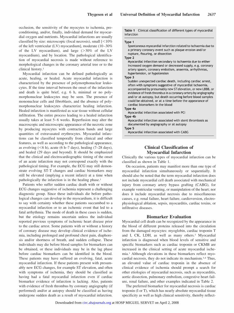
Clinical Classification ofMyocardial Infarction
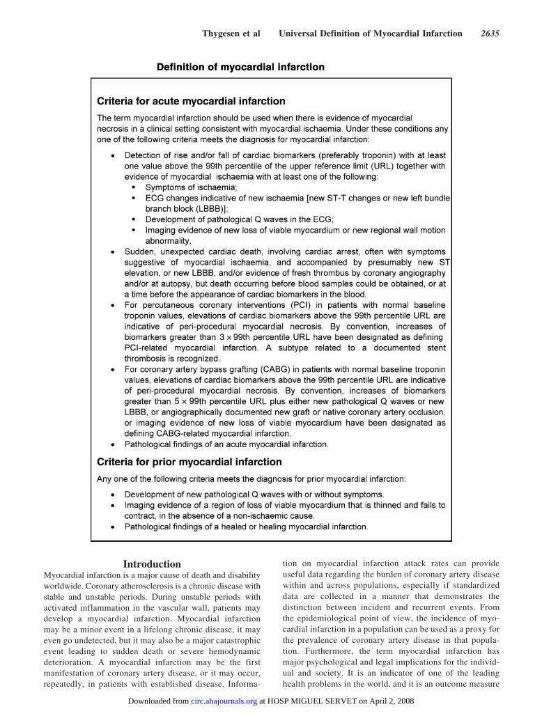
Clinically the various types of myocardial infarction can beclassified as shown in Table 1.
On occasion, patients may manifest more than one type ofmyocardial infarction simultaneously or sequentially. Itshould also be noted that the term myocardial infarction doesnot include myocardial cell death associated with mechanicalinjury from coronary artery bypass grafting (CABG), forexample ventricular venting, or manipulation of the heart; nordoes it include myocardial necrosis due to miscellaneouscauses, e.g. renal failure, heart failure, cardioversion, electro-physiological ablation, sepsis, myocarditis, cardiac toxins, orinfiltrative diseases.
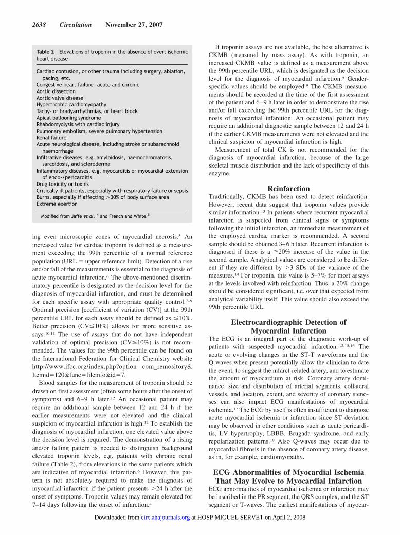
Biomarker EvaluationMyocardial cell death can be recognized by the appearance inthe blood of different proteins released into the circulationfrom the damaged myocytes: myoglobin, cardiac troponin Tand I, CK, LDH, as well as many others.3 Myocardialinfarction is diagnosed when blood levels of sensitive andspecific biomarkers such as cardiac troponin or CKMB areincreased in the clinical setting of acute myocardial ische-mia.1 Although elevations in these biomarkers reflect myo-cardial necrosis, they do not indicate its mechanism.3,4 Thus,an elevated value of cardiac troponin in the absence ofclinical evidence of ischemia should prompt a search forother etiologies of myocardial necrosis, such as myocarditis,aortic dissection, pulmonary embolism, congestive heart fail-ure, renal failure, and other examples indicated in Table 2.
The preferred biomarker for myocardial necrosis is cardiactroponin (I or T), which has nearly absolute myocardial tissuespecificity as well as high clinical sensitivity, thereby reflect-
Thygesen et al Universal Definition of Myocardial Infarction 2637
at HOSP MIGUEL SERVET on April 2, 2008 circ.ahajournals.orgDownloaded from
ing even microscopic zones of myocardial necrosis.3 Anincreased value for cardiac troponin is defined as a measure-ment exceeding the 99th percentile of a normal referencepopulation (URL � upper reference limit). Detection of a riseand/or fall of the measurements is essential to the diagnosis ofacute myocardial infarction.6 The above-mentioned discrim-inatory percentile is designated as the decision level for thediagnosis of myocardial infarction, and must be determinedfor each specific assay with appropriate quality control.7–9
Optimal precision [coefficient of variation (CV)] at the 99thpercentile URL for each assay should be defined as �10%.Better precision (CV�10%) allows for more sensitive as-says.10,11 The use of assays that do not have independentvalidation of optimal precision (CV�10%) is not recom-mended. The values for the 99th percentile can be found onthe International Federation for Clinical Chemistry websitehttp://www.ifcc.org/index.php?option�com_remository&Itemid�120&func�fileinfo&id�7.
Blood samples for the measurement of troponin should bedrawn on first assessment (often some hours after the onset ofsymptoms) and 6–9 h later.12 An occasional patient mayrequire an additional sample between 12 and 24 h if theearlier measurements were not elevated and the clinicalsuspicion of myocardial infarction is high.12 To establish thediagnosis of myocardial infarction, one elevated value abovethe decision level is required. The demonstration of a risingand/or falling pattern is needed to distinguish backgroundelevated troponin levels, e.g. patients with chronic renalfailure (Table 2), from elevations in the same patients whichare indicative of myocardial infarction.6 However, this pat-tern is not absolutely required to make the diagnosis ofmyocardial infarction if the patient presents �24 h after theonset of symptoms. Troponin values may remain elevated for7–14 days following the onset of infarction.4
If troponin assays are not available, the best alternative isCKMB (measured by mass assay). As with troponin, anincreased CKMB value is defined as a measurement abovethe 99th percentile URL, which is designated as the decisionlevel for the diagnosis of myocardial infarction.9 Gender-specific values should be employed.9 The CKMB measure-ments should be recorded at the time of the first assessmentof the patient and 6–9 h later in order to demonstrate the riseand/or fall exceeding the 99th percentile URL for the diag-nosis of myocardial infarction. An occasional patient mayrequire an additional diagnostic sample between 12 and 24 hif the earlier CKMB measurements were not elevated and theclinical suspicion of myocardial infarction is high.
Measurement of total CK is not recommended for thediagnosis of myocardial infarction, because of the largeskeletal muscle distribution and the lack of specificity of thisenzyme.
ReinfarctionTraditionally, CKMB has been used to detect reinfarction.However, recent data suggest that troponin values providesimilar information.13 In patients where recurrent myocardialinfarction is suspected from clinical signs or symptomsfollowing the initial infarction, an immediate measurement ofthe employed cardiac marker is recommended. A secondsample should be obtained 3–6 h later. Recurrent infarction isdiagnosed if there is a �20% increase of the value in thesecond sample. Analytical values are considered to be differ-ent if they are different by �3 SDs of the variance of themeasures.14 For troponin, this value is 5–7% for most assaysat the levels involved with reinfarction. Thus, a 20% changeshould be considered significant, i.e. over that expected fromanalytical variability itself. This value should also exceed the99th percentile URL.
Electrocardiographic Detection ofMyocardial Infarction
The ECG is an integral part of the diagnostic work-up ofpatients with suspected myocardial infarction.1,2,15,16 Theacute or evolving changes in the ST-T waveforms and theQ-waves when present potentially allow the clinician to datethe event, to suggest the infarct-related artery, and to estimatethe amount of myocardium at risk. Coronary artery domi-nance, size and distribution of arterial segments, collateralvessels, and location, extent, and severity of coronary steno-ses can also impact ECG manifestations of myocardialischemia.17 The ECG by itself is often insufficient to diagnoseacute myocardial ischemia or infarction since ST deviationmay be observed in other conditions such as acute pericardi-tis, LV hypertrophy, LBBB, Brugada syndrome, and earlyrepolarization patterns.18 Also Q-waves may occur due tomyocardial fibrosis in the absence of coronary artery disease,as in, for example, cardiomyopathy.
ECG Abnormalities of Myocardial IschemiaThat May Evolve to Myocardial Infarction
ECG abnormalities of myocardial ischemia or infarction maybe inscribed in the PR segment, the QRS complex, and the STsegment or T-waves. The earliest manifestations of myocar-
2638 Circulation November 27, 2007
at HOSP MIGUEL SERVET on April 2, 2008 circ.ahajournals.orgDownloaded from
dial ischemia are typical T-waves and ST segment chang-es.19,20 Increased hyper-acute T-wave amplitude with promi-nent symmetrical T-waves in at least two contiguous leads isan early sign that may precede the elevation of the STsegment. Increased R-wave amplitude and width (giantR-wave with S-wave diminution) are often seen in leadsexhibiting ST elevation, and tall T-waves reflecting conduc-tion delay in the ischemic myocardium.21 Transient Q-wavesmay be observed during an episode of acute ischemia orrarely during acute myocardial infarction with successfulreperfusion.22
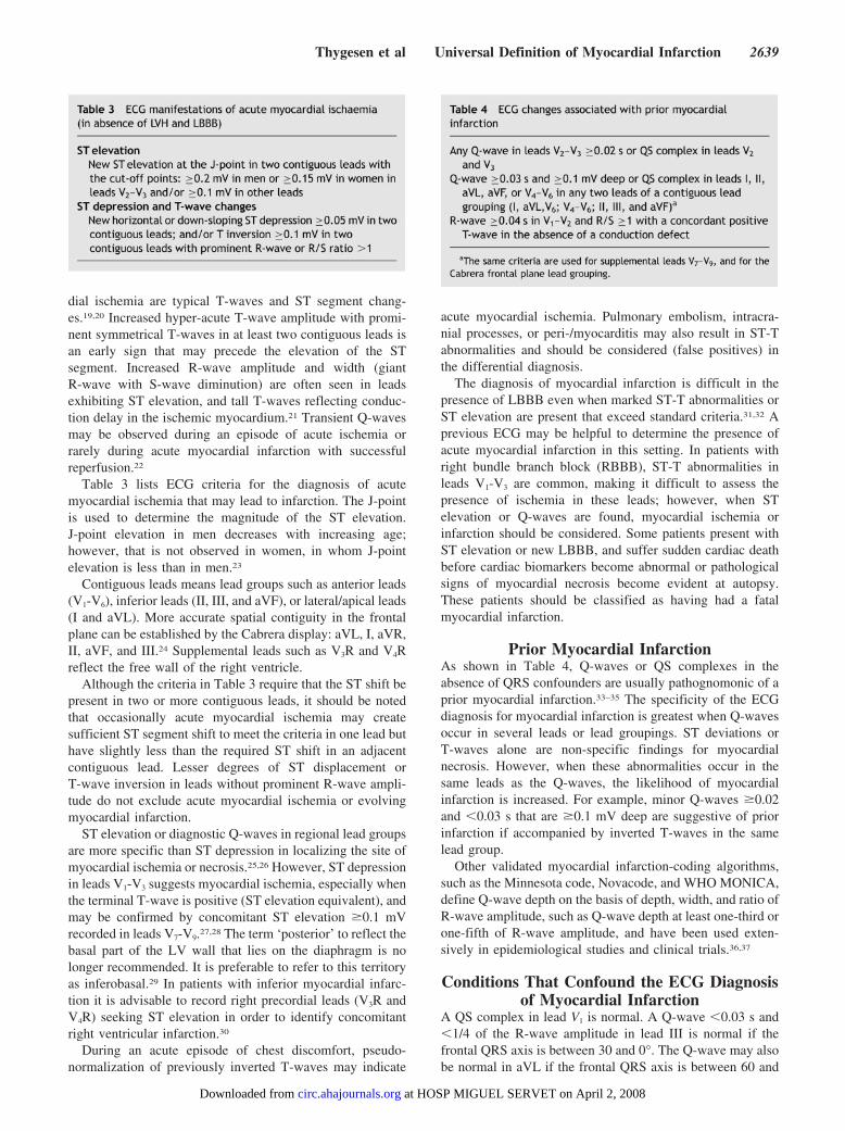
Table 3 lists ECG criteria for the diagnosis of acutemyocardial ischemia that may lead to infarction. The J-pointis used to determine the magnitude of the ST elevation.J-point elevation in men decreases with increasing age;however, that is not observed in women, in whom J-pointelevation is less than in men.23
Contiguous leads means lead groups such as anterior leads(V1-V6), inferior leads (II, III, and aVF), or lateral/apical leads(I and aVL). More accurate spatial contiguity in the frontalplane can be established by the Cabrera display: aVL, I, aVR,II, aVF, and III.24 Supplemental leads such as V3R and V4Rreflect the free wall of the right ventricle.
Although the criteria in Table 3 require that the ST shift bepresent in two or more contiguous leads, it should be notedthat occasionally acute myocardial ischemia may createsufficient ST segment shift to meet the criteria in one lead buthave slightly less than the required ST shift in an adjacentcontiguous lead. Lesser degrees of ST displacement orT-wave inversion in leads without prominent R-wave ampli-tude do not exclude acute myocardial ischemia or evolvingmyocardial infarction.
ST elevation or diagnostic Q-waves in regional lead groupsare more specific than ST depression in localizing the site ofmyocardial ischemia or necrosis.25,26 However, ST depressionin leads V1-V3 suggests myocardial ischemia, especially whenthe terminal T-wave is positive (ST elevation equivalent), andmay be confirmed by concomitant ST elevation �0.1 mVrecorded in leads V7-V9.27,28 The term ‘posterior’ to reflect thebasal part of the LV wall that lies on the diaphragm is nolonger recommended. It is preferable to refer to this territoryas inferobasal.29 In patients with inferior myocardial infarc-tion it is advisable to record right precordial leads (V3R andV4R) seeking ST elevation in order to identify concomitantright ventricular infarction.30
During an acute episode of chest discomfort, pseudo-normalization of previously inverted T-waves may indicate
acute myocardial ischemia. Pulmonary embolism, intracra-nial processes, or peri-/myocarditis may also result in ST-Tabnormalities and should be considered (false positives) inthe differential diagnosis.
The diagnosis of myocardial infarction is difficult in thepresence of LBBB even when marked ST-T abnormalities orST elevation are present that exceed standard criteria.31,32 Aprevious ECG may be helpful to determine the presence ofacute myocardial infarction in this setting. In patients withright bundle branch block (RBBB), ST-T abnormalities inleads V1-V3 are common, making it difficult to assess thepresence of ischemia in these leads; however, when STelevation or Q-waves are found, myocardial ischemia orinfarction should be considered. Some patients present withST elevation or new LBBB, and suffer sudden cardiac deathbefore cardiac biomarkers become abnormal or pathologicalsigns of myocardial necrosis become evident at autopsy.These patients should be classified as having had a fatalmyocardial infarction.
Prior Myocardial InfarctionAs shown in Table 4, Q-waves or QS complexes in theabsence of QRS confounders are usually pathognomonic of aprior myocardial infarction.33–35 The specificity of the ECGdiagnosis for myocardial infarction is greatest when Q-wavesoccur in several leads or lead groupings. ST deviations orT-waves alone are non-specific findings for myocardialnecrosis. However, when these abnormalities occur in thesame leads as the Q-waves, the likelihood of myocardialinfarction is increased. For example, minor Q-waves �0.02and �0.03 s that are �0.1 mV deep are suggestive of priorinfarction if accompanied by inverted T-waves in the samelead group.
Other validated myocardial infarction-coding algorithms,such as the Minnesota code, Novacode, and WHO MONICA,define Q-wave depth on the basis of depth, width, and ratio ofR-wave amplitude, such as Q-wave depth at least one-third orone-fifth of R-wave amplitude, and have been used exten-sively in epidemiological studies and clinical trials.36,37
Conditions That Confound the ECG Diagnosisof Myocardial Infarction
A QS complex in lead V1 is normal. A Q-wave �0.03 s and�1/4 of the R-wave amplitude in lead III is normal if thefrontal QRS axis is between 30 and 0°. The Q-wave may alsobe normal in aVL if the frontal QRS axis is between 60 and
Thygesen et al Universal Definition of Myocardial Infarction 2639
at HOSP MIGUEL SERVET on April 2, 2008 circ.ahajournals.orgDownloaded from
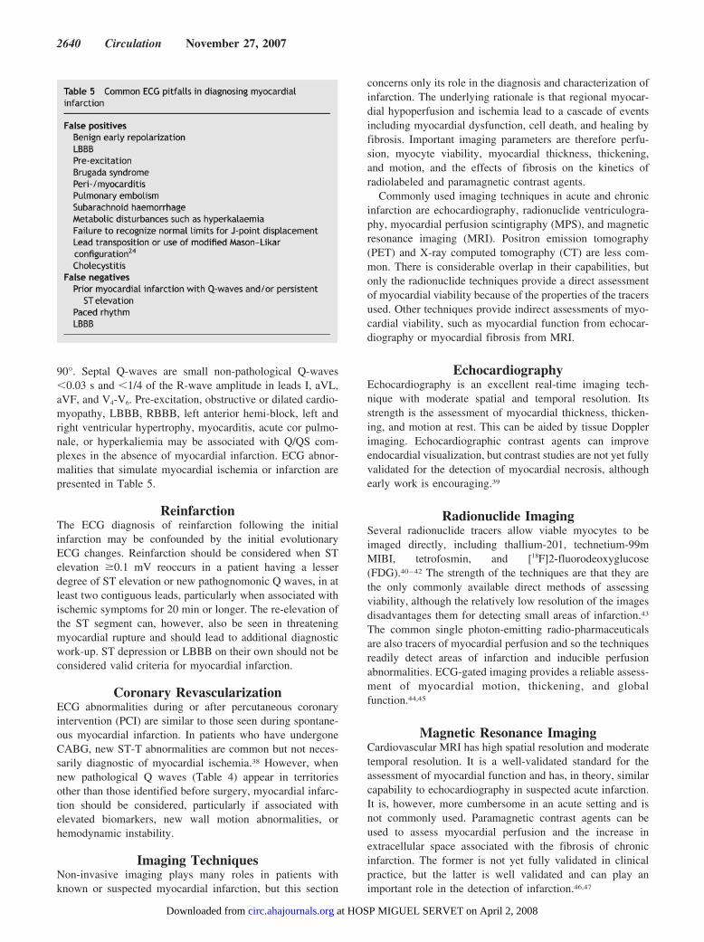
90°. Septal Q-waves are small non-pathological Q-waves�0.03 s and �1/4 of the R-wave amplitude in leads I, aVL,aVF, and V4-V6. Pre-excitation, obstructive or dilated cardio-myopathy, LBBB, RBBB, left anterior hemi-block, left andright ventricular hypertrophy, myocarditis, acute cor pulmo-nale, or hyperkaliemia may be associated with Q/QS com-plexes in the absence of myocardial infarction. ECG abnor-malities that simulate myocardial ischemia or infarction arepresented in Table 5.
ReinfarctionThe ECG diagnosis of reinfarction following the initialinfarction may be confounded by the initial evolutionaryECG changes. Reinfarction should be considered when STelevation �0.1 mV reoccurs in a patient having a lesserdegree of ST elevation or new pathognomonic Q waves, in atleast two contiguous leads, particularly when associated withischemic symptoms for 20 min or longer. The re-elevation ofthe ST segment can, however, also be seen in threateningmyocardial rupture and should lead to additional diagnosticwork-up. ST depression or LBBB on their own should not beconsidered valid criteria for myocardial infarction.
Coronary RevascularizationECG abnormalities during or after percutaneous coronaryintervention (PCI) are similar to those seen during spontane-ous myocardial infarction. In patients who have undergoneCABG, new ST-T abnormalities are common but not neces-sarily diagnostic of myocardial ischemia.38 However, whennew pathological Q waves (Table 4) appear in territoriesother than those identified before surgery, myocardial infarc-tion should be considered, particularly if associated withelevated biomarkers, new wall motion abnormalities, orhemodynamic instability.
Imaging TechniquesNon-invasive imaging plays many roles in patients withknown or suspected myocardial infarction, but this section
concerns only its role in the diagnosis and characterization ofinfarction. The underlying rationale is that regional myocar-dial hypoperfusion and ischemia lead to a cascade of eventsincluding myocardial dysfunction, cell death, and healing byfibrosis. Important imaging parameters are therefore perfu-sion, myocyte viability, myocardial thickness, thickening,and motion, and the effects of fibrosis on the kinetics ofradiolabeled and paramagnetic contrast agents.
Commonly used imaging techniques in acute and chronicinfarction are echocardiography, radionuclide ventriculogra-phy, myocardial perfusion scintigraphy (MPS), and magneticresonance imaging (MRI). Positron emission tomography(PET) and X-ray computed tomography (CT) are less com-mon. There is considerable overlap in their capabilities, butonly the radionuclide techniques provide a direct assessmentof myocardial viability because of the properties of the tracersused. Other techniques provide indirect assessments of myo-cardial viability, such as myocardial function from echocar-diography or myocardial fibrosis from MRI.
EchocardiographyEchocardiography is an excellent real-time imaging tech-nique with moderate spatial and temporal resolution. Itsstrength is the assessment of myocardial thickness, thicken-ing, and motion at rest. This can be aided by tissue Dopplerimaging. Echocardiographic contrast agents can improveendocardial visualization, but contrast studies are not yet fullyvalidated for the detection of myocardial necrosis, althoughearly work is encouraging.39
Radionuclide ImagingSeveral radionuclide tracers allow viable myocytes to beimaged directly, including thallium-201, technetium-99mMIBI, tetrofosmin, and [18F]2-fluorodeoxyglucose(FDG).40–42 The strength of the techniques are that they arethe only commonly available direct methods of assessingviability, although the relatively low resolution of the imagesdisadvantages them for detecting small areas of infarction.43
The common single photon-emitting radio-pharmaceuticalsare also tracers of myocardial perfusion and so the techniquesreadily detect areas of infarction and inducible perfusionabnormalities. ECG-gated imaging provides a reliable assess-ment of myocardial motion, thickening, and globalfunction.44,45
Magnetic Resonance ImagingCardiovascular MRI has high spatial resolution and moderatetemporal resolution. It is a well-validated standard for theassessment of myocardial function and has, in theory, similarcapability to echocardiography in suspected acute infarction.It is, however, more cumbersome in an acute setting and isnot commonly used. Paramagnetic contrast agents can beused to assess myocardial perfusion and the increase inextracellular space associated with the fibrosis of chronicinfarction. The former is not yet fully validated in clinicalpractice, but the latter is well validated and can play animportant role in the detection of infarction.46,47
2640 Circulation November 27, 2007
at HOSP MIGUEL SERVET on April 2, 2008 circ.ahajournals.orgDownloaded from
X-Ray Computed TomographyInfarcted myocardium is initially visible to CT as a focal areaof decreased LV enhancement, but later imaging showshyperenhancement as with late gadolinium imaging byMRI.48,49 This finding is clinically relevant because contrast-enhanced CT may be performed for suspected embolism andaortic dissection, conditions with clinical features that overlapwith those of acute myocardial infarction.
Application in the Acute Phase ofMyocardial Infarction
Imaging techniques can be useful in the diagnosis of myo-cardial infarction because of the ability to detect wall motionabnormalities in the presence of elevated cardiac bio-markers.If for some reason biomarkers have not been measured ormay have normalized, demonstration of new loss of myocar-dial viability alone in the absence of non-ischemic causesmeets the criteria for myocardial infarction. However, ifbiomarkers have been measured at appropriate times and arenormal, the determinations of these take precedence over theimaging criteria.
Echocardiography provides assessment of many non-ische-mic causes of acute chest pain such as peri-myocarditis,valvular heart disease, cardiomyopathy, pulmonary embo-lism, or aortic dissection. Echocardiography is the imagingtechnique of choice for detecting complications of acuteinfarction including myocardial free wall rupture, acuteventricular septal defect, and mitral regurgitation secondaryto papillary muscle rupture or ischemia. However, echocar-diography cannot distinguish regional wall motion abnormal-ities due to myocardial ischemia from infarction.
Radionuclide assessment of perfusion at the time of patientpresentation can be performed with immediate tracer injec-tion and imaging that can be delayed for up to several hours.The technique is interpreter dependent, although objectivequantitative analysis is available. ECG gating provides simul-taneous information on LV function.
An important role of acute echocardiography or radionu-clide imaging is in patients with suspected myocardial infarc-tion and a non-diagnostic ECG. A normal echocardiogram orresting ECG-gated scintigram has a 95–98% negative predic-tive value for excluding acute infarction.50–54 Thus, imagingtechniques are useful for early triage and discharge of patientswith suspected myocardial infarction.55,56
A regional myocardial wall motion abnormality or loss ofnormal thickening may be caused by acute myocardialinfarction or by one or more of several other ischemicconditions including old infarction, acute ischemia, stunning,or hibernation. Non-ischemic conditions such as cardiomy-opathy and inflammatory or infiltrative diseases can also leadto regional loss of viable myocardium or functional abnor-mality, and so the positive predictive value of imagingtechniques is not high unless these conditions can be ex-cluded and unless a new abnormality is detected or can bepresumed to have arisen in the setting of other features ofacute myocardial infarction.
Application in the Healing or Healed Phase ofMyocardial Infarction
Imaging techniques are useful in myocardial infarction foranalysis of LV function, both at rest and during dynamicexercise or pharmacological stress, to provide an assessmentof remote inducible ischemia. Echocardiography and radio-nuclide techniques, in conjunction with exercise or pharma-cological stress, can identify ischemia and myocardial viabil-ity. Non-invasive imaging techniques can diagnose healing orhealed infarction by demonstrating regional wall motion,thinning, or scar in the absence of other causes.
The high resolution of contrast-enhanced MRI means thatareas of late enhancement correlate well with areas of fibrosisand thereby enable differentiation between transmural andsubendocardial scarring.57 The technique is therefore poten-tially valuable in assessing LV function and areas of viableand hence potentially hibernating myocardium.
Myocardial Infarction Associated WithRevascularization Procedures
Peri-procedural myocardial infarction is different from spon-taneous infarction, because the former is associated with theinstrumentation of the heart that is required during mechan-ical revascularization procedures by either PCI or CABG.Multiple events that can lead to myocardial necrosis aretaking place, often in combination, during both types ofintervention.58–61 While some loss of myocardial tissue maybe unavoidable during procedures, it is likely that limitationof such damage is beneficial to the patient and theirprognosis.62
During PCI, myocardial necrosis may result from recog-nizable peri-procedural events, alone or in combination, suchas side-branch occlusion, disruption of collateral flow, distalembolization, coronary dissection, slow flow or no-reflowphenomenon, and microvascular plugging. Embolization ofintracoronary thrombus or atherosclerotic particulate debriscannot be entirely prevented despite current antithromboticand antiplatelet adjunctive therapy or protection devices.Such events induce extensive inflammation of non-infarctedmyocardium surrounding small islets of myocardium necro-sis.63–67 New areas of myocardial necrosis have been dem-onstrated by MRI following PCI.68 A separate subcategory ofmyocardial infarction is related to stent thrombosis as docu-mented by angiography and/or autopsy.
During CABG, numerous additional factors can lead toperi-procedural necrosis. These include direct myocardialtrauma from sewing needles or manipulation of the heart,coronary dissection, global or regional ischemia related toinadequate cardiac protection, microvascular events related toreperfusion, myocardial damage induced by oxygen freeradical generation, or failure to reperfuse areas of the myo-cardium that are not subtended by graftable vessels.69–71 MRIstudies suggest that most necrosis in this setting is not focal,but diffuse and localized to the sub-endocardium.72 Someclinicians and clinical investigators have preferred usingCKMB for the diagnosis of peri-procedural infarction be-cause of a substantial amount of data relating CKMB eleva-tions to prognosis.73,74 However, an increasing number ofstudies using troponins in that respect have emerged.59,75
Thygesen et al Universal Definition of Myocardial Infarction 2641
at HOSP MIGUEL SERVET on April 2, 2008 circ.ahajournals.orgDownloaded from
Diagnostic Criteria for Myocardial InfarctionWith PCI
In the setting of PCI, the balloon inflation during a procedurealmost always results in ischemia whether or not accompa-nied by ST-T changes. The occurrence of procedure-relatedcell necrosis can be detected by measurement of cardiacbiomarkers before or immediately after the procedure, andagain at 6–12 and 18–24 h.76,77 Elevations of biomarkersabove the 99th percentile URL after PCI, assuming a normalbaseline troponin value, are indicative of post-proceduralmyocardial necrosis. There is currently no solid scientificbasis for defining a biomarker threshold for the diagnosis ofperi-procedural myocardial infarction. Pending further data,and by arbitrary convention, it is suggested to designateincreases more than three times the 99th percentile URL asPCI-related myocardial infarction (type 4a).
If cardiac troponin is elevated before the procedure and notstable for at least two samples 6 h apart, there are insufficientdata to recommend biomarker criteria for the diagnosis ofperi-procedural myocardial infarction.77 If the values arestable or falling, criteria for reinfarction by further measure-ment of biomarkers together with the features of the ECG orimaging can be applied.
A separate subcategory of myocardial infarction (type 4b)is related to stent thrombosis as documented by angiographyand/or autopsy. Although iatrogenic, myocardial infarctiontype 4b with verified stent thrombosis must meet the criteriafor spontaneous myocardial infarction as well.
Diagnostic Criteria for Myocardial InfarctionWith CABG
Any increase of cardiac biomarkers after CABG indicatesmyocyte necrosis, implying that an increasing magnitude ofbiomarker is likely to be related to an impaired outcome. Thishas been demonstrated in clinical studies employing CKMBwhere elevations five, 10 and 20 times the upper limit ofnormal after CABG were associated with worsened progno-sis.73,78,79 Likewise, the increase of troponin levels afterCABG indicates necrosis of myocardial cells, which predictsa poor outcome, in particular when elevated to the highestquartile or quintile of the troponin measurements.59,75
Unlike the prognosis, scant literature exists concerning theuse of biomarkers for defining myocardial infarction in thesetting of CABG. Therefore, biomarkers cannot stand alonein diagnosing myocardial infarction (type 5). In view of theadverse impact on survival observed in patients with signif-icant biomarker elevations, this Task Force suggests, byarbitrary convention, that biomarker values more than fivetimes the 99th percentile of the normal reference range duringthe first 72 h following CABG, when associated with theappearance of new pathological Q-waves or new LBBB, orangiographically documented new graft or native coronaryartery occlusion, or imaging evidence of new loss of viablemyocardium, should be considered as diagnostic of a CABG-related myocardial infarction (type 5 myocardial infarction).
Definition of Myocardial Infarction inClinical Investigations
A universal definition for myocardial infarction would be ofgreat benefit to future clinical studies in this area since it will
allow for trial-to-trial comparisons as well as accurate meta-analyses involving multiple investigations. In clinical trials,myocardial infarction may be an entry criterion or an end-point. The definition of myocardial infarction employed inthese trials will thus determine the characteristics of patientsentering the studies as well as the number of outcome events.In recent investigations, different infarct definitions havebeen employed, thereby hampering comparison and general-ization among these trials.
Consistency among investigators and regulatory authoritieswith regard to the definition of myocardial infarction used inclinical investigations is essential. The Task Force stronglyencourages trialists to employ the definition described in thisdocument. Furthermore, investigators should ensure that atrial provides comprehensive data for the various types ofmyocardial infarction (e.g. spontaneous, peri-procedural) andincludes the employed decision limits for myocardial infarc-tion of the cardiac bio-markers in question. All data should bemade available to interested individuals in published formator on a website. Data concerning infarctions should beavailable in a form consistent with the current reviseddefinitions of myocardial infarction. This does not necessarilyrestrict trialists to a narrow end-point definition, but ratherensures that across all future trials access to comparable dataexists, thereby facilitating cross-study analyses. The recom-mendations put forward in this section are not detailed andshould be supplemented by careful protocol planning andimplementation in any clinical trial.
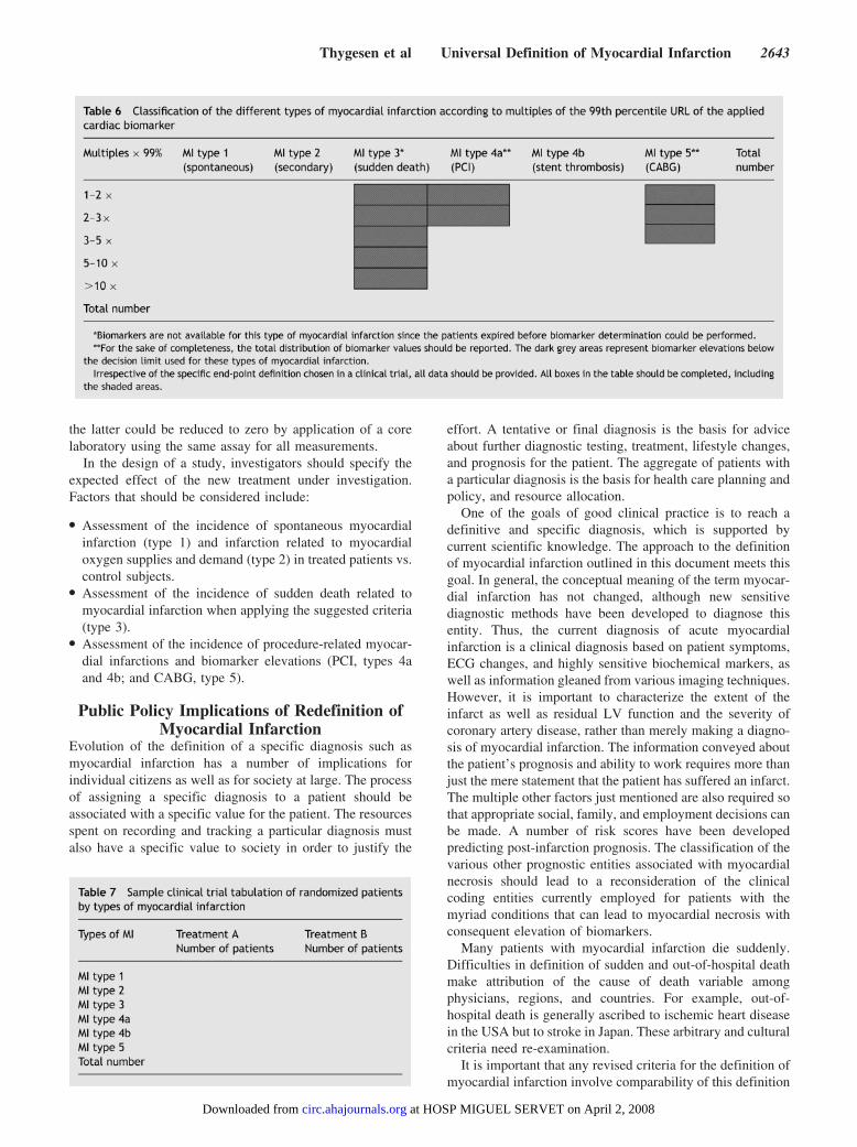
The Task Force strongly endorses the concept of the samedecision limit for each biomarker employed for myocardialinfarction types 1 and 2, and, likewise, the same higher three-and five-fold decision limits in the setting of myocardialinfarction types 4a and 5, respectively78–80 (Tables 6 and 7).In clinical trials, as in clinical practice, measurement ofcardiac troponin T or I is preferred over measurement ofCKMB or other biomarkers for the diagnosis of myocardialinfarction. Assessment of the quantity of myocardial damage(infarct size) is also an important trial end-point. Although thespecific measurements vary depending on the assay andwhether cardiac troponin T or I is used, in most studiestroponin values correlate better with radionuclide-and MRI-determined infarct size than do CK and CKMB.81–83
The use of cardiac troponins will undoubtedly increase thenumber of events recorded in a particular investigationbecause of increased sensitivity for detecting infarction.84–87
Ideally, data should be presented so that future clinicalinvestigations or registries can translate the myocardial in-farction end-point chosen in one study into the end-point ofanother study. Thus, measurements should be presented in auniform manner to allow independent judgment and compar-ison of the clinical end-points. Furthermore, this Task Forcesuggests that data be reported as multiples of the 99thpercentile URL of the applied biomarker, enabling compari-sons between various classes and severity of the differenttypes of myocardial infarction as indicated in Tables 6 and 7.
It is recommended that within a clinical trial all investiga-tors whenever possible should employ the same assay inorder to reduce the inter-assay variability, and, even better,
2642 Circulation November 27, 2007
at HOSP MIGUEL SERVET on April 2, 2008 circ.ahajournals.orgDownloaded from
the latter could be reduced to zero by application of a corelaboratory using the same assay for all measurements.
In the design of a study, investigators should specify theexpected effect of the new treatment under investigation.Factors that should be considered include:
● Assessment of the incidence of spontaneous myocardialinfarction (type 1) and infarction related to myocardialoxygen supplies and demand (type 2) in treated patients vs.control subjects.
● Assessment of the incidence of sudden death related tomyocardial infarction when applying the suggested criteria(type 3).
● Assessment of the incidence of procedure-related myocar-dial infarctions and biomarker elevations (PCI, types 4aand 4b; and CABG, type 5).
Public Policy Implications of Redefinition ofMyocardial Infarction
Evolution of the definition of a specific diagnosis such asmyocardial infarction has a number of implications forindividual citizens as well as for society at large. The processof assigning a specific diagnosis to a patient should beassociated with a specific value for the patient. The resourcesspent on recording and tracking a particular diagnosis mustalso have a specific value to society in order to justify the
effort. A tentative or final diagnosis is the basis for adviceabout further diagnostic testing, treatment, lifestyle changes,and prognosis for the patient. The aggregate of patients witha particular diagnosis is the basis for health care planning andpolicy, and resource allocation.
One of the goals of good clinical practice is to reach adefinitive and specific diagnosis, which is supported bycurrent scientific knowledge. The approach to the definitionof myocardial infarction outlined in this document meets thisgoal. In general, the conceptual meaning of the term myocar-dial infarction has not changed, although new sensitivediagnostic methods have been developed to diagnose thisentity. Thus, the current diagnosis of acute myocardialinfarction is a clinical diagnosis based on patient symptoms,ECG changes, and highly sensitive biochemical markers, aswell as information gleaned from various imaging techniques.However, it is important to characterize the extent of theinfarct as well as residual LV function and the severity ofcoronary artery disease, rather than merely making a diagno-sis of myocardial infarction. The information conveyed aboutthe patient’s prognosis and ability to work requires more thanjust the mere statement that the patient has suffered an infarct.The multiple other factors just mentioned are also required sothat appropriate social, family, and employment decisions canbe made. A number of risk scores have been developedpredicting post-infarction prognosis. The classification of thevarious other prognostic entities associated with myocardialnecrosis should lead to a reconsideration of the clinicalcoding entities currently employed for patients with themyriad conditions that can lead to myocardial necrosis withconsequent elevation of biomarkers.
Many patients with myocardial infarction die suddenly.Difficulties in definition of sudden and out-of-hospital deathmake attribution of the cause of death variable amongphysicians, regions, and countries. For example, out-of-hospital death is generally ascribed to ischemic heart diseasein the USA but to stroke in Japan. These arbitrary and culturalcriteria need re-examination.
It is important that any revised criteria for the definition ofmyocardial infarction involve comparability of this definition
Thygesen et al Universal Definition of Myocardial Infarction 2643
at HOSP MIGUEL SERVET on April 2, 2008 circ.ahajournals.orgDownloaded from
over time so that adequate trend data can be obtained.Furthermore, it is essential to ensure widespread availabilityand standard application of the measures in order to ensurecomparability of data from various geographic regions. Shiftin criteria resulting in a substantial increase or decrease incase identification will have significant health resource andcost implications.86,87 Moreover, an increase in sensitivity ofthe criteria for myocardial infarction might entail negativeconsequences for some patients who are not currently labeledas having had an infarction. On the other hand, increasingdiagnostic sensitivity for myocardial infarction can have apositive impact for a society:
● Increasing the sensitivity of diagnostic criteria for myocar-dial infarction will result in more cases identified in asociety, thereby allowing appropriate secondaryprevention.
● Increasing the specificity of diagnostic criteria for myocar-dial infarction will result in more accurate diagnosis butwill not exclude the presence of coronary artery disease,the cases of which may benefit from secondary prevention.
It should be appreciated that the agreed modification of thedefinition of myocardial infarction may be associated withconsequences for the patients and their families with respectto psychological status, life insurance, professional career, aswell as driving and pilot licenses. Also the diagnosis isassociated with societal implications as to diagnosis-relatedcoding, hospital reimbursement, mortality statistics, sickleave, and disability attestation.
In order to meet this challenge, physicians must be ade-quately informed of the altered diagnostic criteria. Educa-tional materials will need to be created and treatment guide-lines must be appropriately adapted. Professional societiesshould take steps to facilitate the rapid dissemination of therevised definition to physicians, other health care profession-als, administrators, and the general public.
Global Perspectives of the Redefinition ofMyocardial Infarction
Cardiovascular disease is a global health problem. Approxi-mately one-third of persons in the world die of cardiovasculardisease, largely coronary artery disease and stroke, and 80%of these deaths from cardiovascular disease occur in devel-oping countries. The greater proportion of deaths is due toheart disease and specifically coronary heart disease, ofwhich myocardial infarction is a major manifestation. Since itis difficult to measure the prevalence of coronary arterydisease in a population, the incidence of myocardial infarc-tion may be used as a proxy, provided that a consistentdefinition is used when different populations, countries, orcontinents are being compared.
The changes in the definition of myocardial infarction havecritical consequences for less developed and developingcountries. In many countries, the resources to apply the newdefinition may not be available in all hospitals. However,many developing countries already do have medical facilitiescapable of or currently employing the proposed definition ofmyocardial infarction. In the context of the overall cost for a
patient with myocardial infarction, the expense associatedwith a troponin assay would not be excessive and should beeconomically affordable in many hospitals in developingcountries, particularly those where infarcts are frequentevents. The necessary equipment, staff training, and runningcosts may be lacking in some regions, but certainly not inothers. In less advantaged hospitals, the diagnosis of myocar-dial infarction may depend mostly on clinical signs andsymptoms coupled with less sophisticated biomarker analy-ses. Some of these institutions may only have access to CKand its isoenzymes at the present time. The redefinition arisesfrom and is compatible with the latest scientific knowledgeand with advances in technology, particularly with regard tothe use of bio-markers, high quality electrocardiography, andimaging techniques. The definition can and should be used bydeveloped countries immediately, and by developing coun-tries as quickly as resources become available.
The change in the definition of myocardial infarction willhave a substantial impact on the identification, prevention,and treatment of cardiovascular disease throughout the world.The new definition will impact epidemiological data fromdeveloping countries relating to prevalence and incidence.The simultaneous and continuing use of the older WHOdefinition for some years would allow a comparison betweendata obtained in the past and data to be acquired in the futureemploying the newer biomarker approach. Cultural, financial,structural, and organizational problems in the different coun-tries of the world in diagnosis and therapy of acute myocar-dial infarction will require ongoing investigation. It is essen-tial that the gap between therapeutic and diagnostic advancesbe addressed in this expanding area of cardiovascular disease.
Conflicts of InterestThe members of the Task Force established by the ESC, theACCF, AHA and the WHF have participated independentlyin the preparation of this document, drawing on their aca-demic and clinical experience and applying an objective andcritical examination of all available literature. Most haveundertaken and are undertaking work in collaboration withindustry and governmental or private health providers (re-search studies, teaching conferences, consultation), but allbelieve such activities have not influenced their judgment.The best guarantee of their independence is in the quality oftheir past and current scientific work. However, to ensureopenness, their relations with industry, government andprivate health providers are reported in the ESC and Euro-pean Heart Journal websites (www.escardio.org and www.eurheartj.org). Expenses for the Task Force/Writing Commit-tee and preparation of this document were provided entirelyby the above mentioned joint associations.
AcknowledgmentsWe are indebted to Professor Erling Falk MD for reviewing thesection on ‘Pathology’. Furthermore, we are very grateful to thededicated staff of the Guidelines Department of the ESC. We wouldalso like to thank and to acknowledge the contribution of thefollowing companies through unrestricted educational grants, noneof whom were involved in the development of this publication and inno way influenced its contents. Premium observer: GSK. Observer:
2644 Circulation November 27, 2007
at HOSP MIGUEL SERVET on April 2, 2008 circ.ahajournals.orgDownloaded from
AstraZeneca, Beckman-Coulter, Dade Behring, Roche Diagnostics,Sanofi-Aventis, Servier.
References1. The Joint European Society of Cardiology/American College of Car-
diology Committee. Myocardial infarction redefined: A consensusdocument of the Joint European Society of Cardiology/American Collegeof Cardiology Committee for the Redefinition of Myocardial Infarction.Eur Heart J. 2000;21:1502–1513; J Am Coll Cardiol. 2000;36:959–969.
2. Luepker RV, Apple FS, Christenson RH, Crow RS, Fortmann SP, Goff D,Goldberg RJ, Hand MM, Jaffe AS, Julian DG, Levy D, Manolio T,Mendis S, Mensah G, Pajak A, Prineas RJ, Reddy S, Roger VL,Rosamond WD, Shahar E, Sharrett R, Sorlie P, Tunstall-Pedoe H. Casedefinitions for acute coronary heart disease in epidemiology and clinicalresearch studies. A statement from the AHA Council on Epidemiologyand Prevention; AHA Statistics Committee; World Heart FederationCouncil on Epidemiology and Prevention; the European Society of Car-diology Working Group on Epidemiology and Prevention; Centers forDisease Control and Prevention; and the National Heart, Lung, and BloodInstitute. Circulation. 2003;108:2543–2549.
3. Jaffe AS, Ravkilde J, Roberts R, Näslund U, Apple FS, Galvani M, KatusH. It’s time for a change to a troponin standard. Circulation. 2000;102:1216–1220.
4. Jaffe AS, Babuin L, Apple FS. Biomarkers in acute cardiac disease. J AmColl Cardiol. 2006;48:1–11.
5. French JK, White HD. Clinical implications of the new definition ofmyo-cardial infarction. Heart. 2004;90:99–106.
6. Jaffe AS. Chasing troponin: how low can you go if you can see the rise?J Am Coll Cardiol. 2006;48:1763–1764.
7. Panteghini M, Gerhardt W, Apple FS, Dati F, Ravkilde J, Wu AH.Quality specifications for cardiac troponin assays. Clin Chem Lab Med.2001;39: 175–179.
8. Apple FS, Jesse RL, Newby LK, Wu AHB, Christenson RH. NationalAcademy of Clinical Biochemistry and IFCC Committee for Standard-ization of Markers Cardiac Damage Laboratory Medicine Practice Guide-lines: analytical issues for biochemical markers of acute coronary syn-dromes. Circulation. 2007;115:e352–e355.
9. Morrow DA, Cannon CP, Jesse RL, Newby LK, Ravkilde J, Storrow AB,Wu AHB, Christenson RH. National Academy of Clinical BiochemistryLaboratory Medicine Practice Guidelines: clinical characteristics andutilization of biochemical markers of acute coronary syndromes. Circu-lation. 2007;115:e356–e375.
10. Apple FS, Parvin CA, Buechler KF, Christenson RH, Wu AHB, Jaffe AS.Validation of the 99th percentile cutoff independent of assay imprecision(CV) for cardiac troponin monitoring for ruling out myocardial infarction.Clin Chem. 2005;51:2198–2200.
11. Panteghini M, Pagani F, Yeo KT, Apple FS, Christenson RH, Dati F,Mair J, Ravkilde J, Wu AHB on behalf of the Committee on Standard-ization of Markers of Cardiac Damage of the IFCC. Evaluation of impre-cision for cardiac troponin assays at low-range concentrations. ClinChem. 2004; 50:327–332.
12. MacRae AR, Kavsak PA, Lustig V, Bhargava R, Vandersluis R, PalomakiGE, Yerna M-J, Jaffe AS. Assessing the requirement for the six-hourinterval between specimens in the American Heart Association classifi-cation of myocardial infarction in epidemiology and clinical researchstudies. Clin Chem. 2006;52:812–818.
13. Apple FS, Murakami MM. Cardiac troponin and creatine kinase MBmonitoring during in-hospital myocardial reinfarction. Clin Chem.2005;51: 460–463.
14. Westgard JO, Klee GG. Quality management. In: Burtis CA, AshwoodER, Bruns DE, eds. Tietz Textbook of Clinical Chemistry and MolecularDiagnostics, 4th ed. St Louis, Mo: Elsevier Saunders; 2006:498–499.
15. Antman EM, Anbe DT, Armstrong PW, Bates ER, Green LE, Hand M,Hochman JS, Krumholz HM, Kushner FG, Lamas GA, Mullany CJ,Ornato JP, Pearle DL, Sloan MA, Smith SC Jr. ACC/AHA guidelines forthe management of patients with ST-elevation myocardial infarction: areport of the American College of Cardiology/American Heart Asso-ciation Task Force on Practice Guidelines (Committee to Revise the 1999Guidelines for the Management of Patients with Acute MyocardialInfarction) 2004. J Am Coll Cardiol. 2004;44:671–719; Circulation.2004;110:588– 636.
16. Braunwald E, Antman EM, Beasley JW, Califf RM, Cheitlin MD,Hochman JS, Jones RH, Kereiakes D, Kupersmith J, Levinet TN, PepineCJ, Schaeffer JW, Smith EA III, Steward DE, Theroux P. ACC/AHA
2002 guideline update for the management of patients with unstableangina and non-ST-segment elevation myocardial infarction: a report ofthe American College of Cardiology/American Heart Association TaskForce on Practice Guidelines (Committee on the Management of Patientswith Unstable Angina) 2002. J Am Coll Cardiol. 2002;40:1366–1374;Circulation. 2002;106:1893–1900.
17. Zimetbaum PJ, Josephson ME. Use of the electrocardiogram in acutemyocardial infarction. N Engl J Med. 2003;348:933–940.
18. Wang K, Asinger RW, Marriott HJ. ST-segment elevation in conditionsother than acute myocardial infarction. N Engl J Med. 2003;349:2128–2135.
19. Holland RP, Brooks H. Precordial and epicardial surface potentials duringmyocardial ischemia in the pig. A theoretical and experimental analysis ofthe TQ and ST segments. Circ Res. 1975;37:471–480.
20. Richeson JF, Akiyama T, Schenk E. A solid angle analysis of the epi-cardial ischemic TQ-ST deflection in the pig. A theoretical and experi-mental study. Circ Res. 1978;43:879–888.
21. Ekmekci A, Toyoshima H, Kwoczynski JK, Nagaya T, Prinzmetal M.Angina pectoris V. Giant R wave and receding S wave in myocardialischemia and certain non-ischemic conditions. Am J Cardiol. 1961;7:521–532.
22. Matetzky S, Barbash GI, Rabinowitz B, Rath S, Zahav YH, Agranat O,Kaplinsky E, Hod H. Q-wave and non Q-wave myocardial infarction afterthrombolysis. J Am Coll Cardiol 1995:26:1445–1451.
23. Mcfarlane PW. Age, sex, and the ST amplitude in health and disease. JElectrocardiol. 2001;34:S35–S41.
24. Kligfield P, Gettes LS, Bailey JJ, Childers R, Deal BJ, Hancock EW, vanHerpen G, Kors JA, Macfarlane P, Mirvis DSc, Pahlm O, Rautaharju P,Wagner GS. Recommendations for the standardization and interpretationof the electrocardiogram. Part I: the electrocardiogram and its technology.A scientific statement from the American Heart Association Electro-cardiography and Arrhythmias Committee, Council on Clinical Car-diology; the American College of Cardiology Foundation; and the HeartRhythm Society. Circulation. 2007;115:1306–1324; J Am Coll Cardiol.2007;49:1109–1127; Heart Rhythm. 2007;4:394–412.
25. Zimetbaum PJ, Krishnan S, Gold A, Carrozza JP II, Josephson ME.Usefulness of ST-segment elevation in lead III exceeding that of lead IIfor identifying the location of the totally occluded coronary artery ininferior wall myocardial infarction. Am J Cardiol. 1998;81:918–919.
26. Engelen DJ, Gorgels AP, Cheriex EC, De Muinck ED, Ophuis AJO,Dassen WR, Vainer J, van Ommen VG, Wellens HJ. Value of theelectrocardiogram in localizing the occlusion site in the left anteriordescending coronary artery in acute anterior myocardial infarction. J AmColl Cardiol. 1999;34:389–395.
27. Matetzky S, Freimark D, Feinberg MS, Novikov I, Rath S, Rabinowitz B,Kaplinsky E, Hod H. Acute myocardial infarction with isolated STsegment elevation in posterior chest leads V7–V9. Hidden ST-segmentelevations revealing acute posterior infarction. J Am Coll Cardiol. 1999;34:748–753.
28. Agarwal JB, Khaw K, Aurignac F, LoCurto A. Importance of posteriorchest leads in patients with suspected myocardial infarction, but non-di-agnostic 12-lead electrocardiogram. Am J Cardiol. 1999;83:323–326.
29. Bayés de Luna A, Wagner G, Birnbaum Y, Nikus K, Fiol M, Gorgels A,Cinca J, Clemmensen PM, Pahlm O, Sclarowsky S, Stern S, Wellens H.A new terminology for the left ventricular walls and for the location ofmyocardial infarcts that present Q wave based on the standard of cardiacmagnetic resonance imaging. A statement for healthcare professionalsfrom a Committee appointed by the International Society for Holter andNoninvasive Electrocardiography. Circulation. 2006;114: 1755–1760.
30. Lopez-Sendon J, Coma-Canella I, Alcasena S, Seoane J, Gamallo C.Elec-trocardiographic findings in acute right ventricular infarction: sen-sitivity and specificity of electrocardiographic alterations in right prec-ordial leads V4R, V3R, V1, V2 and V3. J Am Coll Cardiol. 1985;6:1273–1279.
31. Sgarbossa EB, Pinsky SL, Barbagelata A, Underwood DA, Gates KB,Topol EJ, Califf RM, Wagner GS. Electrocardiographic diagnosis ofevolving acute myocardial infarction in the presence of left bundle branchblock. N Engl J Med. 1996;334:481–487.
32. Wong C-K, French JK, Aylward PEG, Stewart RAH, Gao W, ArmstrongPW, Van De Werf FJJ, Simes RJ, Raffel OC, Granger CB, Califf RM,White HD. Patients with prolonged ischemic chest pain andpresumed-new left bundle branch block have heterogenous outcomesdepending on the presence of ST-segment changes. J Am Coll Cardiol.2005;46:29–38.
Thygesen et al Universal Definition of Myocardial Infarction 2645
at HOSP MIGUEL SERVET on April 2, 2008 circ.ahajournals.orgDownloaded from
33. Savage RM, Wagner GS, Ideker RE, Podolsky SA, Hackel DB. Corre-lation of postmortem anatomic findings with electrocardiographicchanges in patients with myocardial infarction: retrospective study ofpatients with typical anterior and posterior infarcts. Circulation. 1977;55:279–285.
34. Horan LG, Flowers NC, Johnson JC. Significance of the diagnostic Qwave of myocardial infarction. Circulation. 1971;43:428–436.
35. Pahlm US, Chaitman BR, Rautaharju PM, Selvester RH, Wagner GS.Comparison of the various electrocardiographic scoring codes for esti-mating anatomically documented sizes of single and multiple infarcts ofthe left ventricle. Am J Cardiol. 1998;81:809–815.
36. Rautaharju PM, Park LP, Chaitman BR, Rautaharju F, Zhang Z-M. TheNovacode criteria for classification of ECG abnormalities and their clin-ically significant progression and regression. J Electrocardiol. 1998;31:157–187.
37. Porela P, Helenius H, Pulkki K, Voipio-Pulkki LM. Epidemiologicalclassification of acute myocardial infarction: time for a change? EurHeart J. 1999;20:1459–1464.
38. Yokoyama Y, Chaitman BR, Hardison RM, Guo P, Krone R, Stocke K,Gussak I, Attubato MJ, Rautaharju PM, Sopko G, Detre KM. Associationbetween new ECG abnormalities after coronary revascularization and fiveyear cardiac mortality in BARI randomized and registry patients.Am J Cardiol. 2000;86:819–824.
39. Korosoglou G, Labadze N, Hansen A, Selter C, Giannitsis E, Katus H,Kuecherer H. Usefulness of real-time myocardial perfusion imaging inthe evaluation of patients with first time chest pain. Am J Cardiol.2004;94:1225–1231.
40. Dakik HA, Howell JF, Lawrie GM, Espada R, Weilbaecher DG, He Z-X,Mahmarian JJ, Verani MS. Assessment of myocardial viability with99mTc-sestamibi tomography before coronary bypass graft surgery: cor-relation with histopathology and postoperative improvement in cardiacfunction. Circulation. 1997;96:2892–2898.
41. Medrano R, Lowry RW, Young JB, Weilbaecher DG, Michael LH, AfridiI, He Z-X, Mahmarian JJ, Verani MS. Assessment of myocardial viabilitywith 99mTc-sestamibi in patients undergoing cardiac transplantation: ascintigraphic/pathological study. Circulation. 1996;94:1010–1017.
42. Klein C, Nekolla SG, Bengel FM, Momose M, Sammer A, Haas F,Schnackenburg B, Delius W, Mudra H, Wolfram D, Schwaiger M.Assessment of myocardial viability with contrast-enhanced magnetic res-onance imaging: comparison with positron emission tomography. Circu-lation. 2002;105:162–167.
43. Wagner A, Mahrholdt H, Holly TA, Elliott MD, Regenfus M, Parker M,Klocke FJ, Bonow RO, Kim RJ, Judd RM. Contrast-enhanced MRI androutine single photon emission computed tomography (SPECT) perfusionimaging for detection of subendocardial myocardial infarcts: an imagingstudy. Lancet. 2003;361:374–379.
44. Wackers FJ, Berger HJ, Johnstone DE, Goldman L, Reduto LA, LangouRA, Gottschalk A, Zaret BL. Multiple gated cardiac blood pool imagingfor left ventricular ejection fraction: validation of the technique andassessment of variability. Am J Cardiol. 1979;43:1159–1166.
45. Mahmarian JJ, Moye L, Verani MS, Eaton T, Francis M, Pratt CM.Criteria for the accurate interpretation of changes in left ventricularejection fraction and cardiac volumes as assessed by rest and exercisegated radionuclide angiography. J Am Coll Cardiol. 1991;18:112–119.
46. Lima JAC. Myocardial viability assessment by contrast-enhancedmagnetic resonance imaging. J Am Coll Cardiol. 2003;42:902–904.
47. Isbell DC, Kramer CM. Cardiovascular magnetic resonance: structure,function, perfusion, and viability. J Nucl Cardiol. 2005;12:324–336.
48. Gosalia A, Haramati LB, Sheth MP, Spindola-Franco H. CT detection ofacute myocardial infarction. Am J Roentgenol. 2004;182:1563–1566.
49. Mahnken AH, Koos R, Katoh M, Wildberger JE, Spuentrup E, BueckerA, Günther RW, Kühl HP. Assessment of myocardial viability inreperfused acute myocardial infarction using 16-slice computedtomography in comparison to magnetic resonance imaging. J Am CollCardiol. 2005;45: 2042–2047.
50. Buda AJ. The role of echocardiography in the evaluation of mechanicalcomplications of acute myocardial infarction. Circulation. 1991;84(supplI):I-109–I-121.
51. Peels C, Visser CA, Kupper AJ, Visser FC, Roos JP. Usefulness oftwo-dimensional echocardiography for immediate detection of myo-cardial ischemia in the emergency room. Am J Cardiol. 1990;65:687–691.
52. Sabia P, Abbott RD, Afrookteh A, Keller MW, Touchstone DA, Kaul S.Importance to two-dimensional echocardiographic assessment of left ven-
tricular systolic function in patients presenting to the emergency roomwith cardiac-related symptoms. Circulation. 1991;84:1615–1624.
53. Saeian K, Rhyne TL, Sagar KB. Ultrasonic tissue characterization fordiagnosis of acute myocardial infarction in the coronary care unit.Am J Cardiol. 1994;74:1211–1215.
54. Tatum JL, Jesse RL, Kontos MC, Nicholson CS, Schmidt KL, RobertsCS, Ornato JP. Comprehensive strategy for the evaluation and triage ofthe chest pain patients. Ann Emerg Med. 1997;29:116–125.
55. Udelson JE, Beshansky JR, Ballin DS, Feldman JA, Griffith JL, HandlerJ, Heller GV, Hendel RC, Pope JH, Ruthazer R, Spiegler EJ, WoolardRH, Selker HP. Myocardial perfusion imaging for evaluation and triageof patients with suspected acute cardiac ischemia: a randomized con-trolled trial. JAMA. 2002;288:2693–2700.
56. Stowers SA, Eisenstein EL, Wackers FJT, Berman DS, Blackshear JL,Jones AD Jr, Szymanski TJ Jr, Lam LC, Simons TA, Natale D, Paige KA,Wagner GS. An economic analysis of an aggressive diagnostic strategywith single photon emission computed tomography myocardial perfusionimaging and early exercise stress testing in emergency departmentpatients who present with chest pain but non-diagnostic electrocardio-grams: results from a randomized trial. Ann Emerg Med. 2000;35:17–25.
57. Kim RJ, Fieno DS, Parrish TB, Harris K, Chen E-L, Simonetti O, BundyJ, Finn JP, Klocke FJ, Judd RM. Relationship of MRI delayed contrastenhancement to irreversible necrosis, infarct age, and contractile function.Circulation. 1999;100:1992–2002.
58. Harris BM, Nageh T, Marsden JT, Thomas MR, Sherwood RA. Com-parison of cardiac troponin T and I and CK-MB for the detection of minormyocardial damage during interventional cardiac procedures. Ann ClinBiochem. 2000;37:764–769.
59. Januzzi JL, Lewandrowski K, MacGillivray TE, Newell JB, Kathiresan S,Servoss SJ, Lee-Lewandrowski E. A comparison of cardiac troponin Tand creatine kinase-MB for patient evaluation after cardiac surgery. J AmColl Cardiol. 2002;39:1518–1523.
60. Holmvang L, Jurlander B, Rasmussen C, Thiis JJ, Grande P, ClemmensenP. Use of biochemical markers of infarction for diagnosing perioperativemyocardial infarction and early graft occlusion after coronary arterybypass surgery. Chest. 2002;121:103–111.
61. Miller WL, Garratt KN, Burritt MF, Reeder GS, Jaffe AS. Timing of peaktroponin T and creatine kinase-MB elevations after percutaneouscoronary intervention. Chest. 2004;25:275–280.
62. Anderson KM, Califf RM, Stone GW, Neumann F-J, Montalescot G,Miller DP, Ferguson JJ III, Willerson JT, Weisman HF, Topol EJ.Long-term mortality benefit with abciximab in patients undergoing per-cutaneous coronary intervention. J Am Coll Cardiol. 2001;37:2059–2065.
63. Akkerhuis KM, Alexander JH, Tardiff BE, Boersma E, Harrington RA,Lincoff AM, Simoons ML. Minor myocardial damage and prognosis: arespontaneous and percutaneous coronary intervention-related events dif-ferent? Circulation. 2002;105:554–556.
64. Saadeddin SM, Habbab MA, Sobki SH, Ferns GA. Minor myocardialinjury after elective uncomplicated successful PTCA with or withoutstenting: detection by cardiac troponins. Cathet Cardiovasc Intervent.2001;53: 188–192.
65. Ricciardi MJ, Wu E, Davidson CJ, Choi KM, Klocke FJ, Bonow RO,Judd RM, Kim RJ. Visualization of discrete microinfarction after percu-taneous coronary intervention associated with mild creatine kinase-MBelevation. Circulation. 2001;103:2780–2783.
66. Ricciardi MJ, Davidson CJ, Gubermikoff G, Beohar N, Eckman LJ,Parker MA, Bonow RO. Troponin I elevation and cardiac events afterpercutaneous coronary intervention. Am Heart J. 2003;145: 522–528.
67. Herrman J. Peri-procedural myocardial injury: 2005 update. Eur Heart J.2005;26:2493–2519.
68. Selvanayagam JB, Porto I, Channon K, Petersen SE, Francis JM,Neubauer S, Banning AP. Troponin elevation after percutaneous coronaryintervention directly represents the extent of irreversible myocardialinjury: insights from cardiovascular magnetic resonance imaging. Circu-lation. 2005;111:1027–1032.
69. Benoit MO, Paris M, Silleran J, Fiemeyer A, Moatti N. Cardiac troponinI: its contribution to the diagnosis of perioperative myocardial infarctionand various complications of cardiac surgery. Crit Care Med. 2001;29:1880–1886.
70. Kovacevic R, Majkic-Singh N, Ignjatovic S, Otasevic P, Obrenovic R,Paris M, Vilotijevic B, Guermonprez JL. Troponin T levels in detectionof perioperative myocardial infarction after coronary artery bypasssurgery. Clin Lab. 2004;50:437–445.
2646 Circulation November 27, 2007
at HOSP MIGUEL SERVET on April 2, 2008 circ.ahajournals.orgDownloaded from
71. Noora J, Ricci C, Hastings D, Hills S, Cybulsky I. Determination oftroponin I release after CABG surgery. J Card Surg. 2005;20:129–135.
72. Selvanayagam JB, Pigott D, Balacumaraswami L, Petersen SE, NeubauerS, Taggart DP. Relationship of irreversible myocardial injury to troponinI and creatine kinase-MB elevation after coronary artery bypass surgery:insights from cardiovascular magnetic resonance imaging. J Am CollCardiol. 2005;45:629–631.
73. Costa MA, Carere RG, Lichtenstein SV, Foley DP, de Valk V, Lin-denboom W, Roose PCH, van Geldorp TR, Macaya C, Castanon JL,Fernández-Avilèz F, Gonzáles JH, Heyer G, Unger F, Serruys PW.Incidence, predictors, and significance of abnormal cardiac enzyme risein patients treated with bypass surgery in the arterial revascularizationtherapies study (ARTS). Circulation. 2001;104:2689–2693
74. Ioannidis JPA, Karvouni E, Katritsis DG. Mortality risk conferred bysmall elevations of creatine kinase-MB isoenzyme after percutaneouscoronary intervention. J Am Coll Cardiol. 2003;42:1406–1411.
75. Croal BL, Hillis GS, Gibson PH, Fazal MT, El-Shafei H, Gibson G,Jeffrey RR, Buchan KG, West D, Cuthbertson BH. Relationship betweenpostoperative cardiac troponin I levels and outcome of cardiac surgery.Circulation. 2006;114:1468–1475.
76. Gustavsson CG, Hansen O, Frennby B. Troponin must be measuredbefore and after PCI to diagnose procedure-related myocardial injury.Scand Cardiovasc J. 2004;38:75–79.
77. Miller WL, Garratt KN, Burrit MF, Lennon RJ, Reeder GS, Jaffe AS.Baseline troponin level: key to understanding the importance of post-PCItroponin elevations. Eur Heart J. 2006;27:1061–1069.
78. Klatte K, Chaitman BR, Theroux P, Gavard JA, Stocke K, Boyce S,Bartels C, Keller B, Jessel A. Increased mortality after coronary arterybypass graft surgery is associated with increased levels of postoperativecreatine kinase-myocardial band isoenzyme release. J Am Coll Cardiol.2001;38:1070–1077.
79. Brener SJ, Lytle BW, Schneider JP, Ellis SG, Topol EJ. Associationbetween CK-MB elevation after percutaneous or surgical revasculariza-tion and three-year mortality. J Am Coll Cardiol. 2002;40:1961–1967.
80. Chew DP, Bhatt DL, Lincoff AM, Wolski K, Topol EJ. Clinical end pointdefinitions after percutaneous coronary intervention and their relationshipto late mortality: an assessment by attributable risk. Heart. 2006;92:945–950.
81. Panteghini M, Cuccia C, Bonetti G, Giubbini R, Pagani F, Bonini E.Single-point cardiac troponin Tat coronary care unit discharge after myo-cardial infarction correlates with infarct size and ejection fraction. ClinChem. 2002;48:1432–1436.
82. Licka M, Zimmermann R, Zehelein J, Dengler TJ, Katus HA, Kubler W.Troponin T concentrations 72 h after myocardial infarction as aserologi-cal estimate of infarct size. Heart. 2002;87:520–524.
83. Gallegos RP, Swingen C, Xu XJ, Wang XM, Bianco R, Jerosch-HeroldM, Bolman RM III. Infarct extent by MRI correlates with peak serumtroponin level in the canine model. J Surg Res. 2004;120:266–271.
84. Meier MA, Al-Badr WH, Cooper JV, Kline-Rogers EM, Smith DE, EagleKA, Mehta RH. The new definition of myocardial infarction: diagnosticand prognostic implications in patients with acute coronary syndromes.Arch Intern Med. 2002;162:1585–1589.
85. Kontos MC, Fritz LM, Anderson FP, Tatum JL, Ornato JP, Jesse RL.Impact of the troponin standard on the prevalence of acute myocardialinfarction. Am Heart J. 2003;146:446–452.
86. Salomaa V, Koukkunen H, Ketonen M, Immonen-Räihä P , Kärjä-Koskenkari P, Mustonen J, Lehto S, Torppa J, Lehtonen A, TuomilehtoJ, Kesäniemi YA, Pyörälä K. A new definition for myocardial infarction:what difference does it make? Eur Heart J. 2005;27:1719–1725.
87. Roger VL, Killian JM, Weston SA, Jaffe AS, Kors J, Santrach PJ,Tunstall-Pedoe H, Jacobsen SJ. Redefinition of myocardial infarction:prospective evaluation in the community. Circulation. 2006;114:790–797.
Thygesen et al Universal Definition of Myocardial Infarction 2647
at HOSP MIGUEL SERVET on April 2, 2008 circ.ahajournals.orgDownloaded from
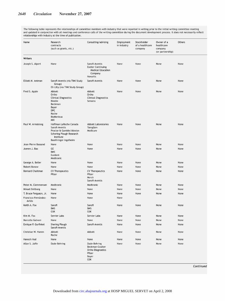
2648 Circulation November 27, 2007
at HOSP MIGUEL SERVET on April 2, 2008 circ.ahajournals.orgDownloaded from
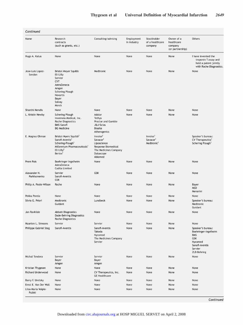
Thygesen et al Universal Definition of Myocardial Infarction 2649
at HOSP MIGUEL SERVET on April 2, 2008 circ.ahajournals.orgDownloaded from
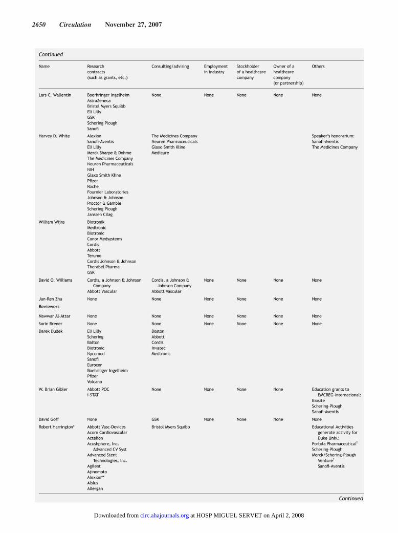
2650 Circulation November 27, 2007
at HOSP MIGUEL SERVET on April 2, 2008 circ.ahajournals.orgDownloaded from
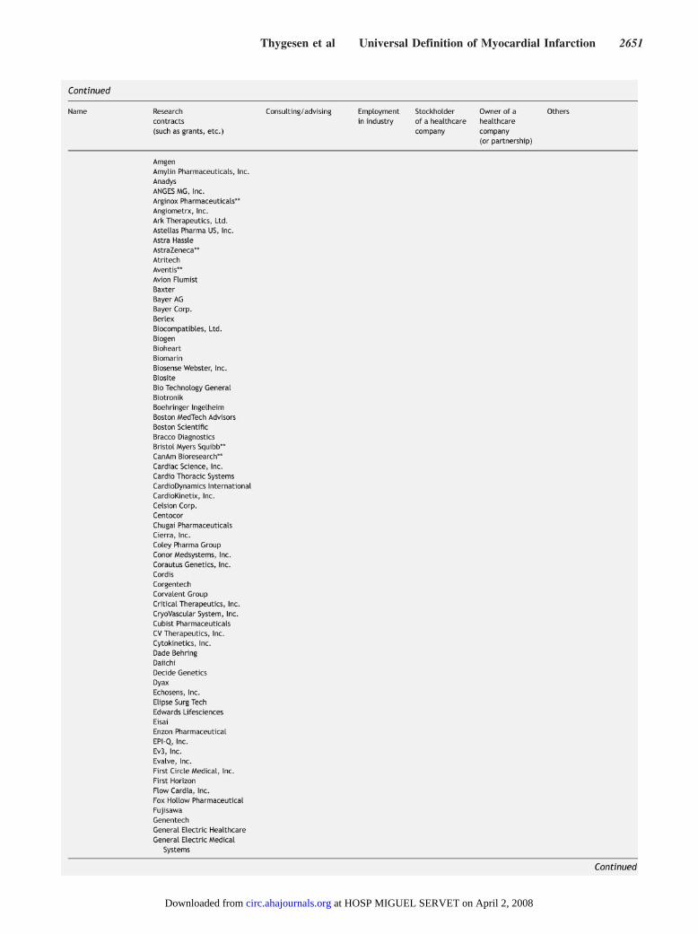
Thygesen et al Universal Definition of Myocardial Infarction 2651
at HOSP MIGUEL SERVET on April 2, 2008 circ.ahajournals.orgDownloaded from
2652 Circulation November 27, 2007
at HOSP MIGUEL SERVET on April 2, 2008 circ.ahajournals.orgDownloaded from
Thygesen et al Universal Definition of Myocardial Infarction 2653
at HOSP MIGUEL SERVET on April 2, 2008 circ.ahajournals.orgDownloaded from