too tall - repub, erasmus university repository
TRANSCRIPT

INFLUENCING THE EXTREMES OF GROWTH
too tall - too small

Cover illustration: Wedding of my grandparents
CIP-DATA KONINKUJKE BIBUOTHEEK, DEN HAAG
De Waal, Wouler J.
Influencing Ihe extremes of growth; too tall, too small I Wouter J. de Waal. - Rotterdam, Erasmus
Universiteit, Afdellng Kindergeneeskunde - [S.I.: s.n.]. - III.
Thesis Rotterdam. - With ref. - With summaI)' In Dutch.
ISBN 90-9009205·6
Subject heading: - child, growth, growth hormone.
No part of this thesis may be reproduced or transmitted in any form. by any means, electronic or
mechanical, Including photocopying, recording or any information storage and retrieval system,
without permission In writing from the author.
~inted by Offsetdrukkerij Haveka B.V" Alblasserdam

INFLUENCING THE EXTREMES OF GROWTH
too tall - 100 small
BEINVLOEDING VAN EXTREME LENGTEGROEI
te groot, te klein
PROEFSCHRIFT
ter verkrijging van de graad van doctor
aan de Erasmus Universiteit Rotterdam
op gezag van de Rector Magnificus
Prof. dr. P.W.C. Akkermans M.A.
en volgens besluit van het College voor Promoties
De openbare verdediging zal plaatsvinden op
woensdag 28 februari 1996 om 13.45 uur
door
WOUTER J. DE WAAL
geboren Ie Egmond aan Zee

PROMOTIECOMMISSIE:
Promotor
Co-promotor
Overige leden
Prof. dr. S.L.S. Drop
Mw. dr. S.M.P.F. de Muinck Keizer-Schrama
Mw. dr. A.C.S. Hokken-Koelega
Prof. dr. HKA. Visser
Prof. dr. F.H. de Jong
Prof. dr. J.M. Wit
The printing of this thesis and the studies described were financially supported by
Novo Nordisk Farma av., Zoeterwoude; Novo Nordisk NS, Denmark; Stichting
Kinderpostzegels, Leiden; Slichtlng Menselijke Voortplanting, Rotterdam; Organon,
Oss.

groot klein
Ik zie een spin
Klein voor mij
Voor aen vlieg een monster
Ik zle aen buizerd
En ik denk wat klein
'Wat groot', dacht de muis
nag even
Peter van Ark, groep 6, Midwolde
Ult: De wereld om mij haen, dichtbundel verschenen onder
ausplclQn van de Stichtlng Werelddag voor Kinderen en
Po~zle, Eel, Vianen 1993
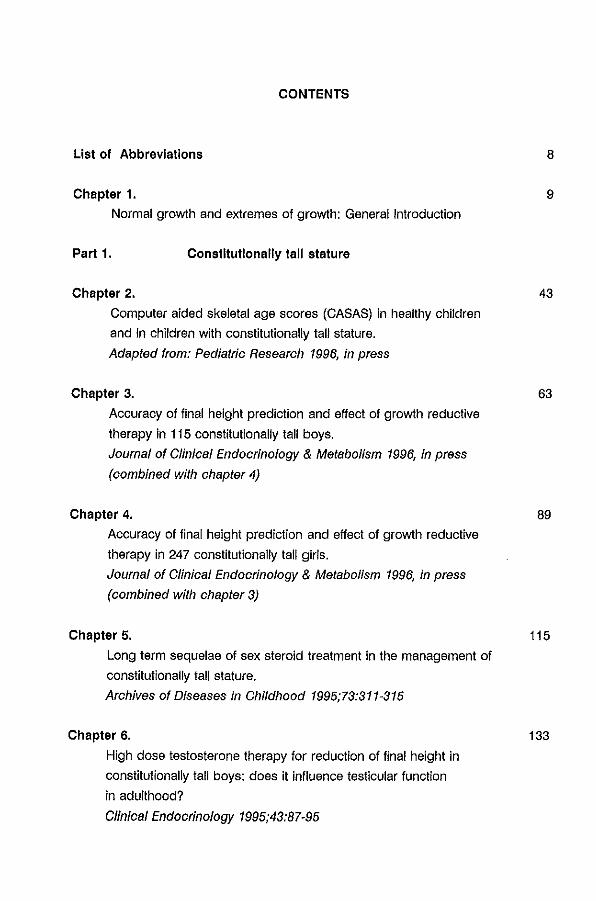
CONTENTS
List of Abbreviations
Chapter 1.
Normal growth and extremes of growth: General Introduction
Part 1. Constitutionally tall stature
Chapter 2.
Computer aided skeletal age scores (CASAS) in healthy children
and in children with constitutionally tall stature.
Adapted from: Pediatric Research 1996, in press
Chapter 3.
Accuracy of final height prediction and effect of growth reductive
therapy in 115 constitutionally tall boys.
Journal of Clinical Endocrinology & Metabolism 1996, in press
(combined with chapter 4)
Chapter 4.
Accuracy of final height prediction and effect of growth reductive
therapy in 247 constitutionally tall girls.
Journal of Clinical Endocrinology & Metabolism 1996, in press
(combined with chapter 3)
Chapter 5.
Long term sequelae of sex steroid treatment in the management of
constitutionally tall stature.
Archives of Diseases in Childhood 1995;73:311-315
Chapter 6.
High dose testosterone therapy for reduction of final height in
constitutionally tall boys: does it influence testicular function
in adulthood?
Clinical Endocrinology 1995;43:87-95
8
9
43
63
89
115
133
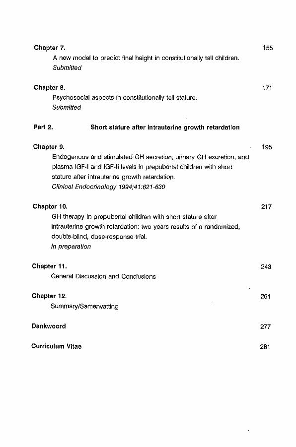
Chapter 7.
A new model to predict final height in constitutionally tall children.
Submitted
Chapter 8.
Psychosocial aspects in constitutionally tall stature.
Submitted
Part 2. Short stature after intrauterine growth retardation
Chapter 9.
Endogenous and stimulated GH secretion, urinary GH excretion, and
plasma IGF-I and IGF-lileveis in prepubertal children with short
stature after intrauterine growth retardation.
Clinical Endocrinology 1994;41:621-630
Chapter 10.
GH-therapy in prepubertal children with short stature after
intrauterine growth retardation: two years results of a randomized,
double-blind, dose-response trial.
In preparation
Chapter 11.
General Discussion and Conclusions
Chapter 12.
Summary/Samenvatting
Dankwoord
Curriculum Vitae
155
171
195
217
243
261
277
281
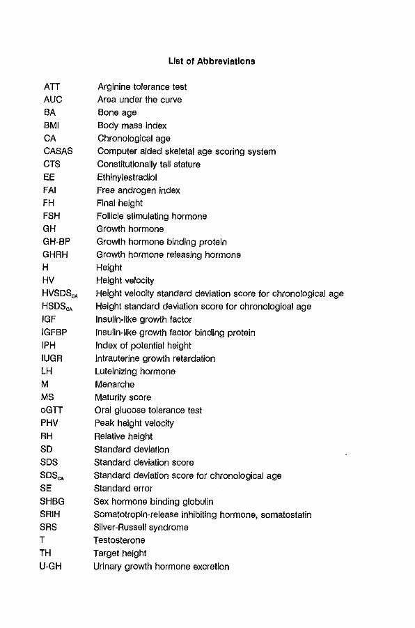
ATI AUC
BA
BMI
CA
CASAS
CTS
EE
FAI
FH
FSH
GH
GH-BP
GHRH
H HV HVSOSCA
HSOS"" IGF
IGFBP
IPH
IUGR
LH
M
MS
oGTI PHV
RH
SO
SOS
SOS",
SE
SHBG
SRIH
SRS
T TH
U-GH
List of Abbreviations
Arginine tolerance test
Area under the curve
Bone age
Body mass index
Chronological age
Computer aided skeletal age scoring system
Constitutionally tall stature
Ethlnylestradiol
Free androgen index
Final height
Follicle stimulating hormone
Growth hormone
Growth hormone binding protein
Growth hormone releasing hormone
Height
Height velocity
Height velocity standard deviation score for chronological age
Height standard deviation score for chronological age
Insulin-like growth factor
Insulin-like growth factor binding protein
Index of potential height
Intrauterine growth retardation
Luteinizing hormone
Menarche
Maturity score
Oral glucose tolerance test
Peak height velocity
Relative height
Standard deviation
Standard deviation score
Standard deviation score for chronological age
Standard error
Sex hormone binding globulin
Somatotropin-release inhibiting hormone, somatostatin
Silver-Russell syndrome
Testosterone
Target height
Urinary growth hormone excretion

CHAPTER 1
General Introduction
General Introduction
NORMAL GROWTH AND EXTREMES OF GROWTH
INTRODUCTION
Growth is an important indicator of physical and emotional well-being in
childhood. Deviations from the normal range both for height and for height velocity
may Indicate an underlying congenital or acquired problem. A thorough understan
ding of the factors influencing the process of normal growth is essential in order to
understand the pathophysiology of extremes of growth.
What is normal growth and what are extremes of growth? This question can
only be adequately addressed when related to population standards. It has been
well established that populations of various ethnic origin may differ considerably in
growth and development (1). Therefore, reference growth curves are obtained by
measuring healthy individuals longitudinally or cross-sectionally (or both). In The
Netherlands, such reference growth curves have been constructed (2). In fact,
Dutch men and women belong to the world's tallest people. Knowing the normal
variance of growth of the reference population, extremes of growth can be defined.
Usually, an individual whose height differs more than 2 standard deviations from
the population mean, I.e. a child with a height above the 97" percentile of the
growth curve is considered too tall and a child growing below the 3" percentile is
considered too short. It needs to be emphasized though, that most of the children
who grow beyond these percentiles are part of the continuum of a normal
distribution curve and only a minority will have a defined abnormality. In addition,
these cut-off points are quite arbitrary and other percentiles may be used.
Children growing at the extremes of height are assumed to encounter
several psychosocial difficulties. In our culture, tallness is generally valued positive.
An association between physical stature and achievement has been documented:
tall individuals score higher on intelligence tests than short individuals (3,4) and
persons who achieved higher social status tend to be taller than those of lower
status (5). In contrast, short statured individuals are perceived to be less
10
Chapter 1
competent, are seen less positively by peers, and are likely to be in lower positions
within a given profession (6). Therefore, tall stature in childhood usually generates
less anxiety initially than shortness. However, many tall adolescents feel different
from their peers and may develop coping mechanisms such as kyphotic posture
(in order to mask their tallness) and social withdrawal. In addition, practical
problems such as clothing and shoeing, fear about future compatible partnering
and careerplanning are also frequently reported problems faced by tall adolescents
(7). In short stature, the main consequences are being infantilized and teased. In
response the child may adopt several behavior patterns. Babyish behavior,
becoming the clown or mascot, social withdrawal, low self-esteem and passiveness
are just a grip of the problems described in children with short stature (8-10).
Growth is the result of complex processes. Multiple factors such as genetic
constitution, nutrition, endocrine function and psychosocial well-being are involved
in the processes of growth (11,12). The genetic component of height has been
estimated to be 0.5 to 0.9; that is, 50% to 90% of the height variation is accounted
for by genetic factors and therefore 50% to 10% is due to environmental factors
(12). Assessment of the parental height as an indicator of the genetic component
of the growth and development of a child is therefore of clinical interest (13). Impor
tant socio-economic factors that are associated with growth and development in
children are social class, family size, birth rank, housing and crowding. Improved
socio-economic conditions and more widespread health have lead to the manife
station of a positive secular trend in growth and development over the past few
centuries (1,2,11). In 1865, the median height among Dutch recruits was 165 cm.
One century later in 1965, the mean adult height in boys was 178 cm. Again, fifteen
years later in 1980, mean adult height had increased by another 4 cm to 182 cm. In
addition, in the middle of the 19th century, age of menarche in European girls was
at about 16 to 17 years. Nowadays, the mean age of menarche is at 13 to 14
years. In contrast, deprivations and poor living conditions, stress, etc., could
strongly affect the sensitive growing years in a negative way, causing an absence
of a positive secular trend or even a negative tendency (1). Remarkably, studies of
11
General Introduction
fossil remains of our hominid ancestors demonstrate that the stature of Individuals
living during the last hundred-thousands of years reached the range of heights
seen today: the mean stature of early anatomically modern H.sapiens in Europe
(around 100,000 years ago) was 184 cm in males and 167 cm in females (14,15).
Probably, similar phenomena responsible for positive and negative secular trends
have affected human height throughout all our history.
Human growth appears to follow a typical pattern irrespective of ethnic
origin or geographical region: from birth a high height velocity is observed until
about 3 years of age; this is followed by a period with a lower and slowly decrea
sing velocity up to puberty. During puberty a sharp increase in height velocity
occurs up to peak height velocity. Thereafter a decrease is noted until adult height
is reached, in girls around 16 years and in boys around 18 years (16,17). Growth
hormone (GH) and thyroid hormone are essential endocrine components for the
regulation of physiological growth during childhood (16-18), whereas during
pubertal growth independent and additional effects of gonadal steroid hormones
are evident (19,20).
In the following paragraphs, we will discuss the endocrine regulation of
normal growth during childhood and focus on the GH/IGFaxis. Thereafter, a short
synopsis of two examples of extremes of growth will be given: constitutionally tall
stature and short stature after intrauterine growth retardation.
Growth hormone physiology
Human GH is synthesized by the anterior pituitary gland as a pre-GH
molecule consisting of 217 amino acids. Subsequent cleavage yields the 191 amino
acid peptide hormone with a molecular weight of 22 kDa, which forms about 80%
of the secreted GH. The other 20% is a smaller variant of about 20 kDa (21). GH
molecules circulate in both bound and free forms. The bound hormones are
complexed to binding proteins (BP). These GH-BPs are identical to the extracellular
domain of the GH receptor (22). They probably act as a modulator of release and
12
Chapter 1
distribution of GH at tissue level.
GH is secreted in a pulsatile manner as a result of a complex of interacting
neuroendocrine pathways (23). Among these are two antagonistic neurohormones:
GH·releasing hormone (GHAH) and somatostatin (Somatotropin Aelease Inhibiting
Hormone, SAl H). GHAH is synthesized in the hypothalamic arcuate and
ventromedial nuclei and contains GH stimulatory activity. SAIH is produced in the
periventricular and amygdaloid nuclei of the medial basal hypothalamus and has
potent inhibitory properties on GH release. Axons from GHAH- and SAIH·
containing neurons terminate in the median eminence and release their hormones
in the vascular network of the portal system. The pulsatile pattern of GH secretion
is orchestrated by episodic increases and decreases in the release of GHAH and
somatostatin, respectively (24,25). In addition, many other neurotransmitters (such
as adrenaline, acetylcholine) and neuropeptides (such as opioid peptides, galanin)
are involved in the neural control of GH secretion with stimulatory or inhibitory
effects on GH release (23).
Many physiological factors are known to affect GH secretion (24,26). These
effects are achieved mainly by altering the secretion of GHAH and/or SAIH, rather
than by direct action at the pituitary level. These physiological factors include
endogenous rhythms, external stimuli and feedback by insulin-like growth factors
(IGFs) and GH itself. Endogenous rhythms include surges of secretion during sleep
and episodiC secretion with peaks at 3 - 4 hours interval during day and night. The
episodic nature of GH secretion varies considerably between physiological states,
sexes and age. External stimuli for GH secretion include physical and emotional
stress, exercise, starvation and metabolic substrates, including basic amino acids
such as arginine and hypoglycaemia. Provocative tests based on these stimuli, and
on administration of various stimulators or blockers of neurotransmitter action, have
been developed to test GH secretion in children. Gonadal steroid hormones also
play a critical role in the regulation of GH secretion by exerting their effects on
multiple sites of the somatotrope axis (19,20). Feedback regulation of GH secretion
is effected by IGF-I by inhibition at both hypothalamic and pituitary sites (27). There
13
General Introduction
is evidence for an inhibitory action of IGF-I on hypothalamic GHRH release and a
stimulatory effect on the secretion of SRIH from the medial basal hypothalamus
(19,24). In addition, local IGF-I production may also inhibit GH release from the
pituitary via paracrine or autocrine mechanisms (28). GH can autoregulate its own
secretion (short loop feedback) by inhibition of GHRH secretion rather than by
enhancement of somatostatin release (29.30).
The GH/IGFaxls
In 1957 Salomon and Daughaday demonstrated that GH stimulated sulphate
incorporation into cartilage indirectly through a serum factor, initially termed
sulphation factor, later designated somatomedin-C or IGF-I (31). This finding
formed the basis of the so called somatomedin hypothesis, stating that circulating
IGFs, produced at distant sites in response to GH, mediate the effect of GH by
endocrine mechanisms (32). With advances in the understanding of biochemistry,
physiology and clinical aspects of the IGFs (33), it is clear that this is an overly
simplistic view. Various lines of evidence point to paracrine and/or autocrine actions
for IGFs as well (34).
There are two different forms of IGF-peptides: IGF-I (70 aminoacids (aa))
and IGF-II (67 aa) with an aa-sequence homology of 60% (35). The IGFs interact
with the cell by binding to specific receptors which have been well characterized
(36). The type I IGF receptor is structurally related to the insulin receptor, binds
IGF-I with higher affinity than IGF-II and binds insulin only weakly. The type II IGF
receptor is structurally and functionally different from the insulin and type I IGF
receptor. It is identical to the mannose-6-phosphate receptor, binds IGF-II with
greater affinity than IGF-I and does not bind insulin. The IGFs are present in the
circulation and throughout the extracellular space almost entirely bound to binding
proteins, IGFBPs. To date, six IGFBPs have been characterized (37). The vast
majority (approximately 90%) of total IGF circulates as a 150 kDalton complex, con
sisting of IGF-I or IGF-II plus IGFBP-3 and an acid labile subunit (38). The
14
Chapter 1
concentration of IGFBP-3 has been found to be GH dependent, while levels of
IGFBP-1 and IGFBP-2 are inversely correlated with GH status (39).
The IGFs have a wide array of biological effects in many tissues and cells.
The best-known effects of IGFs relate to their acute anabolic effects on protein and
carbohydrate metabolism and to their longer term effects on cell replication and
differentiation (33,40,41). These biological actions may be influenced by the
IGFBPs. Possible functions of the IGFBPs are 1) acting as transport proteins, 2)
prolonging the half-lives of the IGFs, 3) providing a means of tissue- and cell type
specific localization, and 4) directly modulating the interaction of the IGFs with their
receptors and thereby indirectly controlling their biological action (37,41,42).
Longitudinal bone growth
GH has a profound effect on longitudinal growth. With respect to the growth
promoting effect of GH on the epiphyseal growth plates a dual effector theory of
GH action has been advocated (43). GH stimulates bone growth directly by
promoting the differentiation of precursor cells in the growth plate, and GH
stimulates bone growth indirectly by inducing IGF-I responsive chondrocytes and
by stimulating the local production of IGF-I. On the other hand, gonadal steroids
also influence skeletal growth as shown by an increased growth rate at the time of
gonadal maturation (19). Sex steroids have both indirect and direct effects on
skeletal growth. The indirect action is mediated by GH and IGF-I, since sex
hormones stimulate GH and IGF-I secretion (19,20). Evidence is present that sex
steroids also have a direct growth effect, independent of IGF-I and GH (44,45).
The effects of GH and sex hormones on longitudinal bone growth become
clear from clinical data of children presenting with growth disorders that can be
conceived as experiments of nature (19,46). GH-deficient or -insensitive children are
characterized by severe growth retardation. The long bones are relatively short
compared to the spine length and head size. In contrast, children with
hypogonadism have relatively long limbs. In this condition the growth plates of the
15
General Introduction
long bones remain open for GH action over a longer period of time due to lack of
sex hormones. Patients with Laron dwarfism suffer from GH-insensitivity due to a
functionally defective GH receptor gene. These patients have high circulating values
of GH but no endocrine generation of IGF-I while a definite pubertal growth spurt is
observed, suggesting a direct sex steroid effect on bone growth (19). Children with
precocious puberty have elevated levels of sex hormones at a prepubertal age
which cause early fusion of the epiphyseal growth plates. They will thus end up
shorter than expected and show relatively short long bones (47).
Beside the effect of GH on longitudinal growth, it also exerts other metabolic
effects on carbohydrate, lipid and mineral metabolism. These items have been
ex1ensively described in a number of reviews and will not be further discussed here
(48-50).
Insight in the complex orchestration of physiological growth, mediated by
variables such as GH secretion, IGFs and IGFBPs, which in turn have complex
biological actions by itself, is essential for possible therapeutic intervention in
children presenting with ex1remes of growth. In this thesis, we will focus on two
examples of extremes of growth: constitutionally tall stature and short stature
associated with intrauterine growth retardation. Both entities will be reviewed briefly.
CLINICAL ASPECTS OF THE EXTREMES OF GROWTH
Constitutionally tall stature
Natural growth pattern
Constitutionally tall stature (CTS) is a variant of the normal pattern of
childhood growth and development and constitute 3 - 10 percent of the normal
popUlation, depending on the definition used. Usually, one or both parents are also
tall, thus genetic and familial factors appear to play the most important role in
16
Chapter 1
etiology and pathogenesis. Mean birth length is at the 75" percentile, and tall stature
becomes evident at the age of three to four years. Growth velocity is accelerated in
early childhood but slows down after four or five years of age when the- growth
curve starts to parallel the normal curves (51). The diagnosis is generally made
from family history, record of growth and physical examination. No apparent
abnormalities are present at physical examination, which makes it possible to
distinct from other excessive growth syndromes such as Marian syndrome,
homocysteinuria, and Klinefelter syndrome (52).
Endocrinology
Endocrinological studies of tall children indicate that tall stature is due, at
least partly, to increased GH secretion. Studying children with various heights, a
significant positive correlation was found between growth and GH secretion (18,53).
Paradoxical GH responses to glucose loading and to administration of thyrotropin
releasing hormone similar to those seen in acromegaly have been observed in
constitutionally tall children (54,55). Furthermore, elevated serum levels of IGF-I
have been found (54,55). However, not all children show signs of relative
hypersecretion of GH. A rscent study showed a clear heterogeneity of GH secreti
on in tall children, with even the presence of low GH secretors (56). Therefore,
other mechanisms than GH hypersecretion may play an important role in the
etiology of tall stature, such as hypersensitivity to GH (56), enhanced pituitary
responsiveness to GHRH (57) or a decreased inhibitory effect of somatostatin (58).
Height prediction
Height prediction plays a key role in the management of children with
growth disorders and thus In children with CTS. In fact, possible therapeulic
intervention is based on the estimated height prognosis: whenever the height
prognosis exceeds a certain limit (usually 2 standard deviations above the mean of
the population), treatment will be considered. Hence, accurate techniques for
reliable height predictions are needed. In clinical practice various methods have
17
General Introduction
been developed of which the methods developed by Tanner et al (59) and Bayley
and Pinneau (60) are most commonly used. They both share the use of bOne age
as an indicator of skeletal maturity in order to estimate final adult height. The first
prediction method uses the bone age method developed by Tanner et al (59),
whereas the latter utilizes the bone age method of Greulich and Pyle (61). This
implies that many of the potential problems underlying the bone age determination
methods have to be considered (62). For instance, both techniques make use of
subjective processes and discontinuous scales which result in considerable inter
and intra·rater variability (63-65). In order to improve the objectiveness of bone age
estimation the method of Tanner et al has recently been transformed into a
computerized image analysis system using a continuous scale (66). However,
before integrating this computer system into clinical practice, validation studies are
needed. Thus far only a limited number of studies have been performed testing the
reliability of height prediction methods in large groups of untreated children with tall
stature (67-69). Nevertheless, knowledge about the specific advantages and
disadvantages of the various methods is of utmost importance since it may
influence clinical practice.
Short stature after Intrauterine growth retardation
Fetal growth
Fetal growth can be considered in two major phases: that relating to
embryogenesis and organogenesis which Is essentially the first half of gestation,
and growth of the late gestation fetus. Growth in the first half of gestation shows
minimal variation, except when associated with pathology e.g. chromosomal
abnormalities or infections. Fetal growth in the second half of gestation is greatly
constrained by the uterine environment and largely mediated by changes in fetal
substrate supply (70). Therefore, reduction in fetal substrate supply by maternal
disease or placental dysfunction causes a prompt reduction in fetal growth. This is
accompanied by a redistribution of fetal blood flow to favour vital organs such as
18
Chapter 1
the brain and heart. Important interactions are present between nutritional state and
the interaction of maternal, placental and fetal hormones and growth factors (71-
75). Most attention has been focussed on the role of insulin and insulin-like growth
factors I and II as regulators of fetal somatic growth (76-81). Marked changes in
insulin secretion are associated with clinical abnormalities of fetal growth:
pancreatic agenesis leads to profound intrauterine growth retardation (IUGR) and
fetal hyperinsulinemia due to maternal diabetes results in fetal overgrowth. In
addition, levels of IGF-I in fetal and cord blood correlate with birth size. In fetuses
with IUGR, circulating levels of IGF-I and IGF-II are reduced. GH on the other hand
has a limited role in fetal growth. Thus, optimal growth can only occur if the
nutritional and endocrine milieu is appropriate.
Postnatal growth
Children with intrauterine growth retardation (IUGR) comprise a heteroge
neous group. As stated above, various causes may underlie the stunted growth at
birth varying from chromosomal disorders, congenital infections, placental
dysfunction to maternal disease (smoking, hypertension, alcohol abuse). However,
in the majority of cases the etiology is not clear (idiopathic) (82). After IUGR, most
of the children born without an underlying disorder do show catch-up growth within
the first years of life. However, the percentage of children who fail to show catch-up
growth after birth is different in various studies ranging from 10-30% (83-85). This
variability is not only caused by the heterogeneity in etiology underlying the
prenatal growth retardation, but also by the differences in definitions of IUGR. In
practice various criteria have been used to define IUGR, including measures of
absolute size, such as low birth weight «2500 g) or very low birth weight « 1500
g), and measures of relative size (small for gestational age), variably defined as
birthweight or birthlength less than the third or tenth percentile, and less than two
SO below the population mean for gestational age and sex (68). However, since
the postnatal growth of children with IUGR is usually related to height, it seems
appropriate to define IUGR in terms of birthlength rather than birthweight. In a
19
General Introduction
recent Swedish study on postnatal growth in 5111 children 13% of the 111 children
born with a short birth length «-2 SO below the mean for gestational age) showed
insufficient catch-up growth in the first two years of life (86). Hokken et al (87)
reported on the postnatal growth of 724 children born with IUGR (birth length < P3
for gestational age) from three academic hospitals In The Netherlands and found a
similar percentage of 15% of children without catch-up growth up to a height > P3
within the first two years of life. In this respect, they observed no significant
difference between preterm and full-term babies. Therefore, one of the long-term
effects of IUGR may be persistent short stature. Indeed, various studies on final
adult height show that these children have shorter adult heights compared to the
normal population and compared to their genetic potential (target height) (85,88-
90).
Silver-Russell syndrome
The growth pattern of children with the Silver-Russell syndrome (SRS) shows
a striking resemblance with that of IUGR children without dysmorphic signs (91,92).
SRS is mainly characterized by short stature, low birth weight and dysmorphic
features such as small triangular face, body asymmetry, clinodactyly, and down
curved corners of the mouth (93). Therefore, many studies on growth of children
with short stature after IUGR usually include patients with SRS as well. There are
indications that the natural history of children with IUGR includes an increase in
skeletal maturation towards the latter half of the first decade of life (88,91,92). In
addition, some patients with IUGR or SRS may develop an early puberty (91,93,94).
Both phenomena may play an important role In the restriction of the final adult
height of these children.
Endocrinology
The endocrine control of postnatal growth differs from that which applies
prenatally. Whereas the intrauterine growth of the fetus is largely independent of
the pattern of GH secretion, the role of the GH axis is dominant during childhood.
20
Chapter 1
The pathophysiological mechanism underlying the failure to catch·up growth of
children with short stature after IUGR is not completely understood. It is possible
that persisting defects in the GH/IGFaxls may be debit to the stunted growth.
However, IUGR children, with and without dysmorphic signs, tend to be tiny and
lack the typical signs of GH·deficient children such as abdominal fat distribution.
Nevertheless, endocrinological studies did find evidence for disturbances in GH
secretion: abnormal GH secretion patterns during 12 or 24 hours of physiological
testing and/or Insufficient GH responses to provocative tests have been observed
{95·97}. Few data are available on IGF·I and IGF-II levels in IUGR children during
childhood. Normal IGF·I levels have been reported, but without correction for
chronological age {96,98}.
Other pathophysiological mechanisms
Factors Involved in the pathophysiological state of IUGR itself, may also
affect organ development. It is known that the brain undergoes a period of rapid
weight gain during the later half of the fetal period with a climax at the time of birth
{99}. This growth spurt of the brain decreases during the first year of life. Besides,
there are at least two periods of rapid brain cell multiplication. One occurs at 15·20
weeks of fetal development and concerns development of neuroblasts. A second
period concerns glia cell multiplication, and starts at 25 weeks of gestation and
ends probably in the second year of life {I DO}. During these growth spurts, the
brain is presumably vulnerable to disturbances in its nutritional supply. Thus, brain
damage may occur due to the underlying factors responsible for the intrauterine
growth retardation. In fact, animal studies gave arguments in support of this
hypothesis {101}. Other lines of evidence are derived from clinical studies in
children with IUGR showing an association between IUGR and an increased
incidence of mental handicap, lower intelligence, neurological deficits, poor
academic performance and behavior problems {I 02-1 06}. Therefore, the GH
secretion disturbances observed in children with IUGR may reflect a form of brain
dysfunction. More research is required to elucidate the pathophysiological
21
General Introduotlon
meohanism of the failure to catch-up growth in children with IUGR.
TREATMENT OPTIONS FOR THE EXTREMES OF GROWTH
Treatment of constitutionally tall stature
Psyohology
Treatment of tall stature is generally based on psychological grounds. Some
children with excessive growth may suffer considerably from being much taller than
others. They feel different from their peers and are subject to hurtful remarks about
their height. Coping mechanisms such as kyphotic posture (In order to mask their
tallness). social withdrawal and even depression may develop. Practical problems
concerning clothing and shoeing. fear about future partnering (especially in girls)
and careerplanning are also frequently reported problems faced by tall children.
This has lead to a search for height intervening therapy in children with CTS.
Sex hormone treatment
In 1956. Goldzieher used high doses of sex steroids in 14 adolescent girls
with tall stature in order to reduce their final height (107). The basis for the use of
sex hormones to limit adult height came from observations in children with
precocious puberty. These children show early closure of the epiphyses due to
premature secretion of gonadal steroids. which limit their eventual adult height
(108.109). Since then. many reports have appeared describing the height reducing
effect of administration of high doses of sex hormones in girls (110-128) and in
boys (129-131). There is general agreement that a favorable effect on ultimate
height results from such pharmacologioal therapy. with possible much greater
effects In selected patients. Some studies have shown that the height reduction is
greater when treatment has begun at a younger age and/or bone age
(111.112.119.123.125.127.128). However. the establishment of the claimed effects is
open to debate. since a well designed. prospective controlled study has never
22
Chapter 1
been performed. This Implies that the observed results may have been biased by
several factors. For instance, results on height reduction had only been derived
from comparison of the achieved height with the height prediction prior to
treatment, and with no correction for the error of the prediction method used. In
addition, when control groups had been used they tended to be small. Besides,
while assessing the ultimate height reduction, differences in initial clinical data
between treated children and controls such as age, bone age and height predic
tion, had not been taken into account. Furthermore, in many studies adult height
had been assumed to be reached at a relatively young age. Therefore,
interpretation of the height reducing effect of sex hormone treatment in children
with CTS has to be seen with reservation.
Long-term side effects
An important issue concerning the treatment with high doses of sex steroids
Is the possibility of unwanted side effects. In this respect special attention has been
focussed on hemostasis (132), lipid metabolism (133) and functioning of the
hypothalamic-gonadal axis (134,135). So far, unwanted side effects have only been
reported during treatment or shortly after discontinuation of therapy (92-131,136-
140). Most side effects were found to be mild and reversible. Suppression of the
hypothalamic-gonadal-axis induced by the pharmacological doses of sex steroids
(via a negative feedback mechanism) was found to be reversible (134,135).
However, the possibility of a long-term suppressive effect of sex hormone therapy
on reproductive functioning in boys has been postulated (141,142). In girls, the
ultimate 'proof' for complete reversibility of hypothalamic-gonadal suppression,
pregnancy, has been reported in various single cases (111,118,120,124,127).
However, systematic long-term follow-up studies on possible unwanted side effects
are lacking. In this respect, the considerable amount of data on the association
between long-term oral contraceptive use and possible health risks (reviewed in
references: 143,144) are indicative as they may form a reflection of the prospective
risks in estrogen-treated girls.
23
General Introduction
Other treatment modalities
Another approach in the management of tall stature relates to interference in
the regulation of GH secretion. It is based on the assumption that tall stature is
related to GH hypersecretion. In order to suppress endogenous GH secretion
bromocriptlne therapy has been used. Bromocriptine is a dopamine agonist and
inhibits GH secretion in patients with acromegaly by binding to dopamine receptors
at the pituitary level (145). In children with CTS, however, results are conflicting and
the effectiveness of bromocriptine has not been SUbstantiated (146-148). Recently,
an alternative strategy has been proposed to limit growth with administration of a
somatostatin-analogue. Preliminary studies showed an effective suppression of GH
secretion and a significant reduction in growth rate by somatostatin therapy (149-
151). However, final results on height reduction remains to be established.
Moreover, the optimum mode of administration and the presence of serious side
effects (such as asymptomatic gall bladder stones) need to be considered.
Treatment of short stature after IUGR
Psychology
Short stature, irrespective of the underlying cause, may encounter several
psychological difficulties (6,8-10). A tendency to lower Intelligence scores, behavior
problems, low self-esteem, unemployment and lower social success have been
reported (8,9,152-154). Apart from this, IUGR per se is associated with an
increased prevalence of psychological disabilities. Several studies have been
performed on this subject and associations have been found between IUGR and an
increased incidence of mental handicap, lower intelligence, neurological deficits,
poor academic performance and behavior problems (102-106).
GH treatment
In order to improve growth in children with short stature after IUGR, GH
24
Chapter 1
treatment using human GH has been explored since the early 1970s (155-157).
Initial results were disappointing probably due to the low dose and frequency of
GH administration (2-3 times weekly). With the availability of biosynthetic GH, the
efficacy and safety of daily treatment with GH has been tested in children with non
GH deficient short stature (158-160), inclusing in children with short stature after
IUGR (95-97,160). In the study by Albertsson-Wikland (95) GH treatment with doses
of 0.1 IU/kg/day of recombinant GH (.0.7 IU/kg/week) resulted in a significant
improvement of growth rate in five out of six children with SRS (height .velocity
standard deviation score (HVSDS) +0.85 during the first year and HVSDS +0.58
during the second year of treatment) and in seven out of ten IUGR patients
(HVSDS + 1.21 during the first year of treatment). Other studies showed similar re
sults. In the study by Rochiccioli et al (96) nine children with IUGR were treated
with a pituitary GH dose of 0.3 IU/kg/week for one year. The height velocity
Increased from a baseline level of 3.5 ± 0.8 cm/year to 7.0 ± 0.9 cm/year. The
study by Stanhope et al (97,161) was a dose-response study comparing treatment
with recombinant GH doses of 15 and 30 IU/m'/week (. 0.5 and 1.0 IU/kg/week,
respectively) in the first year of treatment. In the next two years of treatment all 24
children received the higher GH dose of 30 IU/m'/week. The study included IUGR
patients with and without dysmorphic features. In the first year of treatment a dose
dependent increase in height velocity was observed: HVSDS increased from -0.8 to
+1.4 with the lower dose and from -0.8 to +3.6 with the higher dose. After three
years of treatment, mean HVSDS was + 1.1, irrespective of which initial treatment
dose had been administered during the first year. There was no difference in the
growth response of children with or without dysmorphic features. However, despite
the sustained increase in growth rate, no significant change in height for bone age
SD score was present. This may point to an unaltered height prognosis. Yet, in
untreated IUGR patients, a natural decrease in height prognosis may be present
associated with an inappropriate advance of epiphyseal maturation. Therefore it is
possible, that GH treatment does lead to a greater adult height than would have
been attained otherwise (161). Longer term studies with substantial greater
25
Generallntroduotion
numbers of patients are required to answer the outstanding question whether GH
treatment may be indioated for children with short stature after IUGR.
AIMS OF THE STUDY
Constitutionally tall stature
The studies presented in this thesis were undertaken to evaluate the value of
height prediction methods and the effect of sex hormone treatment in children with
CTS. In the Sophia Children's Hospital, constitutionally tall children have been
treated with high doses of sex steroids since 1968. In girls oestrogens have been
used (ethinyloestradiol 200 Ilg/day, orally, range 100-300llg/day) in combination
with progestagens (medroxyprogesterone 5-10 mg/day, orally) every 5-10 days of
the month. Tall boys have been treated with androgens: testosterone esters in
various regimens with a total monthly dose up to 1000 mg).
26
The following questions were addressed:
How reliable is adult height prediction in children with tall stature?
What is the reliability of a computer aided skeletal scoring system in bone
age assessment and Is it applicable to bone age assessment in children with
tall stature?
What Is the ultimate height reducing effect of sex hormone treatment in the
management of tall stature?
Are there long-term side effects of administration of high doses of sex
steroids to pubertal children, especially with respect to gonadal function and
fertility?
Can we improve height prediction in tall children by using a prediction
method based on growth data of tall subjects?
Do previously treated children differ in psychosocial status from tall subjects
who had not chosen for height reducing therapy?
Chapter 1
Short stature after IUGR
The studies presented In the second part of this thesis were undertaken to
obtain more insight in the pathophysiological mechanism of the stunted growth in
children with IUGA. In addition, the efficacy and safety of recombinant GH therapy
was evaluated.
The following questions were addressed:
Are there disturbances in the GH/IGFaxis explaining the growth retardation
in children with short stature after IUGR?
What is the effect of GH administration on linear growth and bone matura
tion in children with persistent short stature after IUGR?
Is there a relationship between the GH secretory status and the growth
response to GH treatment?
Are there adverse effects of GH therapy on carbohydrate metabolism in
children with IUGR?
Another important Issue concerned the Impact of GH treatment on various
psychological aspects. Questions like: What Is the psychological status of children
with short stature after IUGR?, and: Does GH have a beneficial effect on
psychological development? easily come into mind. These Items will be subject of
the thesis of Mrs. E.A. van der Reijden-Lakeman, psychologiste, entitled: "Growing
pains: psychological evaluation of short stature in intrauterine growth retarded
children, before and after two years of GH treatment" (162).
27
General Introduction
STUDY DESIGN
Constitutionally tall stature
To evaluate the questions raised above, all men and women, who had been
seen at adolescence for evaluation of their constitutionally tall stature, and who
reached the age of 18 were contacted by mail and asked to participate in a large
follow-up study. In total, 247 men and 423 women were contacted, of whom 102
men and 249 women had received hormonal treatment for reducing their final adult
height. Hundred and forty-five men and 174 women had not chosen for treatment
and served as controls. Second mailings were sent to those who did not respond
to the first mailing. Subjects were asked to participate in three substudies: 1) an
auxologlcal evaluation, 2) an evaluation on long-term effects, and 3) a psychosocial
evaluation.
In the auxologlcal part of the study, subjects were recalled to our outpatient
clinic for final height assessment. In addition, auxologlcal data were collected from
the hospital charts and radiographs of the left hand and wrist were retrieved for
bone age determinations and final height predictions.
To evaluate long-term effects of hormonal treatment, all subjects were
interviewed in a standardized way. In women, this interview included questions
about satisfaction with and possible side effects of hormone treatment, oral contra
ception, menstrual cycle characteristics, pregnancy and gynecological complaints.
Men were asked about satisfaction with and possible side effects of hormone
treatment and about their offspring. In addition, men were asked to participate in
an andrological study. For this study, aI/ participants were called in twice at the
outpatient clinic of the Department of Andrology, Dijkzigt Hospital, Rotterdam. At
these visits, every man underwent a medical history and a physical examination
Including testicular volume measurement and screening for varicocele. Fimilly, two
semen samples were obtained for semen analysis and blood was taken for
determination of plasma hormones.
28

Chapter 1
To obtain Insight in psychosocial aspects of constitutionally tall stature, five
different, standardized questionnaires were sent to all participants by mail. Special
areas of interest concerned self-esteem, social anxiety and assertiveness, .general
psychological well-being and body satisfaction.
The following inclusion- and exclusion criteria were used for these studies:
constitutionally tall stature, defined as a height above the P90 according to
Dutch references
age of at least 18 years
if treated, a treatment period of at least 6 months
no endocrine or metabolic disorders, such as acromegaly or homocystei
nuria
no chromosomal abnormalities or syndrome, such as Klinefelter syndrome,
Marfan syndrome or Sotos syndrome.
no use of other growth intervening therapy
Table 1 presents the response rate of all subjects to the various substudies:
Subjects Non-respon- Responders
dars
Auxology Psychology long-term effects Non-particIpants
Interview Andrology
Men
Treated 102 7 6. 76 64 43 20
Untreated 145 31 62 69 61 32 3'
All 247 36 127 14. 125 75 55
Women
Treated 249 46 177 174 160 9
Untreated 174 51 95 106 92 16
All 423 97 272 280 272 25
29
Generallntroduotion
Short stature after IUGR
To evaluate the questions raised above a multicenter study was set up In
which four centers in the western part of The Netherlands participated:. Sophia
Children's Hospital, Rotterdam, Wilhelmina Children's Hospital, Utrecht, Academic
Hospital of Free University, Amsterdam, and Juliana Children's Hospital, The
Hague. The study population comprises 79 prepubertal children with short stature
after lUG A. IUGR was diagnosed when birth length was 2 SD or more below the
mean for gestational age according to the standards of Usher and McLean (163).
In addition, the following Inclusion- and exclusion criteria were used:
no catch-up growth, defined as not obtaining a height equal to or above the
P3
growth rate equal to or below the P50 for chronological age
uncomplicated neonatal period, i.e. without signs of severe asphyxia
(defined as Apgar score below 3 after 5 minutes), without complicated
sepsis neonatorum and without long-term complications of respiratory
ventilation such as bronchopulmonary dysplasia
chronological age 3.00 to 8.99 in girls and 3.00 to 10.99 in boys
prepubertal stage
no chromosomal abnormalities or other organic causes for growth
retardation, except for SRS
no previous use of growth intervening therapy
To evaluate aspects of the GH/IGFaxis, GH secretion was studied in a total
of 40 IUGR children by 24-hour plasma GH profiles and standard arginine
provocation tests. In addition, plasma IGF-I and IGF-lileveis and urinary GH excreti
on were measured.
To assess the effects of GH therapy on linear growth, bone maturation,
pubertal development and final height in children with short stature after IUGR, a
randomized, double-blind, dose-response study was set up. After inclusion,
30
Chapter 1
patients were randomly and blindly assigned to either 3 or 6 IU/m'/day (~ 0.1 and
0.2 IU/kg/day) of recombinant GH (Norditropin,,), administered by subcutaneous
injection. In this ongoing study, patients have been examined at enrollment and
subsequently every 3 months at the four participating centers. Measurements
include auxological parameters, biochemical parameters and safety parameters.
To evaluate the psychological effects of GH treatment on children with short
stature after IUGR, a parallel study was also performed. The results of this study
before and after two years of GH treatment will be described in the thesis' of Mrs.
E.A. van der Reijden-Lakeman, psychologiste (163).
STRUCTURE OF THE THESIS
In Chapters 2 to 8 studies on tall stature are discussed: Chapter 2 describes
the reliability of a recently developed computerized skeletal age scoring system
and its applicability in children with CTS. This system can be used for assessment
of bone age and final height predictions. The accuracy of various height predictions
in the management of tall stature are addressed In the next two chapters (ohapters
3 and 4). In addition, the effect of sex hormone therapy on height reduction in
constitutionally tall children is evaluated in the same chapters (ohapter 3 and 4),
while adjusting for differences between treated and untreated tall children. Chapters
5 and 6 describe the long-term effects of sex steroid treatment in pharmacological
doses and focus on functioning of the hypothalamic-pituitary-gonadal axis. In
chapter 7 a new prediction model is presented to predict adult final height in
children with tall stature based on growth data derived from a sample of untreated
tall subjects. Finally, chapter 8 describes psychosocial aspects in constitutionally
tall stature.
Chapters 9 and 10 concern studies In children with short stature after IUGR.
In Chapter 9, several aspects of the GH/IGFaxis are studied. Chapter 10 presents
31

General Introduction
the two-years results of the randomized, double-blind, dose-response multicenter
trial in 79 prepubertal IUGA children comparing the efficacy and safety of two
different doses of GH (3 and 6 IU/m'/day).
Chapter 11 discusses the significance of the presented data. In addition, final
conclusions and recommendations are made and suggestions for future research
are given.
Chapters 12 and 13 present a summary of the thesis in the English and
Dutch language, respectively.
32

Chapter 1
REFERENCES
1. Eveleth PB, Tanner JM: Worldwide variation In human growth. London, Cambridge University Press, 1990
2. Roede MJ, Van Wieringsn JC: Growth diagrams 1980. Netherlands third nation-wide survey. Tljdschr Soc Gezondheidsz 1985;63(SupPO:1-34
3. Humpreys LG, Davey Te, Park AK: Longitudinal correlation analysis of standing height and intelligence. Child Develop 1985;56:1465·1478
4. Taasdale TWo Owen DR, Sornssen TIA: Intelligence and educational level in adult males at the extremes of stature. Hum Bioi 1991:63:19-30
5. Hensley WE, Cooper A: Height and occupational sueces: a review and critique. Psychol Reports 1987;60:843·849
6, Underwood LE: The social cost of being short: social perceptions and biases, Review paper. Acta Paedlatr Scand 1991 ;377(SuppO:3.8
7. Pradar A, Zachmann M: Treatment of excessively taU girls and boys with sex hormones. Pediatrics 62:1202·1210
8. Law CM: The disability 01 short stature. Arch Dis Child 1987;62:855·859 9. Skuse 0: The psychological consequences of being small. J Child Psychol psychlatr
1987;28:641·650 10. Siegel PT, Clopper R, Stabler B: Psychological Impact 01 slgnillcantly short stature. Review
paper. Acta Paedlatr Scand 1991;377(Suppl)14·18 11. Tanner JM: Growth at adolescence, 2nd edition. Blackwell, Oxford, 1962
12. Mascie-Taylor CGN. Biosoclallnfiuences on stature: a review. J Biosoc Sci 1991;23:113-128 13. Sorva R, Tolppanen EM, Lanklnen S, Perheentupa J: Growth evaluation: parent and child
specilic height standards. Arch Dis Child 1989;64:1463 ·1487 14. Garralda MD: Evolution of human height. In: Hernandez M, Argente J (eds): Human growth:
basic and clinical aspects. Amsterdam, Elsevier Science Publishers B. V., 1992, P 135·142 15. Styne OM, McHenry H: The evolution 01 stature In humans. Horm Res 1993;39(Suppl):3.6 16. Karlberg J: A blologically·orlented mathematical model (ICP) for human growth. Acta
Paedlatr 1989;350(Suppl):70·94 17. Karlberg J: On the construction 01 the Infancy·Chlldhood·Puberty growth standard. Acta
Paediatr 1989;356(Suppl):26·37
18. Albertsson·Wikland, Rosberg S: Analyses 01 24·hour growth hormone profiles In children: relation to growth. J Clln Endocrinol Metab 1988; 67:493·500
19. Bourguignon JP: Linear growth as a function of age at onset of puberty and sex steroid dosage: therapeutic Implications. Endocr Rev 1988;9:467·488
20. Kerrigan JR, Rogol AD: The Impact of gonadal steroid hormone action on growth hormone secretion during childhood and adolescence. Endocr Rev 1992;13:281-298
21. Baumann G: Growth hormone heterogeneity: genes, isohormones, variants and binding proteins. Endocr Rev 1991;12:424·449
22. Leung OW, Spencer SA, Cachlanes G, Hammonds RG, Collins C, Henzel WJ, Barnard R, Waters MJ, Wood WI: Growth hormone receptor and serum binding protein: purification, cloning and expression. Nature 1987;330:537·543
33
General Introduction
23. Bertherat J, Bluet-Palot MT, Epelbaum J: Neuroendocrine regulation of growth hormone. Mlnlreview. Eur J EndocrinoI1995;132:12-24
24. Hartman ML, Veldhuis JD, Thorner MO: Normal control of growth hormone secretion, Horm Res 1993;40:37-47
25. Tannenbaum GS, Ling N: The interrelationship 09 growth hormone (GH)-releasing factor and somatostatin in generation of the ultradlan rhythm of GH secretion, Endocrinology 1984; 115: 1952-1957
26. Johnston DG, Davies RRJ Prescott RWG: Regulation of growth hormone secretion In man: a review. J Roy Soc Med 1985;78:319-327
27. Berelowitz M, Szabo M, Frohman LA, Firestone 8, Chu l, Hintz RL: Somatomedin-C mediates grow1h hormone negative feedback by effects on both the hypothalamus and the pttuttary, Science 1981 ;212:1279·1281
28. Fagin JA, Pixley S, Slanina S, Ong J, Melmed S: Insulin·like grow1h factor I gene expression
In GH3 rat pituitary cells: messenger ribonucleic acid content, immunochemistry, and secretion. Endocrinology 1987;120:2037-2043
29. Conway S, MaCann SM, Krulich L: On the mechanism of grow1h hormone autofeedback regulation: possible role of somatostatin and growth hormone·releaslng factor. Endocrinology 1985;117:2284·2292
30. MOiler EE: Neural control of somatotropic function. Physlol Rev 1987; 67:962-1053 31. Salomon WD, Daughaday WH: A hormonally controlled serum factor which stimulates sulfate
Incorporation by cartilage in vitro. J Lab Clin 1957;49:825·836 32. Gluckman PO, Douglas RG, Ambler GR, Breier BH, Hodgkinson SC, Koea JB, Shaw JHF:
The endocrine role of Insulin·like growth factor-I. Review paper. Acta Paediatr Scand 1991 ;372(Suppl):97·1 05
33. Herington AC: Insulln·like grow1h factors: biochemlstl)l and physiology. Balliere's Clin Endo· crinol Metab 1991;5:531-551
34. Chatelain p, Naville 0, Avallet 0, Penhoat A, Jaillard C, Sanchez P, Saez J: Paracrine and autocrine regulation of insulin-like growth factor·1. Review paper. Acta Paediatr Scand 1991 ;372(Suppl):92-95
35. Daughaday WH, Rotwein P: Insulin·like grow1h factors I and II. Peptide, messenger ribonucleic acid and gene structures, serum, and tissue concentrations. Endocr Rev 1989;10:68-91
36. Neely EK, Beukers MW, Oh Y, Cohen P, Rosenfeld RG: Insulin·like grow1h factor receptors. Review paper. Acta Paedlatr Scand 1991;372 (SuppQ:116·123
37. Drop SLS, Schuller AGP, Lindenbergh·Kortleve OJ, Groffen C, Brinkman A, Zwarthoff EC: Structural aspects of the IGFBP family. Mini review. Grow1h Regulation 1992;2:69·79
3S. Baxter RC, Martin JL: Structure of the M, 140,000 of the grow1h hormone dependent insulin·
like growth factor binding protein complex: determination by reconstitution and affinitylabeling. Proc Natl Acad Sci USA 1989;86:6896·6902
39. Blum WF: Insulln·like grow1h factors and their binding proteins. In:Ranke MB (ed.): Functio· nal endocrinologic diagnostics in children and adolescence. Vertag, Mannheim 1992, pp102-117
34

Chapter 1
40. Cohick WS, Clemmons DR; The insulln·like growth factors. Ann Rev PhysloI1993;55:131·153 41. Jones JI, Clemmons DR: Insulin·like growth factors and their binding proteins: Biological
actions. Endocr Rev 1995; 16:3-34 42. Rutanen EM, Pekonen F: Insulln·like growth lactors and their binding proteins. Mlnlrevlew.
Acta EncodrinoI1990;123:7·13 43. Issakson OGP. Lindahl A, Nilsson A, Isgaard J: Mechanism of the stimulatory effect of growth
hormone on longitudinal bone growth. Endocr Rev 1987;8:426-438 44. COlVol MT, Carrascosa A, Tsagris L, Blanchard 0, Rappaport A: Evidence for a direct in vitro
action of sex steroids on rabbit cartilage cells during skeletal growth: Influence of age and sex. Endocrinology 1987; 120:1422·1429
45. Blanchard 0, Tsagrls L, Rappaport R, Duval·Beaupere G, Corvol M: Age.dependent responsiveness of rabbit and human cartHage cells to sex steroids In vitro. J Steroid Biochern Malec Bioi 1991;40:711·716
46. Nilsson A, Ohlsson C, Isaksson OGP, Lindahl A, Isgaard J: Hormonal regulation of longitudi·
nal bone growth. Eur J Clin Nutr 1994;48 (Suppl):SI50·S160 47. Martinez L, Preece MA, Grant DB: Body proportions In precocious puberty. Acta paedlatr
Scand 1984;73:185·188
48. Davidson MB: Effect of growth hormone on carbohydrate and lipid metabolsm. Endocr Rev 1987;8:115·131
49, Gustafsson J: Possible metabolic side effects of high dose growth hormone therapy. Acta Paedlatr Scand 1989;362(Suppl):50·55
50. Ho, KKY, Kelly JJ, O'Sullivan AJ: Metabolic actions of growth hormone in man. In: Cowell CT, Ho KKY, Werther GA (eds): Growth and sexual development. Chur, Harwood Academic Publishers, 1993, p47·53
51. Dickerman Z, Loewlnger J, Laron Z: The pattern of growth In children with constitutionally tali stature from birth to age 9 years. A longitudinal study. Acta Paedlatr Scand 1984;73:530·536
52. Frasier SO: Tall stature and excessive growth syndromes. In: Pediatric Endocrinology. A clinical guide. Lifshitz F. (eds). Marcel Dekker Inc., New York, 1990
53. Rochlccioll P, Messina A, Tauber MT, Enjaume C: Correlation of parameters of 24-hour growth hormone secretion with growth velocity in 93 children of varying height. Horm Res 1989;31:115·118
54. Evaln·Brion D, Garnier P, Schimpff RM, Chaussaln JL, Job JC: Growth hromone response to
thyrotropln·releasing hormone and oral glucose. Loading tests in tali children and adoles· cents. J Clin Endocrinol Metab 1983;56:429·432
55. Hindmarsh PC, Stanhope R, Kendali BE, Brook CDG: Tali stature: a clinical, endocrinological
and radiological study. Clln EndocrinoI1986;25:323·331 56. Tauber M, Pienkowski C, Rochiccioli P: Growth hormone secretion in children and adoles
cents with familial tali stature. Eur J Pediatr 1994;153:311-316 57. Batrlnos M, Georgiadis E, Panltsa·Faflia Ch, Straligopoulos S: Increased GH response to
GHRH In normal tali men. Clln EndocrinoI1989;30:13·17
58. Garcia Blanco M, Evaln Brion D: Studies In constitutionally tali adolescents: somatostatin decrease associated with growth hormone increase after TRH Injection. Clin Endocrinol 1984;21 :459·483
35
General Introduction
59. Tanner JM, Whitehouse RH, Cameron N, Marshall WA, Healy MJR, Goldstein H: Assessment of skeletal maturity and prediction of adult height (TW2-method). Academic Press, London, 1983
60_ Bayley N, Pinneau S: Tables for predicting adull height from skeletal age. J Pedlatr 1952;40:423-441
61. Greulich WW, Pyle SI: Radiographic atlas of skeletal development of the hand and wrist, 2nd edition. Stanford, Stanford University Press, 1959
62. Preece MA: Prediction of adull height: methods and problems. Acta Paedlatr Scand 1988;347(Suppl):4-11
63. Roche AF, Davila GH, Eyman SL: A comparison between Greulich-Pyle and Tanner-Whttehouse assessments of skeletal maturity. Radiology 1971 ;98:272-280
64. Kemperdlck HF: SkeleHaiter-Bestimmung bel Klndern mit normalen und abwelchendem Wachtumsverlauf. Fortschr Med 1981;99:152-156
65. Wenzel A, Melsen B: Repllcabllity of assessing radiographs by the Tanner Whitehouse-2
method. Hum Bioi 1982;54:575-581 66. Tanner JM, Gibbons RD: Automatic bone age measurement using computerized Image
analysis. J Pedlatr EndocrinoI1994;7:141-145 67. Willig AP, Christiansen OJ Kuhn N, Schaefer E, Stahnke N: Vorraussetzungen und
ergebnisse der Ostrogenbehandlung extrem groBer Madchen. Monatschr Kinderheilkd 1980;128:787-788
68. Bramswlg JH, Hermeling W, von Petrykowski W, Schellong G: Comparison of height predictions with final adult height In boys with constitutional tall stature. In: Borms J, Hauspie R, Sand A, Suzanne C, Hebbellnck M (eds). Human growth and development. Plenum Press,
New York, 1984; p 423-429 69. Joss EE, Temperll R, Mullis PE: Adult height In constitutionally tall stature: accuracy of five
different height prediction methods. Arch Dis Child 1992;67:1357-1362 70. Gluckman PD, Breier BH, Oliver M, Harding JE, Bassett N: Fetal growth In late gestation. A
constrained paHern of growth. Acta Paedlatr Scand 1990;356(SuppQ:105-110 71. Robinson JS, Falconer J, Owens JA: Intrauterine growth relardatlon: Clinical and experimen
tal. Acta Paedlatr.Scand. 1985;319(SuppQ:135-142 72. Gluckman po: Fetal growth: an endocrine perspective. Review paper. Acta Paediatr.Scand.
1989;349(suppl):21-25
73. Warshaw JB: Intrauterine growth restriction revisited. Growth, Genetics and Hormones 1992;8:5-8
74. Gluckman PD, Harding JE: The regulation of fetal growth. In: Hernandez M, Argente J (eds): Human growth: basic and Clinical aspects. Amsterdam, Elsevier Science PUblishers B.V., 1992, P 253-259
75. Gluckman PD, Harding JE, Oliver MH, Llu L, Ambler GR, Klempt M, Breier BH: Mechanisms ot Intrauterine growth retardation: role of fetal and maternal hormones. In: MOiler EE, Cocchi D, Locatelli V (Eds): Growth hormone and somalomedlns during IITespan. Berlin, Springer Verlag, 1993, p147-160
76. Underwood LE, O'Ercole AJ: Insulin and Insulin·like growth factorsjSomatomedins In fetal and neonatal development. Clin Endocrlnol MelaboI1984;13:69-89
36

Chapter 1
77. Bennett A, Wilson DM, Uu F, Nagashlma A, Rosenfeld'RG, Hintz RL: Levels of Insulin-like growth faclors I and II In human cord blood. J Clln Endocrinol Melab 1983;57:609·612
78. Ashlon IK, Zapf J, Elnsckenk I, MacKenzie IZ: Insulln·like growth faclors (IGF) 1 and 2 in human foetal plasma and relationshIp to gestational age and foetal size during midpregnancy. ACla EndocrinoI1985;110:558 ·583
79. Lasarre C, Hardoul" 8, Oaffes F. Forest/ar F I Frankenne F. Blnoux M: Serum insulin-like growth factors and insulin-like growth factor binding proteins In the human fetus. Relationship wilh growth In normal subJecls and in subJecls with Inlrauterine growth relardation. Pedlalr Res 1991;29:219·226
80. Siraus OS, 001 GT, Orlowski CC, Rechler MW: Expression of Ihe genes for insulin·like growth faclor-I (IGF·I), IGF·II, and IGF·Binding Prolelns-1 and ·2 In felal ral under conditions of Intrauterine growth retardation caused by maternal fasting. Endocrinology 1991;128:518-525
81. Unlerman TG, Simmons RA, Glick RP, Ogala ES: Circulating levels of insulin, Insulln·llke growth faclor·1 (IGF·I), IGF·II and IGF·Blndlng Prolelns In Ihe small for geslational age felal ral. Endocrinology 1993; 132:327-337
82. Heinrich UE: Inlraulerine growth relardation and familial short slalure. Ballisra's Clin Endocrlnol Melab 1992;6:589-601
83. Tenovuo A, Kero P, Plekkala P, Korvenranla H, SlIIanpaa, Erkkola R: Growth of 519 small for geslationallnfanls during Ihelr firsl two years of life. Acla Paedlatr Scand 1987;76:836·646
84. Filzhardlnge PM, Inwood S: Long-Ierm growth in small-for-dale children. ACla Paediatr Scand 1989;349(Suppl):27 -33
85. Albertsson-Wikland K, Wennergren G, Wennergren M, Vllbergsson G, Rosberg S: Longitudi
nal follow-up of growth in children born small for gestational age. Acta Paediatr $cand
1993;82:438·443 86. Albertsson-Wikland K, Karlberg J: Nalural growth in children born small for geslalional age
with and wtthoul calch-up growth. Acla Paediatr Scand 1994;399(Suppl):64·70 87. Hokken·Koelega ACS, de Ridder MAJ, van Lemmen RJ, den Hartog H, de Muinck Kelzer
Schrama SMPF, Drop SLS: Children born small for geslational age: do Ihey calch-up? Pedialr Res 1995;38:267-271
88. Job JC, Rolland A: Hlsloire nalurelle des relards de croissance ~ debut inlra-ulerin. Arch Fr Pedlatr 1986;43:301-306
89. Paz I, Seidman OS, Danon YL, Laor A, Slevenson OK, Gale R: Are children born for
geslational age al increased risk for short slalure? Am J Dis Child 1993;147:837-339 90. Chaussaln JL, Colle M, Ducrel JP: Adutt helghl in children wilh prepubertal short slalure
secondary 10 inlraulerlne growth relardation. Acla Paedlatr Scand 1994;399(Suppl):72-73 91. Tanner JM, Lejarraga H, Cameron N: The nalural hlslory of Ihe Silver-Russell syndrome: A
longitudinal study of thirty-nine cases, Pediatr Res 1975;9;611·623
92. Davies PS, Valley R, Preece MA: Adolescenl growth and pubertal progression in Ihe Silver· Russell syndrome. Arch Dis Child 1988;83:130·135
93. Marks LJ, Bergeson PS: The Silver-Russell syndrome. A case wilh sexual ambiguity, and a review of Ihe lileralure. Am J Dis Child 1977; 131:447-451
94. Arlsaka 0, Arisaka M, Kiyokawa N, Shimizu T, Nakayama Y, Yabula K: Intraulerine growth
relardation and early adolescent growth spurt In two slslers. Clln Pedlatr 1986;25:559·661
37
Generallntroduotion
95. Albertsson·Wikland K: Growth hormone secretion and growth hormone treatment in chlldren w~h Intrauterine growth retardation. Acta Paedlatr Scand 1989;349(SuppQ:35-41
96, Rochiccioli P, Tauber M, Moisan V, Pinkowski C: Investigation of growth hormone secretion In patients w~h intrauterine growth retardation. Acta Paedlatr Scand 1989;349(Suppl}:42-46
97. Stanhope R, Ackland F, Hamill G, Clayton J, Jones J, Preece MA: Physiological growth hormone secretion and response to growth hormone treatment in children with short stature and Intra-uterine growth retardation. Acta Paedlatr Scand 1989;349(SuppQ:47-52
98. Grunt JA, Howard CP, Daughaday WH: Comparison of growth and somatomedlc C responses following growth hormone treatment in children with smal·for-data short stature, significant Idiopathic short stature and hypopitu~arlsm. Acta EndocrinoI1984;106:168-174
99. Dobbing J, Sands J: Timing of neuroblast multiplication In developing human brain, Nature 1970;226:639·640
100. Dobbing J, Sands J: Comparative aspects of the brain growth spurt. Early Hum Dev 1979;3:79-83
101 Thomas YM, Bedl KS, Davies RA, Dobbing J: A stereological analysis of the neuronal and synaptic content of the frontal and cerebellar cortex of weaning rats undernourished from birth. Early Hum Dev 1979;3: 1 09-126
102. Fitzhardlnge PM, Steven EM: The small for dates Infanl II: neurological and intellectual
sequelae. Pediatrics 1976;50:30-57 103. Harvey 0, Prince J, Bunton J, Parkinson C, Campbell S: Abilities of children who were small
for-gestational age babies. Pediatrics 1982;69:269-300 104. Westwood M, Kramer MS, Munz 0, Lovett JM, Watters GV: Growth and development of full
term nonasphyxlated small-for-gestational-age newborns: follow-up through adolescence. Pediatrics 1983;71 :376-382
105. Henrichsen L, Sklnhoj K, Andersen GE: Delayed growth and reduced intelligence in 9-17 year old intrauterine growth retarded children compared with their monozygous co-twins. ACla Paedlatr Scand 1986; 75:31-35
106. Mallialnen R, Heinonen K, Siren-Tlusanen H: Effect of Intraulerine growth retardalion (IUGR) on the psychological pertormance of prelerm children al preschool age. J Child Psychol Psychlal 1988;29:601-609
107. Goldzieher M: Treatment of excessive growth In the adolescent female. J elln Endocrinol 1956; 16:249-252
108. Schoevaart CE, Drop SLS, Otten BJ, Slijper FME, Degenhart HJ: Growth analysis up to final height and psychosocial adjustment of treated and untreated patients with precocious puberty. Horm Res 1990;34:197-203
109. Brauner A, Adan L, Malandry F, Zantleifer 0: Adult height in girls with idiopathic true precocious puberty. J Clln Endocrinol Metab 1994;79: 415·420
110. Zachmann M, Ferrandez A, MOrset G, Prader A: Estrogen treatment of excessively tall girls. Helv Paedlat Acta 1975;30: 11-30
111. Wettenhall HNB, Cahill C, Roche AF: Tall girls: A survey of 15 years of management and treatment. J Pedlalr 1975;86:602-610
112. Von Puttkamer K, Bierich JR, Brugger F, Hlrche W, SchOnberg 0: 6strogentherapie bel MMchen mit konstilutlonellem Hochwuchs. DIsch Med Wochenschrft 1977; 102:983-988
38
Chapter 1
113. Kuhn N, Blunck W, Slahnke N, Wiebel J, Willig AP: ESlrogen Irealmenl in lall girls. Acla Paedlalr Scand 1977;66:161-167
114. Colle ML, Alperin H, Greenblatt AB: The lall girl. Prediction of malure helghl and managemen!. Arch Dis Child 1977;52:118-120
115. Blerich JR: Estrogen treatment of girls with constitutional tall stature. Pediatrics 1978;62: 1196-1201
116. Aeeser HM, Heremans GFP, van Gelderen HH: Aeduction of adull helghl in lall girls. Eur J Pedialr 1979;132:37-41
117. Willig RP, Christiansen 0, Kuhn N, Schaefer E, Stahnke N: Voral1Ssetzungen und Ergebnisse der OSlrogenbehandlung exlrem groBer Madchen. Monalsschr Klnderheilkd 1980;128:787-788
118. Andersen H, Jacobsen BB, Kaslrup KW, Krabbe S, Pettersen B, Pelersen KE, Thamdrup E, Wichmann R: Treatment of girls with excessive height prediction. Acta Paediatr Scand 1980;69:293-297
119. John G, Schellong G: oSlrogentheraple hochwOchslger Madchen. Monalsschr Klnderheilkd 1980; 128:545-550
120. SlOver B, Kollmann F: Ergebnisse der Behandlung hochwOchslger Madchen mit conjuglerten OSlrogenen. Monalsschr Klnderhellkd 1982; 130:36-40
121. Sorgo W, Scholler K, Heinze F, Heinze E, Teller WM: Critical analysis of helghl reduction In oeslrogen-Irealed lall girls. Eur J Pedialr 1984;142: 206-265
122, Schambach H, Nitschke U: Ole Behandlung des konstitutionellen Hochwuchses bei MAdchen mit physiologischen Cstrogendosen in der PrApuberta.t, Monatsschr Kin~erhellkd 1985;133:32-37
123. Bartsch 0, Weschke B, Weber B: Oestrogen treatment of constitutionally tall girls with 0.1
mg/day elhinyl oeslradiol. Eur J Pedialr 1988;147:59-63 124. GrOters A, Heidemann P, Schlaler H, Siubbe P, Weber B, helge H: Effecl of differenl
oestrogen doses on final height reduction in girls with constitutional tall stature. Eur J Ped
1989;149:11-13 125. Ignatius A, Lenko Hl, Perheentupa J: Oestrogen treatment of tall girls: effect decreases with
age. ACla Paedlalr Scand 1991;80:712-717 126. Svan H, Ailzen EM, Hall K, Johansson L: Oeslrogen Irealmenl of lall girls: dose dependency
of effecls on subsequent growth and IGF-Ilevels In blood. ACla Paedlalr Scand 1991;80:328-
332 127. Normann EK, Trygslad 0, Larsen S, Dahl-Jorgensen K: Helghl reduction In 539 lall girls
Irealed wilh Ihree different dosages of elhinyloeslradiol. Arch Dis Child 1991;66:1271-1278 128. Joss EE, Zeuner J, ZurbrOgg AP, Mullis PE: Impact of different doses of ethinyl oestradiol on
reduction of final heighlln conslilutionally lall girls. Eur J Pedialr 1994;163:797-801 129. Zachmann M, Ferrandez A. MOrset G, Gnehm HE, Pradar A: Testosterone treatment of
excessively lall boys. J Pedialr 1976;88:116-123 130. Bramswlg JH, Schellong G, Borger HJ, Breu H: Teslosleron-Theraple hochwOchslger
Jungen. Ergebnisse bel 25 Jungen. DIsch Med Wschr 1981;106:1656-1661
39
Genera//ntroduction
131. Bramswig JH, von Lengerke HJ, Schmidt H, Schellong G: The results of short-term (6 months) high dose·testosterone treatment on bone age and adult height In boys of excessively tall stature. Eur J Pedialr 1988;148:104-106
132. Muntean W, Borkensteln M: Hemostatic changes In tall girls treated with high doses of ethlnyleslradlol. Eur J Pedlatr 1980;134:245-248
133. Hinkel GK, Hanefeld M, Jaross W, Leonhardt W, TrObsbach A: Effect of high doses of oestrogens and androgens on lipoproteins: observations in the treatment of ~xcessive
growih with sexual hormones. Exp Clin EndocrlnoI1985;86:17-25 134. Hanker JP, Schellong G, Schneider HPG: The funclional slate of the hypothalamo-pltuital)'
axis after high dose oestrogen therapy In excessively tall girls. Acta Endocrlnol 1979;91:19-
29 135. Bramswlg JH, Nieschlag E, Schellong G: Pituital)'-gonadal funclion In boys after high dose
testosterone treatment for excessively tall stature. Acta EndocrinoI1984;107:97-103 136. Trygstad 0: Oestrogen treatment of adolescent tall girls; short term side effects. Acta
Endocrinol 1986(SuppQ279: 170-173 137. Werder EA, Waibel P, Sege D, Flul)' A: Severe thrombosis during oestrogen treatment for tall
slature. EurJ Pedlatr 1990;149:389-390 138. Panteon E, Loumaye E, Mass M, Malvaux P: Occurrence of prolactinoma after estrogen
treatment in a girl with constitutionally tall stature. J Pedlatr 1988;113:337-339 139. Traupe H, MOhlendahl von KE, Bramswlg JH, Happle A: Acne of the fulminans type following
testosterone therapy in three excessively tall boys. Arch DermatoI1988;124:414-417 140. Commentz JC, Conrad C, Stahnke N, Willig AP: Priapism during testosterone treatment for
reducing final height In tall stalure. Horm Aes 1994:41:103 141. Willig AP, Brod A, Slahnke N, MOiler-MOhring G, Schirren C: Testicular funclion after
teslosterone treatment In tall adolescents. Ped Aes 1988;24:537 (abstract) 142. Willig AP, Bettendori M, Hinkel GK, Schwarz HP, Scllulze W: Androgen trealmenl of tall
slalure during puberty may reduce sperm quality in adult life. Horm Aes 1992;37(Suppl.4):3 (abstract)
143. Grimes DA: The safety of oral contraceptives: epidemiologic Insights from the first 30 years. Am J Obstet Gynecol 1992; 166: 1950-1954
144. Kaunitz AM: Oral contraceptives and gynecologic cancer: an update for the 1990s. Am J Obstet GynecoI1992;167:1171-1176
145. Melmed S: Acromegaly. Aevlew. N Eng J Med 1990;322:966-977 146. Evain·Brion D, Garnier P, Blanco-Garcia M, Job JC: Studies In constitutionally tall adoles
cents. II. Effects of bromocryptine on growth hormone secretion and adult height prediction. J Clin Endocrinol Metab 1984;58:1022-1026
147. Schwarz HP, Joss EE, Zupplnger KA: Bromocriptine trealment In adolescent boys wHh familial tall stalure: A pair-matched controlled sludy. J Clln Endocrinol Metab 1987;65:136-
140 148. Schoenle EJ, Theintz G, Torresani T, Prader A, illig A, Sizonenko PC: Lack of bromocriptine
Induced reduction of predlcled height in tall adolescents. J Clln Endocrinol Melab 1987;65:355-358
149. Tauber MT, Tauber JP, Vigonl F, Harris AG, Aochiccloll P: Effect of long-acting somatostatin analogue SMS 201-995 on growth rate and reduction of predicted adult height in ten tall
adolescents. Acta Paedlatr Scand 1990;79:176-181
40
Chapter 1
150. Hindmarsh PC, Pringle PJ, DI Silvo L, Brook CGD: A preliminary report on the role of somatostatin analogue (8MS 201-995) In the management of children with tall stature. elin EndocrinoI1990;32:83-91
151. Hindmarsh PC, Pringle PJ, Stanhope R, Brook CGD: The effect of continuous Infusion of a somatostatin analogue (actreoUde) for two years on groy/th hormone secretion and height prediction In tall children. Clln EndocrinoI1995;42:509-515
152. Lee PDK, Rosenfeld RG: Psychosocial correlates of short stature and delayed puberty. Pedlatr Clln N Am 1987;34:851-883
153. Gordon M, Crouthamel C, Post EM, Richman RA: Psychosocial aspects of constitutional short stature; social competence, behaviour problems, self-esteem and family functioning. J Pedlatr 1982;101:477-480
154. Westphal 0: Is short stature a psychosocial handicap? Acta Peadiatr Scand 1989;362 (Suppl):24-26
155. Tanner JM, Whitehouse RH, Hughes PCR, Vince FP: Effect of human growth hormone treatment for 1 to 7 years on growth of 100 children eith growth hormone deficiency, low blrthwelght, inherited smallness, Turner's syndrome and other complaints. Arch Dis Child 1971;46:745-782
156. Grunt JA, Enriquez AR, Daughaday WH, Budd S: Acute and long-term responses to hGH in children with Idiopathic small-for-dates dwartlsrn. J Clin Endocrinol Metab 1972;35:157-168
157. Foley TP Jr, Thompson RG, Shaw M, Baghdassarian A, Nissley SP, Blizzard RM: Growth responses to human growth hormone In patients with intrauterine growth retardation, J Pedlatr 1974;84:535·641
158, Ad hoc committee on growth hormone usage, the lawson Wilkins Pediatric Endocrine Society, and Committee on Drugs: Growth hormone treatment of children with short stature. Pediatrics 1983;72:891-894
159. Frasier SD, Lippe BM: Clinical review 11: The rational use of growth hormone during childhood. J Clln Endocrinol Metab 1990;71 :269-273
160. Hlndmarsh PC, Bridges NA, Brook CGD: Wider Indications for treatment with blosynthetlc human growth hormone In children. Review. Clln EndocrinoI1991;34:417-427
161. Stanhope R, Preece MA, Hamill G: Does growth hormone treatment Improve final height attainment of children with Intrauterine growth retardation? Arch Dis Child 1991;66:1180-1183
162. Van der Reyden·Lakeman EA: Growing pains? Psychological evaluation of short stature In intrauterine growth retarded children, before and after two years of GH treatment. Thesis, Rotterdam, 1996
163. Usher A, Maclean F: Intrauterine growth of liveborn caucasian infants at sea level: Standard obtained from measurements of infants born between 25 and 44 weeks of gestation. J Pedlatr 1969;74:901-910
41


CHAPTER 2
Computer aided skeletal age scores (CASAS) In healthy
children and In children with constitutionally tall stature.
A. van Teunenbroek', W. de Waal', A. Roks" P. Chinafo" M. Fokker',
P. Mulder', S. de Muinck Keizer·Schrama" S. Drop'.
1 Department of Pediatrics, Division of Endocrinology, Sophia Children's
Hospifal and 2Department of Epidemiology and Biostatistics,
Erasmus University Rotterdam, The Netherlands.
Adapted from Pediatric Research 1996 (March), in press
Computer Aided Skeletal Age Scoring System
ABSTRACT
The manual Tanner-Whitehouse 2 method has recently been transformed
into a computer-aided skeletal age scoring system (CASAS), which rates either the
complete TW-RUS score (13b model) or a subset consisting of radius, ulna and the
4 bones of the third finger (6b model).
In this study the reliability of CASAS was evaluated in healthy children, the
13b model was compared with the manual ratings and with the 6b model in
(subgroups on 151 healthy children, and 362 children with constitutionally tall
stature (CTS). In addition, reference curves for bone maturation in CTS are
presented.
Some of mean differences in methods were statistically significant, however,
since these mean differences were less than 0.4 SA 'year', they are clinically not
significant. In all comparisons the range of the difference between the methods
(either with the 6b or the 13b model) was considerable, but the combined within
and between components of variance (0.7%) were in the same order of magnitude
as reported for the manual readings. In general, the percentage of equal stage
ratings on duplicate assessments was high (± 90%).
Our data indicate that this computerized method is applicable in these
groups of children. The use of the 6b model seems preferable because it is less
time consuming than rating 13 bones. In view of the percentages of manual
insertions of a stage (up to 8% in all groups) the clinical use of this CASAS version
(3.5) seems to be, in particular, with longitudinal studies. Manual substitution of a
stage should be avoided and when performed its percentage and the limits for the
acceptance of disagreement should be reported.
44
Chapter 2
INTRODUCTION
It is common practice to determine the biological maturity of children by
means of an estimation of the bone age (SA) from a radiograph of the hand and
wrist. Tanner et al (1) described maturity indicators for the epiphyses of each bone
of the hand and wrist (TW2-method). Each bone progresses through a series of
specific stages with attributed weighted scores. These scores are summed to form
the maturity score (MS), which in turn can be converted to a corresponding SA by
means of a table. Although the TW2-method is widely used, the reliability is limited
(2-5). Furthermore, this method uses an interval scale and consequently a
difference of one stage In the rating of a particular bone may result in an increase
of 0.3 SA 'years'. In order to diminish the errors in the interpretation of maturity
stages and to improve the SA ratings, the TW2-(RUS) method has recently been
transformed by the original author into a computerized image-analysis system
using a continuous scale (6,7). This system rates each of the 13 bones in the TW
RUS classification system (13b model). A shortened model, using a 6 bone subset
consisting of radius, ulna and the 4 bones of the third finger, is also available (6b
model).
This study evaluates [1] the reliability of a computer aided skeletal age
scoring system (CASAS) in healthy children. A comparison is made with manual
ratings in healthy children [2], as well as in children with a particular growth
condition [3], i.e. children with constitutional tall stature (CTS). In order to
determine whether the easier 6b model could substitute the more laborious 13b
model, the 13b model of CASAS Is compared with the 6b model in these groups of
children. In addition, reference curves for bone maturation in CTS as determined by
CASAS are presented [4].
45
computer Aided Skeletal Age Scoring System
POPULATIONS AND METHODS
Populations
Healthy children
81 boys and 70 girls with chronological age (CA) 2-22 years were recruited
from the urban area of Rotterdam, the Netherlands. Children on medication or with
conditions or disorders compromising growth and/or bone metabolism had been
excluded from the study.
Constitutionally tall stature (CTS)
115 tall boys (aged 8.8-17.2 year) and 247 tall girls (aged 9.0-16.9 year) had
visited our out-patient clinic for final height prediction. All children had heights at
the 90th percentile or above. In total, 154 and 267 radiographs of the hand were
gathered from boys and girls, respectively.
Study designs
1. Reliability study in healthy children
In order to determine between and within observer variance of CASAS (13b
model) two observers made duplicate assessments of twenty blinded radiographs,
which were selected by taking every first healthy child in a CA age-class of one
year.
2. CASAS in healthy children
In a subgroup of 40 children (20 boys and 20 girls) radiographs rated with
the 13b model of CASAS were compared for assessment of agreement with the 6b
model. The radiographs of all 151 children were rated manually and with CASAS
(6b model). For each sex the ratings were compared for assessment of agreement.
46
Chap/ar 2
3. CASAS in CTS
Four-hundred twenty-one radiographs of all children with CTS were rated
manually and with CASAS (6b model). In a subgroup of 40 children (20 boys and
20 girls) radiographs rated by the 13b model of CASAS were compared with the
6b model. Ratings were compared for assessment of agreement.
4. Bone maturation CUNes in CTS
In every yearly chronological age interval (rounded to the nearest integer
year) only one X-ray of the hand from each patient was used, leaving 411 X-rays of
all children with CTS (146 and 265 X-rays for boys and girls, respectively) for analy
sis with CASAS (6b model). Separate regression equations were estimated for
boys and girls in CTS.
All protocols were approved by the Medical Ethical Committee of the Erasmus
University Medical School and the Academic Hospitals involved in these studies,
and informed consent was obtained from the participants or the guardians.
Methods
Radiographs and bone ratings
All radiographs have been taken by standard radiological techniques (TW2)
and identification details were removed. Radiographs were rated manually (TW2
RUS method (1)) and by a computerized image analysis system (CASAS, version
3.5) (6,7). In combination with the
radius and ulna, the TW2-RUS score uses only three rays - each comprising a
metacarpal and the accompanying phalanges - to avoid excessive weighting on the
finger bones (13 bones-score). Apart from a 13 bones rating (13b), CASAS has a
short model which analyses only 6 bones (6b; radius, ulna, and the bones of the
third ray) to form a complete TW2-RUS score. The grades (A-I) from the third ray
are then extrapolated to the first and fifth rays and the matching maturity scores
47

Computer AJded Ske'eta' Age Scoring System
are calculated based on the original TW2 tables for that grade. If a grade allocated
by CASAS does not conform to the expected grade of the observer it can be
inserted manually. In our studies a manual insertion of a stage was only allowed
after 3 attempts with a difference between expected and determined stage of two
integer stages or more.
For each design comparing methods two different observers performed the
readings, except for both the manual vs 13b and manual vs manual designs in TS,
which were rated by one observer. Hence, inter-method and inter-observer
variability may be inextricably compounded, as is often the real situation in practice.
Statistical analysis
Data are expressed as maturity scores (MS) or as RUS-BA 'years'. In order
to assess the degree of agreement between manual and CASAS ratings of maturity
score (and subsequently BA) we used the approach described by Bland & Altman
(8). In short, this method uses simple calculations and graphical techniques instead
of correlation coefficients to describe the degree of agreement. Plots of the
difference between methods against their average are given to illustrate the range
of agreement by level.
Within and between observer variance of CASAS (13b model) was
determined with an analysis of variance using the BMDP statistical package,
module 8V. All other comparisons between methods were performed by paired
Student's t-tests. Differences between sexes were tested with the Mann-Whitney-U
test. P-values <0.05 were considered significant.
The regression equations for the CTS reference groups were calculated
using a repeated measures ANOVA (BMDP, module 5V). The within-subject
covariance matrix was assumed to have a first order autoregressive structure after
reordering the data on an equidistant time axis.
48
Chapter 2
RESULTS
1. Reliability study on 13 bones model
The estimated total component of between observer, within observer, and
observer/radiograph interaction variance was, for both CASAS 13b and the manual
ratings,only 0.7% of the total variance, leaving 99.3% for the between radiograph
component of variance. The standard deviation of the within observer component
of variance using CASAS was 0.36 BA 'years', for the manual ratings 0.25 BA
'years',
On average, a single observer using CASAS 13b gave the same stage rating
(rounded to the integer stage) on two occasions in 88% of instances. For the
manual readings the equal staging within observers was 90%. CASAS 13b showed
most inconsistencies with regard to equal staging for the fifth metacarpal and
proximal phalanx, and the fifth distal phalanx. Using CASAS 13b the average
percentage of equal stage ratings between two observers was 88%, with the
manual ratings 89%.
Most inconsistencies between observers were noted for the first proximal and distal
phalanx. The CASAS 13b ratings resulted occasionally In a difference of 2 stages
(1 %), the manual ratings only showed differences between adjacent stages.
The percentage of manual insertions per radiograph was 8.1 % and 5.6% for
the first and second rating, respectively, which is equivalent to about one bone in
every radiograph. In 89% of instances the same bone was manually inserted on
both occasions. In the first rating period most manual insertions were performed for
the radius, ulna, first metacarpal, and the distal phalanx of the fifth finger. In the
second period only the first three bones accounted for most of the manual
insertions. The bones which showed inconsistency with regard to manual insertions
on either of the two rating sessions were the radius, ulna, the first metacarpal, and
the distal phalanx of the fifth finger.
We then excluded X-rays of five fully matured children (manual MS=1000),
because of the excess of extreme stages ('H' or 'I') which are more difficult to rate
49
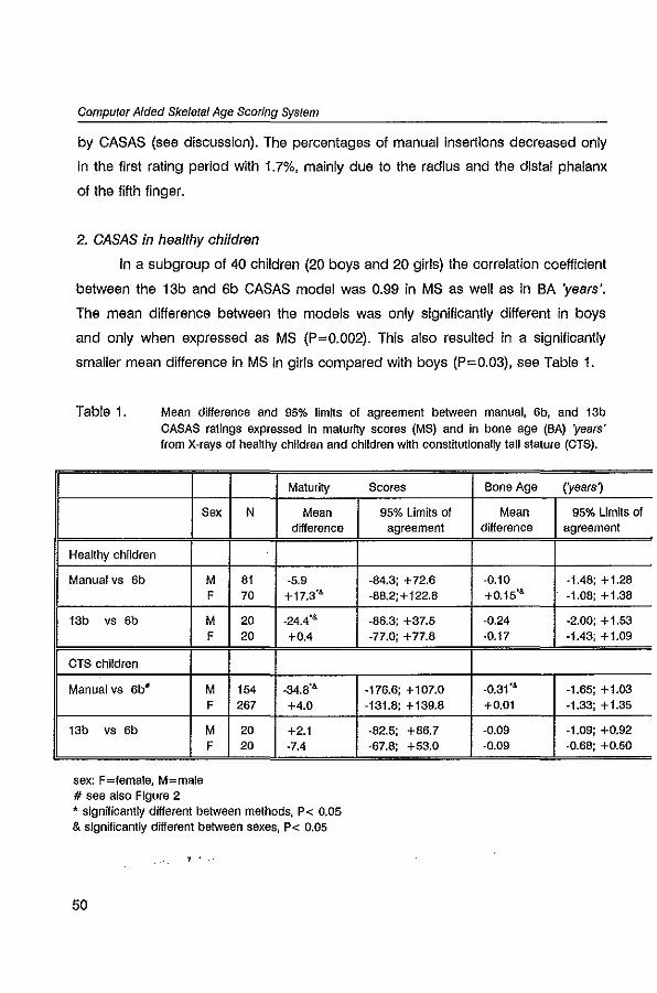
Computer Aided Skeletal Age Scoring System
by CASAS (see discussion). The percentages of manual insertions decreased only
in the first rating period with 1.7%, mainly due to the radius and the distal phalanx
of the fifth finger.
2. CASAS in healthy children
In a subgroup of 40 children (20 boys and 20 girls) the correlation coefficient
between the 13b and 6b CASAS model was 0.99 in MS as well as in BA 'years'.
The mean difference between the models was only significantly different in boys
and only when expressed as MS (P=0.002). This also resulted in a significantly
smaller mean difference in MS in girls compared with boys (P=0.03), see Table 1.
Table 1. Mean difference and 95% limits of agreement between manual, 6b, and 13b CASAS ratings expressed In maturity scores (MS) and in bone age (SA) 'years' from X·rays of healthy children and children with constitutionally tall stature (CTS).
Sex
Healthy children
Manual vs 6b
13b vs 6b
CTS children
Manual vs 6b'
13b vs 6b
sex: F=female, M=male # see also Figure 2
M F
M F
M F
M F
Maturity
N Mean difference
81 -5.9 70 +17.3'&
20 -24.4'& 20 +0.4
154 -34.8'&
267 +4.0
20 +2.1 20 ·7.4
• slgnnicantly different between methods, P< 0.05 & significantly different between sexes, P< 0.05
, .
50
Scores Bone Age ('years)
95% Limits of Mean 95% Limits of agreement difference agreement
·84.3; + 72.6 ·0.10 -1.48; + 1.28 ·88.2;+122.8 +0.15'& -1.08; + 1.38
·86.3; +37.5 ·0.24 -2.00; + 1.53 -77.0; +77.8 ·0.17 -1.43; +1.09
-176.6; +107.0 ·0.31'& -1.65; + 1.03 -131.8; +139.8 +0.01 ·1.33; +1.35
·82.5; +86.7 -0.09 -1.09; +0.92 -67.S; +53.0 ·0.09 ·0.68; +0.50
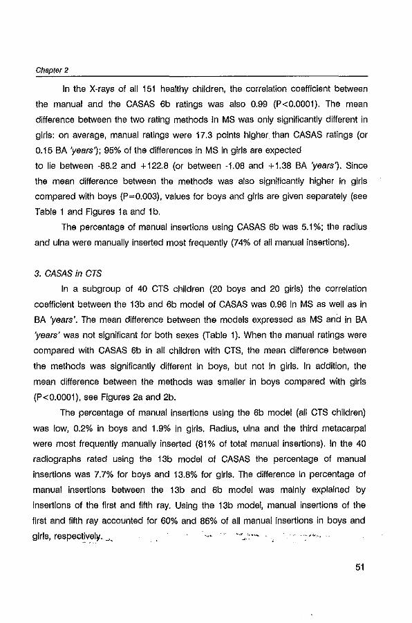
Chapter 2
In the X-rays of all 151 healthy children, the correlation coefficient between
the manual and the CASAS 6b ratings was also 0.99 (P<0.0001). The mean
difference between the two rating methods in MS was only significantly different in
girls: on average, manual ratings were 17.3 points higher than CASAS ratings (or
0.15 BA 'years'); 95% of the differences in MS in girls are expected
to lie between -88.2 and +122.8 (or between -1.08 and +1.38 BA 'years'). Since
the mean difference between the methods was also significantly higher in girls
compared with boys (P=0.003), values for boys and girls are given separately (see
Table 1 and Figures 1a and 1b.
The percentage of manual insertions using CASAS 6b was 5.1 %; the radius
and ulna were manually inserted most frequently (74% of all manual insertions).
3. CASAS in CTS
In a subgroup of 40 CTS children (20 boys and 20 girls) the correlation
coefficient between the 13b and 6b model of CASAS was 0.96 in MS as well as in
BA 'years'. The mean difference between the models expressed as MS and in BA
'years' was not significant for both sexes (Table 1). When the manual ratings were
compared with CASAS 6b in all children with CTS, the mean difference between
the methods was significantly different in boys, but not in girls. In addition, the
mean difference between the methods was smaller in boys compared with girls
(P<0.0001), see Figures 2a and 2b.
The percentage of manual insertions using the 6b model (all CTS children)
was low, 0.2% in boys and 1.9% in girls. Radius, ulna and the third metacarpal
were most frequently manually inserted (81% of total manual insertions). In the 40
radiographs rated using the 13b model of CASAS the percentage of manual
insertions was 7.7% for boys and 13.8% for girls. The difference in percentage of
manual insertions between the 13b and 6b model was mainly explained by
insertions of the first and fifth ray. Using the 13b model, manual insertions of the
first and fifth ray accounted for 60% and 86% of all manual insertions in boys and
girls, respectively. _,_ .. - ""--" .
51
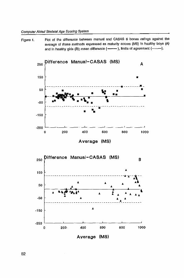
computer Aided Sketetal Age Scoring System
Figure 1.
52
Plot of the difference between manual and CASAS 6 bones ratings against the average of these methods expressed as maturity scores (MS) In heallhy boys (A) and in heallhy girls (B); mean difference (--), IImlls of agreement ( ••••••••• ).
250 Difference Manual-CASAS (MS) A
150
50
·50
·150
o
-- _________________________________ a _______ Jl_
o •
". -"II.. • • I
• o
••
------------------------.---------------------• o.
·250 '------'-----'-----"----'-----' o 200 400 600 800 1000
Average (MS)
250 Difference Manual-CASAS (MS) B
t50 -------------------------------------k.--~\--
50 .. .l ... Ai .. 4 ..
.. ·50 .. .. ..
-----------------------------------~---------
·150 ..
.250 '-----'-----"------''-----'-----'
o 200· 400 600 800 1000
Average (MS)
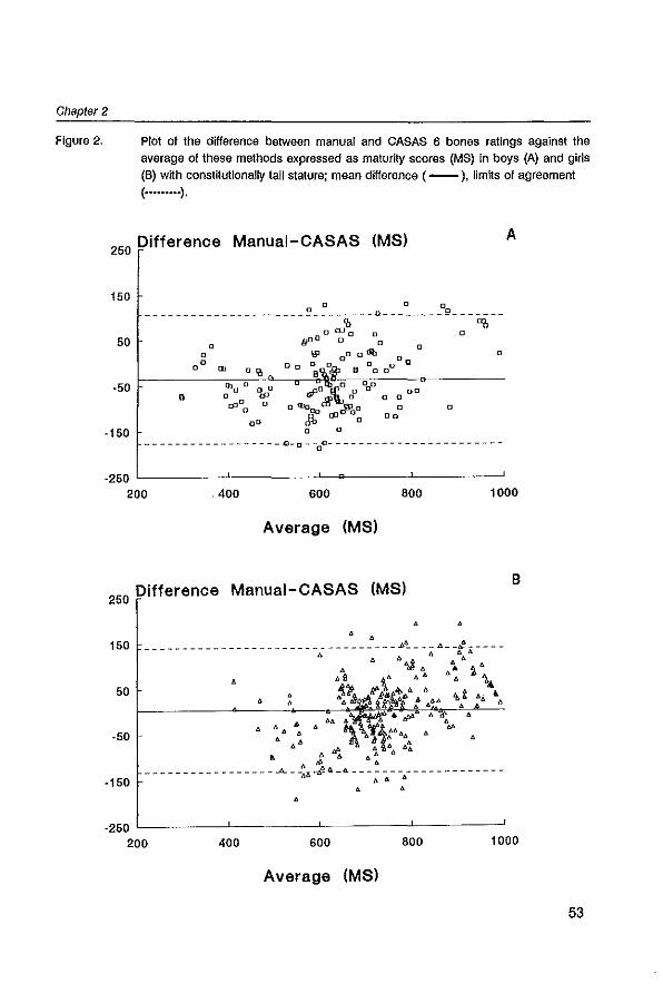
Chapter 2
Figure 2. Plot of the difference between manual and CASAS 6 bones ratings against the average of these methods expressed as maturity scores (MS) in boys (A) and girls (8) wtth constitutionally tali stature; mean difference ( -). limtts of agreement ( ......... ).
250 Difference Manual-CASAS (MS) A
150 o 0 0 _____________________ ~ ________ o ________ g ______ _
50
'b "0 e(J]u D 0
o {}OD 0 [] 0
a §I ooodb 0
0 0 III DO 0 On, • 0 0 0 0'0 il'lt' 00
o
·50 • o
00
·150 ------------------Q-o-o~-----------------------
.250 L-___ ---'-____ -'-~ ___ _L ___ ----'
200 .400 600 800 1000
Average (MS)
250 Difference Manual-CASAS (MS) B
150
50
·50
·150 • • • . 250 L-___ ---'-____ -'-____ --'--___ ----'
200 400 600 800 1000
Average (MS)
53
computer Aided Skeletel Age Scoring System
4. Bone maturation CUNes in CTS
Figures 3a and 3b depict the CASAS 6b ratings versus CA in CTS for boys
and girls, respectively. The estimated regression models are given by the following
equations:
For boys: BA (predicted)= 1.32*CA +0.0002*CA' -0.00009*CA'.
For girls: BA (predicted)= 1.46*CA -0.023*CA' -0.00005*CA'.
DISCUSSION
CASAS in healthy children
The reliability study of CASAS 13b showed very small estimated between
and within observer components of variance. However, when expressed in BA
'years' the range seems considerable and appears even slightiy higher compared
with the manual ratings. In an earlier
reliability study with the traditional manual X-ray ratings of healthy children from 7-
17 years of age (3) the standard deviation of the within observer component of
variance of two observers was similar (0.30-0.35 BA 'years') compared with those
in our manual ratings (0.25 BA 'years') and with CASAS (0.36 BA 'years'). It has
been stated, though, that the reliability varies somewhat in different parts of the age
span; as a result of a change of one stage of a single bone the BA may 'jump' by
0.3 'years' (1). The interval scale in the manual method has been replaced by a
continuous scale in CASAS and, therefore, reliability should be better and equal at
all ages. A further improvement would be the use of MS instead of BA. The
correlation between biological maturation and CA varies between populations (9).
Therefore, ideally every population should have its own standards for· MS and BA.
In fact, BA is a crude determination derived from the MS table.
A comparison of the staging in duplicate assessments by the same
observers (within observer comparison) showed similar percentages for CASAS
54

Chapter 2
Figure 3. Plot of bone age (SA) determined using CASAS 6 bones versus chronological age (CA) in boys (A) and girls (6) with constitutionally tali stature. The regression line ( •..... ) Is depicted In comparison with the assumed y=x line for SA and CA (_).
18 RUS bone age (lyra')
17 0
0
16 II o [] ~@
15 0 Cl 0g: .. f .. ~D .. Q,"...rr°tSIl
14 o 0 ...... 0 0 o ~.. 0
13 .. " 0 ". .. t
12 .' .' n ...... 0
11 .' n 0 ClO
.' • 10
9
8 8 9 10 11 12 13 14
Chronologioal age
RUS bone age (lyra') 16
15
14
13
12
11
10
9 •
'" 0 0
0
15
(yra)
Chronologioal age (yra)
0
0 0
0 0
0
A
16 17 18
55
computer AJded Skeletal Age Scoring System
13b (88%) and the manual ratings (90%). Ten percent of the ratings differed one
stage and using CASAS occasionally a difference of two stages was observed.
Noticeably, there were hardly any differences between the individual bones
included in the 13b model of CASAS. Our percentage of different stages within
observers using CASAS 13b is higher than reported by Tanner et al (2·5%) (5);
however, in their study the CASAS 6b model was used. Using the manual method,
Beunen et al (3) also found that a single observer gave the same stage rating on
two occasions in about 90% of the cases (within observer comparison), whereas
different observers gave the same stage in 75·85% of the cases. In our study, the
latter (between observer comparison) percentage was similar for both CASAS and
the manual ratings, 88% and 89% respectively. Again, using CASAS occasional
differences of 2 stages were observed.
As a part of the CASAS grading process each bone Is compared with
reference standards which represent each stage of bone maturity. The best fitting
stage together with the four adjacent stages are being compared with the standard
by means of their root mean square errors. The extreme stages of each bone ('A'
and 'H'/'I') do not have two adjacent stages on both sides. Although the analysis of
covariance is adjusted for these extreme stages (personal communication), the
extreme computer· rated stage scores, i.e. 1.0 and 8.0/9.0, are not always attained.
To illustrate this, when all epiphyseal plates are visually closed and CASAS rates
the stages with a deficit of minus 0.15, the difference with the expected MS of 1000
can mount to about 44 MS in boys and 33 MS in girls, i.e. 0.85 and 0.5 BA 'years',
respectively. A comparable deficit of 0.15 of a grade to stage 'A' results in a
difference of about 15 MS in boys and girls; at this very young CA also a
substantial difference. It seems less important, however, since skeletal maturity is
less often determined in the first years of life.
The percentage of manual insertions was slightly lower in the second rating
period compared with the first period. The data are in good agreement with the
CASAS 6b model ratings performed with X·rays from all healthy children (second
study), in which 5% manual insertions were performed. There was a high degree of
56
Chapter 2
consistency for each bone between the two rating sessions. When a smaller range
was chosen for manual insertions, i.e. a difference between expected and
determined stage of one integer stage or more, the percentage of manual
insertions on the same X-ray series rated by the same observers increased to 11-
12%, but the between and within observer variances did not show a marked
change (data not shown).
The possibility of inserting a manual score with CASAS creates the problem
of when to make use of this option. To what extent a difference between the
expected and determined rating will be accepted is likely to depend on the
experience of the user and on the reliability of the computer rating of the individual
bones. In addition, the magnification of the Individual bones using CASAS may
induce distinct differences in expected ratings compared to manual ratings usually
performed without magnification. Therefore, cut-off limits for such a manual
expected rating remain very subjective, In particular when one keeps the evident
within observer variability of manual ratings in mind. Ideally, one should not give a
manual insertion at all and the computer should analyse the radiograph fully
automatically. However, since CASAS is based on the same classification of
developmental stages of each bone, one cannot omit to give a manual insertion
whenever the computer rating is far from logical (e.g. stage'S' instead of 'G') due
to e.g. positioning, imaging problems, or software imperfectness. Consequently,
CASAS is not yet completely independent of the observer. In addition, in our
experience, individual CASAS ratings may sometimes vary considerably just by
repositioning of the X-ray and without giving a warning to the user. The designers
of the programme underlined this by stating that "correct assessment depends
crucially on correct positioning" (5). The choice of acceptance of a single bone
rating by the user is also dependent on the subjectiveness (experience) of the
user. Therefore, in order to compare study results we consider that the percentage
of manual Insertions and the limits for the acceptance of disagreement must be
reported, at least until the computer operating system is considered to be the gold
standard.
57
Computer Aided Skeletal Age Scoring System
The designers of CASAS suggest that prior knowledge is not needed;
however, one should familiarize oneself with the sytem and some knowledge on the
developmental stages of the various bones Is needed to avoid evident mistakes in
the rating. Although this Is seldom due to the quality of the X-ray, it is obvious that
the TW2 radiological technique (1) should be used, in particular with regard to the
positioning of the hand.
The use of the 6b model seems preferable because it is less time
consuming than rating 13 bones, 5-6 minutes versus 10 minutes or more,
respectively. Although experienced TW2 'raters' might be able to perform the
manual procedure quicker, it must be noted that after rating the stages data entry
in the computer and subsequent calculations still have to follow, in contrast to
CASAS.
The mean difference between the 13b and the 6b model of CASAS in
healthy children also showed a significant gender difference only when expressed
as MS; however, 95% limits of agreement were again wide.
The mean difference between the manual ratings and CASAS in healthy
children was only significantly different in girls, although the 95% limits of
agreement were considerable.
When X-rays of fully matured children were excluded from the analysis the 95%
limits of agreement were only slightly 'narrower'. The mean difference between
methods was significantly different between boys and girls. A comparable
difference between the sexes was found In CTS children. We have no satisfying
explanation for this phenomenon.
CASAS in CTS
On average, ratings of the 13b model were comparable with those of the 6b
model in both boys and girls with CTS, although individual differences may be
considerable. In girls with CTS the mean difference between the manual ratings
and CASAS was also almost zero. In boys, ratings with CASAS were significantly
higher compared with the manual ratings. These mean differences between
58
Chapter 2
methods were also significantly different between the sexes. For both sexes,
however, the 95% limits of agreement were of the same magnitude and
comparable with that in healthy children.
The percentage of manual insertions In the 6b model of CASAS was
remarkably low in CTS. Using the 13b model the percentage of manual insertions
increased rapidly; it appeared that most of the manual insertions were due to the
first and fifth rays, which are not included in the short 6b model. The use of the 6b
model will thus reduce the number of difficulties in rating. This might explain the
slight, but not significant difference in BA when the 6b model was compared with
the 13b model.
Bone maturation CUNes CTS
Our mixed cross-sectional and longitudinal data of bone age in children with
CTS show that BA is advanced at younger ages in boys as well as in girls. This
discrepancy weakens as CA progresses. To our knowledge, there are no cross
sectional or longitudinal data on bone maturation in children with CTS. However,
our findings are in agreement with our own clinical experiences. In addition, in
various clinical studies the mean manual TW2 BA prior to the start of treatment was
always higher compared with the mean CA of the patients, whereas the standard
deviation of the BA was always smaller than the standard deviation of the CA (14-
18). This seems to be In concert with our present findings.
CONCLUSIONS
In healthy girls and in boys with CTS the semi-automated CASAS 6b ratings
were significantly different from manual ratings. The difference between the 13b
and 6b ratings was only significant in healthy boys. In all comparisons the mean
differences between methods (either with the 6b or the 13b model) were less than
0.4 BA 'year'. The range of the difference between methods was considerable, but
59
Compute! Aided Skeletal Age Scoring System
the within and between observer variations are in the same order of magnitude as
reported for the manual readings. It should be noted, that both methods only
estimate SA; a measure of the 'true' SA Is unknown. Therefore, we think that this
computerized method is applicable in these groups of children. The use of the 6b
model seems preferable because it is less time consuming than rating 13 bones.
In view of the percentages of manual insertions the clinical use of this
CASAS version seems to be with longitudinal studies of patients In particular.
Manual substitution of a stage should be avoided and when performed its
percentage and the limits for the acceptance of disagreement should be reported.
Our data indicate that CASAS is applicable in children with CTS. The bone
maturation curves in boys and girls with CTS tend to be advanced at younger
ages; however, as CA progresses this discrepancy weakens.
ACKNOWLEDGEMENTS We would like to acknowledge the kind coorporation with Dr. Roland Aarsen
for referring the CTS patients. We are Indebted to Ares Services SA and
Discerning Systems for supplying a CASAS system and Dr. Serthon Rikken for the
development of additional software applications. This study was financially
supported by Novo Nordisk Pharma S.v"
60

Chapter 2
REFERENCES
1. Tanner JM, Whitehouse RH, Cameron N, Marshall WA, Healy MJA, Goldstein H: Assessment of skeletal maturity and prediction of adult height (TW2-method). A,cademic Press, 1983, London
2. Taranger J, Bruning at Claasson I, Karlberg P, Landstr6m B: A new method for the assessment of skeletal maturity - the MAT method (mean appearance time of bone stages). Acta Paediatr Scand t976;(Suppl)258:109·120
3. Beunen G, Cameron N: The reproducibility of TW2 skeletal age assessments by a self-taught assessor. Hum Bioi 1980;7:155-162
4. Wenzel A, Melsen B: Replicability of assessing radiographs by the Tanner Whitehouse·2 method. Hum Bioi 1982;54:575-581
5. Tanner JM, Gibbons AD: Automatic bone age measurement using computerized Image analysis. J Pediatr EndocrinoI1994;7:141-145
6. Tanner JM, Gibbons RG, Bock DA: An image analysis system for TW2 skeletal maturily. Harm Res 1992;37 (SuppI4):11
7. Tanner JM, Gibbons AD, Bock RD: Advantages of the computer-aided Image analysis system for estimating tw skeletal maturity: increased reliability and a continuous scale, Pedlatr Aes 1993;33:s35
8. Bland JM, Altman DG: Statistical methods for assessing agreement between two methods of clinical measurement. Lancet 1986;1:307--310
9. Eveleth PB, Tanner JM: World-wide variation in human growth. University Press, 1990, Cambridge
10. Zachmann M, Ferrandez A, Mursel G, Pradar A: Estrogen treatment of excessively tali girls. HeJv Paedlatr Acta 1975;30: 11-30
11. Zachmann M, Ferrandez A, MOrset G, Gnahm HE, Pradar A: Testosterone treatment of excessively tall boys. J Pediatr 1976;88:116-123
12. John G, Schellong G: Oestrogen theraple hochwOchsiger MMchen. Monatsschr Klnderheilk 1980; 128:545-550
13. StOver B, Kollmann F: Ergebnisse der Behandlung hochwOchsiger MMchen mtt conjuglerten Oestrogenen. Monatsschr Kinderheilk 1983;130:36-40
14. Bramswlg JH, Hermellng W, Petrykowskl WV, Schellong G: Comparison of height predictions with final aduk height In boys with consmutional tall stature. In: Borms J, Hausple R, Sand A, Susanne C, Hebbellnck M (eds) 1984, Human growth and development. Plenum Publishing Co, pp 423-429
61


CHAPTER 3
Accuracy of final height prediction and effect of
growth reductive therapy In 115 constitutionally tall boys.
A comparison of nine prediction methods.
W.J. de Waal'. M.H. Greyn-Fokker'. E.A.F.J. van Gurp'. Th. Stijnen'.
S.M.P.F. de Muinck Keizer-Schrama'. R.S.A. Aarsen'. and S.L.S. Drop'.
1 Department of Pediatrics, "Division of Endocrinology, Sophia Children's
Hospital and 'Department of Epidemiology and Biostatistics,
Erasmus University Rotterdam. The Netherlands.
Journal of Clinical Endocrinology and Metabolism
1996. in press (combined with chapter 4)
Height predict/on and treatment in tall boys
ABSTRACT
Height reduction by means of treatment with high doses of androgens in
constitutionally tall stature is a well-known, though still controversial therapy. The
establishment of the effect of such therapy is dependent on the height prediction
method applied. We evaluated the reliability of nine prediction methods in 55
untreated boys with constitutionally tall stature and the effect of height reductive
therapy in 60 tall boys treated with high doses of androgens (cases). For this
purpose we compared the predicted adult height with the attained height at a
mean adult age of 25.1 yrs and adjusted the therapeutic effect for possible
confounding variables.
At the time of the height prediction, controls were significantly shorter, had
more advanced estimated bone ages {except for the bone age (BA) according to
Greulich & Pyle (BAop), had lower target heights, and had smaller adult height
predictions compared with cases (P-values <0.05). At the time of the follow-up
cases were significantly taller than controls; mean (SD) final adult height was 199.7
(4.1) for cases and 196.0 (4.9) for controls (P<0.001).
In controls, a large variability was found for the errors of prediction of the
various prediction methods and in relation to chronological age {CAl. The BP-,
olinical- and RH-predictions' systematically overestimated adult height in
constitutionally tall boys, whereas the other prediction methods sytematically
underestimated final height. The mean (SD) absolute errors of the prediction
methods varied from 2.3 (1.8) to 5.3 (4.3) cm. They were significantly negatively
correlated with CA (r:-0.27 to -0.54, P-values < 0.05) , except for the TW-, and
CASAS-prediotion', Indicating that height prognosis Is more reliable with increasing
CA. The extrapolation of the height SDS for BA (BAop) (IPHap-prediction') was found
to be the most reliable method in predicting adult height. In addition, experienced
clinicians gave an accurate height prediction by evaluating the growth chart of the
child while taking into account various clinical parameters such as CA, BA and
pubertal stage. ' For explanation of abbreviations: see Methods.
64
Chapter 3
The effect of androgen therapy was assessed by means of multiple
regression analysis, while adjusting for the confounding variables height prediction,
CA and BA at start of therapy. The mean (SD) adjusted effect varied from -1.7 (1.4)
to 0.7 (1.4) cm. with ranges from -4.6 to 15.8 cm. The adjusted height reduction
was dependent on the BA at the time of start of androgen therapy: the effect was
more pronounced when treatment was started at a younger BA. The treatment
effect was significantly negative at BAs exceeding 14 to 15 years. After cessation of
therapy a mean (SD) additional growth of 2.4 (1.2) cm was observed. The mean
(SD) BAop at that time was 17.1 (0.7) years.
These data demonstrate, that height prognosis in boys with constitutionally
tall stature is rather inaccurate, the IPHGP-prediction being the most reliable
prediction method. Overall, the height reductive effect of treatment with high doses
of androgens in tall boys is limited. However, a significant height reduction was
found when treatment was started at BAs less than 14 to 15 years, depending on
the method of BA assessment. Remarkably, at older BAs treatment was found to
be contra-Indicated, since androgen administration caused extra growth instead of
growth inhibition. It is recommended that referral should take place early, preterably
before puberty, for careful monitoring of growth and prediction. Moreover, when
treatment is initiated, it is recommended not to discontinue therapy too early in
order to avoid serious additional growth.
INTRODUCTION
Prediction of adult height is commonly used in the management of children
with growth disorders. Over the years various methods have been developed to
predict adult height of which the methods of Bayley and Pinneau (1) and of Tanner
et al (2,3) are most commonly used. In general, height prediction models are
based on growth data derived from normal growing children of normal stature. This
implies that these methods may not give accurate results when applied to children
65
Height prediction and treatment in tall boys
with growth disturbances. Tanner and co-workers have tried to overcome this
problem and revised their original prediction model by including samples of
children with growth disorders (2,3). With respect to constitutionally tall children,
however, only a sample of 19 tall girls had been included.
In the management of children with constitionally tall stature, height
prognosis plays a crucial role since it influences to a large extent the decision for
therapeutic intervention with pharmacological doses of sex steroids. It is essential
therefore, that the accuracy of the prediction technique applied is clinically
acceptable. If treatment is applied, the effect of therapy is evaluated by subtracting
the achieved final height from the predicted adult height. Therefore, the \.Iltimate
effect of the treatment relies on the reliability of the prediction method as well. In
addition, it is to be expected that separate prediction models will provide different
estimates of final adult height within the same person and thus a different result in
height reduction. This is caused by the fact that the prediction methods refer to
different norm populations and use different methodologies.
The height reducing effect of sex steroid therapy in constitutionally tall
children has extensively been studied. However, to our knowledge, a well
designed, prospective controlled study on the effect of such a therapy has never
been performed. This implies that the reported results have been biased by several
factors. For instance, results on height reduction have been derived from
comparison of the achieved height with the height prediction prior to treatment
without correction for the error of the prediction. In addition, when control groups
have been used they tended to be small. Besides, while assessing the \.Iltimate
height redUction, differences in initial clinical data between treated children and
controls such as age, bone age and height prediction, have not been taken into
account. Furthermore, in many studies adult height have been assumed to be
reached at a relatively young age. Therefore, we evaluated the applicability of nine
prediction methods in untreated children with constitutionally tall stature and, with
respect to these findings, the effect of height reductive therapy in children treated
with high doses of sex steroids. For this purpose, we measured final adult height at
66
Chapter 3
a mean age of 25 years and adjusted the effect of sex hormone therapy for
possible confounding variables. In this article we describe our results in tall boys;
the evaluation of girls with tall stature will be reported in an adjoining paper.
To date, only a few studies are available on the reliability of height
predictions in boys with constitutionally tall stature (4-7). These studies show some
conflicting results concerning the tendency to over- or underestimate adult height
depending of the prediction method used. They indicate that there is no major
advantage of any of the prediction methods studied and that there Is considerable
age and bone age related variation. Studies on the effect of androgen therapy on
height reduction have shown different results using different prediction methods
(4,8,9). Overall, these studies indicated that height reductive therapy was effective
in tall boys irrespective of the prediction method used. However, considerable age
and bone age related variation was present and individual effects overlapped the
range of the prediction error.
In this study the reliability of various prediction methods and the ~ffect of
height reductive therapy in a total of 115 constitutionally tall boys was evaluated.
For this purpose we compared the predicted adult height with the attained height
at a mean adult age of 25.1 yrs (range 18.7 - 34.4 yrs.) and adjusted the
therapeutic effect for possible confounding variables.
PATIENTS AND METHODS
Patients
Since the introduction of height reductive therapy in our institute in 1968 a
total of 247 men, who were seen at adolescence for evaluation of their
constitutionally tall stature, had reached the age of 18 at the time of our follow-up
study. All were contacted by mail to participate in this study. Second mailings were
send to those who did not respond to the first mailing. This study was part of a
large follow-up study including psychosocial assessments and long-term sequelae.
67
Height prediction and treatment in tall boys
Reports on psychology and long-term sequelae will be reported elsewhere.
During their puberty, 102 men had been treated for their tall stature (cases).
Hundred and forty-five men had chosen not to undergo treatment for various
reasons such as satisfaction with the given height prognosis or uncertainty about
possible side-effects, and served as controls. Two hundred and nine men (95
cases and 114 controls) responded to our mailing. Of the responders, 127 men (65
cases and 62 controls) agreed to participate in this study. All participants gave
informed consent.
We defined constitutionally tall stature as having a height equal to or above
the 90th percentile (Dutch references (10)) at the time of referral. By this definition 5
boys were excluded as their heights were less than the 90th percentile. In addition,
from 7 boys no radiograph could be retrieved from the archives for reassessment
of bone age and height prediction. Therefore, 115 boys (60 cases and 55 controls)
were included In this study.
In cases, most of the men (n=52) had received intramuscular preparations
of testosterone ester mixtures (SustanonA = testosterone proprionate, -
fenylpropionate, -isohexanoate and -decanoate) 250 mg every week (n=45), 250
mg every two weeks (n=4) or 500 mg every two weeks (n=3). Four men used an
oral testosterone ester (AndriolA = testosterone undecanoate) 240-320 mg per day;
one 'of these patients switched to SustanonA every two weeks after 8 months.
Another four patients used daily Injections of a single testosterone ester (Neo
hombreolA = testosterone propionate) 25-30 mg per day for 6 - 30 months after
which they switched to SustanonA 250 mg every two weeks. Treatment started at a
mean (SO) age of 14.2 (1.3) yrs. with a range of 9.7 to 17.2 yrs. The mean duration
of therapy was 1.4 (0.6) yrs. ranging from 0.7 - 3.7 yrs. In general, treatment was
stopped when radiographs of hand and/or knee were considered to show nearly
complete closure of the epiphyses. The mean (SO) length of the follow-up period
after cessation of androgen therapy lasted 8.7 (4.1) years [range: 3.2 - 18.5 yrs].
68
Chapter 3
METHODS
Auxology
Patients were recalled to our outpatient clinic for measurement of final
height. Height measurements were performed by one investigator (yVdW) using a
Harpenden stadiometer. In addition, auxological data were collected from the
hospital charts and radiographs of the left hand and wrist were retrieved and re
used for bone age determinations and final height predictions. For cases, only two
radiograph were used: the one just before start of treatment and the one at the
time treatment was stopped. For controls all radiographs were used from ihe age
of 8, provided that the interval between every consecutive X-ray was at least 6
months.
Bone age
Bone age (BA) was rated manually according to the methods decribed by
Greulich and Pyle (11) and Tanner et al (3)(lW2-RUS BA) by one investigator (MF).
In cases, the BA at time of stop of therapy was rated according to Greulich and
Pyle (11) by another investigator (yVdW). In addition, lW2-RUS BA was determined
by a third Investigator using the short version (6 bones model) of a computer aided
skeletal age scoring (CASAS) system, as described previously (12,13). BAs were
rated unaware of the treatment design or the post treatment growth.
Final height prediotions
Final height predictions were performed using the methods of Bailey and
Pinneau (BP-prediction)(1) and Tanner et al (7W-prediction, mark /I) (3). In addition,
the relative height (RH-prediotion) and the index of potential height (IPH
prediction)(14) were calculated.
BP-prediction: Using the BA as estimated by Greulich and Pyle (10), the
percent of final height achieved was read in a table (1), and adult final height was
calculated.
69
Height prediction and treatment In tall boys
TW-prediction: From the regression equations provided by Tanner at al (3)
final height was calculated using BA as well as height and chronological age (CA)
as variables. For this study, we performed TW-predictions using the manual TW2-
RUS bone ages (TW-prediction) as well as the TW2-RUS bone ages as determined
by CASAS (CASAS-prediction) (12).
RH-prediction: Final height was calculated with the assumption that the
height expressed as standard deviation score (SOS) for CA equals to adult height
SOS.
IPH-prediction: Final height was predicted based on the assumption that the
height SOS for BA remains constant up to final height. For this study we performed
IPH-predictions using the bone ages as estimated by Greulich and Pyle (IPHa,
prediotlon) , Tanner et al (IPHTW-prediotion) and CASAS (IPH""",,-prediction). In
addition, we predicted final height using a theoretical bone age (IPH"""",,
prediotion) derived from the observed relationship between BA (as estimated by
CASAS) and CA, as described previously (13). For boys this relationship was
BA= 1.32*CA + 0.0002*CA' - 0.00009*CA'.
RH-predictions and IPH-predictions were performed using Dutch references
for height and SO-scores (10).
Beside these objective prediction methods we added a subjective clinical
prediction. These clinical-predictions were performed by two experienced pediatric
endocrinologists separately (SOMKS, SO). For this purpose, a growth chart was
constructed for each individual. The following data were extracted from the hospital
records and, if available, plotted on the growth chart: height measurements
available at time of referral, BA (estimated according to Greulich & Pyle by one
investigator (MF) as stated above), father's height, mother's height, target height,
and pubertal stage. Target height was calculated according to the (ormula:
(Mother's height (cm) + Father's height (cm) + 12)/2 + 3 (15). Growth charts were
then blindly and randomly rated for final height predictions. If separate final height
predictions differed more than 2 cm among the raters, the prediction was repeated
for consensus.
70
Chapter 3
The study protocol was approved by the ethical committee of Academic
Hospital, Erasmus University Rotterdam, The Netherlands.
Statistics
To assess the effect of treatment in cases all height predictions were used
for analysis, since predictions were based on one radiograph per sUbject by
inclusion. In controls, more than one radiograph was available for accuracy
assessment of the various prediction methods. Using repeated predictions of one
control subject for assessment of the accuracy of the prediction method and
comparison with a treated group in which only one radiograph per subject was
taken could induce bias. Therefore we used only one radiograph per control boy.
The radiographs were selected such that the age distribution of treated and
untreated boys were as alike as possible.
In controls, accuracy of the height prediction methods was expressed
mathematically as predicted adult height minus actual adult height. Therefore,
positive values indicate overestimation whereas negative values indicate
underestimation of the predicted adult height. In addition, the reliability of each
height prediction method was also expressed in absolute errors. The absolute
errors demonstrate the method's overall predictive error and are Independent of
over- or underestimation. In cases, the calculation of predicted adult height minus
actual final height was used to express the -uncorrected- effect of growth reductive
therapy. Consequently, positive values indicate growth reduction whereas negative
values indicate lack of growth reduction. Multiple regression analysis was used to
assess the corrected height reducing effect of androgen therapy (see below). For
the clinical-prediction, the mean of the two separate predictions was used for
statistical analysis.
Differences between groups were tested with the Mann-Whitney-U-test. In
controls, Friedman's test was used to test the difference in absolute errors of the
various prediction methods. If this test was significant, Wilcoxon's matched pairs
signed ranks test was used to test the difference in absolute errors between
71
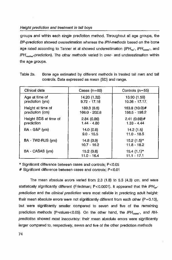
Height prediction and treatment in tali boys
groups and within each single prediction method. Throughout all age groups. the
BP-prediction showed overestimation whereas the IPH-methods based on the bone
age rated according to Tanner et al showed underestimation (IPHTW-. IPHCASAS-. and
IPH".,,,,,-prediction). The other methods varied in over- and underestimation within
the age groups.
Table 2a. Bone age estimated by different methods In treated tall men and tall controls. Data expressed as mean (SO) and range.
Clinical data Cases (n=60)
Age at time of 14.20 (1.32) prediction (yrs) 9.72-17.18
Height at time of 189.3 (6.6) prediction (cm) 166.0 - 202.8
Height SOS at time of 2.84 (0.86) prediction 1.44 - 4.80
BA - G&P (yrs) 14.0 (0.9) 9.0 - 15.5
BA - TW2-RUS (yrs) 14.8 (0.9) 10.7 - 16.2
BA - CASAS (yrs) 15.2 (0.8) 11.0-16.4
, Significant difference between cases and controls; P<0.05 # Significant difference between cases and controls; P<O.Ol
Controls (n=55)
13.90 (1.50) 10.36 - 17.17.
183.8 (10.0)# 158.5 - 198.2
2.41 (0.69)# 1.33 - 4.44
14.2 (1.5) 11.0-18.5
15.2 (1.5)' 11.8 - 18.2
15.4 (1.1)' 11.1 - 17.1
The mean absolute errors varied from 2.3 (1.8) to 5.3 (4.3) cm. and were
statistically significantly different (Friedman; P<O.OOOl). It appeared that the !PHop-
prediction and the clinical prediction were most reliable in predicting adult height:
their mean absolute errors were not significantly different from each other (P=0.13).
but were significantly smaller compared to seven and five of the remaining
prediction methods (P-values<0.05). On the other hand. the IPHCASAS-. and RH
prediction showed most inaccuracy: their mean absolute errors were significantly
larger compared to. respectively. seven and five of the other prediction methods
74
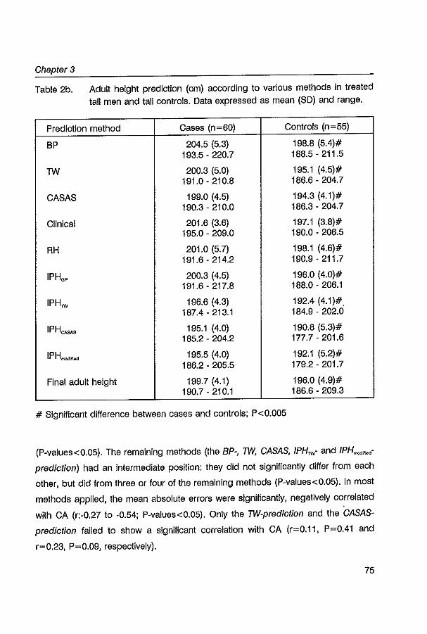
Chapter 3
Table 2b. Adult height prediction (cm) according to various methods in treated tall men and tall controls. Data expressed as mean (SO) and range.
Prediction method Cases (n=60) Controls (n=55)
BP 204.5 (5.3) 198.8 (5.4)# 193.5 - 220.7 188.5 - 211.5
TW 200.3 (5.0) 195.1 (4.5)# 191.0 - 210.8 186.6 - 204.7
CASAS 199.0 (4.5) 194.3 (4.1)# 190.3 - 210.0 186.3 - 204.7
Clinical 201.6 (3.6) 197.1 (3.8)# 195.0 - 209.0 190.0 - 206.5
RH 201.0 (5.7) 198.1 (4.6)# 191.6 - 214.2 190.9 - 211.7
IPHo, 200.3 (4.5) 196.0 (4.0)# 191.6 - 217.8 188.0 - 206.1
IPH", 196.6 (4.3) 192.4 (4.1)# 187.4 - 213.1 184.9 - 202.0
IPHCASAS 195.1 (4.0) 190.8 (5.3)# 185.2 - 204.2 177.7 - 201.6
IPH_ ... 195.5 (4.0) 192.1 (5.2)# 186.2 - 205.5 179.2 - 201.7
Final adult height 199.7 (4.1) 196.0 (4.9)# 190.7 - 210.1 186.6 - 209.3
# Significant difference between cases and controls; P<0.005
(p-values<0.05). The remaining methods (the BP-, TW, CASAS, IPH",- and IPH""",..,
prediction) had an intermediate position: they did not significantly differ from each
other, but did from three or four of the remaining methods (p-values<0.05). In most
methods applied, the mean absolute errors were significantly, negatively correlated
with CA (r:-0.27 to -0.54; P-values<0.05). Only the TW-prediction and the CASAS
prediction failed to show a significant correlation with CA (r=0.11, P=0.41 and
r=0.23, P=0.09, respectively).
75
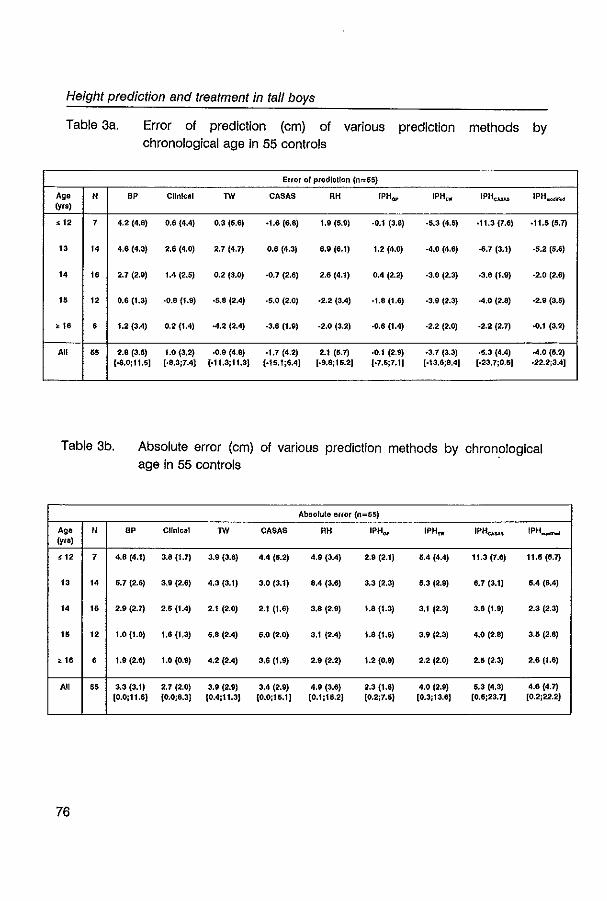
Height prediction and treatment in tall boys
Table 3a. Error of prediction (cm) of various prediction methods by chronological age in 55 controls
Error 01 predIction (0=55)
Ag. N BP Clinical TW CASAS RH iPHQp IPHUf IPHcuu IPH.,.".nw (yrs)
< 12 7 4.2 (4.8) 0.6 (4.4) 0.3 (5.6) ·1.6 (&.8) 1.9 (5.9) ·0.1 (3.8) ·5.3 (4.6) ·11.3 (7.6) ·11.5 (5.7)
13 1. 4.6 (4.3) 2,6 (4.0) 2.7 (4.7) 0.6 (4.3) 6.9 (6.1) 1.2 (4.0) -4.0 (4.6) ·6.7 (3.1) ·5.2 (5.6)
1. 1. 2.7 (2.9) 1.4 (2.5) 0.2 (3.0) ·0.7 (2.6) 2.6 (4.1) 0.4 (2.2) ·3.0 (2.3) .3.6(1.9) -2.0 (2.6)
16 12 0.6 (1.3) ·0.8(1.9) ·5,8 (2.4) ·5.0 (2.0) .2.2 (3.4) ·1.8 (1.6) ·3.9 (2.3) -4.0 (2.8) -2.9 (3.5)
,16 • 1.2 (3.4) 0.2 (1.4) -4.2 (2.4) -3.6(1.9) ·2.0 (3.2) ·0.6 (1.4) ·2.2 (2.0) ·2.2 (2.7) -0.1 (3.2)
All 65 2.8 (3.6) 1.0 (3.2) ·0.9 (4.8) -1.7 (4.2) 2.1 (5.7) ·0.1 (2.9) ·3.7 (3.3) ·5.3 (4.4) -4.0 (6.2) ,·6.0;11.6] (·8.3;7.4) [·11.3j11.3) (·15.1:6.4) (-9.8;16.2) (-7.6;7.1) (·13,6:8.4) (-23.7;0,5) ·22.2;3.4J
Table 3b. Absolute error (cm) of various prediction methods by chronological age in 55 controls
Absolute error (n=55)
Ag. N BP Clinical TW CASAS RH IPHap 1PHIl!' IPHCA$.lS IPH~ (yra)
< 12 7 4,8 (4,1) 3.8 (1.7) 3.9 (3.8) 4.4 (5,2) 4.9 (3.4) 2.9 (2.1) 6.4 (4.4) 11.3 (7.6) 11.6 (6,7)
13 1. 6.7 (2,6) 3,9 (2.6) 4,3 (3,1) 3.0 (3.1) 8.4 (3.6) 3.3 (2.3) 5.3 (2.9) 6,7 (3.1) 6,4 (6.4)
,. ,. 2,9 (2.7) 2.5 (1.4) 2.1 (2.0) 2.1 (1.6) 3,8 (2.9) 1.8 (1.3) 3,1 (2.3) 3,6 (1,9) 2.3 (2.3)
16 12 1.0 (1.0) 1,6 (1,3) 5,8 (2.4) 5,0 (2.0) 3,1 (2.4) 1.8 (1.6) 3,9 (2,3) 4,0 (2.8) 3.6 (2.8)
,1. • 1.9 (2,6) 1.0 (0.9) 4,2 (2.4) 3.6 (1,9) 2.9 (2.2) 1.2 (0.9) 2.2 (2.0) 2.5 (2.3) 2.6 (1.6)
All 65 3.3 (3.1) 2.7 (2.0) 3,9 (2.9) 3.4 (2.9) 4.9 (3.6) 2.3 (1.8) 4.0 (2.9) 5.3 (4.3) 4.6 (4.7) [0.0;11.5] (0,0;8.3) (0,4;11.3) (0.0;15.1J (0.1;15.2) [0.2;7.5) [0,3,13.6) (0,6;23,7] (0.2;22.2)
76
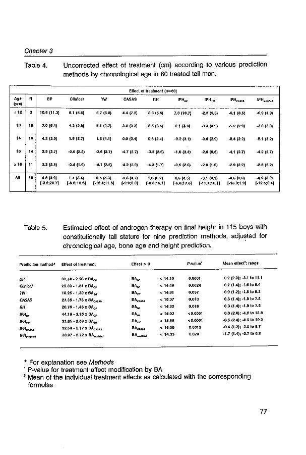
Chapter 3
Table 4.
Ag_ N SP (yro)
< 12 3 10,6 (11.3)
13 16 7.0 (5.4)
14 1. 4.2 (3.6)
1. 14 2.9 (3.7)
> 1. 11 3.2 (2.6)
All 6. 4.8 (4.9) [-3.2;20.7]
Table 5.
PredlcUon method-
BP
ClinlC61
1W
CASAS
RH
1PHGI'
IPHrw
IPHcuu
IPH_
Uncorrected effect of treatment (cm) according to various prediction methods by chronological age in 60 treated tall men.
Effect ollreaimenl (n=60)
Clinical 1W CASAS RH IPH{ll' rPH TW IPHe.u.o,s IPH..-"
6.1 (5.5) 5.7 (6.9) 4.4 (7.2) 8.6 (5.6) 7.3 (10.7) ·2.3 {5.8} ·6.1 (8.6) .6.9 (4.9)
4.3 (2.8) S.1 (3.1) 3,4 (3.3) 8.5 (3.5) 2.1 (4.9) ·3.3 (4.9) ·5.2 (2.9) ·3.8 (3.0)
1.9 (2.7) 1.8 (4.1) 0.0 (3.4) 0.6 (4,4) -0.2 (3.1) -3.8 (2.9) -5.4 (2.2) ·6.1 (3.2)
·0,6 (2,3) .3.6 (3.7) -4.7 (2.7) ·3.3 (2.6) ·1.0 (3.4) ·2.6 {6.6} -4.1 (2.7) -4.2 (2.7)
·0,4 (1.9) -4.1 (2.6) -4.2 (2.9) -4.3 (1.7) ·0,6 (2.6) ·2.9 (1.8) -2.9 (2.2) -2.8 (2.2)
1.7 (3.4) 0.5 (6.3) ·0,8 (4.7) 1.3 (6,2) 0.6 (4.6) -3.1 (4.1) -4.6 (3.0) -4.2 (3.0) [.5.6;10.6J [·12.4;1'.8J (·9.9;9.0) (·8.3iI6.1) ,·6.6iI7.6] ,·11.7,16.1) (016.0;1.9] [·12.5;0.4]
Estimated effect of androgen therapy on final height in 115 boys with constitutionally tall stature for nine prediction methods, adjusted for chronological age, bone age and height prediction.
EHecl 01 treatmenl EHecl > 0 P·value' Mean effecl'; range
30.74 - 2.16 x BAa,. BA",. < 14.10 0.0001 0.2 (2.0), ·3.1 to 11.1
22.30 - 1.64 x BAa,. SA",. < 14.46 0.0024 0.7 (1.4); -1.6108.4
19.25 ·1.30 x BAM SA~ < 14.81 0.037 0.0 (1.2); ·1.810 5.3
27.35.1.78xB~ S ........ < 15.37 0.013 0.3 (1.4); ·1.810 7.8
20.76 -1.46 x BAw SA",. < 14.22 0.018 0.3 (1.4); ·1.910 7.6
44.19.3.15xB~ SA",. < 14.03 <0.0001 0.0 (2.8); -4.6 to 15.8
37.61 ·2.56 x BAM SA~ < 14.66 <0.0001 ..0.5 (2.4); ·4.0 to 10.2
32.56 - 2.17 x B~ B ........ < 15,00 0.0012 ·0.4 (1.7); ·3.0 10 B.7
38.97·2.72 x BA..-.... S ....... < 14.33 0.029 .1.7 (1.4); .2.710 6.2
* For explanation see Methods , P-value for treatment effect modification by BA , Mean of the individual treatment effects as calculated with the corresponding
formulas
77
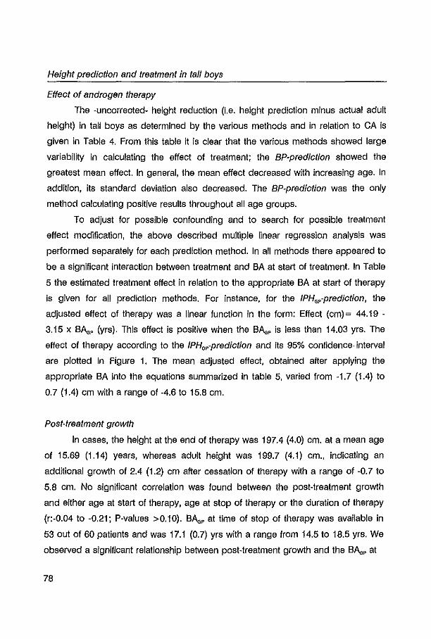
Height prediction and treatment in tall boys
Effect of androgen therapy
The -uncorrected- height reduction (Le. height prediction minus actual adult
height) in tall boys as determined by the various methods and in relation to CA is
given in Table 4. From this table it is clear that the various methods showed large
variability in calculating the effect of treatment; the BP-prediction showed the
greatest mean effect. In general, the mean effect decreased with increasing age. In
addition, its standard deviation also decreased. The BP-prediction was the only
method calculating positive results throughout all age groups.
To adjust for possible confounding and to search for possible treatment
effect modification, the above described multiple linear regression analysis was
performed separately for each prediction method. In all methods there appeared to
be a significant interaction between treatment and BA at start of treatment. In Table
5 the estimated treatment effect in relation to the appropriate BA at start of therapy
is given for all prediction methods. For instance, for the IPHGP-prediction, the
adjusted effect of therapy was a linear function In the form: Effect (cm)= 44.19 -
3.15 x BAap (yrs). This effect is positive when the BAa, is less than 14.03 yrs. The
effect of therapy according to the IPHop-prediction and its 95% confidence· interval
are plotted in Figure 1. The mean adjusted effect, obtained after applying the
appropriate BA into the equations summarized in table 5, varied from -1.7 (1.4) to
0.7 (1.4) cm with a range of -4.6 to 15.8 cm.
Post-treatment growth
In cases, the height at the end of therapy was 197.4 (4.0) cm. at a mean age
of 15.69 (1.14) years, whereas adult height was 199.7 (4.1) cm., Indicating an
additional growth of 2.4 (1.2) cm after cessation of therapy with a range of -0.7 to
5.8 cm. No significant correlation was found between the post-treatment growth
and either age at start of therapy, age at stop of therapy or the duration of therapy
(r:-0.04 to -0.21; P-values >0.10). BAa, at time of stop of therapy was available in
53 out of 60 patients and was 17.1 (0.7) yrs with a range from 14.5 to 18.5 yrs. We
observed a significant relationship between post-treatment growth and the BAa, at
78
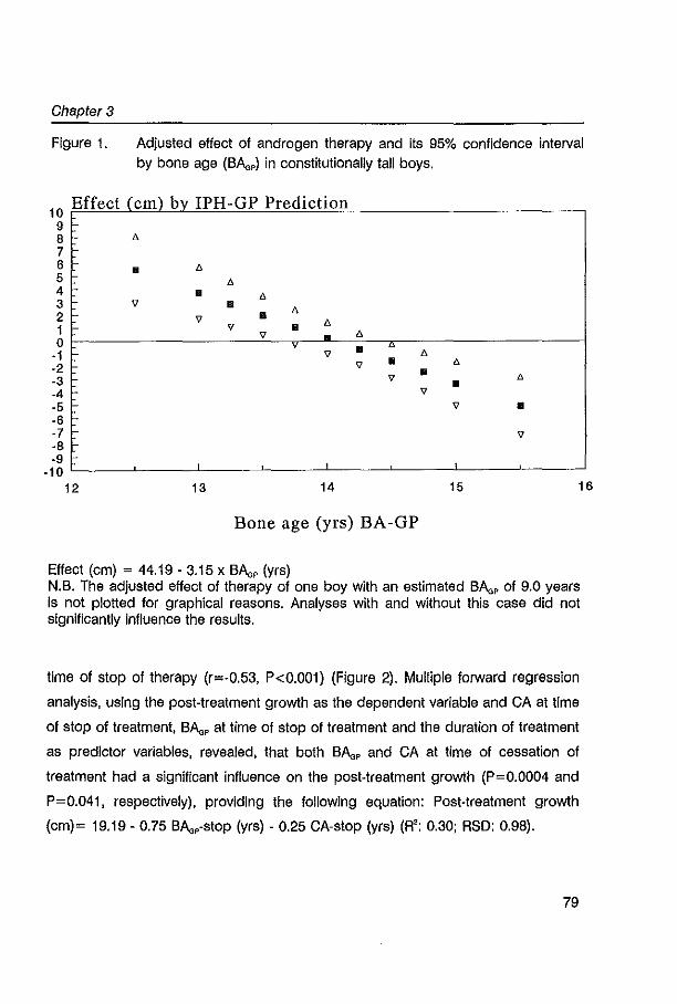
Chapter 3
Figure 1. Adjusted effect of androgen therapy and its 95% confidence interval by bone age (BAa,) in constitutionally tall boys.
10 Effect (em) by IPH-GP Prediction 9 8 7 6 I:-5 r 4 I-3
• n
• • • 2 n 1 99· • o ~------------------~-v--~--~~~----------------~ n
• u
-1 -2 -3 ·4 I:·5 r ·6 I-7 -8
9 • 9
" n • " • 9
9 • 9
-9 ·10L-____ L-____ L-____ L-____ L-____ L-____ l-____ ~ __ ~ ., 12 13 14 15 16
Bone age eyrs) BA-GP
Effect (cm) = 44.19-3.15 x BAa, (yrs) N.B. The adjusted effect of therapy of one boy with an estimated BAa, of 9.0 years is not plotted for graphical reasons. Analyses with and without this case did not significantly influence the results.
time of stop of therapy (r=·0.53, P<0.001) (Figure 2). Mulliple forward regression
analysis, using the post·treatment growth as the dependent variable and CA at time
of stop of treatment, BAa, at time of stop of treatment and the duration of treatment
as predictor variables, revealed, that both BAa, and CA at time of cessation of
treatment had a significant influence on the post·treatment growth (P=O.0004 and
P=0.041, respectively), providing the following equation: Post·treatment growth
(cm)= 19.19-0.75 BAa,·stop (yrs). 0.25 CA·stop (yrs) (R': 0.30; RSD: 0.98).
79
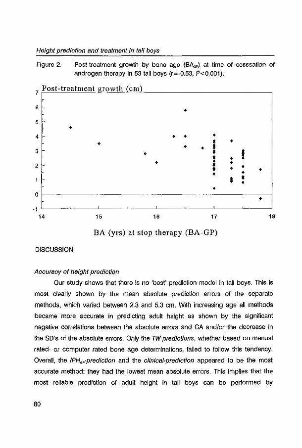
Height prediction and treatment in tall boys
Figure 2. Post-treatment growth by bone age (BAap) at time of cesssation of androgen therapy in 53 tall boys (r=-0.53, P<O.OOI).
7 Post-treatment growth (em)
6 • 5 c
• 4 • • •
• ! • • • 3 • ! i • 2 • • • • • • • • • • • 0 •
-1 1
14 15 16 17 18
BA (yrs) at stop therapy (BA-GP)
DISCUSSION
Aocuracy of height prediot/on
Our study shows that there is no 'best' prediction model in tall boys. This is
most clearly shown by the mean absolute prediction errors of the separate
methods, which varied between 2.3 and 5.3 cm. With increasing age all methods
became more accurate in predicting adult height as shown by the significant
negative correlations between the absolute errors and CA and/or the decrease in
the SO's of the absolute errors. Only the TW-prediotions, whether based on manual
rated- or computer rated bone age determinations, failed to follow this tendency.
Overall, the IPHap-prediotion and the olinioal-prediotion appeared to be the most
accurate method: they had the lowest mean absolute errors. This implies that the
most reliable prediction of adult height in tali boys can be performed by
80
Chapter 3
extrapolation of the height SOS for the bone age according to Greulich and Pyle. In
addition, it shows that experienced clinicians are capable of giving an accurate
prediction by evaluating the growth chart of the child while taking into account age,
bone age (G&P), target height and pubertal stage. This 'weighing" mental process
appears to be as reliable as a population based, mathematical prediction model.
The RH-prediction and the remaining IPH-prediotions (i.e. IPHTW-, IPHc",.-, and
IPH __ -prediotion) appeared to be of minor importance in constitutionally tall boys
as their mean absolute errors were > 4.0 cm.
As shown, every single method had a systematic tendency to over- or
underestimate final height, which varied with CA as well. Knowledge of the
method's tendency to over- or underestimate final height is of clinical importance
since it contributes to the decision of possible therapeutic intervention in tall boys.
In practice, one may increase the accuracy of a height prediction simply by
correcting it for its systematic error as also suggested by Joss et al (6). However,
there are considerable individual variations as shown by the large standard
deviations of the mean errors of the prediction applied. This means that for
instance for a tall boy of 13 years using the BP-prediotion, the method gives a
mean overpredlction of 2.7 cm, but in individual cases this may vary from an
underprediction of -2.9 cm (-2S0) to an overprediotion up to 8.5 cm (+2S0).
To date, only a few studies have included a substantial number of subjects
in order to assess the reliability of various prediction methods In tall boys.
Bramswig and co-workers (5) reported a systematic overprediction of 2.1 (4.8) cm.
of the BP-prediotion and an absolute error of 4.0 (3.3) cm in 42 tall subjects. These
are of the same order of magnitude as found by us. Joss et al (6) also described a
systematic overpredlctlon of the BP-prediotion studying 32 tall boys. Unfortunately,
exact data and absolute prediction errors were not given. In addition, they reported
a systematic overprediction using the TW-prediotion, which was even more
pronounced at an older bone age. We also observed overprediction of the TW
prediotion up to the age of 14 years. At older ages however, a clear
underprediction was present in our study which is in contrast to the findings of
81

Height prediction and treatment in tall boys
Joss et al (6), but in agreement with the findings of Bramswig et al (5) using the
former lW-method (2). The relative inaccuracy of the IPHlW-prediction was found by
Joss et al (6) as well. Both studies used repeated predictions in the same subject
for accuracy assessment, which may have induced bias in reported means and/or
standard deviations. It is hard to judge the influence of the possible bias, but at
least one could expect less variation in the accuracy of the prediction method
tested reflected by smaller standard deviations of the estimates. This is due to the
fact that the estimate of final height will be more accurate and less variable by
increasing age.
Effect of androgen therapy
As expected the -uncorrected- effect of height reductive therapy (i.e. height
prediction minus achieved adult height) varied with the prediction method applied
(Table 4). Since every single prediction method had its own prediction error, the
mean effect of treatment might be "corrected" by subtraction of the corresponding
mean prediction errors, which has been done in the literature (4,8,9). In this way, in
our study the BP-prediction showed the greatest mean "corrected" effect of 2.0 cm.
The IPHGPprediction and the clinical-prediction, being the most accurate methods,
calculated a mean "corrected effect" of only 0.6 and 0.7 cm, respectively. Overall,
these results are much less than reported previously. Zachmann et al (8) reported
an overall mean ("corrected") reduction of 5.4 cm. as determined by the former lW
method (2). Bramswig and co-workers (4) described an achieved mean
("corrected") height reduction varying from 5.3 cm. (former TW-prediction) to 4.7
em. (BP-prediction). In a later paper (9), they reported a mean effect of 7.6 cm.
using a short-term (6 months) treatment design and a mean effect of 9.6 cm. while
using a long-term treatment design (mean 14.3 months) calculated with !he BP
prediction. These results drop to 5.5 and 7.5 cm., respectively, when their reported
overprediction of 2.1 cm. has been taken into account (5). Bettendorf et al (16) on
the other hand, recently reported that short-term androgen therapy failed to reduce
final adult height in constitutionally tall boys. These conflicting results may be due
82
Chapter 3
to differences In CA at the time final adult height was measured and to differences
in BA at the time of cessation of therapy. According to our own data, a mean post
treatment growth of 2.4 (1.2) cm. was observed. It appeared that at the time
treatment was stopped the corresponding BAa, was on average about 17 yrs. This
means that at that point in time approximately 98.8% of the adult final height had
been achieved (1), which mainly explained the observed additional growth. In
addition, not surprisingly, post-treatment growth was more pronounced when
treatment was stopped at an ealier bone age (Figure 2). At the time therapy had
been stopped, the mean height reduction (after "correction" for the mean error of
the prediction) was 4.4 cm according to the BP-prediction and 3.0 cm according to
the IPH.,-prediction. This is in the same order of magnitude as the results reported
by Zachmann et al (8), and the first report of Bramswig et al (4), but is less
compared to the latest study of Bramswig et al (9).
This approach of calculating the effect of treatment after "correcting" for the
mean errors of the prediction will undoubtedly induce bias, because the control
population on which the accuracy of the method is established is usually not strictly
comparable to the treated cases on which the effect of treatment is assessed. We
therefore chose for a more direct and conceptually simple approach using multiple
linear regression. Basically in this approach the final adult height of treated and
untreated boys Is compared having equal CA, BA and height prediction. Separate
analyses were done for each prediction method. All these multiple regression
analyses revealed a significant treatment effect with a size depending on the BA at
start of treatment. The mean adjusted effect for our treated boys varied from -1.7 to
0.7 cm. This means that on average the height reducing effect of androgen therapy
on final height In our study is quite limited. However, the marked variation of the
Individual -uncorrected- effects of treatment as shown by its large standard
deviations already suggested that there was certainly a subgroup of patients that
has experienced more benefit from therapy than others. In fact, multiple regression
analysis clearly showed that height reduction was more pronounced when
treatment was started at a younger BA (Table 5). The finding of a bone age-related
83
Height prediction and treatment in tall boys
effect is in concert with previous studies (5,9).
Beside the fact that the effect of androgen therapy was more pronounced at
a younger BA, our study results also implicate that when treatment was started at a
later BA final adult height significantly exceeded the height prognosis (Figure 1). It
means that androgen therapy had induced extra growth instead of growth
inhibition. Apparently, at an older BA the stimulating effect of androgens on bone
maturation will not counterbalance its anabolic growth stimulating effect. We
thought this to be quite an unexpected finding, but we believe that it limits the use
of androgen therapy in the management of tall stature. The study of Blanchard et al
(17) demonstrated that cartilage cells derived from 2-10 year old boys responded
very well to the stimulating effect of sex steroids, while cells from children up to
one year of age did not respond. Unfortunately, data from cartilage samples of
older children were not available. It may be speculated that this age-dependent
responsiveness of human cartilage cells to sex steroids may also be present during
puberty and in some way be responsible for the net extra growth at older BAs.
We therefore believe that there is no general indication for height reductive
therapy with high doses of androgens in constitutionally tall boys. But whenever
considered, it should be restricted to boys with estimated BAs less than 14 (BAo,)
to 15 (BAo"",,) yrs. In our study, treatment had been started at a BAo, > 14 yrs in
about 52% of the cases, indicating that on average boys had been treated at a
rather late stage for optimal effect. It is recommended therefore to start treatment
earlier and we stress the fact that referral should take place before the age of onset
of puberty in order to be able to monitor growth and height prediction carefully.
Furthermore, if treatment is initiated, it is essential not to discontinue therapy too
early. Serious post-treatment growth can be expected when treatment is stopped
at earlier (bone)ages: every additional treatment year in BA and CA will count for
approximately 1 em less additional growth. The latter is in contrast to the opinion of
Bramswig and co-workers (9) who promoted short term (6-months) therapy and
reported significant height reduction (uncorrected: 7.6 cm.) with a mean BA of 15.3
(0.8) yrs at stop of treatment. In our opinion, however, these results seem too
84
Chapter 3
optimistic, since final height assessment was performed at a relatively young mean
(bone) age. Others failed to show any height reducing effect using the same
therapeutic strategy, but with assessment of final height at a definite later point in
time (16).
Administration of high doses of androgens may lead to adverse effects, such
as aggravation of acne and gynaecomastia, which are mostly mild In course
(18,19). Occasionally, a serious complication such as priapism or acne fulminans
has been reported. Up till now there is no evidence that pharmacological doses of
sex steroids induce long-term side effects on reproductive functioning (18-20). It
should be noted however, that in previously treated men slightly, though
significantly, higher plasma levels of FSH were found compared with controls
(mean (SD) plasma FSH: 3.3 (2.2) and 2.1 (0.8) lUll, respectively). The meaning of
this finding remains to be established since it coexisted in the presence of normal
sperm quality, normal plasma testosterone levels and normal testes volume (19).
In summary, our study demonstrated that height prediction in constitutionally
tall boys is rather Inaccurate, the IPH.p-prediction being the most reliable prediction
method. Overall, the height reductive effect of high doses of androgens in tall boys
is limited. However, a significant effect of treatment is expected when treatment is
started at BA's less than 14 to 15 years, depending on the method of BA
assessment. At older BAs, treatment was found to be contra-indicated, since
androgen administration caused extra growth instead of growth inhibition. It is
recommended that referral should take place early, preferably before puberty, for
careful monitoring of growth and prediction. Moreover, when treatment is initiated, it
is recommended not to discontinue therapy too early in order to avoid serious
post-treatment growth.
85

Height predict/on and treatment in tall boys
ACKNOWLEDGEMENTS
We kindly thank the Foundation for Children's Stamps, Leiden, The
Netherlands, for their generous gift to support this study. It was presented in part
at the annual meeting of the European Society of Pediatric Endocrinology in
Edinburgh, England, July 1995. Dr. Berthon Rikken Is greatly acknowledged for his
assistance in developing additional software applications. We thank Frank
Bekkerlng, Patricia Chinafo, Vincent Kappendijk, Irene Lucas, Paulien Toolens and
Martin Torn, medical students, for their help In coliecting the data.
86

Chapter 3
REFERENCES
1. Bailey N, Pinneau SA: Tables for predicting adun height from skeletal age, revised for Greulich and Pyle hand standards. J Pediatr 1952;40:423·441
2. Tanner JM, WhHehouse RH, Marshall WA, Healy MJA, Goldstein H: Assessment of skeletal maturity and prediction of adun height (TW2 method). Academic Press, New York 1975
3. Tanner JM, WhHehouse AH, Cameron N, Marshall WA, Healy MJA, Goldstein H: Assessment of skeletal maturity and prediction of adult height (TW2 method). Academic
Press, London 1983 4. Bramswig JH, Schellong G, Borger HJ, Breu H: Testosteron·Therapie hochwOchslger
Jungen. Ergebnisse bel 25 Jungen. Dtsch Med Wschr 1981;106:1656·1661 5. Bramswlg JH, Hermellng W, von Petrykowskl W, Schellong G: Comparison of height
predIctions with final adult height in boys with constitutional tall stature. In: Barms J, Hausple A, Sand A, Susanne C, Hebbelinck M (eds) Human growth and development. Plenum Press, New York 1984
6. Joss EE, Temperli A, Mullis PE: Adult height In constitutionally tall stature: accuracy of five different height prediction methods. Arch Dis Child 1992;67:1357·1362
7. Zachmann M, SobradiUo a, Frank M, Frisch H, Pradar A: Bayley-Plnneau, Roche-WalnerThissen, and Tanner height predictions in normal children and in patients with various pathologic condHions. J Pedlatr 1978;93:749·755
8, Zachmann M, Ferrandez A, MOrset 0, Gnehm HE, Pradar A: Testosterone treatment of excessively tall boys. J Pediatr 1976;88:116·123
9. Bramswlg JH, von Lengerke HJ, Schmidt H, Schellong G: The results of short·term (6 months) high dose-testosterone treatment on bone age and adult height In boys of excessively tall stature. Eur J Pediatr 1988; 148: 1 04·1 06
10, Roede MJ, van Wieringen JC: Growth diagrams 1980. Netherlands third nation-wide biometric sUivey. Tijdschr Soc Gezondheldszorg 1985;83 (Suppl):1·34
11. Greulich WW, Pyle SI: Aadiographic atlas of skeletal development of the hand and wrist. Stanford University Press, Stanford 1959
12. Tanner JM, GIbbons AD: Automatic bone age measurement using computerized Image analysis. J Pedlatr EndocrinoI1994;7:141·145
13. Van Teunenbroek A, de Waal WJ, Aoks A, Chlnafo P, Fokker MH, Mulder P, de Muinck Keizer·Schrama SMPF, Drop SLS: Computer aided skeletal age scores (CASAS) in healthy children, girls with Turner syndrome and In children with constitutionally tali stature. Ped Aes 1996; In press
14. Lenko HL: Prediction of adult height with various methods In Finnish children. Acta Paedlatr Scand 1979;68:85·92
15. Van den Brande JVL, Degenhart HJ: Endocrinoiogie en groeistoornlssen. In: Klndergeneeskunde, Van de Brande, Monnens (eds). Wetenschappelljke uitgeverlj Bunge, Utrecht 1993
16. Bettendorf M, Grulich-Henn J, SchOnberg 0, Heinrich U: Failure of short·term, high dose testosterone treatment to reduce adult height In boys with constitutional tall stature. Horm Aes 1994;41:102
87

Height prediction and treatment in tall boys
17. Blanchard 0, Tsagris L, Rappaport R, Duval·Beaupere G, Corvol M: Age·dependent responsiveness of rabbit and human cartHage cells to sex steroids In vitro. J Steroid Blochem Mol Bioi 1991;40:711-716
18. Fyrand 0, Fiskaadal HJ, Trygstad 0: Acne In pubertal boys undergoing lreatment wHh androgens. Acta Derm Venereol 1992;72: 148·149
19. De Waal WJ, Vreeburg JTM, Bekkertng F, Jong de FH, de Muinck Kelzer-Schrania SMPF, Drop SLS, Weber RFA: High lestosterone therapy for reduction of final height In constitutionally tall boys: does it Influence testicular function in adulthood? Clln Endocrinel 1995;43:87-95
88

CHAPTER 4
Accuracy of final height prediction and effect of
growth reductive therapy In 247 constitutionally tall girls.
A comparison of nine prediction methods.
W.J. de Waal', M.H. Greyn·Fokker', A.M.P. Toolens', Th. Stijnen',
S.M.P.F. de Muinck Keizer·Schrama', R.S.R. Aarsen', and S.L.S. Drop'.
'Department of Pediatrics, 20ivision of Endocrinology, Sophia Children's Hpspital
and 'Department of Epidemiology and Biostatistics, Erasmus University Rotterdam,
The Netherlands.
Journal of Clinical Endocrinology and Metabolism
1996, in press (combined with chapter 3)
Height prediotion and treatment in tall girls
ABSTRACT
The administration of high doses of estrogens to constitutionally tall girls has
been found to be effective in reducing final height. The establishment of the effect
of such therapy, however, is open to debate. We evaluated the reliability of nine
prediction methods in 88 untreated girls with constitutionally tall stature and the
effect of height reductive therapy in 159 tall girls treated with high doses of
estrogens (cases). For this purpose we compared the predicted adult height with
the attained height at a mean adult age of 25.3 yrs and adjusted the therapeutic
effect for possible confounding variables.
At the time of the height prediction, controls were significantly shorter, had
more advanced estimated bone ages, had lower target heights, and had smaller
adult height predictions compared with cases (P-values <0.05). At the time of the
follow-up cases were significantly taller than controls; mean (SO) final adult height
was 181.7 (3.2) for cases and 180.5 (3.8) for controls (P=0.02).
In controls, the mean (SO) errors of the single prediction methods varied
from -3.3 (3.9) to 0.6 (3.9) cm. Six of the nine methods applied showed a
systematic error within 1 cm of the adult final height. The mean (SO) absolute
errors of the prediction methods varied from 1.9 (1.7) to 3.7 (3.5) cm. In all
methods, we found a significant negative correlation between the absolute error
and chronological age (CA) (r:-0.25 to -0.65, P-values < 0.02) , indicating that height
prognosis is more reliable with increasing CA. The mean absolute errors were
significantly higher in pre menarcheal girls compared 10 poslmenarcheal girls. This
difference disappeared after correction for CA. Five prediction methods were found
to be most reliable in predicting final height and were mutually comparable: the BP
, TW-, CASAS-, olinioal- and IPHop-prediotion (for explanation of abbreviations: see
Methods).
The effect of estrogen therapy was assessed by means of multiple linear
regression analysis, while adjusting for the confounding variables height prediction,
menarche, CA and bone age (BA) at start of treatment. The mean (SO) adjusted
effect varied from 1.1 (1.0) to 2.4 (1.4) cm with ranges from -2.6 to 6.2 cm. The
90
Chapter 4
adjusted height reduction was mainly dependent on the BA at the time of start of
estrogen therapy: the effect was more pronounced when treatment was started at
a younger BA. After cessation of therapy a mean (SD) additional growth of 2.7 (1.1)
cm was observed. The mean (SD) BA" at that time was 15.2 (0.6) years.
These data demonstrate that the accuracy of various methods to predict
adult height In constitutionally tall girls is clinically acceptable. With increasing age,
height prognosis became more accurate. We found a significant height reductive
effect of high doses of estrogens in tall girls. This effect was more pronounced
when treatment had been started at a younger BA. Serious additional growth was
observed after cessation of therapy, which could be explained only partly by
the BA" at that time; additional spinal growth may also account for part of the
additional growth.
INTRODUCTION
Prediction of adult height is an important clinical tool in the management of
children with tall stature. It not only forms the basis for therapeutic intervention with
pharmacological doses of sex hormones, but it has also a key position in
monitoring the ultimate effect of such a treatment. Therefore, secure techniques for
reliable height predictions are needed. In general practice various prediction
methods are being used (1-4). Since most methods are based on growth data
derived from normal growing children, a critical appraisal of their qualities Is
required when applied to children with constitutionally tall stature. In chapter 3 we
evaluated the reliability of. nine prediction methods and the effect of height
reductive therapy in tall boys (5). In this chapter we will discuss our findings in girls.
To date, the reliability of height prediction methods In constitutionally tall girls
have been rarely studied in large untreated groups. Only Willig et al (6) and Joss et
al (7) reported accuracy of height prediction methods in a substantial number of
subjects (92 and 100 untreated tall girls, respectively). Others documented data on
untreated groups varying from 9 to 28 tall girls in controlled studies on the effect of
91
Height prediction and treatment in tali girls
estrogen therapy (8-16). In the various reports, the systematic errors of the
prediction methods applied have been studied, which gave information about the
tendency of the method to under- or overestimate final adult height. Unfortunately,
absolute errors, which provide information about the reliability of the prediction
method independently of its under- or overestimation, have never been reported. In
addition, interpretation of the results Is hampered by the fact that final adult height
was measured at different points in time.
Studies on the effect of height reduction in tall girls have shown various
results using different prediction models within the same study population (9-
11,13,14,16-18). However, comparison of the ultimate height reducing effect
between the numerous studies is limited; there are significant differences in study
design, therapeutic regimen, initial clinical data and use of adult final height (11,13).
Nevertheless, height reductive therapy in tall girls is considered to be effective
Irrespective of the prediction method used. However, it is acknowledged that
considerable -age and bone age (BA) related- variation is present and individual
effects may overlap the range of the prediction error (5,8-25). As In boys (5),
claimed results on height reduction in tall girls are based on the prediction method
itself or on comparison with relatively small control groups. Correction for
differences between treated and untreated girls In for example chronological age,
bone age and height prediction have never been done. We therefore evaluated the
reliability of nine prediction methods in 88 untreated tall girls and calculated the
ultimate effect of height reductive therapy in 159 tall girls after estrogen treatment.
For this purpose we compared the predicted adult height with the attained height
at a mean adult age of 25.28 yrs (range 18.65 - 35.47 yrs) and took into account
possible confounding variables.
PATIENTS AND METHODS
Patients
Since the introduction of height reductive therapy in our institute in 1968 a
92
Chapter 4
total of 423 women, who were seen at adolescence for evaluation of their
constitutionally tall stature, had reached the age of 18 at the time of our follow-up
study. All were contacted by mail to participate in this study. Second mailings were
send to those who did not respond to the first mailing.
Two-hundred forty-nine women had undergone treatment for tall stature
(cases). Hundred and seventy-four women had not chosen for hormonal therapy
for various reasons such as satisfaction with the given height prognosis or
uncertainty about possible side-effects, and served as controls. Three hundred
twenty-six women (203 cases and 123 controls) responded to our mailing. Of the
responders, 272 women (177 cases and 95 controls) agreed to participate in this
study. All participants gave informed consent.
We defined constitutionally tall stature as having a height equal to or above
the 90th percentile (Dutch references (26)) at time of referral. Therefore 13 girls
were excluded. In addition, from 12 girls no radiographs could be retrieved from
the archives for re-assessment of BA and height prediction. Thus, 247 girls (159
cases and 88 controls) were included in this study.
In cases, most of the women (n = 143) had been treated with 200 I'g ethinyl
estradiol (EE)/day orally; thirteen women had received 300 1'9 EE/day and 3
women 100 I'g EE/day. Every 5-10 days of the months progestagens (medroxy
progesterone 5-10 mg/day, orally) had been added. Treatment started at a mean
(SO) age of 12.72 (1.22) yrs with a range of 9.88 to 16.91 yrs. The mean duration
of therapy was 1.91 (0.63) yrs ranging from 0.60 - 3.62 yrs. In general, treatment
had been stopped when radiographs of hand and/or knee were considered to
show nearly complete closure of the epiphyses. The mean (SO) length of the
follow-up period after cessation of estrogen therapy lasted 10.86 (4.76) years
[range: 3.26 - 22.92 yrsJ.
Methods
Study protocol
The study protocol has been extensively described in the previous chapter
(5). In short, patients were recalled to our outpatient clinic for assessment of adult
final height. Auxological data were collected from the hospital charts and
radiographs of the left hand and wrist were retrieved for BA determinations and
93
Height prediction and treatment in tal/ girls
final height predictions. Bone age was rated manually according to the methods of
Gruelich and Pyle (27) and Tanner et al (3) (lW2-RUS BA). In addition, a computer
aided skeletal age scoring system (CASAS) was used for determination of lW2-
RUS BA (28,29).
A detailed explanation of the various final height prediction methods has
been given in chapter 3. In summary, final height predictions were performed using
the well-known methods of Bailey and Pinneau (BP-predlction)(l) and Tanner et al
(TW-prediction, mark II) (3). In addition, the relative height (RH-prediction) and the
index of potential height (/PH-prediction)(4) were calculated. For the latter this was
done based on all separate BA determination methods (/PH.,-, IPHm -, IPHCMAS- and
IPH"",,,,,-prediction). With regard to the IPH"""",-prediction, a theoretical BA was
used derived from the observed relationship BA=I.46'CA - 0.023'CA' -
0.00D05'CN (29). RH-predictions and IPH-predictions were performed according
to Dutch references (25). Finally, a subjective clinical-prediction was given by two
experienced pediatric endocrinologists separately (SOMKS, SO). For this purpose,
a growth chart was constructed for each individual containing height
measurements available at the time of referral, BA (estimated according to Greulich
& Pyle by one investigator (MF), father's height, mother's height, target height, and
pubertal stage. Target height was calculated according to the formula: (Mother's
height (cm) + Father's height (cm) - 12)/2 + 3 (30). Growth charts were blindly
and randomly rated for final height predictions. If separate final height predictions
differed more than 2 cm among the raters, the prediction was repeated for
consensus.
The study protocol was approved by the ethical committee of Academic
Hospital, Erasmus University Rotterdam, The Netherlands.
Statistics
In cases, all height predictions were used to assess the effect of estrogen
treatment, since predictions were based on one radiograph per subject: the one
prior to start of therapy. In controls, more than one radiograph was available for
accuracy assessment of the various prediction methods. Using repeated
94
Chapter 4
predictions of one control subject for assessment of the accuracy of the prediction
method and comparison with a treated group in which only one radiograph per
subject was taken could induce bias. Therefore we used only one radiograph per
control girl. The radiographs were selected such that the age distribution of treated
and untreated girls were as alike as possible.
In controls, accuracy of the height prediction methods was expressed
mathematically as predicted adult height minus actual adult height. Therefore,
positive values indicate overestimation whereas negative values indicate
underestimation of the predicted adult height. In addition, the reliability of each
height prediction method was also expressed in absolute errors. The absolute
errors demonstrate the method's overall predictive error and are independent of
over- or underestimation. In cases, the calculation of predicted adult height minus
actual final height was used to express the -uncorrected- effect of growth reductive
therapy. Consequently, positive values indicate growth reduction whereas negative
values indicate lack of growth reduction. Multiple regression analysis was used to
assess the corrected height reducing effect of estrogen therapy (see below). For
the clinical-prediction, the mean of the two separate predictions was used for
statistical analysis.
Differences between groups were tested with the Mann-Whitney-U-test. In
case of a contingency distribution chi-square tests were used. In controls,
Friedman's test was used to test the difference in absolute errors of the various
prediction methods. If this test was significant, Wilcoxon's matched pairs signed
ranks test was used to test the difference in absolute errors between separate
prediction methods. Correlations between variables were tested with Spearman's
correlation test. Multiple linear regression was used to adjust the treatment effect
for possible confounding variables and to study treatment effect modification by
other variables. In the analysis final height was the dependent variable, while
treatment, CA, SA, height prediction and menarche (O=no, 1 =yes) were
independent variables. Interactions of CA, SA, height prediction and menarche with
the treatment were added if significant. This approach was repeated for all
prediction methods separately. In controls, multiple regression analysis was used to
95
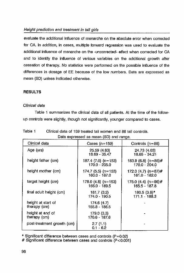
Height prediction and treatment in tall girls
evaluate the additional Influence of menarche on the absolute error when corrected
for CA. In addition, in cases, multiple forward regression was used to evaluate the
additional influence of menarche on the -uncorrected- effect when corrected for CA
and to identify the influence of various variables on the additional growth after
cessation of therapy. No statistics were performed on the possible influence of the
differences in dosage of EE because of the low numbers. Data are expressed as
mean (SO) unless indicated otherwise.
RESULTS
Clinical data
Table 1 summarizes the clinical data of all patients. At the time of the follow
up controls were slightly, though not significantly, younger compared to cases.
Table 1 Clinical data of 159 treated tall women and 88 tall controls. Data expressed as mean (SD) and range
Clinical data Cases (n=159) Controls (n=88)
Age (yrs) 25.59 (4.83) 24.73 (4.03) 18.69 - 35.47 18.65 - 34.21
height father (cm) 187.4 (7.0) (n=153) 183.9 (6.8) (n=86)# 170.0 - 205.0 170.0 - 204.0
height mother (cm) 174.7 (5.5) (n=153) 172.0 (4.7) (n=87)# 160.0-187.0 161.0 - 182.0
target height (cm) 178.0 (4.8) (n=153) 175.0 (4.4) (n=86)# 166.0 - 189.5 165.5-187.8
final adult height (cm) 181.7 (3.2) 180.5 (3.8)' 174.0 - 190.5 171.1 - 188.3
height at start of 174.6 (4.7) -therapy (cm) 155.8 - 186.5
height at end of 179.0 (3.3) -therapy (cm) 170.6 - 187.6
post-treatment growth (cm) 2.7 (1.1) -0.1 - 6.2
, Significant difference between cases and controls (P=0.02) # Significant difference between cases and controls (P<O.OOl)
96
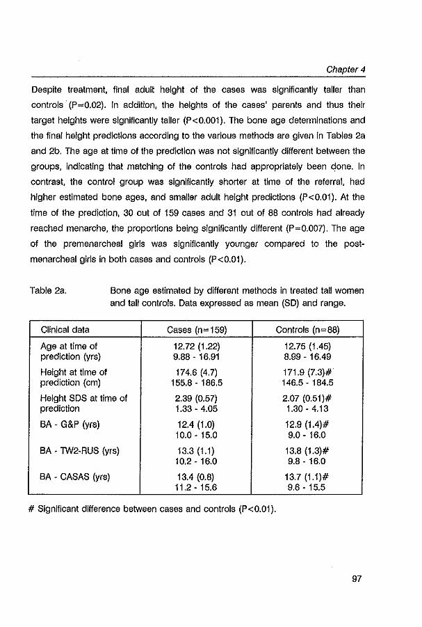
Chapter 4
Despite treatment, final adult height of the cases was significantly taller than
controls· (P=0.02). In addition, the heights of the cases' parents and thus their
target heights were significantly taller (P<0.001). The bone age determinations and
the final height predictions according to the various methods are given in Tables 2a
and 2b. The age at time of the prediction was not significantly different between the
groups, indicating that matching of the controls had appropriately been clone. In
contrast, the control group was significantly shorter at time of the referral, had
higher estimated bone ages, and smaller adult height predictions (P<0.01). At the
time of the prediction, 30 out of 159 cases and 31 out of 88 controls had already
reached menarche, the proportions being significantly different (P=0.007). The age
of the premenarcheal girls was significantly younger compared to the post
menarcheal girls in both cases and controls (P<0.01).
Table 2a. Bone age estimated by different methods in treated tall women and tall controls. Data expressed as mean (SO) and range.
Clinical data Cases (n= 159) Controls (n=88)
Age at time of 12.72 (1.22) 12.75 (1.45) prediction (yrs) 9.88 - 16.91 8.99 - 16.49
Height at time of 174.6 (4.7) 171.9 (7.3)# prediction (cm) 155.8 - 186.5 146.5 - 184.5
Height SDS at time of 2.39 (0.57) 2.07 (0.51)# prediction 1.33 - 4.05 1.30-4.13
BA - G&P (yrs) 12.4 (1.0) 12.9 (1.4)# 10.0 - 15.0 9.0 - 16.0
BA - TW2-RUS (yrs) 13.3 (1.1) 13.8 (1.3)# 10.2-16.0 9.8 - 16.0
BA - CASAS (yrs) 13.4 (0.8) 13.7 (1.1)# 11.2 - 15.6 9.6 - 15.5
# Significant difference between cases and controls (P<0.01).
97
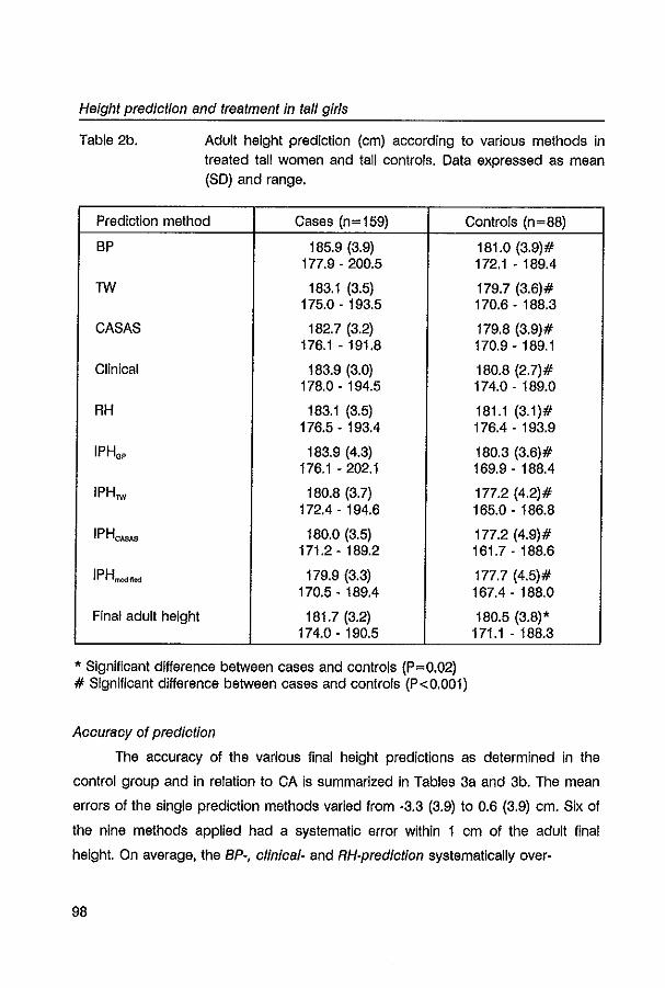
Height prediotion and treatment in tall girls
Table 2b. Adult height prediction (cm) according to various methods in treated tall women and tall controls. Data expressed as mean (SO) and range.
Prediction method Cases (n=159) Controls (n=88)
BP 185.9 (3.9) 177.9 - 200.5
TW 183.1 (3.5) 175.0 - 193.5
CASAS 182.7 (3.2) 176.1 - 191.8
Clinical 183.9 (3.0) 178.0 - 194.5
RH 183.1 (3.5) 176.5 - 193.4
IPH.? 183.9 (4.3) 176.1 -202.1
IPHTW 180.8 (3.7) 172.4 - 194.6
IPHCASAS 180.0 (3.5) 171.2 - 189.2
IPHm"""" 179.9 (3.3)
170.5 - 189.4
Final adult height 181.7 (3.2) 174.0 - 190.5
, Significant difference between cases and controls (P=0.02) # Significant difference between cases and controls (P<O.OOl)
Aoouraoy of prediotion
181.0 (3.9)# 172.1 - 189.4
179.7 (3.6)# 170.6 - 188.3
179.8 (3.9)# 170.9 - 189.1
180.8 (2.7)# 174.0 - 189.0
181.1 (3.1)# 176.4 - 193.9
180.3 (3.6)# 169.9 - 188.4
177.2 (4.2)# 165.0 - 186.8
177.2 (4.9)# 161.7 - 188.6
177.7 (4.5)# 167.4 - 188.0
180.5 (3.8)' 171.1 - 188.3
The accuracy of the various final height predictions as determined in the
control group and In relation to CA is summarized In Tables 3a and 3b. The mean
errors of the single prediction methods varied from -3.3 (3.9) to 0.6 (3.9) em. Six of
the nine methods applied had a systematic error within 1 em of the adult final
height. On average, the BP-, olinioal- and RH-prediotion systematically over-
98
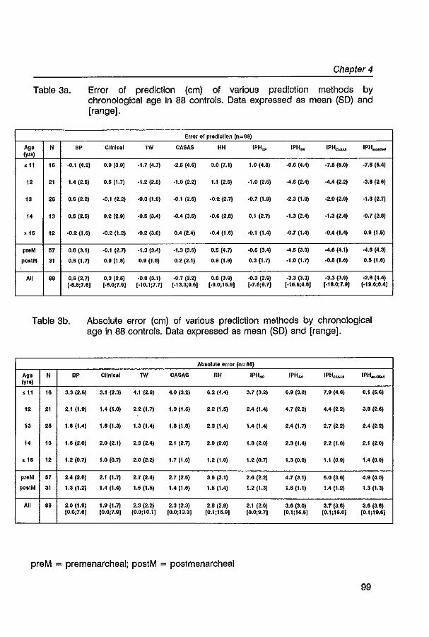
Chapter 4
Table 3a. Error of prediction (cm) of various prediction methods by chronological age in 88 controls. Data expressed as mean (SD) and [range].
Error of predlcllon (n::88)
Ag_ N BP ClinIcal lW CASAS RH 1PHOI' IPH rw IPHCMM 1PH..-It<t (yra)
," I. -0.1 (4.2) 0.9 (3.9) .1.7 (4.7) .2,5 (4.6) 3.0 (7.1) 1.0 (4.8) ·8.6 (4.4) -7.8 (6.0) -7.5 (6.4)
12 21 1.4 (2.5) 0.5 (1.7) .1.2 (2.a) .1.0 (2.2) 1.1 (2,6) .1.0 (2.6) -4,6 (2.4) -4.4 (2.2) ·3.8 (2.6)
13 2. 0.6 (2.2) ·0.1 (2.2) -0,3 (1.9) ·0.1 (2.6) -0.2 (2.7) -0.7 (1.9) ·2.3 (1.9) ·2.0 (2.9) ·1.8 (2.7)
" 13 0.6 (2.5) 0.2 (2.9) ·0.6 (3.4) -0.4 (3,6) ·0,6 (2.8) 0.1 (2.7} ·1.3 (2.4) ·1.3(2.011) ·0.7 (2.8)
,,, 12 ·0.2 (J.6) ·0.2(1.2) ·0.2 (3.0) 0.4 (VI) -0.4 (1.6) ·0.1 (IA) ·0.7 (1.4) .0.4 (1.4) 0.8(1.6)
proM ., 0.6 (3.1) ·0.1 (2.7) -1.3 (3.4) ·1.3 (3.5) 0.5 (4.7) ·0.6 (3.4) -4.5 (3,3) -4,6 (4.1) -4.6 (4.3)
pOBIM 31 0.5 (1.7) O,g (1,8) 0.9 (1.8) 0.2 (2.1) 0.9 (1.9) 0.3 (1.7) ·1.0 (1.7) ·0.8 (1.6) 0.5 (t.8)
A1I •• 0.5 (2.7) 0.3 (2.6) ·0.8 (3.1) ·0.7 (3.2) 0.6 (3.9) ·0.3 (2.9) -3.3 (3.3) ·3.3 (3.9) ·2.8 (4,4) [.5.8;7.6) [·6.0;7.9) [·10.1:7.7) {.13.3j9.6J [.9.0;15.9J [-7.6;9.7) [·15,1;;4.6) [.18.0;7.9) (-19.6;6.4)
Table 3b. Absolute error (cm) of various prediction methods by chronological age in 88 controls. Data expressed as mean (SD) and [range].
Ab80lute error (n=88)
Ag_ N BP Clinical lW CASAS AH IPHOI' IPH rw lPH.... IPH __ tyra)
," 16 3.3 (2.5) 3.1 (2.3) 4.1 (2.9) 4.0 (3.2) 6.2 (4.4) 3.7 (3.2) 6.9 (3.8) 7.9 (4.8) 8.1 (6.6)
12 21 2.1 (1.9) 1.4 (1.0) 2.2 (1.7) 1.9 (1.6) 2.2 (1,5) 2,4 (1,4) 4.7 (2.3) 4,4 (2.2) 3.8 (2.6)
13 26 1.8 (1.4) 1.8 (1.3) 1.3 (1,4) La (1.8) 2.3 (1,4) 1,4(1.4) 2.4 (1.7) 2.7 (2.2) 2.4 (2.2)
I. 13 1,6 (2.0) 2.0 (2,1) 2.3 (2.4) 2,1 (2.7) 2.0 (2.0) 1.8 (2.0) 2.3 (1,4) 2.2 (1.6) 2.1 (2.0)
,,, 12 1,2 (0.7) 1,0 (0,7) 2.0 (2.2) 1.7 (1.6) 1,2 (1.0) 1.2 (0.7) 1.3 (0.9) 1.1 (0.9) 1.4 (0.9)
preM 67 2.4 (2.0) 2.1 (1.7) 2.7 (2.4) 2.7 (2.6) 3.5 (3,1) 2.6 (2.2) 4.7 (3.1) 5.0 (3.6) 4.9 (4.0)
poSIM 31 1.3 (1.2) 1.4 (1.4) 1.5 (1.5) 1.4 (1,6) 1.5 (1.4) 1,2 (1.3) 1,6 (1,1) 1.4 (1,2) 1.3 (1,3)
A1I 6. 2.0 (1.9) 1.9 (1.7) 2.3 (2.2) 2.3 (2.3) 2.8 (2.8) 2.1 (2.0) 3.6 (3.0) 3.7 (3.6) 3.6 (3.8) (0.0;7.6) [0,0:7.9) (0.0;10.1] (0.0:13.3) [0.1;15.9J (0.0;9.7) [0.1;16.6] (O.I;la.O) (0.1;19.6)
preM = pre menarcheal; postM = postmenarcheal
99
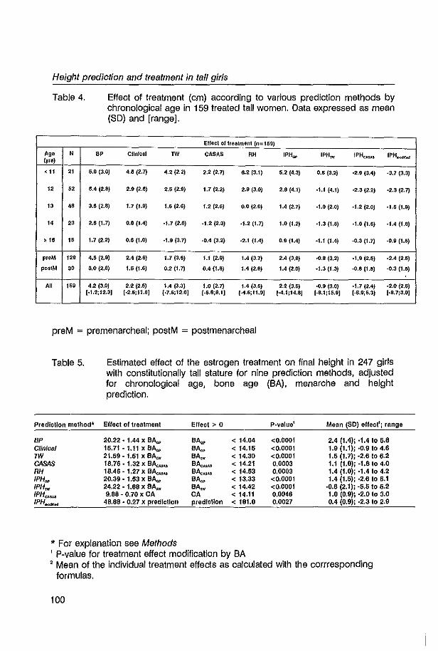
Height prediction and treatment in tall girls
Table 4.
Ag. N (yrs)
<11 21
12 62
'3 48
14 23
>15 '5 preM ,2'
pO$IM 3.
All ,5'
Effect of treatment (cm) according to various prediction methods by chronological age in 159 treated tall women. Data expressed as mean (SO) and [range].
Elfect of treatment (0=159)
BP CHnlcal TW CASAS RH IPH(1p IPHrw IPHCASJ.$ IPH..-.>Od
5,9 (3.0) 4.5 (2.7) 4.2 (2.2) 2.2 (2.7) 6.2 (3.1) 5.2 (4.3) 0.5 (3.2) .2,9 (3.4) -3.7 (3.3)
5.4 (2.8) 2.9 (2.6) 2.5 (2.9) 1.7 (2.2) 2.9 (3.0) 2.8 (4.1) -1.1 (4.1) -2.3 (2.2) ·2.3 (2.7)
3.6 (2.8) 1.7 (1.9) 1.6 (2.6) 1.2 (2.6) 0.0 (2.0) 1.4 (2.7) ·1.0 (2.0) -1.2 (2.0) .1.5 (1.9)
2.6 (1.7) 0.8 (M) -1.7 (2.6) -1.2 (2.3) .1.2 (1.7) 1.0 (1.2) ·1.3 (1.5) -1.0 (1.6) ·1.4 (1.8)
1.7 (2.2) 0.6 (1.0) -1.9 (3.7) .0.4 (3.2) -2.1 (f.4) 0.9 (1.4) -1.1 (1.4) -0.3 (1.7) -0.9 (1.5)
4.6 (2.9) 2.4(206) 1.7 (3.6) 1.1 (2.9) 1.4 (3.7) 2.4 (3.8) -0.8 (3.2) .1.9 (2.6) ·2.4 (2.5)
3.0 (2.8) 1.5 (1.6) 0.2 (1.1) 0.4 (1.8) 1.4 (2.6) 1.4 (2.0) ·1.3 (1.3) ·0.8 (1.5) '0,3 (1.5)
4.2 (3.0) 2.2 (2.5) 1.4 (3.3) 1.0 (2.7) 1.4 (3.5) 2.2 (3.5) ·0.9 (3.0) -1.7 (2.4) -2.0 (2.5) [·1.2;12.3J (·2.5;11.8J [·7.6;12.6J [·6.8;8.1] {·4.5;11.9] [-4.1;14.8] [.8.1;15.9J [.8.9;5.3] [·8.7;3.0]
preM = premenarcheal; postM = postmenarcheal
Table 5.
Prediction method"
BP Clinical 1W CASAS RH 1PHIM' IPHnt IPHC,I.$,I.$ IPH~
Estimated effect of the estrogen treatment on final height in 247 girls with constitutionally tall stature for nine prediction methods, adjusted for chronological age, bone age (BA), menarche and height prediction.
Effect of treatment Effect> 0 P-valuel Mean (SO) effectlj range
20.22 • 1.44 x B~ BA." < 14.04 <0.0001 2.4 (1.4); ·1.4 I. 5.8 15.71 - 1.11 x B~ BA.". < 14.16 <0.0001 1.9 (1.1); ·0.9104.8 21.59-1.61 x BArw BA~ < 14.30 <0.0001 1.5 (1.7); -2.610 6.2 18.76 - 1.32 x BAc.u,l.$ BA.".,u < 14.21 0.0003 1.1 (1.0)j -1.8 to 4.0 18.46 - 1.27 x B~ B~ < 14.53 0.0003 1.4 (1.0)j ·1.4 to 4.2 20.39 • 1.53 x BA.:.,. BA.". < 13.33 <0.0001 1.4 (1.5); ·2.6106.1 24.22 - 1.68 x BArw BAM < 14.42 <0.0001 ·0.6 (2.1)j -5.5 to 5.2 9.88 - 0.70 x CA CA < 14.11 0.0046 1.0 (0.9); -2.0 10 3.0
48.88 - 0.27 x prediction prediction < 181.0 0.0027 0.4 (O.9)j '2,3 to 2.9
* For explanation see Methods , P-value for treatment effect modification by BA , Mean of the individual treatment effects as calculated with the corrresponding
formulas.
100
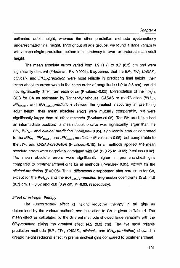
Chapter 4
estimated adult height, whereas the other prediction methods systematically
underestimated final height. Throughout all age groups, we found a large variability
within each single prediction method in its tendency to over· or underestimate adult
height.
The mean absolute errors varied from 1.9 (1.7) to 3.7 (3.5) cm and were
significantly different (Friedman: P< 0.0001). It appeared that the Bp·, TW·, CASAS·,
clinical·, and IPHGp·prediction were most reliable in predicting final height: their
mean absolute errors were in the same order of magnitude (1.9 to 2.3 cm) and did
not significantly differ from each other (P·values>0.05). Extrapolation of the height
SOS for BA as estimated by Tanner·Whitehouse, CASAS or modification (IPHlW·,
IPHCASAS·, and IPH __ ·prediction) showed the greatest inaccurary in predicting
adult height: their mean absolute errors were mutually comparable, but were
significantly larger than all other methods (P·values<0.05). The RH·prediction had
an intermediate position: its mean absolute error was significantly larger than the
Bp·, IHPGp·, and clinical predictIon (P·values<0.05), significantly smaller compared
to the IPHlW·, IPHCASAS·, and IPH"",,_·prediction (P·values <0.05), but comparable to
the TW·, and CASAS·prediction (P·values>0.15). In all methods applied, the mean
absolute errors were negatively correlated with CA (r:-0.25 to ·0.65; P·values<0.02).
The mean absolute errors were significantly higher in premenarcheal girls
compared to postmenarcheal girls for all methods (P·values<0.05), except for the
clinical·prediction (P=0.06). These differences disappeared after correction for CA,
except for the IPHTW·, and the IPH"""",,·prediction (regression coefficients (SE): ·1.5
(0.7) cm, P=0.02 and ·2.0 (0.9) cm, P=0.03, respectively).
Effect of estrogen therapy
The ·uncorrected· effect of height reductive therapy in tall girls as
determined by the various methods and in relation to CA is given in Table 4. The
mean effect as calculated by the different methods showed large variability with the
BP·predlction giving the greatest effect (4.2 (3.0) cm). The five most reliable
prediction methods (BP·, TW·, CASAS·, clinical·, and IPHGp·prediction) showed a
greater height reducing effect in premenarcheal girls compared to postmenarcheal
101
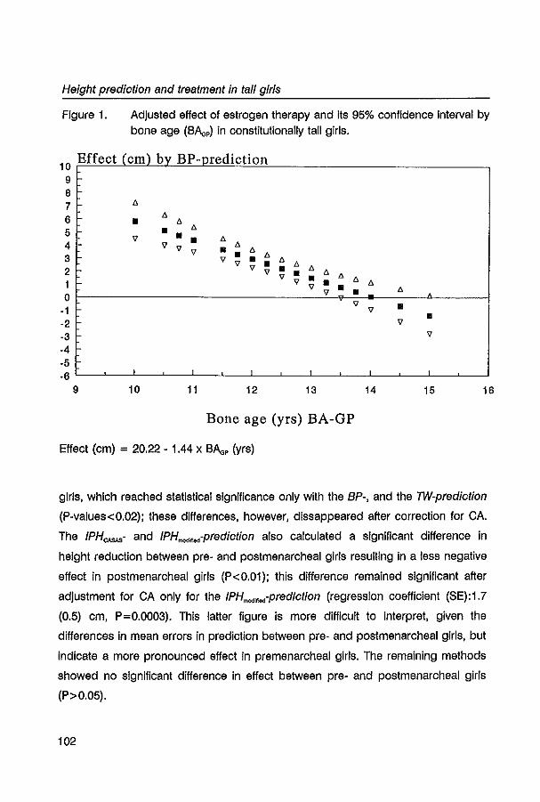
Height prediotion and treatment in ta/l girls
Figure 1. Adjusted effect of estrogen therapy and its 95% confidence interval by bone age (BAa,) in constitutionally tall girls.
10 Effect (em) bv BP-predietion 9 8 7 6
6 - • 6 6
5 • 6
V • • 6 4 v 6 V V • 6 3 • 6
V V • • 6 6 2 v v • 6 6
V • 6 1 v • • 6 6 V • 6 0 V •
V • -1 v • ·2 v ·3 v
-4 -5 ~ -6 1
9 10 11 12 13 14 15 16
Bone age (yrs) BA-GP
Effect (cm) = 20.22 - 1.44 x BAa, (yrs)
girls, which reached statistical significance only with the BP-, and the TW-prediotion
(P-values<0.02); these differences, however, dissappeared after correction for CA.
The IPH""",- and IPH __ -prediction also calculated a significant difference In
height reduction between pre- and postmenarcheal girls resulting in a less negative
effect in postmenarcheal girls (P<0.01); this difference remained significant after
adjustment for CA only for the IPH"",,_-prediotion (regression coefficient (SE): 1. 7
(0.5) cm, P=0.0003). This latter figure Is more difficult to interpret, given the
differences In mean errors in prediction between pre- and postmenarcheal girls, but
indicate a more pronounced effect in pre menarcheal girls. The remaining methods
showed no significant difference In effect between pre- and postmenarcheal girls
(P>0.05).
102
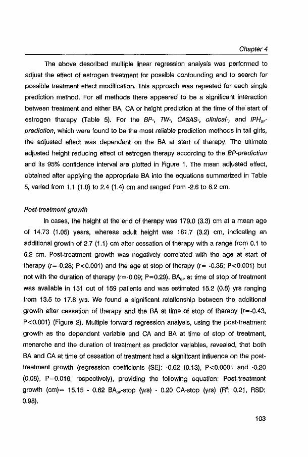
Chapter 4
The above described multiple linear regression analysis was performed to
adjust the effect of estrogen treatment for possible confounding and to search for
possible treatment effect modification. This approach was repeated for each single
prediction method. For all methods there appeared to be a significant interaction
between treatment and either BA, CA or height prediction at the time of the' start of
estrogen therapy (Table 5). For the BP-, TW-, CASAS-, clinical-, and IPH.,
prediction, which were found to be the most reliable prediction methods in tall girls,
the adjusted effect was dependent on the BA at start of therapy. The ultimate
adjusted height reducing effect of estrogen therapy according to the BP-prediction
and its 95% confidence Interval are plotted In Figure 1. The mean adjusted effect,
obtained after applying the appropriate BA into the equations summarized in Table
5, varied from 1.1 (1.0) to 2.4 (1.4) cm and ranged from -2.6 to 6.2 cm.
Post-treatment growth
In cases, the height at the end of therapy was 179.0 (3.3) cm at a mean age
of 14.73 (1.05) years, whereas adult height was 181.7 (3.2) cm, indicating an
additional growth of 2.7 (1.1) cm after cessation of therapy with a range from 0.1 to
6.2 cm. Post-treatment growth was negatively correlated with the age at start of
therapy (r=-0.28; P<O.OOl) and the age at stop of therapy (r= -0.35; P<O.OOl) but
not with the duration of therapy (r=-0.09; P=0.29). BAa, at time of stop of treatment
was available in 151 out of 159 patients and was estimated 15.2 (0.6) yrs ranging
from 13.5 to 17.8 yrs. We found a significant relationship between the additional
growth after cessation of therapy and the SA at time of stop of therapy (r=-0.43,
P<O.OOl) (Figure 2). Multiple forward regression analysis, using the post-treatment
growth as the dependent variable and CA and BA at time of stop of treatment,
menarche and the duration of treatment as predictor variables, revealed, that both
SA and CA at time of cessation of treatment had a significant influence on the post
treatment growth (regression coefficients (SE): -0.62 (0.13), P<O.OOOl and -0.20
(0.08), P=0.016, respectively), providing the following equation: Post-treatment
growth (cm)= 15.15 - 0.62 BAa,-stop (yrs) - 0.20 CA-stop (yrs) (R': 0.21. RSD:
0.98).
103
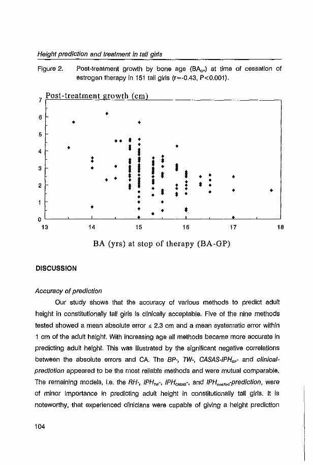
Height prediction and treatment in tall girls
Figure 2. Post-treatment growth by bone age (BAa,) at time of cessation of estrogen therapy in 151 tall girls (r=-0.43, P<0.001).
7 Post-treatment growth (em)
6 • • •
5 l-•• • • • • • 4 • • • : • • • t • 3 l- • • • • • • • t • • • • • • • • •
• I • • 2 • • • • • • • • • • • • • • t • • • • • • • • • 0 • I •
13 14 15 16 17 18
BA (yrs) at stop of therapy (BA-GP)
DISCUSSION
Accuracy of prediction
Our study shows that the accuracy of various methods to predict adult
height in constitutionally tall girls is clinically acceptable. Five of the nine methods
tested showed a mean absolute error < 2.3 cm and a mean systematic error within
1 cm of the adult height. With increasing age all methods became more accurate in
predicting adult height. This was Illustrated by the significant negative correlations
between the absolute errors and CA. The BP-, TW-, CASAS-IPH.,- and clinica/
prediction appeared to be the most reliable methods and were mutual comparable.
The remaining models, i.e. the RH-, IPHTW-, IPHCASAS-, and IPH"""",-prediction, were
of minor importance in predicting adult height In constitutionally tall girls. It is
noteworthy, that experienced clinicians were capable of giving a height prediction
104
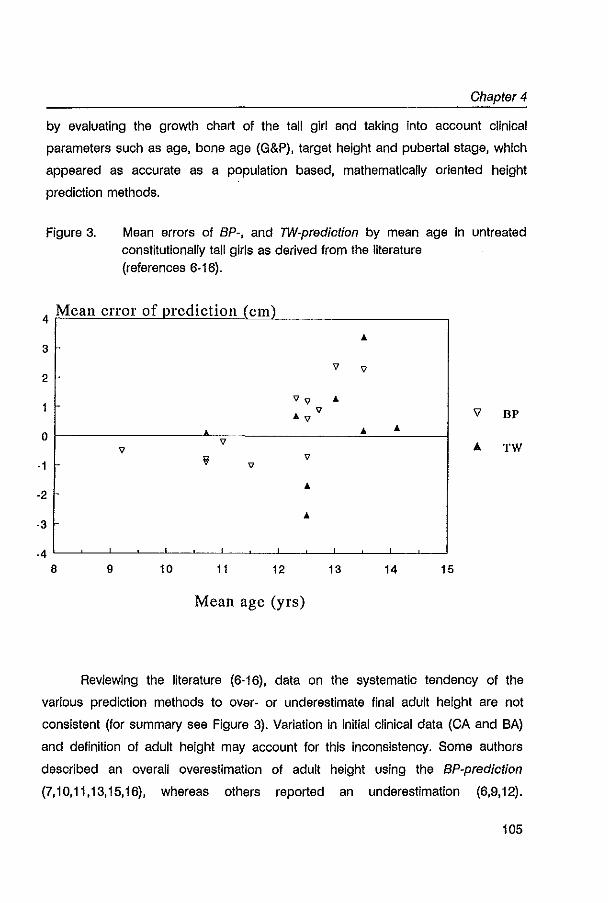
Chapter 4
by evaluating the growth chart of the tall girl and taking into account clinical
parameters such as age. bone age (G&P). target height and pubertal stage. which
appeared as accurate as a population based. mathematically oriented height
prediction methods.
Figure 3. Mean errors of BP-. and TW-prediction by mean age in untreated constitutionally tall girls as derived from the literature (references 6-16).
4 Mean error of prediction (em)
.. 3
v v 2
Vv .. .. v
v 'V BP .. .. 0 v A TW
\I v -1 v
.. -2
.. -3
-4 , ,
8 9 10 11 12 13 14 15
Mean age eyrs)
Reviewing the literature (6-16). data on the systematic tendency of the
various prediction methods to over- or underestimate final adult height are not
consistent (for summary see Figure 3). Variation In Initial clinical data (CA and BA)
and definition of adult height may account for this inconsistency. Some authors
described an overall overestimation of adult height using the BP-prediction
(7.10.11.13.15.16). whereas others reported an underestimation (6.9.12).
105
Height prediction and treatment In tall girls
Nevertheless, the mean errors were rather accurate ranging from a mean
underprediction of 0.9 cm to a mean overprediction of 2.4 cm. When the age of the
various study populations was taken Into account, it seemed that the BP-predlctlon
underestimates adult height in tall girls at younger ages, whereas from the age of
11 a slight overestimation was present, which was confirmed by our study.
Concerning the TW-predlction, most workers have used the older prediction
method of Tanner et al (3) and described an overall overestimation of adult height
ranging from 0.1 to 3.4 cm, which varied with the age of the study population (8-
11). Only Willig et al (6) reported a mean underestimation of ·1.7 to -2.7 cm Joss
and co·workers (16), using the revised TW2 prediction method (4), calculated a
mean overprediction of 1.3 cm. In contrast, we found a mean underestimation of -
0.8 cm, which was more pronounced at younger ages. Knowledge of the
systematic errors of the single prediction methods is of clinical importance since it
may increase the accuracy of a height prediction by correcting it for its prediction
error at a given CA, as also suggested by Joss et al (7). However, considerable
Individual variation will still be present as implied by the large standard deviations
of the mean errors of the prediction method used. This can be illustrated by an
example of a 12 year old girl using the BP-predlctlon in which the method,
according to our study, revealed a mean overprediction of 1.4 cm, but which may
vary In individual cases from an underprediction of ·3.6 cm (-28D) to an
overpredlction up to 6.5 cm (+ 28D).
Absolute errors are independent of a method's tendency for under· or over·
estimation and are more sensitive to deviations from the prediction. Therefore, it is
more indicative to calculate the absolute error of a prediction model in a control
population. Unfortunately, to our knowledge, absolute errors on the prediction
methods applied have never been described in tall girls. Our study showed that the
mean absolute errors of the BP-, TW,- CASAS·, IPH.,- and clinical-prediction were
clinical acceptable (s 2.3 cm). However, large errors in prediction are stili possible
in individual cases as the standard deviations were quite large. In addition, at ages
younger than 11 years absolute errors were considerable indicating less reliability.
In our hands, height prognosis was more reliable in tall girls compared to tali
106
Chapter 4
boys (5). This Is illustrated by our findings that the mean errors were more close to
zero and the mean absolute errors were smaller in girls than in boys. Differences In
timing and intensity of the adolescent growth spurt between boys and girls (31,32)
may be a possible cause for this finding; in our study height prediction was
performed at about the expected age of the peak height velocity (PHV) in the
untreated tall boys, whereas in the untreated girls a height prognosis was
calculated after the expected age of PHV. For both boys and girls, the IPHa,
prediot/on gave a satisfying height prognosis; this method can easily be
implemented in clinical practice. In addition, experienced clinicians may also rely on
their own judgement as the clinical-predictIon showed acceptable reliability in both
boys and girls.
Effect of estrogen therapy
The present study showed that the effect of estrogen treatment in tall girls
varied with the prediction method applied (Table 4). One may "correct" the mean
calculated effect of therapy by substraction of the systematic prediction errors,
which has been commonly done in the literature. In this way, all prediction methods
calculated a positive mean effect, suggesting that, overall, treatment was effective.
The five most reliable methods (i.e. the BP-, TW,- CASAS-, IPHa,- and clinioal
predict/on) Showed a mean "corrected" height reduction varying from 1.2 to 3.7 cm.
These findings support previously published data In which the mean reported
height redUction ("corrected" and uncorrected) ranged from 2.1 cm to 10.0 cm (6,8-
25). A clear comparison, however, is hampered by differences in Initial clinical data
(especially CA and BA), duration of treatment, therapeutic regimen (different doses
and estrogen preparations), and the point in time of adult height assessment. For
example, in our study a mean additional growth of 2,.7 (1.1) cm was observed after
cessation of therapy, which Is in the same order of magnitude as reported by
others (13,18,21). It Implies that at the time therapy was stopped the mean
"uncorrected" height reduction amounted to 6.4 cm using the BP-prediction,
stressing the importance of defining adult height. The cause of the observed
additional growth is not quite clear. In our study, the BAa, at time of cessation of
107

Height prediction and treatment in tall girls
estrogen therapy was estimated on average at about 15 years, Indicating that
approximately 99.3% of the adult final height had been achieved (1). It explains
therefore only part of the post-treatment growth. Since sex steroids in lall girls
reduce spinal growth to a much lesser extent than long bone growth (32,33), it is
plausible that part of the remaining post-treatment growth at a mean age of 14.73
(1.05) years reflects additional spinal growth.
As discussed previously in chapter 3, evaluating the effect of estrogen
treatment after "correction" for the mean errors of the separate prediction methods
will induce bias. We therefore chose for a more direct and conceptually simple
approach using multiple linear regression. In this way, analyses revealed a
significant treatment effect with a size depending on either SA, CA or the height
prediction (Table 5). In the five most reliable prediction methods (i.e. the BP-, 7W,
CASAS-, IPHG,- and clinical-prediction) the treatment effect was related to the
corresponding SA, resulting in a mean height reduction varying from 1.1 to 2.4 cm
and ranging from -2.6 to 6.2 cm. These mean results are less than claimed In the
literature (6,8-25). However, this is probably due to differences in statistical
approach and differences in study design, as explained above. Our study clearly
Indicated that the effect of treatment was more pronounced when treatment had
been started at a younger SA (see Figure 1), which is in concert with previous
studies (10,14,19,21,24,25), although others could not find such relationship (8).
Inconsistency is present whether or not premenarcheal girls may experience more
height reduction compared to postmenarcheal girls. Some reports are in favour
with this finding (16,20-22), while others observed no difference (8,13,23,24). In the
present study, premenarcheal girls seemed to benefit more from therapy than
postmenarcheal girls. However, this was likely to be due to the differences in CA
between these groups since we found no additional effect of menarche over CA
explaining the variability in the ·uncorrected· effect of treatment. This may, at least
partly, explain the contradictive results.
In our experience, tall girls benefited more from sex steroid therapy than tall
boys (5). In fact in boys, even a significant growth Induction was observed at an
older SA (5). It is possible that this was due to the timing of the start of therapy in
108
Chapter 4
the adolescent growth phase. Our treated tall girls had started therapy at a mean
age of 12.7 yrs, which is about 1.2 to 0.7 yrs after the age of PHV. In contrast, our
treated tall boys started therapy at a mean age that was only 0.5 to 0.2 yrs after
the age of PHV (31,32). Another explanation might be differences in mechanism of
action by which estrogens and androgens limit linear growth. The mechanisms by
which sex steroids act, however, are not fully understood. Part of the growth
reduction may be mediated via the GH/lGF-axis (35,36). Indeed, in tall girls' a dose
dependent reduction of IGF-I plasma levels had been observed during estrogen
treatment (15,20), although this decrease was not significant using a dosage of 250
I1g EE/day (15). Therefore, the decrease in growth velocity observed after start of
treatment with doses ~ 200 I1g EE (24,25,37) is hard to relate to changes in IGF-I
concentration, but suggests that other factors are involved. Another important
factor playing a role in the height reducing process is the influence of sex
hormones on bone maturation. In fact, the observation that premature secretion of
gonadal steroids in precocious puberty cause acceleration of bone maturation
which leads to short adult stature due to early fusion of the epiphyseal growth
plates (38-40), is one of the rationales for sex hormone therapy In tall stature.
Indeed, in the management of tall stature sex steroid treatment resulted in an
advanced bone maturation. The regulation of bone growth is very complex and is
beyond the scope of this article. It is generally agreed, however, that the steroid
hormone effects on bone maturation are due to an Indirect action mediated by the
GH/IGF-I axis combined with a direct effect at tissue level (35,41,42). In addition,
several studies point to the possibility that there are sex-specific and age
dependent responses to sex steroids present in cartilage cells in vitro (43-45).
Moreover, it is possible that In males androgens may partly act on cartilage after
their metabolic conversion into estrogens (35,43). A recent report of a 28-year old
adult man with absence of functional estrogen receptors together with unfused
epiphyseal growth plates (46) confirms the need of estrogens for bone maturation
in the male. All these aspects may underlie the differences in therapy response
between boys and girls, although the precise mechanism is far from clear.
High dose estrogen therapy may cause minor side effects, which are likely
109
Height prediction and treatment in ta// girls
to be dose related (47,48). Occasionally a serious complication as thrombosis has
been reported. Up till now there is no evidence that there are long-term side effects
on reproductive function (48). However, concerning a possible association between
estrogen therapy and malignancy based on results in oral contraceptive studies
(49), definite conclusions cannot be drawn at this point In time. It Is Important
therefore to balance all pros and cons In dialogue with the tall girl and her parents
in order to come to a careful decision regarding possible therapeutic intervention.
In summary, these data demonstrate that the accuracy of various methods
to predict adult height In constitutionally tall girls is clinically acceptable. With
increasing age, height prognosis became more accurate. A significant height
reductive effect of high doses of estrogens was found in tall girls. This effect was
mainly dependent on the BA at the start of therapy: the effect was more
pronounced when treatment had been started at a younger BA. Serious additonal
growth was observed after cessation of therapy, which could be explained only
partly by the B.A.p at that time; additional spinal growth may have also accounted
for part of the post-treatment growth.
ACKNOWLEDGEMENTS
We kindly thank the Foundation for Children's Stamps, Laiden, The
Netherlands, for their generous gift to support this study. It was presented In part
at the annual meeting of the European Society of Pediatric Endocrinqlogy in
Edinburgh, England, July 1995. Dr. Berthon Rikken is greatly acknowledged for his
assistance In developing additional software applications. We thank Frank
Bekkering, Patricia Chinafo, Evelien van Gurp, Vincent Kappendijk, Irene Lucas,
and Martin Torn, medical students, for their help in collecting the data.
110

Chapter 4
REFERENCES
1. Bailey N, Plnneau SR: Tables for predicting adult height from skeletal age, revised for Greulich and Pyle hand standards. J Pediatr 1952;40:423441
2. Tanner JM, Whitehouse RH, Marshall WA, Healy MJR, Goldstein H: Assessment 01 skeletal maturHy and prediction of adult height: TW2 method. Academic Press, New York 1975
3. Tanner JM, Whitehouse RH, Cameron N, Marshall WA, Healy MJR, Goldstein H: Assessment of skeletal maturHy and prediction of adull height (lW2 method). Academic Press, London 1983
4. Lenko HL: Prediction of adull height with various methods in Finnish children. Acta Paedlatr Scand 1979;68:85·92
5. De Waal WJ, Fokker MH, van Gurp EAFJ. Stilnen Th, de Muinck Kelzer.$chrama SMPF, Aarsen RSR, Drop SLS: Accuracy of final height prediction and effect of growth reductive Iherapy in 115 constitutionally tall boys. A comparison of nine prediction methods. Chapter 3, thesis 1996.
6. Willig AP, Christiansen 0, Kuhn N, Schaefer E, Stahnke N: Voraussetzungen und Ergebnisse der Cstrogenbehandlung extrem groBer Madchen. Monatsschr K1nderhellkd 1980;128:787-788
7 Joss EE, Temperll R, Mullis PE: Adult height In constnutlonally tall stature: accuracy of five different height prediction methods. Arch Dis Child 1992;67:1357·1362
8. Zachmann M, Ferrandez A, MOrset G, Prader A: Estrogen treatment of excessively tall girls. Heiv Paedlat Acta 1975;30:11-30
9. Aeeser HM, Heremans GFP, van Gelderen HH: Aeduction of adult height In tall girls. Eur J Pedlatr 1979;132:37·41
10. John G, Schellong G: (istrogentheraple hochwOchsiger Madchen. Monatsschr Kinderhellkd 1980; 128:545·550
11. Sorgo W, Scholler K, Heinze F, Heinze E, Teller WM: Critical analysis of height reduction In oestrogen·treated tall girls. Eur J Pedlatr 1984;142:206·265
12. Schambach H, Nitschke U: Ole Behandlung des konstllutlonellen Hochwuchses bel Madchen mil physlologlschen (istrogendosen In der Prapubertat. Monatsschr K1nderheilkd 1985;183:32-37
13. Bartsch 0, Weschke B, Weber B: Oestrogen treatment of consthutlonally tall girls wllh 0.1 mg/day ethlnyl oestradiol. Eur J Pedlatr 1988;147:59·83
14. Ignatius A, Lenko HL, Perheentupa J: Oestrogen treatment of tall girls: effect decreases wllh age. Acta Paedlatr Scand 1991;80:712-717
15. Svan H, Aitz~n EM, Hall K, Johansson L: Oestrogen treatment of tall girls: dose dependency of effects on subsequent growth and IGF·I levels In blood. Acta Paedlatr Scand 1991;80:328-332
16. Joss EE, Zeuner J, ZurbrOgg AP, Mullis PE: Impact of different doses of ethlnyl oestradiol on reduction of final height in constitutionally tall girls. Eur J Pediatr 1994;153:797-801
17. Colle ML, Alperin H, Greenblatt RB: The tall girl. Prediction of mature height and management. Arch Dis Child 1977;52:118·120
18. Andersen H, Jacobsen BB, Kastrup ~, Krabbe S, Peitersen B, Petersen KE, Thamdrup E, Wichmann A: Treatment of girls with excessive height prediction. Acta Paedlatr Scand 1980;69:293·297
111

Height prediction and treatment in tall girls
19. Wettenhall HNB, Cahill C, Roche AF: Tall girls: A survey of 15 years of management and treatment. J Pedlatr 1975;86:602·610
20. Von Puttkamer K, Bierich JR, Brugger F, Hlrche W, SchOnberg D: (istrogentheraple bel Mfidchen mtt konstitutlonellem Hochwuchs. Dtsch Med Wochenschrft 1977;102:983·988
21. Kuhn N, Blunck W, Stahnke N, Wlebel J, WIllig RP: Estrogen treatment In tall girls. Acta Paedlatr Scand 1977;66:161·167
22. Bierlch JR: Estrogen treatment of girls with constitutional tall stature. Pediatrics 1978;62:1196·1201
23. StOver B, Kollmann F: Ergebnisse der Behandlung hochwOchslger Mfidchen mit conjuglerten (istrogenen. Monatsschr Klndrheilkd 1982;130:36·40
24. GrOters A, Heidemann p, SchlOter H, Stubbe P, Weber B, helge H: Effect of different oestrogen doses on final height reduction in girls with constitutional tall stature. Eur J Pad 1989;149:11·13
25. Normann EK, Trygstad 0, Larsen S, Dahl-Jorgensen K: Height reduction in 539 tall girls treated with three different dosages of ethlnyloestradlol. Arch Dis Child 1991;66:1271·1278
26. Roede MJ, van Wleringen JC: Growth diagrams 1980. Netherlands third nation·wlde
biometric survey. Tljdschr Soc Gezondheldszorg 1985;63(Suppl):1-34 27. Greulich WW & Pyle SI: Radiographic atias of skeletal development of the hand and wrist.
Stanford Universlly Press, Stanford 1959 28. Tanner JM, Gibbons RD: Automatic bone age measurement using computerized Image
analysis. J Pediatr EndocrinoI1994;7:141·145 29. Van Teunenbroek A, de Waal WJ, Roks A, Chinafo P, Fokker MH, Mulder P, de Muinck
Kelzer·Schrama SMPF, Drop SLS: Computer aided skeletal age scores (CASAS) in heahhy children, girls with Turner syndrome and in children with constitutionally tall stature. Pad Res 1996; in press
30. Van den Brande JVL, Degenhart HJ: Endocrin%gle en groeistoornissen, In: Klndergeneeskunde, Van de Brande, Monnens (eds). Wetenschappelijke uttgeverij Bunge, Utrecht 1993
31. Preece MA: Prediction of adult helght:methods and problems. Acta Paedlatr Scand 1988;347(SuppO:4.11
32. Buckler J: A longttudlnal study of adolescent growth. Springer·Verlag, London 1990 33. Hermanussen M, Gelger·Benoit K, Burmeister J: Differential effects of oestrogen ireatment
on the proportlonaltty of growth In tall girls. Eur J Pedlalr 1989;149:14·17 34. Brlnkers JM, LamonI PJ, Gevers EF, Boersma M, Wtt JM: The effect of oestrogen treatment
on body proportions In constitutionally tall girls, Eur J Pedlatr 1994:163:237·240
35. Bourguignon JP: Linear growth as a function of age at onset of puberty and sex steroid dosage: TherapeutiC implications. Endocr Rev 1988;9:467·488
36. Kerrigan JR, Rogol AD: The Impact of gonadal steroid action on growth hormone secretion during childhood and adolescence. Endocr Rev 1992;13: 281·298
37. Van der Wertt ten Bosch JJ, Bot A: Growth of tall girls without and during oestrogen treatment. Neth J Med 1981;24:52·57
38. Schoevaart CE, Drop SLS, Otlen BJ, SlIjper FME, Degenhart HJ: Growth analysis up to final height and psychosocial adjustment of treated and untreated patients with precocious puberty. Horm Res 1990;34:197·203
112
Chapter 4
39. Kletter GB, Kelch RP: Effecls of gonadotropln·releaslng hormone analog therapy on adult stature In precocious puberty. J Clln Endocrinol Metab t994;79:331·334
40. Brauner R, Adan L. Malandry F, Zantieifer 0: Adult height In girls with Idiopathic true precocious puberty. J Clln Endocrlnol Metab 1994;79:415·420
41. Turner RT, Riggs BL, Spelsberg TC: Skeletal effects 01 estrogen. Endocr Rev 1994;15:275· 300
42, Nilsson A, Ohlsson C, Isaksson OGP, Lindahl A, Isgaard J: Hormonal regulation of longitudinal bone growth. Eur J Clln Nutr 1994;48(SuppI.1): S150-6160
43. Blanchard 0, Tsagrls L, Rappaport R, Duval·Beaupere G, Corvol M: Age·dependent responsiveness of rabbit and human cartilage cells to sex steroids In vitro. J Steroid Blochem Malec Bioi 1991;40:711-716
44. Nasatzky E, Schwartz Z, Boyan BD, Soskolne WA, Ornoy A: Sex-dependent effects of 176 estradiol on chondrocyte differentiation in culture. J Cell PhysloI1993;154:359-362
45. Schwartz Z, Nasatzky E, Ornoy A, Brooks BP, Soskolne WA, Boyan BD: Gender-specific, maturation-dependent effects of testosterone on chondrocytes in culture. Endocrinology 1994;134:1640-1647
46. Smith EP, Boyd J, Frank GR, Takahashi H, Cohen RM, Specker B, Williams TC, Lubahn DB, Korach KS: Estrogen resistance caused by a mutation in the estrogen receptor gene in a man. N Engl J Med 1994;331:1056-1061
47. Trygstad 0: Oestrogen treatment of adolescent tall gIrls; short term side effects. Acta Endocrinol 1986;279(Suppij: 170-173
48. De Waal WJ, Torn M, de Muinck Keizer-Schrama SMPF, Aarsen RSR, Drop SLS: Long term sequelae of sex steroid treatment In the management of constitutionally laH stature. Arch Dis child 1995;73:311-315
49. Kaunitz AM: Oral contraceptives and gynecologic cancer: an update for the 1990's. Am J Obstet Gynecol 1992;167:1171-1176
113


CHAPTER 5
Long term sequelae of sex steroId treatment In
the management of constitutionally tall stature.
W.J. de Waal', M. Torn', S.M.P.F. de Muinck Keizer-Schrama',
R.S.R. Aarsen" S.L.S. Drop'.
1 Department of Pediatrics, 2Division of Endocrinology,
Sophia Children's Hospital, Erasmus University Rotterdam, The Netherlands.
Archives of Diseases in Childhood
1995;73:311-315
Long term sequelae of sex steroid treatment
ABSTRACT
We evaluated possible long term side effects of high doses of sex steroids
in the management of constitutionally tall stature and paid special attention to
hypothalamic-pituitary-gonadal functioning_ For this purpose, 64 tall adult men and
180 tall adult women, who received supraphysiological doses of sex hormones
during puberty, were interviewed in a standardized way at a mean follow-up period
of 10 years after cessation of treatment. Sixty-one untreated tall adult men and 94
untreated tall adult women served as controls. The majority of the subjects were
satisfied with their decision regarding hormonal therapy. Seventy-seven percent of
the women and 78 percent of the men reported one or more side effects during
therapy. Most side effects were mild. In women, only 3% stopped treatment
because of an adverse event; in men, the reported side effects were no reason to
stop treatment. The frequency of reported side effects in women was higher during
treatment with high doses of estrogens than during oral contraceptive use,
indicating a dose dependent relationship. Amenorrhoea of longer than 6 months
after cessation of therapy was found in 5%. Menstrual cycle characteristics of
previously treated women were comparable with controls. Overall, malignancy was
not reported. Information about a total of 127 pregnancies was obtained and
revealed no distinct differences in details and outcome between previously treated
women and men, and controls. In conclusion, at a mean follow-up of 10 years
there is no evidence that pharmacological doses of sex hormones may have a
long-term effect on reproductive function. However, this period is still too short to
draw definite conclusions.
INTRODUCTION
Treatment of tall adolescent boys and girls with high doses of sex steroids in
order to reduce their final height is a widely used method (1-10). In our hospital we
116
Chapter 5
have been treating constitutionally tall children since 1968. In girls, estrogens
(ethinylestradiol (EE) have been used (200 "g/day, range: 1 00 - 300 "g/day) in
combination with progestagens (medroxyprogestrone 5·10 mg/day, orally) every 5·
10 days of the month; boys have been treated with androgens (testosterone esters
in various therapeutic regimens with a total monthly dosage up to 1000 mg.). Over
the years height reduction by means of supraphysiological doses of sex hormones
has been a matter of discussion, not only because of the problems in assessing
the real growth reductive efficacy and the lowest effective dosage, but also
because of the possibility of unwanted side effects. In this respect special attention
focussed on haemostasis (11), lipid metabolism(12) and functioning of the
hypothalamic·gonadal axis (13,14). In addition, the impressive bulk of available data
on the association between long term oral contraceptive use and possible health
risks (reviewed in 15,16) are indispensable as they form an excellent reflection of
the prospective risks in estrogen·treated girls.
So far, unwanted side effects have only been reported during treatment or
shortly after discontinuation of therapy (1-10,17-21). Most side effects were found
to be mild and reversible. Supression of the hypothalamic'gonadal-axis induced by
the pharmacological doses of sex steroids was found to be reversible (13,14).
However, the possibility of a long·term suppressive effect of sex hormone therapy
on reproductive functioning in boys has been postulated (22). In a recent study in
43 previously treated tall men and 30 untreated tall controls we could not confirm
this postulation (23). In girls, the ultimate 'proof' for complete reversibility of
hypothalamlc·gonadal suppression, pregnancy, has been reported in various single
cases (1-4,6,8,9).
To offer further data on acceptance and possible long term effects of high
doses of sex steroids in the management of constitutionally tall stature we
interviewed tall adult men and women at a mean follow·up period of 10 years after
discontinuation of height reductive therapy by means of a self·constructed
questionnaire. Our main field of interest was the functioning of the hypothalamic·
gonadal axis. Since our study was intended to be non· invasive, this was covered
117
Long term sequelae of sex steroid treatment
by questions about menstruation, gynaecological complaints and childbearing. Tall
adult men and women who had not received treatment during their pubertal period
served as controls.
PATIENTS AND METHODS
Patients
Since the introduction of height reductive therapy in our Institute in 1968 a
total of 247 men and 423 women, who were seen at adolescence for evaluation of
their constitutionally tall stature, had reached the age of 18 at the time of our follow
up study. All were contacted by mall to participate in this study. Second mailings
were sent to those who did not respond to the first mailing. This study was part of
a large follow-up study in constitutionally tall stature, including auxological and
psychosocial assessments. Reports on auxology and psychology will be reported
elsewhere.
During their puberty, 102 men and 249 women had been treated for their tall
stature (cases). Usually, the indication for the therapy was a predicted final height
> 2,5 SO (> 180 cm for girls and> 200 cm for boys). Occasionally, therapy was
Initiated In Idiopathic scoliosis. Hundred and forty-five men and 174 women had not
chosen to undergo treatment for various reasons such as satisfaction with the
given height prognosis or uncertainty about possible side-effects, and served as
controls. Two hundred and nine men (95 cases and 114 controls) and 326 women
(203 cases and 123 controls) responded to our mailing. Of the responders, 125
men (64 cases and 61 controls) and 272 women (180 cases and 92 controls)
agreed to participate in this study. Clinical data of the participants are summarized
In table 1. All participants gave informed consent.
118
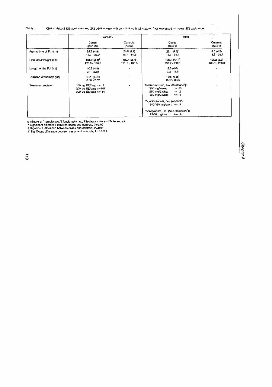
Table 1. Clinical data of 125 adult men and 272 adult women with constitutionally tall stature. Data expressed as mean (SO) and range.
WOMEN
Cases Controls (n=180) (n=92)
Age at time of FU (yrs) 25.7 (4.9) 24.8 (4.1) 18.7 - 35.9 18.7 - 34.2
Final adult height (cm) 181.6 (3.4)$ 180.4 (3.7) 173.8 - 190.5 171.1 -188.3
Length of the FU (yrs) 10.9 (4.9) 3.1 - 22.9
Duration of therapy (yrs) 1.91 (0.61) -0.60 - 3.62
Treatment regimen 100 I-I-g EEJday: n= 9 -200 I1g EEJday: n=157 300 I1g EE/day: n= 14
a Mixture of T-propionate, T-fenylpropionate, T-isohexanoate and T -decanoate. 'II Significant difference between cases and controls, P=0.02 $ Significant difference between cases and controls, P=0.01 # Significant difference between cases and controls, P=0.OO01
CD
MEN
Cases (n=64)
26.1 (4.3}" 18.7 - 34.4
199.8 (4.1)# 190.7 - 210.1
8.6 (4.0) 3.2 -18.5
1.36 (0.56) 0.67 - 3.68
T -ester mixture-, i.m. (Sustanon~: 2SO mg/week: n= 50 250 mg/2 wkS: n= 2 500 mg/2 wks: n= 4
T-undecanoaat, oral (Andriol~: 24Q.320 mg/day: n= 4
T_propionate, Lm. (Neo-HombreoJf): 25-30 mg/day n= 4
Controls (n=61)
4.3 (4.2) 18.6 - 34.1
196.2 (5.0) 186.6 - 209.3
-
-
-
i ~ 0.
Long term sequelae of sex steroid treatment
Methods
All participants were interviewed In a standardized way by one investigator
ryvdW). In women, the standardized interview included questions about satisfaction
and possible side effects of hormonal therapy, oral contraception (OC), menstrual
cycle characteristics (without OC use), pregnancy and gynaecological complaints.
Men were asked about satisfaction and possible side effects of hormonal therapy
and about offspring. The standardized possibilities of adverse effects during sex
hormone therapy were extracted from the literature. Most of the participants were
interviewed In an outpatient clinic set1ing. Five women and one man (all cases)
were interviewed by telephone.
If a subject had sought gynaecological consultation because of suspected
subfertillty additional Information was asked for after informed consent was
obtained.
The protocol was approved by the ethical committee of Academic Hospital,
Erasmus University, Rotterdam, The Netherlands.
Statistios
Differences between groups were tested using Wilcoxon's two sample test.
In women, cases were grouped according to their total daily EE dosage.
Differences between these 3 groups were tested with the Kruskall-Wallis test. In
men, no statistics were performed on the various therapeutic regimens because of
the low numbers In the groups. Chisquare tests (with Yates' correction when
appropriate) were used in case of a contingency distribution. Incidence rates of
adverse events, pregnancy outcome and gynaecological consultation were
compared using Fisher's exact test.
RESULTS
GIRLS
Therapy and adverse effeots
120
Chapter 5
In the control group, four women (4%) regretted their decision not to have
undergone hormonal treatment for height reduction. Ninety-three percent of the
cases were satisfied with the result of their treatment; 7 % showed dissatisfaction.
The main reason for dissatisfaction was the little effect of the therapy compared
with the given height prediction. Eleven percent of the cases would not apply for
height reductive therapy again; they were worried about uncertainties of long term
side effects or thought that tall stature in girls is less a problem nowadays.
Separate possible adverse effects during hormonal therapy and their
reported frequency are listed in table 2. Hundred thirty-nine women (77%) gave
notice being troubled by one or more side-effect during treatment. Forty-one
women (21%) could not remember any adverse effect. Five women (3%) stopped
treatment because of an adverse event: persisting nausea, suspected ovarian cyst,
enlarged uterus, amaurosis fugax, and headache. We found no relationship with
the total daily dosage of EE and the number of reported adverse effects.
Menstruation and oral contraception (OC)
Reported recurrence of menstruation after cessation of therapy is listed in
table 3. Eighty-two percent of the cases reported a regular menstrual cycle
compared to 74 percent of the controls (p~O.15). The reported duration of the
menstrual cycle was 31.6 (12.2) days for cases and 32.4 (11.3) days for controls
(P~O.ll). When we calculated the reported cycle length of the regularly
menstruating women only, the mean duration became shorter: 28.6 (2.2) days and
29.3 (5.3) for cases and controls, respectively (P~0.42) This was due to the finding
that women with irregular menstrual cycles had a higher frequency of very long
menstrual cycles. The reported duration of the menses was slightly, though
significantly, different between cases and controls: 5.3 (1.5) days and 6.1 (1.7)
days, respectively (P<O.Ol). When we calculated the duration of the menses of the
regular menstruating women only, this difference became smaller, but was still
significant (5.3 (1.4) days and 5.9 (1.4) days for cases and controls, respectively,
P<O.Ol). No difference was found in the proportion of intermenstrual bleedings
121
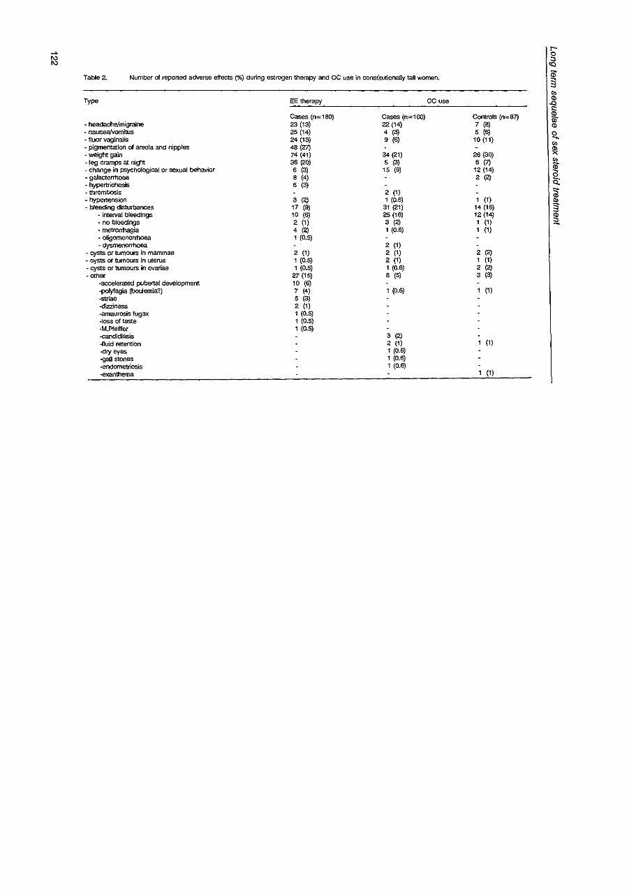
~ Table 2. Number of reported adverse effects (%) during estrogen therapy and OC use in constitutionally tall women.
Type
• headache/migraine · nausea/vomitus - fluor vagina/is • pigmentation of areola and nipples - weight gain - leg cramps at night - change in psychOlogical or sexual behavior - galactorrhoea - hypertrichosis - thrombosis - hypertension - bleeding cflsturbances
• interval bleedings - no bleedings - metrOrrhagia - oligomenOrrhoea - dysmenorrhoea
- cysts or tumours in mammae - cysts or tumours in uterus - cysts or tumours in ovariae - other
--accelerated pubertal development -polyfagia (boulemia?) -striae -dizziness ..amaurosis fugax -loss of taste -M.Pfeiffer -candidiasis -fluid retention -<:try eyes ..gall stones -endometriosis ..exanthema
EE therapy OCuse
Cases (n=180) Cases (n=160) 23 (13) 22 (14) 25 (14) 4 (3) 24 (13) 9 (6) 48 (27) 74 (41) 34 (21) 36 (20) 5 (3) 6 (3) 15 (9) 8 (4) 6 (3)
2 (1) 3 (2) 1 (0.6) 17 (9) 31 (21) 10 (6) 25(16) 2 (1) 3 (2) 4 (2) 1 (0.6) 1 (0.5)
2 (1) 2 (1) 2 (1) 1 (0.5) 2 (1) 1 (0.5) 1 (0.6)
Z7 (15) 8 (5) 10 (6) 7 (4) 1 (0.6) 5 (3) 2 (1) 1 (0.5) 1 (0.5) 1 (0.5)
3 (2) 2 (1) 1 (0.6) 1 (0.6) 1 (0.6)
ControlS (n=87) 7 (8) 5 (6)
10 (11)
26 (30) 6(7)
12 (14) 2 (2)
1 (1) 14(16) 12 (14) 1 (1) 1 (1)
2 (2) 1 (1) 2 (2) 3 (3)
1 (1)
1 (1)
(1)
r-
~ a; § ~
~ ;;; <!)
Q. Co
~
~ a c: ~ Q)
3 ~
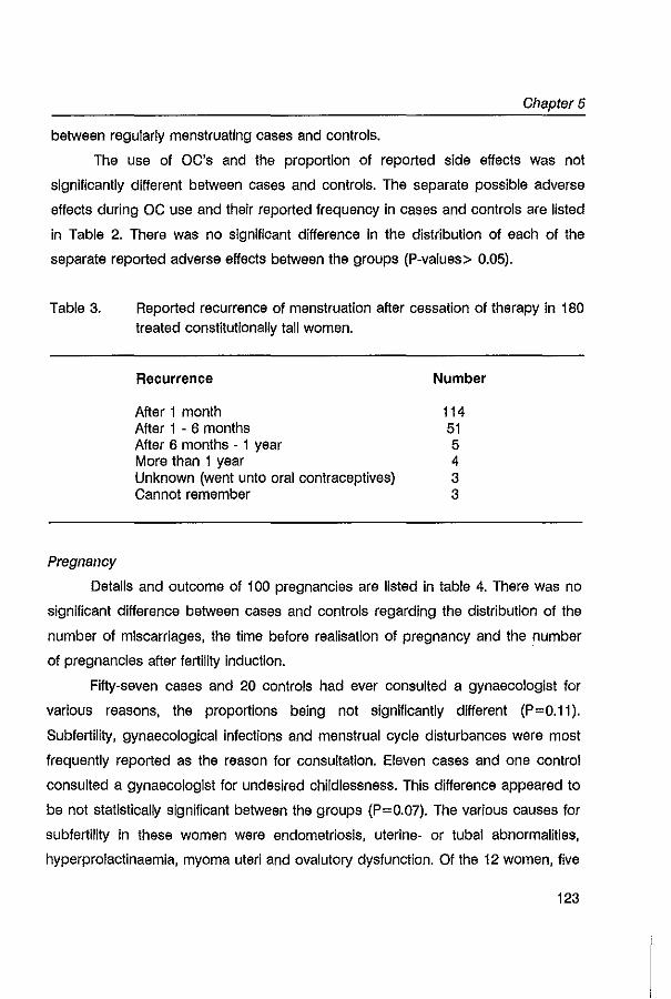
Chapter 5
between regularly menstruating cases and controls.
The use of OC's and the proportion of reported side effects was not
significantly different between cases and controls. The separate possible adverse
effects during OC use and their reported frequency in cases and controls are listed
in Table 2. There was no significant difference in the distribution of each of the
separate reported adverse effects between the groups (P-values> 0.05).
Table 3.
Pregnancy
Reported recurrence of menstruation after cessation of therapy in 180 treated constitutionally tall women.
Recurrence
After 1 month After 1 - 6 months After 6 months - 1 year More than 1 year Unknown (went unto oral contraceptives) Cannot remember
Number
114 51 5 4 3 3
Details and outcome of 100 pregnancies are listed in table 4. There was no
significant difference between cases and controls regarding the distribution of the
number of miscarriages, the time before realisation of pregnancy and the number
of pregnancies after fertility induction.
Fifty-seven cases and 20 controls had ever consulted a gynaecologist for
various reasons, the proportions being not significantly different (P=0.11).
Subfertility, gynaecological infections and menstrual cycle disturbances were most
frequently reported as the reason for consultation. Eleven cases and one control
consulted a gynaecologist for undesired childlessness. This difference appeared to
be not statistically significant between the groups (P=0.07). The various causes for
subfertility In these women were endometriosis, uterine- or tubal abnormalities,
hyperprolactinaemla, myoma uteri and ovalutory dysfunction. Of the 12 women, five
123

Long term sequelae of sex steroid treatment
(4 cases and 1 control) succeeded to become pregnant by means of fertility
induction (table 4).
Table 4. Details and outcome of pregnancies in constitutionally tall men and women.
WOMEN
Cases (n=40) Controls (n=16)
Number of pregnancies: 63 27
Outcome - Abortus provocatus 4 1 - Miscarriage 8 1 · Extra uterine pregnancy 3 - Mola hydatidosa f - Pregnant at time of study 4 1 - Normal birth 43 23
- 1 child 22 7 - 2 children 10 (1 twin) 5 - 3 children 1 2
Details - Toxicosis 4 - Symphysiolysis 1 - Prenatal blood loss - Manual placenta removal · Fluxus post partem 1 · Premature contractions 1 - Fertility induction 4
Duration of realisation of pregnancy - less than 1 year 51 25 - between 1-2 years 2 1 • more than 2 years 8 1
124

Chapter 5
Table 4 (continued)
MEN
Cases (n=6) Controls (n=10)
Number of pregnancies: 11 16
Outcome • Miscarriage 1 1 • Normal birth 10 15
• 1 child 10 15
Details · Asphyxia durante partem 2
Duration of realisation of pregnancy · less than 1 year 10 15 · between 1 . 2 years 1 · more than 2 years 1
In addition, two cases spontaneously became pregnant. The remaining four cases
were stili under gynaecological control at the time of the interview. In cases, women
who consulted a gynaecologist did not receive a significantly different total daily
dosage of EE compared to women who had not consulted a gynaecologist
(P=0.11). In addition, the total daily dosage of EE in women who sought
gynaecological consultation because of subfertility did not differ from that of women
who consulted a gynaecologist for other reasons.
BOYS
Therapy and adverse effects
In the control group, 6 men (10%) regretted their decision not to have
undergone androgen therapy for height reduction. They appeared to be
significantly talier than those who showed no regret (mean (SO) final height 202.8
(5.4) versus 195.5. (4.4), P=0.004).
125

Long term sequelae of sex steroid treatment
Eighty-six percent of the cases were satisfied with the result of their
treatment; 14 % showed dissatisfaction. The main reason for dissatisfaction was the
little effect of the therapy compared with the given height prediction. Eight percent
would not apply for height reductive therapy again. because of treatment
discomfort or uncertainty of long term side effects.
Fifty men (78%) gave notice being troubled by one or more side-effect
during treatment. Forteen men (22%) could not remember any adverse effect.
Aggravation of acne was most frequently reported (39%). followed by painfulness
of the injection (16%). weight gain (14%). gynaecomastia (13%). muscle ache
(13%). oedema (9%) and change in psychological or sexual behavior (5%).
Pregnancy
Details and outcome of 27 pregnancies are listed in table 4. Men who had
no children yet also had no wish for children at the time of the interview. There was
no significant difference between cases and controls regarding outcome of
pregnancy and the time before realisation of pregnancy.
DISCUSSION
Long term follow-up data of sex steroid therapy of constitutionally tall
adolescents are lacking. In order to obtain more Insight in possible long term side
effects we performed a non-invasive controlled study and focussed on the
functioning of the hypothalamic-gonadal axis. In spite of the non-invasive character
of our study. cases were more willing to participate in this follow-up study than
controls. It is possible that this may have biased the results. Cases would probably
have more linked particular conditions with their past hormonal treatment what may
have lead to over-reporting. Therefore results must be interpreted with some
reserve.
126
Chapter 5
Our study showed, that many patients experienced one or more side effects
during therapy. Fortunately, most of them were mild and led to cess<ltion of
therapy in only 3% of the women. In men, therapy was not discontinued because
of adverse effects. Our findings are in concert and additional to the side effects
reported by others (1-10,17-21).
In girls, adverse effects of estrogens occurred more frequently during
therapy than during OC use. This reflects a dose dependent effect of estrogens on
the incidence of adverse events. The scala of reported side effects reported during
OC use was not significantly different between cases and controls and was of the
same order as reported in the literature (24,25). Two cases reported thrombosis
during OC use. Despite the fact that the risk of thromboembolism increases with
the estrogen dose (26), we did not find thrombosis during therapy. One girl who
had a history of amaurosis fugax might have had thromboembolism. However, no
haemostatic changes were found at that time. In general, thrombosis is found to be
an uncommon side effect of height reductive therapy (1,17,18); whenever
thrombosis ocurred it mostly coincided with other risk factors for thromboembolism
such as immobilisation. Amenorrhoea of longer than 6 months after cessation of
height reductive therapy was reported in about 5 %. The incidence of amenorrhoea
following cessation of pill intake Is about 0.5% (27). In addition, the overall
prevalence of secondary amenorrhoea of more than 6 months in women aged 15 -
34 is about 1.3% (28). This may suggest an increase of amenorrhoea after height
reductive therapy. It should be noted however, that there are no convincing data
that OC use is causally related to amenorrhoea and that other risk factors for
amenorrhoea, such as smoking, nutrition and exercise, were not adequately
adressed (27,29). We found no important differences in menstrual cycle
characteristics between cases and controls. This implies that no important
hormonal imbalance is present in previously treated tall women. Malignancy was
not reported in our study. The possibility of a dose· dependent effect' and a
relationship with OC use at a young age and duration of OC use with increased
risks on breast cancer (30) raises the need for long term follow-up in patients
127

Long term sequelae of sex steroid treatment
Methods
All participants were interviewed In a standardized way by one investigator
ryvdW). In women, the standardized interview included questions about satisfaction
and possible side effects of hormonal therapy, oral contraception (OC), menstrual
cycle characteristics (without OC use), pregnancy and gynaecological complaints.
Men were asked about satisfaction and possible side effects of hormonal therapy
and about offspring. The standardized possibilities of adverse effects during sex
hormone therapy were extracted from the literature. Most of the participants were
interviewed In an outpatient clinic set1ing. Five women and one man (all cases)
were interviewed by telephone.
If a subject had sought gynaecological consultation because of suspected
subfertillty additional Information was asked for after informed consent was
obtained.
The protocol was approved by the ethical committee of Academic Hospital,
Erasmus University, Rotterdam, The Netherlands.
Statistios
Differences between groups were tested using Wilcoxon's two sample test.
In women, cases were grouped according to their total daily EE dosage.
Differences between these 3 groups were tested with the Kruskall-Wallis test. In
men, no statistics were performed on the various therapeutic regimens because of
the low numbers In the groups. Chisquare tests (with Yates' correction when
appropriate) were used in case of a contingency distribution. Incidence rates of
adverse events, pregnancy outcome and gynaecological consultation were
compared using Fisher's exact test.
RESULTS
GIRLS
Therapy and adverse effeots
120
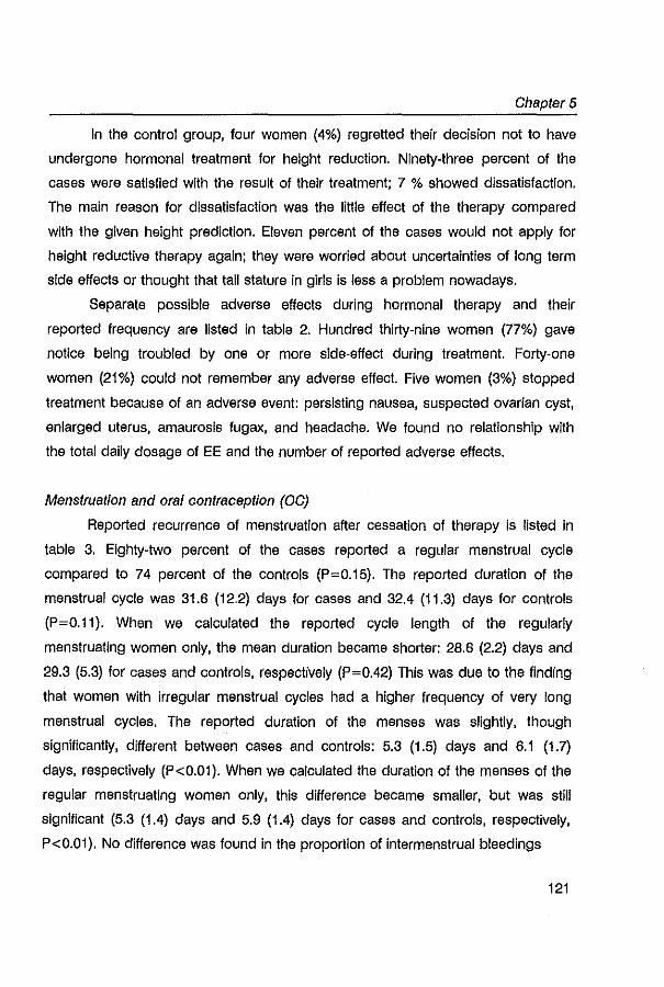
Chapter 5
In the control group, four women (4%) regretted their decision not to have
undergone hormonal treatment for height reduction. Ninety-three percent of the
cases were satisfied with the result of their treatment; 7 % showed dissatisfaction.
The main reason for dissatisfaction was the little effect of the therapy compared
with the given height prediction. Eleven percent of the cases would not apply for
height reductive therapy again; they were worried about uncertainties of long term
side effects or thought that tall stature in girls is less a problem nowadays.
Separate possible adverse effects during hormonal therapy and their
reported frequency are listed in table 2. Hundred thirty-nine women (77%) gave
notice being troubled by one or more side-effect during treatment. Forty-one
women (21%) could not remember any adverse effect. Five women (3%) stopped
treatment because of an adverse event: persisting nausea, suspected ovarian cyst,
enlarged uterus, amaurosis fugax, and headache. We found no relationship with
the total daily dosage of EE and the number of reported adverse effects.
Menstruation and oral contraception (OC)
Reported recurrence of menstruation after cessation of therapy is listed in
table 3. Eighty-two percent of the cases reported a regular menstrual cycle
compared to 74 percent of the controls (p~O.15). The reported duration of the
menstrual cycle was 31.6 (12.2) days for cases and 32.4 (11.3) days for controls
(P~O.ll). When we calculated the reported cycle length of the regularly
menstruating women only, the mean duration became shorter: 28.6 (2.2) days and
29.3 (5.3) for cases and controls, respectively (P~0.42) This was due to the finding
that women with irregular menstrual cycles had a higher frequency of very long
menstrual cycles. The reported duration of the menses was slightly, though
significantly, different between cases and controls: 5.3 (1.5) days and 6.1 (1.7)
days, respectively (P<O.Ol). When we calculated the duration of the menses of the
regular menstruating women only, this difference became smaller, but was still
significant (5.3 (1.4) days and 5.9 (1.4) days for cases and controls, respectively,
P<O.Ol). No difference was found in the proportion of intermenstrual bleedings
121

Chapter 5
between regularly menstruating cases and controls.
The use of OC's and the proportion of reported side effects was not
significantly different between cases and controls. The separate possible adverse
effects during OC use and their reported frequency in cases and controls are listed
in Table 2. There was no significant difference in the distribution of each of the
separate reported adverse effects between the groups (P-values> 0.05).
Table 3.
Pregnancy
Reported recurrence of menstruation after cessation of therapy in 180 treated constitutionally tall women.
Recurrence
After 1 month After 1 - 6 months After 6 months - 1 year More than 1 year Unknown (went unto oral contraceptives) Cannot remember
Number
114 51 5 4 3 3
Details and outcome of 100 pregnancies are listed in table 4. There was no
significant difference between cases and controls regarding the distribution of the
number of miscarriages, the time before realisation of pregnancy and the number
of pregnancies after fertility induction.
Fifty-seven cases and 20 controls had ever consulted a gynaecologist for
various reasons, the proportions being not significantly different (P=0.11).
Subfertility, gynaecological infections and menstrual cycle disturbances were most
frequently reported as the reason for consultation. Eleven cases and one control
consulted a gynaecologist for undesired childlessness. This difference appeared to
be not statistically significant between the groups (P=0.07). The various causes for
subfertility In these women were endometriosis, uterine- or tubal abnormalities,
hyperprolactinaemla, myoma uteri and ovalutory dysfunction. Of the 12 women, five
123
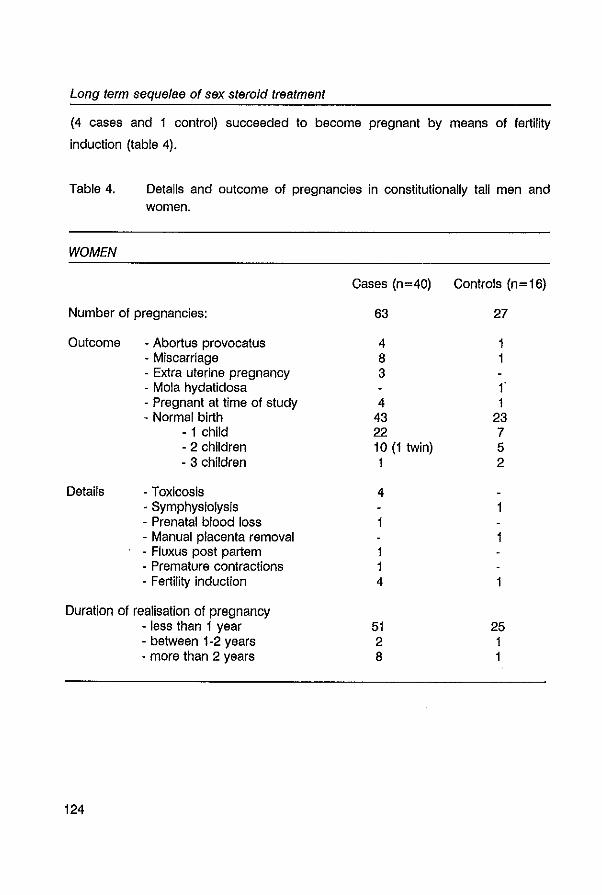
Long term sequelae of sex steroid treatment
(4 cases and 1 control) succeeded to become pregnant by means of fertility
induction (table 4).
Table 4. Details and outcome of pregnancies in constitutionally tall men and women.
WOMEN
Cases (n=40) Controls (n=16)
Number of pregnancies: 63 27
Outcome - Abortus provocatus 4 1 - Miscarriage 8 1 · Extra uterine pregnancy 3 - Mola hydatidosa f - Pregnant at time of study 4 1 - Normal birth 43 23
- 1 child 22 7 - 2 children 10 (1 twin) 5 - 3 children 1 2
Details - Toxicosis 4 - Symphysiolysis 1 - Prenatal blood loss - Manual placenta removal · Fluxus post partem 1 · Premature contractions 1 - Fertility induction 4
Duration of realisation of pregnancy - less than 1 year 51 25 - between 1-2 years 2 1 • more than 2 years 8 1
124
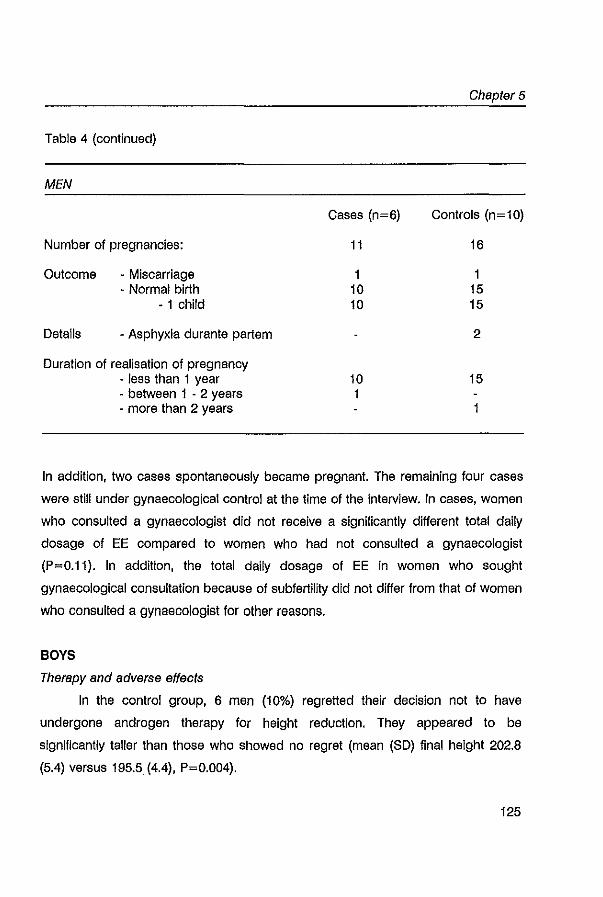
Chapter 5
Table 4 (continued)
MEN
Cases (n=6) Controls (n=10)
Number of pregnancies: 11 16
Outcome • Miscarriage 1 1 • Normal birth 10 15
• 1 child 10 15
Details · Asphyxia durante partem 2
Duration of realisation of pregnancy · less than 1 year 10 15 · between 1 . 2 years 1 · more than 2 years 1
In addition, two cases spontaneously became pregnant. The remaining four cases
were stili under gynaecological control at the time of the interview. In cases, women
who consulted a gynaecologist did not receive a significantly different total daily
dosage of EE compared to women who had not consulted a gynaecologist
(P=0.11). In addition, the total daily dosage of EE in women who sought
gynaecological consultation because of subfertility did not differ from that of women
who consulted a gynaecologist for other reasons.
BOYS
Therapy and adverse effects
In the control group, 6 men (10%) regretted their decision not to have
undergone androgen therapy for height reduction. They appeared to be
significantly talier than those who showed no regret (mean (SO) final height 202.8
(5.4) versus 195.5. (4.4), P=0.004).
125
Long term sequelae of sex steroid treatment
Eighty-six percent of the cases were satisfied with the result of their
treatment; 14 % showed dissatisfaction. The main reason for dissatisfaction was the
little effect of the therapy compared with the given height prediction. Eight percent
would not apply for height reductive therapy again. because of treatment
discomfort or uncertainty of long term side effects.
Fifty men (78%) gave notice being troubled by one or more side-effect
during treatment. Forteen men (22%) could not remember any adverse effect.
Aggravation of acne was most frequently reported (39%). followed by painfulness
of the injection (16%). weight gain (14%). gynaecomastia (13%). muscle ache
(13%). oedema (9%) and change in psychological or sexual behavior (5%).
Pregnancy
Details and outcome of 27 pregnancies are listed in table 4. Men who had
no children yet also had no wish for children at the time of the interview. There was
no significant difference between cases and controls regarding outcome of
pregnancy and the time before realisation of pregnancy.
DISCUSSION
Long term follow-up data of sex steroid therapy of constitutionally tall
adolescents are lacking. In order to obtain more Insight in possible long term side
effects we performed a non-invasive controlled study and focussed on the
functioning of the hypothalamic-gonadal axis. In spite of the non-invasive character
of our study. cases were more willing to participate in this follow-up study than
controls. It is possible that this may have biased the results. Cases would probably
have more linked particular conditions with their past hormonal treatment what may
have lead to over-reporting. Therefore results must be interpreted with some
reserve.
126

Chapter 5
Our study showed, that many patients experienced one or more side effects
during therapy. Fortunately, most of them were mild and led to cess<ltion of
therapy in only 3% of the women. In men, therapy was not discontinued because
of adverse effects. Our findings are in concert and additional to the side effects
reported by others (1-10,17-21).
In girls, adverse effects of estrogens occurred more frequently during
therapy than during OC use. This reflects a dose dependent effect of estrogens on
the incidence of adverse events. The scala of reported side effects reported during
OC use was not significantly different between cases and controls and was of the
same order as reported in the literature (24,25). Two cases reported thrombosis
during OC use. Despite the fact that the risk of thromboembolism increases with
the estrogen dose (26), we did not find thrombosis during therapy. One girl who
had a history of amaurosis fugax might have had thromboembolism. However, no
haemostatic changes were found at that time. In general, thrombosis is found to be
an uncommon side effect of height reductive therapy (1,17,18); whenever
thrombosis ocurred it mostly coincided with other risk factors for thromboembolism
such as immobilisation. Amenorrhoea of longer than 6 months after cessation of
height reductive therapy was reported in about 5 %. The incidence of amenorrhoea
following cessation of pill intake Is about 0.5% (27). In addition, the overall
prevalence of secondary amenorrhoea of more than 6 months in women aged 15 -
34 is about 1.3% (28). This may suggest an increase of amenorrhoea after height
reductive therapy. It should be noted however, that there are no convincing data
that OC use is causally related to amenorrhoea and that other risk factors for
amenorrhoea, such as smoking, nutrition and exercise, were not adequately
adressed (27,29). We found no important differences in menstrual cycle
characteristics between cases and controls. This implies that no important
hormonal imbalance is present in previously treated tall women. Malignancy was
not reported in our study. The possibility of a dose· dependent effect' and a
relationship with OC use at a young age and duration of OC use with increased
risks on breast cancer (30) raises the need for long term follow-up in patients
127

Long term sequelae of sex steroid treatment
treated with pharmacological doses of estrogens. In our study, only 49 of the 180
cases were older than 30 years at time of the follow-up. Therefore, our mean
follow-up period in women of almost 11 years is still too short to give any answer
to this important issue.
In boys, aggravation of acne was by far the most reported side effect. This is
in agreement with others (3,5). A causal relationship with androgen therapy is likely,
as shown by Fyrand et al (31). Gynaecomastia occurred in 13 % of the cases,
probably due to the Increased peripheral aromatization of androgens into
estrogens. Since gynaecomastia is rather prevalent in population studies in boys
(32), it is hard to say whether this condition is increased. In one patient, surgical
correction was necessary several years after cessation of therapy.
Thus far, only casuistic data have been available on successful pregnancies
after height reductive therapy (1-4,6,8,9). In our follow-up study information about a
total of 127 pregnancies was obtained and revealed no distinct differences in
details and outcome of pregnancies between treated women and men and
controls. These results indicate, that long term effects of high doses of sex
hormones on fertility are unlikely. It is tempting to draw definite conclusions at this
time, but one should not forget the social trend to have children at a later age and
the fact that the control group in men was significantly younger. In addition, the
finding in women that a higher, though not significantly, proportion of cases had
sought gynaecological consultation because of subfertility should not be ignored.
However, this latter finding is hard to interpret. There is always the possibility of
bias by over-reporting In cases, as discussed above. Furthermore, additional
information about the causes for subfertility in these women showed no clear
systematical "pattern", indicating that a certain pathophysiological mechanism is
unlikely. And finally, the prevalence of infertility in the normal population is in the
same order of magnitude (10-25%, depending on the definition used (33)).
Besides, the meaning of significantly higher plasma levels of FSH In previously
treated tall adult men In the presence of normal sperm quality, normal plasma
testosterone levels and normal testes volume, remains to be established (23).
128
Chapter 5
Therefore, definite conclusions on the influence of pharmacological doses of sex
hormones on fertility cannot be drawn yet. But for now, our data are reassuring
since there is no clear evidence that treatment with high doses of sex steroids does
induce any harmful effect on reproductive function In man or woman.
In summary, treatment of tall adolescent boys and girls with high doses of
sex hormones In order to reduce their final height is satisfying to most of the
patients, despite the fact that many patients experience side·effects during therapy.
Most side-effects during therapy are mild. The incidence of side-effects is probably
dose-dependent. Post-treatment amenorrhoea of longer than 6 months was found
in 5% of the women. No important differences in menstrual cycle characteristics
were found between cases and controls. Information about a total of 127
pregnancies was obtained and revealed no distinct differences in details and
outcome between treated men and women and controls. At a mean follow-up
period of 10 years, there is no evidence that pharmacological doses of sex
hormones may have a long-term effect on reproductive function or cancer risks.
However, this period is still too short to draw definite conclusions.
ACKNOWLEDGEMENTS
We kindly thank the Foundation for Children's Stamps, Leiden, The
Netherlands for their generous gift to support this study. Dr. B.C.J.M. Fauser,
gynaecologist, is greatly acknowledged for his criticial comments. We thank Frank
Bekkering, Eveline van Gurp, and Paulien Thoolens, medical students, for their help
in collecting the data.
129

Long term sequelae of sex steroid treatment
REFERENCES
1. Wettenhall NH, Cahill C, Roche AF: Tall girls: A sUivey of 15 years of management and treatment. J Pediatr 1975;86:602·610
2. Pradar A. Zachmann M: Treatment of excessively tall girls and boys with sex hormones. Pediatrics 1978;62: 1202-1210
3, Crawford JD: Treatment of tall girls with estrogen. Pediatrics 1978;62:1189·1195
4. Andersen H, Jacobsen BB, Kastrup !<iN, Krabbe S, peitersen B, Petersen KE, Thamdrup E, Wichmann R: Treatment of girls with excessive height prediction. Acta Paediatr SCBnd
1980;69:293-297 5. Bramswlg JH, Schellong G, Borger HJ, Breu H: Testosteron-Therapie hochwOchslger
Jungen. Dtsch Med Wschr 1981;106:1656-1661 6. Stover B, Kollmann F: Ergebnisse der Behandlung hochOchslger Madchen mtt conjuglerten
Ostrogenen. Monatsschr Klnderhellkd 1982;130:36-40 7. Bramswlg JH, Lengerke v. HJ, Schmidt H, Schellong G: The results of short-term (6
months) high-dose testosterone treatment on bone age and adult height in boys of excessively tall stature. Eur J Pediatr 1988;148:104-106
8. GrOters A, Heidemann p, SchlUter H, Stubbe p, Weber B, Helge H: Effect of different oestrogen doses on final height reduction In girls with constitutional tall stature. Eur J Pedlatr 1989; 149: 11-13
9. Normann EK, Trygstad 0, Larsen S, Dahl-Jorgensen K: Height reduction in 539 tall girls treated with three different dosages of ethinyloeslradlol. Arch Dis Child 1991;66:1271-1278
10. Joss EE, Zeuner J, ZurbrOgg RP, Mullis PE: Impact of different doses of ethinyl oestradiol on reduction of final height In constttutionally tall girls. Eur J Pedlatr 1994;153:797-801
11. Muntean W, Borkensteln M: Hemostatic changes in tali girls trealed wtth high doses of ethlnylestradlol. Eur J Pedlatr 1980;134:245-248
12. Hinkel GK, Hanefeld M, Jaross W, Leonhardt W, TrObsbach A: Effect of high doses of oestrogens and androgens on lipoproteins: observations In the treatment of excessive growth with sexual hormones. Exp Clin EndocrinoI1985;86:17-25
13. Hanker JP, Schellong G, Schneider HPG: The functional state of the hypothalamo-pttuitary axis after high dose oeslrogen therapy in excessively tall girls. Acta Endocrinol 1979; 91: 19-29
14. Bramswig JH, Nieschlag E, Schellong G: Pituttary-gonadal function in boys after high dose testosterone treatment for excessively tall stature. Acta EndocrinoI1984;107:97-103
15. Grimes DA: The safety of oral contraceptives: epidemiologic Insights from the first 30
years. Am J Obstet GynecoI1992;166:1950-1954 16. KaunHz AM: Oral conlraceptives and gynecologic cancer: an update for the 1990s. Am J
Obstet GynecoI1992;167:1171-1176 17. Trygstad 0: Oestrogen treatment of adolescent tall girls; short term side effects. Acta
Endocrinol 1986;(SuppQ279: 170-173 18. Werder EA, Waibel P, Sege D, Flury R: Severe thrombosis during oeslrogen Ireatment for
tall stalure. Eur J Pediatr 1990; 149:389-390 19. Panteon E, Loumaye E, Maes M, Malvaux P: Occurrence of prolactinoma after estrogen
treatment In a girl wtth constitutionally tali stature. J Pedlatr 1988;113:337-339
130
Chapter 5
20. Traupe H, MOhlendahl von KE, Bramswig JH, Happle A: Acne of the fulminans type following testosterone therapy In three excessively tall boys. Arch Dermatol 1988; 124:414· 417
21. Commentz JC, Conrad C, Stahnke N, Willig AP: Priapism during testosterone treatment for reducing final height In tall stature. Harm Aes 1994:41 :103
22. Willig AP, Bettendort M, Hinkel GK, Schwarz HP, Schulze W: Androgen treatment of tall stature during puberty may reduce sperm quality In adult life. Harm Aes 1992;37 (Suppl.4) :3
23. De Waal WJ, Vreeburg JTM, Bekkering F, de Jong JH, de Muinck Kelzer-Schrama SMPF, Drop SLS, Weber AFA: High dose testosterone therapy for reduction of final height In constitutionally tall boys: does it Influence testicular function in adutthood? Clln Endocrlnol 1995;43:87-95
24. Hillard PJA: The patient's reaction to side effects of oral contraceptives. Am J Obstet GynaecoI1989;161:1412-1415
25. Akerlund M, Aode A: Comparative profiles of reliability, cycle control and side effects of two oral contraceptive formulations containing 150 ~g desogestrel and either 30 ~g or 20 ~g ethlnyloestradlol. Br J Obstet GynaecoI1993;100:832-838
26. Iman WHW, Vessey MP, Westerholm B, at al: Thromboembolic disease and the'steroldal content of oral contraceptives: a report to the committee on safety of drugs. Br Mad J 1970;2:203-209
27. Hull MGA, Bromham DA, Savage PE, Barlow TM, Hughes AO, Jacobs HS: Post-pili amenorrhea: a causal study. Fertll Steril1981;36:472-476
28, MOnster K. Schmidt L, Helm P: Secondary amenorrhoea: prevalence and medIcal contact-a cross-sectional study from a Danish county. Br J Obstet GynaecoI1992;99:430-433
29. Gold EB, Bush T, Chee E: Alsk factors for secondary amenorrhea and galactorrhea. Int J Fertil1994;39:177-184
30. Rushton L, Jones DR: Oral contraceptive use and breast cancer risk: a meta-analyses of variations with age at diagnosis, partly and total duration of oral contraceptive use. Br J Obstet Gynaecol 1992;99:239-246
31. Fyrand 0, Fiskaadal HJ, Trygstad 0: Acne in pubertal boys undergoing treatment with androgans. Acta Derm VanareoI1992;72:148-149
32. Braunstein GO: Gynecomastia. N Engl J Med 1993;328:490·495 33. Greenhall E, Vessey M: The prevalence of subfertllity: a review of the current confusion
and a report of two new studies. Fertil Steril1990;54:978-983
131


CHAPTER 6
High dose testosterone therapy for reduction of final
height In constitutionally tall boys:
does It Influence testicular function In adulthood?
W. J. de Waal', J.T.M. Vreeburg', F. 8ekkering', F. H. de Jong',
S.M.P.F. de Muinck Keizer-Schrama', S.L.S. Drop', R.FA Weber'·'
1 Department of Pediatrics, Division of Endocrinology, Sophia Children's Hospital,
Erasmus University Rotterdam, 2Department of Endocrinology & Reproduction,
Erasmus University Rotterdam, "Department of Andro/ogy, University
Hospital/Erasmus University Rotterdam, The Netherlands.
Clinical Endocrinology 1995;43:87-95
Testioular funotion in treated tall boys
ABSTRACT
We have studied the effect of treatment with high doses of androgens during
puberty on testicular function in adult men with constitutionally tall stature, taking
into account confounding factors interfering with sperm quality, since existing
published data do not include whether testicular function is impaired by such
treatment. Forty three previously androgen treated tall men (cases) and thirty non
treated tall men (controls) agreed to participate In our study. The study work-up
comprised physical examination, semen analysis and measurements of plasma
hormone levels (LH, FSH, testosterone (T), sex steroid binding globulin (SHBG)
and inhlbln). It appeared that sperm quality and testis volume were comparable
between cases and controls. Mean sperm concentration was 66.4 x 10'lml in cases
and 66.2 x 10'lml in controls. A left-sided varicocele was found in 45% of the oases
and 37% of the controls. In cases we observed a significant effect of the' age at
start of androgen therapy on sperm motility (regr. coeff. (SE): 4.92 (2.41%),
P=0.048). In addition, testis size at start of therapy had a significant effect on
sperm concentration (regr. coeff. (SE): 5.57 (1.54 x 10'lml), P=0.0012) and on total
sperm count (regr. coeff. (SE): 43.1 (7.73 x 10~, P=O.OOOI). Plasma levels of T,
SHBG and Inhibin were not statistically different between both groups. Cases had
significantly higher FSH levels (mean (SD): 3.3 (2.2) versus 2.1 (0.8) lUll, P=0.004)
and significantly lower LH levels (mean (SD): 2.3 (0.9) versus 3.1 (1.4) lUll,
P=0.019). We found a significant effect of the age at start of therapy on plasma
FSH level In the treated men (regr. coeff. (SE): -0.73 (0.18 lUll), P=0.0003j. In
conclusion, treatment with high doses of androgens for reduction of final height in
constitutionally tall stature has no long-term side-effect on sperm quality, testicular
volume and plasma testosterone levels. However, treated men had significantly
higher plasma levels of FSH compared with controls. The meaning' of this
difference remains to be established. Varicocele in adult tall men Is present in 42%.
134
Chapter 6
INTRODUCTION
Treatment of tall adolescent boys with high doses of testosterone is a widely
used method to reduce their final height (1,2,3.4). This therapy induces
suppression of the hypothalamic-pituitary-gonadal axis (1,3). In adult men, low
levels of gonadotropins caused by high doses of androgens are known to reduce
sperm production to oligospermia or azoospermia (5). Contraceptive studies have
shown that androgen-induced suppression of spermatogenesis is reversible in
adult men (6,7). However, extrapolation of these data to the management of tall
stature in pubertal boys must be viewed with caution since factors which regulates
spermatogenesis in the normally functioning adult testis may not be the same as
during puberty (8). Androgen therapy in tall boys is usually initiated with the first
signs of puberty and it is in this peripubertal period that important maturational
processes are taking place in the testis (8-13). Influenced by complex hormonal
actions, these maturational processes eventually lead to initiation of
spermatogenesis. Onset of spermatogenesis (spermarche) as detected by urine
analysis (spermaturia) appears to be an early pubertal event: the median age of
spermarche has been estimated to be 13-14 years (14-17). In addition, it is
noteworthy that administration of testosterone esters at high doses can cause
morphological and cytological changes, as shown in rat and human adult testes
(18-20).
It has not been known whether the suppressive effects of high dose
testosterone therapy on maturing gonads in puberty may affect pituitary-gonadal
functioning in adulthood. Bramswig et al (1) demonstrated normalization of the
gonadotropin levels in 100 tall boys after discontinuation of treatment with follow-up
periods up to 48 months, although transient hypergonadotropic LH- and FSH
secretory patterns occurred. Zachmann, Prader and co-workers (3,4) briefly
reported that testicular volume and sperm count were normal 1.5 years after
discontinuation of treatment, although in individual cases recovery periods up to
five years had been observed. In contrast, Willig et al (21,22) recently reported
135
Testicular function in treated tall boys
reduced sperm concentration in treated tall men compared to untreated controls
and postulated the possibility of long-term suppresive effects of high doses of
androgens on reproductive function. Unfortunately, however, none of the latter
reports corrected for confounding factors interfering with parameters of sperm
quality. It has been well established that varicocele {23-26}, smoking {27} , sexually
transmitted diseases {28} and cryptorchism {29} are likely to affect sperm quality
and/or plasma hormone levels. The purpose of this study was to investigate
whether treatment with high doses of androgens in tall boys during puberty may
induce long-term effects on pituitary-gonadal functioning while taking into account
interfering confounding factors.
MATERIALS AND METHODS
Patients
Two hundred and forty-seven adult men, who were seen at adolescence for
evaluation of tall stature in the Sophia Children'S Hospital, Rotterdam, were
contacted by mail to participate in an andrological follow-up study. Of the 247 men,
102 had been treated for their tall stature during puberty {cases}. The 145 non
treated men served as controls. Ninety-five out of 102 cases responded to our
mailing {response rate {RR}:93%} and 43 out of 95 {45% of the resppnders}
participated in the andrological study. For controls, the RR was 79% {114/145} and
32 out of 114 participated in this study {28% of the responders}. All participants
gave informed consent.
Androgen therapy started at a mean {SO} age of 14.4 {1.2} years with a
range of 12.1 to 17.2 years. Pubertal stages {according to Tanner (55» and
testicular volume {mean of the two testes} at the time of start of treatment were
recorded. In 9 cases, testicular volume could not be retrieved {Table I}. The
duration {SO} of therapy was 17.1 {7.2} months, ranging from 8.7 to 44.2 months.
Most patients used intramuscular preparations of testosterone ester mixtures
136
Chapter 6
(Sustanon '250'" =testosterone propionate, -fenylproplonate, -isohexanoate and -
decanoate) 250 mg every week (n=32), 250 mg every two weeks (n=2) or 500 mg
every two weeks (n=4). Three patients used an oral testosterone ester (Andriol"
=testosterone undecanoate) 240-320 mg per day; one of these patients switched
to Sustanon '250'", 500 mg every two weeks after 8 months. Two patients used
daily injections of the testosterone ester testosterone propionate (Neo-hombreol")
in a dosage of 30 mg per day for at least one year after which they switched to
Sustanon '250'", 250 mg every two weeks. The mean (SO) age at follow-up was
24.2 (4.1) years (range 18.2-33.0 years), i.e. a mean (SO) follow-up period of 8.3
(3.9) years (range 2.8-17.2 years) after discontinuation of treatment. Data on the
growth reductive efficacy of the androgen therapy will be published elsewhere.
Two controls were excluded from the study because of known testicular
impairment. Mean (SO) age at follow-up of the 30 control subjects was 26.4 (4.4)
years with a range from 19.2 to 34.0 years.
All participants were called in twice at the outpatient clinic of the department
of Andrology. Clinical assessment at the first visit comprised medical history and
physical examination. Height was measured by a Harpenden Stadiometer.
Testicular volume was estimated using Prader's orchidometer (30). The diagnosis
of varicocele was made by palpation (all subjects) and/or Doppler measurement
(Doppler stethoscope: 8.2 MHz blood flow detector) (67/73 patients). We graded
the varicocele as follows: grade 0: subclinical varicocele only detectable by Doppler
stethoscope, grade 1: varicocele detectable by palpation with Vasalva's
manoeuvre, grade 2: varicocele detectable by palpation but not visible, and grade
3: visible varicocele. In addition, blood was taken for determination of plasma
hormones and the first semen sample was obtained. A second semen sample was
obtained during the last visit.
The study protocol was approved by the ethical committee of Erasmus
University Hospital, Rotterdam.
137
Testicular function in treated tall boys
Semen analysis
All semen samples were collected after a period of sexual abstinence of at
least three days. Semen was obtained at the hospital by masturbation. The mean
(SO) time interval between the two semen samples was 1.4 (0.5) months (range
0.9-3.0 months).
Semen analysis was performed within 30 minutes after ejaculation according
to standard World Health Organisation (WHO) procedures (31). Volume, sperm
concentration, total sperm count, motility and morphology were assessed. Sperm
concentration was separately determined by two experienced technicians using the
Makler chamber (32). The mean of the two counts was used, provided that the
difference between the two counts did not exceed 20% of the mean; otherwise, the
counting was repeated. In 108 samples (26 controls and 28 treated men) the
concentration of sperm was also determined using the Improved Neubauer
haemocyfometer as described in the WHO manual (31). Sperm motility, calculated
as the percentage of progressive motile spermatozoa (classes a + b, WHO manual
(31)), was determined by the same technicians in all semen samples. If the
difference between the two observations exceeded 20%, motility assessment was
repeated. Sperm morphology was evaluated after Diff-Quick staining using strict
criteria (33).
Hormone measurements
LH and FSH were measured using a commercially available enhanced
luminescence immunometric method (Kodak Clinical Diagnostics, Amersham, UK).
The intra- and interassay variability were < 6.2% and < 12.6% for LH and < 5.2%
and < 12.7% for FSH, respectively. Testosterone (T) was measured by radio
immunoassay, using the antiserum described previously by Verjans et al (34). The
Intra- and interassay variability were < 9.2% and < 10.0%, respectively. Sex
hormone binding globulin (SHBG) was measured using a commercially available
immunoradiometric assay (Diagnostic Products Corporation, Los Angeles, USA).
The Intra- and interassay variability of this kit were < 5.3% and < 10.8%,
138
Chapter 6
respectively. The ratio of T to SHBG was calculated In order to obtain an estimate
of the free androgen fraction (FAI). Inhibin was measured by radioimmunoassay as
described by Robertson et al (35), using an antiserum against purified 32 kDa
bovine follicular fluid (bFF) inhibin and Iodinated 32 kDa bFF Inhibin. All samples
were measured in one assay. The intra-assay variability was 16.6%. This relatively
high value was likely to be caused by the fact that all concentrations were ·read in
the lower range of the standard curve for which CV values of 12% were reported
by others using the same assay (36).
Statistical analysis
The agreement of the sperm concentration as determined by the Makler
chamber and by the improved Neubauer haemocytometer was evaluated using the
method described by Bland & Altman (37). The sperm concentration as measured
by the Makler chamber was used for further statistical analysis. For parameters of
sperm quality, the mean of the two semen samples was used for statistical
analysis; In two previously treated tall men only one semen sample was obtained
and used for statistical analysis. Differences between cases and controls were
tested with Wilcoxon's two sample test. Cases were divided into five different
groups according to the therapeutic regimen used: group 1 (testosterone esters
250 mg/week, n=32), group 2 (testosterone esters 250 mg/2 weeks, n=2), group 3
(testosterone esters 500 mg/2 weeks, n=4), group 4 (testosterone-undecanoate,
n=3), and group 5 (testosterone-propionate, n=2). Differences between these
groups were tested with the Kruskall-Wallis test. Chi-square tests were used in
case of a contingency distribution. Correlations between variables were tested with
Spearman's rank correlation test. Multiple linear regression analysis was used to
correct for confounding factors (analysis of covariance). For this purpose we
created two groups: one group included subjects who had at least one of the
following interfering conditions: varicocele, smoking, cryptorchism or venereal
disease; the other Including subjects with no confounding factor at all. When
correlation analysis between variables showed a significant association, multiple
139
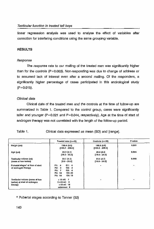
Testicular function in treated tall boys
linear regresssion analysis was used to analyse the effect of variables after
correction for interfering conditions using the same grouping variable.
RESULTS
Response
The response rate to our mailing of the treated men was significantly higher
than for the controls (P=0.003). Non-responding was due to change of address or
to assumed lack of interest even after a second mailing. Of the responders, a
significantly higher percentage of cases participated in this andrological study
(P=0.015).
Clinical data
Clinical data of the treated men and the controls at the time of follow-up are
summarized in Table 1. Compared to the control group, cases were significantly
taller and younger (P=0.021 and P=0.044, respectively). Age at the time of start of
androgen therapy was not correlated with the length of the follow-up period.
Table 1. Clinical data expressed as mean (SD) and [range].
Treated men (n=43) Controls (n=30) P·value
Height (em) 199.4 (4.0) 196.9 (6.0) 0.021 (190.7 . 208.3] [188.3·209.3J
Age (yra) 24.2 (4.1) 26.4 (4.4) 0.044 [18.2.33.0J [19.4 • 34,0]
Testicular volume (ml) 22.1 (4.1) 23.1 (2.7) 0.448 (mean of two testes) [9.5· 25.0J [15.0.25.0J
Pubertalslsges' at lime of start PI: • 01: 4 0' androgen therapy P2: 3 02: 2
P3: 7 03: 4 P4: 18 04: 23 P5: 10 05: 10
Tesllcular volume (mean of two ~ 10ml: 7 tesles) al slart 01 androgen 11-15 ml: • therapy >15 mT: 18
unknown: 9
* pubertal stages according to Tanner (32)
140
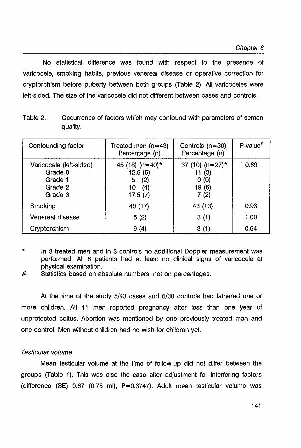
Chapter 6
No statistical difference was found with respect to the presence of
varicocele, smoking habits, previous venereal disease or operative correction for
cryptorchism before puberty between both groups (Table 2). All varicoceles were
left-sided. The size of the varicocele did not different between cases and controls.
Table 2. Occurrence of factors which may confound with parameters of semen quality.
*
#
Confounding factor Treated men (n=43) Controls (n=30) P-value' Percentage (n) Percentage (n)
Varicocele (left-sided) 45 (18) (n=40)* 37 (10) (n=27)* . 0.69 Grade 0 12.5 (5) 11 (3) Grade 1 5 (2) o (0) Grade 2 10 (4) 19 (5) Grade 3 17.5 (7) 7 (2)
Smoking 40 (17) 43 (13) 0.93
Venereal disease 5 (2) 3 (1) 1.00
Cryptorchism 9 (4) 3 (1) 0.64
In 3 treated men and in 3 controls no additional Doppler measurement was performed. All 6 patients had at least no clinical signs of varicocele at physical examination. Statistics based on absolute numbers, not on percentages.
At the time of the study 5/43 cases and 6/30 controls had fathered one or
more children. All 11 men reported pregnancy after less than one year of
unprotected coitus. Abortion was mentioned by one previously treated man and
one control. Men without children had no wish for children yet.
Test/cular volume
Mean testicular volume at the time of follow-up did not differ between the
groups (Table 1). This was also the case after adjustment for interfering factors
(difference (SE) 0.67 (0.75 ml), P=0.3747). Adult mean testicular volume was
141
Testicular function in treated tall boys
moderately correlated with parameters of semen quality (sperm density and
motility) for both cases and controls (r:0.39·0.52, P<0.05). In cases, adult mean
testicular volume was not significantly correlated with either the age at start of
androgen therapy, duration of therapy, length of the follow·up period or testicular
volume at time of start of androgen therapy. However, adult testis size significantly
correlated with the level of plasma FSH in the treated group (r=-0.33, P=0.031).
This association was still significant after adjustment for interfering conditions
(regression coefficient (SE): -0.1 (0.06 lUll), P=0.03).
Semen analysis
For the sperm concentration data, the linear correlation coefficient (r)
between the Makler chamber and the Neubauer haemocytometer was r=0.94
(P<0.001). The mean difference between both methods (Makler minus Ne~bauer)
was -3.9 x 10'/ml. Ninety-five percent of the differences were between -44.1 x 10'/ml
and 36.3 x 10'/ml.
The results of the semen analyses are summarized in Table 3. We found no
significant difference for any of the semen parameters between cases and controls.
Six out of 43 cases and 4 out of 30 controls had oligozoospermia (sperm
concentration less than 20 x 10'/ml, WHO 1992) In both semen samples (NS). An
additional 4 cases and 2 controls showed oligozoospermia in one semen sample.
In cases, we found no significant correlations between parameters of semen quality
and the age at start of treatment, except for sperm motility (r= 0.34; P=0.025).
Duration of treatment and the length of the follow-up period showed no significant
correlation with sperm quality. A significant correlation was found between mean
testicular volume at start of therapy and sperm concentration as well as total sperm
count (r=0.46, P=0.007 and r=0.45, P=0.007, respectively). Age at start of lherapy
was correlated with testicular volume at that time (r=0.34, P=0.048). Semen quality
did not differ statistically between the five therapeutic regimen groups. We did not
observe any significant difference in semen parameters between men with or
without varicocele for both cases and controls.
142

Chapter 6
Analysis of the data after correction for Interfering clinical conditions also
revealed no Significant differences between cases and controls In parameters of
sperm quality or adult testicular volume. In cases, age at start of therapy still had a
significant effect on sperm motility (regression coefficient (SE):4.9 (2.4%), P=0.048).
Adding testicular volume at start of therapy to the model showed no additional
effect on sperm motility. Testicular volume at start of therapy still showed a
significant effect on sperm concentration and on total sperm count (regression
coefficients (SE): 5.6 (1.5 xl0'/ml), P=0.0012 and 34.1 (7.7 xl O'/ml) , P=O.OOOI,
respectively). Adding age at start of treatment to these regression models showed
no additional effect on either sperm concentration or total sperm count. However,
plasma FSH level did show an additional significant effect on sperm concentration
over testis size at start of therapy (regression coefficient (SE):-14.6 (4.4 x 10'/ml),
P=0.0027).
Table 3. Results of semen analysis in 43 treated and 30 untreated tall men. Data expressed as mean (SO) [range].
Perameters of semen quaUty# Trealed lall men (0=43) Controls (0=30) P·value
Volume (mQ 4,3 (1.7) 4.0 (1.1) 0.79 [1.6.7.8J [2.1 .7.3]
sperm Concenlrallon 66.4 (59.f) 66.2 (43.6) 0.56 (10'/mQ (1.5. 315.0J [0.1 • 170.0)
median: 47.5 median: 62.5
Tolel sperm count 290.5 (263.0) 250.8 (157.6) 0.87 (10'/eJIQuhlte) (6.9.960.0] (0.7 • 586.0)
median: 191.3 medIan! 243.6
Motility (%) 44.6 (18.7) 48,0 (fS.f) 0.44 [7.5.80.0J (0.0.80.0]·
Morphology (%) 20.8 (6.5) 18.5 (8,5) 0.21 (6.0.32.0J [0.0 . 34.0)*
# Mean results of two samples * Including two men showing very low sperm concentration «0.1 x 10'/ml) In
which sperm motility and sperm morphology could not be assessed
143
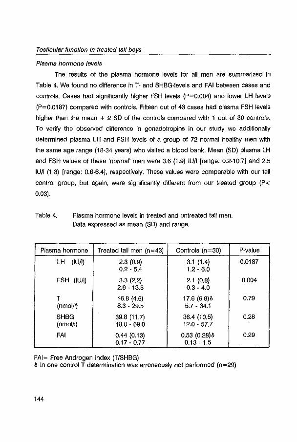
Testicular function in treated tall boys
Plasma hormone levels
The results of the plasma hormone levels for all men are summarized in
Table 4. We found no difference in T- and SHBG-Ievels and FAI between cases and
controls. Cases had significantly higher FSH levels (P=0.004) and lower LH levels
(P=0.0187) compared with controls. Fifteen out of 43 cases had plasma FSH levels
higher than the mean + 2 SD of the controls compared with lout of 30 controls.
To verify the observed difference in gonadotropins in our study we additionally
determined plasma LH and FSH levels of a group of 72 normal healthy men with
the same age range (18-34 years) who visited a blood bank. Mean (SD) plasma LH
and FSH values of these 'normal' men were 3.6 (1.9) lUll [range: 0.2-10.71 and 2.5
lUll (1.3) [range: 0.6·6.41, respectively. These values were comparable with our tali
control group, but again, were significantly different from our treated group (P<
0.03).
Table 4. Plasma hormone levels in treated and untreated tali men. Data expressed as mean (SD) and range.
Plasma hormone Treated tali men (n=43) Controls (n=30)
LH (lUll) 2.3 (0.9) 3.1 (1.4) 0.2 - 5.4 1.2 - 6.0
FSH (lUll) 3.3 (2.2) 2.1 (0.8) 2.6 - 13.5 0.3 - 4.0
T 16.8 (4.6) 17.6 (6.8)& (nmol/I) 8.3 - 29.5 5.7 - 34.1
SHBG 39.8 (11.7) 36.4 {I 0.5) (nmol/l) 18.0 - 69.0 12.0 - 57.7
FAI 0.44 (0.13) 0.53 (0.28)& 0.17 - 0.77 0.13 - 1.5
FAI= Free Androgen Index (T/SHBG) & In one control T determination was erroneously not performed (n=29)
144
P-value
0.0187
0.004
0.79
0.28
0.29
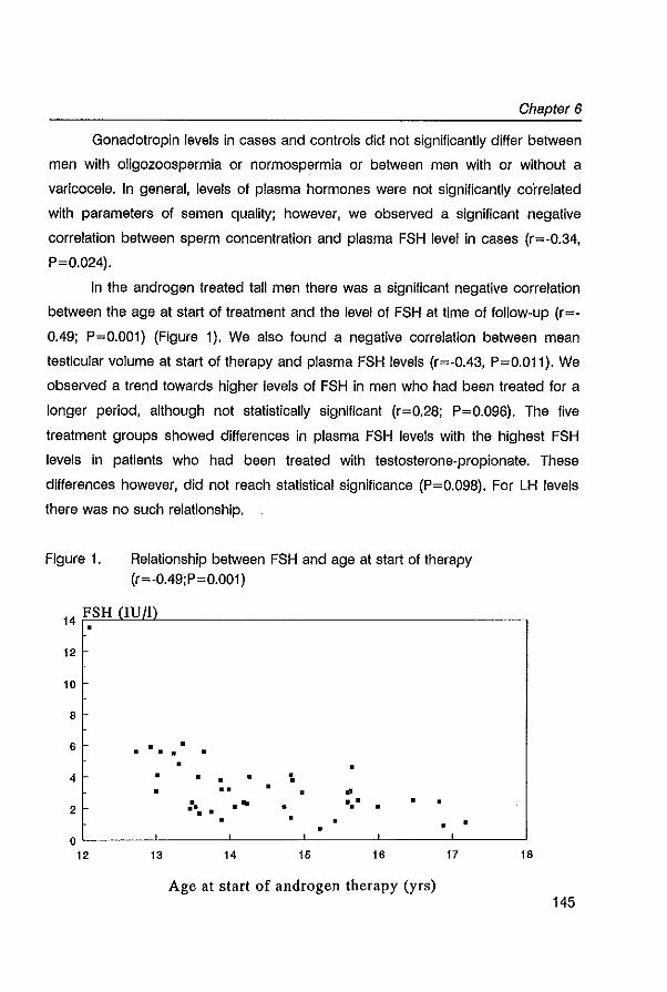
Chapter 6
Gonadotropin levels in cases and controls did not significantly differ between
men with oligozoospermia or normospermia or between men with or without a
varicocele. In general, levels of plasma hormones were not significantly correlated
with parameters of semen quality; however, we observed a significant negative
correlation between sperm concentration and plasma FSH level in cases (r=-0.34,
P=0.024).
In the androgen treated tall men there was a significant negative correlation
between the age at start of treatment and the level of FSH at time of follow-up (r=-
0.49; P=O.OOI) (Figure I). We also found a negative correlation between mean
testicular volume at start of therapy and plasma FSH levels (r=-0.43, P=O.OII). We
observed a trend towards higher levels of FSH in men who had been treated for a
longer period, although not statistically significant (r=0.28; P=0.096). The five
treatment groups showed differences in plasma FSH levels with the highest FSH
levels in patients who had been treated with testosterone-propionate. These
differences however, did not reach statistical significance (P=0.098). For LH levels
there was no such relationship.
Figure I. Relationship between FSH and age at start of therapy (r=-0.49;P=0.001)
14 FSH !lum •
12
10 ~
8
• • • • • • 6
• • • • • • • • • •• • • ..
• ." •• • . ' • • • • • • • •
12 13 14 15 16 17
Age at start of androgen therapy (yrs)
18
145
Testicular function in treated tall boys
The difference (SE) in mean plasma FSH between cases and controls,
corrected for interfering factors, was 1.1 (0.3) lUll, P=0.0014; the difference (SE) in
mean plasma LH was -0.8 (0.3) IU/I, P=0.0071. Testosterone, SHBG and FAI were
not significantly different between cases and controls. We found a significant effect
of the age at start of therapy on plasma FSH level of the treated men (regression
coefficient (SE): -0.7 (0.2 IU/I), P=0.0003) aiter correction for interfering conditions.
Adding testicular volume at start of therapy or testicular size at adulthood to the
regression model showed no significant additional effect over age at start of
therapy on plasma FSH level.
In a subgroup of 30 men (18 cases and 12 controls) we had the opportunity
to measure inhibin levels in saved blood samples. We did not find a significant
difference in inhibin levels between cases and controls in this subgroup (mean (SD)
inhibin levels: 2.10 (0.73 U/ml) and 2.53 (1.27 U/ml) for cases and controls,
respectively, P=0.27). This was also the case after adjustment for confounding
conditions (difference (SE): -0.42 (0.36 U/ml), P=0.26). Inhibin levels did not
significantly correlate with FSH levels.
DISCUSSION
Our study shows that sperm quality in previously androgen treated tall men
is comparable with a control group of untreated tall men. In our control population,
mean sperm concentration as determined by the Makler chamber was 66.2 x
10'/ml. That is in the same range of the normal population as reported recently by
Carlsen et al (38). The considerable variability in semen quality seen in our control
group confirms the work of other studies in 'normal' men (39,40). The fact that
previously treated men showed semen quality comparable to controls, even after
correction for possible interfering conditions, implies that severe long-term
complications on spermatogenesis are unlikely, at least after a mean follow-up
period of 8 years.
146
Chapter 6
The selection of our population may have biased the results; treated tall men
were more willing to participate in this follow-up study than untreated tall men.
However, none of the men participated because of suspected infertility. Therefore,
we have no reason to believe that a structural bias has occurred.
Our findings on sperm quality are in agreement with the experiences briefly
reported by Prader et al (3,4). They found that most of their tall patients had
normospermia (sperm counts more than 20 x 10'/ml) between a few months and a
few years after treatment. In addition, studies in hypogonadotrophic hypogonadism
of prepubertal onset have shown that androgen treatment for viralization Induction
did not impair subsequent spermatogenesis (41,42) although one may argue that
the recommended dose for this replacement therapy is much less. Our results are
in contrast with the findings of Willig and co-workers (21,22), who found reduced
sperm concentration in previously treated tall men compared to controls. Their
control group, however, showed a mean value of sperm concentration of 120.2 ±
111.9 x 10'/ml. Their treated group showed a mean sperm concentration of 63.4 ±
50.6 x 10'/ml, which is comparable with the normal population (38) and with our
study. In an attempt to stress the possibility of unwanted long-term side effects of
androgen treatment, Willig et al (22) subsequently reported that 16/60 of their
patients showed abnormal sperm quality (considering not only sperm concentration
but also sperm morphology and motility) in contrast to 2/20 of their controls. In our
opinion this difference is not statistically significant (Fisher's exact test: P>0.10).
Moreover, it is known that even in normal fertile men abnormal semen results can
be found in a large proportion of samples (39). It Is possible that differences in
patient selection, semen analysis methodology and treatment regimens may
account for the observed differences. In addition, the extent In which interfering
conditions are present may cause important bias as well.
We found an overall prevalence of varicocele of 42% (12% subclinical and
30% clinical). It is difficult to interpret these figures as there is no consensus about
the definition of a varicocele and various epidemiological studies have used
different methods to diagnose this condition (42). Nevertheless, our finding of
147
Testicular function in treated tall boys
clinical varicocele is higher than reported in the normal population (12.4-25.8 %)
but more or less in the same range as in men being evaluated for infertility (19-
41%) (43,44). A relation with androgen treatment is unlikely since no difference in
prevalence of varicocele was observed between cases and controls. One could
speculate about the impact of stature on the pathogenesis of varicocele as our
study subjects have heights in the upper range of the normal Dutch population.
We observed different levels of gonadotropins in previously treated tall men
compared to controls (tall and 'normal' men). How can this observation be
explained considering the comparable semen output? In the management of tall
stature. androgen treatment usually starts after the onset of puberty. It is known
that spermarche Is an early pubertal event that can occur without other signs of
puberty and with small testicular volume (15,45,46). In the pubertal period. the
complex maturational processes that take place within the gonads are not fully
completed (9.12,47). In our study grouP. the mean age at the start of treatment
was 14.4 years. At that time about 15% of the patients had a pubertal Tanner stage
less than G3 and/or less than P3. In addition. 21 % of the boys had a mean
testicular volume " 10 ml. This means that supraphysiological doses of androgens
have been given in a period of testicular maturation and maybe even prior to
spermarche. Theoretically. these high levels of exogenous androgens may have
influenced intratesticular maturational processes directly or via the suppression of
gonadotropins. In fact. it is known that FSH plays an important role in the initiation
of spermatogenesis by effects on Sertoli cell replication and function. the regulation
of the replication of differentiated spermatogonia. and on the degeneration process
of germ cells (reviewed in: Sharpe. 1994 (8)). In addition. morphological and
cytological changes have been described after androgen administration in adult
testes of rat and human (18.19.20). and it has not extensively been studied whether
these changes are reversible. Obviously. our study gives no insight into the nature
of the dynamic processes during androgen therapy or into the presence of any
intratesticular. morphological or histological changes. It is possible. however. that
the higher levels of FSH reflect intratesticular changes (48). due to androgen
148
Chapter 6
treatment received in a period of maturation. These Increased FSH levels may
compensate for partially disturbed germinal function in order to maintain normal
sperm quality (49). On the other hand, the difference in gonadotropins may also
reflect a change in responsiveness at the hypothalamic-pituitary level. It is known
that steroids exert negative feedback effects at the level of the hypothalamus as
well as the pituitary (50,51). At least part of this negative feedback is mediated by
aromatization of testosterone to oestradiol (52). Animal models show regulating
effects of oestradiol on neuronal development (53). Although conclusive data are
lacking it may be possible that the high levels of steroids during puberty have
induced alterations at the hypothalamic-pituitary level. In an attempt to get more
insight into the underlying mechanism we determined inhibin levels in a small
subgroup of 30 subjects. We found no significant difference in inhibin levels
between these groups and observed no significant inverse relationship between
inhibin and FSH concentration. Therefore the exact pathophysiological mechanism
explaining the observed differences in gonadotropins remains to be verified.
One might surmise that treatment started at an early pubertal stage has
more effect on testicular function than treatment started at a later point. Some of
our findings are in concert with this theory as we observed a significant effect of
the age at start of therapy on both the level of FSH and sperm motility. In addition,
testicular size at start of treatment significantly influenced sperm concentration and
total sperm count. On the other hand, semen quality and plasma hormone levels
were not significantly different between the pubertal stages (Tanner stages P and
G) at the start of therapy (data not shown).
Treatment with high doses of androgens induces reduction in testicular
volume in adult men (5) as well as In tall adolescent boys (3.4,54). This implies
major intratesticular changes during therapy such as a decrease in seminiferous
tubule size (18,19). These processes are likely to be reversible as testicular volume
normalizes after discontinuation of therapy (3-5,54). In our study, there was no
difference in mean testicular volume between cases and controls. This is in contrast
with the observations of Willig et al (21,22), who reported significantly smaller
149

Testioular funotion in treated tall boys
testicular sizes in previously treated men.
In summary, our study shows that treatment with high doses of androgens
for reduction of final height In constitutionally tall stature has no long-term side
effeot on sperm quality, testicular volume and plasma testosterone levels. However,
treated men had higher plasma FSH levels compared with controls; the meaning of
this finding remains to be established. A lett-sided varicocele was prevalent in 42%
of the tall men.
ACKNOWLEDGEMENTS
This work was supported by a grant from the Foundation of Human
Reproduction, Rotterdam, The Netherlands. It was presented In part at the Annual
Meeting of the European Society for Paediatric Endocrinology, Maastricht, The
Netherlands, June 1994. We would like to thank Mrs. Marijke Kluit for the
assessment of the semen analysis and Mr. Bert Bierings and Mr. Ronald van der
Wal for the determination of the plasma hormones. We thank Dr. Th. Stijnen for
statistical advice.
150

Chapter 6
REFERENCES
1. Bramswlg JH, Nleschlag E, Schellong G: Phulta'Y'gonadal function In boys after high dose testosterone treatment for excessively tall stature. Acta EndocrinoI1984;107:97-103
2. Bramswlg JH, Von Lengerke HJ, Schmidt H, Schellong G: The results of short term (6 months) high-dose testosterone treatment on bone age and adult height in boys of excessively tall stature. Eur J Ped 1988;148:104·106
3. Pradar A, Zachmann M: Treatment of excessively tall girls and boys with sex hormones. Pediatrics 1978;62:1202·1210
4. Zachmann M. Ferrandez A, MOrset G, Gnehm HE, Pradar A: Testosterone treatment of excessively tall boys. J Pedlatr 1979;88: 116·123
5. Mauss J, BOrsch G, Bormacher K, Richter E, Leyendecker G, Nocke W: Effect ollong·term testosterone oenanthate administration on male reproductive function: clinical evaluation, serum FSH, LH, testosterone, and seminal fluid analysis In normal men. Acta Endocrlnel 1975;78:373-384
6. Matsumoto AM: Effects of chronic testosterone administration In normal men: safety and efficacy of high dosage testosterone and parallel dose-dependent suppression of luteinizing hormone, follicle-stimulating hormone and sperm production. J Clln Endocrinol Metab 1990;70:282·287
7. WHO: Task lorce on methods for the regulation 01 male fertllhy: Contraceptive efficacy 01 testosterone-induced azoospermia in normal men. Lancet 1990;336:955-959
8. Sharpe RM: Regulation of spermatogenesis. In: Knobi! E, Neill JD, ods The physiology 01 Reproduction. 2nd ed. New York: Raven Press 1994:1383·1434
9. Cortes D, MOiler J, Skakkebaek NE: Prolfferation of Sertell cells during development of the human testis assessed by stereologlcal methods. Int J Androl 1987; 10:589·596
10. Kretser de DM, McLachlan RI, Robertson DM, Wrelord NG: Control 01 spermatogenesis by FSH and testosterone. Baliliere's Clin Endocrinol Metab 1992;6:335·354
11. Matsumoto AM, Bremner WJ: Endocrinology of the hypothalamic·phuha'Y·testicular axis with particular reference to the hormonal control of spermatogenesis. 8allllere's Clin Endocrinol Metab 1987;1:71·88
12. MOiler J, Skakkebaek NE: Quantification 01 germ cells and seminiferous tubules by stereologlcal examinalion of testicles from 50 boys who suffered from sudden death. Int J Androl 1983;6: 143·156
13. Steinberger E: Hormonal control 01 mammalian spermatogenesis. Physiol Rev 1971;51:1-22 14. Kulln HE, Frontera MA, Demers LM, Bartholomew MJ, Lloyd TA: The onset 01 sperm
production In pubertal boys: Relationship to gonadotropin excretion. Am J Dis Child 1989;43: 190-193
15. Nielsen CT, Skakkebaek NE, Richardson DW, Darling JAB, Hunter WM, Jorgenson M, Nielsen A, Ingerslev 0, Keldlng N, MOiler J: Onset 01 the (elease of spermatozoa (spermarche) in boys In relation to age, testicular growth, pubic hair, and height Journal 01 Clin Endocrlnol Metab 1986;62:832-835
16. Richardson DW, Short RV: Time 01 onset of sperm production in boys. J Blosoo Science 1978;SuppI5:15·25
151
Testicular function in treated tall boys
17. Schaefer F, Marr J, Seidel C, TUgen W, Scharer K: Assessment of gonadal maturation by evaluation of spermaturia, Arch Dis Child 1990;65:1205-1207
18. Barham SS, Berlin JD: Fine structure and cytochemistry of testicular cells In men treated with testosterone propionate. Cell and Tissue Research 1974:148:159-182
19. Heller CG, Nelson WO, Hill IB, Henderson E, Maddock WO, Jungck EC Paulsen CA, Mortimore GE: Improvement In spermatogenesis following depression of the human testis with testosterone. Fertll SterI1950;1:415-422
20. Jezek D, Simunic-Banek L, Pezerovlc-Panljan R: Effects of high doses of testosterone propionate and testosterone enanthate on rat seminiferous tubules-a stereologlcal and cytological study. Arch ToxlcoI1993;67:131-140
21. Willig RP, Hinkel GK, Brad R, Stahnke N, MOiler-MOhring G, Horn K, Schirren C: Long-term side effects on testicular and pituitary function following testosterone treatment for tall stature In boys. Acta Endocrinol1991;36(Suppl1) (abstract)
22. Willig RP, Bettendorf M, Hinkel GK, Schwarz HP, Schulze W: Androgen treatment of tall stature during puberty may reduce sperm quality In adult Ine. Harm Res 1992;37(Suppl 4) :3 (abstract)
23. Chehval MJ, Purcell MH: Deterioration of semen parameters over time In men with untreated varicocele: evidence of progressive testicular damage. Fertll Steril 1992;57:174-177
24. Gorelick JI, Goldstein M: Loss of fertility in men with varicocele. Ferti! Steri! 1993;59:613-616
25. Haans LCF, Laven JSE, Mall WPTH, te Velde ER, Wensing CJG: Testis volumes, semen quality and hormonal patterns in adolescents with and without a varicocele. Fertil Steri! 1991 ;56:731-736
26. WHO: The Influence of varicocele on parameters of fertility in a large group of men presenting to Infertility clinics. Fertil Steril1992a;57:1289-1293
27. Vine MF, Margolin BH, Morrison HI, Hulka BS: Cigarette smoking and sperm density: a meta-analysis. Fertll Steril1994;61 :35-43.
28. Purvis K, Christiansen E: Infection In the male reproductive tract. Impact, diagnosis and treatment in relation to male Infertility. Review. Int J Androl 1993; 16: 1-13
29. Yavetz H, Harash B, Paz G, Yogev L, Jaffa AJ, Lessing JB, Homonnai ZT: Cryptorchism: Incidence and sperm quality In Infertile men. Andrologla 1992;24:293-297
30. Prader A: Testicular size: assessment and clinical Importance. Triangle 1966;7:240-243 31. WHO: WHO laboratory manual for the examination of human semen and sperm-cervical
mucus Interaction. Cambridge, University Press 1992b 32. Makler A: The improved ten-micrometer chamber for rapid sperm count and motility
evaluation. Ferti! Steril1980;33:337-338 33. Kruger TF, Acosta AA, Simmons KF, Swanson RJ, Matta JF, Oehnlnger S: Predictive value
of abnormal sperm morphology In In-vitro fertilization. Fertll Steri! 1988;49: 112-117 34. Verjans HJ, Cooke BA, de Jong FH, de Jong CMM, van der Molen HJ: Evaluation of a
radioimmunoassay for testosterone estimation. J Steroid Blochem 1973;4:665-676
152

Chapter 6
35. Robertson OM, Hayward S, Irby DC, Jacobson J, Clarke L, McLachlan RI, de Kretser OM Radioimmunoassay of rat serum Inhlbln: changes after PMSG stimulation and gonadectomy. Mol Cell EndocrinoI1988;S8:1·8
36. McLachlan RI, Matsumoto AM, Burger HG, de Kretser OM, Bremner WJ: Folllcle·stimulating hormone Is required for quantitatively normal Inhibin secretion in men. J Clln Endocrinel Metab 1988;67:1305·1408 .
37. Bland JM, Altman DG: Statistical methods for assessing agreement between two methods of clinical measurement. Lancet 1986;1:307-310
38. Carlsen E, Giwercman A, Kelding N, Skakkebaek NE: Evidence for decreasing quality of semen during past 50 years. Br Med J 1992;305:609·613
39. Barratt CLR, Dunphy BC, Thomas EJ, Cooke 10: Semen characteristics of 49 fertile males. Andrologla 1988;20:264·269
40. Hommonnal ZT, Patl G, Weiss JW, David MP: Quality of semen obtained from 627 fertile men. Int J AndroI1980;3:217·228
41. Burger HG, de Kretser OM, Hudson B, Wilson JD: Effects of preceding androgen therapy on testicular response to human piluital)! gonadotropin In hypogonadotroplc hypogonadism: a study of three patients. Fertil Steril 1981 :35:64·68
42. Delemarre·Van de Waal HA: Induction of testicular growth and spermatogenesis by pulsatile, intravenous administration of gonadotrophin-releasing hormone in patients with hypogonadotrophlc hypogonadism. Clln EndocrinoI1993;38:473·480
43. Hargreave TB: Varicocele· A clinical enigma. Review. Br J Ural 1993;72:401·408 44. Laven JSE: The varicocele. Experimental animal models and a randomized clinical study in
adolescents. 1991 . Thesis. 45. Janclewskl Z, Babiak L: Semen characteristics In pubertal boys. III. Semen quality and
somatosexual development. Arch AndroI198Sb;lS:213·218 46. Nysom K, Pedersen JL, Jorgensen M, Nielsen CT, MOiler J, Keldlng N, Skakkebaek NE:
Spermaturia in two normal boys without other signs of puberty. Acta Paedlatr 1994;83:520· 521
47. Janclewskl Z, Babiak L: Semen characteristics in pubertal boys: I. Semen quality after first ejaculation. Arch AndroI198Sa;lS:199·20S
48. Bergmann M, Behre HM, Nleschlag E: Serum FSH and testicular morphology In male infertility. Clin EndocrinoI1994;40:133·136
49. Karpas AE, Matsumoto AM, Paulsen CA, Bremner WJ: Elevated serum folllcle·stlmulating hormone levels In men wilh normal seminal fluid analyses. Fertll Steril1983;39:333-336
50. Finkelstein JS, O'Dea LL, Longcopes C, Schoenfeld DA, Crowley WF: Sex steroid control of gonadotropin secretion In the human male. I. Effects of testosterone administration in normal and gonadotropin-releasing hormone-deficient men. J Clin Endocrinol Metab 1991a;73:609·620
51. Finkelstein JS, O'Dea LL, Whilcomb RW, Crowley WF: Sex steroid control of gonadotropin secretion in the human male. II. Effects of estradiol administration in normal and gonadotropln·releasing hormone·deficlent men. J Clin Endocrlnol Metab 1991b;73: 621· 628
153

Testicular function in treated tall boys
52. Bagatell CJ, Dahl KD" Bremner WJ: The direct pnunal)! effect of testosterone toinhibn gonadotropin secretion in men Is partially mediated by aromatizatlon to estradiol. J Androl 1994;15:15·21
53. Yang SL, Hsu C, Hsu HK, Llu KM, Peng MT: Effects of long·term estradiol exposure on the hypothalamic neuron number. Acta EndocrinoI1993;129:543·547
54. Bramswlg JH, Schellong G, Borger HJ, Breu H: Testosteron-Theraple hochwOchslger Jungen. Deutsche Med Wochenschr 1981;106:1656-1661 .
55. Tanner JM, Whnehouse R: Longlludlnal standards for height, weight-height, height veloclly and stages of puberty. Arch Dis Child 1976;51:170-179
154

CHAPTER 7
A new model to predict final height
In constitutionally tall children.
W.J. de Waal', Th. Slljnen', I.S. Lucas', E.A.F.J. van Gurp',
S.M.P.F. de Muinck Keizer-Schrama', and S.L.S. Drop'.
'Department of Pediatrics, Division of Endoorinology, Sophia Children's Hospital
Rotterdam and 2Department of Epidemiology and Biostatistios, Erasmus University
Rotterdam, The Netherlands.
Acla Paedlalrlca 1996, accepted for publication
A new prediction model in tall stature
ABSTRACT
In order to develop new height prediction models for children with
constitutionally tall stature (CTS) 55 boys and 88 girls with CTS were recalled to our
outpatient clinic for measurement of adult final height (FH). Auxological data,
including height (H), age (CA), and target height (TH), were collected from the
hospital charts and radiographs of the left hand and wrist were retrieved and used
for bone age (BA) determination (BA according to the methods of Greulich and
Pyle (BAa,) and Tanner and Whitehouse (BA,.,.)). Standard multiple regression
techniques were used to develop prediction equations for FH. In addition, to test
the predictive capability of the newly derived prediction models, FH was measured
in a second group of constitutionally tall children (n=32) and compared with the
predicted FH according to our models. In addition, a comparison was made with
other existing prediction methods. Mean (SO) FH was 196.0 (4.9) cm in bqys and
180.5 (3.8) cm in girls. The ultimate regression equation for boys was: FH (cm) =
216.07 + 0.75 x H + 0.25 x TH - 11.09 x CA - 14.02 x BAa, + 0.74 x (CA x BAa,).
For girls the final regression equation was: FH = 161.42 + 0.73 x H + 0.15 x TH -
8.41 x CA - 8.83 x BA"" - 2.45 x M + 0.55 x (CA x BA",,). The models showed
satisfying accuracy in height prognosis: the mean (SO) error of prediction were -1.4
(3.2) cm in boys and -0.5 (3.1) cm in girls with corresponding mean (SO) absolute
errors of 2.7 (2.2) cm and 2.0 (2.4) cm, respectively. Compared to the currently
available prediction methods, the newly derived models were found to be quite
promising. Their clinical additional usefulness has to be ascertained in larger
groups of tall children.
INTRODUCTION
Constitutionally tall stature is a variant of the normal pattern of childhood
growth and constitutes 3-10 percent of the normal population, depending on the
156
Chapter 7
definition used. Prediction of adult height is an important clinical tool in the
management of tall children. In fact, therapeutic intervention is usually based on the
estimated height prognosis. Hence, accurate techniques for reliable height
predictions are needed. In clinical practice various prediction methods are being
used (1-4). Most existing methods are based on growth data derived from normal
growing children. Only the revised prediction equations described by Tanner et al
(3) included longitudinal growth data of a sample of 19 tall girls. Thus applying
height predictions to children with excessive growth may give inaccurate results.
Recently, we have described the accuracy of various height prediction methods in
tall statured boys and girls (5). It appeared that the current prediction models give
more accurate results in tall girls compared to tall boys. Nevertheless, the mean
absolute errors of the single prediction methods were still considerable, varying
from 2.3 to 5.3 cm in boys and from 1.9 to 3.7 cm in girls with individual errors up
to 20 cm, reflecting the relativity of its accuracy.
In this study we describe new prediction models for constitutionally tall
children based on a dataset of 55 untreated tall boys and 88 untreated tall girls. In
addition, we tested the reliability of these models in 32 children (16 boys/16 girls)
and compared them with other existing prediction methods.
PATIENTS AND METHODS
Study protocol
Study sample
Selection of the study popUlation has extensively been described previously
(5). In short, 145 men and 174 women (date of birth before 1975), who were seen
at adolescence for evaluation of constitutionally tall stature at the Sophia Children's
Hospital, Rotterdam, were contacted by mail to participate in a follow-up study. No
subject had received treatment interfering with longitudinal growth. Ultimately, 62
men and 95 women participated in the study after informed consent was obtained.
157
A new prediction model In tall stature
Auxological data including growth data, pubertal stage and parental heights
were collected from the hospital charts. Radiographs of the left hand and wrist
were retrieved and used for bone age (SA) determinations. For each subject every
first available radiograph was used from the age of 8 onwards, providing only one
measurement per subject for analysis.
Constitutionally tall stature was defined as a height equal to or above the
90th percentile according to Dutch references (6) at the time of referral. Seven men
and 7 women were excluded from analysis because their heights were less than
the 90th percentile according to their hospital charts and/or because their
corresponding radiographs could not be retrieved from the archives for re
assessment of skeletal age. Therefore, data of In total 55 men and 88 women were
Included in this study.
Subjects were recalled to our outpatient clinic for measurement of final
height (FH). Height measurements were performed by one investigator using a
Harpenden stadiometer. The mean (SO) age at follow-up was 26.0 (4.4) years
(range: 18.7 - 34.4 years) for men and 24.7 (4.0) years (range: 18.7 - 34.2 years)
for women. SA was rated according to the methods described by Greulich and
Pyle (8) and Tanner et al (3) (TW2-RUS SA) by one investigator.
Validation sample
In a second step, another group of untreated tall subjects (n=55, date of
birth between 1975-1976, i.e. 18 years or older at time of final height measurement)
was contacted by mail to participate in a validation study of our newly developed
regression models. All subjects had visited our institute for constitutionally tall
stature and had eventually received no hormonal treatment. In total 34 patients (16
boys/18 girls) agreed to participate after obtained informed consent. Final height
was measured as described above and radiographs of the hand and wrist were
retrieved for SA-assessment. Again, every first available X-ray was used from the
age of 8 yrs onwards providing one measurement per subject for analysis. Two
girls were excluded from analysis: one because her height at time of referral was
158
Chapter 7
less than the 90· percentile according to Dutch references (6) and another
because her age at time of referral was less than 8 years. Therefore, data of 16
boys and 16 girls were used for this validation study. Final adult height was
predicted using our newly developed regression models and using the methods of
Bailey and Pinneau (1) (BP'prediction), and Tanner et al (3) (TW Mark· II, TW·
prediction). In addition, the index of potential height (4) (IPHo,·prediction) was
calculated based on the assumption that the height SDS for BA remains constant
up to final height. The BA according to Greulich and Pyle was used for the IPH·
prediction since we have shown previously that this method was quite reliable in tall
children (5).
The study protocol was approved by the ethical committee of Academic
Hospital, Erasmus University Rotterdam, The Netherlands.
Statistical procedure
Study sample
Statistical analysis involved the use of standard multiple regression
techniques. Since growth was expected to be different for males and females, men
and women were analyzed separately. Separate regression models for boys and
girls were obtained in which the dependent variable Y (= adult final height (FH in
cm)) was expressed as a combination of predictor variables. In advance we
decided to include the following possible predictor variables: chronological age
(yrs) at time of measurement (CA), BA (yrs) according to Greulich and Pyle (BAo,)
and Tanner et al (BA",,) at time of measurement, height (cm) at time of
measurement (H), target height (cm) (TH), and menarche (only girls) at time of
measurement (M;1 =yes, O=no). The TH was calculated according to the formula's:
(Mother's height + Father's height + 12)/2 + 3 for boys and (Mother's height +
Father's height -12)/2 + 3 for girls (8). Age at time of referral ranged from 8.75 to
17.17 yrs in boys and from 8.99 to 16.49 yrs in girls. Since BA determinations are
estimates of skeletal maturation we included two different techniques of BA
assessment assuming that two estimates will approximate the real value more
159
A new prediotion model in tall stature
reliably.
The first step of the analysis involved a backward procedure on all predictor
variables. We did not investigate every possible interaction between the included
variables because it would greatly increase the risk for spurious findings. However,
interaction terms between age and the remaining variables were thought to be of
importance. Therefore, in the second step, the model was extended in a stepwise,
forward manner adding these interaction terms to the model. The cut-off level for
significance was set at P=0.05. The residuals of the models were analyzed in order
to check its validity. Too small numbers per age· class of a year were present in
order to be able to construct age-class specific equations as done by Tanner and
co-workers (2,3). The prediction equations were in the form:
FH a function of CA, BAo" BARUS, H, TH, (M in girls), and interactions with CA
(CA x BAo, etc.).
Puberty was not entered as a predictor variable since data on pubertal stages as
derived from the hospital records were incomplete.
Validation sample
In a second analysis, data of 32 untreated tall subjects (16 boys/16 girls)
were used to test the prediotive capability of the newly derived prediction models.
In addition, the accuracy of the new models to predict FH in these subjects was
oompared with that of the BP-, TW, and IPHGP-prediotion as described above. For
this purpose, the accuracy of the height prediction was expressed mathematically
as predicted height minus observed FH (in cm). Therefore, positive
values indicate overestimation whereas negative values indicate underestimation of
the adult height by the method applied. In addition, the reliability of each height
prediction method was also expressed in absolute errors. The absolute error
demonstrates the method's overall predictive error ignoring over- or
underestimation. Wilcoxon's matohed pairs signed rank test was used to test the
differences in absolute errors between the separate prediction methods.
160
Chapter 7
RESULTS
Prediction model
BOYS
Mean FH of the 55 untreated tall men was 196.0 (4.9) (range: 186.6 - 209.3
cm). Their mean TH was 187.6 (5.4) cm, ranging from 174.5 to 200.5 cm.
Backward multiple regression analysis revealed that all included variables
had a significant predictive effect on FH (p<0.001), except for CA at time of
measurement (P=0.30) and BARUS (P=0.43) (Table 1a). In the next procedure BA,.,.
was removed from analysis. We decided, however, not to exclude CA from the
model since we thought CA to be an important objective parameter and we wanted
to look for its interactions. Forward, stepwise multiple regression analysis including
the interaction terms between CA and the remaining variables, revealed a
significant additional effect of the interaction CA x BAo? (p<0.001) (Table 1b),
providing the following regression equation for FH prediction: FH= 216.07 + 0.75 x
H + 0.25 x TH - 11.09 x CA - 14.02 x BAo? + 0.74 x (CA x BAo?). In this model, the
percentage explained variability of FH (R') was 77% and the residual standard
deviation (RSD) was 2.5 cm. Analysis of the residuals revealed no evidence for
violations of the model.
GIRLS
Mean FH of the 88 untreated tall women was 180.5 (3.8) cm rangilig from
171.1 to 188.3 cm. Their mean TH was 175.0 (4.4) cm (range: 165.5-187.8 cm).
Backward multiple regression analysis on FH including all variables revealed
that TH, BA,.,. and H had a significant contribution to the model (P-values <0.01),
whereas CA (P=0.87), BAo? (P=0.16) and M (P=0.08) had not. After removal of
BAo?, the remaining variables showed' a significant predictive effect on final adult
height (P<0.05), except for CA at time of measurement (P=0.71) (Table 2a). For
the same reason as mentioned in boys we decided not to exclude CA from the
model. Subsequent forward stepwise multiple regression analysis including the
161
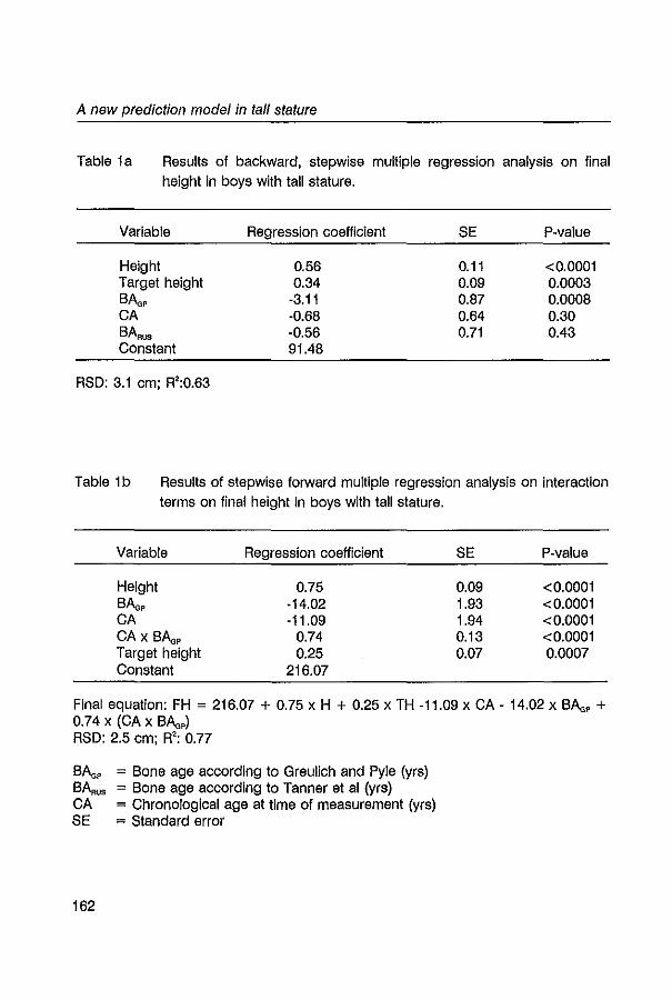
A new prediotion model in tall stature
Table 1a Results of backward, stepwise multiple regression analysis on final height In boys with tall stature.
Variable
Height Target height BAa, CA BA,.,. Constant
RSD: 3.1 cm; R':0.63
Regression coefficient
0.56 0.34
-3.11 -0.68 -0.56 91.48
SE
0.11 0.09 0.87 0.64 0.71
P-value
<0.0001 0.0003 0.0008 0.30 0.43
Table 1b Results of stepwise torward multiple regression analysis on interaction
terms on final height in boys with tall stature.
Variable Regression coefficient SE P-value
Height 0.75 0.09 <0.0001 BAa, -14.02 1.93 <0.0001 CA -11.09 1.94 <0.0001 CA x BAa, 0.74 0.13 <0.0001 Target height 0.25 0,07 0.0007 Constant 216.07
Final equation: FH = 216.07 + 0.75 x H + 0.25 x TH -11.09 x CA - 14.02 x BAa, + 0.74 x (CA x BAa,) RSD: 2.5 cm; R': 0.77
BAa, = Bone age according to Greulich and Pyle (yrs) BAROS = Bone age according to Tanner et al (yrs) CA = Chronological age at time of measurement (yrs) SE = Standard error
162
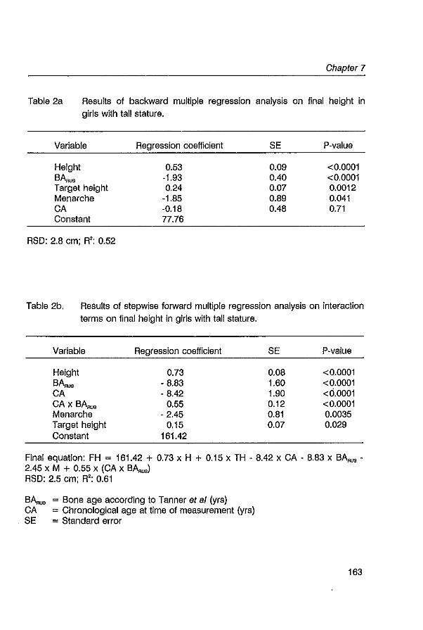
Chapter 7
Table 2a Results of backward multiple regression analysis on final height in girls with tall stature.
Variable
Height BARUS Target height Menarche CA Constant
RSD: 2.8 cm; R': 0.52
Regression coefficient
0.53 -1.93 0.24
-1.85 ·0.18 77.76
SE
0.09 0.40 0.07 0.89 0.48
P-value
<0.0001 <0.0001 0.0012 0.041 0.71
Table 2b. Results of stepwise forward multiple regression analysis on interaction
terms on final height In girls with tall stature.
Variable Regression coefficient SE P-value
Height 0.73 0.08 <0.0001 BARUS - 8.83 1.60 <0.0001 CA ·8.42 1.90 <0.0001 CA x BA"" 0.55 0.12 <0.0001 Menarche - 2.45 0.81 0.0035 Target height 0.15 0.07 0.029 Constant 161.42
Final equation: FH = 161.42 + 0.73 x H + 0.15 x TH - 8.42 x CA . 8.83 x BARUS -2.45 x M + 0.55 x (CA x B~) RSD: 2.5 cm; R': 0.61
BARUS = Bone age according to Tanner et a/ (yrs) CA = Chronological age at time of measurement (yrs) SE = Standard error
163
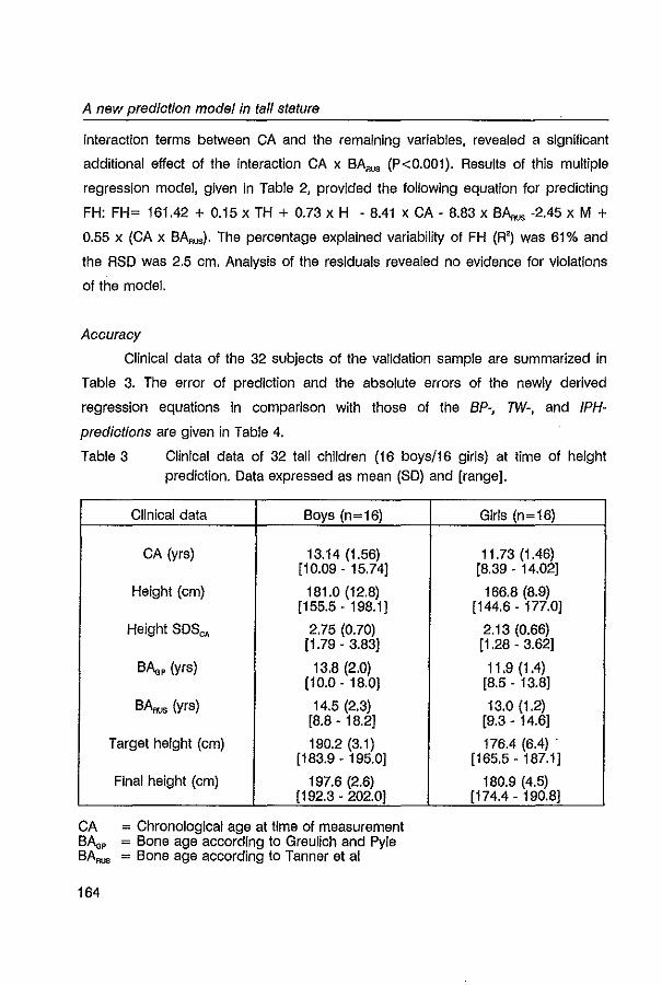
A new prediction mode/In tall stature
interaction terms between CA and the remaining variables, revealed a significant
additional effect of the interaction CA x BA.., (P<0.001). Results of this multiple
regression model, given in Table 2, provided the following equation for predicting
FH: FH= 161.42 + 0.15 x TH + 0.73 x H - 8.41 x CA - 8.83 x BA.., -2.45 x M + 0.55 x (CA x BAROS)' The percentage explained variability of FH (R') was 61% and
the RSO was 2.5 cm. Analysis of the residuals revealed no evidence for violations
of the model.
Accuracy
Clinical data of the 32 subjects of the validation sample are summarized in
Table 3. The error of prediction and the absolute errors of the newly derived
regression equations in comparison with those of the BP-, TW-, and /PH
predictions are given in Table 4.
Table 3 Clinical data of 32 tall children (16 boys/16 girls) at time of height prediction. Data expressed as mean (SO) and [range].
Clinical data Boys (n=16)
CA (yrs) 13.14 (1.56) [10.09 - 15.74]
Height (em) 181.0 (12.8) [155.5 - 198.1J
Height SOSe, 2.75 (0.70) [1.79 - 3.83]
BAa, (yrs) 13.8 (2.0) [10.0-18.0J
BARUS (yrs) 14.5 (2.3) [8.8 - 18.2]
Target height (cm) 190.2(3.1) [183.9 - 195.0J
Final height (cm) 197.6 (2.6) [192.3 - 202.0J
CA = Chronological age at time of measurement BAa, = Bone age according to Greulich and Pyle BA"" = Bone age according to Tanner et al
164
Girls (n=16)
11.73 (1,46) [8.39 - 14.02]
166.8 (8.9) [144.6 - 177.0]
2.13 (0.66) [1.28 - 3.62J
11.9 (1.4) [8.5 - 13.8]
13.0 (1.2) [9.3 - 14.6]
176.4 (6.4) . [165.5 - 187.1]
180.9 (4.5) [174.4 - 190.8]
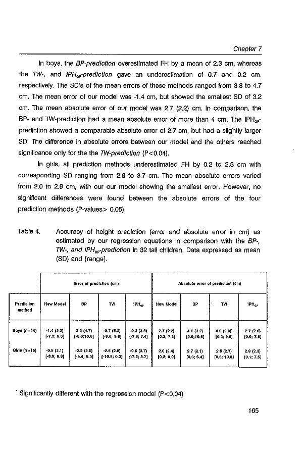
Chaptar 7
In boys, the BP-prediction overestimated FH by a mean of 2.3 cm, whereas
the TW-, and IPHG,-prediction gave an underestimation of 0.7 and 0.2 cm,
respectively. The SD's of the mean errors of these methods ranged from 3.8 to 4.7
cm. The mean error of our model was -1.4 cm, but showed the smallest SD of 3.2
cm. The mean absolute error of our model was 2.7 (2.2) cm. In comparison, the
BP- and TW-prediction had a mean absolute error of more than 4 cm. The IPH.,
prediction showed a comparable absolute error of 2.7 cm, but had a slightly larger
SD. The difference In absolute errors between our model and the others reached
significance only for the the TW-predlctlon (P<0.04).
In girls, all prediction methods underestimated FH by 0.2 to 2.5 cm with
corresponding SD ranging from 2.8 to 3.7 cm. The mean absolute errors varied
from 2.0 to 2.9 cm, with our our model showing the smallest error. However, no
significant differences were found between the absolute errors of the four
prediction methods (P-values> 0.05).
Table 4.
Predlcllon method
80ya (n;;;la)
Girls (ne16)
Accuracy of height prediction (error and absolute error in cm) as estimated by our regression equations in comparison with the BP-, TW-, and IPH.,-predlction in 32 tall children. Data expressed as mean (SD) and [range].
Error 01 predlcllon (em) Absolute error 01 prediction (em)
New Model BP TV! IPHQf' New Model BP TV! IPHQl'
-1.4 (3.2) 2,3 (4.7) ·0.7 (5.2) ·0.2 (3.8) 2.7 (2.2) 4.1 (3.2) 4.2 (2.9)' 2.7 (2.6) [·7.3; 6.6) (.6.6;10.61 (·8.8; 9.6] (.7.a; 7.4J [0.3; 7.3) [0.0;10.6] [0.3: 9.6J [O.Oj 7.8J
-0.6 (3.1) ·0.2 (3.5) -2.6 (2.8) ·0.6 (3.7) 2.0 (2.4) 2.7 (2.1) 2.6 (2.?) 2.9 ('l.3) (.8.0; 5.8) [.6.4; 6.4] [-10.8: 0.3) (.7.S; 5.7] [0.3; 8.0J [0.3; 6.4) (0.3; 10.8) [0.1; 7.61
. Significantly different with the regression model (P<0.04)
165
A new prediction model in tall stature
DISCUSSION
It Is generally agreed, that prediction models derived from a population of
normal growing, normal statured children should be applied to a group children
with growth disorders with considerable caution (3,9). Hence, for children growing
at the upper extremes of normal, that is beyond +2 SD above the mean of the
normal population, It seems to be far better to use prediction models based on
growth data derived from a sample of tall children. So far, only the regression
equations described by Tanner et al have been constructed from data with
inclusion of a small sample of tall children (3). Unfortunately, only girls had been
been Included. We developed a new prediction model for boys and girls with tall
stature based on a set of auxological data derived from 55 tall boys and 88 tall
girls. Our sample of untreated, healthy tall children came from a clinical population
who sought medical advice with respect to their tallness at adolescence. Therefore,
it comprises an excellent reference group for clinical practice. It should be noted,
however, that this group of untreated patients was not strictly comparable with
patients who had ultimately received therapeutic intervention; this latter group had
a significantly higher height prognosis and a significantly younger BA, as shown
previously (5).
The accuracy of a regression model is reflected in its residual standard
deviation (RSO); the smaller the RSD the better the model predicts the dependent
variable (in our case: FH) by the combination of predictor variables. In our final
models, the RSD was 2.5 cm for both boys and girls. This implies that about 95%
of the predictions lie within approximately 5 cm of the real value (± 2 RSD). 'From a
practical point of view, this seems to be a rather large range. However, compared
to other prediction models it is quite acceptable. Tanner et al (3), constructing
regression equations in an age-specific manner, reported RSDs up to 4.1 cm in
boys and 3.6 cm in girls for the same age ranges as in our popUlation; the RSO's
decreased with increasing age. In addition, Karlberg and co-workers recently
described age-dependent regression models based on growth data from birth to
166
Chapter 7
final height of 3650 children (9). Their smallest RSD was 0.69 height SD, which
represents approximately 4 cm.
The explained variability of FH (R') in our models was 0.77 and 0.61 in boys
and girls, respectively. This is in the same order of magnitude as reported by
others (3,9). We used simple anthropometric data and included BAs as estimators
for skeletal maturation In order to predict FH. It must be remembered however, that
linear growth is the resultant of very complex processes influenced by a number of
exogenous and endogenous factors including social class, nutrition and hormones
(10,11). Many of these influencing factors have not been included in our model, nor
in others. In addition, the timing or the intensity of the adolescent growth spurt may
also contribute to the source of prediction error (12). A reliable variable expressing
this phenomenon however, Is lacking. Unless substantial predictor variables are
identified, which, preferably, can be easily obtained in an outpatient clinical setting,
it is unlikely that significant improvement will be made in the prediction of adult
height.
We tested the reliability of the prediction models in a separate group of 32
patients and compared it with three other, widely used prediction methods: the BP
, TW-, and IPHGP-prediction (1-4). In this sample, height prediction in tall girls was
more accurate than in tall boys. This is in concert with our previous report on
height prognosis in children with constitutionally tall stature (5). Our regression
models slightly underestimated FH by approximately 0.5 cm in girls and 1.5 cm in
boys. The absolute errors of the models were 2.0 cm for girls and 2.7 cm for boys.
In comparison with the currently available prediction methods, our model certainly
has a contributive value in height prediction in tall children as shown by the small
absolute errors. In addition, especially in boys, the SDs of both the mean error and
the absolute error of our newly developed models were smaller (or comparable in
girls) compared to the other prediction methods, indicating less individual variation.
Although the reliability of our models had only been tested in a small group of tall
children, these preliminary data are quite promising. They justify the idea that
height prognosis in children with tall stature Is more accurate when based on
167
A new prediction model in tall stature
growth data derived from tall children. Applying the models to a larger group of
children with excessive growth Is recommended to ascertain its clinical additional
use. In addition, combining the dataset of both the study sample and the validation
sample will enlarge the sample size and increase its predictive capability (see note).
In summary, new prediction models are presented for boys and girls with
constitutionally tall stature based on a dataset of 143 untreated tall children. The
reliability of these models was tested In a separate group of 32 patients and
compared to three other prediction methods. The models showed satisfying
accuracy in height prognosis and were found to be quite promising compared to
the currently available prediction methods. Their clinical additional usefulness has to
be ascertained In larger groups of tall children.
Note: the regression equations based on the combined samples of untreated tall
children were:
Boys (n=71): FH= 213.66 + 0.62 x H + 0.29 x TH - 10.49 x CA - 12.98 x BAa, + 0.72 x (CA x BAa,,); RSD = 2.6 em.
Girls (n=103): FH= 130.53 + 0.76 x H + 0.15 x TH - 5.63 x CA - 1.51 x BARUS
6.19 X BAa, + 0.39 x (CA x BAa,); RSD = 2.6 em.
ACKNOWLEDGEMENTS
We kindly thank the Foundation for Children's Stamps, Leiden, The
Netherlands, for their generous gift to support this study. We are grateful to Dr.
Marquita Greyn·Fokker for her bone age assessments and to Dr. Roland Aarsen
for his help in selecting the patients. We thank Frank Bekkering, Paulien Toolens,
and Martin Torn, medical students, for their help in collecting the data.
168

Chapter 7
REFERENCES
1. Bailey N, Pinneau SR: Tables for predicting adulthelght from skeletal age, revised for Greulich and Pyle hand standards. J Pedlatr 1952;40:423-441
2. Tanner JM, Whitehouse RH, Marshall WA, Healy MJR, Goldstein H: Assessment of skeletal maturity and prediction of adun height: TW2 method. Academic Press, New York 1975
3. Tanner JM, Whttehouse RH, Cameron N, Marshall WA, Healy MJR, Goldstein H: Assessment of skeletal maturity and prediction of adult height (TW2 method). Academic Press, London 1983
4. Lenko HL: Prediction of adult height with various methods In Finnish children. Acta Paedlatr Scand 1979;68:85·92 .
5. De Waal WJ, Greyn·Fokker MH, van Gurp EAFJ, Toolens AMP, Stljnen Th, de Muinck Keizer-Schrama SMPF, Drop SLS: Accuracy of final height and effect of growth reductive therapy in 362 constttUllonal1y tall children. J Clln Endocrinol Metab 1996 (March); In press
6. Roede MJ, van Wleringen JC: Growth diagrams 1980, Netherlands. Third nation·wide sUivey. Tljdschr Soc Gez 1985;62(SuppQ:1·34
7. Greulich WW, Pyle SI: Radiographic atlas of skeletal development of the hand and wrist. Stanford University Press, Stanford 1959
8. Van den Brande JVL, Degenhart HJ: Endocrinologle en groelstoornlssen. In: Kindergeneeskunde, van den Brande, Monnens (eds). Wetenschappelijke Uitgeverij Bunge, Utrecht 1993
9. Karlberg J, Lawrence C, Albertsson·Wikland K: Prediction of final height in short, normal and tall children. Acta Paediatr Scand 1994;406(SuppQ:3.9
10. Mascie·Taylor CGN: Blosoclallnlluences on stature: a review. J Blosoc Sci 1991;23:113-128
11. Loveridge N, Farquharson C, Scheven BAA: Endogenous mediators of growth. Proc Nutr Soc 1990;49:443-450
12. Preece MA: Prediction of adult height: methods and problems. Acta Paediatr Scand 1988;347(SuppQ:4·11
169


CHAPTER 8
Psychosocial aspects In constitutionally tall stature.
W.J. de Waal', A. Janssen', J.J. van Bussbach', S.M.P.F. de Muinck
Kelzer-Schrama', R.S.R. Aarsen', F.M.E. Slijper', S.L.S. Drop'.
1 Department of Pediatrics, 2Division of Endocrinology, "Department of Child
and Adolescent Psychiatry, Sophia Children's Hospital, and 4Faculty of Law,
Erasmus University, Rotterdam, The Netherlands.
Submitted
Psychological aspects in tall stature
ABSTRACT
Data on psychological aspects in constitutionally tall stature are scarce. We
studied psychosocial status In 424 adult men and women with constitutionally tall
stature (height> P90). Two hundred forty-three subjects (69 boys and 174 girls)
had been treated for their tallness during childhood with high doses' of sex
hormones (cases). Hundred eighty-two tall subjects (76 boys and 106 girls) had
not chosen for treatment and served as controls. Psychosocial information was
obtained via five different questionnaires. Special areas of interest concerned self
esteem, social anxiety, psychological well-being and body cathexis. In addition,
educational level, employment, marital status and specific height related aspects
were studied. No distinct differences between previously treated tall subjects and
tall controls were found in self-esteem, social anxiety, psychological well-being and
body perception. However, in tall men self-esteem was significantly higher than the
test norms for both cases and controls (P<0.02 and P<O.OOl, respectively). In
addition, previously treated tall men had less social anxiety and showed more
assertiveness compared to tall controls and the norm population (P-values<0.02).
In retrospect, previously treated tall men and women experienced more problems
related to their height than tall controls; they had been teased more frequently and
experienced more remarks or jokes about their stature (P-values<0.03). Tall
subjects did very well in terms of education and employment: they showed higher
levels of education and had a higher percentage of students compared to the
Dutch population of the same age range (P-values <0.01). In conclusion, no major
psychological maladjustment was found in previously treated tall children compared
to tall controls. It is speculative whether the administration of high doses of sex
steroids or possible psychological mechanisms such as denial may have
contributed to these favourable outcomes. Height related problems such as teasing
and hurtful remarks may have played a role for the choice in favour of height
reductive therapy.
172
Chapter 8
INTRODUCTION
Psychosocial problems in tall adolescent boys and girls have been
recognized by pediatricians and endocrinologists. This has led to intervening
therapies with high doses of sex steroids in order to reduce their final adult height
(1-4). Although psychosocial factors form the main reason for treatment extensive
psychological investigation before or during height reductive therapy has never
been performed. Despite this, many clinicians share their experiences that some
children with excessive growth may suffer considerably from being much taller than
others. A commonly heard concern is that these children feel different from their
peers and are subject to hurtful remarks about their height. As a consequence,
coping mechanisms such as kyphotic posture (in order to mask the tall stature),
social withdrawal and even depression have been noticed. Practical problems
concerning clothing and shoeing, fear about future compatible partnering
(especially in girls) and careerplanning are also frequently reported problems faced
by tall children (1-4).
There is no doubt however, that many tall adolescents have no concern at
all about their height. In fact, in our culture, tallness is generally valued positively.
An association between physical stature and achievement has been documented:
taller individuals score higher on intelligence tests, even when various interfering
factors are taken into account (5,6). Individuals who have achieved higher social
status tend to be taller than those of lower status (7,8). In addition, experimental
studies have shown that tall Individuals are regarded to be more competent. For
Instance, In the study of Dannenmaler and Thumin (9) individuals with higher
assigned status were ascribed greater heights. Furthermore, competence
expectations from parents and adults may vary with the physical stature of the child
assigning more difficult tasks to taller children (10). In self-report scales of 82 9-11-
year-old girls, height was linked to superior adjustment and career Importance (11).
Typically, many tall children and their parents seek for medical attention at
the time of puberty. It is commonly believed that the process of pubertal
173

Psyohologioal aspeots in tall stature
development is interrelated with changes in psychological functioning (12,13).
Therefore, changes in physical appearance may affect self-perception. Indeed,
various studies in adolescent boys and girls have shown associations between
physical status and self-esteem or body satisfaction (11,14-16). Studies relating
height and self-esteem, however, showed conflicting results. Coopersmith (17)
found no significant relationship between the actual height and self-esteem in 85
pre-adolescent and adolescent males. Prieto and Robbins (18) on the other hand,
studying 69 young adolescent males, found positive and significant relationships
between their own, peers' and teacher's perceptions of their height and self
esteem, although no relation was observed between the actual height and self
esteem. Nottelman and Welsh (19) examined the relationship between stature and
competence perceptions in 126 sixth- and seventh-grade children. They found that
tall girls in their last year of elementary school rated themselves lowest on general
competence. In secondary school however, short girls rated themselves lowest and
tall stature was regarded more favorably. Results in boys were less pronounced,
but showed that tall boys regarded themselves less competent than short boys in
secondary school. In addition, Hensley (20) studied 85 male and 125. female
undergraduates and revealed no distinct pattern for height and self-esteem for
either gender.
Our study was set out to obtain more insight into the psychosocial status of
a group of constitutionally tall adult men and women, part of whom had been
treated for their tallness during childhood. Main goal of the study was to investigate
whether or not previously treated tall subjects differed in psychological functioning
from tall subjects who had not chosen for any intervening therapy. For this
purpose, psychological questionnaires were send to the participants and
comparisons were made between treated and untreated tall subjects. Special areas
of interest concerned self-esteem, social anxiety and assertiveness, general
psychological well-being and body-cathexis. The latter refers to the degree of
(dis)satisfaction with parts of the body. In addition, we evaluated the influence of
height on socioeconomic status and relationships by comparison with data from
174
the Dutch population.
METHODS
Subjects
Chapter 8
Since the introduction of height reductive therapy in our institute in 1968, 247
men and 423 women, who were seen at adolescence for evaluation of their
constitutionally tall stature, had reached the age of 18 at the time of our follow-up
study. All were contacted by mail to participate in the study. Second mailings were
send to those who did not respond to the first mailing. This study was part of a
large follow-up study in constitutionally tall stature, including auxological
assessments and long-term sequelae. Reports on auxology and long-term
sequelae are reported elsewhere (21-23).
During their puberty, 102 men and 249 women had been treated for their tall
stature (cases). The indication for treatment was usually a predicted final height>
2.5 SO above the mean (> 180 cm for girls and> 200 cm for boys). Occasionally,
treatment was initiated in idiopathic scoliosis. Treatment consisted of high doses of
sex steroids. In girls, estrogens have been used (etl1lnylestradiol 200 Ilg/day orally,
range: 100-300 Ilg/day), while boys have been treated with androgens
(testosterone esters in various regimens with a total monthly dosage up to 1000
mg intramuscular). Hundred and forty-five men and 174 women had not chosen to
undergo treatment for various reasons such as satisfaction with the given height
prognosis or uncertainty about possible side-effects, and served as a comparison
group (controls). Two hundred and nine men and 326 women responded to our
mailing. Of the responders, 145 men (69 cases and 76 controls) and 280 women
(174 cases and 106 controls) agreed to participate in this part of study (see Table
1). All participants gave Informed consent.
In women, the mean (SO) age at the time of the study was 25.5 (5.0) yrs
[range: 18.7 - 35.8 yrs] for cases and 24.6 (4.0) yrs [range: 18.7 - 34.2 yrs] for
controls (P=0.14). Part of the women had also agreed to participate in the
175
Psychological aspects in tall stature
auxological part of our follow-up study in which final adult height was measured
(21). Therefore, final height was available in 158 cases and 92 controls and was
181.7 (3.3) cm and 180.5 (3.8), respectively (P<0.02).
In men, the mean (SO) age at the time of the study was 24.3 (4.1) yrs.
[range: 18.7 - 34.4 yrs.] for cases and 25.7 (4.3) yrs. [range: 18.7 - 34.4 yrs] for
controls (P=0.06). Like in the women, part of the men had participated In the
auxological study so that measured final height was available (21). In 61 cases
mean (SO) final height was 199.7 (3.9) and In 62 controls 196.3 (5.1) (P=0.0001).
Table 1. Response rate of 247 tall males and 423 tall females.
Subjects Number Response (%) Participants (%)'
MALE Cases 102 95 (93) 69 (73)
Controls 145 114 (79) 76 (67)
FEMALE Cases 249 203 (82) 174 (86)
Controls 174 123 (71) 106 (86)
. Percentage of responders
Procedure
In order to obtain psychosocial information five different questionnaires were
sent to the participants by mail. Completion of the questionnaires took
approximately 45 minutes. Most of the subjects returned their questionnaires by
hand, while visiting our outpatient clinic for final height measurement as part of the
auxological study. The remainder was returned by mail. If no reply was received
within two months, subjects were contacted by phone for reminding. Once the
questionnaires were returned, no action was undertaken to fill in missing data.
Measures
1. The "Delft Questionnaire" (24) is a screening instrument for general
unspecified psychological problems and was used to gain a global
176

Chapter 8
psychological impression. It contains 33 items, giving a maximum total score
of 33 and a minimum of zero. A score of 21 or higher is suggested by the
test as a cut-off point for further psychological investigation. The' test is
normed according to a Dutch sample of 665 men and 1167 women.
2. Social anxiety and assertiveness was measured by the "Inventory List on
Association with Others" (25). This Dutch questionnaire is a self-reported
inventory and contains two scales of social activities: overall social
discomfort during contact with others (Discomfort) and overall social
frequency of this contact (Frequency). The scales contain five subscales
each: concerning passing criticism, expressing personal opinions, paying
compliments to others, initiating conversations, and stating positive self
assertiveness. The scales Discomfort and Frequency contain 35 items on a 5
point Likert scale. Total scores of the two scales are obtained by addition of
all 35 items (minimum: 35, maximum: 175). The total scores of the various
subscales are scored with use of a test-key. The test is supplied with a
norm-table of a Dutch reference population of 130 men and 146 women with
a mean (SO) age of 38.6 (13.4) yrs.
3. Self-esteem (positive attitude towards work, flexibility and well-adapted ness)
was measured by a subscale of the "Dutch Personality Questionnaire" (26),
which is a translated and shortened version of the California Psychological
Inventory. The subscale contains 19 items on a three points scale. After
addition, a total score is obtained (maximum: 38, minimum:O) that can be
compared to a Dutch norm scale based on a sample of 2730 men and 2956
women with a mean (SO) age of 34.6 (13.6) years.
4. The degree of satisfaction with various parts of the body was measured by a
Body Cathexis List. This list was originally developed by Secord and Jourard
(27) but it was translated and modified for this study. The list consists of
various body features (25 in boys and 24 in girls) which the subject is asked
to rate on a 5-point Likert scale varying from 'very satisfied (1)' to 'very
dissatisfied (5)'. The total score is calculated by addition of all items divided
177

Psychological aspects In tall stature
by its number.
5. A self·constructed, prestructured questionnaire (Standard Questionnaire)
with open and multiple choice answers was used to gather information
about present socio· economic status including education, employment and
marital status. In addition, specific height related questions had been
included to obtain information about possible psychological distress of
height In the social context. For this purpose we questioned the subjects
about teasing and hurtful remarks, careerplanning, partneririg and
perception of height. Most of these questions were put in a categorical
manner and subjects were asked to choose only one possibility to answer
(e.g. 'yes/no' or 'neverlsometimes/often/always').
The protocol was approved by the ethics committee of Academic Hospital,
Erasmus University, Rotterdam, The Netherlands.
Reference group
For the Body Cathexis List and the self·constructed Questionnaire no
normative data were available. Therefore, a group of 73 students (37 males/36
females) were asked to fill in these questionnaires and were used as a reference
group. Mean (SD) age of the students was 22.2 (2.0) [range: 19.0 - 26.0] for men
and 22.2 (2.0) [range: 19.0 - 27.0] for women, being significantly different
compared to cases and controls for the separate sexes (P·values<0.05). Mean
(SD) adult height of the students was 186.1 (6.7) and 168.2 (7.2) for men and
women, respectively. This was quite comparable to the mean adult height for the
Dutch population (28), but it was significantly shorter compared to our study
population for both sexes (P-values<0.05).
For evaluation of socio·economic status (education, employment and marital
status). reference data of the Dutch population derived from the Central Bureau of
Statistics, The Hague, The Netherlands (29,30), were additionally used for
comparison.
178

Chapter 8
Statistics
Since incomplete questionnaires were present in a very small percentage of
the subjects «1%), only complete questionnaires were used for statistical analysis.
Differences in psychological test variables between groups were tested non·
parametrically (two groups: Mann·Whitney test, three groups: Kruskall-Wallis test).
Comparison with normative data of the various instruments was done using the two
sample t-test, since only mean and SD was available. In case of a contingency
distribution chisquare tests were used. Differences between cases, controls and
students In the height related que~tions of the Standard Questionnaire were tested
with the Kruskall-Wallis test in case of ordinal data. If this test was significant, the
Mann-Whitney test was used to test the differences between groups. In case of
binary data, chisquare tests were used. Comparison of socioeconomic data with
reference data of the Dutch popUlation was done using the Kruskall-Wallis test in
case of ordinal data (educational level) and using chisquare tests in case of binary
data (concerning marital status ("married - unmarried") and employment status
("student - no student")). Correlations between variables were tested with
Spearman's rank correlation test.
RESULTS
Psychological well-being and social anxiety
Results of assessment of psychological well-being and social anxiety are
listed in Table 2. Psychological well-being as determined with the "Delft
Questionnaire" was not significantly different between cases, controls and the norm
population for both men and women. The proportion of subjects with scores > 21
was equal in cases and controls for males (11.5% and 18.9%, respectively; P=0.33)
and females (19.9% and 24.8%, respectively; P=0.42), which was not significantly
different to the norm population (17.1% for males and 20.6% for females).
Male cases scored significantly lower on overall social discomfort as
179
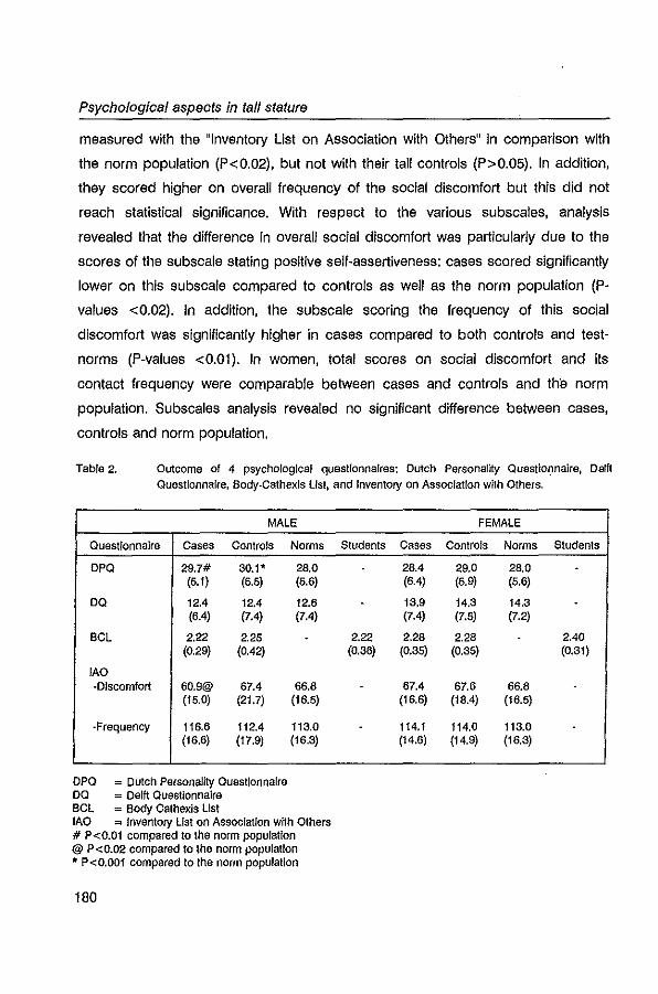
Psychological aspects in tall stature
measured with the "Inventory List on Association with Others" in comparison with
the norm population (P<0.02), but not with their tall controls (P>0.05). In addition,
they scored higher on overall frequency of the social discomfort but this did not
reach statistical significance. With respect to the various subscales, analysis
revealed that the difference in overall social discomfort was particularly due to the
scores of the subscale stating positive self-assertiveness: cases scored significantly
lower on this subscale compared to controls as well as the norm population (P
values <0.02). In addition, the subscale scoring the frequency of this social
discomfort was significantly higher in cases compared to both controls and test
norms (P-values <O.Ol). In women, total scores on social discomfort and its
contact frequency were comparable between cases and controls and the norm
population. Subscales analysis revealed no significant difference between cases,
controls and norm population.
Table 2. Outcome of 4 psychological questionnaires: Dutch Personality Questi0r,1naire, Delft QuestionnaIre, Body·Cathexls list, and Inventory on Association wl1h Others,
MALE FEMALE
Questionnaire Cases Controls Norms Students Cases Controls Norms Students
DPO 29.7# 30.1* 28.0 28.4 29.0 28.0 (5.1) (6.5) (6.6) (6.4) (6.9) (5.6)
DO 12.4 12.4 12.6 - 13.9 14.3 14.3 -(6.4) (7.4) (7.4) (7.4) (7.5) (7.2)
BCL 2.22 2.25 - 2.22 2.28 2.28 2.40 (0.29) (0.42) (0.36) (0.35) (0.35) (0.31)
lAO ·Dlscomfort 60.9@ 67.4 66.8 67.4 67.6 66.8
(15.0) (21.7) (16.5) (16.6) (18.4) (16.5)
-Frequency 116.6 112.4 113.0 114.1 114.0 113.0 (16.6) (17.9) (16.3) (14.6) (14.9) (16.3)
DPQ = Dutch Personality QU8slionnaJre DQ = Defft QUestionnaire BCL ~ Body Cathexis List lAO = Inventory list on AssocIation with Others # P<O.01 compared to the norm population @ P<O.02 compared to the norm population 11: p < 0.001 compared to the norm populaUon
180
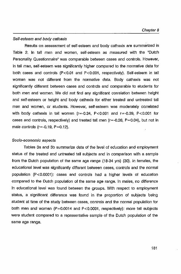
Chapter 8
Self-esteem and body cathexis
Results on assessment of self-esteem and body cathexis are summarized in
Table 2. In tall men and women, self-esteem as measured with the "Dutch
Personality Questionnaire" was comparable between cases and controls. However,
in tall men, self-esteem was significantly higher compared to the normative data for
both cases and controls (P<O.Ol and P<O.OOl, respectively). Self-esteem in tall
women was not different from the normative data. Body cathexis was not
significantly different between cases and controls and comparable to students for
both men and women. We did not find any significant correlation between height
and self-esteem or height and body cathexis for either treated and untreated tall
men and women, or students. However, self-esteem was moderately correlated
with body cathexis in tall women (r=-0.34, P<O.OOl and r=-0.39, P<O.OOl for
cases and controls, respectively) and treated tall men (r=-0.26, P=0.04), but not in
male controls (r=-0.19, P=0.12).
Socia-economic aspects
Tables 3a and 3b summarize data of the level of education and employment
status of the treated and untreated tall subjects and in comparison with a sample
from the Dutch population of the same age range (18-34 yrs) (30). In females, the
educational level was significantly different between cases, controls and the normal
population (P<O.OOOl): cases and controls had a higher levels of education
compared to the Dutch population of the same age range. In males, no difference
in educational level was found between the groups. With respect to employment
status, a significant difference was found in the proportion of subjects being
student at time of the study between cases, controls and the normal population for
both men and women (P=O.0014 and P<O.OOOl, respectively): more tall subjects
were student compared to a representative sample of the Dutch population of the
same age range.
181

Psychological aspects in tall stature
Table 3a. Educational level in treated tall males and females compared to tall controls and the normal population.
MALE (%) FEMALE (%)
Cases Controls Population' Cases Controls Population I n=69 n=76 n=887 n=174 n=106 n=1144
PrimaI)' school 3 4 5 1 - 8
Secondary school 32 21 22 22 21 27
High school 51 54 58 56 56 49
University 15 21 15 21 24 15
= Central Bureau for Statistics (ref. 30)
Table 3b. Employment status of treated tall males and females compared to tall controls and the normal population.
MALE (%) FEMALE (%)
Cases Controls Population' Cases Controls Population 1
n=69 n=76 n=889 n=174 n=106 n=1144
Employed 59 64 73 61 54 39
Unemployed 3 4 4 2 4 2
Studenl 36 26 19 29 36 12
Houswivej-man - 1 7 7 46
Retired - - 1 0.6 1
Other 1 5 2 0.6 - 1
= Central Bureau for Statistics (ref. 30)
Relational aspects
The marital status of cases and controls and compared to that of the Dutch
population of the same age range (18-34 yrs) (31) are given in Table 4. A
significant difference was found in marital status between cases, controls and the
normal population for both men and women (P-values <0.01): less tall subjects
were married at the time of the study compared to the Dutch population. Upon the
182

Chapter 8
question whether or not one has ever had a serious relationship, has had
intercourse and has left one's parental home, an equal proportion of cases and
controls answered confirmative. The mean age at the time these events occurred
for the first time are listed In Table 5 and were not significantly different between
cases and controls for both men and women.
Table 4. Marital status of treated tall males and females compared to tall controls and the normal population.
MALE (%) FEMALE (%)
Cases Controls Population' Cases Controls Population' n=69 n=75' n=2.181.076 n=174 n=106 n=2.088.158
Married 14 24 32 28 25 44
Unmarried 86 76 66 70 74 53
Divorced - 2 2 2 3
Widowed - <1 <1
= Central Bureau for Statistics (ref. 31)
Table 5. Relational aspects of treated and untreated tali males and females. Data expressed as mean (SO) and [range].
MALE FEMALE
Cases Controls Cases Controls
n Age n Age n Age n Age
Age first 61 18.0 (3.2) 58 18.5 (2.8) 157 17.5 (2.3) 98 17.8 (2.2)
serious partner [14.0·32.0] [14.0-28.0] [13.0-25.0] [14.0-26.0]
Age first coUus 59 18.2 (2.7) 56 18.1 (2.4) 145 17.8 (2.2) 95 18.1 (2.0)
[ 15.0-32.0] [12.0-25.0] [10.0-25.0] [14.0-26.0]
Age leaving 34 20.2 (2.2) 46 21.4 (2.7) 120 19.8 (2.2) 77 20.1 (1.9)
parental home [18.0-25.0] [17.0-29.0] [15.0-27.0] [18.0-27.0]
183

Psyohologioal aspeots in tall stature
Height related aspeots
Previously treated tall men and women had been teased more frequently
about their stature at school compared to their tall controls (p~0.0009 and
P~0.0006 for men and women, respectively): in total, only 19% of the previously
treated tall men and 20% of the treated women had never been teased at school
compared to 39% in both tall control groups. In contrast, 70% of the male students
and 69% of the female students reported that they had never been teased about
their stature at school, which was significantly different to both cases and controls
for men as well as women (P·values <0.001).
In men, cases experienced more remarks or jokes about their stature than
controls or students (P~0.0006 and P~0.0082, respectively). In women, cases
were also more subject to jokes and/or remarks about their height, which was
significantly different to tall controls (P~0.029), but not to students (P~O.1 0).
With respect to occupation, an equal proportion of cases, controls and
students reported to be limited by their height to choose a job they wanted. In
addition, there was no difference in the proportion of subjects that, despite
suitability, had ever been rejected for application because of their stature. Height
limited occupations, as recalled by men, varied from aircraft-personnel
(pilot/steward), army-personnel to racing driver. In women, professions like
stewardess, ballet-dancer, nurse, police·officer and jockey had been mentioned.
The question whether or not height had ever played a role in the finding of a
partner was answered confimative by an equal proportion of tall females (49% and
38% for cases and controls, respectively; P~O.1 0) in contrast to 14% of the
controls (cases vs. students:P~0.0003; controls vs. students:P~0.014). In males,
25% of the cases confirmed that height had played a role in partnering, which was
higher than controls (8%, P~0.013), but not significantly different to students (19%,
P~0.67).
Previously treated tall subjects recalled to be more dissatisfied with their
height as a child than students and/or controls. In females, 64% of the cases and
47% of the controls were dissatisfied with their height during childhood in contrast
184

Chapter 8
to 39% of the students (cases vs. students and controls:P<0.0001 and P=0.0004,
respectively; controls vs. students:P=0.071). In males, this percentage was 46 for
cases and 26 for controls, while only 19% of the students recalled dissatisfaction
(cases vs. students and controls:P=O.0072 and P=0.15, respectively; controls vs.
students:P=0.071). At the time of the study, male cases were still more dissatisfied
with their stature than controls and students (cases vs. controls and
students:P=0.020 and P=0.0036, respectively; controls vs. students:P=0.21). Upon
the question whether they would prefer to have a different stature, 46 percent of
the cases answered confirmative, in contrast to 21 % of the controls and 22% of the
students (p-values<0.01); cases (and controls) preferred to be shorter, while
students preferred to be slightly taller. In females, cases were as satisfied as tall
controls with their present height (P=0.39), while students appeared to be more
dissatisfied than cases and controls (P=0.027 and P=0.0086, respectively). In
addition, 45% of the female cases preferred to have a different height, which was a
significantly higher proportion than controls (25%, P=0.0028). In contrast, 55% of
the female stUdents preferred to have a different height, which was in the same
order of magnitude as in cases, but significantly higher than controls (P=0.0021). It
appeared that cases (and controls) would have liked to be shorter, while female
students preferred to be slightly taller.
DISCUSSION
Data on psychosocial aspects in constitutionally tall children are scarce. We
obtained psychosocial information from a total of 145 tall adult men and 280 tall
adult women, part of whom had received height reductive therapy during
childhood. Our main objective was to look for possible differences in psychological
functioning between previously treated tall subjects and tall controls. In addition, we
evaluated the influence of height on various psychosocial aspects by comparing
data from tall subjects with that from the Dutch population.
185

Psychological aspects in tall stature
Screening for psychological well·belng by means of the "Delft Questionnaire"
revealed no evident abnormalities in cases and controls when compared to the
normative data. With respect to social anxiety and assertiveness, as determined by
the "Inventory List on Association with Others", previously treated women reported
to experience no more problems in interpersonal contact than tall controls or the
norm group. Previously treated tall men however, showed less social anxiety and
were more assertive than the norm population and their tall controls, although in
the latter this difference did not reach statistical significance. Subscale analysis
revealed that this was particularly due to the fact that they had less difficulty in
stating positive self-assertiveness. In addition, they tended to avoid this behavior to
a lesser extent than controls and the norm population. it may be speculated that
these differences point into the direction of a psychological mechanism such as
denial. Usually, low scores on self-criticism are interpreted as evidence of
defensiveness saying negative things about oneself. Therefore denial and its
counterpart "accentuating the positive", psychological mechanisms which have
been reported to occur in short statured adult populations (31,32), might explain
the results In tall men. Another explanation could be that the observed differences
may be related to the androgen therapy. Androgens are known to affect human
behaviour. Individual differences in testosterone levels have been linked to
externalizing behaviors such as aggression and assertiveness (13,33·35). Results,
however, are not consistent. Mattson et al (33) found higher plasma levels of
testosterone in forty 14 to 19 years old male delinquents compared to a control
group of fifty-eight 15 to 17 years old healthy boys. The group of Nottelmann
(13,34) on the other hand, found lower rather than higher sex-steroid levels to be
associated with higher degrees of self-image and behavior problems. In addition,
they stated that asynchrony between different aspects of development (e.g.
between hormone levels and age or pubertal stage) may have implications for
adjustment of boys (and girls). These studies concerned physiological plasma
testosterone levels. To our knowledge, there are no studies on the influence of
pharmacological doses of androgens on adolescent behavior. In adult men,
186
Chapter 8
administration of high doses of androgens did not affect aggressive behavior
(36,37). Therefore, the possibility of any causal relationship between androgen
therapy during puberty and adult assertiveness remains speculative.
Self·esteem as measured with a subscale of the "Dutch Personality
Questionnaire" revealed no significant differences between cases and controls.
However, compared to the norm popUlation, tall males scored slightly, though
significantly, higher. In women no such difference was observed. The meaning of
these results Is unclear. In adolescents and adults with various chronic diseases,
the finding of a higher or even a normal self-esteem has been linked with denial of
their ilness (38,39). This does also apply for adults with short stature (31,32). It is
possible that such a defense mechanism might be present in tall adults. However,
additional data supporting this assumption are lacking. We found no relationship
between the actual height and self-esteem in tall men and women. This is in
concordance with others (17,18,20). It is conceivable therefore, that a person's self
esteem may be not so much related to one's actual height but more to one's
perception of height, as suggested by Prieto and Robbins (18).
Tall subjects and students were equally satisfied with their body. We
observed no relationship between the actual height and body satisfaction in tall
men and women and in students. Our findings are In agreement with the study of
Shim and others (40) in adult men, who reported that body satisfaction in tall men
was comparable to average- and big men; short men were found to have the
lowest body satisfaction. Gupta and coworkers (41) selected 174 subjects from a
Canadian shopping mall and revealed an inverse correlation between height and
body dissatisfaction in men, though not in women. In view of the heights of their
study population however, this observation is probably more related to shorter
people. Our finding that self-esteem was positively correlated with body image
supports the theory that body perception is related to self·concept (27).
Since our study was retrospective, it is possible that what we measure now
as aspects of psychosocial status in adulthood may have been different from what
we would have measured in adolescence. For instance, studies in adolescent boys
187

Psychological aspects in tall stature
and girls do indicate that height (and weight) relative to same-age or same-grade
peers may have important implications for self-esteem and self-concept during the
pubertal years (11,14-16,18). In fact in our study, previously treated men and
women recalled to be more frequently teased at school and to be more subjected
to remarks or jokes about their stature than controls and students. In addition, they
reported to be more dissatisfied with their stature as a child. Unfortunately,
psychological evaluation of adolescent tall children prior and during sex steroid
treatment in a controlled manner has never been performed. It remains speculative
therefore, whether the height reductive therapy may have had a positive influence
on the development of psychosocial functioning. Other adaptive mechanisms, such
as denial as discussed above, could have played a contributive role as well. In
addition, transient effects of physical stature on self-regard in tall adolescent
children without height intervening therapy have been described (19).
Several studies point to the possibility that tall people may have more
occupational succes than shorter people, especially in men (6-8). Our study seems
to confirm this finding: a large percentage of tall subjects was still student at the
time of the study, while in a representative sample of the Dutch population of the
same age range most of the people were employed (29). It suggests that taller
people would be able to achieve higher academic positions. In our study, this was
particularly the case in females where the difference in employment was most
strikingly: most of the tall women were employed or still student, while in the normal
population most women run the household at these ages.
The marital status of tall people was different compared to the normal
population: less tall subjects were married. This might be due to the fact that many
of the tall subjects were still students; generally as a student, one keeps from
marriage until one's study is finalized. On the other hand, a low percentage of
marriage might also be seen as a sign of a delayed maturation process resulting in
an unsatisfactory psychosocial maladjustment or social isolation as suggested in
studies from adult patients with short stature (42,43). The possibility of a delayed
psychosocial maturation, however, is contradicted by other findings in our study.
188

Chapter 8
For instance, information about the psychosexual maturation revealed a quite
normal pattern: the age of the first sexual intercourse was comparable to that
reported for the Dutch population (44). In addition, the process of leaving the
parental home (which could also be conceived as a maturational sign) was quite
the same as the trend observed in the Dutch population (45·47) nowadays.
One may argue that the study population could have been biased by
selection. It appeared that more cases responded to our call for the follow-up study
than controls. However, of the responders, an equal proportion of subjects was
willing to participate In this follow-up study. It Is possible that this procedure has
caused some bias, but it is hard to estimate the effect of this bias on the results as
the reason for non-responding was not known. It might also be possible that for
instance more students had been included explaining the differences in
employment and marital status between tall subjects and the normal population.
However, the patients had only been selected by their diagnosis of constitutionally
tall stature unaware of any other demographic or socio-economic data. It is true,
though, that our study concerned referred subjects, who might be different to tall
people from the normal population. To what extent this may account for the
observed differences remains speculative.
In summary, our study revealed no major psychological maladjustment in
previously treated tall children compared to an untreated tall control group. No
distinct differences were found in psychological well-being, social anxiety, self
esteem and body perception. However, tall men showed slightly higher scores on
self-esteem than the norms. In addition, previously treated tall men had less social
anxiety and showed more assertiveness compared to tall controls and the norm
population. It is speculative whether the administration of high doses of sex
steroids or possible psychological mechanisms such as denial may have
contributed to these favourable outcomes. In retrospect, previously treated tall men
and women experienced more problems related to their height than tall controls,
especially during childhood. This may have played a role for the choice In favour of
height reductive therapy. Compared to the normal population tall subjects did very
189

Psychological aspects in tall stature
well in terms of education and employment confirming the idea that tallness is
linked to occupational succes. Further research is needed to establish the
psychological problems In constitutionally tall children and to study the possible
beneficial effects of height intervening therapy.
ACKNOWLEDGEMENTS
This study was supported by a gift from Organon, Oss, The Netherlands.
We thank Frank Bekkering, Evelien van Gurp, Paullen Toolens and Martin Torn,
medical students, for their help in collecting the data.
190

Chapter 8
REFERENCES
1. Wettenhall HNB, Cah11l C, Roche AF: Tall girls: a survey of 15 years of management and treatment. J Pediatr 1975;86:602-610
2. Pradar A, Zachmann M: Treatment of excessively tall girls and boys with sex hormones. Pedlatr 1978;62:1202-1210
3. Bailey JD, Park E, Cowell C: Estrogen treatment 01 girls wHh constHutional tall stature. Ped Clin North Am 1981;28:501-512
4. Zachmann M: Use and risks of androgen therapy: replacement, constitutional delay, and tall stature. In: Forest MG (ed). Androgens In childhood. Pediatric Adolescent Endocrinology. Karger, Basel 1989;19:247-263
5. Humphreys LG, Davey TC, Park RK: LongHudlnal correlation analysis 01 standing height and intelligence. Child Development 1985:56:1465-1478
6. Taesdale TW, OWen DR, Sorenssen TIA: Intelligence and educational level in adult males
at the extremes 01 stature. Hum Bioi 1991;63:19-30 7. Schumacher A: Zur bedeutung der KOrperhOhe In der menschlichen Gesellschaft. I.
Untersuchungen zur sozlalen K6rperhOhedifferenzierung, Zeit Morphol Antropol 1981 ;72: 213-228
8. Hensley WE, Cooper R: Height and occupational succes: a review and, critique. Psychological Reports 1987;60:843·849
9, Dannenmaler WD, Thumin FJ: Authority status as a factor In perceptual distortion of size. J Soc Psychol 1964;63:361-365
10. Brackbill y, Nevill DD: Parental expectations 01 achievement as affected by children's height. Merrill-Palmer Quarterly 1981 ;27:429-441
11. Brooks·Gunn J, Warren MP: The psychological slgnnlcance 01 secondary sexual characteristics In nlne- to eleven-year-old girls. Child Development 1988;59:1061-1069
12. Petersen A: Pubertal development as a cause of disturbance: Myths, realities and unanswered questions. Genetic, Social and General Psychology Monographs 1985; 111 : 205·232
13. Nottelmann ED, Inoff·Germain G, Susman EJ, Chrousos GP: Hormones and behavior at puberty. In: Adolescence and puberty. Bancroft, McChover Relnlsch (eds). Oxford University Press, New York 1990
14. Blyth DA, Simmons RG, Bulcroft R, Felt 0, Van Cleave EF, Bush DM: The effecl 01 physical development on sell-Image and satislaction wHh body-Image lor early adolescent males. Res Comm Health 1982;2:43-73
15. Davies E, Furnham A: Body satislaction in adolescent girls. Br J Med Psychol 1986; 59:279-287
16. Martin S, Housley K, McCoy H, Greenhouse P, Stigger F et al: Sell-esteem 01 adolescent girls as related to weight. Perceptual and Motor Skills 1988;67:879-884
17. Coopersmfth S: The antecedent of seff·esteem. Freeman, San Francisco 1969 18. Prieto AG, Robbins MC: Perceptions 01 height and sell-esteem. Perceptual and Motor Skills
1975;40:395-398
191

Psyohologioal aspeots in tall stature
19. Nottelmann ED, Welsh CJ: The long and the short of physical stature In adolescence. J Early Adolesc 1986;6:15·27
20. Hensley WE: Gender, self·esteem and height. Perceptual Motor Skills 1983;56:235·238 21. De Waal WJ, Fokker MH, Stijnen Th, van Gurp EAFJ, Toolens AMP, de Muink Keizer·
Schrama SMPF, Drop SLS: Accuracy of final height and effect of growth reductive therapy In 362 constitutionally tall children. J CUn Endocrinol Metab 1996, accepted for publication (march)
22. De Waal WJ, Torn M. de Muinck Kelzer.schrama SMPF, Aarsen RSR, Drop SLS: Long term sequelae of sex steroid treatment In the management of constitutionally tall stature. Arch Dis Child 1995;73:311-315
23. De Waal WJ, Vreeburg JTM, Bekkering F, de Jong FH, de Muinck Kelzer.schrama SMPF, Drop SLS, Weber RFA: High dose testosterone therapy for reduction of final height In constitutionally tall boys: does it Influence testicular function in adulthood? elin Endocrinol 1995;43:87-95
24. Appels A: Provisional Manual for the Delft Questionnaire. Voorlopige handlelding blj de
Delftse Vragenlijst. Swets & Zellllnger, Amsterdam 1985 25. Van Dam·Baggen CMJ, Kraaimaat FW: Manual for the Inventory list on Association with
Others. Inventarisatielijst Omgaan met Anderen. Handleiding. Swets & Za/tUngar, Lissa 1987
26. Luteljn F, Starren J, van D1Jk H: Manual for the Dutch Personality Questionnaire. Nederlandse Persoonlljkhelds Vragenlijst. Handleidlng. Swets & Zeitlinger, Amsterdam 1985
27. Secord PF, Jourard SM: The appraisal of body cathexis: body cathexis and the self. J Consull PsychoI1953;17:343-347
28. Roede MJ, van Wieringen JC: Growth diagrams 1980, Netherlands. Third nation-wide sUivey. Tljdschrift Sociale Gezondheidszorg 1985;62(SuppQ:1-34
29. Central Bureau for Statistics. Life style of the Dutch population 1991. Figures. De
leefsituatle van de nederlandse bevolking 1991. Kernci#ers. SDUjUllgeverij, CBS publlcaties, 's Gravenhage 1992
30. Central Bureau for Statistics. Annual statistics. Population of the Netherlands by marital status, sex, age and counlry of nationality, 1 January 1991. Jaarcljfers. Bevolking van Nederland naar burgerlijke staal, geslacht, leeftljd en land van nationaliteit, 1 januari 1991.
Maandstatlstlek der Bevolklng 1991;9:40-41 31. Rotnem D. Cohen D, Hintz R, Genel M: Psychological sequelae of relative 'treatment
failure' for children receiving human growth hormone replacement therapy. J Am Acad Child Psych 1979; 18:505·520
32. Mitchell CM, Joyce S, Johanson AJ, L1bber S, Plotnlsck L, Mlgeon CJ, Blizzard RM: A retrospective evaluation of psychosocial impact of long·term growth hormone therapy. Clln Pedlatr 1986;25: 17-23
33. Mattsson A. Schalling D, Olweus D, LOw H, Svensson J: Plasma testosterone, agresslve behavior and personailly dimensions In young mafe delinquents. J Am Acad Child Psych
1980;19:476·490
192
Chapter 8
34. NoUelmann ED, Susman EJ, Inoff·Germaln G, Culler Jr. GB, Lorlaux DL, Chrousos GP:
Developmental processes In early adolescence: relationships between adolescent adjustment problems and chronological age, pubertal stage, and puberty-related serum hormone levels. J Pediatr 1987;110:473·480
35. Mooradian AD, Morley JEt Korenman SG: Biological effects of androgens. Endocrine Rev 1987;7:1-28
36. Anderson A, Bancroft J, Wu F: The effects of exogenous testosterone on sexuality In normal men. J Clin Endocrinol Metab 1992;75:1503-1507
37. Bagatell CJ, Helman JR, Matsumoto AM, Rivier JE, Bremner WJ: Metabolic and behavioral
effects of high-dose, exogenous testosterone in healhy men. J Clln Endocrinol Metab 1994;79:561-567
38. Kellerman J, Zeltzer L, Ellenberg L, Dash J, Rigler 0: Psychological effects of illness in
adolescence. I. Anxiety, sell-esteem and percepllon 01 control. J Pediatr 1980;97:126-131 39. Utens EMWJ, Verhulst FC, Erdman RAM, Meijboom FJ, Duivenvoorden HJ, Bas E, Roeland
RTC, Hess J: Psychosocial functioning of young adults after surgical correction for congenital heart disease in childhood; a follow-up study. J Psychosomatic Res 1994;38:745-758
40. Shim S, Kotslopulos, Knoll OS: Short, average-height, tall, and big men: Body cathexis,
clothing and retail satisfactions, and clothing behavior. Perceptual Motor Skills 1990;70:83-
96 41. Gupta MA, Schork NJ, Dhaliwal JS: Stature, drive for thinness and body dissatisfaction: a
study of males and females from a non clinical sample. Canad J Psych 1993;38:59-61
42, Dean HJ, McTaggart TL, Fish DG, Friesen HG: The educational, vocational and marital status of growth hormone deficient adults treated with growth hormone during childhood, Am J Dis Child 1985;139:1105-1110
43, Sartorio A, Morabito F, Peri G, Conti A, FagUa G: The social outcome of adults with constitutional growth delay. J Endocrinollnvest 1990;13:593-595
44. Van Zessen G, Sand/art Th (red): Sex in the Netherlands: sexual behavior, risk and
prevention of AIDS. SeksuaUteit in Nederland: seksueel gedrag, rislco en preventie van aids. Swets & Zeltllnger, Amsterdam 1991
45, Central Bureau for Statistics, Visser H: Girls leave home at a younger age than boys,
Melsles eerder het huls uit dan longens. Maandstatlstlek der Bevolking 1988;5:7
46. Central Bureau for Statistics. LaUen JJ: In the eighties young people stayed longer with
their parents again. Development since the sixties. Jongeren bleven In jaran '80 weer
langer bll hun ouders. Ontwlkkelingen slnds de laren '60. Maandstatlstiek der Bevolking
1990; 1 0: 14-24
47. Central Bureau for Statistics. De Jong AH: Boys live two years longer in the parental home
than girls. Jongens wonen twee laar langer thuls dan meisles. Maandstatistlek der
Bevolk/ng 1993;1:8
193


,
CHAPTEA 9
Endogenous and stimulated GH secretion, urinary GH excretion,
and plasma IGF·I and IGF·II levels In prepubertal children with
short stature after Intrauterine growth retardation (lUG A).
W.J. de Waal', A.C.S. Hokken·Koelega', Th. Stijnen', S.M.P.F. de Muinck Kelzer
Schrama', S.l.S. Drop', and the Dutch Working Group on Growth Hormone'.
1 Department of Pediatrics, Division of Endocrinology, Sophia Children's Hospital,
2Department of Biostatistics, Erasmus University, Rotterdam, The Netherlands.
Participating investigators: R.J.H. Odink, Free University, Amsterdam;
J.M. Wit, Wilhelmina Children's Hospital, Utrecht.
Clinical Endocrinology
1994;41 :621-630
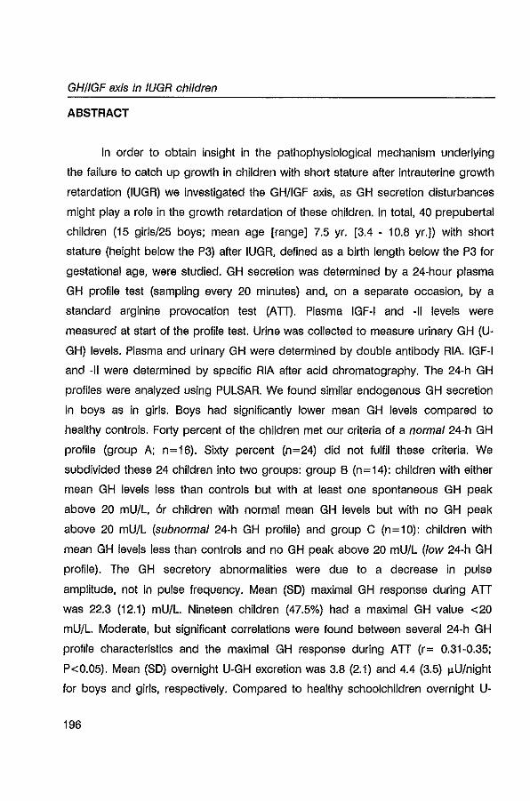
GHllGFaxis in IUGR children
ABSTRACT
In order to obtain insight in the pathophysiological mechanism underlying
the failure to catch up growth in children with short stature after intrauterine growth
retardation (IUGR) we investigated the GH/IGFaxis, as GH secretion disturbances
might playa role in the growth retardation of these children. In total, 40 prepubertal
children (15 girls/25 boys; mean age [range] 7.5 yr. [3.4 - 10.8 yr.]) with short
stature (height below the P3) after IUGR, defined as a birth length below the P3 for
gestational age, were studied. GH secretion was determined by a 24-hour plasma
GH profile test (sampling every 20 minutes) and, on a separate occasion, by a
standard arginine provocation test (All). Plasma IGF-I and -II levels were
measured at start of the profile test. Urine was collected to measure urinary GH (U
GH) levels. Plasma and urinary GH were determined by double antibody RIA. IGF-I
and -II were determined by specific RIA after acid chromatography. The 24-h GH
profiles were analyzed using PULSAR. We found similar endogenous GH secretion
in boys as in girls. Boys had significantly lower mean GH levels compared to
healthy controls. Forty percent of the children met our criteria of a normal 24-h GH
profile (group A; n= 16). Sixty percent (n=24) did not fulfil these criteria. We
subdivided these 24 children into two groups: group B (n=14): children with either
mean GH levels less than controls but with at least one spontaneous GH peak
above 20 mUll, 6r children with normal mean GH levels but with no GH peak
above 20 mUll (subnormal 24-h GH profile) and group C (n=10): children with
mean GH levels less than controls and no GH peak above 20 mUll (low 24-h GH
profile). The GH secretory abnormalities were due to a decrease in pulse
amplitude, not in pulse frequency. Mean (SD) maximal GH response during An
was 22.3 (12.1) mUlL. Nineteen children (47.5%) had a maximal GH value <20
mUlL. Moderate, but significant correlations were found between several 24-h GH
profile characteristics and the maximal GH response during An (r= 0.31-0.35;
P<0.05). Mean (SD) overnight U-GH excretion was 3.8 (2.1) and 4.4 (3.5) flU/night
for boys and girls, respectively. Compared to healthy schoolchildren overnight U-
196
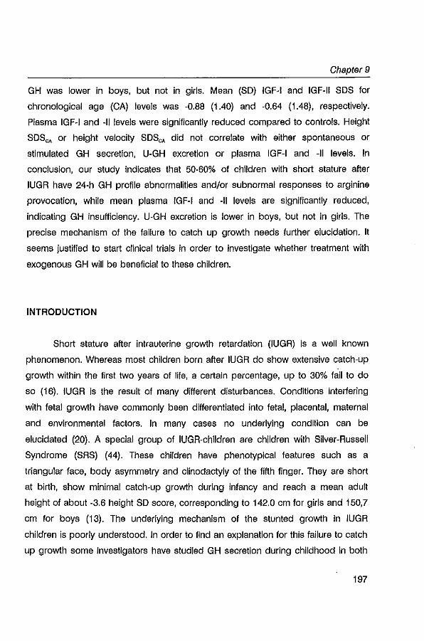
Chapter 9
GH was lower in boys, but not in girls. Mean (SO) IGF-I and IGF-II SOS for
chronological age (CAl levels was -0.88 (1AO) and -0.64 (1A8), respectively.
Plasma IGF-I and -II levels were significantly reduced compared to controls. Height
SOSCA or height velocity SOS", did not correlate with either spontaneous or
stimulated GH secretion, U-GH excretion or plasma IGF-I and -II levels. In
conclusion, our study indicates that 50-60% of children with short stature after
IUGR have 24-h GH profile abnormalities and/or subnormal responses to arginine
provocation, while mean plasma IGF-I and -II levels are significantly reduced,
indicating GH insufficiency. U-GH excretion is lower in boys, but not in girls. The
precise mechanism of the failure to catch up growth needs further elucidation. It
seems justified to start clinical trials in order to investigate whether treatment with
exogenous GH will be beneficial to these children.
INTRODUCTION
Short stature after intrauterine growth retardation (IUGR) is a well known
phenomenon. Whereas most children born after IUGR do show extensive catch-up
growth within the first two years of life, a certain percentage, up to 30% fail to do
so (16). IUGR is the result of many different disturbances. Conditions interfering
with fetal growth have commonly been differentiated into fetal, placental, maternal
and environmental factors. In many cases no underlying condition can be
elucidated (20). A special group of IUGR-children are children with Silver-Russell
Syndrome (SRS) (44). These children have phenotypical features such as a
triangular face, body asymmetry and clinodactyly of the fifth finger. They are short
at birth, show minimal catch-up growth during infancy and reach a mean adult
height of about -3.6 height SO score, corresponding to 142.0 cm for girls and 150,7
cm for boys (13). The underlying mechanism of the stunted growth in IUGR
children is poorly understood. In order to find an explanation for this failure to catch
up growth some investigators have studied GH secretion during childhood In both
197
GH/lGFaxis in IUGR ohildren
dysmorphio and non-dysmorphic IUGR children (1,2,33)_ Abnormal GH secretion
during 12 or 24 hours of physiological testing and/or insufficient GH response to
various provocative stimuli have been described (1,2,33)_ Interpretation of these
results however, is difficult as the various study populations are differently defined.
In addition, little Is known about the plasma IGF-Ievels and urinary GH excretion (U
GH) in these children. We therefore studied spontaneous plasma GH secretion
during 24 hours as well as maximal plasma GH response after arginine stimulation
in 40 prepubertal children with short stature after lUG A. In addition, we measured
U-GH levels during day- and nighttime as well as plasma IGF-I and -II levels.
SUBJECTS AND METHODS
Study group
Forty prepubertal children (25 boys/15 girls) with short stature after IUGR
were studied. The mean (SO) age was 7.5 (2.0) yr, with a range of 3.4-10.8 yr. All
patients fulfilled the following inclusion criteria: 1. IUGR defined as a birthlength
below the third percentile for gestational age (48), 2. an uncomplicated neonatal
period, i.e. without signs of severe asphyxia (defined as an Apgar score below 3
after 5 minutes), without complicated sepsis neonatorum and without long-term
complications of respiratory ventilation such as broncho-pulmonary dysplasia or
pneumothorax, 3. no catch-up growth above the P3 for chronological age (Dutch
references (34)) within the first two years of life, 4. height velocity for chronological
age s P50 (Dutch references (31)), 5. no signs of puberty defined as Tanner stage
M/G 1 or testicular volume < 4 ml. (45), 6. chronological age < 9 yrs for girls and
< 11 yrs for boys, and 7. no chromosomal abnormalities or other organic causes
for growth retardation, except for Silver-Russell syndrome (SRS).
Clinical details of the patients are given in Table 1. In 2 boys the diagnosis
SRS was made. Body mass index (weight(kg)/height(m'»(BMI) was calculated and
expressed in SO scores for chronological age (BMI-SDScJ using a logarithmic
198
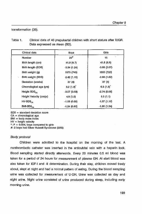
Chapter 9
transformation (35).
Table 1. Clinical data of 40 prepubertal children with short stature after IUGR. Data expressed as mean (SO).
Clinical data
Number
Birth length (em)
Birth length (SOS)
Birth weight (g)
Birth weight (SOS)
Gestation (weeks)
Chronological age (yrs)
Height SOS",
Height Velocity (crn/yr)
HV-SOScA
BMI-SOScA
SOS = standard deviation score CA = chronological age BMI = body mass index HV = height velocity *: p = 0.004; boys compared to girls #: 2 boys had Silver Russell Syndrome (SRS)
Study protocol
Boys Girls
25' 15
4t.9 (4.7) 4t.5 (6.5)
-3.34 (1.24) -3.88 (2.07)
1970 (745) 1850 (720)
·2.42 (1.15) ·2.88 (1.02)
37 (4) 37 (4)
8.2 (1.8)' 6.3 (1.8)'
-3,07 (0.63) ·2.74 (0.58)
4.9 (t.0) 5.5 (1.1)
·1.08 (0.89) ·1.07 (1.12)
·t.24 (0.63) ·1.86 (t.34)
Children were admitted to the hospital on the morning of the test. A
nonthrombotic catheter was inserted in the anticubital vein with a heparin lock.
Blood sampling started directly afterwards. Every 20 minutes 0.5 ml blood was
taken for a period of 24 hours for measurement of plasma GH. At start blood was
also taken for IGF·I and -II determination. During their stay, children moved freely
about, slept at night and had a normal pattern of eating. During the blood sampling
urine was collected for measurement of U·GH. Urine was collected as day and
night urine. Night urine consisted of urine produced during sleep, including early
morning urine.
199
GHllGFaxis in IUGR children
On a separate occasion a standard arginine tolerance test was performed
after an overnight fast. Arginine 0.5 g/l<g was infused in 30 minutes. Blood samples
were taken at T=-15, 0, 15, 30, 45, 60, 90 and 120 minutes for plasma GH
determination.
Blood samples were stored on ice for no longer than 3 hours. After
centrifugation, plasma samples were frozen (_20' C) and stored until assayed.
During collection urine was stored in the refrigerator. After collection, the total
amount of urine was measured and urine samples were frozen (_20' C) until
assayed.
The protocol was approved by the Ethics Committee of each participating
centre.
Hormone assays
All GH assays in plasma and urine were performed in the same laboratory.
All GH samples were analyzed in duplicate in the same assay. Plasma GH was
measured by a double antibody RIA, as described previously (23). Urinary GH was
measured by the same RIA as that described above with a preincubation technique
and an antiserum dilution of 1 :500,000. For the isolation of GH, Millipore filters
(UFPI-LGC-BK) with a mol wt discrimination of 10,000 were used. Overnight U-GH
levels were compared with the mean of 5 consecutive overnight urine samples of a
group of 93 prepubertal healthy schoolchildren aged 4-11 years (OW[l data,
unpublished). Mean (SO) overnight U-GH level of the controls was 4.8 (2.6)
!'U/nlght.
Plasma IGf-1 and -II concentrations were determined by specific RIA after
acid chromatography in order to remove interfering binding proteins, as published
previously (23). Since both growth factors are dependent on age and sex, values
were transformed to SO scores for chronological age using reference values based
on 600 samples from a healthy Dutch population, as described previously (23).
Reference data for I<3F-1I were only available from the age of 7.
200
Chapter 9
Analysis of 24-h GH profiles
The 24-h GH profiles were analyzed using the Pulsar program (29) and
adapted for Quick Basic by Rosberg and Albertsson-Wikland (PC-Pulsar, 1987).
The Pulsar program identifies secretory peaks by height and duration from a
smoothed baseline, using the assay SD as scale factor. Peak selection criteria
appropriate for our own assay conditions and data set were established before the
24-h GH profiles were analyzed (23). With these settings the Pulsar program did
not detect any peaks when 72 consecutive samples from each of 3 different
plasma pools were assayed. From the Pulsar program the following profile
characteristics were extracted: area under the curve above zero (AU Co) and above
baseline (AUC,), total number of peaks, mean and maximal value, mean peak
amplitude. The sum of the amplitude was calculated by multiplying the mean of the
peak amplitude with the number of peaks.
Obtaining control data from a group of healthy children was considered
unethical, as Information on 24-h GH profiles in healthy prepubertal children was
available (4). We have previously shown that the methodology and GH
measurements as performed in this control study are comparable with ours (24).
We therefore compared the mean plasma GH levels of the 24-h GH profiles of our
study patients with the mean GH levels of 62 healthy prepubertal children (18
girls/44 boys) as reported by Albertsson-Wikland and Rosberg (4). Mean (SD) of
mean plasma GH levels of these controls were 5.9 (0.6) mUll for boys and 4.8
(0.6) mUll for girls (4). To correct for sex we also expressed the mean GH values
of our study group as SD scores.
Based on descriptions in the literature (3,4,49) we defined a normal 24-h GH
profile as a profile with: 1. well defined GH peaks returning to baseline levels
between the pulses, 2. at least one major GH peak above 20 mUll, 3. a mean GH
level over 24 hours within the range of 4.8 ± 2 SD mUll for girls and 5.9 ± 2 SD
mUll for boys, and 4. a normal diurnal rhythm.
201
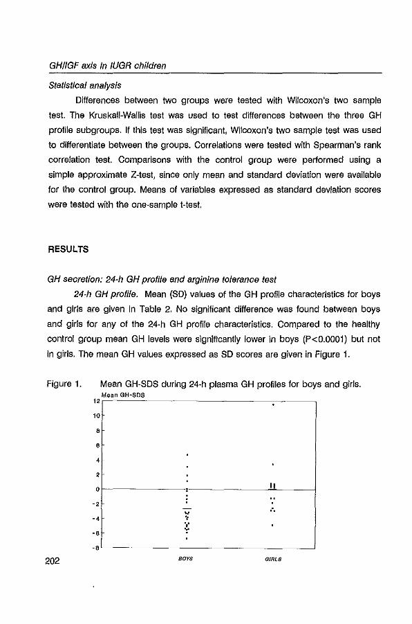
GH/IGFaxis in IUGR children
Statistical analysis
Differences between two groups were tested with Wilcoxon's two sample
test. The Kruskall-Wallis test was used to test differences between the three GH
profile subgroups. If this test was significant, Wilcoxon's two sample test was used
to differentiate between the groups. Correlations were tested with Spearman's rank
correlation test. Comparisons with the control group were performed using a
simple approximate Z-test, since only mean and standard deviation were available
for the control group. Means of variables expressed as standard deviation scores
were tested with the one-sample t-test.
RESULTS
GH secretion: 24-h GH profile and arginine tolerance test
24-h GH profile. Mean (SO) values of the GH profile characteristics for boys
and girls are given in Table 2. No significant difference was found between boys
and girls for any of the 24-h GH profile characteristics. Compared to the healthy
control group mean GH levels were significantly lower in boys (P<0.0001) but not
in girls. The mean GH values expressed as SO scores are given in Figure 1.
Figure 1. Mean GH-SOS during 24-h plasma GH profiles for boys and girls. Mean GH~SDS
12
10
8
8
4
2
0
-2 v
-4 • '," -6 'r
-8
202 BOYS GIRLS
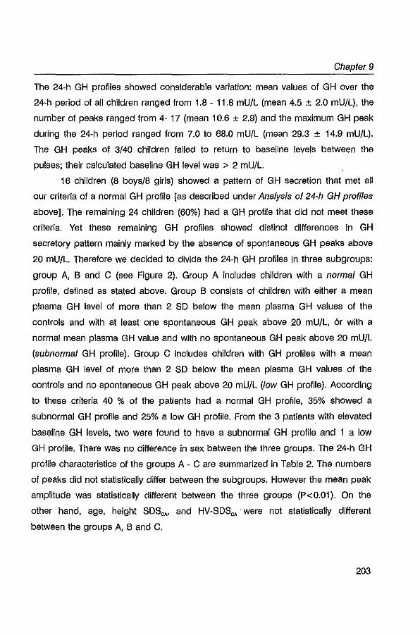
Chapter 9
The 24-h GH profiles showed considerable variation: mean values of GH over the
24-h period of all children ranged from 1.S - 11.S mUll (mean 4.5 ± 2.0 mUll), the
number of peaks ranged from 4- 17 (mean 10.6 ± 2.9) and the maximum GH peak
during the 24-h period ranged from 7.0 to 6S.0 mUll (mean 29.3 ± 14.9 mUll).
The GH peaks of 3/40 children failed to return to baseline levels between the
pulses; their calculated baseline GH level was > 2 mUlL.
16 children (S boys/S girls) showed a pattern of GH secretion that met all
our criteria of a normal GH profile [as described under Analysis of 24-h GH profiles
above]. The remaining 24 children (60%) had a GH profile that did not meet these
criteria. Yet these remaining GH profiles showed distinct differences in GH
secretory pattern mainly marked by the absence of spontaneous GH peaks above
20 mUlL. Therefore we decided to divide the 24-h GH profiles in three subgroups:
group A, Band C (see Figure 2). Group A includes children with a normal GH
profile, defined as stated above. Group B consists of children with either a mean
plasma GH level of more than 2 SO below the mean plasma GH values of the
controls and with at least one spontaneous GH peak above 20 mUll, 6r with a
normal mean plasma GH value and with no spontaneous GH peak above 20 mUll
(subnormal GH profile). Group C includes children with GH profiles with a mean
plasma GH level of more than 2 SO below the mean plasma GH values of the
controls and no spontaneous GH peak above 20 mUll (low GH profile). According
to these criteria 40 % of the patients had a normal GH profile, 35% showed a
subnormal GH profile and 25% a low GH profile. From the 3 patients with elevated
baseline GH levels, two were found to have a subnormal GH profile and 1 a low
GH profile. There was no difference in sex between the three groups. The 24-h GH
profile characteristics of the groups A - C are summarized in Table 2. The numbers
of peaks did not statistically differ between the subgroups. However the mean peak
amplitude was statistically different between the three groups (P<0.01). On the
other hand, age, height SOSCA' and HV-SOSCA ' were not statistically different
between the groups A, Band C.
203
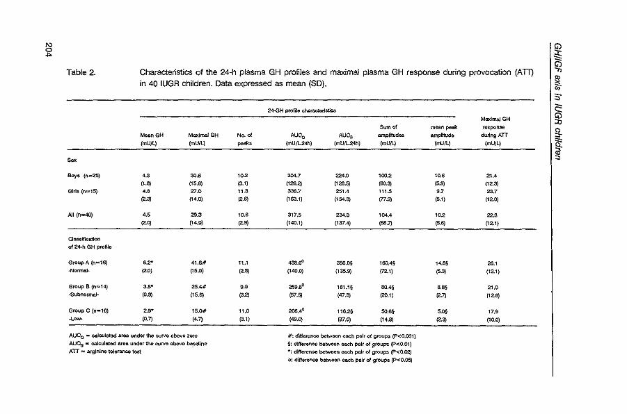
I\)
~
Table 2. Characteristics of the 24-h plasma GH profiles and maximal plasma GH response during provocation (An) in 40 IUGR children. Data expressed as mean (SO).
24-GH profilo cho.rnctor]:stlcs
MaxlmnlGH
Sum of moan poole rO$poMo Moan GH Maximal GH No. of AUeo AUes ampJlbJd8$ amplitudo duringATT
(mUll) (mUll) po,", (mU/L24h) (mU/L24h) (mUll) (mUlL) (mUll)
$0,
~ (n=25) 4.3 30.6 '02 304.7 224.0 '002 10.6 21.4
(1.8) {15.6} (3.1) <126.2) (1213.S) (60.3) la~ (12.3) 61rl$ (0""5) 4.' 27.0 11.3 338.7 251.4 111.5 9.7 23.7
(2.3) (14.0) (2.~ (163.1) (154.3) (77.9) (5.1) (12.0)
All (n .. 4O) 4.5 29.3 10.6 317.5 234.3 104.4 '02 22.3 (2.0) (14.9) (2.9) (140.1) (137.4) (56.7) (5.6) (12.1)
CI~eatlon
of 24-h GH prolllo
Group A (n=16) 6 ... 41.6# 11.1 438.0' 356.0§ 160.4§ 14.8§ 26.' ""0_ (2.0) (15.0) (2.8) (140.0) (135.9) (72.') (5.3) (12,1)
Group B (1"1"'4) 3.S" 25.4# OS 259.8° tSU§ SO.4O .... 21.0 -Subnormal- (0.9) (1S,S) (3.~ 157.~ (47.3) (20.1) (2.n (12.8)
Group C (1"1-10) 2.9* 15.0# 11.0 206,4° 116.2§ SO." S.OS 17.9 ·L_ (O.?) (4.7) (3.1) (49.0) (37.0) (14.8) (2.~ (10.0)
AUeo .. caloulo.tod &roo. undor tho curve above zero II: odferenco betwoon ench pair of groups (P<O.OO1) AUCo - calculated area under the curve nbove bll:Seline §: differonce betwoen each pair of ~roupl3 (P<O.Ol) ATT "" arginine toleranco 'lost w: dlfferenco bctwoen each pair of groupl3 (P<O.Cl2)
0: difference bctwoon el!;ch pair of grouPll (P<O.05j
G) .;s ~ !'i CO· S·
i g. ~ §
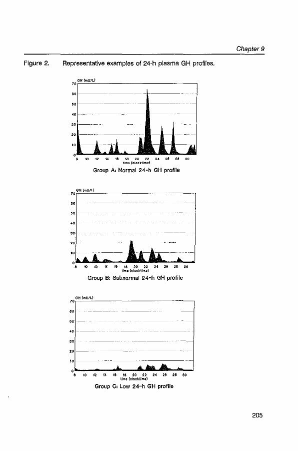
Figure 2. Representative examples of 24-h plasma GH profiles.
OH(mUIl..)
001---- -------
" 30
20
101---1--
8 ~ 12 « ~ u ro 22 24 ~ 28 30 time (elookUmel
Group A: Normal 24-h GH profile
70rOH~~~ ____________________ --,
60
00
"1-----
8 ~ 12 « ffl ffi 20 ~ 24 28 U ~ tIme (olocktlme)
Group B: Subnormal 24-h GH profile
70rOH~~~U~~I ______________ -,
60
00
" ----
30
20--
10-----··-
o~------~---A~~ .. ~--~ 8 ro 12 u ~ m ro 22 24 ~ 28 ~
tlmo (crockUme)
Group C: Low 24-h GH profile
Chapter 9
205
GH/IGFaxis in IUGR children
Arginine tolerance test (ATT). The mean (SO) maximal plasma GH response
during AIT was 22.3 (12.1) mUlL. with a range from 4.5 to 56 mUlL. There was no
difference between the sexes. Nineteen out of 40 children (47.5%) had a maximal
GH-response less than 20 mUlL. The response during A IT was not statistically
different between the groups A - C (see Table 2). In group A, 10 out of 16 patients
showed a normal GH response (maximal GH peak> 20 mUll) during AIT. In
group Band C a normal response was found in 11/24 cases.
We found a moderate but significant correlation between the endogenous
GH secretion and the stimulated GH secretion. The following 24-h GH profile
characteristics correlated with the maximal GH response during A IT: mean GH
value, number of peaks > 20 mUll, AUC" mean peak amplitude, and the sum of
the amplitudes (rsp= 0.31-0.35; P<0.05). On the other hand no correlation was
found between parameters of GH secretion and parameters of growth (expressed
as H-SOSCA or HV-SOSc.J. In addition there was also no correlation between
parameters of GH secretion and severity of IUGR (expressed as birth length SOS).
24-h urinary GH excretion
Results of urinary GH excretion are shown in Table 3. There was no
difference in U-GH excretion between boys and girls. Compared to controls, boys,
in contrast to girls, had lower mean overnight U-GH levels (P<0.05). U-GH
excretion was not different between the GH profile groups A. Band C.
U·GH levels did not significantly correlate with either stimulated or
endogenous GH secretion. In addition, no correlation was seen between the
urinary GH excretion and the growth parameters (H-SOSCA and HV-SOSc.J or the
severity of IUGR (birth length SOS).
Expressing the data corrected for creatinine excretion in U-GH/ng creatinine
did not change any of these findings.
IGF-I and IGF-1/ levels
Plasma IGF-I and -II levels are given in Table 3. Mean (SO) SOSCA for plasma
IGF-I was -0.88 (1.40). For IGF-II, SO score could only be calculated for 29/40
206
'" 0 .....
Table 3. Age, plasma IGF-I and -II levels and urinary GH excretion (U-GH) in 40 IUGR children. Data expressed in mean
(SO).
Age IGF-I SOS~ IGF-II 50S"" U-GH/24h U-GH day U-GH night (years) (n=29) (~U/24h) (~U/day) (~U1night)
Sex
Boys (n=25) 8.2 -0.74 -0.64 (n=21) 9.3 5.5 3.8 (1.8) (1.43) (1.54) (4.3) (3.5) (2.1)
Girls (n=15) 6.3' -1.10 -0.65 (n=8) 9.5 4.9 4.4 (1.8) (1.35) (1.39) (6.4) (3.3) (3.5)
Classification 01 24-h GH profile
Group A (n=16) 7.4 -0.55 -0.11 (n=11) 11.1 6.0 4.9 -Norma/- (2.1) (1.28) (1.10) (6.5) (3.9) (3.5)
Group B (n=14) 7.7 -1.23 -1.18 (n=10) 7.3 4.0 3.5 -Subnorma/- (2.2) (1.36) (1.46) (4.6) (3.4) (1.6)
Group C (n=10) 7.3 -0.91 -0.69 (n=8) 9.1 5.9 3.2 -Low- (1.9) (1.61) (1.84) (3.4) (2.0) (1.9)
* P = 0.004 girls versus boys Ig ~
I~
GHllGFaxis in IUGR children
patients (children > 7 years). Mean (SO) SOSCA for IGF·II was ·0.64 (1.48). Mean
values of IGF-I SOSCA and IGF·II SDSCA were significantly different from zero
(P<0.001 and P<0.05, respectively), i.e. the IGF·levels were significantly reduced
compared to controls (see Figure 3). IGF·I and ·11 levels were not significantly
different between boys and girls. Furthermore IGF·I SDSCA as well as IGF·II SDSCA
did not significantly differ between the three GH profile groups (A·C).
Plasma IGF·I expressed as SDSCA was correlated with the maximal plasma
GH peak during ATT (r,,=0.43; P<0.01) but not with endogenous GH secretion.
Interestingly, the IGF's did not correlate with the parameters of growth (H-SDSCA
and HV·SDScJ or severity of IUGA (birthlength SDS).
Figure 3. IGF·I and ·11 SDS for chronological age. IOF-1/1I SDS for CA 4r-----~~~------------------------,
2
,. '. 0'. j
... : ':'
Or---------------~--------------_7------~
.' 0 0 .... :.: .'. -2
.=.
-4
IGF-II
DISCUSSION
Our study shows that many prepubertal children with short stature after
IUGA have 24·h GH profile disturbances. Forty percent of the children had a 24·h
GH profile that met all our criteria of a normal profile. Consequently, sixty percent of
the children showed an abnormal 24·h GH profile. These findings are comparable
with previous studies (1,33), despite differences in patient selection. The finding of
208
Chapter 9
such a high percentage of GH secretory disturbances in these children is
surprising, because they lack clinical features of GH deficiency. In fact, these
children had a low BMI, thus the low spontaneous GH levels in plasma were not
due to a high BMI as reported in healthy children (27,36). Ten out of the 24
children with an abnormal 24-h GH profile (25% of all patients) secreted a very low
amount of GH and failed to produce a GH peak greater than 20 mUll, indicating a
marked GH insufficiency. This is In accordance with the studies of Albertsson
Wikland and Ackland et ai, who found percentages of 37.5 % and 12.9 %,
respectively (1,2). Assuming a spectrum in GH secretion (4,30), we divided all 24-h
GH profiles into a kind of gliding-scale, ranging from normal -via subnormal- to low
GH secretion (group A, Band C, respectively). By this division it was clear, that this
gradual reduction in GH secretion was caused by a decrease in GH pulse
amplitude, not in pulse frequency. It is known that GH pulse amplitude increases
with age (28,36), however, in our study age did not differ between the groups. The
high percentage of GH secretory abnormalities in our patient group may be
attributed to a disturbance in GH pulse amplitude.
47.5 % of the children showed a subnormal response during arginine
provocation « 20 mUlL). This percentage is somewhat higher than reported
previously (2,33). This difference is probably caused by the pharmacological
stimulation tests used, as it has been shown that different provocation tests have
different Intensities of response (32). Several studies have shown that stimulated
GH hormone levels do not completely reflect endogenous GH levels In children
with short stature and that it is not uncommon for stimulation tests to overestimate
spontaneous GH secretion (7,15,39), although others have argued for the contrary
(37). In our study we found a significant, though moderate, correlation between
various 24-h GH profile characteristics and the maximal GH response during An.
When we combined the results of the 24-h GH profiles and the pharmacological
stimulation tests, the accuracy of the An to categorize the children correctly
according to their 24-h GH profile was only 57.5%. Conversely, stimulated GH
secretion did not reflect endogenous GH secretion In 42.5% of the patients:
209
GH/fGFaxis in fUGR children
underestimation in 6 (15%) and overestimation in 11 patients (27.5%). In addition,
11/24 patients with an abnormal GH profile (groups Band C) showed a normal GH
response to arginine provocation indicating a neurosecretory dysfunction (NSD), as
described by Spiliotis et al (40). Thus NSD accounted for 46% of the GH secretory
abnormalities. This finding is higher than reported by others (33).
Compared to healthy schoolchildren mean overnight U·GH excretion was
lower in boys, but not in girls. The reason for this finding is not clear. It might be
related to the fact that mean plasma GH levels in boys were lower than controls,
but on the other hand, we found no significant correlation between U·GH levels
and parameters of endogenous GH secretion. In addition, measurement of U·GH
levels (over 24 hours as well as during day- or nighttime) did not enable us to
distinguish between the different GH profile groups. Our finding is in contrast with
other studies in short children where urinary GH levels were found to be well
correlated with endogenous GH secretion (12,25,43). Difference in statistical
approach may account for this discrepancy, because in these studies parametric
correlations tests were used. We also found good correlations between parameters
of endogenous GH secretion and U·GH levels by use of a parametric correlation
test (r-values up to 0.60), but we considered it better to use a non·parametric
correlation test as U-GH levels were not normally distributed.
During fetal growth IGFs play an Important role (14) and birth size is
correlated with IGF·I (but not with IGF·II)(5,6,18). In addition, decreased IGF-I and -
II plasma levels are described in IUGA, in man as well as in animal models
(26,42,47). Catch-up growth in IUGA children has also been associated with
normalization of IGF-I levels (46). Few data are available on IGF-I or -II levels in
lUG A children during childhood. Grunt et al and Aochiccioli et al reported normal
IGF-I levels in IUGA children (19,33). In these studies, however, IGF-I levels were
not corrected for age. To our knowledge, there are no data on IGF-II levels during
childhood in IUGA children. In our study, we found significantly reduced mean
levels of IGF-I and -II SDS for CA. 9/40 children had an IGF-I SDSCA value more
than 2 SD below the mean. As IGF-I levels are at least in part GH-dependent, the
210
Chapter 9
reduced IGF-I levels reflect our findings of disturbances in GH secretion in these
children. Indeed, we found a significant correlation (r,p=0.43; P<0.01) between the
maximal GH response during A IT and IGF-I 8DSo. values. Surprisingly, IGF-I levels
did not correlate with endogenous GH secretion. This is in contrast with healthy
children in whom IGF-I levels do reflect endogenous GH secretion (8). We cannot
give a satisfacory explanation for this discrepancy. The cause of the decreased
mean IGF-II levels is less clear, as IGF-II is found to be less GH-dependent.
Reduced levels of IGF-II have also been described In GH-deficient children and
children with normal short stature (38). The physiological meaning of IGF-II,
however, is still unclear, and thus the interpretation of this finding Is speculative.
We could not find any relation between the growth status of the child and
the GH profiles, U-GH excretion or the growth factors. This is in accordance with
Ackland et ai, while others do not mention any relationship (1,2,33). This might be
due to the little variance in growth status of these children. Several studies indicate
the presence of a relationship between the growth during childhood and the level
of GH secretion in as much it concerns the extremes in height (3,22,49); however,
within the normal range of growth rates, this relation is less clear cut (11). The
severity of IUGR was also not related to any of the above parameters. For GH, this
finding was not completely unexpected as the influence of GH on birth length is at
least of minor importance. Apparently, the IGF-Ievels at childhood do not reflect
birth length. As IGF becomes GH dependent, postnatal adaptive mechanisms may
be responsible for this. The heterogeneity of IUGR may also playa role.
The findings outlined do not enable us to elucidate the complete
pathophysiological mechanism in IUGR children. Certainly, we have shown that
many children have GH secretion disturbances, reduced IGF-Ievels and/or reduced
U-GH excretion, but this GH/IGFaxis disorder can only in part explain the fact of
their growth failure. There are several children with no distinct GH/IGFaxis
abnormalities, who still failed to show postnatal catch up growth. One could
speculate about abnormalities of the growth hormone receptor, the IGF-binding
proteins or second messenger systems. In addition, the heterogeneity of the
211
GH/lGFaxis In /UGR children
etiology of IUGR may also play a role in some unexplained findings. Hpwever,
supportive data are lacking.
With the availability of biosynthetic GH the efficacy and safety of GH
treatment in children with non-GH deficient short stature is explored (17,21). This
also accounts for children with short stature after IUGR (2,9,10,41). Our study
results give support to the intention to treat these children in view of the 24-h GH
profiles abnormalities, the subnormal GH responses during An and the reduced
growthfactors. However, whether treatment will be beneficial to all children or only
to those with suspected GH insufficiency can only be investigated within clinical
trials including all children regardless of their GH secretion, as the complete
pathophysiological mechanism is unclear and as there seems to be no clear
relationship between GH secretion status and growth in these children.
In summary, our results indicate that many children with short stature after
IUGR have 24-h GH profile abnormalities and/or subnormal responses to arginine
provocation indicating a GH insufficiency. Mean serum levels of IGF-I and -II are
significantly reduced. Compared to controls mean plasma GH levels and U-GH
excretion are lower in boys, but not in girls. The precise mechanism of the failure to
catch up growth needs further elucidation. It seems justified to start clinical trials in
order to investigate whether treatment with exogenous GH will be beneficial to
these children.
ACKNOWLEDGEMENTS
We would like to thank Mrs. Janneke van Nieuwkasteele for her technical
assistance. Presented in part at the 4th Joint Lawson Wilkins Pediatric Endocrine
Society (LWPES)/ European Society of Pediatric Endocrinology (ESPE) meeting,
San Francisco, June 1993. This study was supported by Novo Nordisk NS,
Denmark.
212
Chapter 9
REFERENCES
1. Ackland FM, Sianhope R, Eyre C, Hamill G, Jones J, Preece MA: Physiological growth hormone secretion In children with short stature and Intra-uterine growth retardation. Harm Res 1988;30:241·45
2. Albertsson-Wikland K: Growth hormone secretion and growth hormone treatment In children wtth intraulerine growth relardallon. Acla Paedlalr Scand 1989;349(SupPO':3541
3. Albertsson·Wikland K, Rosberg S: Analyses 01 24·hour growth hormone proliles in children: relallon 10 growth. J Clin Endocrinol Melab 1988;67:493·500
4. Albertsson·Wikland K, Rosberg S: Melhods of evaluallng sponlaneous growth hormone secretion. In: Functional endocrinologic diagnostics In children and adolescents (ed. Ranke M.B.) 1992:76·101. J & J Verlag, Mannhelm
6. Ashlon IK, Zapf J, Einsckenk I, MacKenzie IZ: Insulin·like growth faclors (IGF) 1 and 2 In human foetal plasma and relationship to gestational age and foetal size during mldpregnancy. ACla Endocrlnol 1985; 11 0:558·563
6. Bennett A, Wilson OM, Liu F, Nagashlma R, Rosenleld RG, Hinlz RL: Levels of insulin·like growth faclors I and II in human cord blood. J Clln Endocrinol Melab 1983;57:609·612
7. Bercu BB, Shulman 0, Rool AW, Spillolis BE: Growth Hormone (GH) provocalive lesling frequently does not reflect endogenous GH secretion. J Clln Endocrlnof Metab 1986;63:709·716
8. Blum WF, AlbertssonWikland K, Rosberg S, Ranke MB: Serum levels of Insulln·like growth factor I (IGF-I) and IGF Binding Protein 3 reflect spontaneous growth hormone secret/on. J Clin Endocrinol Melab 1993;76:1610·1616
9. Chaussaln JL, Landlaf F: Growth hormone treatment of short children born small for geslallonal age: a muilicentre sludy. Ped Res 1993;33(SuppI.5):62
10. Colle M, Maes M, Chalelaln P, Job JC, Vanderschueren·Lodeweyck M, Job B: Three years results 01 two dose·regimens of growth hormone In prepubertal children with Idlopalhic intra·ulerlne growth relardallon. Ped Res 1993;33(Suppl.5):66
11. Costin G, Kaufman FA, Brasel JA: Growth hormone secretory dynamics in subjects with normal slature. J Pedlalr 1989;115:637·544
12. Crowne EC, Wallace WHB, Shalel SM, Addison GM, Price DA: Relalionshlp between urinary and serum growth hormone and pubertai slalus. Arch Dis Child 1992;67:91·95
13. Davies PSW, Valley A, Preece MA: Adolescent growth and pubertal progression in the Sllver·Russell syndrome. Arch Dis Child 1988;63:130·135
14. D'Ercole AJ: The Insulin·like growth faclors and In ulero growth. Growth Genel & Horm 1992;8:1·5
15. Donaldson DL, Pan F, Hollowell JG, Slevenson JL, Gifford RA, Moore WV: Reliability 01 stimulaled and sponlaneous Growth Hormone (GH) levels for Idenlifylng Ihe child with low GH secrelion. J Clln Endocrinol Melab 1991;72:647·652
16. Fllzhardlnge PM, Inwood S: Long·lerm growth in small·for dale children. ACla Paedlalr Scand 1989;349(SupPO:27-33
17. Frasier SO, Lippe BM: Clinical review 11: The rational use of growth hormone during childhood. J Clln Endocrlnol Melab 1990;71:269·273
18. Gluckman PO, Brlnsmead MW: Somalomedln In cord blood: relallonshlp 10 geslatlonal age and birth size. J Clln Endocrinol Melab 1976;43:1378·1381
213
GHllGFaxis in IUGR children
19. Grunl JA, Howard CP, Daughaday WH: Comparison of growth and somalomedln C responses following growth hormone treatment In children with small·for·date short stature slgnHlcanlldlopalhlc short slalure and hypop.u.arlsm. ACla EndocrinoI1984;106:168·174
20. Heinrich UE: Inlraulerine growth relardation and familial short slalure. Baliliere's Clin Endocrlnol Melab 1992;6(3):589·601
21. Hlndmarsh PC, Bridges NA, Brook CGD: Review: Wider Indications for Irealmenl wilh blosynlhetlc human growth hormone In children. Clln Endocrinol1991 ;34:417·427
22. Hlndmarsh PC, Sm.h PJ, Brook CGD, MaHhews DR: The relationship between heighl velocity and growth hormone secretion In short prepubertal children. Clin Endocrinol 1987;27:681·591
23. Hokken·Koelega ACS, Hackeng WHl, Stllnen Th, W. JM, de Muinck Keizer-Schrama SMPF, Drop SlS: Twenty·four·hour Growth Hormone (GH) profiles urinary GH excretion and plasma Insulin·like Growth Factor·' and -II levels in prepubertal ch1fdren with chronic renal Insufficiency and severe growth retardallon. J Clln Endocrlnel Matab 1990;71:688·695
24. Hokken·Koelega ACS, Slijnen Th, de Muinck Keizer-Schrama SMPF, Blum WF, Drop SlS. levels of growth hormone IGF·I and ·11 and cortisol In prednisone trealed children wilh growth relardatlon after renal transplanlation. J Clin Endocrinol Melab 1993;77:932-938
25. Kida K, 110 T, Hayashi M, Kalno Y, GOIO Y, Ikeuchl M, Malsuda H: Urinary excretion of human growth hormone In children with short stature: Correlation with pituitary secretion of human growth hormone. J Ped 1992;120:223·237
26. Lassarre C, Hardouin $, Daffos F, Forestier F. Frankenne F, Binoux M: Serum Insulin-like growth faclors and Insulln·like growth faclor binding prolelns In Ihe human felus Relationships w.h growth in normal subjecls and In subjecls wilh Intrauterine growth relardation. Ped Res 1991 ;29:219·225
27. Martha PM, Gorman KM, Blizzard RM, Rogol AD, Veldhuls JD: Endogenous growth hormone secretion and clearance rates in normal boys as determined by deconvolution analysis: relationship 10 age pubertal slalus and body mass. J Clin Endocrinol Melab 1992;74:336-344
28. Mauras N, Blizzard RM, Link K, Johnson Ml, Rogol AD, Veldhuis JD: Augmenlatlon of growth hormone secretion during puberty: Bvldence for a pulse amplitude·modulated phenomenon. J Clin Endocrinol Melab 1987;64:596·601
29. Merriam GR, Wachter KW; Algorithms for the study of episodic hormone secretion. Am J PhysloI1982;243:E31O-318
30. Ranke MB, Haber P: Growth Hormone stimulation tesls. In: Functional endocrinologic diagnostics in children and adolescenls (ed. Ranke MB) 1992;61·75. J & J Verlag Mannhelm
31. Rikken B, W. JM: Prepubertal helghl velocity references over a wide age range. Arch Dis Child 1992;67: 1277·1280
32. Rochlccloll P, Enjaume C, Tauber MT, Plenkowskl C: Slatistical sludy of 5473 resulls of nine pharmacological stimulation lesls: a proposed weighting Index. Acla Paedlalr Scand 1993;82:245·248
33, Rochlccloll P, Tauber M, Moisan V, Plenkowskl C: Investigation of growth hormone secretion In patients with intrauterine growth retardation. Acta Paedlatr Scand 1989; (Suppl) 349:42-46
214
Chapter 9
34. Roede MJ, van Wieringen JC: Growth diagrams 1980 Netherlands. Third nation-wide survey. Tijdschrift Soclale Gezondheidszorg 1985;62(SuppQ:1-34
35. Rolland-Cachera MF, Semp~ M, Guilloud-Bataille M, Patois E, P~qulqnot-Guggenbuhl F, Fautrad V: Adiposity indices in children. Am J Clin Nutr 1982;36:178-184
36. Rose SR, Municchi G, Barnes KM, Kamp GA, Uriarte M, Ross JL, Cassorla F, Cutler GB: Spontaneous growth hormone secretion Increases during puberty in normal girls and boys. J Clln Endocrlnol Metab 1991;73:428-435
37. Rose SA, Ross JL, Uriarte M, Barnes KM, Cassarla FG, Cutler GB: The advantage of measuring stimulated as compared with spontaneous growth hormone levels In the diagnosis of growth hormone deficiency. N Engl J Med 1988;319:201-207
38. Rosenfeld RG, Wilson DM, Lee PDK, Hintz RL: Insulin-like growth factors I and II in evaluation of growth retardation. J Pedlatr 1986; 1 09:428-433
39. Seigel SF, Becker DJ, Lee PA, Gutal JP, Foley TP, Drash AL: Comparison of physiologic and pharmacologic assessment of growth hormone secretion. Am J Dis Child 1984;138: 540-543
40. Splliotis BE, August GP, Hung W, Sonls W, Mendelson W, Bercu BB: Growth hormone neurosecretol)' dysfunction: a treatable cause of short stature. JAMA 1984;251 :2223-2230
41. Stanhope R, Preece MA, Hamill G: Does growth hormone treatment improve final height attainment of children with Intrauterine growth retardation? Arch Dis Child 1991;66:1180-1183
42. Straus OS, Ooi GT, Orlowski ce, Rachler MW: Expression of the genes for Insulin-like growth factor-I (IGF-I) IGF-II and IGF-Blndlng Protelns-1 and -2 In fetal rat under condnlons of Intrauterine growth retardation caused by maternal fasting. Endocrinology 1991;128:518-525
43. Sukegawa I, Hlzuka N, Takano K, Asawaka K, Horikawa R, Hashlda S, Ishakawa E, Mohr! ZI, Murakami Y, Shlzume K: Urinal)' Growth Hormone (GH) measurements are useful for evaluating endogenous GH secretion. J Clin Endocrlnol Metab 1988;66:1119-1123
44. Tanner JM, Lelarraga H, Cameron N: The natural hlstol)' of the Silver-Russell syndrome: a longitudinal study of thirty-nine cases. Ped Res 1975;9:611-623
45. Tanner JM, Whitehouse R: Longnudinal standards for height weight-height height velocity and stages of puberty. Arch Dis Child 1976;51:170-179
46. Thleriot-Prevost G, Boccara JF, Francoual C, Badoual J, Job JC: Serum Insulin-like growth factor 1 and serum growth promoting-activity during the first postnatal year In infants with intrauterine growth retardation. Ped Res 1989;24:380-383
47. Unterman TG, Simmons RA, Glick AP, Ogata ES: Circulating levels of insulin insulin-like growth factor-I (IGF-I) IGF-II and IGF-Binding Proteins in the small for gestational age fetal rat. Endocrinology 1993;132:327-337
48. Usher A, Maclean F: Intrauterine growth of liveborn caucasian Infants at sea level: Standard obtained from measurements of Infants born between 25 and 44 weeks of gestation. J Pedlatr 1969;74:901-910
49. Zadik Z, Chalew SA, Rani S, Kowarskl AA: Do short children secrete insufficient growth hormone? Pediatrics 1985;76:355-360
215


CHAPTER 10
GH-therapy In prepubertal children with short stature
after Intrauterine growth retardation: two years results
of a randomized, double-blind, dose-response trial.
W.J. de Waal', AC.S. Hokken-Koelega', R.J.H. Odink', M.H. Reeser', J.M. Wit',
S.M.P.F. de Muinck Keizer-Schrama S.L.S. Drop' on behalf of the Dutch Working
Group on Growth Hormone.
1 Department of Pediatrios, Division of Endoorinology, Sophia Children's Hospi
tal/Erasmus University, Rotterdam; 2Department of Pediatrios, Division of
Endoorinology, Aoademio Hospital of Free University, Amsterdam"; 3Department of
Pediatrios, Juliana Children's Hospital, The Hague, 4Department of Pediatrios,
Division of Endoorinology, Wilhelmina Children's Hospital, Utreoht#.
'Present address: Department of Pediatrics, Division of Endocrinology, Academic Hospital,
Groningen
'Present address: Department of Pediatrics, Lelden Unfversity Hospital, Lelden
In preparation
GH therapy in short children after IUGR
ABSTRACT
To evaluate the effect of growth hormone (GH) on linear growth, bone
maturation, pubertal development and final height (FH) in children wilh short stature
after intrauterine growth retardation (IUGR) a multicenter, randomized, double-blind,
dose-response study is being carried out in 79 prepubertal IUGR children (52
boys/27 girls). IUGR was diagnosed when birth length was 2 SO or more below the
mean for gestational age according to the standards of Usher & McLean. Patients
have been assigned to either 3 (group A) or 6 (group B) IU/m'/day of recombinant
GH (Norditropin"). After two years of treatment an interim analysis was performed
on a total of 62 patients (34 in group N28 in group B). A significant increase in
height and height velocity was observed over the two years treatment period,
which was dose-dependent. The mean (SO) increase in height SOS for
chronological age (CA) was + 1.3 for group A and + 1.7 for group B. The mean
Increase in height velocity SOS for CA was +4.0 and +5.1 for groups A and B,
respectively. An increase In bone maturation was found for each group during two
years of GH therapy, which was not dose-dependent. However, a significant
improvement in predicted FH was present after two years of GH treatment,
suggesting that the Increase in height surpassed the advanced bone maturation. In
a subgroup of 30 children who had completed 3 years of GH treatment, bone
maturation decreased over the third year of treatment, increasing the possibility of
an improvement in FH. We stress the fact that data on bone age and height
prediction have to be handled with caution since the natural pattern of growth and
development may be different in children with IUGA. Plasma levels of IGF-I and
IGFBP-3 at the start of the study were significantly reduced compared to healthy
controls for both groups. GH treatment induced a significant increase in IGF-I levels
and normalization of IGFPB-3 levels. Preliminary analysis showed no correlations
between the growth response to GH therapy and pretreatment parameters of GH
secretory status. However, the change in IGF-I SOS for CA and in IGFBP-3 SOS
over two years of GH therapy was significantly correlated with the ch,mge in
218
Chapter 10
HSDSCA over two years of GH treatment (r=0.46 and r=037, respectively; P
values<0.02). Glucose tolerance was maintained at the expense of a significant rise
in fasting insulin concentrations and a significant increase in total insulin production
after oral glucose stimulation for both groups. In conclusion, GH therapy in children
with short stature after IUGR appears to be efficacious in increasing height and
height velocity. In addition, a significant improvement in predicted FH was achieved
without serious adverse effects, despite advanced bone maturation. However, final
results of this ongoing study are required to draw definite conclusions.
INTRODUCTION
Short stature after intrauterine growth retardation (lUGR) is a well known
phenomenon. Although postnatal catch-up growth occurs in most of the IUGR
newborns, about 15% of these children fail to show catch-up growth (1,2). They
present with a height deficit during childhood that may result in short adult stature
(3,4). The etiology of IUGR includes maternal factors, such as toxaemia and
smoking, and fetal and placental factors. In many cases no underlying condition
can be determined. The mechanism of the stunted growth in IUGR children is
poorly understood. However, it has been shown previously by us (5) and others (6-
8) that disturbances in the GH/IGFaxis may account for some of the. growth
retardation: up to 60 percent of the IUGR children have GH-secretory abnormalities
and/or reduced levels of Insulin-like growth factors (IGFs). GH treatment in short
children after IUGR has been explored from the early 1970s (9,10). Initial data were
disapPointing probably due to the low dose and frequency of GH administration.
More recent studies have indicated that daily administration of recombinant human
GH therapy in varying dosages accelerates growth significantly in children with
short stature after IUGR (6,11-14)_ In order to evaluate whether GH treatment will
improve linear growth and final adult height, we set up a randomized, double-blind,
dose-response multicenter trial in 79 prepubertal children with short stature
219
GH therapy in short children after IUGR
secondary to IUGR. We now report on the efficacy and safety of two doses of GH
(3 versus 6 IU/m'/day) after two years of treatment in 62 patients. In addition, we
describe IGF-I and IGFBP-3 plasma levels In relation to GH therapy.
PATIENTS AND METHODS
Patients
The study population comprises 79 prepubertal IUGR children (52 boys, 27
girls). IUGR was diagnosed when length at birth was 2 SD or more below the mean
for gestational age according to the standards of Usher and McLean (15). In
addition, the following inclusion criteria were used: 1) chronological age (CA) of at
least 3.00 years and less than 11.00 years in boys and 9.00 years in girls, 2)
growth retardation determined by a height SDS for CA (HSDSc.J < -1.88 (that is
<P3 for CA according to Dutch references (16)), 3) height velocity (HV) for CA
under the 50· percentile for CA (17). 4) prepuberty defined as Tanner stage I or a
testicular volume < 4ml (18), 5) uncomplicated neonatal period, that is without
signs of severe asphyxia (defined as an Apgar score below 3 after 5 minutes),
without complicated sepsis neonatorum and without long-term complications of
respiratory ventilation such as bronchopulmonary dysplasia or pneumothorax.
Excluded from the study were children with syndromes and chromosomal
disorders, patients with other organic causes for growth retardation such as severe
chronic illness and chondrodysplasia, and patients who had received any previous
hormonal treatment; patients with Silver-Russell syndrome (SRS) , however, were
included in this study.
Study procedure
Four centers in the western part of The Netherlands participated in the
study. The study was approved by the ethics committee of each participating
center. At preinclusion written informed consent was obtained from the parents.
220
Chapter 10
Upon inclusion, patients were stratified according to age and plasma 24-hours GH
profile and then randomly and blindly assigned to either 3 or 6 IU/m'/day (.0.1 and
0.2 IU/kg/day) of recombinant human GH (Norditropin", Novo Nordis!< NS),
administered once daily by subcutaneous injection. Patients were examined at en
rollment and subsequently every 3 months at the four participating centers and
always by one and the same investigator (yVdW).
Height was measured with a Harpenden stadiometer by the same
investigator (yVdW). The height reported at each visit was the mean of four
successive measurements performed by the same observer using the same
stadlometer.
Bone age (BA) was determined at the start of the study and subsequently
every 6 months, using the TW2-method (19). All bone ages (TW-RUS) were rated
by one and the same pediatric radiologist. Final height (FH) prediction was
performed by the method developed by Tanner et al (19).
Blood and urine samples were taken at each visit for determination of
complete blood cell count, serum electrolytes, creatinine, alkaline phosphatase,
ALAT, ASAT, calcium, phosphate, and urine stick analysis. Thyroid function (TSH,
T 4) and Hemoglobin-A" (HbA,,) were measured at start and at 6 monthly intervals.
At start of the study and at 12 months a standard oral glucose tolerance test
(oGTI) was performed after an overnight fast. A high carbohydrate intake had
been given for 3 days prior to testing. Glucose (1.75 g/kg with a maximum of 75 g)
was administered orally. Blood samples were taken at -15, 0, 15, 30, 60, 90 and
120 minutes for glucose determination and at 0, 30, 60 minutes for insulin
determination.
Prior to treatment a standard arginine tolerance test (A TI) was performed
after an overnight fast. Arginine (0.5 g/kg) was infused over 30 minutes. Blood
samples were taken at -15, 0, 15, 30, 45, 60, 90 and 120 minutes for plasma GH
determination.
Before treatment 24-hours GH profiles were performed in the first 40 patients
who enrolled the study, as described previously (5).
221
GH therapy in short children after IUGR
Additional blood samples were taken at the start of the study and at 6, 12
and 24 months for determination of IGF-I and IGFBP-3. After centrifugation, serum
samples were frozen (-20'C) until assayed.
Plasma GH was determined by a double antibody RIA, as described
previously (20). Concentrations of plasma IGF-I were determined by a specific
radioimmunoassay (RIA) after acid chromatography (20). IGFBP-3 was isolated
from human plasma in principle according to the method developed by Martin and
Baxter (21). IGFBP-3 was determined by a specific RIA using an polyclonal
antiserum derived from New Zealand White rabbits. The assay had a sensitivity of
0.5 ng/ml with an intra-assay variation of 6.9% at 2.52 mg/I and 12.9% at 0.83 mg/1.
The interassay variation was 10.8% at 2.88 mg/I and 9.9% at 1.67 mg/1. Since levels
of both IGF-I and IGFBP-3 are dependent on age and sex, values were
transformed to SD scores for CA. For IGF-I reference values were used based on
600 samples from a healthy Dutch population, as described previously (20).
Reference data for IGFBP-3 were available from a healthy Dutch population of 286
children aged 0 - 14 years, provided by the laboratory. Serum glucose was
measured in automatic analyzers using glucose oxidase, with interassay and
intraassay variability < 10%. Insulin was measured by RIA with interassay variability
< 12%. HbA,e was determined by HPLC-method (high pressure. liquid
chromatography) using a Diamat (Biorad. USA).
Methods of expression of growth parameters
Height was expressed as SD score for CA (HSDSc.J according to Dutch
references (16). Pretreatment HV in cm per year was calculated by fitting a
quadratic linear model to the pre-treatment height data collected within four years
before start of treatment; HV was calculated by the differential from the fitted
model. During treatment, growth was assumed to be linear and HV in cm per year
was calculated by piecewise and continuous linear regression for each succeslve 6
month period during the first year and then for each successive 12 month period.
HV was also expressed in SDS for CA (HVSDSc.J using height velocity references
222
Chapter 10
derived from the childhood phase of the Infant-Chlldhood-Puberty model and
adapted for wide age ranges (17). Bone maturation was expressed as the ratio of
Increase in BA to increase in CA (IlBNIlCA). The target height (TH) was calculated
according to the formula: (length father + length mother + 12)/2 + 3 for boys and
(length father + length mother - 12)/2 + 3 for girls, in which 3 cm represents the
secular trend (22). THSDS then was calculated using Dutch references of mean
and SO of adult men and women (16).
Interim analysis
An Interim analysis was planned for a total of at least 60 children having
completed the full treatment period of two years. Sample size calculation had been
based on previously reported growth responses of growth hormone delicient
(GHD) children (23). Sixty children randomized into two groups of 30 to each
dosage and a treatment effect within each dose group equivalent to a change in
the average height velocity of 1.1 cm/year would result in a statistical significance
with a probability of 80% (equivalent to a risk of a type II error of 20%). The
difference in height velocity obtained between the dose groups should be in the
order of 1.9 cm/year to make statistical significance (with a risk of a type II error of
20%) based on the above number of patients. The estimate of the sample size was
based on a within group and a between group comparison of the change in height
velocity by a two-sided t=test for paired and unpaired design, respectively and a
significance level of 5% (equivalent to a risk of a type I error of 5%). In total, 62
patients were included in the interim analysis.
Statistics
Differences between groups were tested using unpaired t-tests or Wilcoxon's
two sample test, as appropriate. The between group comparisons over time were
assessed by repeated measures analysis of variance. Within treatment group
comparisons were made by paired t-tests or Wilcoxon matched-pairs signed-ranks
tests, as appropriate. Correlations between variables were tested using Spearman's
223
GH therapy in short children after IUGR
correlation test. The ratio (;. BM (;. CA was calculated and compared within treatment
to see if the increase was significantly different to unity indicating acceleration in
bone maturation.
During the two years treatment period 10 patients (7 girls/3 boys) entered
into puberty. Due to a discrepancy between vial preparation and mode of
administration, the first 19 children (9 boys, 10 girls) who enrolled in the study
actually received half of the assigned GH dosage in the first three months of
treatment. Subgroup analysis was therefore performed excluding either one or both
of these groups of patients.
Since the study remains double-blinded, statistical analysis was performed
by an independent statistician (N.Baker, Pharmaco LSR Ltd, England) and
therefore data are only expressed as mean (SO).
RESULTS
Table 1 shows the baseline clinical data of all 62 patients (39 boys/23 girls).
No significant differences were found between group A and B in baseline data,
except for mother's height; the children from group A had shorter mothers
compared to group B (P<0.05). Parental height was significantly shorter compared
to the Dutch population (P<0.05) Consequently the target height of the patients
was significantly shorter compared to the normal population. One patient dropped
out after the first year of treatment due to lack of motivation despite marked growth
acceleration. The subcutaneous injections were well tolerated.
Growth
Table 2 and figures 1 and 2 summarize the effect of 3 IU (group A) versus 6
IU (group B) of GH/m'/day on HSDSCA, HV and HVSDSCA for all children. We found
a significant effect of both GH doses on HSDScA and HVSDSCA compared to
pretreatment data (P-values<O.OOl). This effect sustained over the two years
treatment period. In addition, the change in HSDSCA after 2 years of treatment was
224
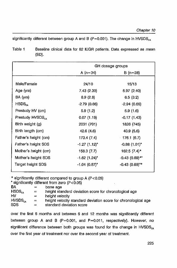
Chapter 10
significantly different between group A and B (P=O.OOl). The change in HVSOSCA
Table 1 Baseline clinical data for 62 IUGR patients. Data expressed as mean (SO).
GH dosage groups
A (n=34)
Male/Female 24/10
Age (yrs) 7.43 (2.30)
BA (yrs) 6.9 (2.8)
HSOS"" -2.79 (0.66)
Prestudy HV (cm) 5.9 (1.2)
Prestudy HVSOSCA 0.07 (1.19)
Birth weight (g) 2031 (701)
Birth length (cm) 42.6 (4.6)
Father's height (cm) 173.4 (7.4)
Father's height SOS -1.27 (1.12)'
Mother's height (cm) 158.3 (7.7)
Mother's height SOS -1.62 (1.24)"
Target height SOS -1.04 (0.87)'
• significantly different compared to group A (P<0.05) , significantly different from zero (P<0.05) BA bone age
B (n=28)
15/13
6.97 (2.40)
6.5 (3.2)
-2.94 (0.66)
5.9 (1.6)
-0.17 (1.43)
1826 (745)
40.9 (5.6)
176.1 (6.7)
-0.88 (1.01)'
162.5 (7.4)'
-0.43 (0.89)"
-0.43 (0.89)"
HSOS"" height standard deviation score for chronological age HV = height velocity HVSOS"" height velocity standard deviation score for chronological age SOS standard deviation score
over the first 6 months and between 6 and 12 months was significantly different
between group A and B (P=O.OOl, and P=O.Oll, respectively). However, no
significant difference between both groups was found for the change in HVSOS""
over the first year of treatment nor over the second year of treatment.
225
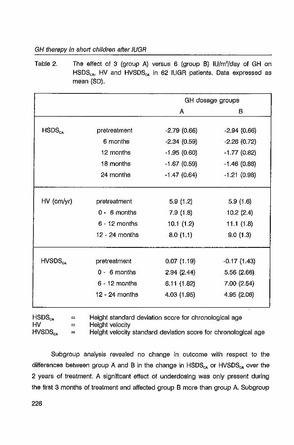
GH therapy in shorf children after fUGR
Table 2.
HSOSCA
HV (cm/yr)
HVSOSCA
The effect of 3 (group A) versus 6 (group B) IU/m'/day of GH on
HSOScA> HV and HVSOSCA in 62 IUGR patients. Data expressed as mean (SO).
GH dosage groups
A B
pretreatment -2.79 (0.66) -2.94 (0.66)
6 months -2.34 (0.59) -2.26 (0.72)
12 months -1.95 (0.60) -1. 77 (0.82)
18 months -1.67 (0.59) -1.46 (0.88)
24 months -1.47 (0.64) -1.21 (0.98)
pretreatment 5.9 (1.2) 5.9 (1.6)
0- 6 months 7.9 (1.8) 10.2 (2.4)
6 - 12 months 10.1 (1.2) 11.1 (1.8)
12 - 24 months 8.0 (1.1) 9.0 (1.3)
pretreatment 0.Q7 (1.19) -0.17 (1.43)
0- 6 months 2.94 (2.44) 5.56 (2.66)
6 - 12 months 6.11 (1.82) 7.00 (2.54)
12 - 24 months 4.03 (1.95) 4.95 (2.06)
HSOSCA
HV HVSOSCA
= Height standard deviation score for chronological age Height velocity Height velocity standard deviation score for chronological age
Subgroup analysis revealed no change in outcome with respect to the
differences between group A and B in the change in HSOSCA or HVSOSCA over the
2 years of treatment. A significant effect of underdosing was only present during
the first 3 months of treatment and affected group B more than group A. Subgroup
226
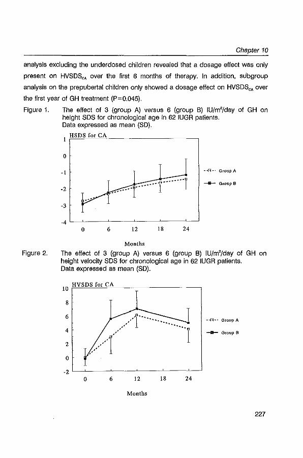
Chapter 10
analysis excluding the underdosed children revealed that a dosage effect was only
present on HVSOSCA over the first 6 months of therapy. In addition. subgroup
analysis on the prepubertal children only showed a dosage effect on HVSOSCA over
the first year of GH treatment (P=O.045).
Figure 1.
Figure 2.
The effect of 3 (group A) versus 6 (group B) IU/m'/day of GH on height SOS for chronological age in 62 IUGR patients. Data expressed as mean (SO).
I HSDS for CA
o
-I --(3-' GroupA
___ Group B
-2
-3
o 6 12 18 24
Months
The effect of 3 (group A) versus 6 (group B) IU/m'/day of GH on height velocity SOS for chronological age in 62 IUGR patients. Data expressed as mean (SO).
10 HVSDS for CA
8
6 --(3-- Group A
4 __ Group B
2
o
o 6 12 18 24
Months
227
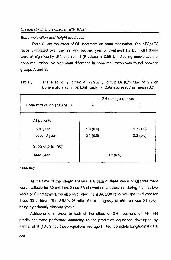
GH therapy in short children after IUGR
Bone maturation and height prediction
Table 3 lists the effect of GH treatment on bone maturation. The !J.BN!J.CA
ratios calculated over the first and second year of treatment for both G H doses
were all significantly different from 1 (P-values < 0.001), indicating acceleration of
bone maturation. No significant difference in bone maturation was found between
groups A and B.
Table 3. The effect of 3 (group A) versus 6 (group B) IU/m'/day of GH on bone maturation in 62 IUGR patients. Data expressed as mean (SD).
GH dosage groups
Bone maturation (!J.BN!J.CA) A B
All patients
first year 1.8 (0.9) 1.7 (1.0)
second year 2.2 (0.9) 2.3 (0.9)
Subgroup (n=30)"
third year 0.6 (0.9)
• see text
At the time of the interim analysis, BA data of three years of GH treatment
were available for 30 children. Since BA showed an acceleration during the first two
years of GH treatment, we also calculated the !J. BN!J. CA ratio over the third year for
these 30 children. The !J.BN!J.CA ratio of this subgroup of children was 0.6 (0.9),
being significantly different from 1.
Additionally, in order to look at the effect of GH treatment on I'H, FH
predictions were performed according to the prediction equations developed by
Tanner et al (16). Since these equations are age-limited, complete longitudinal data
228
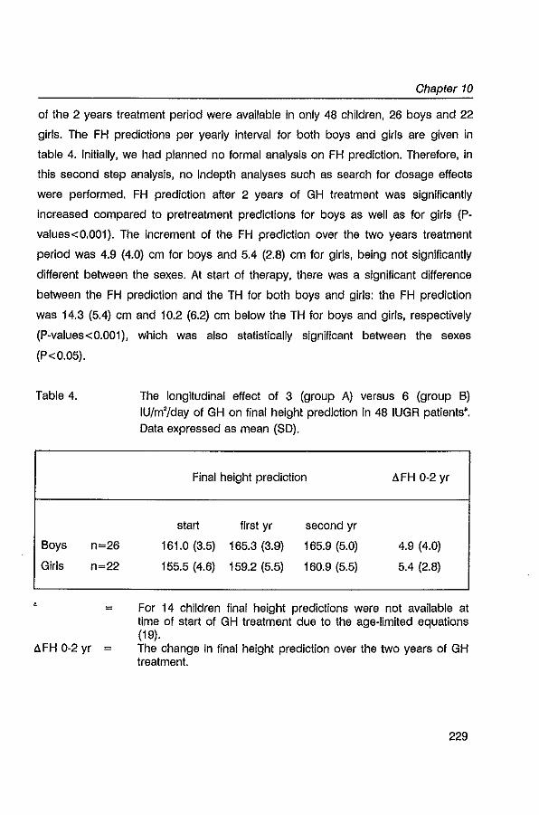
Chapter 10
of the 2 years treatment period were available in only 48 children, 26 boys and 22
girls. The FH predictions per yearly interval for both boys and girls are given in
table 4. Initially, we had planned no formal analysis on FH prediction. Therefore, in
this second step analysis, no Indepth analyses such as search for dosage effects
were performed. FH prediction after 2 years of GH treatment was significantly
increased compared to pretreatment predictions for boys as well as for girls (P
values<0.001). The increment of the FH prediction over the two years treatment
period was 4.9 (4.0) cm for boys and 5.4 (2.8) cm for girls, being not significantly
different between the sexes. At start of therapy, there was a significant difference
between the FH prediction and the TH for both boys and girls: the FH prediction
was 14.3 (5.4) cm and 10.2 (6.2) cm below the TH for boys and girls, respectively
(p-values<0.001), which was also statistically significant between the sexes
(P<0.05).
Table 4. The longitudinal effect of 3 (group A) versus 6 (group B) IU/m'/day of GH on final height prediction in 48 IUGR patients'. Data expressed as mean (SO).
Boys
Girls
n=26
n=22
=
ll.FH 0-2 yr
Final height prediction
start first yr second yr
161.0 (3.5) 165.3 (3.9) 165.9 (5.0)
155.5 (4.6) 159.2 (5.5) 160.9 (5.5)
ll.FH 0-2 yr
4.9 (4.0)
5.4 (2.8)
For 14 children final height predictions were not available at time of start of GH treatment due to the age-limited equations (19). The change in final height prediction over the two years of GH treatment.
229
GH therapy in short children after fUGR
GHlfGFaxis
At start of the study the mean maximal plasma GH response during ATT
was 20.5 (12.2) mU/I in group A and 28.9 (11.1) mUll In group B, which was
significantly different between the groups (P<0.01). In a subgroup of 40 children,
data of 24 hour GH profiles had been collected prior to GH treatment as described
previously (5). We found no significant correlations between parameters of GH
secretion, as determined by 24-hour G H profiles (mean GH, number of peaks
above 20 mUll, peak amplitude and number of peaks) or by pharmacological
testing (maximal GH response during AlT), and the change in HSDSCA over two
years of treatment.
Mean levels of IGF-I and IGFBP-3 at start of the study, expressed as SDS for
CA, were significantly different from zero for both GH groups, indicating reduced
plasma concentrations (table 5) (P-values<0.05). During GH treatment, these
plasma levels increased significantly during the first year in both groups (P-values
< 0.001) and remained so during the second year of treatment. For IGF-I SDSCA
but not for IGFBP-3 SDSCA, a significant difference was observed between groups A
and B for all time points (P<0.001), showing the highest levels in group B. Figure 3
visualizes the change in IGF-I SDSCA and IGFBP-3 SDSCA levels during the 2 yrs of
GH treatment for both groups.
Preliminary analysis showed no correlation between the change in HSDSCA
over the first or second year of GH treatment and baseline levels of IGF-I and
IGFBP-3, expressed as SD scores. However, the change in IGF-I SDS over one
year and over two years of GH therapy correlated significantly with the change in
HSDSCA over two years of treatment (r=0.46, P=0.003 and r=0.49, P=0.001,
respectively). Moreover, the change in IGFBP-3 over two years of GH treatment but
not over the first year of treatment correlated significantly with the change in
HSDSCA over 2 years (r=0.37, P<0.02 and r=0.20, P>0.10, respectively).
Carbohydrate metabolism
Table 5 shows plasma glucose and insulin levels during oGTT. Fasting
230
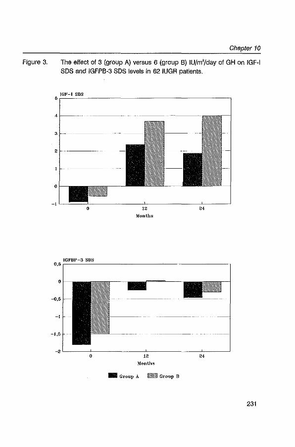
Figure 3.
Chapter 10
The effect of 3 (group A) versus 6 (group B) IU/m'/day of GH on IGF-I SOS and IGFPB-3 SOS levels in 62 IUGR patients.
6~IG~F_-~I~SD=S~ ____________________________________ -,
4
3
2
o
_IL-~~~ __________ -L __________ ~L-__ ~
o 12
Months
24
_IG.=F~B~P-~3~SD=S~ ________________________________ __ 0,6,
o
-0.6
-I
-1.5
_2L-____ -L ____________ -L ____________ -L ____ ~
o 12
Months
_ Group A _ Group B
24
231
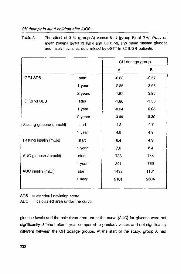
GH therapy in short children after IUGR
Table 5. The effect of 3 IU (group A) versus 6 IU (group B) of GH/m'/day on mean plasma levels of IGF-I and IGFBP-3, and mean plasma glucose and insulin levels as determined by oGIT in 62 IUGR patients.
GH dosage group
A B
IGF-I SOS start -0.88 -0.57
1 year 2.36 3.68
2 years 1.87 3.98
IGFBP-3 SOS start -1.80 -1.50
1 year -0.24 0.03
2 years -0.46 -0.30
Fasting glucose (mmol/l) start 4.5 4.7
1 year 4.9 4.9
Fasting insulin (mUI/I) start 6.4 4.9
1 year 7.6 8.4
AUG glucose (mmol/l) start 786 744
1 year 801 769
AUG insulin (mUll) start 1433 1161
1 year 2101 2634
SOS = standard deviation score AUG = calculated area under the curve
glucose levels and the calculated area under the curve (AUG) for glucose were not
significantly different after 1 year compared to prestudy values and not significantly
different between the GH dosage groups. At the start of the study, group A had
232
Chapter 10
significantly higher levels of fasting insulin than group S (P<O.05). Fasting insulin
levels increased significantly after 1 year of GH treatment for both groups (P<0.01).
In addition, the calculated AUC for insulin increased significantly for both groups
(P<0.001), but was not significantly different between the groups.
Mean HbA" levels were 5.1 (0.3) and 5.0 (0.4) at start of study for group A
and S, respectively. GH therapy induced no significant change in mean HbA"
levels in both groups.
Other safety parameters
Throughout the study there were slight, statistically significant but clinically
non-significant, time-related changes in blood chemistry and thyroid function.
However, all values stayed well within the normal limits, irrespective of GH dosage.
Urinalysis revealed no abnormalities during the study in either study group.
DISCUSSION
The present study shows that 2 years of therapy with recombinant GH gives
a significant improvement of growth In children with persistent short stature after
IUGR. We demonstrated a significant effect of both GH doses (3 (group A) and 6
(group 8) IU/m'/day) on linear growth, expressed as the mean change in HSDSCA
and HVSDSCA over 2 years. In addition, a clear dose-response effect was present
with the highest dose showing a greater change in HSDSCA• In our study, the mean
change in HSDSCA was + 1.3 and + 1.7, whereas the mean change in HVSDSCA was
+4.0 and +5.1 for group A and 8, respectively. A significant, dose-dependent
improvement of linear growth during GH therapy has been described in previous
studies (6,8,11-14). However, these studies showed lower growth reSponses
compared to our study. Chatelain et al (13) reported a mean change in HVSDSCA
after two years treatment of + 1.8 for the highest dose (1.2 IU/kg/week, i.e.
approximately 5 IU/m'/day) and Chaussaln et al (14) described a change In HSDSCA
233
GH therapy in short children after IUGR
of +1.0 and +1.4 after two years of treatment using GH doses of 0.7 and 1.4
IU/kg/week, respectively. These observed differences in growth response to GH
treatment are likely to be related to differences in patient selection. For instance in
our sample, no distinction was made for differences in GH response during
stimulation testing, while others (11-14) have included only children with adequate
GH responses. The reason for us not to exclude children with suspected GH
insufficiency was that in a previous study we did not find a clear relation between
GH secretion status and spontaneous growth in these children (5). Thus, as the
complete pathophysiological mechanism is unclear, we wanted to study whether
there Is a relation between growth response to GH treatment and parameters of
GH secretion and included all patients regardless of their GH secretion, but after
stratification for parameters of GH secretion. Another issue contributing to the
differences In growth response is, that children with short stature secondary to
IUGR probably represent multiple aetiologies; in some way this may be responsible
for a heterogeneous growth response.
In our study, the most remarkable finding was the acceleration in bone
maturation during the 2 years of GH treatment, which appeared to be not dose
dependent. In the recent studies data on bone maturation are not consistent.
Stanhope et al (11,12), studying 24 children with short stature after IUGR,
described that height for SA SDS did not increase after 3 and 4 years of treatment
with 30 IU/m'/week. This implies the presence of an advance in epiphyseal
maturation, since the increase in HVSDS sustained during treatment. Unfortunately,
exact data on bone maturation were not given. In contrast, the combined Belgi
an/French study (13) treating 95 children with either 0.4 or 1.2 IU/kg/week (i.e.
approximately 2 and 5 IU/m'/day, respectively), reported ratios of l!.BNl!.CA varying
from 1.1 to 1.6, which were also not significantly different between both GH dosage
groups. In another French study (14) In which 130 children received either 0.7 or
1.4 IU/kg/week (approximately 3 and 6 IU/m'/day, respectively) or served as
controls, bone maturation over two years of GH treatment did not exceed 1.2 years
per year in either dosage group. The observed differences in bone maturation are
234
Chapter 10
partly due to differences in bone age assessment: some investigators (13,14) have
used bone age determinations according to Greulich and Pyle (24) , while others
(11,12) and we have used the method of Tanner et al (19). Since advanced
progression In bone maturation may attenuate the beneficial effects on final height
we looked at the !J.BN!J.CA ratio over the third year of treatment of a subgroup of
children who had completed 3 years of GH treatment. These data showed that a
decrease in progression of bone maturation is to be expected, increasing the
possibility of an improvement in final height. It should be stressed, however, that
the interpretation of bone maturation is hampered by the clinical impression that
spontaneous epiphyseal maturation is inappropriately advanced between the age
of 6-10 years in many untreated IUGR children with or without dysmorphic features
and that precocious or early puberty Is seen more frequently than in .healthy
children (3,25-28). Therefore, one should be cautious to place too much emphasis
on bone age data, as also mentioned by others (6,11-13).
Despite the observed progression in bone maturation, a significant
improvement In FH prediction was observed during the treatment period. This
suggests that the increase in height surpasses the Increase in bone maturation.
However, as with bone age determination, one should be extremely cautious to rely
completely on height prediction. Height prediction in children with growth disorders
may overestimate adult final height (29,30). Only long-term studies with the
attainment of adult final height will enable definite conclusions on the benefit of GH
treatment in children with short stature after IUGA. Our study is an ongoing,
double-blinded study on the effect of two doses of GH on linear growth and FH.
Therefore, final results have to be awaited.
We have shown previously, that a substantial part of the children with short
stature after IUGR have biochemical signs of GH insufficiency (5). This is confirmed
by this study on a larger group of IUGR children: we found significantly reduced
levels of IGF-I and IGFBP-3 compared to healthy children. Since GH is one of the
most important regulatory factors for IGF-I and IGFBP-3, our findings support the
hypothesis that persistent defects in the GH/IGFaxis are present in children with
235
GH therapy in short children after IUGR
short stature after IUGR. The underlying mechanism, however, remains to be
established. It is speculative that the disturbances In GH secretion observed in
children with IUGR reflect alterations in brain structure and function. In utero, the
brain undergoes a period of Impressive growth and development (31,32). During
this period, it may be vulnerable to nutrititional disturbances. Indeed, animal studies
have shown evidence that intrauterine undernutrition causes alterations in brain
structure and function and affects the postnatal growth programme (33). Other
lines of evidence are derived from clinical studies on children with IUGR showing
an association between IUGR and an increased prevalence of mental handicap,
lower intelligence, neurological deficits, poor academic performance and behavior
problems (34-39). However, several children showed no catch-up growth in the
absence of distinct GH/IGFaxis abnormalities. Therefore, one has to speculate
about other factors beside the GH/IGFaxis responsible for the stunted growth such , as hypothalamic appetite center disturbances (33), reduced cell number (40) or
abnormalities at the level of the GH receptor or secondary messenger systems.
The growth response to GH therapy varies considerably. In a preliminary
analysis, we found no significant correlations between pretreatment parameters of
GH secretory status and the growth response to GH therapy. This Is in contrast to
the study of Albertsson-Wikland (6), who observed an inverse relationship between
the amount of GH secreted over 24 hours, as determined by 24 hour GH profiles,
and the increase in HVSDS. However, evaluating the relation between the growth
response to GH therapy and biochemical changes during therapy we did find
significant correlations. This Implies, that it might be possible to predict the
Individual growth response to GH therapy after a limited period of GH treatment. In
order to predict growth response to GH therapy, Ranke and coworkers (41)
performed a regression analysis on data from 135 children with IUGR using various
auxologlcal predictor variables. It appeared that the GH dosage applied had the
greatest influence on the growth response to GH therapy during the first year of
treatment. However, only 23% of the variability of the growth response could be
predicted from these variables and the GH dose accounted for most of the
236
Chapter 10
explained variability (13%). In future, more indepth analyses including auxological
and biochemical parameters are needed to investigate to what extent the growth
response to GH treatment can be predicted in children with IUGA.
Concern has been expressed, that children treated with pharmacological
doses of GH may develop carbohydrate intolerance and hyperinsulinaemia similar
to those seen in acromegaly (42,43). In our study, glucose tolerance was
maintained at the expense of a rise in fasting insulin concentrations and an
Increase In total Insulin production after oral glucose stimulation. These findings are
in concert with previous findings in short normal children treated with GH (4'4-46). It
is speculated that the hyperinsulinemia may be a direct trophic and/or
insulinotropic effect of GH on beta-cells (44), rather than a secondary consequence
of insulin resistance. It cannot be excluded however, that insulin resistance may
develop with longer duration of GH treatment leading to glucose intolerance or
diabetes. It is therefore recommended to monitor carbohydrate metabolism during
and after the course of GH treatment in these children, especially since it has been
shown that IUGR per se is associated with Insulin resistance and impaired glucose
tolerance at adult life (47-49).
In summary, treatment with recombinant GH in prepubertal children with
short stature after IUGR for two years has resulted in a dose-dependent increase in
growth. However, an increase in bone maturation was observed as well, although
dose-independent. Nevertheless, a significant improvement in FH prediction was
present, suggesting that the increase in height surpassed the increased bone
maturation. It should be stressed, however, that data on bone age and height
prediction have to be handled with caution since the natural pattern of growth and
development may be different in children with IUGR. Preliminary analysis showed
no correlations between pretreatment parameters of GH secretory status and the
growth response to GH treatment. However, significant correlations were observed
between changes in biochemical parameters during GH therapy and the growth
response after two years of GH therapy. Additional analyses are required to study
the possibility of predicting the growth response to GH therapy in IUGR children.
237
GH therapy in short children after IUGR
GH treatment induced no serious adverse effects and glucose tolerance was not
affected. However, GH therapy induced a significant Increase in fasting insulin and
total insulin levels after glucose loading. Monitoring of carbohydrate metabolism is
therefore recommended during and after the course of GH treatment. Final height
results of this ongoing, double-blind study are required before definite conclusions
can be drawn.
ACKNOWLEDGEMENTS
Mrs. Janneke van Nieuwkasteele, research-nurse, is greatly acknowledged
for her assistance. We kindly thank Dr. Theo Stijnen for his statistical advice and
Mr. Dr. C. Lameer for performing ali bone age assessments. This study was
supported by Novo Nordisk NS, Denmark.
238
Chapter 10
REFERENCES
1. Albel1sson-Wikland K. Karlberg J; Natural growth In children born small for gestational age wnh and without catch·up growth. Acta Paediatr Scand 1994;399(SuppQ:64·70
2. Hokken·Koelega ACS, de Aidder MAJ, van Lemmen AJ, den Hartog H, de Muinck KeizerSchrama SMPF, Drop SLS: Children born small for gestational age: do they catch-up? Ped.Aes. 1995;38:267-271
3. Job JC, Rolland A. Histoire naturelle des retards de croissance a debut Intra-utt~~rin. Arch Fr Pedlalr 1986;43:301-306
4. Chaussain JL, Colle M, Ducret Jp: Adull height in children wnh prepubertal short stature secondal)lto intra uterins growth retardalion. Acta Paedlatr Scand 1994;399(Suppl):72-73
5. De Waal WJ, Hokken-Koelega ACS, Slijnen Th, de Muinck Kelzer-Schrama, Drop SLS and the Dutch Working Group on Growth Hormone: Endogenous and stimulated GH secretion, urinary GH excretion, and plasma IOF-1 and IGF-II levels In prepubertal children with short stature after intrauterine growth retardation. Clln Endocrinel 1994;41:621·630
6. Albertsson-Wikland K: Growth hormone secretion and growth hormone Ireatment in children with Intrauterine growth retardalion. Acta Paedlatr Scand 1989;349(SuppQ:35-41
7. Rochlccloll P, Tauber M, Moisan V, Pinkowski C: Investigation of growth hormone secretion In patients with Intrauterine growth retardalion. Acta Paediatr Scand 1989;349(SuppQ :42-46
8. Stanhope A, Ackland F, Hamill G, Clayton J, Jones J, Preece MA: Physiological growth
hormone secretion and response to growth hormone treatment in children with short stature and Intra·uterine growth retardalion. Acta Paediatr Scand 1989;349(Suppl):47-52
9. Tanner JM, Whitehouse RH, Hughes peR, Vince FP: Effect of human growth hormone treatment for 1 to 7 years on the growth of 100 children with growth hormone deficiency, low birth weight, Inherited smallness, Turner's syndrome, and other complaints, ·Arch Dis Child 1971 ;46:745-782
10. Grunt JA, Enriquez AA, Daughaday WH, Budd S: Acute and long-term responses to hGH in children with idiopathic smail-far-dates dwartism. J Clln Endocrinol Metab 1972;35:157-
168 11. Stanhope A, Preece MA, Hamill G: Does growth hormone treatment improve final height
atlainment of children with intrauterine growth retardation? Arch Dis Child 1991 ;66:1180-1183
12. Albanese A. Stanhope R: Growth and metabolic data following growth hormone treatment
of children with intrauterine growth retardation. Horm Aes 1993;39:8-12 13. Chatelain P, Job JC, Blanchard J, Ducret JP, Olivier M, Sagnard L, Vanderschueren
Lodeweyckx M on behalf of the Belgian and French pediatric clinics and Sanofl-Choay (France): Dose·dependent catch·up growth after 2 years of growth hormone trealment In intrauterine growth retarded children. J Clin Endocrinol Metab 1994;1454-1460
14. Chaussain JL, Colle M, Landier F: Effects of growth hormone therapy In prepubertal children with short stature secondary to Intrauterine growth retardation, Acta Paediatr Scand 1994;399(SuppQ:74-75
239
GH therapy in shott children after fUGR
15. Usher A, Maclean F: Intrauterine growth of liveborn caucasian infants at sea level: Standard obtained from measurements of infants born between 25 and 44 weeks of gestation. J Pedlatr 1969;74:901·910
16. Roede MJ, Van Wieringen JC: Growth diagrams 1980, Netherlands. Rhlrd nation·wlde survey. T Soc Gezondheldsz 1985:62(SuppO:1-34
17. Rikken B, Wit JM: Prepubertal height velocity references over a wide age range. Arch Dis Child 1992;67:1277-1280
18. Tanner JM, Whitehouse A: Longitudinal standards tor height, weight-height, height velocity and stages 01 puberty. Arch Dis Child 1976:51:170·179
19. Tanner JM, Whitehouse RH, Cameron N, Marshall WA, Healy MJR, Goldstein H: Assessment 01 skeletal maturity and prediction 01 adult height (TW2·method). Academic Press, London, 1983
20. Hokken·Koelega ACS, Hackeng WHL, Stijnen Th, WIT JM, de Muinck Keizer-Schrama SMPF, Drop SLS: Twenty·lour-hour growth hormone (GH) profiles, urinary GH excretion, and plasma Insulin-like growth factor-I and ·11 levels in prepubertal children with chronic renal Insufficiency and severe growth retardation. J elin Endocrinel Metab 1990;71:688-695
21. Martin JL, Baxter RC: Insulin·like growth lactor·blndlng protein Irom human plasma. Purification and characterization. J Bioi Chern 1986;261:8754·8760
22. Van den Brande JL, Degenhart HJ: Endocrinologie en groelstoornlssen, In: Van de Brande JL, Van Gelderen HH, Monnens LAH (eds). Klndergeneeskunde. Wetenschappelljke UITgeverij Bungen, Utrecht, 1993, p179·219
23. Job JC: Results 01 long.term growth hormone replacement therapy In children: when and how to treat. Horm Res 1990;33(SuppIA):69·76
24. Greulich WW, Pyle SI: Radiographic atlas 01 skeletal development 01 the hand and wrist. Stanlord University Press, Stanlord, 1959
25. Tanner JM, Lejarraga H, Cameron N: The natural history of the Silver-Russell syndrome: a longitudinal study 01 thirty·nlne cases. Pediatr Res 1975;9:611·623
26. Davies PSW, Valley R, Preece MA: Adolescent growth and pubertal progression In the Silver·Russell syndrome. Arch Dis Child 1988;63:130·135
27. Wollmann HA, Ranke MS, Kirchner T, Preece M: Silver Russell syndrome: spontaneous growth and symptoms in 386 patients. Horm Res 1994;41:73
28. Ar/saka 0, Arlska M, Kiyokama N, Shimizu T, Nakayama Y, Yabuta K: Intrauterine growth retardation and early adolescent growth spurt In two sisters. Clin Ped 1986;25:559·561
29. Zachmann M, Sobradillo B, Frank M, Frisch H, Prader A: Bayley-Pinneau, Roche-WainerThissen, and Tanner height prediclions in normal children and In patients with various pathologic conditions. Pediatrics 1978;93:749·755
30. Chaussaln JL, Ducret JF, Job JC: Actual versus predicted final height In patients with
Intrauterine growth retardation. Pedlatr Res 1993;5(SuPPO:S38 31. Dobbing J, Sands J: Timing of neuroblast mulliplication in developing human brain. Nature
1970;226:639·640 32. Dobbing J, Sands J: Comparative aspects of the brain growth spurt. Early Hum Dev
1979;3:79·83
240
Chapter 10
33. Davies DP: Growth of 'smail-far-dates' babies. Review. Early Hum Dev 19B1;5:95-105 34. Fitzhardinge PM, Steven EM: The small for dates infant II: neurological and intellectual
sequelae. Pediatrics 1976;50:30·57 35. HalVey 0, Prince J, Bunton J, Parkinson C, Campbell S: Abilities of children who were
small-far-gestational age babies. Pediatrics 19B2;69:269-300 36. Westwood M, Kramer MS, Munz D, Lovett JM, Watters GV: Growth and development of
full-term nonasphyxlated small-for-gestalional-age newborns: follow-up through
adolescence. Pediatrics 1963:71:376-3B2 37. Henrichsen L, Sklnhoj K, Andersen GE: Delayed growth and reduced intelligence in 9-17
year old intrauterIne growth retarded children compared with their monozygous co-twins, Acta Paedlatr Scand 19B6;75:31-35
38, MaUlalnen A, Heinonen K, Siren-Tiusanen H: Effect of Intrauterine growth retardation (IUGR) on the psychological perlormance of preterm children at preschool age. J Child Psychol Psychlat 19BB;29:601-609
39. Van der RelJden-Lakeman EA: Growing pains? Psychological evaluation of short stature In
intrauterine growth retarded children, before and after two years of GH treatment. Thesis,
Rotterdam, 1996 40. Winick M: Malnutrition and brain development. J Pedlatr 1969;74:667-679 41. Ranke MB, Guilbaud 0, Lindberg A, Cole T: Prediction of the glWoth response In children
with various growth disorders treated with growth hormone: analyses of data from the Kabi
Pharmacia International Growth Study. Acta Paediatr 1993;391 (Suppl):82-BB 42. Davidson MB: Effect of growth hormone on carbohydrate and lipid metabolism. Endocrine
Reviews 1987;8:115-131 43. Gustafsson J: Possible metaboliC side effects of high dose growth hormone treatment.
Acta Paedlatr Scand 19B9; 362(Suppl):50-55 44. Walker J, Chaussain JL, Bourgneres PF: Growth hormone treatment of chlldren with short
stature increases Insulin secretion but does not impair glucose disposal. J Clin Endocrinol
Metab 19B9;69:253-25B 45. Hlndmarsh PC, Pringle PJ1 Di Silva L, Brook CGD: Effects of 3 years of growth hormone
therapy in short normal children. Acta Paediatr Scand 1990:366(Suppl):6-12 46. McCaughey ES, Mulligan J, Voss LD, Betts PR: Growth and metabolic consequences of
growth hormone treatment In prepubertal short normal children. Arch Dis Child
1994;71 :201-206 47. Phipps K, Barker DJP, Hales CN, Fall CH, Osmond C, Clark PM: Fetal growth and impaired
glucose tolerance In men and women. Dlabetologla 1993:36:225-228 4B_ Phillips DIW, Barker DJP, Hales CN, Hirst S, Osmond C: Thinness at birth and insulin
resistance in adult life. Dlabetologla 1994;37:150-154 49. Law CM, Gordon GS, Shiell AW, Barker DJP, Hales CN: Thinnes at birth and glucose
tolerance in seven year old children. Diabetic Mad 1995;12:24-29
241


CHAPTER 11
General Discussion and Conclusions.
General discussion and conclusions
INTRODUCTION
Children growing at the extremes of height may experience psychological,
social, practical and/or physical problems. Hormonal treatment intervening in the
process of growth may be employed to Improve height prognosis. This thesis
describes various clinical studies undertaken to obtain more insight into the
possibilities and limitations of therapeutic intervention in children presenting with
extremes of growth: constitutionally tall stature (CTS) and short stature after
intrauterine growth retardation (IUGR). The major question addressed in these
studies was: is it possible to influence childhood growth by means of hormonal
treatment in such a way that it is beneficial to children with extremes of growth?
EXTREMES OF GROWTH
Constitutionally tall stature
Constitutionally tall stature is a normal variant of childhood growth. Genetic
and familial factors play an important role in etiology and pathogenesis.
Endocrinological studies indicate that tall stature is due, at least partly, to increased
GH secretion. However, other mechanisms might playa role as well. The concept
of treating tall adolescents to reduce final adult height developed from the
observations in children with precocious puberty. In these children, pr~mature
secretion of sex hormones induces a progression in bone maturation, which leads
to early epiphyseal closure and limitation of linear growth. Therefore, sex steroids
have been used for final height reduction since the 1950s (1).
Aoouraoy of height prediction and effeot of treatment
The main question was: Is treatment with pharmacological doses of sex
steroids effective in order to reduce final adult height in children with CTS without
244
Chapter 11
long-term side effects?
Our study gives a more differentiated answer to this question than many
other reports thus far (1-25)_ The generally accepted opinion is, that sex hormone
treatment is effective in reducing final height, although claimed results may vary (1-
25). This variability is mainly due to differences in study design, therapeutic
regimen, inclusion criteria (such as age and bone age at initiation of treatment) and
definition of adult final height (15,17). The main problem in assessing the effect of
sex hormone treatment Is that the effect of treatment depends on the method used
to predict final adult height. A critical appraisal of the quality of the various
prediction methods Is required especially since most prediction methods are based
on growth data derived from normal growing children. The reliability of height
prediction methods in CTS has been rarely studied In large groups of untreated tall
children (26-29). Therefore, our studies on the reliability of nine different prediction
methods In large groups of untreated tall children (55 boys and 88 girls) are
clinically relevant.
We demonstrated that height prediction In tall girls is rather accurate: most
prediction methods showed a mean error below 1 cm with mean absolute errors
varying from + 1.9 to +3.7 cm. In contrast, in tall boys height prediction was found
to be rather inaccurate with mean errors ranging from -5.3 to +2.8 cm and mean
absolute errors varying from +2.3 to +5.3 cm for the various prediction methods.
An important finding was that at the time of referral the control groups (girls and
boys) were significantly different to children who had received sex hormone
treatment. They were shorter, had smaller parents, had a more advanced bone age
and, consequently, had smaller final height predictions. This Implies that if one
wants to establish the effect of sex hormone treatment, one cannot simply correct
the calculated effect of treatment for the error of prediction derived from these tall
control groups. However, this has been done In most previously reported studies.
We used a more direct and conceptually simple approach using multiple linear
regression. Basically in this approach, the treatment effect on final height can be
calculated while adjusting for differences in age, bone age and height between
245
General dlsoussion and oonolusions
treated and untreated children. In this way, the mean adjusted effect for the various
prediction methods varied from -1.7 to +0.7 cm In boys and from +1.1 to +2.4 cm
in girls. These outcomes are clearly less than reported previously (1-25) and
seriously question the applicability of high doses of sex hormones in children with
CTS to limit final adult height; this applies particularly to boys. It should be noted
however, that there was a marked variation of the individual height reducing effects.
For example, in boys a range in effect was found from -4.6 to + 15.8 cm while in
girls the range in effect varied from -2.6 to +6.2 cm. In fact, we clearly showed that
the height reduction was dependent on the bone age at the start of treatment:
height reduction was more pronounced when treatment was started at a younger
bone age. This bone age-related effect is in concert with previous studies
(5,7,13,18,19,21,23,24).
Surprisingly, when treatment in boys had been started at a relatively 'old'
bone age I.e. bone age> 14.0 to 15.5 years depending on the method used, a
growth induction was observed instead of a growth reduction. This finding has not
been reported before and contributes to a limitation of androgen use in boys with
CTS. We speculate that this might be due to the presence of a sex-specific and
age-dependent responsiveness of cartilage and bone cells to sex steroids, as
shown in vitro (30-33).
Another Important issue that caused a significant reduction in the height
limiting effect of sex steroid treatment, was the observation of a marked additional
post-treatment growth after cessation of therapy. It appeared that this post
treatment growth was mainly due to the fact that treatment had been stopped
before complete closure of the epiphyses. Therefore, continuation of treatment until
complete closure of the growth plates has occurred will contribute to a more
favourable effect in height reduction. According to the method of Greulich and Pyle,
this Implies that treatment has to be continued preferably until a bone age 18.0
years in boys and 16.5 years in girls.
246
Chapter 11
Long-term effeots
At a mean follow-up period of 10 years, we found no evidence that
administration of pharmacological doses of sex steroids has long-term side effects.
Although sex hormone treatment induced various side effects during treatment,
these tended to be mild and occasionally led to cessation of treatment. This is in
concert with previous studies (4,5,23,33-35). We obtained information about 127
pregnancies and found no difference in outcome between treated and untreated
tall subjects. In addition, sperm quality in men was not affected. However,
previously treated tall men had significantly higher levels of plasma FSH. The
meaning of this latter finding remains to be established. We speculate that the
higher levels of FSH may reflect intratesticular changes due to androgen treatment
received in a period of testicular maturation. These increased FSH levels may
compensate for partially disturbed germinal function in order to maintain normal
sperm quality. On the other hand, the difference in gonadotropin levels may also
reflect a change In responsiveness at the hypothalamic-pituitary level. We Intend to
study sperm quality and plasma FSH levels in these men with increasing age in
order to see whether sperm quality will be maintained. In women no case of
malignancy was reported. However, the considerable amount of data relating long
term oral contraceptive use to breast cancer cannot be Ignored (36-41). Although
the potential hazard should not be exaggerated, it might raise the need for long
term follow-up in these girls.
Psyohosooial aspeots
One may argue about the effectiveness and applicability of sex hormone
treatment In the management of tall stature. It Is oertain however, that some
children with CTS experience psychosocial problems related to their tallness. In our
study, many tall adults recalled to be subject to hurtful remarks and jokes about
their height; they were frequently teased during childhood. Probably, this plays a
major role in the decision in favour of hormone treatment. As adults, previously
treated men and women showed no major psychological maladjustment compared
247
General discussion and conclusions
to tall controls. Since our study was a retrospective one, it remains speculative,
however, whether treatment with sex hormones did have a beneficial effects on
psychosocial functioning. Only properly designed prospective studies will enlighten
this Important issue. Therefore, inventarisation of psychosocial problems prior to
possible treatment Is urgently needed.
Conclusions and recommendations
Our studies led us to make the following conclusions and recommendations:
1. Whenever a child presents with constitutionally tall stature, a reliable height
prediction is of utmost importance. Extrapolation of the height SDS for bone
age according to Greulich and Pyle has been shown to be an easy. clinical
tool to make a rather accurate height prognosis in tall children. Otherwise,
the regression equations based on a reference group of untreated tall
children can be used, as described in chapter 7.
2. Dependent on the height prognosis, sex steroid treatment for final height
reduction may be considered. However, the expected effect of such a
treatment is significantly dependent on the bone age at the time treatment
may be started. As outlined in chapters 3 and 4, the equations in table 5 can
be used to estimate the expected effect of treatment according to various
prediction methods and methods of bone age assessment.
3. If treatment is given, it should be continued until complete closure of the
epiphyses has occurred. According to the method of Greulich and Pyle, this
implies that treatment has to be continued preferably until a bone age 18.0
years in boys and 16.5 years in girls.
4. In boys, we do not favor the start of sex hormone treatment at a bone age
beyond 14 years according to Greulich and Pyle and beyond 15 years
according to Tanner-Whitehouse. It might Increase final adult height rather
than limit it.
5. Reassurance can be given to patients and parents about the fact that to
date no evidence of long-term side effects have been demonstrated.
248
Chapter 11
However. the presence of elevated FSH levels with normal sperm quality In
previously treated tall men should be discussed.
Considerations
In order to Increase the height reducing effect of hormonal treatment. other
ways of intervention may be explored. Recent approaches have focussed on
suppression of GH secretion In children with CTS using somatostatin analogues
(42-44). These preliminary results are promising. However. final results remain to be
established and the occurrence of adverse effects has to be considered. It may be
worthwile to combine these two modes of height intervening strategies: reduction
of GH secretion by means of somatostatin analogues and progression of bone
maturation via high doses of sex steroids. Hindmarsh et al (44) demonstrated a
shift of the growth curve to the right by administration of octreotide (a somatostatin
analogue) to prepubertal children with CTS. Therefore It might be considered to
treat tall children with octreotide during prepuberty. whereafter sex steroid therapy
can be applied or given as adjuvant. Future studies are required to investigate the
effect and safety of these theoretical modalities on height reduction in children with
tall stature.
Short stature after IUGR
The clinical importance of IUGR is apparent. The association between the
degree of IUGR and perinatal morbidity and mortality and the greater risk of
asphyxia-induced encephalopathy with consequent risks of neurological
dysfunction have been well established. In addition. infants born with IUGR may
have lifelong consequences. Barker and associates have presented considerable
evidence linking abnormal fetal and/or placental growth to development of
cardiovascular diseases (reviewed in reference: 45). Furthermore. an association
has been found between IUGR and both insulin resistance and impaired glucose
intolerance in adulthood (45.46). Finally. persistent growth failure may be present
249
General discussion and conclusions
(47-52). A recent study showed that children born small-for-gestational age with
birth length less than -2 SD below the mean have a 7-fold higher risk for short
stature In adulthood compared to non-SGA children (53). In fact, at 18 yrs of age
22% of the total short population were found to be short at birth «-2SD)(53). The
studies presented in the second part of this thesis mainly adressed the problem of
persistent growth failure. The main question was whether treatment with
recombinant GH would Improve growth and ultimately final height In children with
short stature after IUGR.
Pathophysiology
Although GH therapy is explored in many short children without any sign of
classical GH deficiency such as Turner syndrome (54), supportive evidence for GH
intervening therapy in children with short stature after IUGR came from
endocrinological studies showing GH secretion disturbances in many of these
children (55-57). These studies comprised small groups of children and did not
address IGFs or urinary GH excretion. We studied the GH/IGFaxis in a group of 40
prepubertal IUGR children and demonstrated that 50-60% of these children have
signs of GH insufficiency. The etiology of this remains to be established. Most of
our children experienced growth retardation in early pregnancy (symmetric IUGR).
It is possible that the factors that induce the pathophysiological state that causes
IUGR, may also affect organ development. Especially the brain seems to be
vulnerable to disturbances in its nutrititional supply, since important brain growth
and development occurs in utero (58,59). Indeed, animal studies give arguments
supportive to the hypothesis of neuropsychological dysfunction due to IUGR (60).
Other lines of evidence are derived from clinical studies in children with IUGR
showing an association between IUGR and an increased incidence of mental
handicap, lower intelligence, neurological deficits, poor academic performance and
behavior problems (61-65). In the parallel study on psychological aspects, IUGR
children scored lower in Intelligence tests and showed more disturbances in
attention and concentration, indicating a possible underlying neurophysiological
250
Chapter 11
deficit (66). It Is speculative that the GH secretion disturbances observed in children
with IUGR reflect alterations in brain structure and function. Since the high
percentage of GH secretory abnormalities might be attributable to GH pulse
amplitude disturbances, this dysfunction could be at the hypothalamic level.
However, there were several children with no distinct GH/IGFaxis abnormalities
who still failed to show catch-up growth. Therefore other mechanisms may be
operative as well.
An alternative hypothesis explaining why postnatal growth may be disturbed
due to prenatal factors concerns an alteration in the nuclei and tracts of the
hypothalamic appetite center (67). In the human fetus, the hypothalamic appetite
center develops from the fourth to seventh months in fetal life. Intrauterine
undernutrition may affect this developing process. Indeed, in many children with
IUGR and short stature feeding problems seem to be present. In animal models,
temporary early life dietary protein restriction leads to lack of catch-up growth and
produces long-term alterations in GH and insulin secretion which may underlie the
impairment in subsequent physical growth (68). To our knowledge, there are no
studies evaluating the consequences of possible malnutrition on growth during
childhood In children with IUGA. Finally, malnufrition in early intrauterine life may
Interfere with the rate of cell division, leaving the fetus with fewer cells and thus
limiting the potential for catch-up growth as suggested by Winick (69).
Children with short stature after IUGR should be distinguished from children
with Idiopathic or normal short stature. Short stature after IUGR seems to be a
distinct entity. This is illustrated by the fact that the intrauterine environment seems
to influence postnatal growth in such way that it overrules the genetic potential of
the child. In our study, the predicted adult height at start of GH treatment was more
than 10 cm below the target height. Usually in idiopathic short stature, no such
discrepancy has been found (70). In addition, evidence for GH/IGF disturbances
have been described In many patients with IUGR shortly after birth and during
childhood (55-57,71-73). By definition, children with idiopathic short stature show
no signs of GH insufficiency. However, as in idiopathic short stature, children with
251
General discussion and conclusions
IUGR represent a heterogeneous group. Efforts should be made to categorize
those patients who respond to hormonal therapy from those who are non·
responders.
Effect of GH treatment
Although the pathophysiological mechanism is not quite clear, it seems
justified to start clinical trials to investigate whether treatment with recombinant GH
might be beneficial for IUGR children. Therefore, we set up a randomized, double·
blind, dose dependent, multicenter study to investigate the effect of GH therapy on
linear growth, bone maturation, pubertal development and final height. After two
years of treatment, we demonstrated a significant dose· dependent effect of GH
therapy on growth acceleration, expressed as the change in HSDSCA• However, an
increase in bone maturation was also observed, which was dose· independent. The
bone maturation slowed down during the third year of treatment. Despite the
progression in skeletal maturity, a significant improvement in height prognosi~ was
found during the two years treatment period. Nevertheless, we like to stress that
data on bone maturation and height prediction should be Interpreted with caution.
It is our clinical Impression, supported by thrifty literature data (74-78) that
spontaneous epiphyseal maturation is inappropriately advanced between the age
of 6-10 years In many untreated IUGR children with or without dysmorphic features
and that early puberty may occur more frequently thari in healthy children. This
latter phenomenon might also reflect alterations in brain structure and function as
discussed above. It is obvious though, that the possible acceleration of bone
maturation by GH will negatively Influence the positive effects of GH therapy on final
height. Ultimately, only long· term studies with the attainment of final adult height will
enable definite conclusions. Therefore, the present study will be continued until all
children have attained their final adult height.
In our study, glucose tolerance was maintained at the expense of a rise in
fasting insulin concentrations and an increase In total insulin production after oral
glucose stimulation. These findings are in concert with previous findings in short
252
Chapter 11
normal children treated with GH (79-81). It is speculated that the hyperinsulinemia
may be a direct trophic and/or insulinotroplc effect of GH on beta-cells, rather than
a secondary consequence of insulin resistance (79). It cannot be excluded
however, that insulin resistance may develop with prolonged duration of GH
treatment leading to glucose intolerance or diabetes. Since it has been shown that
IUGR per se is associated with an increased prevalence of insulin resistance and
Impaired glucose tolerance in adulthood (45,46,82,83), monitoring of carbohydrate
metabolism during and after the course of GH therapy is recommended.
The effect of GH therapy on growth rate is known to vary considerably.
Therefore, we have searched for factors which might Influence the growth
response. Obviously this would have clinical implications, as prediction of the
growth response to GH treatment is very useful in the management of short
stature. Unfortunately, we could not identify a significant marker prior to treatment
predicting the growth response to GH therapy in children with short stature after
IUGR. For instance, no correlations were found between pretreatment par?meters
of GH secretory status and either growth or growth response to GH therapy.
Albertsson-Wikland reported an inverse relationship between the amount of
spontaneous GH secretion and the 1-year growth response to GH therapy in
children with IUGR (55). However, preliminary analysis revealed that biochemical
changes during therapy did correlate with the growth response to GH therapy. For
instance, the change in IGF-I SOS and IGFBP-3 SOS over the first two years of
treatment was positively correlated with the change in HSOSCA over the same
period (r=0.49 and r=0.37, respectively). This implies, that it might be possible to
predict the individual growth response to GH therapy after a try-out treatment with
GH during a limited period. Ranke et al (84) demonstrated that the GH dosage
applied had the greatest Influence on the growth response to GH therapy during
the first year of treatment in 135 children with IUGR. In their study, only auxological
parameters had been included, which could explain only 23% of the total variability
In growth response. More in depth analyses are needed Including auxologlcal and
biochemical parameters to investigate to what extent the growth response to GH
treatment can be predicted In children with IUGA.
253
General discussion and conclusions
Conclusions and recommendations
From our studies the following conclusions and recommendations can be
made:
1. In 50-60 percent of the children presenting with short stature after IUGR,
signs of GH insufficiency are present. Although the complete
pathophysiological mechanism has to be established, it seems justified to
study the effect of GH therapy in these children.
2. GH treatment induces a significant increase in height and height velocity,
which is dose-dependent. However, bone maturation Is also increased,
independent of the GH dose. This latter finding has to be interpreted with
caution, since it may be influenced by the natural history of growth and bone
age development of these children. Nevertheless, final adult height
prognosis appeared to be significantly Improved after two years of GH
treatment.
3. To date, it is not possible to predict the growth response to GH treatment in
IUGR children adequately. More indepth analyses are needed to evaluate the
possibility of growth response prediction.
4. No major adverse effects of GH treatment are demonstrated. However,
fasting insulin levels and total insulin production after glucose loading are
significantly increased after 1 year of GH treatment. Therefore, it is
recommended to monitor carbohydrate metabolism during and after GH
treatment.
5. Before GH has proven to be effective, treatment of children with short
stature after IUGR with GH should be limited to investigational settings.
Considerations
To evaluate the ultimate effect of GH treatment on final adult height in
children with short stature after IUGR, studies including auxological measurements
and bone age determinations up to final height in a large group of untreated IUGR
children will be of a great value. Establishment of the natural pattern of growth and
development can be used as a reference for GH intervention studies.
254
Chapter 11
REFERENCES
1. Goldzleher M: Treatment of excessive growth In the adolescent female. J Clln Endocrinol 1956; 16:249·252
2. Schoevaart CE, Drop SLS, Otten BJ, SlIjper FME, Degenhart HJ: Growth analysis up to final height and psychosocial adjustment of treated and untreated patients with precocious puberty. Horm Res 1990;34:197·203
3. Brauner R, Adan L, Malandry F, Zantlener 0: Adutt height In girls with Idiopathic true precocious puberty. J Clln Endocrlnol Metab 1994;79:415·420
4. Zachmann M, Ferrandez A, MOrset G, Pradar A: Estrogen treatment of excessively tall girls. Helv Paedlat Acta 1975;30:11-30
5. Wettenhall HNB, Cahill C, Roche AF: Tall girls: A survey of 15 years of management and treatment. J pedlatr 1975;86:602·610
6. Von Puttkamer K, Blench JA, Brugger F, Hlrche W, SchOnberg 0: Ostrogentherapie bel Madchen mit konsthu·tionellem Hochwuchs. DIsch Med Wochenschrft 1977;102:983·988
7. Kuhn N, Blunck W, Stahnke N, Wlebel J, Willig RP: Estrogen treatment In tall girls. Acta Paedlatr Scand 1977; 66:161·167
8. Colle ML, Alperin H, Greenblatt RB: The tall girl. Prediction of mature height and management. Arch Dis Child 1977;52:118·120
9. Bierich JR: Estrogen treatment of girls with constitutional tall stature. Pediatrics 1978;62:1196·1201
10. Aeeser HM, Heremans GFP, van Gelderen HH: Reduction of adult height In tall glrfs. Eur J Pedlatr 1979;132:37-41
11. Willig RP, Christiansen 0, Kuhn N, Schaefer E, Stahnke N: Voraussetzungen und Ergebnisse der Ostrogenbehandlung extrem groBer Madchen. Monatsschr Kinderheilkd 1980; 128:787·788
12, Andersen H, Jacobsen B8, Kastrup'r<M/, Krabbe S, Peitersen 8, Petersen KE, Thamdrup E, Wichmann R: Treatment of girls with excessive height prediction. Acta Paedlatr Scand 1980;69:293·297
13. John G, Schellong G: Ostrogentheraple hochwOchslger Madchen. Monatsschr Klnderhellkd 1980;128:545·550
14. StOver B, Kollmann F: Ergebnisse der Behandlung hochwOchsiger Madchen mit conjuglerten Oslrogenen. Monatsschr K1ndrheilkd 1982;130:36·40
15. Sorgo W, Scholler K, Heinze F, Heinze E, Teller WM: Critical analysis of height reduction in oestrogen·lreated lall girls. Eur J Pedlatr 1984;142:206·265
16. Schambach H, Nitschke U: Ole Behandlung des konstltullonellen Hochwuchses bel Madchen mit physlologlschen Ostrogendosen In der Prapubertat. Monatsschr Kinderheilkd 1985;133:32·37
17. Bartsch 0, Weschke B, Weber B: Oestrogen treatment of consthutlonally tall girls with 0.1 mg/day ethlnyl oestradiol. Eur J Pedlatr 1988;147:59·63
18. GrOters A, Heidemann P, SchlOter H, Stubbe P, Weber B, Helge H: Effect of different oestrogen doses on final height reduction In girls with consthutlonal tall stature. Eur J Ped 1989;149:11·13
255
General discussion and conclusIons
19. Ignatius A, Lenko HL, Perheentupa J: Oestrogen treatment of tall gIrls: effect decreases wilh age. Acta Paedlatr Scand 1991;80:712-717
20. Svan H, Ritzen EM, Hall K, Johansson L: Oestrogen treatment of tall gIrls: dose dependency of effects on subsequent growth and IGF-I levels In blood. Acta Paedlatr Scand 1991;80:328-332
21. Normann EK, Trygstad 0, larsen S, Dahl.Jorgensen K: Height reduction In 539 tall girls treated with three different dosages of elhlnyloestradlol. Arch Dis Child 1991;66:1271-1278
22. Joss EE, Zeuner J, ZurbrOgg RP, Mullis PE: Impact of different doses of ethlnyl oestradiol on reduction of final height In constttutionally lall girls. Eur J Pediatr 1994;153:797-801
23. Zachmann M, Ferrandez A, MOrset G, Gnehm HE, Prader A: Testosterone treatment of excessively tall boys. J Pedlatr 1976;88:116-123
24. Bramswlg JH, Schellong G, Borger HJ, Breu H: Testosteron-Theraple hochwOchsiger Jungen. Ergebnisse bel 25 Jungen. Dtsch Med Wschr 1981;106:1656-1661
25. Bramswig JH, von Lengerke HJ, Schmidt H, Schellong G: The results of short-term (6 months) high dose-testosterone treatment on bone age and adult height in boys of excessively lall stature. Eur J Pedlatr 1988; 148: 1 04-106
26. Zachmann M, Sobradillo B, Frank M, Frisch H, Prader A: Bayley-Plnneau, Roche-WalnerThissen, and Tanner height predictions In normal children and in patients with various pathologic conditions. Pediatrics 1978;93:749-755
27. Willig RP, Christiansen 0, Kuhn N, Schaefer E, Slahnke N: Vorraussetzungen und ergebnisse der Cstrogenbehandlung extrem groOer Madchen. Monatschr Klnderhellkd 1980; 128:787-788
28. Bramswig JH, Hermellng W, von Petrykowskl W, Schellong G: Comparison of height predictions with final adult height in boys wilh constitutional tall stature. In: Berms J, Hausple R, Sand A, Suzanne C, Hebbellnck M (eds). Human growth and development. Plenum Press, New York, 1984; pg,423-429
29. Joss EE, Temperll R, Mullis PE: Adult height in constilutlonally tall stature: accuracy of five different height prediction methods. Arch Dis Child 1992;67:1357-1362
30. Blanchard 0, Tsagris L, Rappaport R, Duval-Beaupere G, COIvol M: Age-dependent responsiveness of rabbit and human cartilage cells to sex steroids In vitro, J Steroid Blochem Mol Bioi 1991;40:711-716
31. Nasatzky E, Schwarz Z, Boyan BD, Soskolne WA, Ornoy A: Sex-dependent effects of 170 estradiol on chondrocyte differentiation In culture J Cell PhysioI1993;154:359-362
32. Schwarz Z, Nasatzky E, Ornoy A, Brooks BP, Soskolne WA, Boyan BD: Gender specific, maturatlon·dependent effects of testosterone on chondrocytes In culture, Endocrinology 1994;134:1540-1647
33. Hanker JP, Schellong G, Schneider HPG: The functional state of the hypothalamo-pituital)' axis after high dose oestrogen therapy In excessively tall girls. Acta EndocrinoI1979;91:19-29
34. Bramswlg JH, Nieschlag E, Schellong G: Pitullal)'-gonadal function In boys after high dose testosterone treatment for excessively tall stature. Acta EndocrinoI19S4;107:97-103
35. Trygstad 0: Oestrogen treatment of adolescent lall girlS; short term side effects. Acta Endocrlnol1986(SuppQ 279:170-173
256

Chapter 11
36. Olsson H, MOiler TR, Ranstam J: Early oral contraceptive use and breast cancer among premenopausal women: final report from a study In southern Sweden. J NaU Cancer Inst 1989;81:1000-1004
37. Schlesselman JJ: Cancer of the breast and reproductive tract In relation to use of oral contraceptives. Contraception 1989;40:1-38
38. Rushton L, Jones DR: Oral contraceptive use and breast cancer risk: a meta-analysis of variations with age at diagnosis, parity and total duralion of oral contraceptive use. Bf J Obstet GynecoI1992;99:239-246
39. Grimes DA: The safety of oral contraceptives: epidemiologic insights from the first 30 years. Am J Obstet GynecoI1992;166:1950-1954
40. Kaunitz AM. Oral contraceptives and gynecologIc cancer: an update for the 19905. Am J
Obstet Gynecol 1992; 167:1171-1176 41. Malone KE, Dalllng JR, Weiss NS: Oral contraceptives In relation to breast cancer.
Epldemlol Rev 1993;15:80-97 42. Tauber MT, Tauber JP, Vigoni F, Harris AG, Rochiccioli P: Effect of long-acting
somatostatin analogue 8MS 201-995 on growth rate and reduction of predicted adult height In ten tall adolescents. Acta Paedlatr Scand 1990; 79:176-181
43. Hindmarsh PC, Pringle PJ, 01 Slivo L, Brook CGD: A preliminary report on the role of somatostatin analogue (SMS 201-995) In the management of children with tall stature. Clin EndocrlnoI1990;32:83-91
44. Hlndmarsh PC, Pringle PJ, Stanhope A, Brook eGo: The effect of continuous infusion of a somatostatin analogue (aetrecUde) for two years on growth hormone secretion and height prediction In tall children. Clin Endocrinol 1995;42:509-515
45 Barker DJP, Gluckman PO, Godfrey KM, Harding JE, Owens JA, Robinson JS: Fetal nutrition and cardiovascular disease in adult life. Review article. Lancet 1993;341:938-941
46. Phipps K, Barker DJP, Hales CN, Fall CHD, Osmond C, Clark PMS: Fetal growth and Impaired glucose intolerance in men and women. Dlabetologla 1993;36:225-228
47. Westwood M, Kramer MS, Munz 0, Lovett JM, Watters GV: Growth and development of full-term nonasphyxlated small-for-gestaUonal-age newborns: foHow-up through adolescence. Pediatrics 1983;71 :376-382
48. Job JC, Rolland A: Hisloire naturelle des retards de croissance a debut intra-uterin. Arch Fr Pediatr 1986;43:301-306
49. paz I, Seidman OS, Danon YL, Laor A, Stevenson OK, Gale R: Are children born for gestational age at Increased risk for short stalure? Am J Dis Child 1993;147:337-339
50. Chaussaln JL, Colle M, Ducret JP: Adult height In children wilh prepubertal short stature secondary to Intrauterine growth retardation. Acta Paedlatr Scand 1994;399(SuppQ:72-73
51. Tanner JM, Lejarraga H, Cameron N: The natural history of the Silver-Russell syndrome: A longitudinal study of thirty-nine cases. Pedlatr Res 1975;9:611-623
52. Davies PS, Valley R, Preece MA: Adolescent growth and pubertal progression in the SiiverRussell syndrome. Arch Dis Child 1988;63:130-135
53. Karlberg J, Albertsson-Wlkland K: Growth In full-term small-for-gestational-age infants: from birth to final height. Pediatr Res 1995;38:733-739
54. Hlndmarsh PC, Bridges NA, Brook CGD: Wider indications for treatment with biosynthetic human growth hormone In children. Review. Clln EndocrinoI1991;34:417-427
257

General discussion and conclusions
55 Albertsson-Wlkland K: Growth hormone secretion and growth hormone treatment In children with Intrauterine growth retardation_ Acta Paedlatr Scand 1989;349(Suppl):35-41
56 Rochiccioli P, Tauber M, Moisan V. Pinkowski C: Investigation of growth hormone secretion in patients with Intrauterine growth retardation_ Acta Paedlatr Scand 1989;349(SuppQ:42-46
57 Stanhope A, Ackland F, Hamill G, Clayton J, Jones J, Preece MA: Physiological growth hormone secretion and response to growth hormone treatment In children with short stature and Intra-uterine growth retardation. Acta Paedlatr Scand 1989;349(Suppl):47-52
58 Dobbing J, Sands J: Timing of neuroblast multiplication In developing human brain, Nature 1970;226:639-640
59 Dobbing J, Sands J: Comparative aspects of the brain growth spurt. Early Hum Dev 1979;3:79-63
60. Thomas YM, Bedi KS, Davies RA, Dobbing J: A stereologlcal analysis of the neuronal and synaptic content of the frontal and cerebellar cortex of weaning rats undernourished from birth. Early Hum Dev 1979;3:109-126
61. Fitzhardlnge PM, Steven EM: The small for dates Infant II: neurological and intellectual sequelae. Pediatrics 1976;50:30-57
62. Harvey D, Prince J, Bunton J, Parkinson C, Campbell S: Abilities of children who were small-for-gestational age babies. Pediatrics 1982;69:269-300
63. Westwood M, Kramer MS, Munz D, Lovett JM, Watters GV: Growth and development of ful'·term nonasphyxlated small-for-gestallonal-age newborns: follow-up through adolescence. Pediatrics 1963;71 :376-382
64. Henrichsen L, Sklnhoj K, Andersen GE: Delayed growth and reduced intelligence in 9-17 year old Intrauterine growth retarded children compared with their monozygous co-twins. Acta Paedlatr Scand 1986;75:31-35
65. Matilalnen A, Heinonen K, Siren-Tiusanen H: Effect of Intrauterine growth retardation (IUGA) on the psychological periormance of preterm children at preschool age. J Child Psychol Psychlat 1988;29:601-609
66. Van der Aeljden-Lakeman EA: Growing pains? Psychological evaluation of short stature In intrauterine growth retarded children, before and after two years of GH treatment. Thesis, Aotterdam, 1996
67. Davies DP: Growth of small-for-dates babies. Review article. Early Hum Dev 1981;5:95-105 6S. Harel Z, Tannenbaum GS: Long-term alterations In growth hormone and Insulin secretion
after temporalY dletalY protein restriction In early me In the rat. Pedlatr Res 1995;38:747-763
69. Winick M: Malnutrition and brain development. J Pedlatr 1969;74:667-679 70. Wit JM, Aletveld DHF, Drop SLS, Oostdijk W, Gons M, Otten BJ, Delemarre-Van de Waal
HA, Aeeser M, Waelkens JJ, Bot A (Dutch Growth Hormone Working Group): A controlled trial of methlonyl growth hromone therapy In prepubertal children with short stature, subnormal growth rate and normal growth hormone response to secretagogues. Acta Paedlatr Scand 1989;78:426-435
71. Lasarre C, Hardouln S, Daffos F, Forestier F, Frankenne F, Blnoux M: Serum Insulln·llke growth factors and Insulin-like growth factor binding proteins In the human fetus. Aelationshlp with growth In normal subjects and in subjects with Intrauterine growth retardation. Pedlatr Aes 1991;29:219-225
258
Chapter 11
72. Straus OS, Ooi GT, Orlowski CC, Rechler MW: Expression of the genes for Insulin-like growth factor-I (IGF-I), IGF-II, and IGF-Bindlng Protelns-1 and -2 in fetal rat under conditions of intrauterine growth retardation caused by maternal fasting. Endocrinology 1991; 128:518-525
73. Unterman TG, Simmons RA, Glick RP, Ogata ES: Circulating levels of Insulin, insulin-like
growth factor-I (IGF-I), IGF-II and IGF-Binding Proteins In the small for gestational age fetal rat. Endocrinology 1993; 132:327-337
74. Job Je, Rolland A: Histoire naturelle des retards de croissance a debut Intra-uterin. Arch Fr Pediatr 1986;43:301-306
75. Tanner JM, LeJarraga H, Cameron N: The natural history of the Silver-Russell syndrome: A longitudinal study of thirty-nine cases. Pedlatr Res 1975;9:611-623
76. Davies PS, Valley R, Preece MA: Adolescent growth and pubertal progression In the SilverRussell syndrome. Arch Dis Chitd 1988;63:130-135
77. Wollmann HA, Ranke MS, Kirchner T, Preece M: Silver Russell syndrome: spontaneous
growth and symtoms In 386 patients. Harm Res 1994;41:73 78. Arisaka 0, Arisaka M, Kiyokama N, Shimizu T, Nakayama Y, Yabuta K: Intrauterine growth
retardation and early adolescent growth spurt In two sisters. Clin Pedlatr 1986;25:559-561 79. Walker J, Chaussain JL, Bourgneres PF: Growth hormone treatment of children with short
stature Increases insulin secretion but does not impair glucose disposal. J Clin Endocrinol Metab 1989;69:253·258
80. Hindmarsh PC, Pringle PJ, Di Sliva L, Brook CGD: Effects of 3 years of growth hormone therapy in short normal children. Acta Paediatr Scand 1990:366(Suppl):6-12
81. McCaughey ES, Mulligan J, Voss LD, Betts PR: Growth and metabolic consequences of growth hormone treatment In prepubertal short normal children. Arch Dis Child 1994;71 :201-206
82. Phillips DIW, Barker DJP, Hales CN, Hirst S, Osmond C: Thinness at birth and Insulin resistance in adult life. Dlabetologla 1994;37:150-154
83. Law CM, Gordon GS, Shiell AW, Barker DJP, Hales CN: Thinnes at birth and glucose tolerance in seven year old children. Diabetic Med 1995;12:24-29
84. Ranke MB, Gullbaud 0, Lindberg A, Cole T: Prediction of the growth response in children with various growth disorders treated with growth hormone: analyses of data from the Kabi Pharmacla International Growth Study. Acta Paediatr 1993;391 (SuppQ:82-88
259


CHAPTER 12
Summary/Samenvattlng
Summary/Samenvatting
SUMMARY
In this thesis various clinical studies are described on children growing at the
extremes of height. Several aspects of growth intervening therapy in the
management of children growing at the upper extreme of the growth curve,
children with constitutionally tall stature (CTS), and of those growing at tile lower
extreme of the growth curve, children with short stature after intrauterine growth
retardation (IUGR), are studied.
Chapter 1 gives an overview of the hormonal regulation of growth and
focuses on growth hormone (GH) as the most important endocrine factor
responsible for growth during childhood. From this it is clear that there are several
possibilities to interfere in the hormonal regulation in order to influence growth and
extremes of growth. Growth reduction is striven for in children with CTS and this
might be achieved by administration of pharmacological doses of sex hormones. It
is expected that, although sex hormones increase GH secretion and thus stimulate
growth, the acceleration of bone maturation caused by these hormones will
surpass the growth induction and ultimately will result in a decrease of the
expected adult final height. To study the effect of such treatment and to investigate
possible adverse effects, a large group of children with CTS, part of whom had
received high doses of sex steroids in order to reduce their ultimate height, were
contacted by mail and recalled to our hospital. In total, 209 men and 326 women
responded to our call and most of them participated in one or more of our studies.
The results of these studies are discussed in chapters 2 to 8. In children with short
stature after IUGR, promotion of growth is the purpose of clinical intervention. The
unlimited availability of recombinant GH has allowed the possibility to Investigate
whether GH administration is beneficial to these children. To study the effect of GH
on linear growth, bone maturation, pubertal development and adult final height in
children with short stature after IUGR a multicenter, randomized, double·blind, dose
response study was set up. The study population comprises a total of 79 patients.
They had been allocated to a GH dosage of either 3 or 6 IU/m'/day and are
controlled at 3 monthly intervals in one of the four participating centres (Sophia
262
Chapter 12
Children's Hospital, Rotterdam; Wilhelmina Children's Hospital, Utrecht; Academic
Hospital of Free University, Amsterdam; Juliana Children's Hospital, The Hague).
The results of this study are described in chapters 9 and 10.
Chapter 2 describes the reliability of a recently developed computer-aided
skeletal scoring system (CASAS) and the applicability of this system to children
with CTS. CASAS estimates Tanner-Whitehouse (TW) skeletal maturity scores
automatically, although manual insertion is possible. It rates each of the 13 bones
in the TW-RUS classification system (13 bones). A shortened model, using a 6
bone subset is also available. We compared manual ratings and CASAS ratings in
151 radiographs from healthy children and 421 radiographs from 362 children with
CTS. Some of the mean differences between the methods were statistically signi
ficant, however, since these mean differences were less than 0.4 bone age 'year',
they are clinically not significant. In addition, the range of the differences between
the methods was considerable, but the combined within and between components
of variance (0.7%) was in the same order of magnitude as reported for manual
readings. Manual insertions were performed in up to 8%. This study indicates that
CASAS is quite reliable in healthy children and applicable in children with CTS. It
should be noted though, that this system is not yet completely independent of its
user. Therefore, whenever manual substitution is used, the limits for the acceptance
of disagreement should be reported.
In chapter 3, the accuracy of nine different height prediction methods is
evaluated in 55 untreated tall boys and the effect of androgen therapy is evaluated
in 60 previously treated tall boys. For this purpose we compared the predicted
adult height with the attained height measured at a mean age of 25.0 years. In
controls, we found a large variability for the errors of prediction of the various
prediction methods, demonstrating that height prognosis In boys with CTS is rather
inaccurate. The most reliable prediction was performed by extrapolation of the
height SDS for bone age according to Greulich and Pyle. In this study, boys who
had ultimately chosen for hormonal treatment were significantly different from those
who had not chosen for treatment in several aspects. Therefore, the effect of
androgen therapy was evaluated, while adjusting the effect of treatment for dlffe-
263
Summary/Samenvatling
rences between treated and untreated subjects in age, bone age and height
prediction by means of multiple regression analysis. The mean adjusted effect of
treatment according to the various prediction methods varied from -1.7 to +0.7 em,
meaning that the height reducing effect of high doses of androgens In tall boys is
limited. However, we observed a significant treatment effect when treatment was
started at bone ages less than 14 to 15 years. At older bone ages, treatment was
found to be contraindicated, since androgen administration had caused extra
growth instead of growth inhibition. Finally, we demonstrated a significant mean
additional growth of 2.4 cm after cessation of androgen treatment due to the fact
that treatment had been stopped before complete closure of the epiphyses.
In chapter 4, the accuracy of nine different height prediction methods Is
evaluated in 88 untreated tall girls and the effect of oestrogen therapy is evaluated
in 159 previously treated tall girls. As in boys, the accuracy of the various prediction
methods was evaluated by comparing the predicted adult height with the attained
height measured at a mean age of 25.3 years. In tall girls, six of the nine methods
applied showed a systematic error within 1 cm of the adult final height, indicating
that height prediction is rather accurate and clinically acceptable. Again, the effect
of oestrogen therapy was evaluated, while adjusting for differences between treated
and untreated subjects in age, bone age and height prediction by means of
multiple regression analyses. We found a significant mean height reducing effect of
oestrogen treatment varying from + 1.1 to +2.4 cm according to the various
prediction methods. This effect was more pronounced when treatment had been
started at a younger bone age. Serious additional growth of 2.7 cm after cessation
of oestrogen treatment was observed which could only be partly explained by
incomplete closure of the epiphyses; additional spinal growth may also account for
part of the post-treatment growth.
Chapter 5 describes possible long-term side effects of high doses of sex
steroids in the management of CTS with special attention to hypothalamic-pituitary
gonadal function. Two hundred and forty-four tall men and women were
interviewed in a standardized way at a mean follow-up of 10 years after cessation
of sex hormone treatment. Hundred and fifty-five untreated tall men and women
264
Chapter 12
served as controls. Almost 80% percent of the previously treated men and women
indicated to have been troubled by one or more side effects during treatment,
however, most side effects were mild. The incidence of side effects is probably
dose-dependent. Post-treatment amenorrhoea of longer than 6 months was found
In 5% of the women. No differences in menstrual cycle characteristics were found
between treated and untreated subjects. Information about a total of 127 pregnan
cies was obtained and revealed no evidence that pharmacological doses of sex
hormones have long-term effects on reproductive function. However, this period is
still too short to draw definite conclusions.
Chapter 6 evaluates whether treatment with high doses of androgens In tall
boys during puberty may influence testicular function in adulthood. We studied
sperm quality, testis volume and plasma hormone levels in 43 previously androgen
treated tall men and 30 non-treated tall men. It appeared that sperm quality, testis
volume and plasma testosterone levels were comparable between treated and
untreated tall men. Mean sperm concentration was about 66 x 10'lml, which is
comparable to that in the normal population nowadays. However, treated men had
significantly higher plasma levels of FSH compared with controls (3.3 lUll versus
2.1 lUll). The meaning of this finding remains to be established. As a coincidence,
we found a high prevalence of left-sided varicocele in the treated and untreated tall
men (about 40%).
Chapter 7 describes the development of a new model to predict final height
In constitutionally tall children. Standard multiple regression techniques were used
to develop prediction equations for final height based on growth data derived from
a sample of 143 untreated tall children. Separate regression models for boys and
girls were obtained. To test the predictive capability of these newly derived
prediction models, a second group of 32 untreated tall children was used. Final
height was measured and compared to the predicted final height. The regression
models showed satisfying accuracy in height prognosis and, when compared to
the currently available prediction methods, were found to be quite promising.
However, its additional clinical usefulness has to be ascertained In larger groups of
tall children.
265
Summa/y/Samenvafting
In chapter 8 psychosocial aspects of constitutionally tall stature are
described. Psychosocial information was obtained via five different questionnaires
sent to 145 tall men and 280 tall women, part of whom had received hormonal
treatment during puberty. Special areas of interest concerned self-esteem, social
anxiety and assertiveness, general psychological well-being and body perception.
We observed no major psychological maladjustment in previously treated tall
children compared to tall controls. Previously treated tall men scored slightly higher
on self-esteem and lower on social anxiety scales than the norms. It is speculative
whether the administration of high doses of sex steroids may have contributed to
these favourable outcomes. Other psychological mechanisms may have played a
role as well. In retrospect, previously treated tall men and women experienced
more problems related to their height than tall controls, especially during childhood.
This may have played a role for the choice in favour of height reducing therapy.
Chapters 9 and 10 concern studies in children with short stature after IUGR.
Chapter 9 describes the possibility that disturbances in the GH/IGFaxis may
play a role in the mechanism underlying the failure to catch-up growth of these
children. We studied GH secretion by physiological testing (24 hour plasma GH
profiles) and by pharmacological testing (arginine provocation test). In addition,
plasma IGF-I and IGF-II levels were measured and GH urinary excretion was
determined. We demonstrated that 50-60% of children with short stature after IUGR
have 24-hour plasma GH profile abnormalities and/or subnormal responses to
arginine provocation, while mean IGF-I and -II levels are significantly reduced,
Indicating GH inSUfficiency. Compared to controls, urinary GH excretion was lower
in boys, but not in girls. Surprisingly, we found no clear relation between GH
secretory status and growth. These findings did not enable us to elucidate the
complete pathophysiological mechanism In IUGR. A disturbance in the GH/IGFaxis
is only in part responsible for the failure to catch-Up growth; several children had
no distinct GH/IGFaxis abnormalities, but still lacked postnatal catch-up growth.
Chapter 10 describes the two-years results of an ongoing clinical multicenter
trial on the effect of two doses of GH on linear growth, bone maturation, pubertal
development and final height of 79 children with short stature after IUGR. We
266
Chapter 12
demonstrated a significant effect of both GH doses (3 and 6 IU/m'/day) on growth
acceleration, expressed as the change In HSDSCA and HVSDSCAo after two years of
treatment. In addition a clear dose-response effect was present with the highest
dose showing a greater change in HSDSCA (+ 1.3 and + 1.7 with 3 and 6 IU/m'/day,
respectively). An impressive acceleration in bone maturation during the 2 years of
GH treatment was observed, which appeared to be not dose dependent. Despite
this progression in skeletal maturity, a significant improvement In height prognosis
was found during the treatment period. However, data on bone age maturation and
height prediction should be interpreted with much caution, since spontaneous
epiphyseal maturation is inappropriately advanced in untreated IUGR children and
height prediction may overestimate adult final height. IGF-I and IGFBP-3 levels were
low In children with IUGR. GH treatment induced normalization of IGFBP-3 levels
and a marked Increase of IGF-I levels. Preliminary analysis showed no correlations
between pretreatment parameters of GH secretory status and the growth response
to GH treatment. However, significant correlations were observed between changes
In biochemical parameters during GH therapy and the growth response after two
years of GH therapy. Additional analyses are required to study the possibility of
predicting the growth response to GH therapy in IUGR children. In our study,
glucose tolerance was maintained at the expense of a rise In fasting insulin
concentrations and an Increase In total Insulin production after oral glucose
stimulation. In conclusion, GH treatment seems beneficial to children with short
stature after IUGR in promoting growth without serious adverse effects. However,
only long-term follow-up studies such as this with the attainment of final height will
enable definite conclusions. Monitoring of adverse effects on for Instance
carbohydrate metabolism is recommended.
Finally, chapter 11 discusses the results of our studies and their implications
for the management of children growing at the extremes of height. In addition,
recommendations are made and suggestions for future research are given.
267

Summary/Samenvatting
SAMENVATTING
In dit proefschrift worden verschillende klinische studies beschreven welke
betrekking hebben op kinderen met extreme lengtegroei. Diverse aspecten van
groei-be'lnvloedende therapie worden bestudeerd. Dit alles in het kader van de
behandeling van kinderen met extreme lengtegroei, dat wil zeggen kinderen met
een lengtegroel die aan de bovenzijde van de groeicurves verloopt, constitution eel
lange gestalte (CLG), en kinderen die zich presenteren met een lengtegroei aan de
onderzijde van de groeicurves, kleine gestalte na intrauteriene groeivertraging
(IUGV).
In hoofds/uk 1 wordt een overzicht gegeven van de hormonale regulatie van
de normale groei. Hierbij wordt voornamelijk stilgestaan bij groeihormoon (GH) als
de meest belangrijke endocrinologische factor die verantwoordelijk is voor de groei
op de kinderleeftijd. Vanuit dit overzicht wordt het duidelijk dat er verschillende
mogelijkheden bestaan om in te grijpen in de hormonale regulatie om op die
manier de groei van kinderen met extreme lengtegroei te be'lnvloeden. Bij kinderen
met CLG wordt getracht een lengtereductie te bewerksteiligen. Dit kan worden
bereikt door het toedienen van geslachtshormonen in farmacologische doseringen.
De ratio hierachter is, dat hoewel geslachtshormonen de GH secretie en dus ook
de groei stimuleren, ze tevens een versnelling van de botrijping veroorzaken.
Hlerdoor vindt een snell ere sluiting van de epiphysairschijven plaats, zodat de groei
eerder ophoudt en er uiteindelijk een lengte reductie optreedt. Teneinde het effect
van een dergelijke behandeling te bestuderen en na te gaan of er mogelijke
bijwerkingen bestaan werd een grote groep kinderen met CLG gevraagd mee te
werken aan een uitgebreid na-onderzoek. Deze groep, inmiddels jong
volwassenen, was ooit in ons zlekenhuis geweest in verband met hun lange
gestalte en een aantal van hen is destijds behandeld met geslachtshormonen. In
totaal reageerden 209 mannen en 326 vrouwen op onze oproep en de meesten
waren bereid aan ons onderzoek mee te werken. De resultaten van dit follow-up
onderzoek worden beschreven in de hoofdstukken 2 tot en met 8. Bij kinderen met
een kleine gestalte na IUGV wordt groeibevordering nagestreefd. Door de komst
268
Chapter 12
van recombinant GH lijkt het mogelljk onbeperkt GH voor te schrijven. Onderzoek
zal echter moeten aantonen of GH theraple oak bij deze groep kinderen zinvol is.
Teneinde het effect van GH behandeling op kinderen met een klelne gestalte na
IUGV te bestuderen werd een multicenter, gerandomiseerd, dub bel· blind, dosis
respons onderzoek opgezel. In totaal doen 79 prepubertaire kinderen aan dit
onderzoek mee. Zij kregen een GH dosering toegewezen van 3 of 6 IU/m'/dag en
worden vervolgens am de drie maanden gecontroleerd in Mn van de vier
deelnemende centra. De deelnemende centra zijn: het Sophia Kinderziekenhuis te
Rotterdam, het Wilhelmina Kinderziekenhuis te Utrecht, het Academlsch Ziekenhuis
van de Vrlje Universiteit te Amsterdam en het Juliana Kinderziekenhuis te Den
Haag. De resultaten van dit lopende onderzoek worden beschreven In de
hoofstukken 9 en 10.
Hoofdstuk 2 beschrijft de betrouwbaarheld van een recent ontwikkeld
computer systeem voar het automatisch beoordelen van botleeftijden (CASAS).
Daarnaast wordt de toepasbaarheid van dit nleuwe systeem geevalueerd bij
kinderen met een CLG. CASAS beoordeelt automatisch de skeletmaturatie scores
zeals deze zljn ontwikkeld door Tanner en Whitehouse en medewerkers. Er bestaat
echter wei de mogelljkheld tot handmatige correctie. De computer scoort elk van
de 13 handbeenderen van het TW-RUS classlficatiesysteem. Een verkorte versie Is
oak beschikbaar; deze maakt slechts gebrulk van een zestal handbeenderen. Wij
vergeleken handmatige scores met scores van CASAS van 151 handfoto's van een
groep gezonde kinderen en van 421 handfoto's van 362 klnderen met CLG. Enkele
van de gemlddelde verschillen tussen de methoden waren statistisch significant.
Echter, aangezien deze verschillen mlnder waren dan 0.4 'jaar' In botleeftijd, Is dit
kllnisch niet belangrljk. De range van de verschillen tussen de methoden was
aanzienlijk, maar de gecomblneerde Inter- en Intra variantie (0.7%) voor CASAS
was vergelljkbaar met hetgeen beschreven Is voor handmatige scores.
Gebruikmakend van het computersysteem vond handmatlge correctle plaats in
maximaal 8% van de gevallen. Deze stud Ie laat zien dat CASAS betrouwbaar is bij
gezonde klnderen en toepasbaar bij kinderen met CLG. Wei dien! te worden
opgemerkt dat het computersysteem nog nlet volledig onafhankelijk is van de
269

Summary/Samenvatfing
gebruiker. Derhalve dienen bij het toepassen van handmatige correcties de
grenzen van het accepteren van verschillen tussen computer en gebruiker te
worden vermeld.
In hoofdstuk 3 wordt de nauwkeurigheid van negen verschillende
voorspellingsmethoden geevalueerd blj 55 onbehandelde lange jongens. Daarnaast
wordt het effect van androgeenbehandeling op de eindlengte bestudeerd bij 60
behandelde lange jongens. We vergeleken de voorspelde eindlengte met de
werkelijke, gemeten eindlengte op een gemiddelde leeftijd van 25.0 jaar. In de
onbehandelde groep vonden we een grote variabiliteit aan predlctiefouten voor de
verschillende voorspelllngsmethoden. Dit toont aan dat de lengtevoorspelling bij
jongens met CLG vrij onnauwkeurig is. De meest betrouwbare voorspelling wordt
verkregen door de lengte uit te drukken in standaard devlatie score volgens de
botleeftijd (bepaald met de Greulich en Pyle methode) en vervolgens te
extrapoleren tot volwassen eindlengte. Het bleek, dat de jongens die uiteindelijk
niet hadden gekozen voor hormoonbehandeling, al voor de behandeling op
verschillende punten significant verschilden van de jongens die wei waren
behandeld. Teneinde nu het effect van de hormoonbehandeling te berekenen werd
daarom rekening gehouden met de bestaande verschillen in leeftijd, botleeftijd en
lengteprognose tussen behandelde en onbehandelde jongens. Hiertoe werd
gebruik gemaakt van multipele regressle analysen. Het gemiddelde, gecorrigeerde
effect van behandellng bepaald voor de diverse voorspellingsmethoden varieerde
van -1.7 cm tot +0.7 cm. Dit houdt In dat hoge dosering androgenen in lange
jon gens slechts een beperkte lengtereductie bewerkstelligt. Echter, wanneer de
behandeling was gestart op een botleeftijd van jonger dan 14 tot 15 'jaar', werd er
een significant effect van de behandeling waargenomen. Wanneer de behandeling
was gestart op een relatief 'oude' botleeftijd, had de androgeen toediening juist
extra groel veroorzaakt In plaats van groelremming. Het op 'oudere' botleeftijd
starten van androgeenbehandeling is daarom gecontraindiceerd. Tenslotte toonden
we aan dat er een aanzienlljk restgroei van gemiddeld 2.4 cm had plaatsgevonden
nadat de groeiremmingsbehandeling was gestopt. Dit komt doordat de
behandeling werd gestaakt voordat volledige sluiting van de epiphysairschijven had
270

Chapter 12
plaatsgevonden.
In hoofdstuk 4 wordt de nauwkeurigheid van negen verschillende
voorspellingsmethoden geevalueerd bij 88 onbehandelde lange meisjes. Daarnaast
wordt het effect van oestrogeenbehandeling op de eindlengte bestudeerd bij 159
behandelde meisjes. Net als bij de jon gens werd de nauwkeurigheld van de
diverse voorspellingsmethoden vastgesteld door de voorspelde elndlengte te
vergelijken met de werkelijke eindlengte, gemeten op een gemiddelde leeftijd van
25.3 jaar. Bij lange meisjes toonden 6 van de 9 geteste voorspellingmethoden een
systematische voorspellingsfout van nog geen centimeter. Dit impliceer! dat de
voorspelling van de eindlengte bij meisjes met CLG vrij nauwkeurig is en klinisch
acceptabel. Ook bij hen werd het groeiremmende effect van de geslachtshormonen
geanalyseerd door middel van muttipele regressie analysen. Hiermede wordt
gecorrigeerd voor de gevonden verschillen in leeftijd, botleeftijd en
eindlengteprognose tussen behandelde en onbehandelde meisjes. We vonden dat
oestrogeenbehandellng een gecorrigeerd groelremmend effect induceerde van
gemiddeld + 1.1 to +2.4 cm afhankelijk van de gebruikte voorspellingsmethode.
Het groelremmende effect was meer uitgesproken naarmate de behandeling op
'jongere' botleeftijd was gestar!. Nadat de hormoonbehandeling was gestaakt, had
een belangrijke restgroei plaatsgevonden van gemlddeld 2.7 cm. Dit kan niet aileen
worden verklaard door het feit dat de behandeling was gestopt voordat complete
sluiting van de epiphysairschijven had opgetreden; een gedeelte van de restgroei
wordt mogelijk ook veroorzaakt door additlonele spinale groel.
Hoofdstuk 5 beschrijft de mogelijke lange termijn effecten van de
behandeling met hoge dosering geslachtshormonen bij kinderen met CLG.
Speciale aandacht gaat daarbij uit naar het functioneren van het hypothalame
hypofysaire-gonadale systeem. 244 Lange mannen en vrouwen werden op
gestandaardiseerde wljze ge'lnterviewd na een gemiddelde follow-up periode van
10 jaar na het staken van een behandeling met geslachtshormonen. 155
Onbehandelde lange mannen en vrouwen dienden als controlegroep. Bijna 80%
van de mann en en vrouwen die behandeld waren, gaven aan last te hebben gehad
van 1 of meer bijwerkingen gedurende de hormoonbehandeling. De incidentie van
271

SummarylSamenvatting
bijwerkingen is daarbij waarschijnlljk dosls afhankelljk. Een amenorrhoe van langer
dan 6 maanden na het staken van de behandeling trad op bij 5% van de vrouwen.
Er werd geen verschil gezien in menstruatlepatroon tussen behandelde en
onbehandelde vrouwen. In totaal werd informatie verkregen van 127
zwangerschappen. Hieruit kwamen geen aanwijzingen naar voren dat he!
toedienen van geslachtshormonen in farmacologische dosering lange termijn
ellecten heelt op de vruchtbaarheld. Ech!er, deze follow-up periode is nog steeds
te kort om definitieve conclusies !e kunnen trekken.
Hoofdstuk 6 evalueert de vraag of behandeling me! hoge dosering
androgenen gedurende de pubertel! de testlculaire functie bei'nvloedt op de
volwassen leeltijd blj jongens met CLG. Hiertoe werden de spermakwaliteit, het
testikelvolume en plasmawaarden van verschillende hormonen bestudeerd van 43
behandelde en 30 onbehandelde lange mannen. Het bleek dat de spermakwaliteit,
het testikelvolume en de testosteronspiegels in het plasma niet significant
verschilden tussen de behandelde en de onbehandelde groep. De gemiddelde
sperma concentratie bedroeg 66 x 10'/ml en da! is vergelljkbaar met die van de
normale populatle van deze tljd. Opvallend was, dat behandelde mannen in
vergelljking met de controlegroep significant hog ere plasmawaarden had<:len van
het follikel-stimulerend hormoon (FSH), 3.3 lUll versus 2.1 lUll. De betekenis van
deze bevlnding dient nog te worden bezlen. Ais toevalsbevindlng werd
geconstateerd dat er bij lange mannen (behandeld en onbehandeld) sprake is van
een hoge prevalentie van een IInks-zijdige varicocele (40%) .
Hoofdstuk 7 beschrijlt de ontwikkeling van een nieuw model om de
eindlengte te voorspellen bij klnderen met CLG. Met behulp van standaard
mUltipele regressie methoden werden formules ontwikkeld om de eindlengte te
voorspellen. Deze formules zijn gebaseerd op groelgegevens van een groep van
143 onbehandelde lange kinderen. Voor meisjes en jongens werden aparte
regressiemodellen berekend. Tenelnde de waarde van deze nieuwe
voorspeilingsmethoden te toe!sen werd gebruik gemaakt van een tweede groep
van 32 onbehandelde lange kinderen. Bij deze groep werd de eindlengte gemeten
en vergeleken met de voorspelde eindlengte. De regresslemodellen lieten een
272

Chapter 12
goede nauwkeurigheid zien In het voorspellen van de eindlengte en lijken
veelbelovend In vergell]klng met bestaande voorspellingsmethoden. Echter, de
nadere kllnlsche toepasbaarheid van de nleuw ontwikkelde methoden dient nog
nader bekeken te worden bij grotere groepen lange kinderen.
In hoofdstuk 8 worden de psychosociale aspecten van een constitutioneel
lange gestalte beschreven. Door mlddel van een 5·tal vragenlijsten werd
psychosoclale informatie verkregen van 145 mannen en 280 vrouwen met CLG;
een deel van hen had een hormoonbehandeling ondergaan gedurende de
pubertei!. Speciale Interessegebleden bl] dit onderzoek betroffen aspecten Van zelf
respect, soclale angst en assertiviteit, algemeen psychologlsch welbevinden en
lichaamsperceptie. Vergeleken met de controlegroep vonden we geen aanwi]zingen
voor de aanwezigheid van ernstige psychologische problem en blj behandelde
lange kinderen op de volwassen leeltl]d. 8ehandelde lange mannen scoorden wat
hoger met betrekking tot zelt·respect en hadden lag ere scores voor sociale angst
dan de norm. In hoeverre de toedienlng van hoge doserlng geslachtshormonen
heelt bijgedragen tot deze gunstige resultaten Is een kwestie van speculatie.
Andere psychologlsche mechanismen kunnen eveneens een rol hebben gespeeld.
Retrospectief gesteld ondervonden behandelde mannen en vrouwen meer
problemen van hun lengte dan onbehandelde lange controles, met name
gedurende hun jeugd. Dit kan een belangrijke rol hebben gespeeld bij de keus
voor een groeiremmingsbehandeling.
De hoofdstukken 9 en 10 hebben betrekking op klinlsche studies bij
kinderen met een kleine gestalte na IUGV.
Hoofdstuk 9 beschrijlt de mogelljkheid dat verstoringen in de GH/IGF as een
rol spelen bij het onderliggend mechanisme dat veranwoordelijk Is voor het
uitblijven van inhaalgroei groei bij deze klnderen. WI] bestudeerden de GH secretle
door mid del van fysiologlsche testen (24-uurs GH profielen) en farmacologische
testen (arginine stimulatle test). Daarnaast werden plasma waarden van IGF·I en
IGF·II gemeten en werd de uitscheiding van GH in de urine bepaald. We toonden
aan dat bij 50·60% van de kinderen met een kleine gestalte na IUGV er sprake is
van 24·uurs GH profiel stoornissen en/of een subnormale oploop van GH na
273

Summary/Samenvatfing
arginine provocatie. Tevens zijn de gemlddelde waarden van IGF·I en IGF-II in het
bloed verlaagd. Dit alles wijst op GH insufficientie. In vergelijking met een
controlegroep bleek de uitschelding van GH in de urine verlaagd te zljn bij jongens
maar niet blj meisjes. Tot onze verrassing vonden we geen duidelijke relatie tussen
de GH secretie en de groei. Met de resultaten van deze studie kon het gehele
pathofyslologische mechanlsme bij IUGV niet worden opgehelderd. Het blijkt dat
stoornissen In de GH/IGF as slechts ten dele verantwoordelljk zijn voor het
uitblijven van inhaalgroel; verschillende kinderen hadden geen duidelijke stoornis in
hun GH/IGF as maar vertoonden toch geen Inhaalgroei na de geboorte.
Hoofdstuk 10 beschrijft de twee·jaars resultaten van een klinlsch multicenter
onderzoek naar het effect van twee doseringen GH op de lengtegroei, de
botrijplng, de puberteitsontwikkeling en de eindlengte van 79 prepubertalre
kinderen met een kleine gestalte na IUGV. Wij toonden aan dat beide GH
doseringen (3 en 6 IU/m'/dag) na twee jaar een significante groeiversnelling,
uitgedrukt als de veranderlng in HSDS"" en HVSDS"", teweegbrachten. Daarnaast
was er een duidelijk dosis·respons effect waarneembaar waarbij de hogere
doserlng een grotere veranderlng in HSDS"" liet zien (+ 1.3 en + 1.7 met
respectievelijk 3 en 6 IU/m'/dag). Gedurende de twee jaar van GH behandeling
werd een versnelling in botrijplng waargenomen, welke niet dosis-afhankelijk bleek
te zijn. Ondanks deze progressie in botrijping werd er een signiflcante toe name
gezlen van de lengteprognose gedurende de behandelingsperlode. Echter, de
gegevens met betrekklng tot de botrijping en de lengteprognose dienen met de
nodige voorzlchtigheid te worden bekeken, aangezien ook de spontane botrijping
bij onbehandelde IUGV klnderen onevenredig kan toenemen en de
lengtevoorspellingen de werkelijke eindlengte kunnen overschatten. De waarden
van IGF·I en IGFBP-3 waren laag bij kinderen met IUGV. Tijdens GH behandeling
normaliseerden de IGFBP·3 waarden en namen de IGF-I waarden aanzienlijk toe. In
een voorloplge analyse vanden we geen correlatie tussen de uitgangswaarden van
de diverse GH secretie parameters en de groeirespons tijdens GH behandeling.
Wei bleek dat er significante correlaties bestonden tussen veranderingen in
blochemische parameters tijdens GH theraple en de GH gei"nduceerde
274

Chapter 12
groeirespons na twee jaar behandeling. Aanvullende analyses zullen nodig zijn om
te bestuderen of het mogelijk is de groeirespons door GH therapie te voorspellen
bij kinderen met IUGV. In onze studie werd de glucose tolerantie behouden ten
koste van een stijging van de nuchtere insuline concentratie en een toename van
de totale insuline produktie na een orale glucose belastlng. Concluderend kunnen
we stellen dat GH behandeling zinvol lijkt bij kinderen met een kleine gestalte na
IUGV teneinde de groei te bevorderen zonder dat daarbij ernstige bijwerkingen
optreden. Echter, definitieve conclusies kunnen aileen worden getrokken aan de
hand van lange termijn studies zoals deze waarbij een en ander tot en met
eindlengte wordt vervolgd. Het verdlent daarbij aanbeveling de nevenwerkingen
van GH op bijvoorbeeld het koolhydraal-metabolisme Ie blijven vervolgen.
Tenslolte wordt in hoofdstuk 10 de resultaten van onze studies besproken
en de implicaties ervan voor hel beleid bij kinderen mel extreme lengtegroei.
Daarnaast worden aanbevelingen gedaan en worden suggesties gegeven voor
verder toekomstig onderzoek.
275


DANKWOORD
Het doen van klinlsch wetenschappelijk onderzoek Impliceert de betrokken
held van en samenwerklng met velen. Graag wil ik een aantal van hen persoonlijk
bedanken.
Mijn eerste dank gaat uit naar aile kinderen en volwassenen die de b.eschre
ven onderzoeken met hun medewerking mogelijk maakten. Aile kinderen uit de
groelhormoonstudie wll ik met name bedanken voor de enorme verrassing tljdens
het Ahoy Kerstclrcus 1994. Ik wens jullie aile goeds voor de toekomst.
Pro!. dr. S.L.S. Drop, promotor, beste Sten, ik wll je hartelijk danken voor de
mogelijkheid die je me hebt geboden onderzoek te verrichten naar kinderen met
extreme lengtegroei. Ik kreeg van jou aile vrijheid tot zelfstandlg werken en je
toonde je altijd be reid om ontstane problem en, het lIefst direct, op te lossen ("loop
maar even mee .. "). Ook heb je een inspirerende werklust ("kunnen we dit er niet
nog even bij doen?'~, waarbij doelmatigheld en efficientle voorop staan. De prettige
manler van werken hebben ervoor gezorgd dat ik een fantastische tijd heb gehad
bij "de Endo".
Dr. S.M.P.F. de Muinck Keizer-Schrama, co-promotor, beste Sabine, ik wi! je
graag bedanken voor de fijne samenwerking in de afgelopen jaren. Je enthou
siasme en ervaring waren een stimulans tijdens mijn onderzoek. Ook je interesse In
het persoonlijk leven buiten het onderzoek heb ik altijd zeer gewaardeerd.
Dr. A.C.S. Hokken-Koelega, co-promotor, beste Anita, je was de bedenker
van het groeihormoonprotocol van het eerste uur. Je grote ervaring op het gebled
van klinisch wetenschappelijk onderzoek waren voor mij onontbeerlijk voor de
praktlsche uitvoering van een multicenter trial. Ik wi! je hiervoor dan ook hartelljk
danken.
Prof. dr. H.K.A. Visser, hoofd van de afdellng Kindergeneeskunde tljdens
mijn onderzoeksperiode, ik wi! u hartelijk danken voor het beoordelen van het
manuscript tijdens de altijd drukke decembermaand.
Prof. dr. F.H. de Jong, beste Frank, ik wi! je bedanken voor het beoordelen
van het manuscript en voor de samenwerkingsmogelijkheden op onderzoeksge
bied met jouw afdellng Endocrinologie en Voortplanting.
Pro!. dr. J.M. Wit, beste Jan Maarten, ik ben je zeer erkentelijk voor het

beoordelen van hel manuscript. Daarnaasl wi! ik je danken voor de samenwerking
in de periode dal je verbonden was aan hel WKZ en je verantwoordelijk was voor
'mijn' groeihormoon-prolocol.
Aile leden van de Werkgroep Groeihormoon van de Nederlandse Groei
slichting wi! ik danken voor hun samenwerking voor het uitvoeren van een grote
multicenter studie. Met name prof. dr. HA Delemarre-van de Waal, drs. R.J.H.
Odink, drs. E.CAM. Houdijk, drs. G. Hoorweg-Nijman, dr. M. Jansen, dr. M. de
Vroede, en drs. H.M. Reeser ben ik zeer erkentelijk voor de prettige manier waarop
aile zaken binnen het onderzoek konden worden geregeld. Beste Henriiitte, Roel,
Mieke, Gera, Maarten, Monique en Maarten, voor mij zitten de driemaandelijkse
controlebezoeken erop. Ik hoop jullie nog vaak daarbuiten te ontmoeten.
Janneke van Nieuwkasteele, later Van Spronsen, beste Janneke, jou wi! ik in
hel bijzonder bedanken. Niet in de laatsle plaats voor al hel geregel in het kader
van het onderzoek, maar vooral ook voor de gezellige tijd die we hebben gehad
tijdens aile uurtjes die we samen, reizend of niel reizend, doorbrachlen. Het feit dat
aile kinderen dol op je zljn, geeft aan hoe zeer )e gewaardeerd bent.
lise Lakeman, laler van der Reljden, zelfs al zijn we dan neef en nicht, het feit
dat ik je meer beschouw als een waardevolle vriendin zegt genoeg over onze
verstandhouding. Bedankt voor aile jaren samen met 'onze' 90 kinderen en veel
sterkte op de 28ste.
Prof. dr. F.C. Verhulst en dr. F.M.E. Slijper, beste Frank en Froukje, jullie wi!
ik danken voor de prima samenwerking met de afdeling Kinder- en Jeugdpsychi
atrie gedurende de afgelopen jaren. Jullie deskundigheid en hulp bij het opzetten
van psychologisch onderzoek zijn van grote waarde geweest. Daarnaast heb ik de
informele sfeer binnen ons kontakt altijd als zeer prettig ervaren.
Dr. Th. Stijnen, beste Theo, bedankt voor hel eindeloos geduld waarmee je
lelkenmale opnieuw statistische problemen simpel kon uitleggen. Je enorme kennis
van zaken hebben significanl bijgedragen 101 de vorming van mijn proefschrift.
Dr. R.FA Weber en dr. J.M. Vreeburg, beste Rob en Jan, jullie wi! ik zeer
bedanken voor de prettige manier van samenwerken, zelfs tol in de vrije uurtjes.
Veel heb ik van jullie kunnen leren over de complexiteit van de andrologie. Jullie
gastvrijheid binnen de afdeling heb ik zeer gewaardeerd. Ook Marijke Kluit wi! ik
hartelijk danken voor de inzel in het kader van het onderzoek en de gezelligheid

aan tafel. V~~r spijkerbroeken houd ik mij nog steeds aanbevolen.
Drs. R.S.R. Aarsen, beste Roland, in jouw naam wi! ik aile verantwoordelijke
mensen van de polikllniek Aigemene Kindergeneeskunde danken voor de be reid
heid mee te denken en mee te werken aan het follow-up onderzoek van de
kinderen met een lange gestalte.
Drs. M.H. Greyn-Fokker, beste Marquita, ik wi! je hartelijk danken voor het
scoren van aile handfoto's in het kader van mijn onderzoek. Nu ikzelf in opleiding
ben, waardeer ik je inzet des te meer, wetende dat onderzoek naast opleiding
welhaast een onmogelijke opgave lijkt.
Jacqueline Drost, jou wi! ik enorm bedanken voor de inzet tijdens de laatste
loodjes. Daarnaast ben je ook bijzonder gewaardeerd als 'aanlooppunt', mede
rapper en wat dies meer zij.
Vele (oud)-medewerkers van de subafdeling Endocrinologie wi! ik hartelijk
danken voor hun collegialiteit, gezelligheid en steun tijdens mijn onderzoeksperi
ode. Arne van Teunenbroek, Anne van de Wiel, Annemieke Boot, Anneke Saarloos,
Ingrid van Siobbe, Lydia Velt, en Elly van Brakel: bedankt, ik vond het een prima
tijd. Theo Sas, ik vind het Ie uk dat jij met het onderzoek doorgaat en ik wens je
veel succes met het uitvoeren van het protocol en het verwerken van de resultaten.
Aile research collega's en verpleegkundigen van het eerste en het laatste
uur Corine Koopman, Anneke Boerlage, Niels de Bruin, Arjen van Esch, Rene
Kornelisse, Hans van Goudoever, Eric Sulkers, Hanneke IJsselstein, Marjon
Cnossen, Svati Patandin, Annemie Boehmer, Marja Suur, Henriette van Vuuren, en
Yolanda den Ouden wi! ik allemaal hartelijk bedanken voor de goede werksfeer en
de gezelligheld tijdens en naast het werk.
Met Berthon Rikken, Bart Boersma, LYl'ette Rekers-Mombarg, David van Tijn,
Eveline Grevers, Eelco Schroor, Geert-Jan Blok en Monique de Boer wi! ik tevens
aile medewerkers van het Bureau Groeihormoon en aile 'Ieden' van de YEC
(=Young Endo Club) danken voor de fijne samenwerking, de 'peptalks', de inter
discipllnaire discussles en vooral de gezelligheid.
Jacqueline de Vogel en Riet Visser ben ik zeer erkentelijk voor de gestroom
lijnde gang van zaken aangaande de financien, maar ook voor het prettige kontakt
in de wandelgang.
Prof. dr. H.J. Degenhart en Ingrid Luijendijk wi! ik danken voor hun gastvrij-

held op het lab in het oude Sophie. Ingrid, jouw behulpzaamheld en attenties blj tij
en ontij heb ik ten zeerste gewaardeerd.
De medisch studenten Frank Bekkering, Patricia Chinafo, Eveline van Gurp,
Angelique Janssen, Vincent Kappendljk, Irene Lucas, Paulien Toolens, en Martin
Torn wil ik allen hartelijk danken voor hun enorme inzet, soms zelfs In de vrije
avonduurtjes. Zonder jullie was dit nooit zo gelukt.
Velen die behulpzaam zijn geweest blj zoveel kleine regeldlngetjes wil ik ook
heel hartelijk danken. In de naam van Maarten van de Lippe 'van de microfilm' wil
ik aile mensen van het medisch archlef bedanken voor het feit dat Ik jullie steeds
opnieuw lastlg mocht vallen voor het opvragen van microfilms en medlsche
statussen. Ook Koos Leezer 'van de rontgen' wordt bedankt voor het telkenmale
opzoeken van oud archiefmaterlaal. De polizusters Manon van Paassen en Sophie
Dumas wil ik bedanken voor het gezellige onderhoud tijdens de policontroles. Ook
Eric Herdes en Gertrude de Bruijn wil ik met name bedanken voor de prima
werksfeer 'op lokatie'.
Novo Nordlsk NS, Denemarken en Novo Nordisk Farma B.V., Nederland
dank ik voor de samenwerking en financlele ondersteuning van de groeihor
moonstudie en het ultgeven van het proefschrift.
Drs J.B. Reitsma en drs. J.P. Roubos, paranimfen, beste Hans en Jan Pleter,
ik vind het heel erg leuk dat jullie mlj terzijde staan. Wellicht speel Ik de lastigste
vragen naar jullie door.
Mljn ouders wil ik danken voor de kans die zij mlj gaven te studereil en de
mogelijkheden die zij mij boden om mijn eigen weg te klezen.
Lleve Laura en sinds kort ook lieve Onna, ik heb nu gelukkig weer maer tijd
voor jullie. Jullie doen me altijd weer beseffen hoe betrekkelijk veel dingen zijn. Ik
hoop nog lang met jullie te zljn en veel te delen.

CURRICULUM VITAE
Wouter de Waal was born in Egmond aan Zee, The Netherlands, on
February 13", 1965. He passed his secondary school exam rywO) in 1983 at the
Christelijk Lyceum in Veenendaal. In 1983, he started his medical training at the
Medical Faculty of State University Utrecht. From September to December 1988,
during his study, he performed a literature research on 'CARA and Sports in
children' by order of The Foundation "The Sick Child in Exercise", Utrecht (head:
Drs. F.J.G. Backx). In March 1990, he completed a clinical clerkship at the
Department of Child and Adolescent Psychiatry (supervisor: Drs. D. van Strien) of
the Academic Hospital Utrecht. In May 1990 he obtained his Medical Degree.
From September 1990 to April 1995 he was a research fellow (Assistent In
Onderzoek (Ala)) at the Division of Endocrinology (head: Prof.Dr. S.L.S. Drop) of
the Sophia Children's Hospital, Rotterdam (head: Prof.Dr. H.K.A. Visser). His
research project included several studies on the influencing of extremes of growth.
During this period he was a member of the Dutch Working Group on Growth
Hormone of the Dutch Growth Foundation. In May 1992, during his research
fellowship, he passed his Oxford Examination in English as a Foreign Language
(higher level). He attended the Erasmus Autumn School on Endocrinology of
Erasmus University Medical School, Rotterdam, in October 1992. In October 1993,
he passed his exam after the course on Classical Statistical Methods for Data
Analysis of The Netherlands Institute for Health Sciences of Erasmus University
Medical School, Rotterdam.
In April 1995 he started his specialist training in Pediatrics at the Sophia
Children's Hospital, Rotterdam (head: Prof.Dr. H.K.lI. Visser/Prof.Dr. H.J. Neijens).
In his spare time, he is an amateur ornithologist, a fervent chess player and
he likes ice-skating and gymnastics. In June 1992, during his research period, he
performed three parachute jumps in Texel, The Netherlands, with success. Since
1987 he has been married to Laura Pakvis, oncology nurse, and they have one
son: anne.