the transcription factor encyclopedia
TRANSCRIPT
This Provisional PDF corresponds to the article as it appeared upon acceptance. Copyedited andfully formatted PDF and full text (HTML) versions will be made available soon.
The Transcription Factor Encyclopedia
Genome Biology 2012, 13:R24 doi:10.1186/gb-2012-13-3-r24
Dimas Yusuf ([email protected])Stefanie L Butland ([email protected])
Magdalena I Swanson ([email protected])Eugene Bolotin ([email protected])
Amy Ticoll ([email protected])Warren A Cheung ([email protected])
Xiao YC Zhang ([email protected])Christopher TD Dickman ([email protected])
Debra L Fulton ([email protected])Jonathan S Lim ([email protected])Jake M Schnabl ([email protected])
Oscar HP Ramos ([email protected])Mireille Vasseur-Cognet ([email protected])
Charles N de Leeuw ([email protected])Elizabeth M Simpson ([email protected])
Gerhart U Ryffel ([email protected])Eric W-F Lam ([email protected])
Ralf Kist ([email protected])Miranda SC Wilson ([email protected])
Raquel Marco-Ferreres ([email protected])Jan J Brosens ([email protected])
Leonardo L Beccari ([email protected])Paola Bovolenta ([email protected])
Berenice A Benayoun ([email protected])Lara J Monteiro ([email protected])
Helma DC Schwenen ([email protected])Lars Grontved ([email protected])
Elizabeth Wederell ([email protected])Susanne Mandrup ([email protected])
Reiner A Veitia ([email protected])Harini Chakravarthy ([email protected])Pamela A Hoodless ([email protected])
Michela Mancarelli ([email protected])Bruce E Torbett ([email protected])
Alison H Banham ([email protected])Sekhar P Reddy ([email protected])Rebecca L Cullum ([email protected])
Michaela Liedtke ([email protected])Mario P Tschan ([email protected])
Michelle Vaz ([email protected])
Genome Biology
© 2012 Yusuf et al. ; licensee BioMed Central Ltd.This is an open access article distributed under the terms of the Creative Commons Attribution License (http://creativecommons.org/licenses/by/2.0),
which permits unrestricted use, distribution, and reproduction in any medium, provided the original work is properly cited.

Angie Rizzino ([email protected])Mariastella Zannini ([email protected])
Seth Frietze ([email protected])Peggy J Farnham ([email protected])
Astrid Eijkelenboom ([email protected])Philip J Brown ([email protected])
David Laperriere ([email protected])Dominique Leprince ([email protected])Tiziana de Cristofaro ([email protected])
Kelly L Prince ([email protected])Marrit Putker ([email protected])
Luis del Peso ([email protected])Gieri Camenisch ([email protected])Roland H Wenger ([email protected])
Michal Mikula ([email protected])Marieke Rozendaal ([email protected])
Sylvie Mader ([email protected])Jerzy Ostrowski ([email protected])
Simon J Rhodes ([email protected])Capucine Van Rechem ([email protected])
Gaylor Boulay ([email protected])Sam WZ Olechnowicz ([email protected])
Mary B Breslin ([email protected])Michael S Lan ([email protected])
Kyster K Nanan ([email protected])Michael Wegner ([email protected])
Juan Hou ([email protected])Rachel D Mullen ([email protected])
Stephanie C Colvin ([email protected])Peter J Noy ([email protected])
Carol F Webb ([email protected])Matthew E Witek ([email protected])
Scott Ferrell ([email protected])Juliet M Daniel ([email protected])
Jason Park ([email protected])Scott A Waldman ([email protected])
Daniel J Peet ([email protected])Michael Taggart ([email protected])
Padma-Sheela Jayaraman ([email protected])Julien J Karrich ([email protected])
Bianca Blom ([email protected])Farhad Vesuna ([email protected])
Henriette O'Geen ([email protected])Yunfu Sun ([email protected])
Richard M Gronostajski ([email protected])Mark W Woodcroft ([email protected])
Margaret R Hough ([email protected])Edwin Chen ([email protected])
Genome Biology
© 2012 Yusuf et al. ; licensee BioMed Central Ltd.This is an open access article distributed under the terms of the Creative Commons Attribution License (http://creativecommons.org/licenses/by/2.0),
which permits unrestricted use, distribution, and reproduction in any medium, provided the original work is properly cited.

Nicholas Europe-Finner ([email protected])Magdalena Karolczak-Bayatti ([email protected])
Jarrod Bailey ([email protected])Oliver Hankinson ([email protected])
Venu Raman ([email protected])David P LeBrun ([email protected])
Shyam Biswal ([email protected])Christopher J Harvey ([email protected])
Jason P DeBruyne ([email protected])John B Hogenesch ([email protected])
Robert F Hevner ([email protected])Christophe Heligon ([email protected])
Xin M Luo ([email protected])Marissa C Blank ([email protected])
Kathleen J Millen ([email protected])David S Sharlin ([email protected])Douglas Forrest ([email protected])
Karin Dahlman-Wright ([email protected])Chunyan Zhao ([email protected])
Yuriko Mishima ([email protected])Satrajit Sinha ([email protected])
Rumela Chakrabarti ([email protected])Elodie Portales-Casamar ([email protected])
Frances M Sladek ([email protected])Philip H Bradley ([email protected])
Wyeth W Wasserman ([email protected])
ISSN 1465-6906
Article type Software
Submission date 2 February 2012
Acceptance date 29 March 2012
Publication date 29 March 2012
Article URL http://genomebiology.com/2012/13/3/R24
This peer-reviewed article was published immediately upon acceptance. It can be downloaded,printed and distributed freely for any purposes (see copyright notice below).
Articles in Genome Biology are listed in PubMed and archived at PubMed Central.
For information about publishing your research in Genome Biology go to
Genome Biology
© 2012 Yusuf et al. ; licensee BioMed Central Ltd.This is an open access article distributed under the terms of the Creative Commons Attribution License (http://creativecommons.org/licenses/by/2.0),
which permits unrestricted use, distribution, and reproduction in any medium, provided the original work is properly cited.

http://genomebiology.com/authors/instructions/
Genome Biology
© 2012 Yusuf et al. ; licensee BioMed Central Ltd.This is an open access article distributed under the terms of the Creative Commons Attribution License (http://creativecommons.org/licenses/by/2.0),
which permits unrestricted use, distribution, and reproduction in any medium, provided the original work is properly cited.

The Transcription Factor Encyclopedia
Dimas Yusuf1, Stefanie L Butland1, Magdalena I Swanson2, Eugene Bolotin3, Amy
Ticoll4, Warren A Cheung5, Xiao Yu Cindy Zhang1, Christopher T D Dickman6, Debra L
Fulton7, Jonathan S Lim1, Jake M Schnabl8, Oscar H P Ramos9, Mireille Vasseur-
Cognet10, Charles N de Leeuw1, Elizabeth M Simpson1, Gerhart U Ryffel11, Eric W-F
Lam12, Ralf Kist13, Miranda S C Wilson12, Raquel Marco-Ferreres14, Jan J Brosens15,
Leonardo L Beccari16, Paola Bovolenta14, Bérénice A Benayoun17, Lara J Monteiro12,
Helma D C Schwenen12, Lars Grontved18, Elizabeth Wederell19, Susanne Mandrup18,
Reiner A Veitia20, Harini Chakravarthy21, Pamela A Hoodless19, M. Michela
Mancarelli22, Bruce E Torbett23, Alison H Banham24, Sekhar P Reddy25, Rebecca L
Cullum19, Michaela Liedtke26, Mario P Tschan27, Michelle Vaz28, Angie Rizzino29,
Mariastella Zannini30, Seth Frietze31, Peggy J Farnham31, Astrid Eijkelenboom32, Philip J
Brown33, David Laperrière34, Dominique Leprince35, Tiziana de Cristofaro30, Kelly L
Prince36, Marrit Putker37, Luis del Peso38, Gieri Camenisch39, Roland H Wenger39,
Michal Mikula40, Marieke Rozendaal41, Sylvie Mader42, Jerzy Ostrowski40, Simon J
Rhodes43, Capucine Van Rechem44, Gaylor Boulay35, Sam W Z Olechnowicz45, Mary B
Breslin46, Michael S Lan47, Kyster K Nanan48, Michael Wegner49, Juan Hou19, Rachel D
Mullen50, Stephanie C Colvin36, Peter John Noy51, Carol F Webb52, Matthew E Witek53,
Scott Ferrell54, Juliet M Daniel55, Jason Park56, Scott A Waldman57, Daniel J Peet58,
Michael Taggart59, Padma-Sheela Jayaraman60, Julien J Karrich61, Bianca Blom61, Farhad
Vesuna62, Henriette O'Geen63, Yunfu Sun64, Richard M Gronostajski65, Mark W
Woodcroft66, Margaret R Hough67, Edwin Chen68, G Nicholas Europe-Finner59,
Magdalena Karolczak-Bayatti69, Jarrod Bailey70, Oliver Hankinson71, Venu Raman72,
David P LeBrun48, Shyam Biswal73, Christopher J Harvey73, Jason P DeBruyne74, John B
Hogenesch75, Robert F Hevner76, Christophe Héligon77, Xin M Luo78, Marissa Cathleen
Blank79, Kathleen Joyce Millen80, David S Sharlin81, Douglas Forrest81, Karin Dahlman-
Wright82, Chunyan Zhao82, Yuriko Mishima80, Satrajit Sinha83, Rumela Chakrabarti83,
Elodie Portales-Casamar1, Frances M Sladek8, Philip H Bradley4 and Wyeth W
Wasserman1*

*Corresponding author: [email protected]
1 Department of Medical Genetics, Faculty of Medicine, Centre for Molecular Medicine and Therapeutics, Child and Family Research Institute, University of British Columbia, 950 West 28th Avenue, Vancouver, British Columbia V5Z 4H4, Canada
2 Evaluation and Research Services, Fraser Health Authority, 300 - 10334 152A Street, Surrey, British Columbia V3R 7P8, Canada
3 Children's Hospital Oakland Research Institute, 5700 Martin Luther King Junior Way, Oakland, California 94609-1809, United States
4 Computational Biology Program, Public Health Sciences Division, Fred Hutchinson Cancer Research Center, 1100 Fairview Avenue North, Seattle, Washington 98109, United States
5 Department of Bioinformatics, Centre for Molecular Medicine and Therapeutics, Child and Family Research Institute, University of British Columbia, 950 West 28th Avenue, Vancouver, British Columbia V5Z 4H4, Canada
6 Department of Biology, University of Western Ontario, 1151 Richmond Street, London, Ontario N6A5B7, Canada
7 Genetics Program, Centre for Molecular Medicine and Therapeutics, Child and Family Research Institute, University of British Columbia, 950 West 28th Avenue, Vancouver, British Columbia V5Z 4H4, Canada
8 Cell Biology and Neuroscience, Institute of Integrated Genome Biology, University of California at Riverside, 2115 Biological Sciences Building, Riverside, California 92521, United States
9 SIMOPRO, Laboratory of Life Sciences (Laboratoire de Sciences du Vivant), CEA (Commissariat à l'Énergie Atomique), Gif-sur-Yvette, Saclay, Île-de-France 91191, France
10 Department Endocrinology, Metabolism and Cancer, INSERM (Unité 1016), Institut Cochin, 24 Rue du Faubourg Saint Jacques, Paris, Île-de-France 75014, France
11 Institut für Zellbiologie, Universitätsklinikum Essen, Universität Duisburg-Essen, Hufelandstrasse 55, Essen, Nordrhein-Westfalen 45122, Germany
12 Department of Surgery and Cancer, Division of Cancer, Imperial College London, Du Cane Road, London, London W12 0NN, United Kingdom
13 Centre for Oral Health Research, School of Dental Sciences, Newcastle University, Medical School, Framlington Place, Newcastle upon Tyne, Tyne and Wear NE2 4BW, United Kingdom
14 Department of Development and Differentiation, Centro de Biologia Molecular Severo Ochoa (CBMSO), Consejo Superior de Investigaciones Científicas (CSIC) and CIBER de Enfermedades Raras (CIBERER), Nicolas Cabrera 1, Cantoblanco, Madrid, Madrid 28049, Spain
15 Division of Reproductive Health, Warwick Medical School, University of Warwick, Clifford Bridge Road, Coventry, West Midlands CV2 2DX, United Kingdom
16 Neurobiologia Molecular Celular y del desarrollo, Centro de Biologia Molecular Severo Ochoa (CBMSO), Centro de Biologia Molecular Severo Ochoa and
CIBER de Enfermedades Raras (CIBERER), Nicolas Cabrera 1, Cantoblanco, Madrid, Madrid 28049, Spain
17 Department of Molecular and Cellular Pathology, Institut Jacques Monod, Université Paris Diderot (Paris 7), 15 rue Hélène Brion, Paris, Île-de-France 75013, France
18 Department of Biochemistry and Molecular Biology, University of Southern Denmark, Campusvej 55, Odense, Region Syddanmark 5230, Denmark
19 Terry Fox Laboratory, BC Cancer Agency, Provincial Health Services Authority, 675 West 10th Avenue, Vancouver, British Columbia V5Z 1L3, Canada
20 Molecular and Cellular Pathology Program, Institut Jacques Monod, Université Paris Diderot (Paris 7), 15 rue Hélène Brion, Paris, Île-de-France 75013, France
21 Eppley Institute for Research in Cancer and Allied Diseases, University of Nebraska Medical Center, University of Nebraska, 985950 Nebraska Medical Center, Omaha, Nebraska 68198-5950, United States
22 Department of Molecular Experimental Medicine, Scripps Research Institute, 10550 North Torrey Pines Road, La Jolla, California 92037, United States
23 Departments of Molecular and Experimental Medicine and Immunology and Microbial Sciences (MEM 131), Scripps Research Institute, 10550 North Torrey Pines Road, La Jolla, California 92037, United States
24 Nuffield Department of Clinical Laboratory Sciences, John Radcliffe Hospital, Oxford NIHR Biomedical Research Centre, University of Oxford, Level 4 Academic Block, John Radcliffe Hospital, Headington, Oxford, Oxfordshire OX3 9DU, United Kingdom
25 Department of Pediatrics, College of Medicine, University of Illinois at Chicago, 840 South Wood Street (M/C 856), Chicago, Illinois 60612, United States
26 Department of Medicine/Hematology, Stanford University School of Medicine, Stanford University, 875 Blake Wilbur Drive, Stanford, California 94305, United States
27 Department of Medicine, University of Bern, Hochschulstrasse 4, Bern, Bern-Mittelland CH-3012, Switzerland
28 Department of Oncology, Sidney Kimmel Comprehensive Cancer Center at Johns Hopkins, Johns Hopkins University School of Medicine, 1650 Orleans Street Room 530, Baltimore, Maryland 21237, United States
29 Eppley Institute for Research in Cancer and Allied Diseases, University of Nebraska Medical Center, University of Nebraska, 986805 Nebraska Medical Center, Omaha, Nebraska 68198-6805, United States
30 Institute of Experimental Endocrinology and Oncology (IEOS), CNR - National Research Council, via Pansini 5, Naples, Naples 80131, Italy
31 Department of Biochemistry and Molecular Biology, Norris Comprehensive Cancer Center, University of Southern California, 1450 Biggy Street, Los Angeles, California 90089, United States
32 Department of Molecular Cancer Research, University Medical Center Utrecht, Utrecht University, Universiteitsweg 100, Utrecht, Utrecht 3584 CG, The Netherlands
33 Nuffield Department of Clinical Laboratory Sciences, Medical Sciences Division, University of Oxford, Level 4 Academic Block, John Radcliffe Hospital, Headington, Oxford, Oxfordshire OX3 9DU, United Kingdom
34 Molecular Targeting in Breast Cancer research unit, Institute for Research in Immunology and Cancer, Université de Montréal, 2950 Chemin de Polytechnique, Montréal, Québec H3T 1J4, Canada
35 Institut de Biologie de Lille, Institut Pasteur de Lille, Centre National de la Recherche Scientifique (CNRS) UMR 8161, 1 Rue du Pr Calmette, Lille, Nord-Pas-de-Calais 59021, France
36 Department of Cellular and Integrative Physiology, Indiana University School of Medicine, Indiana University-Purdue University Indianapolis, 635 Barnhill Drive, Indianapolis, Indiana 46202, United States
37 Department of physiological chemistry, University Medical Centre Utrecht, Utrecht University, Universiteitsweg 100, Utrecht, Utrecht 3584 CG, The Netherlands
38 Department of Biochemistry, School of Medicine, Universidad Autonoma de Madrid, Arzobispo Morcillo, 4, Madrid, Madrid 28029, Spain
39 Institute of Physiology, Zurich Center for Integrative Human Physiology, University of Zurich, Winterthurerstrasse 190, Zurich, Zurich CH-8057, Switzerland
40 Department of Oncological Genetics, Medical Center of Postgraduate Education, Maria Sklodowska-Curie Memorial Cancer Center and Institute of Oncology, Roentgena 5, Warsaw, Mazovia 02-781, Poland
41 Department of Biochemistry, Institute for Research in Immunology and Cancer, Université de Montréal, PO Box 6128, Station Centre-Ville, Montréal, Québec H3C 3J7, Canada
42 Department of Biochemistry, Institute for Research in Immunology and Cancer, Université de Montréal, 2950 Chemin de Polytechnique, Montréal, Québec H3T 1J4, Canada
43 Department of Biology, School of Science, Indiana University-Purdue University Indianapolis, LD222, 402 North Blackford Street, Indianapolis, Indiana 46202, United States
44 Department of Medicine, Cancer Center, Massachusetts General Hospital, Harvard Medical School, 13th Street, Building 149, Room 7.103, Charlestown, Massachusetts 02129, United States
45 Department of Biochemistry, School of Molecular and Biomedical Science, University of Adelaide, North Terrace, Adelaide, South Australia 5005, Australia
46 Department of Pediatrics and Biochemistry and Molecular Biology, Research Institute for Children, Children's Hospital at New Orleans, Louisiana State University Health Sciences Center, 200 Henry Clay Avenue, New Orleans, Louisiana 70118, United States
47 Departments of Pediatrics and Genetics, Research Institute for Children, Children's Hospital at New Orleans, Louisiana State University Health Sciences Center, 200 Henry Clay Avenue, New Orleans, Louisiana 70118, United States
48 Department of Pathology and Molecular Medicine, Queen's Cancer Research Institute, Queen's University, 18 Stuart Street, Botterell Hall, Kingston, Ontario K7L 3N6, Canada
49 School of Medicine, Institut fuer Biochemie, Emil-Fischer-Zentrum, Friedrich-Alexander Universitaet Erlangen-Nuernberg, Fahrstrasse 17, Erlangen, Bavaria 91096, Germany
50 Department of Molecular Biology and Biochemistry, Indiana University School of Medicine, Indiana University-Purdue University Indianapolis, 635 Barnhill Drive, Indianapolis, Indiana 46202, United States
51 Department of Immunity and Infection, School of Medical and Dental Sciences, University of Birmingham, Wolfson Drive, Edgbaston, Birmingham, West Midlands B15 2TT, United Kingdom
52 Immunobiology and Cancer Program, Oklahoma Medical Research Foundation, 825 NE 13th Street, Oklahoma City, Oklahoma 73104, United States
53 Radiation Oncology, Department of Pharmacology and Experimental Therapeutics, Jefferson University Hospital, 1020 Locust Street, Philadelphia, Pennsylvania 19107, United States
54 Department of Microbiology and Immunology, University of Oklahoma Health Sciences Center, University of Oklahoma, 100 North Lindsay Avenue, Oklahoma City, Oklahoma 73104, United States
55 Department of Biology, McMaster University, LSB-331, 1280 Main Street West, Hamilton, Ontario L8S4K1, Canada
56 School of Medicine, Johns Hopkins University, 720 Rutland Avenue, Baltimore, Maryland 21205, United States
57 Department of Pharmacology and Experimental Therapeutics, Jefferson Medical College, Thomas Jefferson University, 132 South 10th Street, 1170 Main, Philadelphia, Pennsylvania 19107, United States
58 Discipline of Biochemistry, School of Molecular and Biomedical Science, University of Adelaide, North Terrace, Adelaide, South Australia 5005, Australia
59 Institute of Cellular Medicine, Faculty of Medicine, Newcastle University, Medical School, Framlington Place, Newcastle upon Tyne, Tyne and Wear NE1 7RU, United Kingdom
60 Department of Immunity and Immunology, School of Medical and Dental Sciences, University of Birmingham, Wolfson Drive, Edgbaston, Birmingham, West Midlands B15 2TT, United Kingdom
61 Department of Cell Biology and Histology, Center for Immunology Amsterdam, Academic Medical Center, University of Amsterdam, Meibergdreef 15, Amsterdam, Noord Holland 1105 AZ, The Netherlands
62 Division of Cancer Imaging Research, Department of Radiology, School of Medicine, Johns Hopkins University, 720 Rutland Avenue, Baltimore, Maryland 21205, United States
63 Genome Center, University of California at Davis, 1 Shields Avenue, Davis, California 95616, United States
64 University of California at San Diego, 9500 Gilman Drive, San Diego, California 92093, United States
65 Department of Biochemistry and Developmental Genomics Group, Center of Excellence in Bioinformatics and Life Sciences, State University of New York at Buffalo, 701 Ellicott Street B3-303, Buffalo, New York 14203, United States
66 Department of Pathology and Molecular Medicine, Queen's Cancer Research Institute, Queen's University,
18 Stuart Street, Botterell Hall, Kingston, Ontario K7K 4G4, Canada
67 Department of Molecular and Cellular Biology, Department of Laboratory Medicine and Pathobiology, Sunnybrook Health Sciences Centre, University of Toronto, 2075 Bayview Avenue, Toronto, Ontario M4N 3M5, Canada
68 Department of Molecular and Cellular Biology, Sunnybrook Health Sciences Centre, University of Toronto, 2075 Bayview Avenue, Toronto, Ontario M4N 3M5, Canada
69 Institute of Cellular Medicine, Newcastle University, Medical School, Framlington Place, Newcastle upon Tyne, Tyne and Wear NE2 4HH, United Kingdom
70 Faculty of Medical Sciences, Institute of Cellular Medicine, Newcastle University, Medical School, Framlington Place, Newcastle upon Tyne, Tyne and Wear NE2 4AA, United Kingdom
71 Department of Pathology and Laboratory Medicine, David Geffen School of Medicine, University of California at Los Angeles, 10833 Le Conte Avenue, Los Angeles, California 90095-1732, United States
72 Radiology and Oncology, School of Medicine, Johns Hopkins University, 720 Rutland Avenue, Baltimore, Maryland 21205, United States
73 Department of Environmental Health Sciences, Johns Hopkins Bloomberg School of Public Health, Johns Hopkins University, 615 North Wolfe Street, Baltimore, Maryland 21205, United States
74 Department of Pharmacology and Toxicology, Neuroscience Institute, Morehouse School of Medicine, 720 Westview Drive Southwest, Atlanta, Georgia 30310, United States
75 Department of Pharmacology, Perelman School of Medicine, University of Pennsylvania, 10-124
Translational Research Center, 3400 Civic Center Boulevard Building 421, Philadelphia, Pennsylvania 19104-5158, United States
76 Department of Neurological Surgery, Seattle Children's Research Institute, University of Washington, 1900 Ninth Avenue, Seattle, Washington 98101, United States
77 Faculty of Biology and Medicine, Center for Integrated Genomics, University of Lausanne, CH-1015 Lausanne, Lausanne, Vaud CH-1015, Switzerland
78 Department of Biomedical Sciences and Pathobiology, VA-MD Regional College of Veterinary Medicine, Virginia Polytechnic Institute and State University, Duck Pond Drive, Blacksburg, Virginia 24061, United States
79 Department of Molecular Genetics and Cell Biology, University of Illinois at Chicago, 920 East 58th Street, Chicago, Illinois 60637, United States
80 Center for Integrative Brain Research, Seattle Children's Research Institute, University of Washington, 1900 Ninth Avenue, Seattle, Washington 98101, United States
81 Clinical Endocrinology Branch, National Institute of Diabetes, Digestive, and Kidney Disorders, National Institutes of Health, 10 Center Drive, Bethesda, Maryland 20892-1772, United States
82 Department of Biosciences and Nutrition, Novum, Karolinska Institutet, Hälsovägen 7-9, Huddinge, Stockholm SE-141 83, Sweden
83 Department of Biochemistry, University of Buffalo School of Medicine and Biomedical Sciences, State University of New York at Buffalo, 701 Ellicott Street, Buffalo, New York 14203, United States

Abstract
Here we present the Transcription Factor Encyclopedia (TFe), a new web-based
compendium of mini review articles on transcription factors (TFs) that is founded on the
principles of open access and collaboration. Our consortium of over 100 researchers has
collectively contributed over 130 mini review articles on pertinent human, mouse and rat
TFs. Notable features of the TFe website include a high-quality PDF generator and web
API for programmatic data retrieval. TFe aims to rapidly educate scientists about the TFs
they encounter through the delivery of succinct summaries written and vetted by experts
in the field. TFe is available at http://www.cisreg.ca/tfe.

Background
As modulators of gene expression, transcription factors (TFs) act on all eukaryotic
biochemical systems, driving “networks” or “regulatory programs” that define the
developmental stages of life and maintain cells in dynamically changing
microenvironments. From regulating muscle differentiation in embryonic development
(MYOD)[1] to helping the kidneys reclaim water at times of dehydration (NR3C2)[2] and
even instigate oncogenesis (MYC)[3], the pervasive roles of TFs are becoming
increasingly appreciated and experimentally characterized. TFs are amongst the most
highly studied class of proteins. Even though TFs comprise fewer than 5% of human
protein-encoding genes[4, 5], over 16% of gene-related papers address members of this
critical class (Figure 1).
Increasingly, TFs are the focus of research aimed at deciphering the complex regulatory
programs that allow a single genome to specify hundreds of phenotypically distinct cell
types. The study of stem cell differentiation is dominated by efforts to understand how
the activation of individual TFs can direct the progression to specific lineages. Perhaps
the most important of these advances in recent years is the realization that by introducing
specific “sets” of TFs into terminally differentiated cells, one can induce these cells to
return to a pluripotent capacity[6, 7]. A complete understanding of TFs and the processes
that alter their activity is a fundamental goal of modern life science research.
Rapidly advancing knowledge in TFs is nearly impossible to track, with over 8,000 TF-
related papers published in 2009 alone (Figure 1). In this light, the authors of this work
believe that non-TF researchers are sometimes confronted with the need to understand the
properties of certain TFs that they come across with in their research, as a potential
participant in a some differentiation, signaling or regulatory pathway they are studying.
In this scenario, an accessible, high quality synopsis of the TF can catalyze rapid progress
in the study, allowing researchers to chart an efficient approach. Such synopses have
traditionally been obtained from published review articles, but the need for timely

information about the growing pool of actively studied genes has increasingly led
researchers to online information sources.
In the Internet Age, gene-specific resources have emerged that present information
gathered from highly specialized biomedical databases. Examples of such resources
include Entrez Gene[8] and GeneCards[9]. While automated content can be useful, many
researchers seek summary descriptions of the proteins. The classic UniProt/Swiss-
Prot[10, 11] model for curated content is often viewed as a gold standard, while
automated systems have emerged to extract key sentences from the research literature,
such as iHOP[12] and WikiGenes[13]. The community participation model for
maintaining current information exemplified by Wikipedia has arguably not been proven
successful for small communities with specialized interests and need for peer-reviewed
content, perhaps reflecting the limited time available from the small cadre of qualified
experts. The Gene Wiki project within Wikipedia has been the most advanced effort,
providing automated stub articles for many genes within the confines of Wikipedia[14].
However, the absence of a rigorous and enforced peer review process and the lack of
oversight in monitoring contributor qualifications makes the model less than ideal for
scientists who seek bona fide information in the digital realm.
TFs are proteins with special abilities and attributes not found in other classes of proteins.
For example, they often work in pairs or networks to modulate specific regulatory
pathways. They directly or indirectly bind to DNA. Some also interact with ligands or
hormones. In short, the unique properties of TFs place special demands on—and presents
opportunities for innovation with regards to—the kind of information TF-specific
biomedical resources can offer, and how this information can be displayed to users such
that it is intuitive, sensible, and helpful. There are many different kinds of TF-specific
useful data that can be captured. Sequence-specific DNA binding TFs act on target genes,
interact with other TFs to achieve specificity in action, and have structural characteristics
that are predictive of DNA interaction mechanisms. A well-characterized TF will be
represented by a binding profile that defines the target sequences to which it can bind.
These class-specific properties have spurred the development of key databases, such as

JASPAR[15], PAZAR[16] and TRANSFAC®[17]. These efforts, however, are
constrained by limited capacity to identify and curate data from the scientific literature.
Based on the importance of TFs, the rapid accumulation of research advances in the
scientific literature, and the need to provide class-specific information, we have created a
new web-based platform called the Transcription Factor Encyclopedia (TFe). TFe’s
mission is to facilitate the curation, evaluation, and dissemination of TF data. TFe
espouses the principles of open access and promotes collaboration within the TF research
community. It rewards scientists for contributing their data, and aims to optimize content
quality ensuring expert editorship and multiple levels of peer review, both internal and
external. TFe is curated and managed by the TFe consortium, a collaboration of over 100
TF researchers from throughout the world (see Figure 2 for the list of completed mini
review articles which they contributed, Figure 3 for their distribution by country). The
objective of the TFe consortium is to produce concise mini review articles on pertinent
human and mouse TFs, and to accelerate the curation of TF-specific data.
To date, the TFe consortium has prepared over 800 TF mini review articles, 136 of which
are sufficiently complete to be presented here in the inaugural paper. Overall, the TFe
database contains 184 original tables and diagrams, 221 TF binding site profiles, 3,083
non-redundant binding sequences, 2,334 genomic targets, 212 three-dimensional
structural predictions, 6,308 protein-protein and protein-ligand interactions, 42,500 TF-
to-disease predictions based on Medical Subject Headings (MeSH), and more.
The long-term goal of TFe is to create an online encyclopedic collection about well-
studied TFs, combining a mixture of both expert-curated and automatically populated
content.
Resource content
In this paper we present a collection of 136 mini review articles about human and mouse
TFs. These articles are available on the TFe website[18]. Two versions of every article
are available. A definitive version can be viewed online, while an abridged version can be

downloaded in PDF format from the website. A sample PDF article is enclosed in
Additional file 1, while Additional file 2 contains the raw data files.
The completed articles represent 15% of all TF articles that have been pre-populated with
automated content in TFe. As for the remaining articles, most are awaiting an expert
volunteer author or remain at a preliminary state of development. An ongoing effort aims
to recruit appropriate authors to curate these “orphaned” articles. In total, TFe currently
hosts 803 TF articles, 216 of which are human, 585 of which are mouse, and 2 of which
are rat. While TFs that bind directly to DNA are considered for inclusion in TFe at this
time, a few contributed articles have addressed other TFs. Recent research has suggested
that there are well over 1,300 TFs in the human genome[4, 5]. With the increasing
availability of data, our goal is to eventually characterize all TFs in the human and mouse
genomes. See Additional file 3 for an inventory of all TF articles currently available in
TFe alongside their classification, which is discussed in further detail below.
Article structure
To ensure uniformity, all TF articles in TFe are written in a standardized format that was
established in response to input and feedback from consortium members. The style
emphasizes relatively short articles—accompanied by a few figures and up to 75
references. These articles are concise, informative, and cater to a broad audience of life
science researchers.
The article page (shown in Figure 4b) is the cornerstone of the TFe website, as it is where
articles are accessed. Articles in TFe are organized into ten tabbed sections titled
“Summary”, “Structure”, “TFBS” (TF binding site), “Targets”, “Protein”, “Interactions”,
“Genetics”, “Expression”, “Ontologies”, and “Papers” (i.e. references) (see Figure 5).
Above the tabs lies a standard header that displays pertinent information regarding the
TF, including the TF symbol, species, classification, the date of the most recent revision,
and an article completion score bar (Figure 5). Sections generally contain a mixture of
author-curated and automatically populated content, typically in the form of an expert-
written overview text—the author-curated portion—followed by several additional
headings filled with a mixture of author-curated and automatically populated content. See

Figure 6 for a comprehensive list of all automatically populated and manually curated
content available in the article page. The automatically populated content represents data
that we have incorporated into TFe from 2nd and 3rd party resources, including:
BioGRID[19], Ensembl[20], Entrez Gene[8], Gene Ontology[21], MeSH[22], the Mouse
Genome Database[23], OMIM[24], PAZAR[25], RCSB Protein Data Bank[26], the
UCSC Genome Browser[27] and the Allen Brain Atlas[28]. More details on the software
tools and data repositories utilized in the generation of automatically populated content
found in each tab are presented in Table 1.
Each section—with the exceptions of the sections “Ontologies” and “Papers”—begins
with a brief, expert-written summary statement from the authors followed by relevant
figures, lists, and tables. For instance, the “Summary” section is designed to begin with a
500-word (maximum) overview followed by one or two captioned figures. The “Targets”
section contains a 200-word overview focusing on the TF’s regulatory role, followed by a
table of genomic targets populated by the author and additional data automatically
extracted from PAZAR. The expert-written summaries in TFe are meant to provide the
reader with some perspective, highlight key points, and reveal tacit knowledge. For a
complete list of features available in each section, please see Additional file 4.
Here we discuss each of the ten tabbed sections—“Summary”, “Structure”, “TFBS”,
“Targets”, “Protein”, “Interactions”, “Genetics”, “Expression”, “Ontologies”, and
“Papers”—in greater detail.
Summary tab
The “Summary” tab presents insightful overview text written by expert authors, one or
more figures as supplied by them, and a list of relevant references. Authors also have the
option to post noteworthy links—for instance, to a Wikipedia entry for the TF.
Like every other tab, the “Summary” tab user interface is a content viewer and editor
combined into one. When expert authors wish to implement changes to their articles, they
may “sign in” to TFe using their personalized user accounts. After this is done, they are
able to see the normally hidden editing interface that allows them to upload text, figures,
figure captions, references, external links, and data, depending on the tab. The editing
interface supports the widely used wiki syntax to allow basic text formatting such as
bolding, italicizing, underlining, and the creation of bulleted and numbered lists. All text
entered in wiki syntax is converted to HTML by a local installation of the MediaWiki
software. Authors also have the option to add PubMed references anywhere in their text
by using special tags that look like “(pmid:16371163)”—without the quotes. These tags
are automatically converted to a proper citation (Vancouver style) by the TFe software.
Figures can be uploaded in many different image formats, while figure captions are
submitted as text. PubMed citations are also supported in figure caption text.
Structure tab
The “Structure” tab contains author-provided overview text regarding the structural
properties of the TF, followed by—if available—the predicted three-dimensional
structure of the TF’s DNA binding domain. These “structural predictions”, which were
created by the consortium using a custom-made pipeline, are available for download as
both high-resolution Portable Network Graphics (PNG) images and Protein Data Bank
(PDB) formatted files. The materials and methods used in their construction are discussed
in the materials and methods section of this paper.
TFBS tab
A key property of TFs is the DNA sequences to which they bind. In the world of TF
research, such DNA sequences are often called “transcription factor binding sites”, or
“TFBS” for short. Knowledge of TFBS patterns is key to identifying putative binding
sites in genomic sequences and to the identification of sets of genes regulated by the TF
in promoter analysis.
In light of this, disseminating TFBS data is a crucial part of TFe’s mission. The “TFBS”
tab contains a summary of the DNA binding characteristics of the TF, alongside one or
more DNA binding target site data, when sufficient data is available. A graphical
depiction of the target site pattern is displayed in the form of a sequence logo, along with
a brief summary text from the author. This information is extracted from the PAZAR
regulatory sequence database.
TFe authors are able to create new binding models by inputting a list of binding sites,
experimental evidence, and references in the “TFBS” tab through a submission interface
that is visible to authors only. It is possible for authors to submit target sequences that
exist in a genome, or artificial sites, such as those generated in a SELEX experiment.
When we receive a submission through this TFBS form system, we forward the supplied
information to a team of curators who review the information for errors and, if
appropriate, deposit the annotation into the PAZAR database. Because PAZAR and TFe
are programmatically linked, the annotation deposited in PAZAR will also appear in TFe.
Targets tab
Related to the “TFBS” tab, the “Targets” tab presents users with an introductory text
followed by a list of genes directly regulated by the subject TF sourced from the PAZAR
database. At a minimum, the “Targets” list recapitulates the information in the “TFBS”
tab, but oftentimes, expert authors provide additional genes known to be regulated by the
TF but for which the specific DNA target sequence is unknown. Authors can add
additional targets by using a specialized editing interface that is accessible upon sign in.
Protein tab
The “Protein” tab presents information about the functional consequences of protein
modifications or distinctions between protein isoforms. Authors summarize such
information in free text entries. As a late addition to the system identified as a need
during the beta-testing process, the section has yet to be populated for many entries.
Interactions tab
Interactions between TFs and ligands or proteins are reported in this tab. While
automated content from the BioGRID database is included, authors may also provide
information about additional interactions not reported in the external system through a
specialized submission interface. Authors have a limited set of interaction types (Table 2)
from which to pick labels. If the gene encoding the TF is subject to transcriptional
regulation in a selective manner, the regulating TFs are reported in this section.
Genetics tab
TFs perform powerful genetic roles in the development and physiology of organisms.
Therefore the genetic properties of TFs can have powerful consequences upon the
phenotype of an organism. The “Genetics” tab presents two sets of data linking TFs to
phenotype, in addition to the prerequisite expert-written summary. The first is a “cloud”
of TF-to-disease associations composed with MeSH terms. The second set of data linking
TFs to phenotype is a list of Mouse Genome Database (MGD) mammalian phenotype
terms associated with the mouse homolog of the TF protein.
Expression tab
The “Expression” tab reports expression data from the GNF Expression Atlas, sourced
from the UCSC Genome Browser, and observed regional expression in the brain
according to the Allen Brain Atlas. Authors are encouraged to provide a text description
of known expression properties of the TF gene.
Ontologies tab
Annotated characteristics of the TF are reported in the “Ontologies” tab. Gene Ontology
terms linked to the gene are extracted from Entrez Gene for display. The automatically
populated TF-to-MeSH associations for all MeSH terms outside of diseases are reported,
following the same procedure as introduced in MeSHOP.
Papers tab
The “Papers” tab provides a set of recommended articles pertinent to the TF. Authors
indicate the most useful introductory readings and other key papers with a two circle
rating system. Two full circles indicate an excellent paper in the author’s opinion, while
no circles still indicate a very good and noteworthy paper.
System features
In this section of the paper, we discuss the important features of our platform. These
features include: (1) our system of classifying TFs; (2) our concept of “content

inheritance”, or how articles of very closely related TFs may derive content from each
other when biologically appropriate; (3) our structural prediction system; (4) our data on
TF binding sites; (5) our TF-to-disease association predictions; (6) our PDF rendering
system; (7) TFe’s data export capabilities; and (8) the article completion score.
Classification of transcription factors
The classification of TFs into “groups”, “families” and “subfamilies” is a very important
feature of TFe. Over the past few years, there have been efforts to identify and classify all
TFs within the human and mouse genomes[4, 5]. While there are potentially several
different strategies for classifying TFs, one promising approach is to group them based on
DNA binding domain (DBD) structures. Building upon the work of Fulton et al in the
Transcription Factor Catalog (TFCat) project[4], we have organized all TFs in TFe into
various groups, families and subfamilies as previously mentioned (Additional file 3) .
Content inheritance
When comparing orthologous TFs, or recently evolved paralogs within a species, it is
commonly observed that homologous TFs are well conserved structurally and
functionally[29]. Indeed, some homologous TFs are so well conserved that there is often
no information that distinguishes the homologs. However, in TFe we have opted to create
separate articles for all TFs, including homologous TFs. For instance, we have several
articles for the TF NFE2L2—one each for human, mouse, and rat. In doing so, we aim to
provide maximum flexibility to our authors who may wish to discuss key subtle
differences between closely related proteins.
The drawback to this approach is that, in some cases, we end up with multiple articles for
what could be considered as functionally synonymous TFs. These TFs, due to their
extreme likeness, would inevitably share common attributes such as binding site profiles,
interactors, and target genes. In this situation, it becomes important to keep all shared
attributes current and synchronized across the different articles. To assist with this
information management process, we implemented a content inheritance system that
enables authors to define small clusters of homologous TFs for which certain data may be
automatically shared. Under this system, the article that is more annotated—the “parent
article”—donates text, figures, and data as appropriate to the article that is less annotated,
the “child article”. However, authors and editors are able to override the automatic
sharing of data when it is not reflective of the underlying biology.
Structural predictions
We have developed a custom computational pipeline for predicting the three dimensional
protein structures of the DBDs of TFs. The final output of our pipeline is a PDB
formatted file of the predicted structure, alongside a short segment of double stranded
DNA for positional reference. The DNA molecules are stylistic and do not represent
particular sequences such as the consensus sequence for the TF. We have generated
standardized PNG image renderings of these PDB files for web and print purposes.
Figure 7 contains a representative sample of the structural predictions, one from each
family of TFs featured in our first release. To date, 212 structural predictions have been
generated, with the emphasis of effort focused on TFs with articles that are nearing
completion. All structural predictions are available for download in PDB format under
the “Structure” section in the articles of their respective TFs. A brief summary of the
materials and methods used in our protocol can be found in the materials and methods
section below.
Transcription factor binding site data
One of the goals of TFe is to encourage experts to assist in the curation of TF binding site
sequences and generation of binding profiles. Working in partnership with PAZAR[25],
an open source and open access TF and regulatory sequence annotation database, our
consortium gains access to a powerful curation platform with which it can store, annotate,
and manage data, as well as retrieve additional data from other projects in PAZAR. Our
initial collection of 100 reviews collectively contain 3,083 unique binding site sequences
from the PAZAR database, of which a total of 452 sequences have been donated to
PAZAR by the consortium. From this set of binding site sequences, we have generated
221 binding models and extracted 1,436 genomic targets for 199 different TFs. In
addition, 898 genomic targets that have been entered manually by our authors to
supplement this genomic target dataset. See Additional file 2 to see the binding data of

released articles and Additional file 5 for key binding profiles that have been generated in
the TFe project.
Disease associations
Many TFs are implicated in disease. Out of a growing list of 1,321 human TFs we
compiled from the work of Vaquerizas et al[5] and Fulton et al[4], 197 are currently
linked to one or more diseases in the OMIM Morbid Map[30]. In light of the strong
connection between TFs and disease, we have predicted 42,500 TF-to-disease
associations. This was done by using the “Entrez Gene to PubMed” (“gene2pubmed”)
and Medical Subject Headings (MeSH) datasets that are available at NCBI. With mainly
these two datasets, with additional datasets such as OMIM and GeneRIF to further
strengthen our predictions, we developed a protocol that makes the connection between
TF-encoding genes, papers that discuss these genes, and the MeSH terms that are tagged
to the papers. By indirectly mapping disease-oriented MeSH terms to TF-encoding gene
identifiers, we are able generate a list of MeSH terms that are associated with each TF.
Statistical analysis is applied to the raw connections to determine their strength—mainly
by reflecting the frequency of TF-term co-occurrence in light of the number of papers
that refer to either the TF or the term. This information can be viewed as a table or as a
“cloud” under the “Genetics” tab.
PDF rendering
We have built a PDF rendering engine in TFe that transforms articles into condensed,
four-page PDF “mini summaries” available for printing (see Figure 8). These summaries
can be downloaded by clicking on the “Download article (PDF)” link that is prominently
displayed on all article pages on the TFe website. We have included a sample in
Additional file 1.
While the articles as they appear on the TFe website permits great flexibility in terms of
length and variety of content, the PDF format is more structured and compact. Thus, the
PDF version of the articles can be described as the “abridged” form of the article. When
necessary, we are keen to remind users that there is be additional content on the TFe
website which cannot be incorporated into the abbreviated PDF article.
In our effort to encourage authors to write more balanced articles that fulfill the
prescribed style, we ration the available space for each section. For instance, one third of
the last page is strictly allocated to the Genetics and Expressions paragraphs. If an author
chooses not to comment on those sections, that space will remain blank—to motivate
authors to do something about it. Conversely, if the author provides more text than
allowed, the surplus text will simply be trimmed to the nearest sentence.
The PDF feature was created to produce an article format that more closely resembles a
“journal paper”, with pleasant typesetting and pagination. Indeed, many open source
journals that publish exclusively online still invest significant resources to generate
definitive PDF copies for all of their articles, even when HTML versions are adequate for
practical purposes. We envision that for some users, once a TF has significantly piqued
their interest for further perusal, they would be inclined to review the web version to
access the most complete and up-to-date information.
Behind the scenes, our PDF rendering engine is based on in-house code and the dompdf
0.5.1 open source module. It uses fuzzy logic to handle the modifications necessary to
determine the best solution of text, images, captions, and data tables to make the page
layouts as aesthetically pleasing as “machinely” possible. These modifications include
changing the sizes of the figures, truncating excess text, reformatting the references, and
calculating trade-offs between having larger figures and more data in data tables at the
expense of less text, or keeping more text at the expense of having fewer figures and
sparser data tables.
Data export
One of the goals of TFe is to make TF data easily accessible to all. To support this goal,
we built a web-based application programming interface (API) to facilitate a
straightforward approach for extracting data from the TFe website. In addition to the TFe
API, we have built a spreadsheet generator that allows visitors to download Excel (.xls)
formatted files containing all of the information that is available through the web API, as
a service for users who are not inclined to use the programmer-oriented web API. In
short, virtually all forms of data available in TFe, including binding sequences, genomic

targets, interactors, key papers, and even ontology terms, can be downloaded through the
API, the spreadsheet generator, or PDF renderer. The TFe web-based API and its
accompanying documentation can be found on the TFe website.
The presence of a machine-friendly API is what sets TFe apart from most other
biomedical wikis. For easy parsing, the API sends data in tab-delimited plain text format.
Since the API is web-based and communicates through the ubiquitous HTTP protocol, it
is compatible with all common scripting and programming languages including PHP,
Perl, and Python. See Figure 9 for an illustration of how the data retrieval process works
when using the TFe web API.
Article completion score
An article completion score (ACS) is automatically computed for every article in TFe.
The ACS can range from 0% to 100%. Its purpose is to reflect the depth of annotation
present in the article—the article’s level of completeness. To illustrate, nearly complete
articles typically have an ACS of 90% or more. The ACS is prominently displayed in the
header of all articles on the TFe website in the form of a “progress bar” that changes from
orange to green as the score approaches 100% (see Figure 5). The ACS system is
designed to help authors determine whether their articles are sufficiently complete, and
more importantly, identify article sections that are in need of more attention. By clicking
on the “see what’s missing” link to the right of the progress bar, authors can view a list of
suggestions that they can undertake to increase the score of their article, such as “please
provide more information in the Overview section of the Summary tab.”
The ACS evaluates the completeness of TFe articles based on several factors, which
include, among others, the amount of text, figures, references, and data contributed for
each article. Over all, 19 attributes (see Table 3) are taken into account in the
computation of the ACS.
The ACS was implemented during TFe’s beta testing period, when we observed that
authors need guidance as to the expected level of article content. Prior to the
implementation of the ACS, a large majority of “completed articles” were deemed
substantially incomplete. The ACS metric has established a standard for author
contribution and helps authors attain this standard by highlighting sections of deficient
articles which require further attention, and notifies authors how the deficiency can be
remedied. While developed de novo, subsequent feedback indicates that the progress
tracking scores are reminiscent of content tracking scores utilized in the LinkedIn social
networking system. The response to the ACS has been positive. Within six months of
implementation, the completion scores of all articles increased from 40.6% to 60.2% (see
Figure 10). The scoring metrics for computing the ACS are presented in Table 3.
User authentication
The user authentication system of TFe, which handles the “sign in” and “sign out”
functions, is built upon the Perl CGI::Session module. All account passwords stored in
the TFe database are encrypted to safeguard the privacy and security of TFe users.
Software
In this section, we discuss in technical detail the software that runs the TFe website,
mainly its user interface and system architecture.
Overview
The TFe software is a database-driven website application that runs the TFe website. For
end users, the TFe website is an information resource where researchers can read peer-
reviewed, expert-written summaries on pertinent TFs as well as obtain a wide variety of
TF-related data including binding sequences, genomic targets, and TF-to-disease
associations. See Figure 6 for a complete list of all types of information available on TFe.
As previously discussed, the TFe website also features a password-protected user
interface that allows expert authors to create and edit TF articles, upload data, report
technical problems (i.e. bugs), and submit anonymous peer reviews of other articles. It
also features a built-in Customer Relationship Management (“CRM”)-like tool to help the
administrators recruit new authors, as well as manage and communicate with the rest of
the consortium. In short, the TFe website is a specialized and integrated software

platform that has been custom-built to facilitate a community-curated transcription factor
wiki project.
User interface
The TFe website, which can be accessed here[18], features a familiar and streamlined
graphical user interface that is written in Extensible Hypertext Markup Language
(XHTML) 1.0 Transitional, Cascading Style Sheets (CSS), and JavaScript.
On the homepage, a large “universal” search box dominates the center of the screen
(Figure 4a). This search box allows users to quickly access TFe’s built-in search engine
that accepts 18 different types of queries including gene symbols, fragments of binding
sequences, and the names of researchers who are associated with particular TFs through
their publication records. Alternatively, visitors can click on the “go to a random article”
link to view a random article on the article page.
Displayed in Figure 4b and as previously discussed, the article page is the centerpiece of
the TFe-user interaction as it is where the bulk of TFe content lies. It features a compact
yet informative and graphically rich header with key pieces of information about the TF,
followed by the described ACS indicating the TF article’s level of completeness or
“depth”. Below the ACS, the contents of the article are divided into ten tabs labeled
“Summary”, “Structure”, “TFBS”, “Targets”, “Protein”, “Interactions”, “Genetics”,
“Expression”, “Ontologies”, and “Papers”. A row of navigation links is placed
unobtrusively on the left side of the page. Other noteworthy pages on the TFe website
include the classification page (Figure 4c) and the browse page. The classification page
presents an organized hierarchy of TFs based on the Transcription Factor Catalog
(TFCat) and the extended TF classification system of Vaquerizas et al[4, 5]. The browse
page allows users to browse for TF articles based on various attributes such as name,
classification, and level of completeness.
System architecture
The TFe website software is written almost entirely in the Perl programming language,
using the “LAMP” (Linux, Apache, MySQL, Perl/PHP) paradigm for developing web-

based applications. The Perl programming language was chosen for its robust text-
manipulation capabilities and widespread support within the bioinformatics research
community. In developing the website software, we have incorporated Perl and PHP
modules and software packages to handle specialized tasks—such as reading the
database, generating Portable Document Format (PDF) files, and resizing images. See
Table 4 for a list of Perl and PHP modules and software packages incorporated into the
TFe software.
The TFe website software is designed to run quickly and efficiently, yet remain relatively
simple for programmers and system administrators to maintain. One challenge we had to
overcome during the development process was keeping the software fast and responsive
despite its size and complexity. One solution was to purposefully fragment the TFe
website software into over 40 independent components. Each component serves a single
unique purpose—for instance, to generate the home page, or to search the database, or to
display articles. Each component can be summoned separately and without disturbing the
other components. This fragmentation allows us to improve the speed and responsiveness
of the TFe website, as at any given time only a fraction of the entire TFe software is
being executed by the server.
To reduce code repetition, we placed shared functions—such as those that generate the
page header or navigation links—in a shared module that can be summoned by any
component as needed. We call this module the “TFe core module” because it forms the
nucleus of the TFe website software. To further increase speed, we divided this TFe core
module into three separate components: (1) a component that contains the vast majority
of shared functions called “tfe.pm”; (2) a component that contains only those functions
involved with database reads and writes called “db.pm”; and (3) a component that deals
with maintenance and update functions called “update.pm”. See Figure 11 for a
schematic representation of the TFe website software.
With regards to hardware architecture, the TFe website software is currently implemented
in Linux-based (CentOS) environment using a dedicated virtual server. The TFe software
stores data in both the UNIX file system (i.e. for images and PDF files) and a MySQL
database, both of which are physically located in a proximal Storage Area Network
(SAN). To share computational load and optimize service responsiveness, a dedicated
database server executes all complex database queries. This database server is connected
to both the primary TFe web server and SAN via fiber optic.
Development process
The TFe system was developed over a period of three years. Early prototypes in 2007
were subjected to intense testing and continuous refinement by the programming team
during what we refer to as the “pre-alpha stage”. In the 2008 “alpha stage”, external
quality control (QC) testing was initiated by inviting 10 authors to provide feedback on
the software’s design, features, and usability. By 2009, the software had evolved to a
more stable and mature form. At this “beta stage”, we invited over 100 TF experts from
around the world to contribute articles.
Over the next six months, TF experts responded to our invitations and began producing
articles. To cope with the influx of feedback, we implemented an online feedback form.
We upgraded our bug tracking process by adopting MantisBT, a web-based system that is
available here[31]. All feedback was reviewed and prioritized for system modification if
justified. Small changes were addressed immediately.
A rigorous backup regimen occurs on a daily and weekly basis to help us quickly and
fully recover in the event of catastrophic system failure.
Discussion
Three prominent systems have been introduced that rely more heavily on the community-
contributed content wiki model. These are: (1) WikiProteins[32]; (2) WikiGenes[13]; and
(3) Gene Wiki[14]. WikiProteins uses automated procedures to extract information from
multiple resources, a text-based procedure to summarize this data, and a wiki-based
format to collect user-supplied information. Similarly, WikiGenes uses a text-based
procedure based on the iHOP service to present automated content organized under
categorized subjects, and users are encouraged to provide content and corrections to the
system, with their identities displayed to acknowledge contributions. Gene Wiki, the
product of which resides within Wikipedia, automate the creation and maintenance of

“stub” articles on genes, thus creating a systematic framework for gene content. Despite
the quality of these systems, examples of deep community commitment to contribute
content are rare. By visual inspection, most entries in these systems still contain mostly
automated content.
A striking divergence from the classic model is GeneTests[33], in which expert authors
are recruited for each subject gene, taking intellectual ownership of an article of
substantial importance to the clinical genetics community. When contrasting GeneTests
to the aforementioned wiki-based systems, two qualities contribute prominently to the
success of the former. First, GeneTests addresses a niche, allowing content to be tailored
to the needs of a target audience. Second, the scientists who write articles on GeneTests
are strongly acknowledged, allowing them to receive recognition for their intellectual
contributions. While lasting participation in—and the continuing evolution of—
GeneTests may ultimately derive from the intense commitment of the project’s directors,
it stands out as one of the rare cases in which prominent genetics researchers contribute
original content to a community resource.
TFe represents a new direction in scientific communication of gene-specific information.
Combining automated data presentation with expert-user reviews, the wiki-based system
provides succinct reports about TFs, one of the most highly studied classes of proteins.
The highly engaged efforts by researchers worldwide demonstrate that a wiki-based
system can attract active participation and meet high quality standards of scholarly
content. With over 100 mini reviews presented in the initial release, TFe represents one
of the largest community participation in a gene-focused wiki project.
While the term wiki has become loosely applied over the years, in reality the term refers
to a specific class of software that allows shared development of a document. However,
in its most basic sense the term is commonly used to reflect the philosophy that
information is best made accessible and editable by anyone pro bono. The wiki model has
caught the attention of some scientists, who see it as a powerful tool that can hasten the
pace of scientific communication. In the wake of Wikipedia’s success, there emerged a
high profile rallying call to create a gene-function wiki for scientists[34], and several

groups have heeded this call by creating various scientific wikis, some built from the
ground up[13] and some derived from existing general purpose wiki engines.[32, 35]
Unfortunately, as evidenced by WikiProteins[32], WikiGenes[13], and to a lesser extent
Gene Wiki[14], scientific wikis have generally struggled to attract the level of
community involvement envisioned by their founders. There are several contributing
influences for the observed low rate of participation. The success of Wikipedia is in part
attributable to the enthusiasm of a tiny fraction of the large global community of Internet
users who are willing to contribute content. The scientific community with expertise on a
specific topic, on the other hand, is small. Thus, even if the participation rate among these
scientists remains comparable to the participation rate of the global community of
Internet users who contribute content to Wikipedia, there would still be far fewer
scientists contributing. To make matters worse for proponents of scientific wikis,
scientists seem generally less willing to participate in these sorts of endeavors than the
average user, reflecting perhaps the enormous demands on their time or the relative age
of the experts. For many, their limited time is dedicated to rewarding tasks, such as
performing experiments and reporting on the results in peer-reviewed journals. Earning
new publications appears to be a strong motivator for many scientists. Few are willing to
spend the same amount of time and effort to expand a wiki article that resides in the
public domain and from which they would not receive any substantial credit.
Recognizing these constraints, a critical component of the success of TFe is the provision
for authorship credit. Furthermore, we strive to actively identify and recruit authors, as
opposed to waiting for contributors to contact us. Without addressing these two aspects,
we doubt that we would be able to attain the same level of community involvement.
Ultimately, the support of a journal willing to publish the resulting mini reviews in the
form of this article (subject to passing a peer-review process) was a key motivator for
many authors to participate in the project.
The retention of peer-review within the wiki-based article development process is
scientifically critical. Readers of the system must hold high confidence in the quality of
the reports. To meet this standard, all participating authors were encouraged to provide

anonymous peer review reports for a set of articles. Approximately 40% of TFe authors
participated in this voluntary peer review program as peer reviewers of other TFe articles.
Author identification was a challenge. We initially sought participation from existing
collaborators and subsequently from peer referrals. During this early part of the project,
we were able to recruit a core team of about 10 authors who also became our de facto
“alpha testers”, thus allowing us to incorporate user feedback during the application
development process. These authors—and eventually other authors as well—had
significant input into the TFe system throughout its formation.
Given the large number of characterized TFs, we ultimately needed a larger-scale
approach. To this end, we identified researchers who frequently appeared as the senior
author in publications that discuss a specific human or mouse TF (using an automated
analysis of articles in PubMed). Overall, 251 authors were individually contacted via
email. About 59% (149 authors) agreed to participate, in addition to 10 authors who were
directly invited at the outset of the project, and 2 authors who expressed interest and
joined without invitation. About 65% of the participants developed articles sufficiently
for inclusion in this report.
Moving forward TFe can be expanded, advancing the effort to the ultimate goal of a
high-quality article for every human TF. For the future, we plan to adopt a more targeted
approach by working with communities of authors who represent specific structural
groups of TFs (e.g. nuclear receptors) or TFs that function within a specific biological
context (e.g. diabetes). Such efforts can be partnered with sponsoring journals that agree
to reward the community efforts with a citable publication.
Citing the resource
To cite TFe as a concept or software tool, cite this paper. To cite specific mini review
articles found on the TFe website, please use the following format when possible:
Author(s) last name followed by initials: <TF symbol in bold and proper
capitalization>. In Yusuf D et al: The Transcription Factor Encyclopedia. Genome
Biology 2012, 13:<this article’s number as assigned by Genome Biology>.

Example:
Bolotin E, Schnabl JM, Sladek FM: HNF4a. In Yusuf D et al: The Transcription
Factor Encyclopedia. Genome Biology 2012, 13:000*.
* Where “000” refers to the number assigned to this paper by Genome Biology.
Conclusions
TFe is a new web-based platform for facilitating the collection, evaluation, and
dissemination of TF data. It is organized and curated by a consortium of TF experts from
around the world whose goal is to develop concise mini review articles on pertinent
human and mouse TFs. TFe contains a wealth of TF information consisting of both
automatically populated and manually curated content. There are over 100 released
articles currently available, with more to come. By offering multiple data export options
that include the web API, the PDF generator, and spreadsheet generator, TFe strives to be
a convenient and accessible resource. The TFe is available at http://www.cisreg.ca/tfe.
Materials and methods
TFe is an amalgamation of several different and highly involved projects. For the sake of
brevity, here we present only the most important key points regarding the materials and
methods we employed in creating TFe. Thus, we selectively describe the materials and
methods used in creating: (1) our TF classification system; (2) our transcription factor
binding profiles; (3) our TF protein structure predictions; and (4) our TF-to-disease
associations. We describe the latter two in greater detail.
Transcription factor classification system
With few exceptions, all TF genes and classification information in TFe were sourced
from TFCat, a large collection of predicted and confirmed mouse TF genes[4]. This
collection is based on Entrez Gene identifiers. However, not all TF genes described in
TFCat were added to TFe, as TFe is focused on those TFs that bind directly to DNA in a
sequence-specific fashion. Thus, with few exceptions, only TFs tagged with the function-
based taxonomy of “DNA-Binding: sequence specific” in TFCat were added. Ultimately,
out of about 1,764 mouse TF genes catalogued in TFCat, 585 were suitable enough
imported from TFCat to TFe.
TFs in TFe are organized into “groups” and “families” based on their DNA binding
evidence and transcriptional activation functions. This method of TF classification is
inherited from TFCat. “Groups” of TFs represent the highest level of organization in this
classification system. Within each group exists different “families” of TFs. For nuclear
receptors, this classification system is further extended with a “subfamily” category.
Placement of nuclear receptors within the subfamily category is guided by
recommendations from the Nuclear Receptors Nomenclature Committee[36]. For a
comprehensive list of the groups, families, and subfamilies that are represented in TFe,
refer to Additional file 3.
Transcription factor binding profiles
Most of the profiles in TFe are generated through manual curation. Binding site data from
our authors are submitted via a web-based form. Submissions were processed by the
curatorial staff of the PAZAR database who confirm the quality of the submitted
information and enter the data into the TFe division of the PAZAR database. Authors
may submit either genomic coordinates or TF binding motifs, such as those generated in
selection and amplification experiments.
Protein structure predictions
In summary, DNA binding transcription factors have been extensively studied and can be
grouped according to a structural classification system[4]. For each of the small set of
structural domains known to facilitate sequence-specific protein-DNA interactions,
solved protein structures have been reported. Thus, it is feasible to produce homology-
based models for many DNA-binding domains of proteins represented in TFe, by using
these solved protein structures as templates.
We generated a set of 202 predicted protein structures—homology-based predictions of
the DNA binding domains of TFs. To do this, we developed a custom pipeline, written in

Python, that incorporates several tools well-known in the realm of protein studies:
HMMER[37] and Modeller[38]. Our protocol is based on the work of Morozov and
Siggia, in which templates are selected to optimize similarity of DNA-binding
residues[39]. This method has been shown to increase modeling accuracy at the DNA-
binding interface.
There are three main steps in generating the structural predictions: (1) building the
template library; (2) finding a suitable template for each unsolved structure we would like
to model; and (3) creating the structural prediction using the template as a guide.
Building the template library
We downloaded the entire Research Collaboratory for Structural Bioinformatics (RCSB)
Protein Data Bank (PDB) database[40] and the Protein Families (Pfam) Pfam-A HMMs
database[41]. Using a custom Python script, we identified and extracted records from the
PDB database that appear to contain a DNA binding domain and depict a protein-DNA
binding interface (see Additional file 6 for a list of PDB records extracted). Each record
is fragmented into one or more files, such that each file contains only one chain and the
DNA residue. Using HMMER and the Pfam-A HMMs database, we analyzed each
fragmented PDB record to catalogue all Pfam domains contained in the protein sequence.
The result of this exercise is a list of relationships between Pfam domains and PDB
records (see Additional file 7). This constitutes our template library.
Finding a suitable template for each unsolved structure
For each unsolved TF protein structure, we looked for Pfam domains in the protein
sequence by reviewing protein domain annotations provided by Entrez Gene. Since we
are focused on modeling just the DNA binding domain of the TF protein, we removed the
rest of the protein sequence. We then looked for templates in our PDB set which contain
the same Pfam binding domains. We take these matching templates and compare each
individually with our unsolved protein structure until the most suitable template is found.
Our comparisons, which are done by an alignment tool, are scored based on similarity of
the DNA-binding domain residues. For TFs known to form homodimers, a homodimeric
template is selected.

Creating the structural prediction
After the most appropriate template is found, we input the unsolved protein sequence and
the chosen template to Modeller 9v2, which constructs the predicted structure. After the
structure is complete, we transfer the DNA residue from the template to the model by
superimposing the two protein structures in three dimensional space to find the most
optimal superimposition, copying the DNA residue from the template to the model, and
transposing the DNA residue per the superimposition coordinates. As mentioned earlier,
the DNA molecules are stylistic and do not represent particular sequences—for
example—the consensus sequence for the TF. The final predicted structure is rendered
using iMol for presentation on the website.
Transcription factor to disease associations
TFs are a class of proteins that are highly implicated in disease. Thus, we have made
disease annotations an important component of TFe. Under the “Genetics” section of all
TFe articles, we have implemented a “cloud” report of associated MeSH disease terms,
along with their respective p-values. These annotations were generated in-house using a
novel pipeline. Conceptually, the pipeline works as follows.
In PubMed, most—if not all—articles are tagged with a list of MeSH terms by NCBI
curators. Some of these terms refer to diseases such as “Diabetes Mellitus, Type 2” or
“Aniridia”. In addition, articles are often tagged with the identifiers of genes that are
featured prominently in the report. Ultimately, an article is tagged with a list of MeSH
terms, and also a list of genes. In TFe, we have leveraged these annotations to infer
associations between TFs and certain diseases. For instance, mutations in the TF PAX6
are causal for the genetic disorder Aniridia[42]. An automated analysis of all PAX6-
referring articles identifies the term Aniridia as appearing far more often than expected
by chance (Fisher’s exact p-value 3.2 × 10–184).
In the end, We generated 58,807 predicted TF-to-disease associations for the TFs in TFe
(mean of 74.2 associations per TF) with a scoring threshold of 0.05. These associations
can be viewed under the “Genetics” tab on the TFe website. An overview of our
approach is as follows.
Creating the associations
We derived these associations utilizing data from PubMed and Entrez Gene. In the
PubMed database, the publications indexed in PubMed are associated with MeSH terms.
For instance, in PubMed, a publication about the well-characterized gene TP53 may be
associated with the MeSH terms “Cell Line, Tumor”, “Oncogene Proteins, Fusion”,
“Tumor Suppressor Protein p53”, and the like. We refer to this set of data as
“mesh2pubmed” as it links MeSH terms to PubMed references.
In the Entrez Gene database, there similarly exists the “gene2pubmed” dataset that
associates PubMed references with genes. Given these resources, it is possible to create a
link between genes and MeSH terms through PubMed references. The end result is a set
of “many-to-many” associations between MeSH terms and genes, such that each MeSH
term is associated with numerous genes, and vice versa. As MeSH is a hierarchical
controlled vocabulary maintained by curators and only the most specific relevant terms
are ultimately associated with each PubMed article, each MeSH term is expanded to
include all of its more generic parent terms. For instance, the MeSH term “Diabetes
Mellitus, Type 2” would be expanded to include “Diabetes Mellitus”, “Glucose
Metabolism Disorders”, “Metabolic Diseases”, and “Nutritional and Metabolic Diseases”
(the latter being the broadest and most generic term).
Following this exercise we are left with millions of gene to MeSH associations, and—in
particular—662,163 associations between TF-encoding genes and MeSH terms. Yet, not
all associations are informative. For instance, the MeSH term “Humans” is associated
with many genes and—in practice—offers little annotation value. On the other hand, the
association—or multiple associations—of a relatively rare term such as “Leukemia,
Erythroblastic, Acute” with a TF-encoding gene may offer greater insight into the
function of that gene.
To evaluate the quality of these associations, we computed Fisher’s Exact Test p-value
scores (see [Equation 1]) for each TF to MeSH term association. For the background set,
we compute the average rate of occurrence for all possible gene to MeSH term
associations, taking into account not only whether an association exists, but also how
often the same association occurs in each gene. In short, relatively rare MeSH terms that
are associated multiple times with the same gene will yield a low (significant) p-value,
while relatively common MeSH terms that are associated a few times with the same gene
will yield a high (insignificant) p-value. We have not applied corrections for multiple
hypothesis testing in our p-values, although we plan to implement this option in the
future.
Pr(K ≤ k) =
m
i
N −m
n− i
N
n
i=0
k
∑ (1)
Equation 1. The Fisher's Exact Test is used to compute the p-value for the occurrence of a
MeSH term in association with a gene under the assumption of MeSH term
independence. Letter n is the number of articles associated with the gene via
“gene2pubmed”. Letter k is the number of n articles associated with the gene annotated
with the MeSH term. Letter N is the number of articles in PubMed. Letter m is the
number of articles in PubMed annotated with the MeSH term.
On the TFe website, this data is displayed to the user through the use of MeSH term
clouds, where clicking on a term in the cloud launches a PubMed search displaying the
relevant articles.
This procedure resulted in 662,163 TF to MeSH term associations, 333,909 of which
return a p-value of ≤ 0.05. About 58,807 of these associations are of TFs in the TFe
database. Overall, TFs are significantly linked to 2,121 out of over 4,400 disease terms
described in the MeSH vocabulary.
Abbreviations
API: application programming interface; CFI: Canada Foundation for Innovation; CIHR:
Canadian Institutes of Health Research; CRM: customer relationship management; DBD:
DNA-binding domains; DNA: deoxyribonucleic acid; GO: Gene Ontology; LAMP:

Linux Apache MySQL Perl/PHP; MeSH: Medical Subject Headings; MGI: Mouse
Genome Informatics; NCBI: National Center for Biotechnology Information; NIH:
National Institutes of Health; NLM: National Library of Medicine; PDB: Protein Data
Bank; PDF: Portable Document Format; PHP: PHP Hypertext Preprocessor; RSCB:
Research Collaboratory for Structural Bioinformatics; SELEX: Systematic Evolution of
Ligands by Exponential Enrichment; TF: transcription factor; TFBS: transcription factor
binding site; TFCat: Transcription Factor Catalog; TFe: Transcription Factor
Encyclopedia.
Competing interests
No competing interests declared.
Authors’ contributions
WWW conceptualized and led the project. WWW, DY and SLB defined TFe content.
DY designed, developed, and maintained the database, interface and software. WWW,
SLB, MS and DY invited authors, corresponded with collaborators, and managed day-to-
day operations. WWW, MS, WAC, SLB and DY devised and implemented the author
recruitment and relationship management system. AT and PB envisioned and designed
the structural prediction pipeline. AT, PB, and DY produced all native structural
predictions. XYCZ and CTDD annotated the binding site profiles with the assistance of
EP. WAC performed the TF to MeSH term association analysis. EP and SLB managed
the process of importing binding site data into PAZAR for eventual use in TFe. DLF
provided early suggestions on the creation of a wiki-based system and provided the TF
classification system. DY and WWW drafted the manuscript with the assistance of WAC,
AT, and SLB. All other authors contributed content and provided feedback. All authors
read and approved the final manuscript for publication.
Acknowledgements
We thank all authors who have made this project possible, especially our alpha and beta
testers who have worked closely with us to refine the contents and user interface of this

resource. We thank David J. Arenillas, and Miroslav Hatas for providing programming
assistance and systems administration support. We highlight author Frances M. Sladek
(University of California, Riverside) for extensive suggestions about the system interface
and contents. We thank Frederick Pio (Simon Fraser University) for early assistance with
TF structural modeling. TFe was supported by the Pleiades Promoter project, which is
funded by Genome Canada, Genome British Columbia, GlaxoSmithKline R&D Limited,
the British Columbia Mental Health and Addiction Services, Child and Family Research
Institute, the University of British Columbia Institute of Mental Health, and the
University of British Columbia Office of the Vice President Research. It is now
supported by funding from the National Institutes of Health (Grant No. R01GM084875 to
WWW). Computer hardware resources utilized in this project were supported by the
Gene Regulation Bioinformatics Laboratory funded by the Canada Foundation for
Innovation (CFI). We sincerely thank the multitude of funding agencies throughout the
world that have turned this project into reality. Here are our funding acknowledgments.
DY is supported by funding from the University of British Columbia Work Study
Program as well as the Faculty of Medicine of the University of British Columbia. EB is
funded by T32 training grant. WAC is funded by the National Sciences and Engineering
Research Council of Canada, the Michael Smith Foundation for Health Research
(MSFHR) and the Canadian Institutes of Health Research/MSFHR Strategic Program in
Bioinformatics. DLF is supported by a CIHR Frederick Banting and Charles Best Canada
Graduate Scholarship Doctoral Award and Michael Smith Foundation for Health
Research (MSFHR) Graduate Doctoral Scholarship award. OHPR is funded by
CHAARM in Europe. MVC is funded by INSERM, EFSD, and ANR. CNdL is funded by
a CIHR CGS-M Graduate Award and a MSFHR Junior Graduate Scholarship. EMS is
currently funded by Canadian Institutes of Health Research, Sharon Stewart Trust, and
Genome British Columbia. GUR's work is supported by funding from Deutsche
Forschungsgemeinschaft. EWFL's work is supported by Cancer Research UK and Breast
Cancer Campaign. RK was funded by a research fellowship of the Faculty of Medical
Sciences, Newcastle University. MSW is funded by a CASE Studentship from the
BBSRC in partnership with Novartis. RMF's work is supported by funding from
Ministerio de Economía y Competitividad (MEyC) in Spain, CAM, and private

foundations Fundaluce, ONCE, and Retina España. JJB is funded by the Biomedical
Research Unit in Reproductive Health, University Hospitals Coventry and Warwickshire
NHS Trust. LLB's work is supported by funding from Ministerio de Economía y
Competitividad (MEyC) in Spain, CAM, and private foundations Fundaluce, ONCE, and
Retina España. PB's work is supported by funding from Ministerio de Economía y
Competitividad (MEyC) in Spain, CAM, and private foundations Fundaluce, ONCE, and
Retina España. BAB was supported by an AMN and Université Paris Diderot-Paris 7
PhD fellowship. LG is funded by the Danish Natural Science Research Council. SuM is
funded by the Danish Natural Science Research Council. RAV's laboratory is funded by
the Centre national de la recherche scientifique (CNRS), Université Paris-Diderot,
Association pour la recherche contre le cancer (ARC), Fondation pour la recherche
médicale (FRM), and Institut Universitaire de France (IUF). PAH is supported by
funding from the Michael Smith Foundation for Health Research, Genome Canada,
Genome British Columbia and Canadian Institutes for Health Research. BET is funded
by grants number HL091219 and R24 EY017540. AHB is funded by Leukaemia and
Lymphoma Research and Cancer Research UK. SPR is funded by the National Institutes
of Health (NIH) grants ES11863, ES018998 and HL66109. RLC is funded by CIHR and
the Heart and Stroke Foundation of Canada. ML is funded by the National Institutes of
Health (NIH) grant K08 CA120349. MV is funded by the National Institutes of Health
(NIH) grants ES11893 and HL 66109. AR is funded by the National Institute for General
Medical Sciences, Nebraska Department of Health and the National Cancer Institute. MZ
is funded by the Italian Association for Cancer Research (AIRC) and the Italian Ministry
of Education, University and Research (MIUR-PRIN). SF is funded by the National
Institutes of Health (grants CA45250, 1U54HG004558, GM007377). PJF is funded by
the National Institutes of Health (grants CA45250, 1U54HG004558, and U01
ES017154). PJB is funded by Leukaemia and Lymphoma Research. KLP is funded by
the National Institutes of Health (NIH) grant F32HD068113, and SJR is funded by NIH
grant HD42024. LdP is funded by the Spanish Ministry of Science and Innovation, as
well as the 7th Framework research Program of the European Union. RHW is funded by
the EU FP7 and the Swiss National Science Foundation (grant 31003A_129962/1). MM
is supported by the Ministry of Science and Higher Education (grant IP 2010 026770).

MR was supported by studentships from the Faculté des Études Supérieures at Université
de Montréal and from the Montreal Center for Experimental Therapeutics in Cancer.
SyM is funded by the Canadian Institutes for Health Research (grant MOP 13-147) and
holds the CIBC Breast Cancer Research Chair at Université de Montréal. JO is supported
by the Ministry of Science and Higher Education (grant N N401 071439) SJR is funded
by the National Institutes of Health grant R01 HD42024. MBB is funded by the Research
Institute for Children, Children's Hospital New Orleans, LA. MBB performed this work
in collaboration with the Diana Helis Henry Medical Research Foundation. MSL is
funded by the Research Institute at Children's Hospital, New Orleans, and the National
Institutes of Health (NIH) grant R01DK061436. KKN and JMD are funded by Canadian
Institutes of Health Research (grant MOP-84320). MW is funded by Deutsche
Forschungsgemeinschaft and Fonds der Chemischen Industrie. JH is funded by Canadian
Institutes of Health Research (CIHR) (grant MOP-89806). PN is funded by the
Department of Medical and Dental Sciences, University of Birmingham. CFW is funded
by the National Institutes of Health (grant 044215) and the Oklahoma Center for Adult
Stem Cell Research. JMD is supported by funding from the Canadian Institutes of Health
Research (CIHR grant MOP-84320) and the Natural Sciences and Engineering Research
Council of Canada (NSERC grant RGPIN/238700-2010). SW is funded by the National
Institutes of Health grants CA75123, CA95026, and CA146033. DJP is funded by
Australian Research Council and the National Health and Medical Research Council of
Australia PSJ is funded by the Welcome Trust and Breast Cancer Campaign. JJK is
funded by the Dutch Organization for Medical Research (ZonMW). BB is funded by the
Netherlands organization for health research and development ZonMW (grant VIDI-917-
66-310), the Landsteiner foundation (grant 0608), and the National Institutes of Health
(grant NIH/NIAID R01-AI080564-01) . RMG is supported by funding from the National
Heart, Lung and Blood Institute (NHLBI) at the National Institutes of Health (NIH), and
New York State Stem Cell Science (NYSTEM). MWW is a recipient of a Doctoral
Award from the Canadian Institutes of Health Research. MRH is funded by the Canadian
Institutes of Health Research and the Cancer Research Society. EC is funded by the Dina
Gordon-Malkin Ontario Graduate Scholarship in Science and Technology. GNEF have
been supported by funding from the Medical Research Council (MRC) in the United

Kingdom, the British Heart Foundation, the Wellcome Trust, and Action Medical
Research. MKB is funded by the Medical Research Council (MRC) in the United
Kingdom, grant G0800202. OH is funded by the National Institutes of Health (NIH)
grant R01CA28868. VR is funded by the NIH grant 5RO1 CA097226-03. DPL is funded
by the Canadian Institutes of Health Research, Cancer Research Society, and the
Leukemia and Lymphoma Society of Canada. SB is funded by the National Institutes of
Health grants GM079239 and HL081205, National Heart, Lung, and Blood Institute
Specialized Centers of Clinically Oriented Research grant P50HL084945, Center for
Childhood Asthma in the Urban Environment grant P50ES015903, National Institute on
Environmental Health Sciences Center grant P30 ES003819, and a Clinical Innovator
Award from the Flight Attendant Medical Research Institute. CJH is funded by NIEHS
grant ES07141. RFH is funded by the National Institutes of Health (grant R01
MH058869). MCB was funded by National Institutes of Health (NIH) training grant
T32GM007183 and also by the NIH grant R01 NS050386 (through KJM). KJM is funded
by the National Institutes of Health (grants R01NS050386, R01NS044262). KD is funded
by the Swedish Medical Research Council, the Swedish Cancer Foundation, and the
Novo Nordisk Foundation. CZ's work was supported by grants from the Swedish Cancer
Society (Cancerfonden), grant CAN 2007/1113. SS is funded by the National Institutes of
Health (grant R01GM069417). FMS is funded by the National Institutes of Health (NIH)
grants R01 DK053892 and R21 MH087397. PHB is funded by a new development
funding from the Fred Hutchinson Cancer Research Center (FHCRC) and the National
Institutes of Health (Grant No. R01 GM088277-01).
References
1. Brand-Saberi B: Genetic and epigenetic control of skeletal muscle
development. Ann Anat 2005, 187:199-207.
2. Balsamo A, Cicognani A, Gennari M, Sippell WG, Menabo S, Baronio F, Riepe FG: Functional characterization of naturally occurring NR3C2 gene
mutations in Italian patients suffering from pseudohypoaldosteronism type 1. Eur J Endocrinol 2007, 156:249-256.
3. Field JK, Spandidos DA: The role of ras and myc oncogenes in human solid
tumours and their relevance in diagnosis and prognosis (review). Anticancer
Res 1990, 10:1-22.
4. Fulton DL, Sundararajan S, Badis G, Hughes TR, Wasserman* WW, Roach* JC, Sladek* R: TFCat: The Curated Catalog of Mouse and Human Transcription
Factors. Genome Biol 2009, 10:R29 (*Co-Supervisors).
5. Vaquerizas JM, Kummerfeld SK, Teichmann SA, Luscombe NM: A census of
human transcription factors: function, expression and evolution. Nat Rev
Genet 2009, 10:252-263.
6. Takahashi K, Yamanaka S: Induction of pluripotent stem cells from mouse
embryonic and adult fibroblast cultures by defined factors. Cell 2006, 126:663-676.
7. Takahashi K, Tanabe K, Ohnuki M, Narita M, Ichisaka T, Tomoda K, Yamanaka S: Induction of pluripotent stem cells from adult human fibroblasts by
defined factors. Cell 2007, 131:861-872.
8. Maglott D, Ostell J, Pruitt KD, Tatusova T: Entrez Gene: gene-centered
information at NCBI. Nucleic Acids Res 2007, 35:D26-31.
9. Safran M, Solomon I, Shmueli O, Lapidot M, Shen-Orr S, Adato A, Ben-Dor U, Esterman N, Rosen N, Peter I, Olender T, Chalifa-Caspi V, Lancet D: GeneCards 2002: towards a complete, object-oriented, human gene
compendium. Bioinformatics 2002, 18:1542-1543.
10. UniProt Consortium: The Universal Protein Resource (UniProt) in 2010. Nucleic Acids Res 2010, 38:D142-148.
11. Boeckmann B, Bairoch A, Apweiler R, Blatter MC, Estreicher A, Gasteiger E, Martin MJ, Michoud K, O'Donovan C, Phan I, Pilbout S, Schneider M: The
SWISS-PROT protein knowledgebase and its supplement TrEMBL in 2003. Nucleic Acids Res 2003, 31:365-370.
12. Fernandez JM, Hoffmann R, Valencia A: iHOP web services. Nucleic Acids Res
2007, 35:W21-26.
13. Hoffmann R: A wiki for the life sciences where authorship matters. Nat Genet
2008, 40:1047-1051.

14. Huss JW, 3rd, Lindenbaum P, Martone M, Roberts D, Pizarro A, Valafar F, Hogenesch JB, Su AI: The Gene Wiki: community intelligence applied to
human gene annotation. Nucleic Acids Res 2010, 38:D633-639.
15. Portales-Casamar E, Thongjuea S, Kwon AT, Arenillas D, Zhao X, Valen E, Yusuf D, Lenhard B, Wasserman WW, Sandelin A: JASPAR 2010: the greatly
expanded open-access database of transcription factor binding profiles. Nucleic Acids Res 2010, 38:D105-110.
16. Portales-Casamar E, Kirov S, Lim J, Lithwick S, Swanson MI, Ticoll A, Snoddy J, Wasserman WW: PAZAR: a framework for collection and dissemination of
cis-regulatory sequence annotation. Genome Biol 2007, 8:R207.
17. Matys V, Kel-Margoulis OV, Fricke E, Liebich I, Land S, Barre-Dirrie A, Reuter I, Chekmenev D, Krull M, Hornischer K, Voss N, Stegmaier P, Lewicki-Potapov B, Saxel H, Kel AE, Wingender E: TRANSFAC and its module
TRANSCompel: transcriptional gene regulation in eukaryotes. Nucleic Acids
Res 2006, 34:D108-110.
18. Transcription Factor Encyclopedia [http://www.cisreg.ca/tfe]
19. Breitkreutz BJ, Stark C, Reguly T, Boucher L, Breitkreutz A, Livstone M, Oughtred R, Lackner DH, Bahler J, Wood V, Dolinski K, Tyers M: The
BioGRID Interaction Database: 2008 update. Nucleic Acids Res 2008, 36:D637-640.
20. Hubbard TJ, Aken BL, Ayling S, Ballester B, Beal K, Bragin E, Brent S, Chen Y, Clapham P, Clarke L, Coates G, Fairley S, Fitzgerald S, Fernandez-Banet J, Gordon L, Graf S, Haider S, Hammond M, Holland R, Howe K, Jenkinson A, Johnson N, Kahari A, Keefe D, Keenan S, Kinsella R, Kokocinski F, Kulesha E, Lawson D, Longden I, et al: Ensembl 2009. Nucleic Acids Res 2009, 37:D690-697.
21. Ashburner M, Ball CA, Blake JA, Botstein D, Butler H, Cherry JM, Davis AP, Dolinski K, Dwight SS, Eppig JT, Harris MA, Hill DP, Issel-Tarver L, Kasarskis A, Lewis S, Matese JC, Richardson JE, Ringwald M, Rubin GM, Sherlock G: Gene ontology: tool for the unification of biology. The Gene Ontology
Consortium. Nat Genet 2000, 25:25-29.
22. Lipscomb CE: Medical Subject Headings (MeSH). Bull Med Libr Assoc 2000, 88:265-266.
23. Bult CJ, Kadin JA, Richardson JE, Blake JA, Eppig JT: The Mouse Genome
Database: enhancements and updates. Nucleic Acids Res 2010, 38:D586-592.
24. McKusick VA: Mendelian Inheritance in Man and its online version, OMIM. Am J Hum Genet 2007, 80:588-604.
25. Portales-Casamar E, Arenillas D, Lim J, Swanson MI, Jiang S, McCallum A, Kirov S, Wasserman WW: The PAZAR database of gene regulatory

information coupled to the ORCA toolkit for the study of regulatory
sequences. Nucleic Acids Res 2009, 37:D54-60.
26. Berman H, Henrick K, Nakamura H: Announcing the worldwide Protein Data
Bank. Nat Struct Biol 2003, 10:980.
27. Rhead B, Karolchik D, Kuhn RM, Hinrichs AS, Zweig AS, Fujita PA, Diekhans M, Smith KE, Rosenbloom KR, Raney BJ, Pohl A, Pheasant M, Meyer LR, Learned K, Hsu F, Hillman-Jackson J, Harte RA, Giardine B, Dreszer TR, Clawson H, Barber GP, Haussler D, Kent WJ: The UCSC Genome Browser
database: update 2010. Nucleic Acids Res 2010, 38:D613-619.
28. Jones AR, Overly CC, Sunkin SM: The Allen Brain Atlas: 5 years and beyond. Nat Rev Neurosci 2009, 10:821-828.
29. Lowry JA, Atchley WR: Molecular evolution of the GATA family of
transcription factors: conservation within the DNA-binding domain. J Mol
Evol 2000, 50:103-115.
30. Hamosh A, Scott AF, Amberger JS, Bocchini CA, McKusick VA: Online
Mendelian Inheritance in Man (OMIM), a knowledgebase of human genes
and genetic disorders. Nucleic Acids Res 2005, 33:D514-517.
31. Mantis Bug Tracker [http://www.mantisbt.org/]
32. Mons B, Ashburner M, Chichester C, van Mulligen E, Weeber M, den Dunnen J, van Ommen GJ, Musen M, Cockerill M, Hermjakob H, Mons A, Packer A, Pacheco R, Lewis S, Berkeley A, Melton W, Barris N, Wales J, Meijssen G, Moeller E, Roes PJ, Borner K, Bairoch A: Calling on a million minds for
community annotation in WikiProteins. Genome Biol 2008, 9:R89.
33. Pagon RA: GeneTests: an online genetic information resource for health care
providers. J Med Libr Assoc 2006, 94:343-348.
34. Wang K: Gene-function wiki would let biologists pool worldwide resources. Nature 2006, 439:534.
35. Maier H, Dohr S, Grote K, O'Keeffe S, Werner T, Hrabe de Angelis M, Schneider R: LitMiner and WikiGene: identifying problem-related key players of gene
regulation using publication abstracts. Nucleic Acids Res 2005, 33:W779-782.
36. A unified nomenclature system for the nuclear receptor superfamily. Cell
1999, 97:161-163.
37. Eddy SR: A probabilistic model of local sequence alignment that simplifies
statistical significance estimation. PLoS Comput Biol 2008, 4:e1000069.
38. Eswar N, Webb B, Marti-Renom MA, Madhusudhan MS, Eramian D, Shen MY, Pieper U, Sali A: Comparative protein structure modeling using
MODELLER. Current protocols in protein science 2007, Chapter 2:Unit 2.9.

39. Morozov AV, Siggia ED: Connecting protein structure with predictions of
regulatory sites. Proc Natl Acad Sci U S A 2007, 104:7068-7073.
40. Berman HM, Westbrook J, Feng Z, Gilliland G, Bhat TN, Weissig H, Shindyalov IN, Bourne PE: The Protein Data Bank. Nucleic Acids Res 2000, 28:235-242.
41. Finn RD, Tate J, Mistry J, Coggill PC, Sammut SJ, Hotz HR, Ceric G, Forslund K, Eddy SR, Sonnhammer EL, Bateman A: The Pfam protein families
database. Nucleic Acids Res 2008, 36:D281-288.
42. Lee H, Khan R, O'Keefe M: Aniridia: current pathology and management. Acta Ophthalmol 2008, 86:708-715.
43. Released mini review articles in the TFe website [http://www.cisreg.ca/cgi-bin/tfe/browse.pl]

Figure legends
Figure 1. New journal articles associated with human or mouse TFs
Over the past five years, 216,421 journal articles associated with human or mouse genes
have been published and indexed in NCBI PubMed. This amount represents 5.59% of all
articles published and indexed during the same time frame (3,871,190 articles). Out of the
216,421 articles associated with human or mouse genes, at least 34,943 are associated
with human or mouse TFs, or 16.15%. This is astounding when considering that known
TFs represent only 5% of the genome. The proportion of journal articles associated with
TFs has also been rising steadily over the past five years—from 15.47% in 2005 to
16.81% in 2009. These figures were determined with a conservative set of approximately
3,200 human and mouse TF genes derived from the works of Fulton et al and Vaquerizas
et al and the publicly available “gene2pubmed” annotation from NCBI.
Figure 2. Released mini review articles
Here are the mini review articles—listed in alphabetical order—which have been
sufficiently completed and released by their respective authors. These articles can be
accessed here[43].
Figure 3. Worldwide distribution of authors by country
The TFe consortium comprises 114 authors from 13 countries. The exact distribution is
as follows: Australia 2; Canada, 25; Denmark, 2; France, 6; Germany, 2; Italy, 2; Poland,
2; Spain, 4; Sweden, 2; Switzerland, 4; the Netherlands, 4; the United Kingdom, 14; and
the United States, 45.
Figure 4. TFe user interface
Shown in this figure are three screenshots from the TFe web-based user interface. Built
around HTML, JavaScript and CSS standards, the TFe user interface is a quick and
powerful method of viewing, downloading, and editing TFe data. Pages visualized in this
figure are: (a) the home page; (b) the article page; and (c) the classification page.
Figure 5. Tour of the user interface
(A) The project logo links back to the homepage. (B) The “quick search” and “sign in”
widgets are conveniently placed near the top of the page. (C) The vertical site navigation
bar offers fast access to all available pages in the site. (D) The official symbol, name, and
authors are prominently placed to immediately grab the user’s attention. Beneath the
authors’ names is the date of the most recent revision. (E) When available, a thumbnail of
the structural prediction rendering is displayed in the header area. (F) Two drop-down
menus provide easy access to the top ten most recently visited and updated articles. (G)
Vital information on the transcription fact, such as its classification, homologs, genomic
links, and synonyms, occupy the top right corner of each page. (H) An article completion
score bar provides immediate feedback to the author on the progress of their articles. (I)
Articles in TFe are organized into ten tabs. Immediately underneath, the tabs are links to
data downloads in PDF and Excel file formats. A “view content, comments” toggle
allows the user to view comments that have been attached to the article. By default,
comments are hidden from sight. (J) Most tabbed sections start with an author-
contributed “summary” paragraph that range in length from 150 to 500 words.
Figure 6. Content available in TFe
This diagram demonstrates the diverse range of TF-related content available in TFe.
Articles in TFe are organized into ten tabs. In this diagram, the ten tabs are represented
by the ten horizontal columns labeled “Summary”, “Structure”, “TFBS”, and so forth.
Under each tab in the article, there exist one or more relevant subheadings. In this
diagram, these subheadings are represented by beige or grey boxes, which contain partial
screenshots of the actual content—whether they are text, figures, or tables. Beige boxes
represent content that has been composed by TFe authors, while grey boxes represent
content that are largely automatically populated. Below each screenshot box is the name
of the subheading and a brief description of the subheading. Below the description are a
series of blue, red, green, and yellow icons labeled “WEB”, “PDF”, “XLS”, and “API”.
As the names suggest, these icons indicate whether the content of that particular
subheading is available in various formats. All subheadings are available in web
format—on the TFe website. Thus, we consider the TFe website format as the most

comprehensive format available. Select content is available in redacted form in the PDF
format. Content that is in the form of “data” can be downloaded as an Excel spreadsheet
(“XLS”) or retrieved using the TFe web API (“API”) from the TFe website.
Figure 7. Structural predictions of transcription factor DNA binding
domains
To date, we have created 212 structural predictions of the active sites of select TFs in
TFe. We focused on TFs for which a structural prediction is most feasible and whose
articles are nearing completion. These predictions were generated with an in-house,
custom-made pipeline that finds the most similar, experimentally determined protein
structure for each unsolved TF, and uses that experimentally determined “template” to
guide the prediction of the unknown structure.
Figure 8. Format of the PDF article
The PDF mini summaries are composed of four pages. The first page features basic
information such as the TF name, gene identifiers and classification, as well as author
information. Also on the first page are the names and affiliations of the authors, an
overview of the TF, an image of its active site protein structure that is accompanied by a
brief commentary, and a featured TF binding profile selected by the author. The second
and third pages contain a mixture of figures, paragraph text, and tables of genomic targets
and protein as well as ligand interactors. The last page contains two brief paragraphs, a
MeSH cloud, and selected references.
These are the first two pages of a four-page PDF mini summary generated by the TFe
system software. Our PDF creation tool, based on in-house code and the dompdf 0.5.1
open source module, is able to format a TFe article of any length and annotation depth as
a standardized four-page PDF article. A fuzzy logic algorithm does all of the
modifications necessary to make the conversion. These modifications may include
changing the sizes of the figures, truncating excess text, reformatting the references, and
calculating trade-offs between having larger figures and data tables in the expense of
fewer text, or keeping more text in the expense of having fewer figures and smaller data
tables.
Figure 9. Using the TFe web API
Adventures in bioinformatics often involve large amounts of data retrieval and
computation not amenable to manual labor. Thus, in place of humans, software is written
to automate the grunt work, which may include computing vast quantities of data or
obtaining large amounts of information from resources in the cloud, such as NCBI. To
give researchers the option to retrieve data from TFe in an automated fashion, we have
implemented a simple yet powerful web API. This figure provides a summary of what a
data transaction may look like when using the TFe web API. In this case, the goal of the
data retrieval exercise is to obtain all MeSH disease terms associated with the
transcription factor “ATF3”.
Figure 10. The completion scores of authored articles in TFe
The y-axis of this graph is the Article Completion Score (ACS), while the bars on the x-
axis represent the 176 authored TF articles in TFe (some of which are still works-in-
progress), ordered such that higher scoring articles are positioned on the right (for
clarity). In this graph, the completion scores of the 176 articles from three different
periods—Q2 2009, Q4 2009, and Q2 2010—are superimposed to demonstrate that the
scores have been increasing over time. Within six months of the implementation of the
ACS system in Q2 2009, the completion scores of authored TFe articles have increased
from 40.6% to 60.2%, thus attesting to the effectiveness of this feedback mechanism (see
Q2 2009 versus Q4 2009).
Figure 11. Software architecture
This schematic demonstrates the conceptual structure of the TFe software. Written
mainly in the Perl programming language, the software is essentially a collection of Perl
“scripts” that runs on an Apache web server, in a UNIX-compatible environment. The
software relies on MySQL for data storage, and a number of 3rd party modules. Over
forty “front line” scripts (shown as the red rectangle) generate individual pages such as

the home page and article page. These front line scripts are backed by a cluster of three
TFe Perl modules (shown as the green circles): (1) the “database updater”, which is
summoned pro re nata whenever the TFe database needs to be maintained or updated
with new content from external sources such as NCBI; (2) the “main module”, which
contains shared subroutines such as those that generate page headers; and (3) the
“database handler” which forms the gateway between all components of the TFe software
and the TFe database. The database (shown as the yellow cylinder) is stored on a separate
database server and communicates with the rest of the TFe software via fiber optic. It
contains cached copies of third party resources so that the TFe software does not have to
constantly retrieve data from the “cloud”. This optimizes performance. The web API
(shown as the purple rectangle) is directly connected to the ultra small and efficient
database handler module. In bypassing activation of the large main module and database
updater, the web API is able to run faster than the web-based interface.
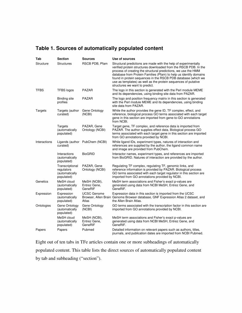
Table 1. Sources of automatically populated content
Tab Section Sources Use of sources
Structure Structures RSCB PDB, Pfam Structural predictions are made with the help of experimentally verified protein structures downloaded from the RSCB PDB. In the process of creating the structural predictions, we use the HMM database from Protein Families (Pfam) to help us identify domains found in protein sequences in the RSCB PDB database (which we use as templates) as well as the protein sequences of putative structures we want to predict.
TFBS logos PAZAR The logo in this section is generated with the Perl module MEME and its dependencies, using binding site data from PAZAR.
TFBS
Binding site profiles
PAZAR The logo and position frequency matrix in this section is generated with the Perl module MEME and its dependencies, using binding site data from PAZAR.
Targets (author curated)
Gene Ontology (NCBI)
While the author provides the gene ID, TF complex, effect, and reference, biological process GO terms associated with each target gene in this section are imported from gene-to-GO annotations from NCBI.
Targets
Targets (automatically populated)
PAZAR, Gene Ontology (NCBI)
Target gene, TF complex, and reference data is imported from PAZAR. The author supplies effect data. Biological process GO terms associated with each target gene in this section are imported from GO annotations provided by NCBI.
Ligands (author curated)
PubChem (NCBI) While ligand IDs, experiment types, natures of interaction and references are supplied by the author, the ligand common name and image are provided from PubChem.
Interactions (automatically populated)
BioGRID Interactor names, experiment types, and references are imported from BioGRID. Natures of interaction are provided by the author.
Interactions
Transcriptional regulators (automatically populated)
PAZAR, Gene Ontology (NCBI)
Regulating TF complex, regulating TF, genomic links, and reference information is provided by PAZAR. Biological process GO terms associated with each target regulator in this section are imported from GO annotations provided by NCBI.
Genetics MeSH cloud (automatically populated)
MeSH (NCBI), Entrez Gene, GeneRIF
MeSH term associations and Fisher’s exact p-values are generated using data from NCBI MeSH, Entrez Gene, and GeneRIF.
Expression Expression (automatically populated)
UCSC Genome Browser, Allen Brain Atlas
Expression data in this section is imported from the UCSC Genome Browser database, GNF Expression Atlas 2 dataset, and the Allen Brain Atlas.
Gene Ontology (automatically populated)
Gene Ontology (NCBI)
GO terms associated with the transcription factor in this section are imported from GO annotations provided by NCBI.
Ontologies
MeSH cloud (automatically populated)
MeSH (NCBI), Entrez Gene, GeneRIF
MeSH term associations and Fisher’s exact p-values are generated using data from NCBI MeSH, Entrez Gene, and GeneRIF.
Papers Papers Pubmed Detailed information on relevant papers such as authors, titles, journals, and publication dates are imported from NCBI Pubmed.
Eight out of ten tabs in TFe articles contain one or more subheadings of automatically
populated content. This table lists the direct sources of automatically populated content
by tab and subheading (“section”).
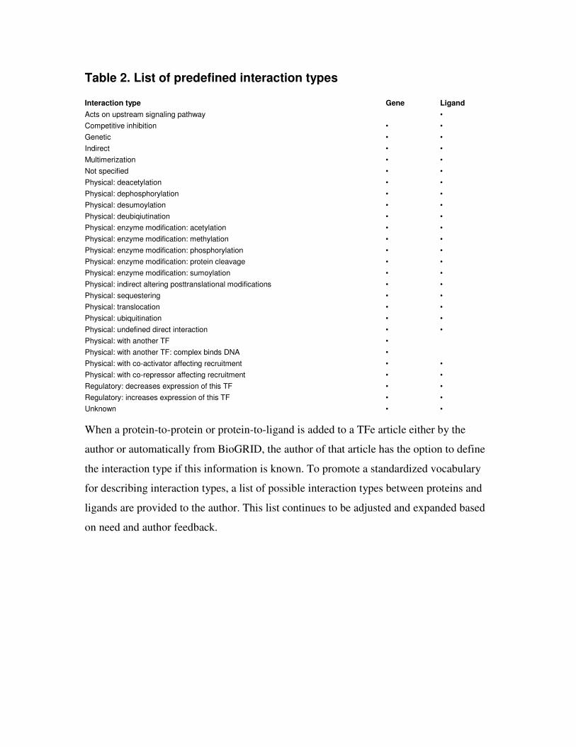
Table 2. List of predefined interaction types
Interaction type Gene Ligand
Acts on upstream signaling pathway •
Competitive inhibition • •
Genetic • •
Indirect • •
Multimerization • •
Not specified • •
Physical: deacetylation • •
Physical: dephosphorylation • •
Physical: desumoylation • •
Physical: deubiqiutination • •
Physical: enzyme modification: acetylation • •
Physical: enzyme modification: methylation • •
Physical: enzyme modification: phosphorylation • •
Physical: enzyme modification: protein cleavage • •
Physical: enzyme modification: sumoylation • •
Physical: indirect altering posttranslational modifications • •
Physical: sequestering • •
Physical: translocation • •
Physical: ubiquitination • •
Physical: undefined direct interaction • •
Physical: with another TF •
Physical: with another TF: complex binds DNA •
Physical: with co-activator affecting recruitment • •
Physical: with co-repressor affecting recruitment • •
Regulatory: decreases expression of this TF • •
Regulatory: increases expression of this TF • •
Unknown • •
When a protein-to-protein or protein-to-ligand is added to a TFe article either by the
author or automatically from BioGRID, the author of that article has the option to define
the interaction type if this information is known. To promote a standardized vocabulary
for describing interaction types, a list of possible interaction types between proteins and
ligands are provided to the author. This list continues to be adjusted and expanded based
on need and author feedback.
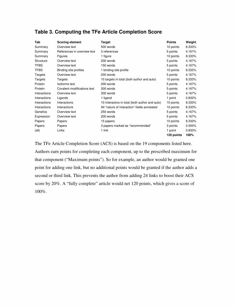
Table 3. Computing the TFe Article Completion Score
Tab Scoring element Target Points Weight
Summary Overview text 500 words 10 points 8.333%
Summary References in overview text 3 references 5 points 4.167%
Summary Figures 1 figure 10 points 8.333%
Structure Overview text 200 words 5 points 4.167%
TFBS Overview text 150 words 5 points 4.167%
TFBS Binding site profiles 1 binding site profile 10 points 8.333%
Targets Overview text 200 words 5 points 4.167%
Targets Targets 10 targets in total (both author and auto) 10 points 8.333%
Protein Isoforms text 200 words 5 points 4.167%
Protein Covalent modifications text 200 words 5 points 4.167%
Interactions Overview text 200 words 5 points 4.167%
Interactions Ligands 1 ligand 1 point 0.833%
Interactions Interactions 10 interactors in total (both author and auto) 10 points 8.333%
Interactions Interactions All “nature of interaction” fields annotated 10 points 8.333%
Genetics Overview text 250 words 5 points 4.167%
Expression Overview text 200 words 5 points 4.167%
Papers Papers 15 papers 10 points 8.333%
Papers Papers 3 papers marked as “recommended” 3 points 2.500%
(all) Links 1 link 1 point 0.833%
120 points 100%
The TFe Article Completion Score (ACS) is based on the 19 components listed here.
Authors earn points for completing each component, up to the prescribed maximum for
that component (“Maximum points”). So for example, an author would be granted one
point for adding one link, but no additional points would be granted if the author adds a
second or third link. This prevents the author from adding 24 links to boost their ACS
score by 20%. A “fully complete” article would net 120 points, which gives a score of
100%.
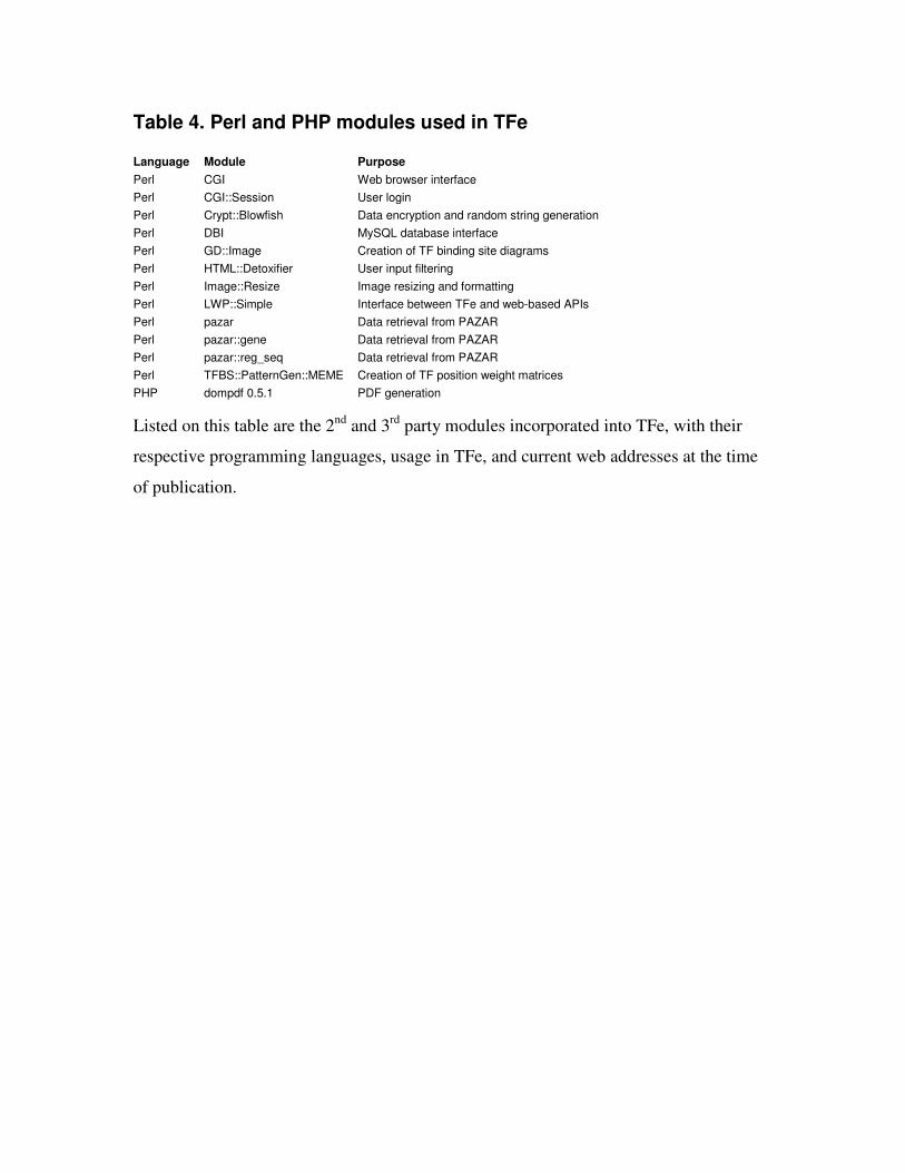
Table 4. Perl and PHP modules used in TFe
Language Module Purpose
Perl CGI Web browser interface
Perl CGI::Session User login
Perl Crypt::Blowfish Data encryption and random string generation
Perl DBI MySQL database interface
Perl GD::Image Creation of TF binding site diagrams
Perl HTML::Detoxifier User input filtering
Perl Image::Resize Image resizing and formatting
Perl LWP::Simple Interface between TFe and web-based APIs
Perl pazar Data retrieval from PAZAR
Perl pazar::gene Data retrieval from PAZAR
Perl pazar::reg_seq Data retrieval from PAZAR
Perl TFBS::PatternGen::MEME Creation of TF position weight matrices
PHP dompdf 0.5.1 PDF generation
Listed on this table are the 2nd and 3rd party modules incorporated into TFe, with their
respective programming languages, usage in TFe, and current web addresses at the time
of publication.

Additional files
Additional file 1: PDF of released article. The PDF version of the human FOXL2 article.
Other PDF versions of released TFe articles can be accessed on the TFe website.
Additional file 2: Data of released articles. Additional data related to the released mini
review articles to supplement the four-page PDF versions, arranged in alphabetical order
by TF name.
Additional file 3. Classification of transcription factors in TFe. There are 803 human,
mouse, and rat articles in TFe, most of which are organized into groups, families and
subfamilies of TFs. The classification scheme utilized in TFe is derived from the work of
Fulton et al in TFCat. There are 8 large groups, which are further subclassified into 34
families. Several TFs, namely nuclear receptors, are even further subclassified into
subfamilies.
Additional file 4: The TFe article structure. Articles in TFe are organized into 10 tabs
labeled “Summary”, “Structure”, “TFBS”, “Targets”, “Protein”, “Interactions”,
“Genetics”, “Expression”, “Ontologies”, and “Papers”. Each tab, with the exception of
the Ontologies and Papers tabs, typically begins with a brief overview written by the
authors, followed by a mixture of tables and figures that features data from both the
authors and 2nd (i.e. PAZAR) or 3rd party (i.e. BioGRID) sources.
Additional file 5: Binding models produced in the TFe project. Images of the binding
models produced in TFe that are sufficiently characterized to be used in a study.
Additional file 6: PDB records depicting protein-DNA binding interface. A list of PDB
records which depict a protein-DNA binding interface.
Additional file 7: Relationship Between Pfam Domains and PDB Records. A list of Pfam
binding domains followed by PDB records in which the domains can be found.

Figure 1
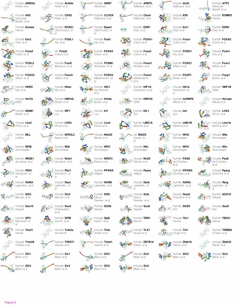
human ARID3aFerrell et al
mouse Arid3aFerrell et al
human ARNTHankinson
human ARNTLDeBruyne et al
mouse ArntlDeBruyne et al
human ATF2Karolczak-Bayatti et al
mouse Atf2Karolczak-Bayatti et al
human CDX2Witek et al
human CLOCKDeBruyne et al
mouse ClockDeBruyne et al
mouse Elf5Sinha et al
human EOMESHevner
mouse EomesHevner
human EPAS1Olechnowicz et
al
mouse Epas1Olechnowicz et
al
human ESR1Zhao et al
mouse Esr1Zhao et al
human ESR2Zhao et al
mouse Esr2Zhao et al
human FOSL1Vaz et al
mouse Fosl1Vaz et al
human FOXA1Cullum et al
mouse Foxa1Cullum et al
human FOXA2Cullum et al
mouse Foxa2Cullum et al
rat Foxa2Cullum et al
human FOXA3Cullum et al
mouse Foxa3Cullum et al
human FOXH1Hou et al
mouse Foxh1Hou et al
human FOXL2Benayoun et al
mouse Foxl2Benayoun et al
human FOXM1Schwenen et al
mouse Foxm1Monteiro et al
human FOXO1Wilson et al
mouse Foxo1Wilson et al
human FOXO3Wilson et al
mouse Foxo3Wilson et al
human FOXO4Eijkelenboom et
al
mouse Foxo4Eijkelenboom et al
human FOXP1Brown et al
mouse Foxp1Brown et al
human HHEXNoy et al
mouse HhexNoy et al
human HIC1Van Rechem et al
human HIF1ACamenisch et al
mouse Hif1aCamenisch et
al
human HNF1BRyffel
mouse Hnf1bRyffel
human HNF4ABolotin et al
mouse Hnf4aBolotin et al
human HNF4GRyffel
human hnRNPKMikula et al
mouse HnrnpkMikula et al
human INSM1Breslin et al
human IRF1Luo
mouse Irf1Luo
human ISL1Sun
mouse Isl1Sun
human LHX3Prince et al
mouse Lhx3Prince et al
human LHX4Prince et al
mouse Lhx4Prince et al
human LMX1AMishima et al
human LMX1BMishima et al
mouse Lmx1bMishima et al
human MLLLiedtke
human NFE2L2Biswal et al
mouse Nfe2l2Biswal et al
rat Nfe2l2Harvey et
al
human NFIAGronostajski
mouse NfiaGronostajski
human NFIBGronostajski
mouse NfibGronostajski
human NFICGronostajski
mouse NficGronostajski
human NFIXGronostajski
mouse NfixGronostajski
human NR2E1de Leeuw et al
mouse Nr2e1de Leeuw et al
human NR2F2Ramos et al
mouse Nr2f2Ramos et al
human PAX8de Cristofaro et al
mouse Pax8de Cristofaro et al
human PBX1Woodcroft et
al
mouse Pbx1Woodcroft et
al
human PPARAHéligon
mouse PparaHéligon
human PPARGGrontved et al
mouse PpargGrontved et al
human RARALaperrière et al
mouse RaraLaperrière et
al
human RARBLaperrière et al
mouse RarbLaperrière et
al
human RARGLaperrière et al
mouse RargLaperrière et
al
human SIX3Beccari et al
mouse Six3Beccari et al
human SIX6Marco-Ferreres et al
mouse Six6Marco-Ferreres et al
mouse Snai2Chaudhuri et al
human SOX10Wegner
mouse Sox10Wegner
mouse Sox2Chakravarthy et al
human SOX8Wegner
mouse Sox8Wegner
human SOX9Kist
mouse Sox9Kist
human SPI1Mancarelli et
al
human SPIBKarrich et al
mouse SpibKarrich et al
human TBR1Hevner
mouse Tbr1Hevner
human TBX21Hevner
mouse Tbx21Hevner
mouse Tcfe2aWoodcroft
mouse ThrbSharlin et al
human TLX1Hough et al
mouse Tlx1Hough et al
human TRIM28O'Geen et al
mouse Trim28O'Geen et al
human TWIST1Vesuna et al
mouse Twist1Vesuna et al
human ZBTB16Frietze et al
mouse Zbtb16Frietze et al
mouse Zbtb33Nanan et al
human ZIC1Blank et al
mouse Zic1Blank et al
human ZIC2Blank et al
mouse Zic2Blank et al
human ZIC3Blank et al
mouse Zic3Blank et al
human ZIC4Blank et al
mouse Zic4Blank et al
human ZIC5Blank et al
mouse Zic5Blank et al
Figure 2
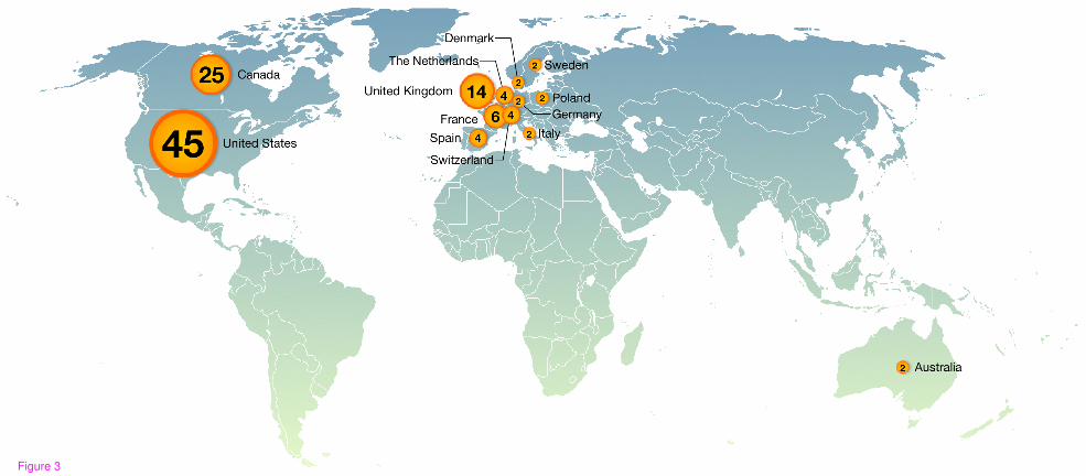
Figure 3

Figure 4

Figure 5

Figure 6
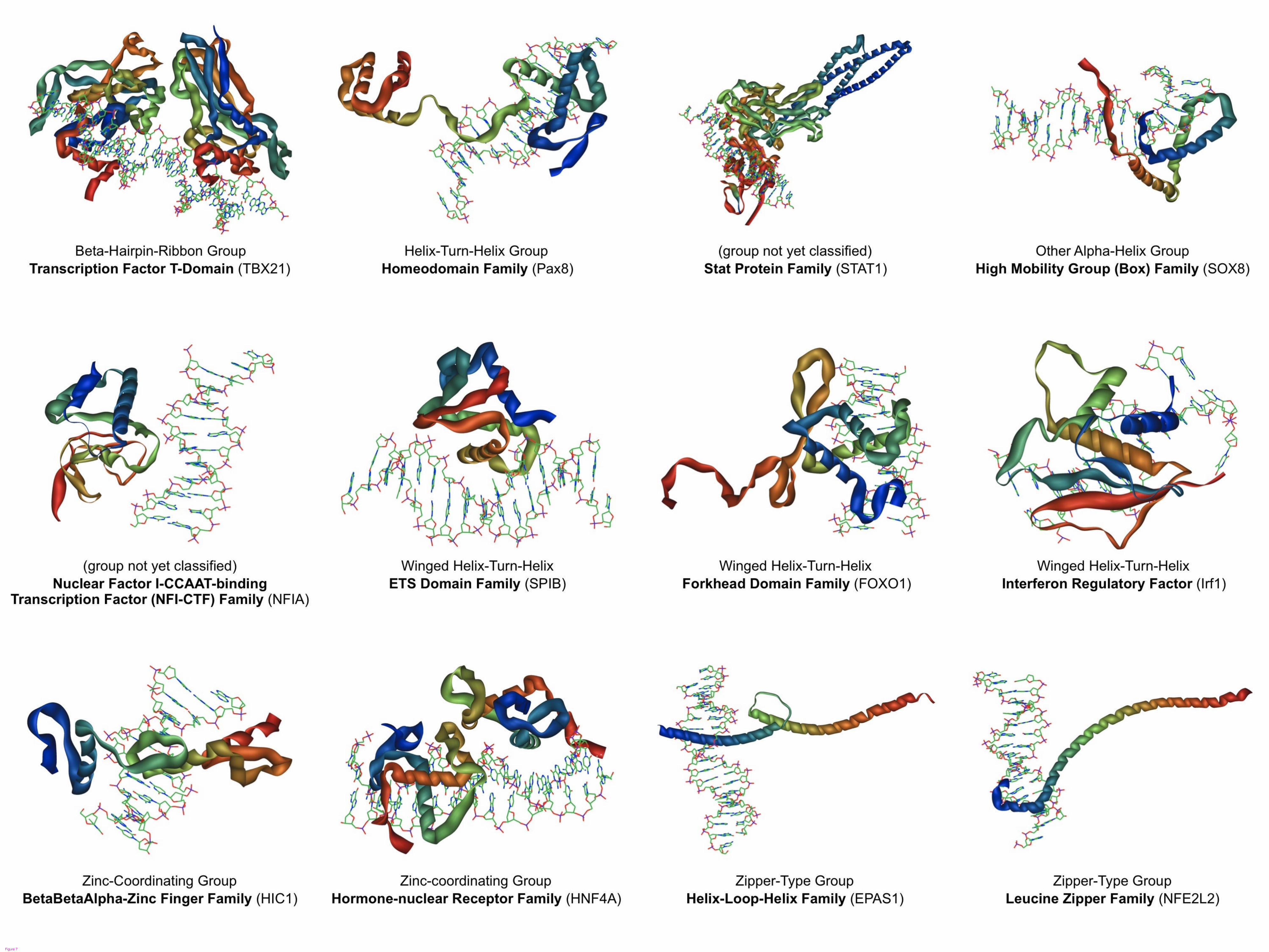
Figure 7
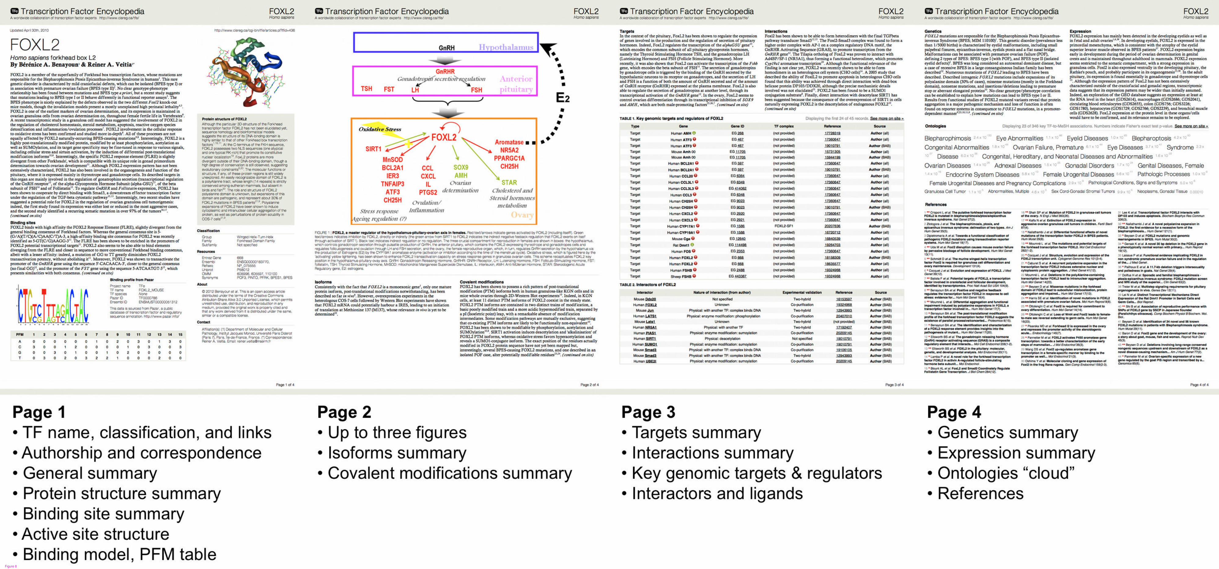
Figure 8

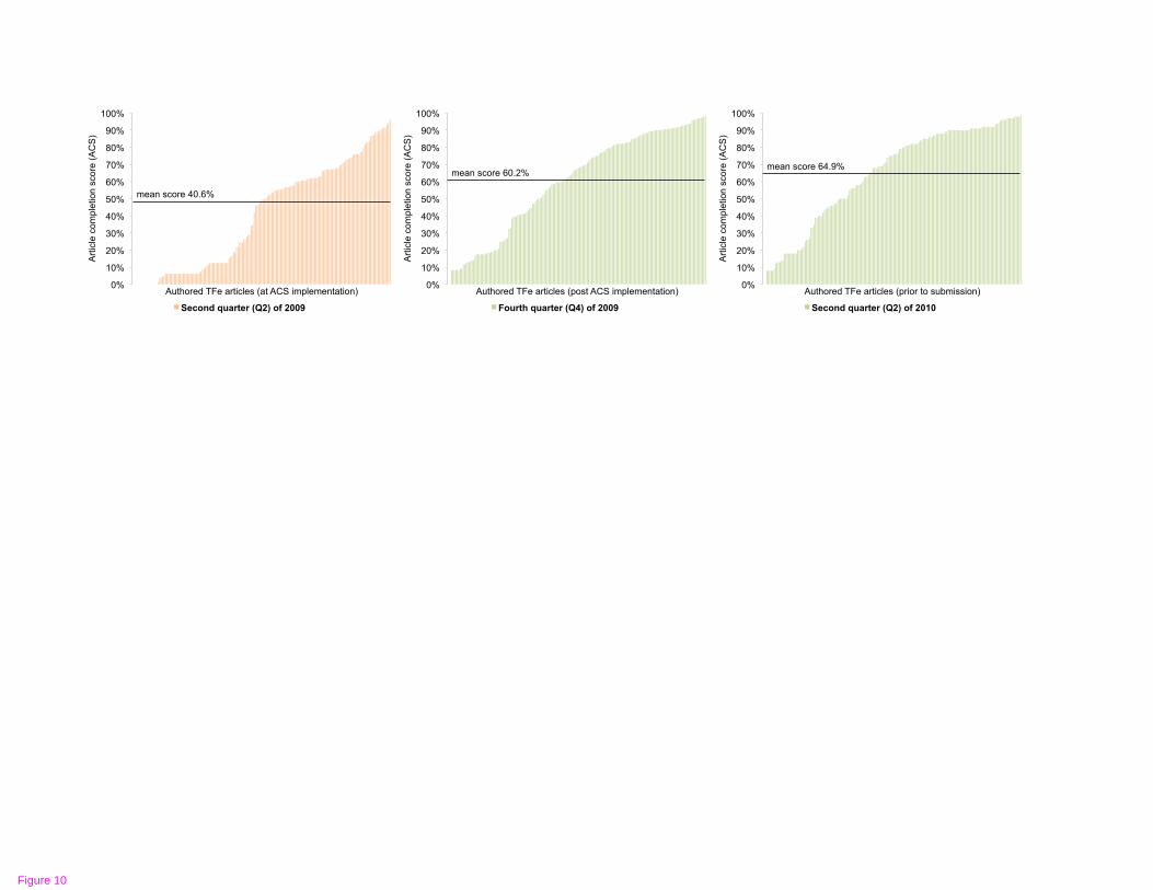
0%
10%
20%
30%
40%
50%
60%
70%
80%
90%
100%
Art
icle
co
mp
letio
n s
co
re (
AC
S)
Authored TFe articles (post ACS implementation)
Fourth quarter (Q4) of 2009
0%
10%
20%
30%
40%
50%
60%
70%
80%
90%
100%
Art
icle
co
mp
letio
n s
co
re (
AC
S)
Authored TFe articles (at ACS implementation)
Second quarter (Q2) of 2009
0%
10%
20%
30%
40%
50%
60%
70%
80%
90%
100%
Art
icle
co
mp
letio
n s
co
re (
AC
S)
Authored TFe articles (prior to submission)
Second quarter (Q2) of 2010
mean score 40.6%
mean score 60.2% mean score 64.9%
Figure 10
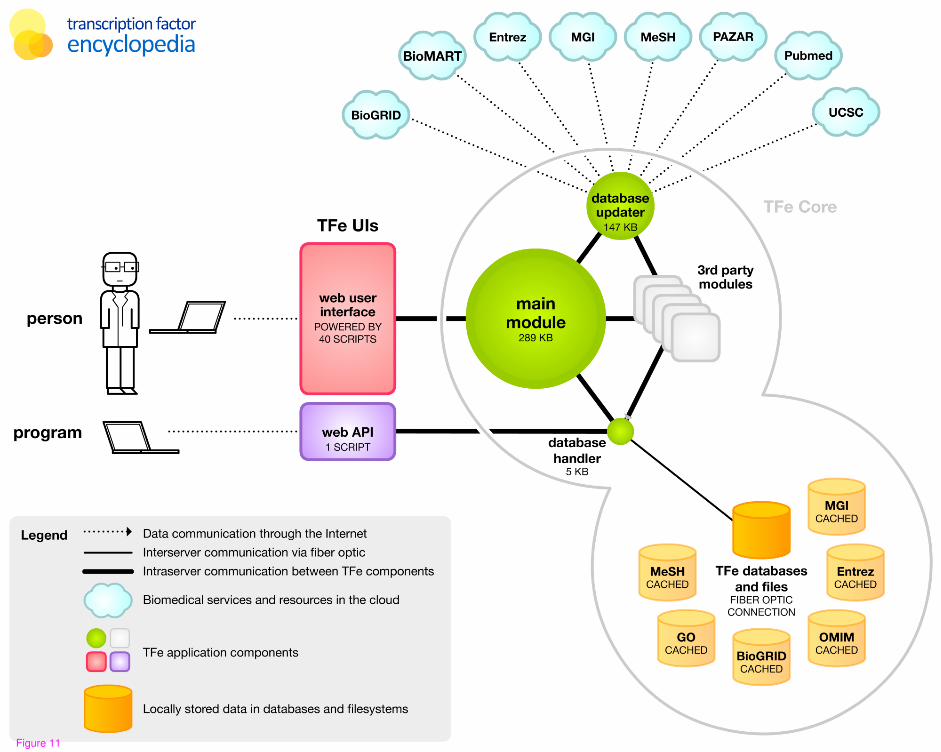
Figure 11

Additional files provided with this submission:
Additional file 1: Additional File 1 - PDF of released article.pdf, 1924Khttp://genomebiology.com/imedia/1264008786702480/supp1.pdfAdditional file 2: Additional File 2 - Data of released articles.zip, 9051Khttp://genomebiology.com/imedia/2080466286702480/supp2.zipAdditional file 3: Additional File 3 - Classification of transcription factors in T,123Khttp://genomebiology.com/imedia/5928500570248061/supp3.xlsAdditional file 4: Additional File 4 - The TFe article structure.pdf, 46Khttp://genomebiology.com/imedia/9469406967024806/supp4.pdfAdditional file 5: Additional File 5 - Binding models produced in the TFe project.p,1008Khttp://genomebiology.com/imedia/7991413087024806/supp5.pdfAdditional file 6: Additional File 6 - PDB records depicting protein-DNA binding in,7Khttp://genomebiology.com/imedia/5243377437024806/supp6.txtAdditional file 7: Additional File 7 - Relationship Between Pfam Domains andPDB Re, 32Khttp://genomebiology.com/imedia/1076505177702480/supp7.txt