transcription factor pax6 regulates cell cycle progression and
TRANSCRIPT

TranscriptionfactorPax6regulates
cellcycleprogressionandcellfate
determination:themodularlogicof
complextranscriptionalcontrol
Dissertation der Graduate School of Systemic Neurosciences
der Ludwig-Maximilians-Universität München
Submitted by
Stefania Petricca
München, 2nd of July 2015

ii

iii
Supervisor: Dr. Jovica Ninkovic
Second reviewer: Prof. Dr. Magdalena Götz
Third reviewer: Prof. Dr. Federico Calegari
Date of submission: 02.07.2015
Date of oral defense: 29.09.2015

iv

Abstract
v
Abstract
The development of the central nervous system relies on the tight regulation of the
neural progenitor proliferation and differentiation in order to generate new neurons.
The transcription factor Pax6 coordinates these functions during the development of
the mammalian forebrain, using the paired DNA binding domain. This is a bipartite
DNA-binding domain constituted by two subdomains, the PAI and the RED, binding the
DNA in a cooperative or independent manner in order to control specific targets.
Focusing on the activity of Pax6 as regulator of cell proliferation and of cell fate
determination, I aimed to understanding how these functions are regulated at the
molecular level, using the developing ventral forebrain as a model.
In this work the role of the RED domain of Pax6 as a regulator of cell cycle progression
is investigated. The mutation of the RED subdomain leads to an increase of progenitors
in active mitosis (phospho-histone3 positive cells) in the ventral telencephalon at
midneurogenesis. Similar result was obtained in the progenitors of the dorsal
telencephalon. The increment in the phospho-histone3 positive cells is followed by the
increase in cell death in both dorsal and ventral forebrain. These evidences suggest
impairments in the cell cycle progression of the progenitor cells in the RED domain
mutant.
The importance of full activity of the RED domain of Pax6 for the proper progression of
the cell cytokinesis is shown via ex-vivo live imaging, performed on ventral developing
forebrain of Pax6Leca2
mutant animals (RED domain mutant).
To elucidate the molecular mechanisms underlying the observed phenotype,
transcriptome of the Pax6Leca2
mutants and their age matching siblings is analyzed,
identifying a potential candidate gene: the Holliday junction recognition protein
(HJURP). The overexpression of the HJURP protein in the wild type progenitors in vitro
resembles the impairment of cytokinesis observed in Pax6Leca2
mutant.
In summary, my data suggest new mechanisms for the regulation of cytokinesis in
progenitors mediated by the RED domain of Pax6 and indicate that the full
functionality of the paired domain is a prerequisite for Pax6 to function as fate
determinant.

vi

Table of contents
vii
Tableofcontents
ABSTRACT.................................................................................................................................V
TABLEOFCONTENTS.........................................................................................................VII
TABLEOFFIGURES...............................................................................................................IX
LISTOFABBREVIATIONS...................................................................................................XI
1 INTRODUCTION...............................................................................................................1
1.1 Early brain development ........................................................................................................... 1 1.2 Ventral telencephalon .............................................................................................................. 4
1.2.1 Lateral Ganglionic Eminence specification and neurogenesis ................................................... 5 1.3 Adult brain from the LGE .......................................................................................................... 9
1.3.1 Transit amplifying progenitors ................................................................................................. 10 1.3.2 Neuroblasts .............................................................................................................................. 11
1.4 The transcription factor Pax6 .................................................................................................. 12 1.4.1 Pax6 in development ................................................................................................................ 14 1.4.2 Pax6 protein structure.............................................................................................................. 16
2 MATERIALSANDMETHODS.....................................................................................23
2.1 Materials ................................................................................................................................ 23 2.1.1 Chemicals ................................................................................................................................. 23 2.1.2 Kits ............................................................................................................................................ 24 2.1.3 Tissue culture reagents ............................................................................................................ 25 2.1.4 Standard solution and media ................................................................................................... 26 2.1.5 Solutions and media ................................................................................................................. 28 2.1.6 Cell lines ................................................................................................................................... 30 2.1.7 Cloning and expression plasmids ............................................................................................. 31 2.1.8 Primary antibodies ................................................................................................................... 31 2.1.9 Primers used for qPCR analysis ................................................................................................ 32
2.2 Methods ................................................................................................................................. 34 2.2.1 In vivo methods ........................................................................................................................ 34 2.2.2 Methods in cell biology ............................................................................................................ 38 2.2.3 Methods in molecular biology .................................................................................................. 42 2.2.4 Data analysis ............................................................................................................................. 48
3 AIMOFTHETHESIS....................................................................................................49
4 RESULTS..........................................................................................................................51
4.1 Pax6 in the lateral ganglionic eminence .................................................................................. 51 4.1.1 Pax6 isoforms expression in the developing telencephalon .................................................... 51 4.1.2 Proliferation of progenitors in Pax6 RED domain mutants ...................................................... 54 4.1.3 Cell death analysis in RED domain mutants ............................................................................. 58 4.1.4 Cell cycle progression in Pax6
Leca2 ventral telencephalon: loss and gain of function
experiments ........................................................................................................................................... 60 4.1.5 Candidate gene: HJURP ............................................................................................................ 67
4.2 Adult brain .............................................................................................................................. 74 4.2.1 Pax6
Leca2 in adult brain .............................................................................................................. 74
4.3 Domain specific functions of Pax6........................................................................................... 85 4.3.1 Embryonic and Adult neurospheres ......................................................................................... 85
5 DISCUSSION...................................................................................................................89
5.1 The Pax6 RED subdomain is responsible for the cell cycle progression of apical progenitors .. 89 5.2 PAI and RED domains are necessary for Pax6 neurogenic function ......................................... 93

Table of contents
viii
5.3 Pax6 RED domain affects the development of the postnatal neurogenic niche ...................... 95
6 REFERENCES..................................................................................................................97
AFFIDAVIT...........................................................................................................................111
LISTOFCONTRIBUTIONS...............................................................................................111
ACKNOWLEDGMENTS.....................................................................................................113
CURRICULUMVITAE........................................................................................................115

Table of figures
ix
Tableoffigures
Figure 1: Vertebrate brain and spinal cord development ................................................ 2
Figure 2: Organization of the telencephalon .................................................................... 3
Figure 3: Glial nature of embryonic and adult neural stem cells ..................................... 4
Figure 4: Dorsal and ventral telencephalon specification ................................................ 5
Figure 5: Production and migration of different neurons in the ventral telencephalon . 6
Figure 6: Progenitor cells in the cortex vs LGE ................................................................. 8
Figure 7: Neural stem cells and their progeny in adult brain ......................................... 10
Figure 8: Pax6 expression in embryonic and adult brain................................................ 12
Figure 9: Pax6 expression in the SEZ and SGZ population .............................................. 13
Figure 10: Pax family and Pax6 domains ........................................................................ 17
Figure 11: PD domain mutants Pax6Leca2
and Pax6Leca4
.................................................. 21
Figure 12: Pax6 isoforms expression in the cortex and in the LGE................................. 53
Figure 13: Proliferating cells in LGE ................................................................................ 55
Figure 14: Progenitor proliferation in Pax6Leca2
LGE ....................................................... 56
Figure 15: Pax6 expression in the progenitors of the LGE. ............................................. 57
Figure 16: Survival of progenitors in Pax6Leca2
................................................................ 59
Figure 17: Ex vivo life imaging: ....................................................................................... 60
Figure 18: LGE ex vivo live imaging ................................................................................. 63
Figure 19: Time of cytokinesis definition and measuring ............................................... 64
Figure 20: Cell division in vivo ......................................................................................... 65
Figure 21: In vivo overexpression of the RED domain mutation of Pax6 ....................... 66
Figure 22: HJURP candidate gene ................................................................................... 68
Figure 23: In vitro live imaging of P19 cells.upon HJURP overexpression ...................... 72
Figure 24: Working model .............................................................................................. 73
Figure 25: Gross morphology of Pax6Leca2
....................................................................... 74
Figure 26: Cells accumulation in the Pax6Leca2
background ............................................ 75
Figure 27: Neuroblasts and TAPs proliferation analysis ................................................. 78
Figure 28: HJURP expression in the adult brain .............................................................. 80
Figure 29: Neuroblasts migration ability in Pax6Leca2
adult brains ................................. 83
Figure 30: Migrating neuroblasts accessory structures .................................................. 84
Figure 31: Embryonic ad adult neurosphere .................................................................. 87

x

List of abbreviations
xi
Listofabbreviations
% Percent
°C Degree Celsius
µg Microgram(s)
µl Microlitre(s)
µM Micromolar
AP Apical Progenitor
bHLH Basic helix-loop-helix
BP Basal Progenitor
Bp Base pairs
cDNA Complementary Deoxyribonucleic Acid
CNS Central Nervous System
CO2 Carbon Dioxide
Ctx Cortex
DAPI 4',6-diamidino-2-phenylindole
dATP DeoxyAdenosine TriPhosphate
dCTP DeoxyCytidine TriPhosphate
Dcx Doublecortin
dGTP DeoxyGuanosine TriPhosphate
DMEM Dulbecco's Modified Eagle Medium
DNA Deoxyribonucleic Acid
dNTP DeoxyNucleoside TriPhosphate
dNTP DeoxyNucleoside TriPhosphate(s)
dTTP DeoxyThymidine TriPhosphate
E Embryonic Day
e.g. For example (Latin ‘exempli gratia’)
EDTA Ethylene-Diamine-tetra-Acetic-Acid
EGF Epidermal Growth Factor
ENU N-ethyl-N-nitrosourea

List of abbreviations
xii
FCS Fetal Calf Serum
FGF Fibroblast growth factor
G Gram
GAPDH GlycerAldehyde 3-Phosphate DeHydrogenase
GE Ganglionic Eminence
GFAP Glial Fibrillary Acidic Protein
GFP Green Fluorescent Protein
H Hour(s)
HBSS Hank’s balanced salt solution
HCl Hydrochloric acid
HEPES 4-(2-Hydroxyethyl)-1-piperazineethanesulfonic acid
HJURP Holliday Junction recognition protein
Kb Kilobase
kDa Kilodalton
Kg Kilogram(s)
L Litre
LB Luria-Bertani medium
Leca Lens Corneal Adhesion
LGE Lateral Ganglionic Eminence
MGE Medial Ganglionic Eminence
Min Minute
miRNA Micro Ribonucleic Acid
mM Millimolar
mRNA Messenger Ribonuicleic Acid
NaCl Sodium Chloride
Ng Nanogram(s)
OB Olfactory Bulb
Olig2 Oligodendrocyte transcription factor2
P Postnatal day
Pax6 Paired Box 6

List of abbreviations
xiii
PBS Phosphate Buffered Saline
PCR Polymerase Chain Reaction
PDL Poly-D-Lysine
PFA Paraformaldehyde
PSB Pallial-subpallial boundary
qPCR Quatitative Polymerase Chain Reaction
RG Radial Glia
RMS Rostral Migratory Stream
RNA Ribonucleic Acid
rpm Revolutions per minute
RT Room Temperature
Sec Seconds
SEM Standard Error of the Mean
SEZ Subependymal Zone
SP Subapical Progenitor
Str Striatum
SVZ Subventricular Zone
Taq DNA polymerase of bacterium Thermus aquaticus
U Enzyme unit
VZ Ventricular Zone
w/v Weight/Volume
WT Wild Type

xiv

Introduction
1
1 Introduction
The mammalian brain comprises a variety of cell types. The two major populations are
the neurons, specialized in conducting signals to other cells, and the glia cells
comprising various cells important to preserve homeostasis and provide support and
protection to the neurons.
During brain development the neural stem cells represent the source of all the
neurons in the mammalian brain as well as of the astrocytes and oligodendrocytes
(Gotz and Huttner, 2005). The commitment of a cell to a specific fate can be
determined by the environment surrounding the cells providing different cues (non-
cell autonomous) and by a gene expression program pre-specified in the cells (cell
autonomous). The proper development of the brain requires a fine regulation of the
combination of these extrinsic and intrinsic fate determinants in order to define the
correct number of each cell type at the right time and place (Guillemot et al., 2006;
Johansson et al., 2010; Lui et al., 2011; Tiberi et al., 2012).
A cell intrinsic factor, that is regulating the commitment of the neural stem cells to the
neuronal fate, is Pax6. In the developing cortex in fact the transcription factor Pax6 is
expressed in neural stem cells and controls their proliferation as well as the time they
exit the cell cycle in order to proceed to differentiation (Warren et al., 1999). Pax6 is
important for the generation of superficial cortical neurons (Schuurmans et al., 2004;
Georgala et al., 2011b). In addition Pax6 is also important for the generation of
olfactory bulb interneurons originating from the ventral part of the telencephalon and
are produced lifelong in the adult lateral wall of the subependymal zone (Merkle et al.,
2004; Hack et al., 2005; Merkle and Alvarez-Buylla, 2006). More insights on the
functions of this transcription factor will be described further in the chapter.
1.1 Earlybraindevelopment
After the neurulation process and prior to the onset of neurogenesis (embryonic days
E7-9 in mice), the neural plate and the neural tube are composed by a single layer of
neuroepithelial cells (NE).

Introduction
2
The NE cells, characterized by apical- basal polarity, form a pseudostratified layer of
cells. The appearance of the neuroepithelium in layers is given by the nuclei of the cells
that are performing the so called interkinetic nuclear migration (INM). During this
process the nucleus of every NE cell moves in the direction of the basal lamina (a thin
layer of extracellular matrix at the pia surface) during G1 and S phase of the cells cycle
and moves back to the apical side (at the ventricular surface) in G2 phase undergoing
to mitosis directly at the apical side (Sauer, 1935; Takahashi et al., 1993).
Figure 1: Vertebrate brain and spinal cord development
Schemes taken from Sanes, Reh and Harris; Development of the Nervous System.
(A) The primary structures appearing are the forebrain (prosencephalon), midbrain (mesencephalon), and hindbrain
(rhombencephalon). These structures further differentiate and the forebrain generates the telencephalic vesicles
and the diencephalon, while the rhombencephalon divides into the metencephalon and the myelencephalon (B).
The basic brain divisions are then maintained in the adult one (C).
The rostral region of the neural tube differentiates in three primary vesicles: forebrain
(prosencephalon), midbrain (mesencephalon) and hindbrain (rhombencephalon)
(Figure 1 A). These three regions further subdivide in five secondary brain vesicles. The
forebrain gives rise to the anterior telencephalon and the more caudal diencephalon.
The hindbrain further differentiates into an anterior metencephalon, forming the pons
and the cerebellum, and a posterior myelencephalon giving rise to the medulla

Introduction
3
oblongata (Figure 1 B) (Melton et al., 2004). The anterior-posterior regionalization
finally ends in the caudal part of the neural tube where it specifies the spinal cord.
Figure 2: Organization of the telencephalon
Scheme taken from (Puelles et al., 2013) summarizing the subdivision of the telencephalon area of interest in this
work. (A-B) Diagrams of a lateral view of developing mouse brain. The forebrain is subdivided in the telencephalon
and diencephalon. The telencephalon subdivides into the pallium, in the dorsal side, and subpallial regions ventrally.
The most anterior vesicle, the telencephalon, gives rise to the cerebral cortex or
pallium dorsally whereas the ventral region generates the lateral and the medial
ganglionic eminences (LGE and MGE) which will result in the striatum and the pallidum
respectively (Figure 2).
At the cellular level, while the neurogenic process proceeds, the NE cell populating the
telencephalic area are transformed in the radial glia cells (RG).
The RG cells similarly to the NE have apical- basal polarity and undergo INM. These
cells differentiate from the NE because of the expression of some astroglial markers
like vimentin, the glial glutamate/ aspartate transporter GLAST, the brain lipid binding
protein (BLBP) which are all lacking in the NE (Gotz and Huttner, 2005; Sild and
Ruthazer, 2011).
In the dorsal telencephalon, the RG cells either directly differentiate to neurons or give
rise to basal progenitors (BP), also known as intermediate progenitors, that are located
in the subventricular zone (abventricular region)(Figure 3). These progenitors are
distinguished from the RG cells by the expression of distinct transcription factors
(Englund et al., 2005). Aside the expression of different factors, the BP cells
differentiate morphologically from the RG. BPs are lacking the apical-basal polarity and
do not undergo INM. In the dorsal telencephalon, these progenitors divide in basal
Introduction
4
subventricular area and undergo symmetric cell division which leads to the increase of
the numbers of neurons generated per round of division (reviewed in (Gotz and
Huttner, 2005).
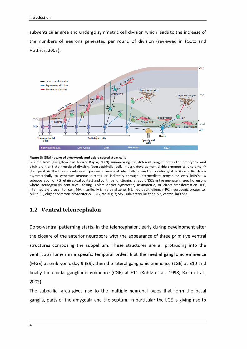
Figure 3: Glial nature of embryonic and adult neural stem cells
Scheme from (Kriegstein and Alvarez-Buylla, 2009) summarizing the different progenitors in the embryonic and
adult brain and their mode of division. Neuroepithelial cells in early development divide symmetrically to amplify
their pool. As the brain development proceeds neuroepithelial cells convert into radial glial (RG) cells. RG divide
asymmetrically to generate neurons directly or indirectly through intermediate progenitor cells (nIPCs). A
subpopulation of RG retain apical contact and continue functioning as adult NSCs in the neonate in specific regions
where neurogenesis continues lifelong. Colors depict symmetric, asymmetric, or direct transformation. IPC,
intermediate progenitor cell; MA, mantle; MZ, marginal zone; NE, neuroepithelium; nIPC, neurogenic progenitor
cell; oIPC, oligodendrocytic progenitor cell; RG, radial glia; SVZ, subventricular zone; VZ, ventricular zone.
1.2 Ventraltelencephalon
Dorso-ventral patterning starts, in the telencephalon, early during development after
the closure of the anterior neuropore with the appearance of three primitive ventral
structures composing the subpallium. These structures are all protruding into the
ventricular lumen in a specific temporal order: first the medial ganglionic eminence
(MGE) at embryonic day 9 (E9), then the lateral ganglionic eminence (LGE) at E10 and
finally the caudal ganglionic eminence (CGE) at E11 (Kohtz et al., 1998; Rallu et al.,
2002).
The subpallial area gives rise to the multiple neuronal types that form the basal
ganglia, parts of the amygdala and the septum. In particular the LGE is giving rise to

Introduction
5
the caudate and the putamen and the MGE is forming amygdala and globus pallidus
(Reiner et al., 1998).
From the subpallial region is also originating a variety of cortical interneurons.
Differently from the cerebral cortex where the projection neurons build up a cortical
plate of 6 layers (Molyneaux et al., 2007), the striatum is organized in a mosaic pattern
of patches and surrounding matrix (Gerfen, 1992). Patch neurons are born in two
waves; the first wave is between E10.5-E11, the second wave of patch neurons is
between E12.5-E13.5. Matrix neurons are born later (Fishell and van der Kooy, 1987;
Mason et al., 2005; Liao et al., 2008).
Figure 4: Dorsal and ventral telencephalon specification
(A) Scheme taken from (Hebert and Fishell, 2008); (B) scheme taken from(Schuurmans and Guillemot, 2002). (A).
The factors that act early to establish broad telencephalic regions are in blue. Sonic hedgehog (SHH) ventralizes the
telencephalon antagonizing the effect of GLI3 (dorsalizing factor). FoxG1 and FGF signaling are necessary to form all
regions of the telencephalon (shown in green), except for the dorsomedial region (shown in orange). Downstream
transcription factors, such as GSH2 and NKx2.1, then form specific subdivisions. (B) The dorsal telencephalon
specification directed by the expression of Pax6 and Ngn1/2 antagonizing with the ventralizing factors Gsh1/2 and
Ascl1/ Mash1.
The development and the specification of the ventral telencephalon identity are
governed by a variety of morphogens and transcription factors like Gsx1/2, Ascl1 and
Dlx1/2 (Schuurmans and Guillemot, 2002; Hebert and Fishell, 2008) (Figure4).
1.2.1 LateralGanglionicEminencespecificationandneurogenesis
The LGE is anatomically defined as the bulge that is protruding into the ventricle in the
area located between the cortex and the MGE. From the molecular point of view, the
LGE is characterized by the expression of Dlx1/2/5 (Bulfone et al., 1993) as well as the

Introduction
6
expression of Isl1, Gsx2, ER81 and Pax6 (Stoykova et al., 1996; Szucsik et al., 1997;
Sussel et al., 1999; Stenman et al., 2003; Flames et al., 2007). The dorsal border of the
LGE with the cortex is defined as ventral pallium (Puelles et al., 2000). In the ventral
pallium Tbr2, Ngn2, and Dbx1 are expressed whereas Dlx2 is absent (Puelles et al.,
2000; Yun et al., 2001). The border between the LGE and the MGE is identified by a
high expression of Nkx2.1 (Puelles et al., 2000; Garcia-Lopez et al., 2008) (Figure 4).
The progenitors in the LGE are distinct in four domains, all extending (except the
ventral pLGE4) along the rostro-caudal extent of the subpallium (Flames et al., 2007).
Two domains of the dorsal LGE are discriminated by the differential expression of Pax6
and ER81 (Stenman et al., 2003; Carney et al., 2006; Waclaw et al., 2006; Flames et al.,
2007). The dorsal LGE is thought to give rise to interneurons migrating to the olfactory
bulb (Toresson et al., 2000; Yun et al., 2001; Stenman et al., 2003; Waclaw et al., 2006)
and to contribute to distinct populations of postmitotic neurons forming the striatum
proper and the central amygdala (Moreno et al., 2009).
The ventral LGE is the most prominent part of the LGE and is divided in two further
domains. These two domains express Nkx6.2 (Flames et al., 2007) and give rise to Isl1
expressing cells in the SVZ (Stenman et al., 2003).
Figure 5: Production and migration of different neurons in the ventral telencephalon
Scheme representing the telencephalic hemispheres from E14 mouse embryos (Wilson and Rubenstein, 2000). (A)
Expression patterns in the progenitor zones of the transcription factors implicated in the regulation of telencephalic
patterning and differentiation. (B) Subdivisions of the telencephalic proliferative zone and location of precursor cells
producing neurons expressing the neurotransmitters, glutamate, GABA, and acetylcholine. (C) Migration pathways
from different progenitors zones.
The progression of neurogenesis in the ventral telencephalon is under the control of
the proneuronal factor Ascl1 (Mash1) (Casarosa et al., 1999). In this context Ascl1 is

Introduction
7
not only acting as neural determinant but it is also conferring ventral neuronal identity
due to the regulation of Dlx and GAD67, the enzyme synthetizing GABA (Casarosa et
al., 1999; Fode et al., 2000; Parras et al., 2002; Poitras et al., 2007; Castro et al., 2011).
Ascl1 is also responsible for the generation of an early population of subpallial
oligodendrocytes cooperating with the transcription factor Olig2 (Parras et al., 2007).
Differently from the pallium, where the projection neurons (glutamatergic neurons)
reach their final position via radial migration from their ventricular progenitor zone, in
the subpallial region the majority of telencephalic interneurons migrate following
tangential routes to integrate into their final destination (Marin and Rubenstein, 2001;
Marin and Rubenstein, 2003) (Figure 5). The streams of migration of the interneurons
are classified according to the timing of migration, the subpallial domain of origin and
the target region (Corbin et al., 2001; Nadarajah and Parnavelas, 2002; Flames and
Marin, 2005; Metin et al., 2006). Hence, cortical GABA interneurons arise through two
main tangential migrations, a superficial pathway, which originates in the MGE
(Anderson et al., 1999; Lavdas et al., 1999; Sussel et al., 1999; Wichterle et al., 1999;
Pleasure et al., 2000; Anderson et al., 2001) and a deep pathway originating from the
LGE (de Carlos et al., 1996; Tamamaki et al., 1997; Pleasure et al., 2000; Anderson et
al., 2001) (Figure 5).
1.2.1.1 ProgenitorcellsdiversityintheLGE
Differently from the cortex where the vast majority of progenitor cells are within the
ventricular zone (VZ), in the LGE the most prominent number of proliferating
progenitors is located abventricularly (Pilz et al., 2013).
The apical progenitors (AP) are the cells undergoing mitosis at the luminal surface of
the VZ. The AP cell population in the ganglionic eminence, is composed by two cells
types that undergo interkinetic nuclear migration: the bipolar radial glia cells (RG) and
the short neural precursors (SNP) (Pilz et al., 2013). The SNPs are unipolar with a
unique apical process. The apically dividing RG cells asymmetrically divide and, besides
self-renewing, they produce either SNPs or a subapically dividing progenitors (SP) (Pilz
et al., 2013) (Figure 6). The SNPs undergo further steps of amplification. The SNPs
amplification represents a peculiar characteristic of the ventral telencephalon (Pilz et

Introduction
8
al., 2013); in the cerebral cortex, in fact, the SNPs directly generate postmitotic
neurons (Gal et al., 2006; Stancik et al., 2010; Tyler and Haydar, 2013).
Both RGs and SNPs are giving rise to SP (Figure 6). This population of cells has been
recently identified in the LGE (Pilz et al., 2013). The SP progenitors undergo mitosis in a
region comprised within 10 rows of cells from the apical surface. Beside the common
proliferative zone, the SP cells are very heterogeneous in terms of morphology.
Figure 6: Progenitor cells in the cortex vs LGE
Scheme illustrating the progenitors present in the LGE and in the cortex.(Pilz et al., 2013). RG are in white, short
neural precursors (SNP) are in light red; they both divide apically but the SNPs present only one process at the apical
side. In light blue the SP dividing within 10cells rows from the ventricle. BPs are depicted in green and divide in the
SVZ. In the cortex RG cells divide at the ventricle and give rise to neurons in yellow directly or to BP in green dividing
in the SVZ. Abbreviations: AS (apical side); VZ (ventricular zone); SVZ (subventricular zone).
In the LGE SPs are positive for radial glia markers like RC2 and GLAST and also express
Sox2, Ascl1, Dlx2 and Pax6 (Pilz et al., 2013). The SPs amplify the pool of proliferating
progenitors in the LGE and they also give rise to basal progenitors (BP) (Pilz et al.,
2013).
In term of cell cycle progression, live imaging experiments in the LGE area revealed
that the RGs are the cells with the longest cell cycle (25h in average). The cell cycle
length of SNPs and SPs is about 17h and the second generation of SPs from SNPs or SPs
themselves is significantly shorter (12h) (Pilz et al., 2013). These observations suggest
that besides a series of division that fasten the cell cycle of the progeny, these fast
divisions are necessary to amplify the number of progeny in a given time; those are all
hallmarks of the adult neural stem cells lineages emerging from this region (Pilz et al.,
2013).

Introduction
9
1.3 AdultbrainfromtheLGE
In mammals, radial glial cells populating the brain during development disappear after
birth even if some RG-like cells persist in few areas during adulthood (Figure 7). The
adult NSCs (aNSC) and the radial glia cells belong to the same lineage (Merkle et al.,
2004). In particular, radial glial cells of the neonatal lateral ventricular wall (Figure 7 C)
occupy the same region as the astrocytic stem cells of the adult subedendymal zone
(SEZ) (Figure 7 D). This led to the conclusion that the primary progenitors for the
postnatal generation of new neurons in the adult SEZ zone are regionally specified and
the organization of the aNSCs in different domains is defined before birth (Figure 7)
(reviewed in(Merkle and Alvarez-Buylla, 2006)).
The adult SEZ is the largest germinal zone in the adult mammalian brain and is located
at the lateral wall of the lateral ventricle. In this region are generated the new neurons
that migrate through the OB via the rostral migratory stream (RMS) (Luskin, 1993; Lois
and Alvarez-Buylla, 1994) and glial cells destined to the corpus callosum (CC) (Hack et
al., 2005; Menn et al., 2006).
The SEZ niche consists of slow proliferating cells, the stem cells (also called Type B
cells), that, via multistep process of fate restriction, switch from multipotent cells
towards more fate-restricted precursors the transit amplifying progenitors (type C) and
neuroblasts (type A), which further differentiate resulting in new neurons that
functionally integrate into the OB system.

Introduction
10
Figure 7: Neural stem cells and their progeny in adult brain
Schematic drawing from (Merkle and Alvarez-Buylla, 2006).
The NSCs of the lateral ventricule, in blue, change their shape and produce different progeny as the brain develops.
They begin as neuroepithelial cells and transform into radial glial cells having NSCs characteristics. RGs contact with
the ventricle, into which they project a primary cilium and produce progeny either directly or via an intermediate
progenitors, in green. (A) At early developmental stages the CNS is a tubular structure, composed of neuroepithelial
cells, which divide symmetrically to expand the stem cell pool. (B) Neuroepithelial cells differentiate into embryonic
radial glial cells, which divide to generate striatal neurons and oligodendrocytes, either directly or via an
intermediate progenitor. The radial processes of radial glial cells support the migration of neuroblasts, in red. (C)
Radial glial cells persist in the neonatal brain, where they generate oligodendrocytes, olfactory bulb interneurons,
and ependymal cells. They also generate astrocytes, some of which remain stem cells in the adult. (D) In the adult
brain, adult NSCs retain a radial process and contact both the ventricle and the basal lamina of blood vessels. They
generate oligodendrocytes and olfactory bulb interneurons. Abbreviations: Stri, striatum; SVZ, subventricular zone;
VZ, ventricular zone.
1.3.1 Transitamplifyingprogenitors
The transit-amplifying progenitors (TAPs or Type C) represent a class of fast cycling
cells within the SVZ. They originate directly from the slow dividing aNSCs.
Clonal analysis highlighted that TAPs undergo 3-4 rounds of symmetric proliferative
divisions (Ponti et al., 2013; Calzolari et al., 2015) and seem to divide in close proximity
to blood vessels (Tavazoie et al., 2008).

Introduction
11
At the molecular level, this cell type can be isolated from the other populations by the
expression the EGF receptor (Morshead et al., 1994; Doetsch et al., 2002).
At least two different lineages of the fast proliferating intermediate progenitors have
been identified within the SVZ: TAPs that generate oligodendrocytes migrating towards
the white matter (Hack et al., 2005; Marshall et al., 2005; Menn et al., 2006; Colak et
al., 2008) and TAPs that give rise to immature neuroblasts migrating via the rostral
migratory stream (RMS) into the OB (Doetsch et al., 2002; Parras et al., 2004; Hack et
al., 2005; Menn et al., 2006; Colak et al., 2008). This difference in TAPs fate is
generated at the level of the aNSCs where they either generate neuronal or
oligodendroglial lineage (Ortega et al., 2013).
1.3.2 Neuroblasts
Neuroblasts or Type A cells originate from the TAPs. These cells are characterized by
their capability to tangentially migrate to the OB. Once the neuroblasts reach the OB,
they radially migrate to reach their final location.
These young neurons are characterized by the expression of factors like polysialic acid
(PSA-NCAM), doublecortine (Dcx), and CD24. They show properties of already
differentiated neurons but continue to divide in the SVZ and on their way to the OB.
For this reason, they appear to be BrdU positive upon short pulses of this compound
similarly to the TAPs (Luskin, 1993).
The main feature of these cells is that they migrate covering long distances from the
lateral wall of the ventricle to reach the OB. The migration along the rostral migratory
stream is mediated by molecules like chemoattractants and chomorepellents, as well
as proteins of extracellular matrix and proteins present on the cell surface (Hagg,
2005). In addition their migration is regulated by the blood vessels delimiting the route
of migration (Snapyan et al., 2009) and the astrocytic tube that avoids the dispersion
of cells in the surrounding tissue (Ghashghaei et al., 2007).
In the SEZ-RMS system, both neuroblasts and some TAPs are characterized by the
expression of the transcription factor Pax6 (Hack et al., 2005; Brill et al., 2008) and this
feature will be relevant in the following chapters.

Introduction
12
1.4 ThetranscriptionfactorPax6
Pax6 is a member of the Pax family of transcriptional regulators. This gene is located
on the chromosome 2 in mice (chromosome 11 in humans) and encodes for a highly
conserved protein of 422 amino acids.
Pax6 protein is involved in the development of the eyes, brain, spinal cord, and
pancreas. Pax6 is additionally expressed in the pituitary, the gut and the olfactory
epithelium (Walther and Gruss, 1991; Callaerts et al., 1997).
In mice, losing the functionality of this protein leads to the death of the animal shortly
after birth (Hogan et al., 1986).
In the developing CNS Pax6 is widely expressed including the forebrain, hindbrain,
cerebellum and spinal cord (Walther and Gruss, 1991; Grindley et al., 1995).
Figure 8: Pax6 expression in embryonic and adult brain
(A-C) Pax6 in situ hybridization from the GenePaint.org the digital atlas of gene expression patterns in the mouse.
(A) Pax6 expression at E10, (B) at E14 and (C) at postnatal stages.
In mice (Figure 8) Pax6 starts to be expressed as early as embryonic day 8.5 (E8.5)
when the neural tube is closing. In the dorsal telencephalon the peak of expression is
reached at E14 (peak of neurogenesis) and declines as gliogenesis starts (E18) (Gotz et
al., 1998; Englund et al., 2005).
In the adult CNS, Pax6 is detected in the neurons of various regions including the
cerebellum, the thalamus, the olfactory bulb and the amygdala (Stoykova and Gruss,
1994; Engelkamp et al., 1999; Kawano et al., 1999; Yamasaki et al., 2001; Tole et al.,
2005).

Introduction
13
Figure 9: Pax6 expression in the SEZ and SGZ population
(A-B) Schemes taken from (Hsieh, 2012). The scheme is depicting the SEZ (A) and SGZ (B) niches respectively and
highlights the expression of Pax6 in certain cells population along the lineage.
Pax6 is expressed in the two most known neurogenic regions of the postnatal brain:
the subgranular zone (SGZ) of the hippocampal dentate gyrus (DG) and the
subependymal zone (SEZ) in the ventricular zone (Hsieh, 2012)(Figure 9).
In the SGZ, the transcription factor is expressed in a subpopulation of neural stem cells,
in early progenitor cells presenting a radial glia like morphology and in a small
population of late progenitors (Maekawa et al., 2005; Nacher et al., 2005) (Figure 9).
In the SEZ, Pax6 is expressed in a subpopulation of TAPs and immature neurons that
migrate via the rostral migratory stream (RMS) to the olfactory bulbs (OB) (Figure 9). In

Introduction
14
the OB Pax6 is relevant for the survival of the dopaminergic neurons (Hack et al., 2005;
Kohwi et al., 2005; Ninkovic et al., 2010).
1.4.1 Pax6indevelopment
Most of the knowledge concerning the functions of Pax6 derives from the analysis of
the phenotype of mutants animals where the Pax6 function is lost. One of these is the
small eye mutant (Pax6Sey
) which is carrying a non-functional Pax6 protein.
The Pax6Sey
is characterized by a natural occurring point mutation that generates a
truncated protein lacking the transactivation domain at the C-terminus and
considered, for this reason, non-functional (Hill et al., 1991).
From the analysis of these mutants was possible to define the necessity of the Pax6
protein for the eye and brain development. In the heterozygotes mice the eye is
smaller, as suggested by the name, but is completely missing when the mutation is in
both alleles of the gene (Hogan et al., 1986; Osumi et al., 2008). Pax6 is also essential
for the development of the lens and the retina, (Ashery-Padan et al., 2000; Ashery-
Padan and Gruss, 2001; Kozmik, 2005; Graw, 2010; Shaham et al., 2012; Cvekl and
Ashery-Padan, 2014).
In the brain, losing Pax6 functionality leads to a reduction of the brain size due to a
smaller cerebral cortex composed by a thinner cortical plate (Schmahl et al., 1993;
Heins et al., 2002; Asami et al., 2011). In the developing telencephalon the
transcription factor regulates essential processes like the proliferation of the cells, the
neurogenic fate determination and the regionalization of the telencephalon defining
the borders between the dorsal and the ventral area (Haubst et al., 2004; Georgala et
al., 2011a).
1.4.1.1 Patterning
The establishment of the dorso ventral border delimited by the so called pallial-
subpallial boundary (PSB), a physical boundary composes by RG cells highly expressing
Pax6, is under the control of the transcription factor Pax6. When this protein is non-
functional (e.g.Pax6Sey
), the PSB is severely disrupted (Stoykova et al., 1997) leading to

Introduction
15
an increase of the cell migrating from the ventral telencephalon to the cerebral cortex
(Chapouton et al., 1999). In addition, the expression of dorsal factors like Ngn2 and
Tbr2 is lost (Stoykova et al., 2000; Toresson et al., 2000; Yun et al., 2001; Haubst et al.,
2004) and ventral factors like Gsx1/2, Ascl1, Dlx1/2 and Olig2 take over spreading in
the dorsal area (Stoykova and Gruss, 1994; Stoykova et al., 1996; Stoykova et al., 1997;
Toresson et al., 2000; Yun et al., 2001; Heins et al., 2002).
It is not only the specification of dorsal versus ventral regions in the telencephalon that
depends on Pax6, but also arealisation of the cerebral cortex (Bishop et al., 2000;
Bishop et al., 2002; Muzio et al., 2002a; Muzio and Mallamaci, 2003).
Pax6 is expressed in a gradient with high expression rostro-lateral and low expression
caudal-medial, in an opposing manner if compared with the expression of Emx1 and
Emx2 (other two dorsally expressed transcription factors). Losing the functionality of
Pax6 leads to the expansion of Emx2 and in turn to the anterior expansion of the
primary visual area. The loss of a functional Emx2 leads to the caudal expansion of
Pax6 and the consequent expansion of domains containing somatosensory and motor
neurons (Bishop et al., 2002; Muzio et al., 2002a; Muzio et al., 2002b; Muzio and
Mallamaci, 2003).
Changes in regionalization due to loss of functional Pax6 have also been observed in
other regions of the CNS, as in the ventral diencephalon, hindbrain and spinal cord
(Briscoe et al., 1999) (Stoykova et al., 1996; Grindley et al., 1997; Osumi, 2001).
1.4.1.2 Fatedeterminant
Pax6 is involved in the regulation of the neurogenic process.
In the dorsal telencephalon of Pax6 deficient mice, neurogenesis is strongly impaired
(Schmahl et al., 1993; Heins et al., 2002), whereas the ventral telencephalon is not
affected. This impairment is due to the fact that at the peak of neurogenesis the RG
cells express the Pax6 protein (Gotz et al., 1998; Hartfuss et al., 2001). The loss of Pax6
functionality in the Pax6Sey
mice leads to a decrease in the RG cells neurogenic
potential (Heins et al., 2002; Haubst et al., 2004) whereas the basal progenitors in the
SVZ are not affected (Haubensak et al., 2004; Miyata et al., 2004; Noctor et al., 2004).
Conversely Pax6 overexpression enhances in vitro neural production from RG (Heins et

Introduction
16
al., 2002; Haubst et al., 2004). This neurogenic potential is not restricted in time and
region since the overexpression of Pax6 results in a fate conversion of already
committed glia cells in postnatal (Heins et al., 2002) and fully adult brain derived cells
(Hack et al., 2004).
1.4.1.3 Proliferation
The neurogenic function of Pax6 and the capability of regulating the cell cycle are very
much linked. The fact that it drives cells to the neuronal fate means that, when
overexpressed, Pax6 forces cells to exit the cell cycle and start differentiating.
In the developing cortex, the deletion of Pax6 leads to an increase of proliferation in
the progenitors located in non-apical positions (Gotz et al., 1998; Estivill-Torrus et al.,
2002; Haubst et al., 2004; Quinn et al., 2007; Tuoc et al., 2009). This increase in
proliferation was explained also by the loss of the dorsal area specification, as
described before. In addition, when Pax6 is not functional, progenitors located apically
delaminate prematurely and contribute to the non-apical increase of proliferating
progenitors (Asami et al., 2011).
Differently from the developing cortex, where loss of Pax6 contribute to increase of
proliferation, in other regions like the diencephalon, the eye and in different type of
cells like the postnatal glia progenitors, when Pax6 is lost proliferation decreases
(Warren and Price, 1997; Marquardt et al., 2001; Sakurai and Osumi, 2008). In the
cerebellum of Pax6Sey
animals, the proliferation of progenitors is not affected
(Engelkamp et al., 1999).
These various responses upon lacking of Pax6 functionality underline the fact that the
capability of Pax6 to regulate proliferation of cells is not only cell type specific but also
region related.
1.4.2 Pax6proteinstructure
The Pax6 protein contains different DNA binding domains: the paired domain (PD) at
the amino terminal, shared by all the members of the Pax family (Chalepakis et al.,
1992); a homeodomain (HD) and a transactivation domain (TA) at the carboxy terminal

Introduction
17
(Glaser et al., 1992). The PD is further differentiated in two DNA binding subdomains:
the N-terminal subdomain or PAI and the C-terminal subdomain or RED (Walther et al.,
1991; Czerny et al., 1993; Epstein et al., 1994b; Epstein et al., 1994a; Jun and Desplan,
1996; Xu et al., 1999). The sequence that is recognized by the PAI is named P6CON
whereas the one recognized by the RED is the 5aCON (Epstein et al., 1994b; Chauhan
et al., 2004; Xie et al., 2013; Xie et al., 2014).
Figure 10: Pax family and Pax6 domains
Graph depicting all the members of the Pax family from (Mahajan et al., 2014) and the structure of the PD and HD
of the human Pax6 gene from crystallographic analysis (Callaerts et al., 1997).
Crystallographic analyses of the human PD DNA complex of Pax6 revealed that the PAI
domain contains two extended β-sheets and three α-helices between 20 to 60 amino
acids residues. The RED subdomain is constituted by three α helical regions (Figure 10).
The HD is predicted to have three α-helices where the third of them is contacting the
major groove of the DNA and is necessary for sequence recognition (Bruun et al.,
2005). Differently from other sequence specific DNA binding proteins that use only one
DNA binding domain, Pax6 can use all its DNA binding parts or a range of their
combinations to bind to the DNA (Xie and Cvekl, 2011).
The Pax6 gene generates different transcripts variants either by alternative splicing or
by the usage of an alternative promoter. One of the most commonly expressed Pax6
variants is the Pax6(5a). This splice variant has an insertion of 14 amino acids in the PAI
subdomain (Walther and Gruss, 1991; Glaser et al., 1992; Puschel et al., 1992) leading
to abolishment of the binding activity of the subdomain with the retention of the RED
domain functionality (Epstein et al., 1994b; Kozmik et al., 1997; Anderson et al., 2002).

Introduction
18
The canonical form of Pax6 together with the splice variant Pax6(5a) are expressed in
the developing brain, eye, spinal cord and in the olfactory epithelium (Walther and
Gruss, 1991; Glaser et al., 1992) even if the Pax6(5a) is expressed at lower levels.
The second major isoforms is missing the PD domain and its activity is restricted to the
functional HD and TA. The PD-less form (Mishra et al., 2002) is present in the adult
brain, pancreas and eye (Walther et al., 1991; Turque et al., 1994; Grindley et al., 1995;
St-Onge et al., 1997) and is found also in the invertebrates differently from the
Pax6(5a) which is specific for vertebrates (Miles et al., 1998).
As mentioned in the previous sections, Pax6 is involved in the regulation of many
developmental processes, such as the regulation of cell proliferation, neurogenesis and
brain patterning. Recently many genes regulating these functions have been identified
(Sansom et al., 2009; Wolf et al., 2009; Coutinho et al., 2011; Xie and Cvekl, 2011) and
it was also shown how Pax6 positively regulates them (Sansom et al., 2009). However,
in order to understand how Pax6 coordinates the regulation of these multiple
functions at the molecular level, the role of the different DNA-binding domains has
been analyzed in loss- and gain- of function approaches in different regions of the
brain (Haubst et al., 2004).
From this analysis was evident that the homeodomain (HD) of Pax6 is not relevant for
any of the above mentioned functions of Pax6 during brain development. In the
mutant line Pax64NEU
, where the DNA-binding properties of the HD are disrupted by a
point mutation leading to an amino acid substitution, no defect was evident in the
brain region specification, in cell proliferation or differentiation (Haubst et al., 2004).
Moreover no defect was detectable also at later stages (Ninkovic et al., 2010). These
observations imply that most of the roles exert by Pax6 during development might be
mediated by the bipartite PD domain.
1.4.2.1 Pax6PDmutants:Pax6Leca2andPax6Leca4
In order to discriminate the role of the two subdomain of the PD in regulating
neurogenesis, brain patterning and cells cycle regulation, two mouse line Pax6Leca2
and
Pax6Leca4
were analyzed during their brain development (Walcher et al., 2013).

Introduction
19
Each of these two mutants is carrying a single point mutation in one of the two
subdomains (Thaung et al., 2002) resulting in a substitution of lysine for asparagine
(N50K) in the PAI domain (Pax6Leca4
) or of cysteine for arginine (R128C) in the RED
domain (Pax6Leca2
). The RED domain mutation has been observed also in patients
(Azuma et al., 1996).
Both these mutations are predicted to affect the binding capacity of the domains to
their consensus site and the milder eye phenotype, compared with the Pax6Sey
(full KO
for Pax6), would be consistent with a selective disruption of only one of the subdomain
at the time (Thaung et al., 2002).
According to the eye phenotype, the craniofacial defects, and the reduction of the OB
sizes both the homozygote mice for the two mutants line show similarities with the
Pax6Sey
(Hill et al., 1991; Tzoulaki et al., 2005). However, the Pax6Leca2
, in contrast with
Pax6Sey
and the PAI domain mutants (Pax6Leca4
), survive to adulthood.
The Pax6Leca4
mutants are more similar to the Pax6Sey
than the Pax6Leca2
. While the
Pax6Leca4
have reduction in brain size due to a reduction in the cortical thickness and
the misspecification of the dorso-ventral borders, the RED domain mutants do not
presents these aberrations. In the Pax6Leca2
the cortical size is comparable with the
wildtype (WT) control and as well the borders between dorsal and ventral area are
maintained. Those are all considered insights that neither the neurogenic capacity of
Pax6 nor its regulation of the brain regionalization is under the control of the RED
domain (Walcher et al., 2013) (Figure 11).
In terms of cell proliferation both the domains (PAI and RED) are important to regulate
this function of Pax6 but in an opposite manner. In the Pax6Leca2
the cells in active
mitosis, both the apical and basal progenitors, are almost doubled in their number
when compared to the WT. Conversely when the same analysis is carried in the
Pax6Leca4
, both progenitors were significantly decreased. Apical and basal progenitors
in both the mutants were still Pax6 and Tbr2/Eomes positive as expected and the RG
cells were normal in their morphology. Hence only the number of cells in mitosis but
not their identity is affected by the mutation of the PAI and the RED domains (Walcher
et al., 2013). Notably this is in contrast with the Pax6Sey
where the apical progenitors
proliferation is unaffected and the increase in non-apical proliferating cells is resulting

Introduction
20
from the migration of cells from the ventral area (Gotz et al., 1998; Haubst et al., 2004;
Tuoc et al., 2009) (Figure 11).
The analysis of the RED and PAI mutants showed that Pax6 can exert opposing
functions on the same cortical progenitor population, having the intact RED domain
the capability of inhibiting proliferation and the PAI domain the capability of
promoting proliferation.
Interesting goals would be unraveling the molecular network controlled by the RED
domain involved in the cell cycle control and, since this mutants survive to postnatal
stages, investigate the repercussion of this mutation also on the adult neurogenesis.
These achievements are partially reached with this work.

Introduction
21
Figure 11: PD domain mutants Pax6Leca2
and Pax6Leca4
(A-D) Summary of the phenotype observed in the developing cortex of Pax6Leca2
and Pax6Leca4
mice in comparison to
the WT and the Pax6Sey
in terms of regulations of progenitors proliferation, patterning and neurogenic functions of
Pax6. (A) WT, (B) Pax6Leca2
that have no defects in patterning and neurogenic potential but have an increase in the
mitotically active cells in SVZ and VZ. Opposite situation in Pax6Leca4
(C) where mitotic figures are decreased, there
are patterning defects and the brain size is reduced with a thinner cortical plate. (D) Pax6Sey
. Abbreviations: AP,
apical progenitors; BP basal progenitors; PD, paired domain, HD, homeodomain; TA, transactivation domain.

Introduction
22

Materials and Methods
23
2 MaterialsandMethods
2.1 Materials
2.1.1 Chemicals
Chemical Company
4',6-diamidino-2-phenylindole
(DAPI) AppliChem
5’-bromo-2’-deoxyuridine (BrdU) Sigma-Aldrich
Acrylamide 30% Roth
Agarose Biozym
Ampicillin Roth
Aqua ad iniectabilia Braun
Bovine Serum Albumin (BSA) Sigma-Aldrich
Difco LB-Agar Hartenstein Laborversand
DNA Ladder (Generuler 1kb) Fermentas
D-Saccarose Roth
EDTA Merck
Ethanol Merck
Glycerol Sigma-Aldrich
Glycine AppliChem
HEPES Roth
Hydrochloric acid Merck
Isopropanol Merck
Kanamycin sulfate Roth
Low melting Agarose Biozym
Mercaptoethanol Merck
Methanol Merck
Milk powder Roth
Normal goat serum (NGS) Vector Laboratories

Materials and Methods
24
N-P40 (Nonidet-P40) ICN Biomedicals
Paraformaldehyde (PFA) Merck
PCR dNTP Mix (25mM each) Fermentas
Protein ladder Page ruler plus Thermo Scientific
Proteinase K Roth
Sodium dodecyl sulphate (SDS) Roth
Sodium phosphate (Na2HPO4•7H2O) Merck
SYBR green Qiagen
SYBR Safe Life Technologies
TEMED Sigma-Aldrich
Tissue Tek Hartenstein Laborversand
Tris Base Sigma-Aldrich
Tris-HCl Roth
Triton-X100 Roth
Tween-20 Sigma-Aldrich
2.1.2 Kits
Kit Company
BigDye Terminator v3.1 Cycle Sequencing Kit Applied
Biosystems
DC Biorad protein Assay Kit Biorad
Dye Ex 2.0 Spin Kit Qiagen
ECL Western Blotting Detection
Reagents(Amersham) GE Healthcare
EndoFree Plasmid Maxi Kit Qiagen
Maxima First Strand cDNA Synthesis Kit Thermo Scientific
QIAquick Gel Extraction Kit Qiagen
QIAquick PCR Purification Kit Qiagen
RNase Free DNase set Qiagen
Materials and Methods
25
RNeasy Mini Kit Qiagen
Taq DNA Polymerase Qiagen
2.1.3 Tissueculturereagents
A summary of the media and reagents, needed to cultivate cells used in this work,
follows in this section.
Reagent Company
B-27 Serum-Free Supplement Gibco (Life technologies)
BD Matrigel Matrix Growth Factor Reduced BD Biosciences
Bovine serum albumin (BSA) Sigma-Aldrich
D-Glucose (Stock 45%) Sigma-Aldrich
DMEM (Dulbecco's Modified Eagel
Medium)+4.5g/ml glucose and GlutaMAX Gibco (Life technologies)
DMEM:F12 (Dulbecco's Modified Eagel
Medium:Nutrient Mixture F12)+ GlutaMAX Gibco (Life technologies)
DMSO Sigma-Aldrich
EGF (Epidermal Growth Factor) Roche
FCS (Fetal Calf Serum) Pan Biotech
FGF2 (Fibroblast Growth Factor-2) Roche
HBSS 1X (Hank's balanced salt solution) Gibco (Life technologies)
HEPES (1M) Gibco (Life technologies)
Horse serum (HS) Pan Biotech
Hyaluronidase Sigma-Aldrich
Laminin Roche
L-Glutamine (200mM) Gibco (Life technologies)
Lipofectamine 2000 Life technologies
MEM Non-Essential Amino Acids Solution
(100X) Gibco (Life technologies)
N2 supplement Gibco (Life technologies)
Materials and Methods
26
Neurobasal Gibco (Life technologies)
Opti-MEM +GlutaMAX Gibco (Life technologies)
Penicillin/Streptomycin (Pen/Strep) Gibco (Life technologies)
Poly-D-Lysine (PDL) Sigma-Aldrich
Poly-L-ornithine Sigma-Aldrich
Trypan Blue Gibco (Life technologies)
Trypsin Sigma-Aldrich
Trypsin (EDTA), 0.05% Gibco (Life technologies)
2.1.4 Standardsolutionandmedia
2.1.4.1 PhosphateBufferSaline(PBS)(0.15M)
1.4M NaCl
27mM KCl
83mM Na2HPO4
15mM KH2PO4
5L of 10X stock solution of PBS are prepared by dissolving 400g NaCl; 10g KCl; 10g
KH2PO4 and 73.66g Na2HPO4 x 2 H2O in ultrapure water followed by adjustments of the
pH to 6.8. After autoclaving, the stock solution is diluted 1:10 with autoclaved water to
have 0.15M 1X PBS with a pH 7.4.
2.1.4.2 Paraformaldehyde(PFA)4%
4% diluted PFA is prepared dissolving 40g of Paraformaldehyde in 800ml ultrapure
water by gentle heating during stirring with the addition of 3NaOH pellets to enable
the PFA powder to dissolve. The solution is cooled down to room temperature and the
volume adjusted to 1L with 1X PBS The pH is then adjusted to 7.2 -7.3. The solution is
then filtered through a paper filter and stored at 4°C.

Materials and Methods
27
2.1.4.3 50XTrisAcetateEDTA(TAE)Buffer
2M Tris acetate
50mM EDTA
To prepare 1L of 50X TAE-buffer 242g Tris Base; 57.1ml Acetic Acid and 100ml 0.5M
EDTA are dissolved in 800ml autoclaved H2O. The pH is then adjusted to 8.0 with HCl
and the solution filled up to 1L with autoclaved H2O.
2.1.4.4 LysisBufferfortailDNAextraction
100mM Tris, pH 8.0-8.5
200mM NaCl
5mM EDTA, pH8.0
2% SDS
1L of Lysis buffer is prepared by mixing 100ml 1M Tris HCl; 10ml 0.5M EDTA; 20ml 10%
SDS; 200ml 1M NaCl and 660ml autoclaved H2O. To proceed to the lysis, 10μl/ml of
Proteinase K (10mg/ml) is added freshly to the Lysis solution.
2.1.4.5 LBmedium(Luria-Bertani)
0.5% (w/v) NaCl
1% (w/v) Bacto-Tryptone
0.5% (w/v) yeast extract
20mM Tris/HCl pH 7.5
The pH value was adjusted to 7.0 with NaOH.

Materials and Methods
28
2.1.5 Solutionsandmedia
2.1.5.1 Exvivoliveimaging
2.1.5.1.1 Dissection Medium
7.8g DMEM/F12 powder
0.6g NaOH
1.45g Glucose
5ml Penicillin/Streptomycin
495ml qH2O
2.1.5.1.2 Reconstitution Medium
10ml HEPES 1M
1.25ml 2M NaOH
1.09g NaHCO3
40ml qH2O
2.1.5.1.3 Collagen Matrix
320μl cellmatrix Type I-A (Nitta Gelatin)
120μl 5xDMEM
60μl Reconstitution medium
2.1.5.1.4 Slice culture Medium
17.4ml Dissection Medium
1ml HS
1ml FCS
400μl B27
200μl N2

Materials and Methods
29
2.1.5.2 ExvivoSEZexplant
2.1.5.2.1 Explants culture Medium
Neurobasal (Gibco-Life Technology)
1ml B27 supplement
400µl HEPES
500µl P/S
500µl D-glucose
2.1.5.3 CellcultureMedia
2.1.5.3.1 Neurosphere preparation and maintenance
2.1.5.3.1.1 Solution1
10% HBSS
1.8% Glucose
Diluted in ddH2O with final pH 7.5
2.1.5.3.1.2 Solution2
30% sucrose dissolved in 0.5x HBSS with pH7.5.
2.1.5.3.1.3 Trypsin inactivating solution (Solution3)
4% BSA
20mM HEPES
Diluted in EBSS with a final pH 7.5

Materials and Methods
30
2.1.5.3.1.4 Dissection medium
1.33mg/ml trypsin
0.7mg/ml hyaluronic acid
Trypsin and hyaluronic acid are dilute in a Solution1
2.1.5.3.1.5 Adult neurosphere Medium
8mM HEPES
1% penicillin/streptomycin
2% B27
10ng/ml EGF/FGF2
Diluted in DMEM:F12 (GlutaMAX)
2.1.5.3.1.6 Embryonic neurosphere Medium
1% D-glucose
1% penicillin/streptomycin
2% B27
0.2% EGF andFGF2
Diluted in DMEM: F12 (GlutaMAX)
2.1.6 Celllines
In this work are used different cell lines for various purposes. These are recorded
below.
Bacterial cell lines:
• DH5α Competent E.Coli (Invitrogen- Life Technologies)
• DB 3.1, ccdB resistant, Competent E. Coli (Invitrogen- Life Technologies)
Eukaryotic cell lines:
• P19 carcinoma cells (kindly provided by Dr. Francois Guillemot)

Materials and Methods
31
2.1.7 Cloningandexpressionplasmids
Plasmid Origin
164-pENTR1A Kindly provided by Dr. Alexandra Lepier
311-pCAGGS-Dest Kindly provided by Dr. Alexandra Lepier
374-pLV(CAG)-Dest-GFP Kindly provided by Dr. Alexandra Lepier
pCAGCre Shitamukai et al. 2011
pCAG-Floxp-mKO2-F Shitamukai et al. 2011
pCAG-GFP-DEST Kindly provided by Dr. Paolo Malatesta
pCAG-HJURP-GFP Stefania Petricca
pCAG-Leca2-GFP Tessa Walcher
pCAG-Leca4-GFP Tessa Walcher
pCAG-Pax6-GFP Kindly provided by Dr. Robert Blum
pENTR6-GW-grandestuffer Kindly provided by Dr. Paolo Malatesta
pmiRNA-1202 Stefania Petricca
pmiRNA-946 Stefania Petricca
2.1.8 Primaryantibodies
Following is the list of primary antibodies used for immunostaining.
Antigen Species-
Isotype Company Catalog # Dilution Treatment
Anti-5-bromo-2'-
deoxyuridine
(BrdU)
Rat Abcam AB6326 1:200 HCl
treatment
Anti-Acetylated
Tubulin
mouse
IgG2b
Sigma
Aldrich 6-11B-1 1:500
Anti-Activated
Caspase3 rabbit Millipore AB3640 1:200
Anti-Doublecortin
(Dcx) guinea pig Millipore AB2253 1:1000

Materials and Methods
32
Anti-Glial fibrillary
acidic protein
(GFAP)
rabbit Dako Z0334 1:500
Anti-Glial fibrillary
acidic protein
(GFAP)
mouse IgG1 Sigma
Aldrich G3893 1:1000
Anti-Green
Fluorescent Protein
(GFP)
Chick Aves Labs GFP-1020 1:1000
Anti-Holliday
Junction
Recognition
Protein (HJURP)
rabbit Sigma
Aldrich
HPA00843
6 1:250
Anti-Paired box
protein6 (Pax6) rabbit Millipore AB2237 1:500
Anti-Paired box
protein6 (Pax6) mouse IgG1 Abcam ab78545 1:200
Anti-Phospho
Histone H3 (Ser10) rabbit Millipore 06-570 1:200
Anti-
Phosphorilated
Vimentin (Ser55)
mouse
IgG2b MBL D076-3 1:100
Anti-T-box brain
protein 2 (Tbr2) Chick Millipore AB15894 1:500
Anti-β-III-Tubulin mouse
IgG2b
Sigma
Aldrich T8660 1:500
2.1.9 PrimersusedforqPCRanalysis
Below are catalogued the primers used to evaluate the levels of specific genes via
qPCR.

Materials and Methods
33
Primer Sequence
Paired forward CAGCTTGGTGGTGTCTTTGT
Paired reverse GCAGAATTCGGGAAATGTCG
Splice forward GCAGATGCAAAAGTCCAGGT
Splice reverse CTCGTAATACCTGCCCAGAA
Homeo forward TCTAATCGAAAGGGCCAAATG
Homeo reverse AGGAGGAGACAGGTGTGGTG
Bub1 forward AGCATGAGCAGTGGGTTAGT
Bub1 reverse TCCTGCTGGGAGCAAGTATT
Bub1 forward_2 GCATGAGCAGTGGGTTAGTG
Bub1 reverse_2 TTCCTGCTGGGAGCAAGTAT
HJURP Forward GCCCCAGGGGTTGAGAGTTT
HJURP Reverse TAAGCTCGTATCCGCACCGC

Materials and Methods
34
2.2 Methods
2.2.1 Invivomethods
2.2.1.1 Mouselines
All the animals used in this work were kept in the animal facility of the Helmholtz
Zentrum München. All the experimental procedures were performed in accordance
with German and European Union guidelines. Animals are maintained on a 12h light-
dark cycle. The day of the vaginal plug is considered as embryonic day 0 (E0) and the
day of birth as postnatal day 0 (P0). In this study, except when specifically expressed,
the Pax6Leca2
mouse line (Thaung et al., 2002) is used.
This line was originally obtained from the GlaxoSmithKlein research.
Animals were crossed two generation with the CD1 wild type strain and afterwards
maintained by interbreeding (original background: 129S6). Currently animals are
backcrossed to C3HeB/Fej background (3rd
generation).
Characteristic of the mouse line used in this work:
Name Symbol Mutation description Mouse strain
Paired box 6; lens
corneal adhesion 2 Pax6
Leca2
Chemically induced (ENU) Single
point mutation (C586T RED
domain)
Original Background:129S6
crossed 2 generations with
CD1WT and maintained by
interbreeding
2.2.1.2 Anesthesia
To perform in utero operations, mice are anaesthetized by intraperitoneal injection of
a “Sleep” solution containing: Fentanyl (0.05 mg/kg), Midazolam (5 mg/kg) and
Medetomidine (0.5 mg/kg). The anaesthesia is terminated with a subcutaneous
injection of an “Awake” solution composed of: Buprenorphine (0.1 mg/kg), Atipamezol
(2.5 mg/kg) and Flumazenil (0.5 mg/kg).

Materials and Methods
35
To perform transcardial perfusion, juvenile and adult animals are anesthetized with an
intraperitoneal injection of Ketamine (Ketamine hydrochloride 100μg per gram of
bodyweight) and Rompun (Xylazinhydrochloride, 20μg per gram of bodyweight)
prepared in 0.9% NaCl solution (Braun).
2.2.1.3 Inuteroelectroporation
Surgeries were performed on animals in accordance with the guidelines of
Government of Upper Bavaria under licence number 55.2-1-54-2532-79/11.
E13 pregnant females are anesthetized as described in the section above and operated
as previously described in (Saito, 2006). In brief, the shaved abdomen is opened by
caesarian section in order to expose the uterine horns. These are kept wet and warm
by continuous application of pre-warmed saline. Endotoxin free plasmids -diluted to
1µg/µl- are mixed to Fast green (2.5 mg/μl, Sigma). 1µl of mix is injected into the
ventricle with the aid of glass capillaries (self-made with a micropipette puller).
DNA is electroporated into the telencephalon (dorsal or ventral according to the
orientation of the electrodes) with four pulses of 38mV and 100ms. At the end of the
electroporation, the uterine horns are repositioned into the abdominal cavity, which is
then filled with pre-warmed saline. The abdominal wall is then sewn closed by surgical
sutures with medical sewing equipment. Anaesthesia is terminated as described above
and animals are monitored while they wake up and recover from anaesthesia.
The day after (E14), operated animals are anesthetized with CO2 and sacrificed by
cervical dislocation. Embryos are placed into HBSS (Hank’s Balanced Salt Solution -
Gibco, Life Technologies-) supplemented with 10mM Hepes (Gibco, Life Technologies).
Embryos are dissected and brains are fixed; used for FACS sorting or used for ex vivo
life imaging.
2.2.1.4 Fixationofembryonicmousebrainandcryosectioning
Embryonic brains are fixed in 4% Paraformaldehyde (PFA) with gentle rotation at 4°C
for 1,5-6h (E10/E12: 1,5h; E14: 2h; E16: 4h; E18: 6h). Brains are then washed with PBS
and cryoprotected in 30% sucrose (diluted in PBS) overnight followed by embedding in

Materials and Methods
36
Tissue-Tek and frozen on dry ice. Tissue is then stored at -20 °C until it is cryosectioned
in 20-30μm slices with a Cryostat (Leica).
2.2.1.5 Exvivoliveimaging
To perform ex vivo live imaging Pax6Leca2
embryos are electroporated in utero as
described before (section 2.2.1.3) and the embryos processed the day after (E14).
Briefly to the procedure: the pregnant mother is sacrificed by cervical dislocation at
day E14 and the uterus with the embryos is placed in the ice cold dissection medium.
After dissection of the brains, with consequent removal of the meninges, the brains
are transferred into a plate with low melting agarose at 42°C.
The brains are then oriented with the olfactory bulb facing the operator and the plate
left in ice to solidify.
The blocks of agarose containing the brains are then fixed on the vibrotome support
(Leica) using the Roti Coll I (Carl Roth) and cut in 300µm tick sections.
The sections are then cleaned of the excess of agarose and embedded in collagen
matrix.
The slices embedded in the matrix are finally placed onto the filter (cell culture insert;
Millicell PICMORG50) and the plate is put in the incubator (37°C, 5%CO2) for 10 min to
allow the collagen to solidify and only at that point the culture medium is added.
The slices are then incubated for at least 30min in order to stabilize the whole culture
system and minimize movements during the movie acquisition.
The movie is performed using a confocal microscope Olympus FV1000 cLSM system
(Olympus), using the FW10-ASW 4.0 software (Olympus).
Images are acquired every 15min for about 24h.
Data processing and analysis are performed with Fiji software, an open source image
processing package based on ImageJ (Schindelin et al., 2012).
2.2.1.6 BrdUadministration
In adult mouse, 100mg/kg bodyweight of DNA base analogue 5 bromodeoxyuridine
(BrdU) is injected intraperitoneally 1h before sacrifice to label fast proliferating cells.

Materials and Methods
37
BrdU is dissolved at a concentration of 10mg/ml in sterile 0.9 % NaCl solution (Braun)
by continuous stirring at 37°C for 2h.
2.2.1.7 Fixationofadultmousebrainandsectioning
Adult animals are anesthetized by intraperitoneal injection as previously described
(section 1.2.1.2). They are transcardially perfused first with a PBS in order to clean up
the circulation from the blood and afterwards with 4% PFA as fixative. The fixed brains
are then removed and post fixed in 4% PFA overnight at 4°C in continuous rotation.
After post fixation, brains are washed in PBS for a few hours and embedded in 3%
agarose diluted in PBS. After the agarose hardened, brains are sliced with a vibrotome
(Zeiss) at thickness of 70-100µm and collected in PBS.
2.2.1.8 ExvivoSEZexplantpreparation
To assess the migratory ability of neuroblasts (cells located in the lateral wall of the
SEZ characterized by the capacity to migrate via the RMS to the OB) of postnatal
animals, explant of the SEZ were taken out from P15 animals and cultured for 7div.
The procedure implies that P15 animals are sacrificed by cervical dislocation and the
brain dissected.
The SEZ is isolated as described before in (Fischer et al., 2011) and the explants are
made according to the guidelines proposed in (Gierdalski et al., 2011)
Briefly, explants of the diameter of about 0.5mm (using Harris Uni-Core) are moved
from ice cold dissection medium (1xHBSS supplemtend with 10mM HEPES), into a drop
(0.25-0.5ml) of ice cold Matrigel. Using a 20µl pipette tip (kept on ice to avoid that the
Matrigel sticks on it) the explants are posed on a glass coverslip and let seed for 5 min
at room temperature. The explants are then covered with additional 30µl of Matrigel
and let it set in the incubator at 37°C for 15min.
Finally 2ml of explants culture medium is added to the explants.
The explant are then incubate at 37°C in 5%CO2, 95%O2 for 3 days, time in which the
migratory behavior is analyzed.

Materials and Methods
38
2.2.2 Methodsincellbiology
2.2.2.1 Cellculture
2.2.2.1.1 Preparation of coated coverslips
Glass cover slips are washed with acetone and boiled in ethanol containing 0.7% HCl
for 30min. After two washing steps in 100% ethanol, these are dried at RT and
autoclaved for 2h at 180°C. To each well of a 24-well plate one sterilized cover slip is
added and pre-wetted with PBS. The PBS was replaced by 1% poly-D-lysine (PDL)
dissolved in PBS or 1% Poly-dL-Ornithine (PORN) dissolved in water and plates are
incubated for at least 2h at 37°C.
The wells are washed three times using autoclaved ultrapure water. The PDL coated
plates are left drying for 2h in the laminar flow before being transferred to the fridge
and stored at 4°C until needed; on the PORN coated plate, Laminin (dissolved in water)
is directly put on them and left for about 3h before use.
2.2.2.1.2 Adult neurosphere preparation
Adult animals are scarified by cervical dislocation and decapitated. The skull is opened;
the brain dissected and collected in ice-cold HBSS containing 10mM HEPES. The adult
subependymal zone (SEZ) is dissected out as described before in (Fischer et al., 2011).
Afterwards the tissue is incubated in the dissociation solution at 37°C for 15min and
after gentle trituration, incubated for another 15min. In order to inactivate trypsin
Solution 3 is added and cells are passed through a 70μm cell strainer. Next cells are
centrifuged at 1300rpm for 5min and suspended in Solution2. Another centrifugation
step follows at 2000rpm for 10 min. The cell pellet is suspended in 2ml of Solution3,
added on top of 10ml of cold Solution3 and centrifuged at 1500rpm for 7min. Finally,
cells are suspended in adult neurosphere medium. The cell concentration is
determined by Trypan Blue dye exclusion and primary sphere forming cells are seeded
at a cell density of 5-10cells/μl.

Materials and Methods
39
Flasks containing the floating cells are incubated at 37°C with 95%O2 and 5% CO2.
Every second day, fresh EGF and FGF2 are added.
Cells are passaged three times before differentiation. The passaging takes place once a
week by centrifugation at 1200rpm for 5min, followed by enzymatic dissociation
(0.05% trypsin [EDTA]) and re-plating in 50% neurosphere conditioned medium and
50% fresh adult neurosphere medium. From the second passage on, cells are seeded
at a clonal density of 1cell/μl.
To differentiate the neurosphere forming cells after the third dissociation step (third
passage) 200.000 cells per well are plated in a 24 well plate (PDL coated) in 1ml of
adult neurosphere medium. After 2h from plating, cells are virally transduced and left
in their medium for 7days before analysis.
2.2.2.1.3 Embryonic neurosphere
E14 embryos are removed from the mother and collected in ice-cold HBSS containing
0.01M HEPES. The brains are dissected out with careful removal of the meninges.
Dorsal telencephalon is cut as well as the ganglionic eminence but separated from the
olfactory bulb and the hippocampal anlage. Cortices and GEs are kept in separate
tubes filled with ice cold HBSS. The tissue is then allowed to settle; the balanced salt
solution is replaced by 1ml 0.05% trypsin (EDTA) and incubated for 15min at 37°C.
Afterwards 2ml of DMEM (4.5g glucose, GlutaMAX) supplemented with 10% FCS and
1% penicillin/streptomycin, is added in order to block the enzymatic activity. The
tissues are then mechanically dissociated with a fire polished Pasteur pipette coated
with serum and centrifuged for 5min at 1200rpm.
The pellet cells are then cultivated in flask in embryonic neurosphere medium.
The neurosphere are passaged 3 times for three consecutive weeks. After the third
passage, the cells are allowed to seed, on PDL coated dishes and let them differentiate
in neurophere medium and normally 2h after plating the cells were transduce with
different viral constructs.

Materials and Methods
40
2.2.2.1.4 P19 cells cultivation
P19 cells (embryonic carcinoma cell line derived from an embryo-derived
teratocarcinoma in mice) are cultivated as described next.
Medium
• DMEM (Dulbecco’s Modified Eagles Medium)
• 10% FCS
• 1% Penicillin/Streptomycin
• 1% MEM Non-Essential Amino Acids Solution (Life Technology)
The cells are cultivated at 37°C and 5% CO2, 95% O2. They are passaged when they
reach the confluence of 80%.
For passaging, cells are washed with sterile 1xPBS once, and let detach with 2ml of
Trypsin-EDTA for 1-2min at 37°C. The detached cells are re-suspended in 2ml fresh
growth medium until a single cell suspension is present. Aliquots (1/20-1/3) of the re-
suspended cells are plated on Petri dish containing 10ml fresh medium and equally
distributed by gentle shaking.
2.2.2.1.5 Cell time lapse imaging
Live imaging of dissociated cultures (2.2.2.1.2,2.2.2.1.3) or from P19 cells (2.2.2.1.4) is
performed with a cell observer (Zeiss) at a constant temperature of 37°C and 5% CO2.
Phase contrast images are acquired every 10min for 2-7 days, according to the type of
culture, using a 20x phase contrast objective (Zeiss) and an AxioCamHRm camera with
a VBA module remote controlling Zeiss AxioVision 4.7 software (Rieger and Schroeder,
2009). Single-cell tracking is performed using a custom made computer program
(TTT)(Rieger and Schroeder, 2009). Movies are analyzed using the TAT player from the
TTT software and assembled using the open source imaging software Fiji (Schindelin et
al., 2012).

Materials and Methods
41
2.2.2.2 Immunostaining
Immunohistochemistry (immunostaining performed on fixed tissue) and
immunocytochemistry (immunostaining performed on cell culture sample) are
performed with the primary antibodies listed in paragraph 2.1.8 diluted in a solution
containing 0.5% TritonX in PBS and 10% NGS (Normal Goat Serum).
In general, primary antibodies are applied overnight at 4°C in humidified chambers to
avoid evaporation of the staining solution. Afterwards the cells coverslips or the tissue
slices are washed with PBS prior the application of the secondary antibody staining
solution for 2h at room temperature. To visualize cell nuclei, DAPI (4', 6-diamidino-2-
phenylindole) is included in the solution with a concentration of 0.1µg/ml.
The secondary antibodies used are isotype specific antibodies conjugated to Alexa-488,
Alexa-543, Alexa-633 (dilution 1:1000 in staining solution –Invitrogen, Life
Technologies-) or Cy2, Cy3, Cy5 (dilution 1:500 in staining solution -Jackson Immuno
Research-).
As last step, cells and sections are again washed in PBS and mounted with Aqua
Poly/Mount, a mounting medium that enhances and retains fluorescent stains.
The mounted samples are kept for long storage at room temperature.
Some antibodies required a specific pre-treatment to retrieve the antigen.
In particular for the anti- BrdU antibody the HCl pretreatment is required. This consists
of a 30min incubation of the samples at room temperature with 2N HCl and two
consequent washing for 15min each with 0.1M sodium-tetraborate.
All the antibodies used, together with the working concentration and the treatment
conditions are listed in the section 2.1.8.
2.2.2.3 Imageacquisitionandprocessing
Immunohistochemistry images are acquired with an Olympus FV1000 cLSM system
(Olympus), using the FW10-ASW 4.0 software (Olympus). The system is equipped with
laser diodes 405nm, 559nm and 635nm. Colour adjustments are performed in the
acquisition software, or using the open source imaging software Fiji (Schindelin et al.,
2012).

Materials and Methods
42
2.2.2.4 Transfectionofcells
The Lipofection, also called liposome transfection, is used to be able to introduce
specific genetic materials into cells. To do so cells are plated on 6 well dishes (when
need form RNA or protein extraction) or 24 well dishes (in case of transfection prior to
2D-imaging) at 30-40% confluency the day before transfection in 2ml total volume.
Before transfection, growth medium is collected and replaced with OptiMEM. Cells are
transfected with Lipofectamine2000 according to the protocol of the manufacturer.
2μg of plasmid DNA is used per well in a 6 well dish format and 1μg is used per well of
a 24 well dish. 2-4 hours after transfection, OptiMEM is replaced by growth medium.
Reporter expression was generally visible 12-16 h later.
2.2.3 Methodsinmolecularbiology
2.2.3.1 MousetailDNAextraction
Parental animals are maintained in a heterozygote allelic state and crossed to obtain
all three possible genotypes (WT/WT; WT/-; -/-) from the same litter.
To sort out the different genotypes in the Pax6Leca2
colony, the homozygote mice are
distinguished by eye phenotype whereas wild type and heterozygote mice are
genotyped by PCR followed by sequencing on tail DNA.
In details, 1-3mm long tails from ear marked mice (4-5 weeks old) are incubated
overnight at 55°C in 300µl of Lysis Buffer supplemented with 2µl of Proteinase K
(1mg/mL) in order to digest contaminating proteins and inactivate nucleases in the
nucleic acid preparation.
The following day, the samples are cooled down to room temperature with the
addition 100µl of protein precipitation solution. Samples are then vortexed and
centrifuged at 13000rpm to remove hairs and tissue residues. The supernatant is
poured in 300µl of Isoporapanol and mixed by inverting. The precipitate is then
washed with 300µl of 70% ethanol and dissolved in 50µl of DNA hydration solution
(Qiagen) and purified DNA is dissolved at 65°C for 1h.

Materials and Methods
43
2.2.3.2 PCRgenotyping
Pax6Leca2
colony is genotyped with a standard PCR mix (Qiagen kit) containing 1nM of
each required primer (Pax6Leca2 forward primer: TCCAGGATGGCTGGGAGCTT;
Pax6Leca2 reverse primer: TTGCGTGGGTTGCCCTGGTA); 1nM of dNTPs (dATP, dCTP,
dGTP, dTTP); 0.5µl of MgCl2; 1.5U of Taq DNA polymerase; 2.5µl of 10X PCR buffer and
2µl of genomic DNA (dilution1:10 in H2O) with a final volume of 25µl per reaction. The
DNA is amplified under these cycling conditions:
94°C 2min
30x 94°C 50sec
57°C 50sec
72°C 50sec
72°C 5min
16°C ∞
2.2.3.3 Sequencing
To be able to genotype the colony, characterized by the presence of a single point
mutation, it is necessary to sequence the Pax6 RED domain to localize the mutation. To
do so, consequently to the first PCR, the products need to be cleaned. The 295bp
amplicone is purified using the QIAquick Gel Extraction Kit (Qiagen) according to the
instructions of the manufacturer. A second PCR is then run using 0.5µl of Big Dye
Terminator (containing the polymerase); 2µl of Big Dye Buffer; 0.5µl of Primer sense or
antisense (from 100µM stock) and 2µl of DNA template with a total of 5µl PCR reaction
volume.
The sequencing PCR is then performed according to the following cycling conditions:
96°C 1min
35x 96°C 10sec
50°C 5sec
60°C 4min
16°C ∞
The sequencing PCR is then purified using Dye Ex 2.0 Spin Kit (Qiagen) according to the
instructions of the manufacturer and filled in a sequencing plate.

Materials and Methods
44
The sequencing is performed at the Sequencing Core Facility of the Helmholtz Zentrum
München and the sequence analyzed with the use of the free available software ApE
(A plasmid Editor by Wayne Davis).
2.2.3.4 RNAextraction
The total RNA for qPCR analysis was extracted from cortices and lateral ganglionic
eminences of WT and Pax6Leca2-/-
littermates in different developmental stages in order
to evaluate the expression of the two isoforms of Pax6 and confirm Microarray data
from previous study (Walcher et al., 2013).
The RNA derived from P19 cell lines was also used in order to evaluate gene expression
changes upon overexpression of different forms of Pax6 and HJURP at different time
points after overexpression.
In all the cases the RNA was extracted using RNeasy Mini Kit (Qiagen) according to the
protocol suggested by the manufacturer.
2.2.3.5 Determinationoftheconcentrationandqualityofnucleicacids
The concentrations of nucleic acids are determined using the Nanodrop (NanoDrop
1000 Thermo Scientific. The ratio of absorbance at 260 nm and 280 nm is used to
assess the purity of DNA and RNA. A ratio around 1.8 is generally accepted as “pure”
for DNA; a ratio around 2.0 is generally accepted as “pure” for RNA.
The quality of RNA was further assessed by using the Agilent Bioanalyzer (Agilent 2100
Bioanalyser). Agilent Bioanalyzer creates electrophorograms measuring the abundance
of ribosomal 5S, 18S, and 28S RNAs. This also assigns RNA Integrity Number (RIN) that
ranges from 1 (totally degraded RNA) to 10 (completely intact RNA). For Real Time PCR
were used high quality RNA samples (RIN > 7).
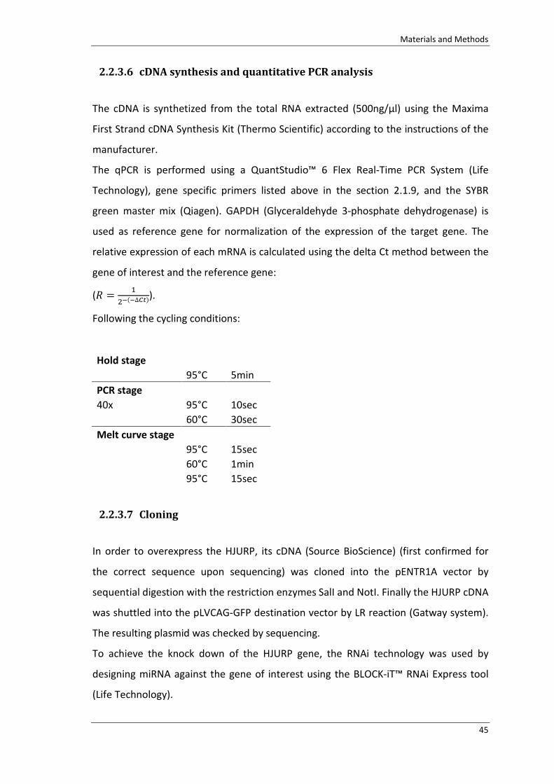
Materials and Methods
45
2.2.3.6 cDNAsynthesisandquantitativePCRanalysis
The cDNA is synthetized from the total RNA extracted (500ng/µl) using the Maxima
First Strand cDNA Synthesis Kit (Thermo Scientific) according to the instructions of the
manufacturer.
The qPCR is performed using a QuantStudio™ 6 Flex Real-Time PCR System (Life
Technology), gene specific primers listed above in the section 2.1.9, and the SYBR
green master mix (Qiagen). GAPDH (Glyceraldehyde 3-phosphate dehydrogenase) is
used as reference gene for normalization of the expression of the target gene. The
relative expression of each mRNA is calculated using the delta Ct method between the
gene of interest and the reference gene:
(� = �����∆�). Following the cycling conditions:
Hold stage
95°C 5min
PCR stage
40x 95°C 10sec
60°C 30sec
Melt curve stage
95°C 15sec
60°C 1min
95°C 15sec
2.2.3.7 Cloning
In order to overexpress the HJURP, its cDNA (Source BioScience) (first confirmed for
the correct sequence upon sequencing) was cloned into the pENTR1A vector by
sequential digestion with the restriction enzymes SalI and NotI. Finally the HJURP cDNA
was shuttled into the pLVCAG-GFP destination vector by LR reaction (Gatway system).
The resulting plasmid was checked by sequencing.
To achieve the knock down of the HJURP gene, the RNAi technology was used by
designing miRNA against the gene of interest using the BLOCK-iT™ RNAi Express tool
(Life Technology).

Materials and Methods
46
The double stranded oligos were generated according to the protocol of the
manufacturer. The two miRNAs (HJURP946 and HJURP1202, according to the designing
coordinates) were cloned into the pENTR6_GW_grandestuffer and finally shuttled into
the pDest-311 vector by LR reaction (Gateway system).
2.2.3.7.1 Transformation into chemically competent E.Coli
The amplification of a specific plasmid is achieved inserting it into chemically
competent cells. The insertion of exogenous genetic material into a bacterial cell
happens by a process called transformation.
To perform a transformation the following materials are needed:
• LB-medium
• Chemo-competent E.coli
• Bacterial agar plates
• Antibiotic:
o Ampicillin stock (100 mg/ml): use 1:1.000
o Kanamycin stock (50 mg/ml): use 1:1.000
An aliquot of 50μl containing chemo-competent cells are thawed on ice. DNA (5-200
ng) is added to it and incubated for 20min on ice. Cells are heat-shocked for 45s at
42°C, and cooled on ice for 2min. 200μl LB medium is added and cells are incubated for
45min at 37 °C on a roller shaker. Afterwards, different aliquots are plated on bacterial
agar plates (50-500μl) and incubated overnight at 37°C.
2.2.3.7.2 Bacterial liquid cultures and DNA purification
For large scale (200ml) and small scale (5ml) liquid bacterial cultures, a single colony
from the agar plate is inoculate into LB buffer, supplement with the appropriate
antibiotic and incubated overnight at 37°C under vigorous shaking.
Plasmid DNA is then isolated with Qiagen Maxi or Mini kits according to the instruction
of the manufacturer.

Materials and Methods
47
2.2.3.7.3 Restriction digestion of DNA
A restriction reaction is required to be able to cut DNA in specific region. This is
prepared as described below:
• 1µg DNA plasmid
• 2µl 10xreaction buffer (NEB)
• 5-10U restriction enzyme (NEB)
• Up to 20µl of H2O
The reaction is incubated at the appropriate temperature according to the restriction
enzyme used and for a time that ranges from 2h to overnight. The DNA digestion is
assessed by gel electrophoresis.
2.2.3.8 Gelelectrophoresis
To perform gel electrophoresis, an appropriate amount of Agarose to prepare 1-2%
(w/v) gel, depending on the size of the nucleic acid of interest, is dissolved in 1X TAE
buffer let to melt in a microwave. The solution is then cooled down and SYBR® Safe
DNA Gel Stain (Life Technology) is added in 1:10000 dilution before pouring the gel in
an electrophoretic chamber. After the gel solidified, DNA samples mixed with 6X
loading dye and a DNA ladder of appropriate size is loaded and electrophoresis
performed at 100-200V. Then the band patterns of the samples are documented under
UV light. In case of further usage of the DNA fragments for downstream applications;
the DNA fragments with the right size are cut under UV light (254nm), transferred into
an Eppendorf tube and purified using the QIAquick gel extraction kit (Qiagen)
according to the protocol of the manufacturer.
2.2.3.8.1 Ligation
In order to allow ligation, the insert of DNA is used in a 5-10 fold excess from the DNA
vector. The ligation reaction is let at 16°C overnight and transformed the following
day. Colonies are picked, DNA purified and the final vector analyzed by sequencing.

Materials and Methods
48
The sequencing is performed at the Sequencing Core Facility of the Helmholtz Zentrum
München and the sequence analyzed with the use of the free available software ApE
(A plasmid Editor by Wayne Davis).
2.2.4 Dataanalysis
Quantifications of cells in mitosis (PH3+ cells) were performed at E14 and E16 across
the entire LGE from lateral to medial using the Olympus Flow View software. All
quantifications were done on images from ventral telencephalon using level matched
sections of at least three stage matched embryos of each genotype.
Quantifications of BrdU+ and Dcx+ cells in the adult brain were performed in P56
sections and total numbers of the different categories defined per field of view. Fields
of view are pictures taken with a 40x magnification 800*800 pixels. 2-3 sections per
animals were measured of at least three staged matched animals of each genotype.
2.2.4.1 Statistics
Calculation of the arithmetic average:
�̅ = 1��������
Standard deviation (SD):
� = �∑ ��� − �̅������ − 1
Standard error of the mean (SEM):
σ� = �√�
were performed with GraphPad Prism5 and the same software used to test statistical
significance. Data were considered as significant with P≤0.05 (*), very significant with
P≤0.01 (**) and highly significant with P≤0.001 (***).

Aim of the thesis
49
3 Aimofthethesis
Neural stem cells proliferation, specification and differentiation are three processes
that need to be tightly regulate in order to allow the production of specific neuronal
subtype at the right time and place in the brain during development and in the
neurogenic regions at postnatal stages.
The transcription factor Pax6 is one of the factors able to orchestrate all these
functions. It is defined as master regulator, being implicated in the control of fate
determination and cell proliferation (Osumi et al., 2008; Georgala et al., 2011a). To
control these aspects of the neurogenic process, Pax6 utilizes the paired (PD) DNA
binding domain (Haubst et al., 2004; Ninkovic et al., 2010).
The PD domain is structured in a modular bipartite manner with two subdomains PAI
and RED that bind cooperatively and independently to different binding sites. Both
these subdomains exert effect on neurogenesis and on the control of cell proliferation
(Walcher et al., 2013).
Given these evidences this work is aiming to dissect the molecular network controlled
by the PD domain which is involved in the regulation of the cell cycle progression.
Particular attention is given to the RED subdomain and how it affects the progenitors
of the ventral telencephalon in the lateral ganglionic eminence (LGE).
In this work will be also explored the influence of the RED subdomain of Pax6 on the
neurogenic lineage progression in the subependymal zone (SEZ), the postnatal
neurogenic region laying at the ventricular surface, which is originating from the
developing LGE.

50

Results
51
4 Results
The transcription factor Pax6 is a pivotal gene in the central nervous system
compelling, among other functions, for the proper patterning of the brain during
development (Stoykova et al., 1996; Stoykova et al., 2000; Toresson et al., 2000).
Pax6 is highly expressed in the developing cortex (Walther and Gruss, 1991; Gotz et al.,
1998; Heins et al., 2002), delineating the major border between the dorsal and the
ventral forebrain with the pallial-subpallial boundary (PSPB).
In the ventral telencephalon, although at lower level if compared with the dorsal
forebrain, the Pax6 protein is expressed in the lateral ganglion eminence (LGE)
(Hallonet et al., 1998). Additionally in the ventral most side of the forebrain it is
expressed in the amygdaloid complex (Tole et al., 2005) and in the latero-cortical
stream (Carney et al., 2006), but these two areas will not be further discussed.
To address the contribution of the RED domain of the Pax6 protein in the regulation of
the proliferation mode of the progenitors cells located at the ventricular surface, we
took advantage of the Pax6Leca2
mutants harboring a single point mutation in the RED
DNA binding domain which is predicted to abolish its binding activity.
In this chapter will be underlined the role of the RED domain in regulating cell division
of progenitor cells and the repercussion of such regulation on the postnatal brain.
4.1 Pax6inthelateralganglioniceminence
4.1.1 Pax6isoformsexpressioninthedevelopingtelencephalon
In vertebrate the Pax6 locus encodes for three different protein isoforms: the
canonical form, the Pax6(5a) and the PD-less form. The Pax6 PD-less form is expressed
higher than the other two isoforms early in development (E10-E12), but it decreases at
the onset of neurogenesis when the PD-containing forms play the major roles (Haubst
et al., 2004).
The levels of expression of the two PD-containing forms of Pax6 are different between
the dorsal and the ventral region. Pax6 and Pax6(5a), consisting of an insertion of 14aa
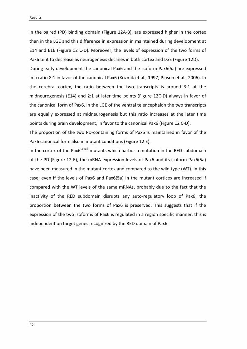
Results
52
in the paired (PD) binding domain (Figure 12A-B), are expressed higher in the cortex
than in the LGE and this difference in expression in maintained during development at
E14 and E16 (Figure 12 C-D). Moreover, the levels of expression of the two forms of
Pax6 tent to decrease as neurogenesis declines in both cortex and LGE (Figure 12D).
During early development the canonical Pax6 and the isoform Pax6(5a) are expressed
in a ratio 8:1 in favor of the canonical Pax6 (Kozmik et al., 1997; Pinson et al., 2006). In
the cerebral cortex, the ratio between the two transcripts is around 3:1 at the
midneurogenesis (E14) and 2:1 at later time points (Figure 12C-D) always in favor of
the canonical form of Pax6. In the LGE of the ventral telencephalon the two transcripts
are equally expressed at midneurogenesis but this ratio increases at the later time
points during brain development, in favor to the canonical Pax6 (Figure 12 C-D).
The proportion of the two PD-containing forms of Pax6 is maintained in favor of the
Pax6 canonical form also in mutant conditions (Figure 12 E).
In the cortex of the Pax6Leca2
mutants which harbor a mutation in the RED subdomain
of the PD (Figure 12 E), the mRNA expression levels of Pax6 and its isoform Pax6(5a)
have been measured in the mutant cortex and compared to the wild type (WT). In this
case, even if the levels of Pax6 and Pax6(5a) in the mutant cortices are increased if
compared with the WT levels of the same mRNAs, probably due to the fact that the
inactivity of the RED subdomain disrupts any auto-regulatory loop of Pax6, the
proportion between the two forms of Pax6 is preserved. This suggests that if the
expression of the two isoforms of Pax6 is regulated in a region specific manner, this is
independent on target genes recognized by the RED domain of Pax6.
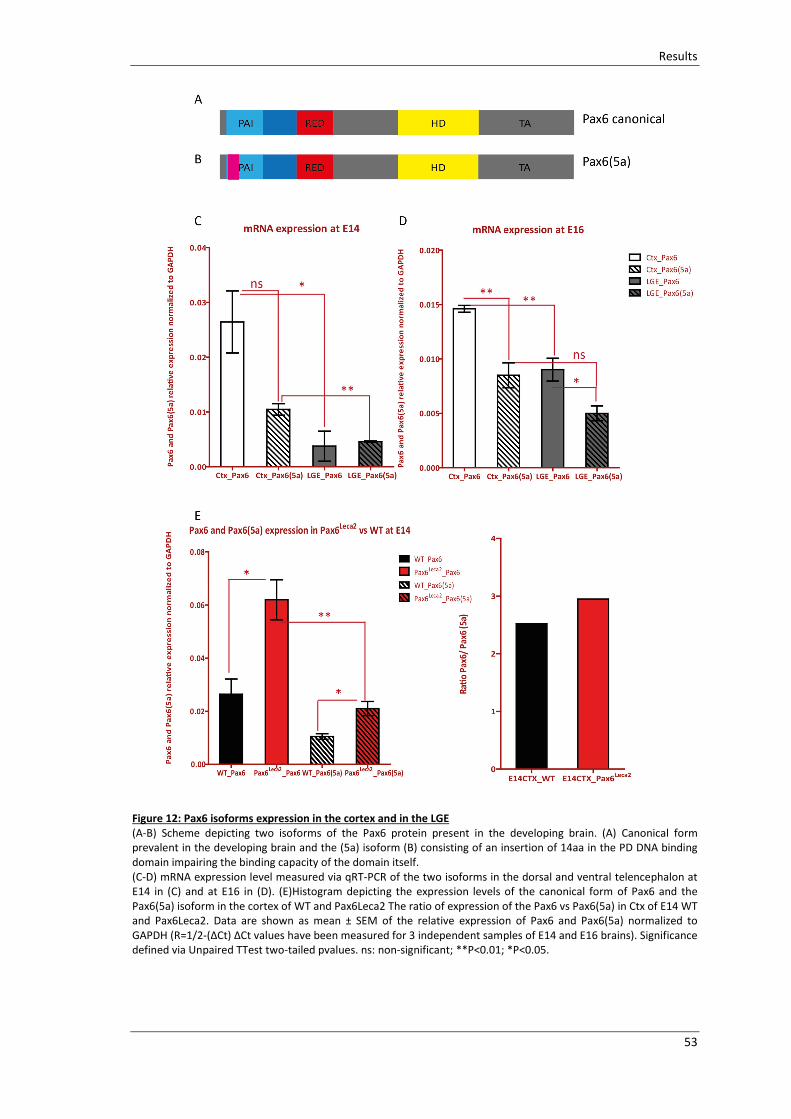
Results
53
Figure 12: Pax6 isoforms expression in the cortex and in the LGE
(A-B) Scheme depicting two isoforms of the Pax6 protein present in the developing brain. (A) Canonical form
prevalent in the developing brain and the (5a) isoform (B) consisting of an insertion of 14aa in the PD DNA binding
domain impairing the binding capacity of the domain itself.
(C-D) mRNA expression level measured via qRT-PCR of the two isoforms in the dorsal and ventral telencephalon at
E14 in (C) and at E16 in (D). (E)Histogram depicting the expression levels of the canonical form of Pax6 and the
Pax6(5a) isoform in the cortex of WT and Pax6Leca2 The ratio of expression of the Pax6 vs Pax6(5a) in Ctx of E14 WT
and Pax6Leca2. Data are shown as mean ± SEM of the relative expression of Pax6 and Pax6(5a) normalized to
GAPDH (R=1/2-(ΔCt) ΔCt values have been measured for 3 independent samples of E14 and E16 brains). Significance
defined via Unpaired TTest two-tailed pvalues. ns: non-significant; **P<0.01; *P<0.05.

Results
54
4.1.2 ProliferationofprogenitorsinPax6REDdomainmutants
Pax6 controls the proliferation of progenitors during the development of the cortex
and the PD domain at the N-terminus is determinant for such function (Warren et al.,
1999; Estivill-Torrus et al., 2002; Haubst et al., 2004).
As previously mentioned, Pax6 expression is not only constrained in the cortical area
but also expressed in the LGE of the ventral telencephalon. To assess the role of Pax6
in controlling the proliferation of the progenitors in the LGE, I analyzed the number of
phospho-histone 3 (PH3)-positive cells in Pax6Leca2
mutants compared to their age
matching siblings.
The antibody PH3 labels cells in the G2/M phase of the cell cycle, allowing the
localization of actively dividing cells (Hendzel et al., 1997). While in the cortex PH3-
positive cells are mostly located in apical positions (the basal division represent around
25% of the total PH3 positive cells (Pilz et al., 2013)), in the LGE around 60% of
progenitors divide in non-apical position (Pilz et al., 2013). These non-surface mitoses
are located within 10 rows of cells from the ventricular side (subapical progenitors SP)
and in more basal positions (basal progenitors BP) (Figure 13).

Results
55
Figure 13: Proliferating cells in LGE
Scheme depicting the position of dividing progenitors in the lateral ganglionic eminence of the ventral
telencephalon classified as apical progenitors (AP) dividing at the ventriclular surface; subapical progenitors (SP)
dividing within 10 rows of cells from the ventricle and basal progenitors (BP) dividing more than 10 rows of cell
away from the ventricle.
In the mutant LGE the PH3-positive cells distribute similarly to the WT littermate and
all the progenitors populations are represented (Figure 14 A-B). The mutation of the
RED domain induces to the increase of mitoses in the population of the apical
progenitors, the cells that divide at the ventricular surface (Figure 14 C-D).
The increased number of PH3-positive AP cells is evident at E14 and then maintained
at later times (E16), suggesting a constitutive effect due to the defective binding of the
RED domain and not only a transient phenomenon.
The increase in apically dividing progenitors is a common feature between the
progenitors in the LGE and the progenitors in the developing cerebral cortex (Walcher
et al., 2013). This suggests a common role for the RED domain in the regulation of the
cell cycle independently from the region of origin of the cells.

Results
56
Figure 14: Progenitor proliferation in Pax6Leca2
LGE
(A-B) Micrographs of E14 ventral telencephalic sections depicting cells immunoreactive for phospho-histone H3
(PH3) in Pax6Leca2
brains and their WT littermates. The insert shows a zoom of the ventricular area where the cells
are mostly affected. Scale bars 50µm. (C-D) Histogram depicting the distribution of PH3+ cells in the LGE of E14 and
E16 Pax6Leca2
. Data are shown as mean ± SEM of the PH3+ cells per LGE length. Significance defined via Mann
Whitney test. ns: non-significant;**
P<0.01; *P<0.05. E14 AP WT vs AP Pax6
Leca2pvalue=0.019. E16 AP WT vs AP
Pax6Leca2
pvalue=0.0031. n≥4 LGE sections per genotype, at least three different animals per genotype.
The exclusive affection of the AP population in the Pax6Leca2
mutants resides on the
fact that all the cells proliferating at the ventricular side express Pax6 (Figure 15).
In the ventricular area (Figure 15) the phospho- vimentin (pVim) positive cells (marker
used to label cells in the M-phase of the cell cycle homologous of PH3) are all
expressing Pax6. This is different from the SP and BP where only some cells express the
Pax6 protein.

Results
57
In the populations of the SP and the BP the effect of the RED domain mutation might
be diluted and masked in the analysis of the all non-apically proliferating progenitors.
Figure 15: Pax6 expression in the progenitors of the LGE.
(A-B) Micrographs depicting the expression of Pax6, in red, and pVim, in green, in WT (A) and in the mutant (B). in
the high magnification examples of different progenitor co-immunostained with Pax6 and pVim (filled arrow heads)
or expressing only pVim (outlined arrow heads). In green AP, in yellow SP and red BP. Scale bars 50µm.

Results
58
4.1.3 CelldeathanalysisinREDdomainmutants
The increment of mitotic progenitors in the Pax6Leca2
is a general effect registered in
the dorsal and ventral telencephalon.
In the cortex the increase in PH3-positive apical progenitors upon the RED mutation
has been explained as the effect of the Pax6 RED domain in controlling cells survival
(Walcher et al., 2013). Pax6 is implicated to play a role in this context (Nikoletopoulou
et al., 2007; Ninkovic et al., 2010).
Supposedly this is a general effect exerted by Pax6 independently from the region of
expression. In order to validate whether Pax6 RED domain is relevant for preserving
progenitors survival, has been analyzed the level of apoptosis in the progenitors of LGE
of the Pax6 mutants and their WT siblings. The immunostaining against activated-
Caspase3 was performed in the ventral telencephalon of E14 animals (Figure 16).

Results
59
Figure 16: Survival of progenitors in Pax6Leca2
(A-B) Micrographs of E14 LGE depicting cells immunoreactive for activated-Caspase3 (green) in WT and mutants
siblings. Scale bar 20µm. (C) Histogram depicting the number of activated-Caspase3 positive cells per total LGE. Data
are shown as total number per 10 sections ±S.E.M. n (number of brains)≥3. *p<0.05; Mann-Whitney test.
This analysis revealed that the mutation of the RED domain of Pax6 leads to a decrease
of the cell survival in the LGE of the ventral telencephalon, similarly to the effect that it
exerts on the progenitors of the developing cortex.
This evidence, together with the increment of cells in mitosis (Figure 16 and Figure 14),
suggested a direct involvement of the RED subdomain on the regulations of the mitotic
progression that, if impaired, leads to cells aberrations and eventually cell death.
Results
60
4.1.4 CellcycleprogressioninPax6Leca2ventraltelencephalon:lossandgain
offunctionexperiments
In LGE of the Pax6Leca2
mutant animals, the increase in apically dividing progenitors and
the reduction of cell survival could be explained by the AP cells failing in completing
the cell cycle and the division of the two daughter cells during the last step of the
cytokinesis. Failure in the cytokinesis would lead to the visualization of more nuclei, as
in the analysis of fixed tissue where there is an increment of about 30% in PH3-positive
cells (Figure 14), but those could be predicted as divided nuclei of daughter cells that
did not completely cleaved their cytoplasm.
To prove this hypothesis, we performed ex vivo live imaging of progenitor cells in the
LGE. This technique offers the ability to follow single cells overtime during their cell
cycle and allows answering if the abundance of mitotic figure in Pax6Leca2
is due to
impairment in cytokinesis.
Figure 17: Ex vivo life imaging:
Scheme depicting the ex vivo live imaging paradigm as described in the method paragraph 2.2.1.5
As described in the method paragraphs 2.2.1.3, 2.2.1.5, progenitor cells in the LGE are
labelled via in utero electroporation (Figure 17) of embryos at E13. Three plasmids are
introduced into the cells of the LGE of WT and mutant animals:
• H2B-Venus is used to identify the nuclear movements since H2B is a member of
the core histones that package DNA in the nucleus.
• pCAG-Floxp-mKO2-F (Shitamukai et al., 2011) combined with a Cre expressing
plasmid are used to highlight the cell membrane and to visualize the phase of
the cytoplasmic scission.
The day after the operation, embryos at E14 are sacrificed, sliced and imaged (Figure
17). The imaging session is running for about 24 hours to be able to visualize at least

Results
61
one cell division. The cell cycle of an apical progenitor cells is in fact lasting about 25h
(Pilz et al., 2013).
During this time was possible to monitor the progenitor cells during their division and
estimate the time required for their nuclear and cytoplasmic division (example of time
series in Figure 18).

Results
62

Results
63
Figure 18: LGE ex vivo live imaging
(A-D) Micrographs depicting examples of cells in the WT (panel A-B, curtesy of Dr Sven Falk) and Pax6Leca2
(panel C-
D). Images are acquired with an interval of 15 minutes with a confocal microscope Olympus FV1000 cLSM system,
20 x magnification ((A-B) 2x zoom as acquisition setting). (A-C) H2B-Venus used to visualize nuclear movements. (B-
D) mkO2 used to visualize the cytoplasm and cell membrane movements. Arrow heads indicate examples of the
cells analyzed.

Results
64
Figure 19: Time of cytokinesis definition and measuring
(A) Scheme depicting the definition of “time of cytokinesis” measured as the time between the division of the
nucleus and the full scission of the cytoplasm in two different daughter cells. (B) Dot blot representing the time that
the cells analyzed need to divide into two daughter cells. Dots in black represent the WT cells measured (9 cells).
The red dot represents the Pax6Leca2
cells that divided in the 150min of observation whereas the red triangles
represent the cells that did not fulfill the requirement in the given time (cell1=510min: cell2=315min: cell3=150min;
cell4=180min; cell5=255min).
For the WT the horizontal bar represents the mean ±SEM.
To prove the idea of a prolonged cytokinesis, we measured the “time of cytokinesis”.
This is defined (Figure 19 A) as the time between the division of the nuclei, right after
the condensation of the heterochromatin that is a synonym of the nuclear division
initiation, and the full division at the membrane level of the two daughter cells. This
analysis, (Figure 19 B) revealed that the time of cytokinesis in Pax6Leca2
mutants is
longer than in the WT. Given an observation time of 150 minutes all the WT cells
managed to complete division (average time: 82.8min) whereas only one Pax6Leca2
completed the cytokinesis in the same time.
These measurements could confirm that cells upon mutation of the RED domain have
a prolongation of the time the cells share their cytoplasm after their nuclear division.
This evidence was further consolidate when E14 brains, derived from Pax6Leca2
background, were analyzed and fixed after the electroporation of the same plasmids
listed above (Figure 20).

Results
65
Also via fixed analysis we could detect that the mutation of the RED domain is
impairing the cell cycle progression of the apical progenitors of the LGE. The
progenitors in the Pax6Leca2
, even if composed by two nuclei (Figure 20), are still
surrounded by a unique membrane. The situation in the WT is different (Figure 20 A),
the cells are mostly in G phase with one nucleus ready to divide or differentiate.
These data underline the importance of the RED domain of Pax6 in controlling the final
step of the cell cytokinesis.
Figure 20: Cell division in vivo
Orthogonal projections depicting examples of radial glia cells after in utero electroporation of membrane protein in
the mutant (B) and WT littermate (A) LGE. Scale bars A 20µm, B 10µm.
The implication of the RED domain mutation affecting cell cycle progression and
compromising the capacity of the cells to complete their mitosis was further supported
by in vivo experiments. In these experiments we performed loss of the RED domain
functions in the WT background. Cells in the WT brains were transduced with retroviral
vectors co-expressing the Pax6Leca2
form and the GFP protein. The viruses were
injected at the E14 stage and the brains analyzed 3 days later (E17). The injection was
directed in the ventricle and labelled cells were found in the cortex.
The cells found, even if they were only few probably due to the low titer of the virus,
were all in duplets (Figure 21).
This is considered an additional evidence that the mutation of the RED domain of Pax6
would lead to impairments in the progression of the mitotic phase.

Results
66
Figure 21: In vivo overexpression of the RED domain mutation of Pax6
(A-B) Micrographs depicting duplets of cells in the cortical area upon in vivo transduction with retro viral vector
Pax6Leca2
CAG-GFP in WT background. (A) GFP immunostaining, (B) GFP immunolabeling merged with the DAPI. Scale
bars 10µm.
Pax6 is known as a regulator of cell division (Asami et al., 2011), but its involvements in
the control of the progression of cytokinesis is a novel role which opens as new
question whether the RED domain controls specific targets sufficient to explain the
observed phenotype.
To have new insights on the molecular partners regulated by the RED domain that
could explain the phenotype in the developing brain, the microarray data derived from
the developing cortex of the Pax6Leca2
(Walcher et al., 2013) were analyzed. From this
analysis the Holliday junction recognition protein (HJURP) emerged as an interesting
candidate.

Results
67
4.1.5 Candidategene:HJURP
The effect of the RED subdomain mutation of Pax6 on the proliferation of progenitors
observed via live imaging, motivates to analyse genome–wide expression changes
previously collected (Walcher et al., 2013), in order to identify possible candidates
suitable to explain the phenotype.
Microarray analysis was carried out using rostral developing cortices from mutants and
wild type animals at the developmental stage E14 (Figure 22). The rostral part of the
cortex was selected based on the knowledge that Pax6 is expressed in a rostral high -
caudal low gradient (Stoykova et al., 1997; Muzio et al., 2002b). In the microarray
analysis, among the most upregulated genes (35 genes out of the 94 differentially
expressed probe sets detected), two seemed particularly promising in explaining the
prolongation of the cell mitosis: Bub1 (Budding uninhibited by benzimidazoles 1) and
HJURP (Holliday junction recognition protein).
The Bub1 gene encodes for a serine/threonine-protein kinase that plays a central role
in mitosis: it is essential for spindle-assembly checkpoint signaling and for correct
chromosome alignment (Elowe, 2011).
Since the upregulation of Bub1 could not be confirmed via quantitative RT-PCR with all
the sets of primers in independent samples (data not shown), the attention was
focused on the second candidate: HJURP.
HJURP is a centromeric protein playing a central role in the incorporation and
maintenance of histone H3-like variant CENPA at centromeres. HJURP is a chaperone
for CENPA and is required for the incorporation of newly synthesized CENPA molecules
into nucleosomes at replicated centromeres. (Stellfox et al., 2013; Muller and
Almouzni, 2014). The proper organization of the centromere is fundamental for the
assembly of the centromere-kinetochore structure and the consequent proper
segregation of the chromatids during mitosis.
The HJURP gene is upregulated in the Pax6Leca2
cortices and such upregulation is
confirmed via quantitative RT-PCR in independent samples derived from the cortices of
mutants and wild types (Figure 22 C).

Results
68
Figure 22: HJURP candidate gene
(A-E) Parts of this figure are adapted from the publication (Walcher et al., 2013). (A) Heat map illustrating the
expression of the differentially regulated genes in the E14 Pax6Leca2
dorsal cortices in comparison with their WT
littermates. Red/blue indicates higher/lower expression values (Walcher et al., 2013). (B) List of up regulated genes
in the Pax6Leca2
, obtained with the genome-wide expression analysis performed by Affymetrix MOE430 2.0 array.
Differences are determined by a filter consisting of statistical significance (FDR<10%), an average expression level
exceeding 50 and at least 1.4-fold difference expression (Walcher et al., 2013). Two possible candidate genes to
explain the phenotype in vivo, 6430706D22Rik also called HJURP and Bub1, are highlighted in red. (C) Histogram
depicting the linear fold changes of the gene HJURP between the Pax6Leca2
and the WT littermates as measured by
Affimetrix array analysis (black bar) and qRT-PCR (red bar). Student TTEST **
P<0.01. (D-E) Micrographs depicting in
situ hybridization in E14 sections of WT and mutants, showing the mRNA expression for the HJURP gene (Walcher et
al., 2013). Abbreviations: Ctx: cortex; LGE: lateral ganglionic eminence.

Results
69
4.1.5.1 ExpressionofHjurp
The implication of the HJURP protein in the brain was never explored before. To
further characterize the expression of HJURP in this system, in situ hybridization was
performed. In the developing cortex (Figure 22 D-E), the mRNA is particularly localized
in the VZ/SVZ. In the Pax6Leca2
mutant (Figure 22 E) the expression of the mRNA for
HJURP is increased in this cortical area. The localization of HJURP mRNA in the VZ/SVZ
is in accordance with the function of HJURP that helps the incorporation of newly
synthesized CENPA at the replicated centromeres during S phase (Stellfox et al., 2013;
Muller and Almouzni, 2014). In this region in fact, together with the proliferating basal
progenitors, there are also the radial glia cells (expressing Pax6) in the S phase of their
cell cycle. Radial glia cells in fact undergo intekinetic nuclear migration which comprise
that the cell nucleus migrates radially to the basal side of the cells during G1 phase of
the cell cycle, undergoes to S-phase in basal position and migrates back to the apical
side for G2-M phase of the cell cycle.
In the LGE of the Pax6Leca2
mutants the increase in the HJURP mRNA appears very
modest and difficult to confirm. The reason of a less clear result might reside in the
characteristic heterogeneity of the progenitors present in the LGE.
4.1.5.2 HJURPinvitro
In order to recapitulate the phenotype observed in vivo in the mutant mice and to
delineate the contribution of HJURP on it, the protein was overexpressed in vitro on
P19 cells.
P19 cells are able to fast proliferate and eventually to differentiate to neurons when
exposed to specific compounds like retinoic acid (Staines et al., 1994). Therefore, this
cell line was used to approximate neuronal progenitors in vivo. In this system the
expression of HJURP was forced via lipofection of plasmid encoding for the protein and
the GFP (green fluorescent protein) that allows the visualization of the cells expressing
the protein of interest. 20h after the lipofection, the time necessary to visualize the
signal from the GFP, the cells were imaged for about 36h until reaching confluency.

Results
70

Results
71

Results
72
Figure 23: In vitro live imaging of P19 cells.upon HJURP overexpression
(A-B) Time series depicting the behavior of cells upon lipofection of the control plasmid (A) and the plasmid
expressing HJURP (B). Pictures are acquired with an inverted Zeiss Cell-Observer Z1 video microscope. 20 x
magnification. In A and B the arrow heads indicate examples of cells analyzed. (C) Dot blot depicting the time of
cytokinesis as previously described for different conditions. Data are represented as mean ± SEM. ***P<0.001,
Mann-Whitney test. The cells analyzed are coming from n≤3 experiment except the case of the OE of Pax6Leca2
where n=1. (D) Micrographs depicting P19 cells upon HJURP OE immunostained 36h after lipofection. Scale bar
5µm.
In the cells expressing HJURP was measured the time of cytokinesis. This was
determined has the time intercurring between the nuclear division and the complete
scission of the cytoplasm of the two daughter cells; the cells in which the cytokinesis
was not completed by the end of the movie where excluded from the analysis.
The overexpression of HJURP (Figure 23 C) leads to a lengthening of the time needed
to complete the cytoplasmic division of the cells. The cell cytokinesis, upon HJURP
overexpression, appears impaired and longer than in the wild type condition. The cells
remain in contact possibly because the DNA in the two nuclei is not completely
segregate in the daughter cells so that they are blocked from proceeding with the last
step of cytokinesis (Figure 23 D).

Results
73
P19 cells were imaged also after overexpression of the RED domain mutant form of
Pax6. In this case a similar effect on cell cycle progression was detected (Figure 23 C).
This supports the idea of the existence of a tight connection between the RED domain
of Pax6 and the HJURP protein, in fact following the cells expressing HJURP during their
division, unravels very similar behaviors than the mutation of the Pax6 RED domain per
se.
The current model (Figure 24) starts hypothesizing an inhibitory capacity of the RED
domain in normal conditions. In the Pax6Leca2
the mutation of the RED domain impairs
this inhibitory function, contributing to the increase of the HJURP mRNA. As a
consequence, the increment in HJURP leads to a prolongation of the cytokinesis in the
apical progenitors cells of the developing telencephalon. What remains to be
elucidated in this regard is the direct implication of Pax6 RED domain on the increase
of HJURP expression.
Figure 24: Working model
(A-B) Scheme depicting the model suggested by the results obtained from the analysis of the RED domain mutation
in the developing brain (A) WT, (B) Pax6Leca2
.

Results
74
4.2 Adultbrain
The LGE region of the ventral telencephalon gives rise, at postnatal stages, to the cells
populating the subependymal zone (SEZ), one of the regions where neurogenesis
persists during adult life (Merkle and Alvarez-Buylla, 2006).
The progenitors cells present in the LGE of the Pax6Leca2
mice are facing defects in cell
cycle progression, this motivated the assumption that the impairment encountered by
the progenitors of the LGE might have repercussion on the cells originating from there.
In addition, giving the fact that the cells populating the SEZ niche express the Pax6
protein in normal condition (Hsieh, 2012), we used the Pax6Leca2
animals as a tool to
investigate the role of the RED domain of Pax6 in the SEZ region of the adult brain.
4.2.1 Pax6Leca2inadultbrain
To investigate the repercussion of the RED domain mutation on the adult
neurogenesis, eight to ten weeks old Pax6Leca2
mice were used. Differently from many
Pax6 mutants which die at birth due to the loss of a protein that has pivotal roles in
various systems, Pax6Leca2
mutants are able to survive after birth and reach adulthood.
Figure 25: Gross morphology of Pax6Leca2
(A) Examples of animals of the two genotyping (Walcher et al., 2013). (B) Histogram depicting the difference in body
weight and brain size between WT (black bar) and Pax6Leca2
(red bar). Data are reported as mean ± SEM *P≤0.05; ns:
non-significant. Unpaired T Test and Upaired TTest with Welch’s correction. (C-D) Gross morphology of the brain in
the mutants and their WT littermates seen from the top (C) and from the bottom (D) (Walcher et al., 2013).
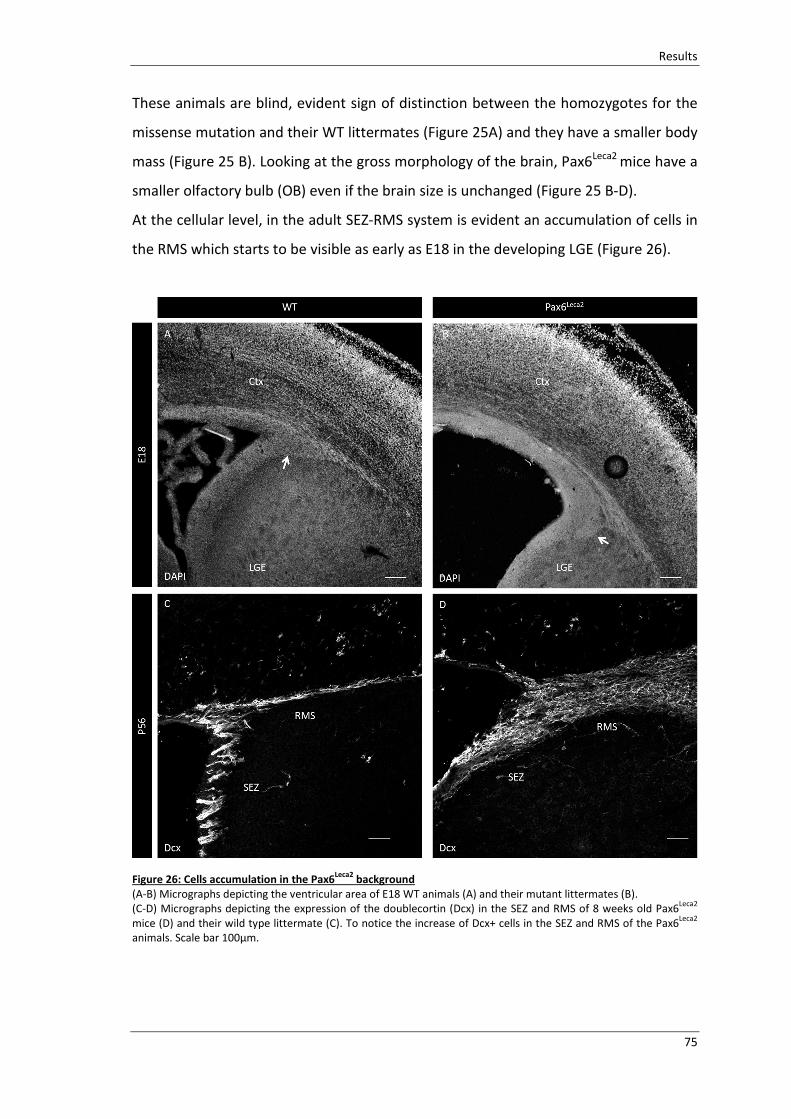
Results
75
These animals are blind, evident sign of distinction between the homozygotes for the
missense mutation and their WT littermates (Figure 25A) and they have a smaller body
mass (Figure 25 B). Looking at the gross morphology of the brain, Pax6Leca2
mice have a
smaller olfactory bulb (OB) even if the brain size is unchanged (Figure 25 B-D).
At the cellular level, in the adult SEZ-RMS system is evident an accumulation of cells in
the RMS which starts to be visible as early as E18 in the developing LGE (Figure 26).
Figure 26: Cells accumulation in the Pax6Leca2
background
(A-B) Micrographs depicting the ventricular area of E18 WT animals (A) and their mutant littermates (B).
(C-D) Micrographs depicting the expression of the doublecortin (Dcx) in the SEZ and RMS of 8 weeks old Pax6Leca2
mice (D) and their wild type littermate (C). To notice the increase of Dcx+ cells in the SEZ and RMS of the Pax6Leca2
animals. Scale bar 100µm.

Results
76
Two possible hypotheses arouse in order to elucidate the cause of this cell
accumulation:
1. The RED domain of Pax6 is relevant for the proper lineage progression in the
SEZ neurogenic region. Pax6 protein is expressed in the progeny of the stem
cells lying in the SEZ lateral wall, namely the transit amplifying progenitors
(TAPs) and the neuroblasts (Hack et al., 2005; Kohwi et al., 2005; Brill et al.,
2008); impairments in their proliferation or differentiation could be the cause
of the accumulation of cells.
2. The RED domain of Pax6 has roles in conferring the migratory ability to the cells
in the SEZ-RMS system. The cells in the RMS, in fact, are characterized by their
capability to tangentially migrate (Ghashghaei et al., 2007) in order to reach the
olfactory bulbs. Losing the ability to migrate could cause the aggregation of
cells in this particular region.
The attempts directed to the understanding of these possibilities will be discussed in
the following paragraphs.
4.2.1.1 NeurogenicprogressioninthePax6Leca2mutant
To investigate the involvement of the Pax6 RED domain in the regulation of the cell
proliferation in the adult SEZ we evaluated the proliferative capacity of TAPs and
neuroblasts in adult (8 weeks old) Pax6Leca2
mutants and their age-matching
littermates.
To elucidate if any of the two populations was affected, I analysed the number Dcx-
positive neuroblasts in S-phase of cell cycle based on incorporation of the DNA-base
analogous BrdU (one hour before sacrificing the animals, 0.5 ml of a 10 mg/ml BrdU
solution are injected intraperitoneally). This approach followed by the colocalization
with Dcx allows to discriminate the proliferating neuroblasts (BrdU+ and Dcx+) from
the TAPs themselves (BrdU only) and locate the origin of the impairment causing
accumulation of cells in the SEZ-RMS area.

Results
77

Results
78
Figure 27: Neuroblasts and TAPs proliferation analysis
(A-B) micrographs representing high magnification pictures of the SEZ-RMS area of the Pax6Leca2
and WT animals at
postnatal stage P56. (C-F) orthogonal picture magnification of cells expressing BrdU and Dcx and BrdU only in WT
(C-D) and Pax6Leca2
(E-F). (G-H) histograms depicting the amount of BrdU+ cells, Dcx+ and of double positive cells for
Dcx and BrdU in the SEZ and RMS. Fields of view are pictures taken with a 40x magnification 800*800 pixels (2-3
sections per animals n (number of animal)=3). (I-J) histogram depicting the proportion of double positive cells
among all the dividing cells and among all the postmitotic neuroblasts in SEZ and RMS. Data are represented as
mean ±SEM. Mann-Whiney test for significance ns: non-significant; **P<0.01; ***P<0.001.
In the SEZ the TAPs population (BrdU only cells), similarly to the neuroblasts, is not
affected by the RED domain mutation. No difference between the mutant animals and
the WT was detectable in this population in the SEZ area.
In the RMS the condition is different. Looking at the absolute number of the cells
positive for the markers is evident that the BrdU only cells are increased (from an
average of 21.06 ± 3.223 cells in the WT to 35.88 ± 4.219 in the Pax6Leca2
).
The hypothesis was that the increase in number in the TAPs population would account
for the increase of the population of the neuroblasts responsible for the enlargement
of the RMS in the Pax6Leca2
.
To investigate this idea we followed the previous approach (short BrdU pulses) and
focus the attention on the proliferating neuroblasts which directly generate from the
TAPs (Doetsch et al., 1999).
In the SEZ the absolute number of proliferating neuroblasts (Dcx+BrdU+) and of
postmitotic neuroblasts (Dcx+ only) is unchanged when the two genotypes are
compared. The difference in neuroblasts number between Pax6Leca2
and the WT is
evident only at the RMS level.
In the RMS, the number of post mitotic neuroblasts (28.65 ± 4.336 cells in the WT,
97.19 ± 14.42 the Pax6Leca2
) and proliferating ones is increased upon RED domain
mutation (3.000 ± 0.6301 cells in the WT, 6.938 ± 0.5513 in the Pax6Leca2
). This
difference nevertheless, disappears when the double positive population (the
proliferative neuroblasts) is put in relation with total population of the post mitotic
neuroblasts (Figure 27) and in relation to all the proliferating cells.
Since no relevant difference could be detected with the short pulse of BrdU paradigm
explaining the enlargement of the RMS, the hypothesis is that the bulge of cells
residing in the RMS is originated earlier in development and for poorly understood
reasons remains in the area being composed by early generated cells and newly

Results
79
supplied cells by the SEZ. In the SEZ of Pax6Leca2
animals, in fact, neurogenesis is
proceeding in a similar manner than in the WT.
The possibility that in the SEZ of the Pax6Leca2
mutants the progression in the cell cycle
might be delayed and other phases, rather than the S phase, might be affected
remains still open.
In the SEZ and RMS system the gene HJURP is in fact expressed in both TAPs and
neuroblasts. HJURP is the gene that, as shown in the previous chapter, is a possible
candidate explaining at the molecular level the phenotype seen in the RED domain
mutant animals having impairments in the cell cycle progression.
Investigating the data obtained from the transcriptome analysis performed on cells
isolated from the SEZ niche (Beckervordersandforth et al., 2010), we could reveal the
presence of HJURP also in the SEZ niche. This gene is primarily present in the stem cells
(hGFAP/GFP and Prominin+) and then maintained in neuronal lineage having very high
levels of expression in the progeny of the stem cells (HGFAP/GFP+ only) which, in this
analysis, is composed by the TAPs and the neuroblasts.
With the in situ hybridization the HJURP expression was highlighted in SEZ and in the
RMS (Figure 28). The HJURP expression is high in the RMS of the Pax6Leca2
animals
where the cells show a blockage during the path through the OB.
The direct connection between HJURP and the enlargement of the RMS is still not
understood and matter of speculation. The HJURP expression is nevertheless
representing a bridging point between the adult and the embryonic phenotype.

Results
80
Figure 28: HJURP expression in the adult brain
(A) Scheme adapted from the original publication (Beckervordersandforth et al., 2010), representing the cell type
examined by FACS sorting isolation present in adult SEZ niche of the lateral ventricle. (B) Histogram depicting the
expression levels of HJURP in adult SEZ cells FACS sorted and analyzed via microarray analysis performed with
Affymetrix MOE430 2.0 arrays (as described in the publication (Beckervordersandforth et al., 2010). hGFAP/GFP+
Prominin+: putative stem cells; hGFAP/GFP+only: stem cell progeny comprising of TAPs, neuroblasts and niche
astrocyte; Diencephalic hGFAP/GFP+: astrocytes outside the niche used as comparison. (C-D) Micrographs depicting
in situ hybridization in adult brains of Pax6Leca2
homozygotes and heterozygotes, which highlight the expression of
the HJURP mRNA in the SEZ and in particular in the RMS, where the neuroblasts reside. Abbreviations: Ctx: cortex;
SEZ: subependymal zone.
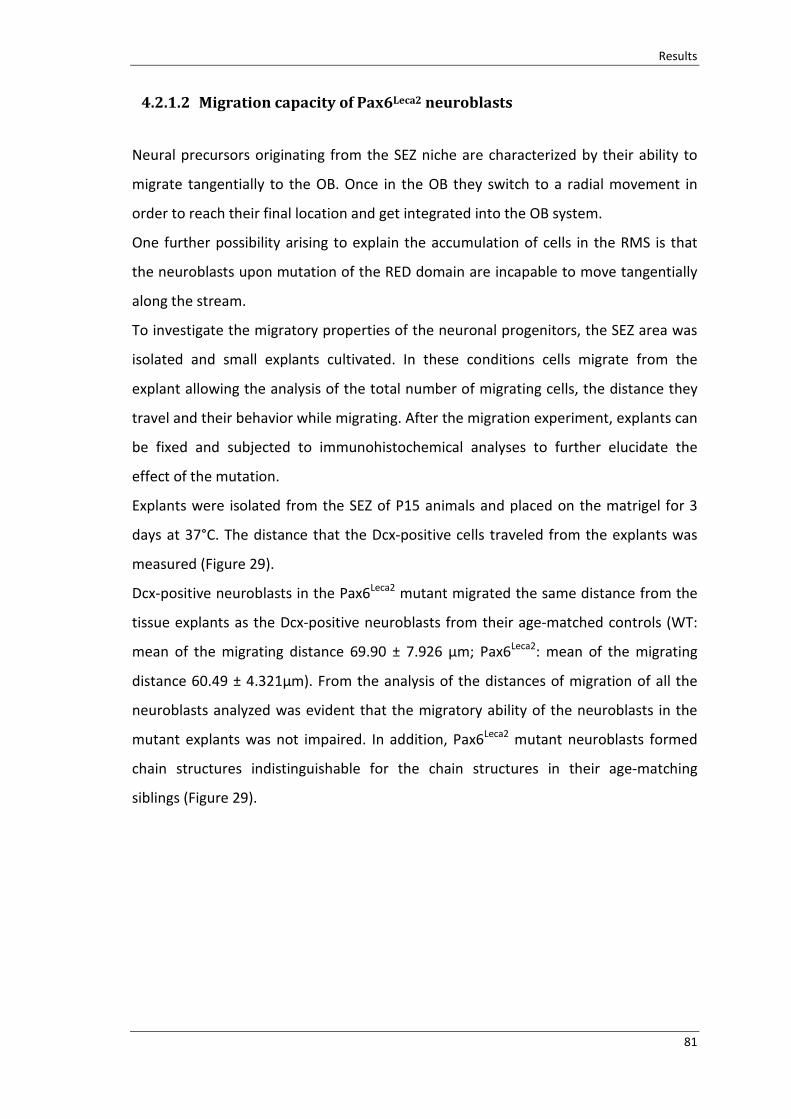
Results
81
4.2.1.2 MigrationcapacityofPax6Leca2neuroblasts
Neural precursors originating from the SEZ niche are characterized by their ability to
migrate tangentially to the OB. Once in the OB they switch to a radial movement in
order to reach their final location and get integrated into the OB system.
One further possibility arising to explain the accumulation of cells in the RMS is that
the neuroblasts upon mutation of the RED domain are incapable to move tangentially
along the stream.
To investigate the migratory properties of the neuronal progenitors, the SEZ area was
isolated and small explants cultivated. In these conditions cells migrate from the
explant allowing the analysis of the total number of migrating cells, the distance they
travel and their behavior while migrating. After the migration experiment, explants can
be fixed and subjected to immunohistochemical analyses to further elucidate the
effect of the mutation.
Explants were isolated from the SEZ of P15 animals and placed on the matrigel for 3
days at 37°C. The distance that the Dcx-positive cells traveled from the explants was
measured (Figure 29).
Dcx-positive neuroblasts in the Pax6Leca2
mutant migrated the same distance from the
tissue explants as the Dcx-positive neuroblasts from their age-matched controls (WT:
mean of the migrating distance 69.90 ± 7.926 µm; Pax6Leca2
: mean of the migrating
distance 60.49 ± 4.321µm). From the analysis of the distances of migration of all the
neuroblasts analyzed was evident that the migratory ability of the neuroblasts in the
mutant explants was not impaired. In addition, Pax6Leca2
mutant neuroblasts formed
chain structures indistinguishable for the chain structures in their age-matching
siblings (Figure 29).

Results
82
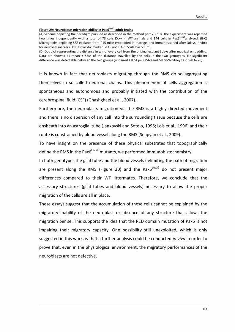
Results
83
Figure 29: Neuroblasts migration ability in Pax6Leca2
adult brains
(A) Scheme depicting the paradigm pursued as described in the method part 2.2.1.8. The experiment was repeated
two times independently with a total of 73 cells Dcx+ in WT animals and 144 cells in Pax6Leca2
analysed. (B-C)
Micrographs depicting SEZ explants from P15 mice embedded in matrigel and immunostained after 3days in vitro
for neuronal markers Dcx, astrocytic marker GFAP and DAPI. Scale bar 50µm.
(D) Dot blot representing the distance in µm of every cell from the original explant 3days after matrigel embedding.
Data are showed as mean ± SEM of the distance travelled by the cells in the two genotypes. No-significant
difference was detectable between the two groups (unpaired TTEST p=0.2568 and Mann-Whitney test p=0.6220).
It is known in fact that neuroblasts migrating through the RMS do so aggregating
themselves in so called neuronal chains. This phenomenon of cells aggregation is
spontaneous and autonomous and probably initiated with the contribution of the
cerebrospinal fluid (CSF) (Ghashghaei et al., 2007).
Furthermore, the neuroblasts migration via the RMS is a highly directed movement
and there is no dispersion of any cell into the surrounding tissue because the cells are
ensheath into an astroglial tube (Jankovski and Sotelo, 1996; Lois et al., 1996) and their
route is constrained by blood vessel along the RMS (Snapyan et al., 2009).
To have insight on the presence of these physical substrates that topographically
define the RMS in the Pax6Leca2
mutants, we performed immunohistochemistry.
In both genotypes the glial tube and the blood vessels delimiting the path of migration
are present along the RMS (Figure 30) and the Pax6Leca2
do not present major
differences compared to their WT littermates. Therefore, we conclude that the
accessory structures (glial tubes and blood vessels) necessary to allow the proper
migration of the cells are all in place.
These essays suggest that the accumulation of these cells cannot be explained by the
migratory inability of the neuroblast or absence of any structure that allows the
migration per se. This supports the idea that the RED domain mutation of Pax6 is not
impairing their migratory capacity. One possibility still unexploited, which is only
suggested in this work, is that a further analysis could be conducted in vivo in order to
prove that, even in the physiological environment, the migratory performances of the
neuroblasts are not defective.

Results
84
Figure 30: Migrating neuroblasts accessory structures
(A-B) Micrographs depicting the rostral migratory stream in WT (A) and mutant sections (B). In green is represented
the glial tube (GFAP positive), in grey the blood vessels highlighted via fluorescent Dextran administered via tail
injection. The white arrowheads highlight the blood vessels. In the insert zoom of the RMS showing that none of the
two structures are impaired. Scale bar 50 µm.

Results
85
4.3 DomainspecificfunctionsofPax6
Pax6 is defined as master regulator gene. This definition underlines its capability to
govern the lineage specification process regulating multiple downstream genes either
directly or via a cascade of gene expression changes The Pax6 function as fate
determinant has been previously elucidated in the adult SEZ system as well as in the
developing cortex, underlying the crucial role of the N-terminal PD domain (Heins et
al., 2002; Hack et al., 2004; Haubst et al., 2004; Hack et al., 2005; Ninkovic et al., 2013).
4.3.1 EmbryonicandAdultneurospheres
In order to understand whether the disruption of the binding capacity of the PD
domain to the DNA is impairing the function of Pax6 in directing neuronal fate
determination, gain-of-function experiments were performed in neurosphere derived
culture.
This culture type allows quantifying the neurogenic capacity of a defined factor.
Neurosphere derived cultures are, in fact, gliogenic cultures which give rise,
independently from the region of origin of the cells cultured, to about 80% of glia cells
(GFAP+ cells) and to 20% of neurons (Dcx+ cells) (Hack et al., 2004).
When these cells are challenged under certain conditions, e.g. the overexpression of
neurogenic factors like Pax6, the outcome is reverted with the majority of cells being
part of the neuronal type (Hack et al., 2004).
In this analysis neurospheres derived from E14 Cortex and LGE, as well as from adult
SEZ, were exposed to Pax6 and its two mutant forms (Pax6Leca2
and Pax6Leca4
), via viral
vector transduction. The different regions and developmental stages were chosen in
order to validate whether the response of the cells upon exposure to Pax6 depends on
the context of origin of the cells themselves.
After the third propagation passage, where the cells were growing in suspension in
medium supplied with growth factors (EGF and FGF2), the cells were plated and
transduces with viral vectors expressing Pax6, Pax6Leca2
and Pax6Leca4
.

Results
86

Results
87
Figure 31: Embryonic ad adult neurosphere
(A-L) Example of cells derived from neurospheres culture after transfection of control (A-D), Pax6Leca2
(E-H) and
Pax6Leca4
viruses (I-L). (M-N) Histogram depicting the number of neurons (Dcx+cells GFP+cells) among the viral
transduced cells (GFP+cells) in the neurosphere cultures derived from the E14 Cortex and the E14 LGE (M) and the
adult SEZ (N). (M-N) Histograms are compared with data derived from (Hack et al., 2004), figure 6 of the original
publication panel E (M), panel D (N). Data are reported as mean± SEM. Unpaired T Test for significance. Non-
significant difference is detected between the cells transfected with a control virus and the Pax6 mutant forms for
the PAI and the RED domain in E14 derived cells as well as in the adult derived cells.
To measure the neurogenic capacity of the Pax6 PD domain mutants, the number of
neurons generated from the glial cells was assessed. When the glia cells are exposed to
the canonical form of Pax6, regardless if they are originating from the cortex, the
ganglionic eminence (Hack et al., 2004) or the SEZ, they give rise to 60-80% of neurons.
Differently, exposing the glia cells to the RED domain or PAI domain mutant forms did
not change the fate of the cells maintaining the number of neurons generated similar
to the control condition (Figure31).
These results suggested that, differently from the overexpression of the full Pax6
protein, the mutated form for the RED domain and the mutated form for the PAI are
not able to direct the glia cell to the neuronal fate. This is valid for cell originating from
the developing and the adult brain.
The activity of only one subdomain of the PD DNA binding domain is not sufficient to
switch the fate of the glial cells to the neuronal one.
However, the lack of capability of RED domain mutation to direct glia cells to
neurogenesis is in contrast with the previous results obtained from primary culture,
composed by cells already primed to the neurogenic lineage, where the Pax6 RED
domain mutation was still capable to induce neurogenesis and failed uniquely when
the PAI domain was impaired (Walcher et al., 2013).
These results suggest that Pax6 neurogenic capacity is cells specific. The transcription
factor Pax6 needs the full activity of its PD domain to induce neurogenesis from glia
cells. In this system both the subdomain PAI and RED are necessary to conduct
neurogenesis.

88

Discussion
89
5 Discussion
During brain development the control of the proliferation of cells and their fate
specification is crucial to preserve the equilibrium in every region of the brain, despite
the high dynamicity of the developing process per se.
In the telencephalon, the transcription factor Pax6 plays a pivotal role coordinating the
proliferation of progenitors and their fate specification (Osumi et al., 2008; Georgala et
al., 2011a).
This work was aiming to the understanding of the role of the RED domain of Pax6 in
controlling the cell cycle progression of the progenitors located in the ventral
telencephalon (LGE) and in the understanding of the relevance of the functionality of
this domain for the proper progression of the neurogenic process in the postnatal
subependymal zone that is originating from the LGE. Towards these ends the
proliferative capacity of the different progenitors in the LGE and the neurogenic
progression in the adult SEZ were analysed in domain specific mutant mice Pax6Leca2
.
The data collected with this work suggest that in the ventral telencephalon the RED
domain mutation of Pax6 brings to a prolongation of the cytokinesis in the apical
progenitors cells. The impairments in the cytokinesis prompt to a reduction of cell
survival. In addition, the RED domain mutation induces the upregulation of the
chaperone protein HJURP (Holliday junction recognition protein). The upregulation of
HJURP in vitro recapitulates the phenotype observed in the progenitor cells of the
Pax6Leca2
animals. Differently, in the postnatal neurogenic niche, the impairments in
binding of the RED domain of Pax6 do not induce failure in the progression through the
neurogenic lineage. This suggests that the accumulation of neuroblasts, evident in the
postnatal brain of Pax6Leca2
animals, has developmental origin rather than emerging at
late stages.
5.1 The Pax6 RED subdomain is responsible for the cell cycle
progressionofapicalprogenitors
The RED DNA binding domain of the transcription factor Pax6 modulates the
progression of the cell cytokinesis in the embryonic apical progenitors of the LGE. In

Discussion
90
the LGE, the exclusive affection of the APs offered the opportunity to resolve the
importance of the RED domain for the regulation of cell cycle progression without
additional influence of the cells in the subapical region. In the APs of the LGE, the
impairment in the RED domain capacity to bind to the DNA leads to a prolongation of
the cell cytokinesis, unraveling a novel role for this subdomain.
Pax6 controls the proliferation of the apical progenitor cells of the cerebral cortex. The
non-functional Pax6 protein leads to alterations in the spindle orientation of the cells
and consequently of their cleavage plane (Asami et al., 2011). The latter generates
daughter cells delaminating from the ventricular side which contribute to the increase
of cells in active mitosis located in subapical positions (Asami et al., 2011). The increase
in this population of cells is in fact one of the most prominent phenotypes upon
disruption of Pax6 protein (Warren et al., 1999).
The PD domain of Pax6 is involved in controlling the proliferative capacity of the RG.
The exclusive impairment of this domain, in fact, resembles the phenotype of the
Pax6Sey
mutant (Haubst et al., 2004). In particular in the cortical progenitors, the two
subdomains of the PD domain of Pax6 antagonize in the regulation of proliferative
genes. Impairing the PAI domain leads to the downregulation of pro-proliferative
factors (Walcher et al., 2013) suggesting that it might promote proliferation in
physiological condition. Conversely when the RED subdomain is defective, the
numbers of PH3 positive progenitors is increased (Walcher et al., 2013), suggesting
that it might inhibit proliferation in normal condition.
The increment of PH3 positive cells upon RED domain mutation is a general phenotype
for the apical progenitor cells expressing Pax6, independently from their location in the
dorsal or ventral side of the telencephalon. In the LGE, the AP cells population
encounters an increment of about 30% if compared with the WT situation (Figure 14),
in the cortex the AP cells almost doubled in comparison to the WT (Walcher et al.,
2013).
The behavior of the non-apical progenitor cells is different. Upon RED domain
mutation, in fact, in the cerebral cortex the basal progenitors increase (Walcher et al.,
2013) whereas in the LGE this increment is not occurring either in the subapical
progenitors or in the basal progenitors. The different behavior of the non-apically

Discussion
91
dividing cells, in the dorsal and ventral telencephalon, could reside in the different
response of the progenitors generating them to the RED domain mutation. In the LGE
SP and BP are originating from the AP that are composed by RG always dividing
asymmetrically having often, as first progenitor generated, a short neural precursor
(SNP) also dividing apically (Pilz et al., 2013). In the LGE, SNPs are able to further
proliferate within the ventricular region before undergoing differentiation (Gal et al.,
2006; Stancik et al., 2010; Tyler and Haydar, 2013) and probably compensate for the
impairments caused by the mutation. The SNPs in the cortex are rather different. The
same population directly generates post mitotic neurons (Pilz et al., 2013) leaving to
the RG task of generating non-apically diving progenitors. The non-apically dividing
progenitors of dorsal and ventral region might respond differently to the RED domain
mutation because different is the population that is originating them.
The increment of PH3 positive mitotic figures is accompanied by a reduction in the cell
survival (Figure 16), suggesting that the cell cycle of the progenitors was affected. This
is a common feature between dorsal (Walcher et al., 2013) and ventral regions
pointing to the necessity of a functional RED domain to properly control the cell cycle
progression of the progenitors.
Ex vivo live imaging revealed the indisputable evidence that the last phase of the cell
division is prolonged upon RED domain mutation and the progenitor cells encounter
difficulties in concluding their cytokinesis (Figure 19). All the cells dividing during the
time lapse are affected, but it is difficult to predict which are the identity of the cells
and their stage of differentiation. The fate of the cells after scission of the two
daughter cells is still a speculation. In fact, some of them might die as a consequence
of not proper DNA segregation but most of them might still be able to conclude
successfully the scission into two daughter’s cells considering that no major defects are
detected in the region at the moment of the analysis and at later stages.
This is the first report suggesting the direct implication of Pax6 in the regulation of cell
cytokinesis. Other transcription factors like the zinc finger transcription factor Sp2
(specificity protein 2) are reported as regulators of cell cycle progression, in particular
Sp2 is suggested to be able to regulate centrosomal proteins in the M-phase
progression (Liang et al., 2013). Knowing the relevance in this context of genes
Discussion
92
encoding for centrosomal associated proteins and knowing that Pax6 is also relevant in
the regulation of such genes (Holm et al., 2007) with for example its capability to
directly target the Spag5 (Asami et al., 2011), I searched for possible targets in the
microarray of RED domain mutants. Nonetheless, since centrosomal proteins are not
significantly differentially regulated in the RED domain context, I moved searching for
targets likable to the found phenotype.
The evidence that the point mutation in the RED domain (R128C) is causing
upregulation of the Pax6 mRNA itself (Figure 12), suggests that this is acting as a
dominant negative mutation This is also further corroborated by two different reporter
assays in vitro (Walcher et al., 2013; Xie et al., 2013), highlighting that the Pax6Leca2
mutation enhances transcriptional activation and that in physiological condition the
RED domain has inhibitory functions.
From these assumptions, the set of upregulated genes in the RED domain mutants in
comparison with the WT was analyzed. In this group the Holliday junction recognition
protein (HJURP) was detected. HJURP gene changes directly with the mutation of the
RED domain. The PAI domain mutation and the non-functional Pax6 do not interfere
with the expression of the protein HJURP (Walcher et al., 2013).
HJURP is a CENP-A specific chromatin assembly factor, important for the deposition of
CENP-A at the centromere (Stellfox et al., 2013). According to the working model, the
Pax6Leca2
mutation leads to the disruption of the inhibitory function of the RED domain
which leads to the upregulation of this chaperone protein.
The in vitro upregulation of this protein recapitulates the impairments in cytokinesis
evident in the RED domain mutant cells. In this work it is suggested that cells with
mutant Pax6 having HJURP upregulated cannot conclude their cytoplasm scission due
to the presence of DNA lagging in intercellular bridge. HJURP dysregulation has been in
fact described as the cause of malfunctioning of the kinetochore which results in
chromosome instability (Kato et al., 2007; Mishra et al., 2011).
The direct transcriptional control of this chaperon protein exerted by the RED domain
of Pax6 is still speculative but nevertheless opens the possibility of a new level of
regulation of the Pax6 protein acting at the centromere level.

Discussion
93
5.2 PAI and RED domains are necessary for Pax6 neurogenic
function
Neurogenesis is linked to proliferation and in the brain, at different developmental
stages, depends on Pax6. In the developing cortex, losing this protein causes the
decrease of cortical neurons (Schmahl et al., 1993; Haubst et al., 2004; Tuoc et al.,
2009); in the adult brain, deletion of Pax6 in the neurogenic zone (SEZ) leads to fate
conversion of cell towards a glia fate (Ninkovic et al., 2013). In order to accomplish to
this function at the molecular level Pax6 utilizes the PD domain (Haubst et al., 2004). In
more details, the improper DNA binding capacity of the PAI subdomain leads to the
reduction of the neuronal output and concomitantly to the reduction of the
proliferation capacity of the progenitors. The RED domain mutants, on the contrary, do
not have defects in neurogenesis (Walcher et al., 2013), indicating that the neurogenic
fate specification of the progenitors in vivo is achieved prominently by the activity of
the PAI DNA binding domain.
The analysis conducted in this work highlighted the importance of both the
subdomains of the PD to fate convert already committed cells to the neuronal fate.
Previously was shown that in primary culture derived cells (cells fully committed to the
neuronal fate from developing cortex) the neurogenic potential of the PD domain of
Pax6 is abolished only when the PAI subdomain is impaired (Walcher et al., 2013). In
this condition the activity of the RED subdomain is sufficient to regulate neurogenesis.
When the cells are in a different context and committed to glia fate, the PAI and RED
domain simultaneously need to be available to convert the cells to the neuronal fate.
In embryonic derived neurospheres from dorsal and ventral telencephalon when the
RED domain of Pax6, or the PAI domain, is impaired the neurogenic capacity of Pax6 is
lost. When the neurosphere derived cells are transfected with the viral vectors
expressing both the two mutant forms of Pax6, in the culture are generated a similar
amount of neurons than in the control conditions (Figure 31). In contrast to the full
length of Pax6 which is giving rise to about 80% of neurons among the transfect cells in
these cultures (Hack et al., 2004), in the PAI and RED domain mutant the number of
neurons is settled around 40% in the embryonic derived cells, and 20% in the adult
derived cells. The incapability of both the subdomain mutations to fate convert the glia

Discussion
94
cells is valid for the embryonic derived neurospheres as well for the adult derived
ones. The comparable reaction of the cells when exposed to neurogenic factors like
Pax6, independently from the region of origin or the developmental stage of the cells
cultivated, it is supported by previous results (Hack et al., 2004). What is known is in
fact that when cells are treated with growth factors (like EGF and FGF), as in the case
of neurospheres, they are induced to lose their dorsal and ventral identity as well as
the age difference. Notably, the region specific differences in gene expression among
cells from cortex or GE are reduced and even neurospheres from adult region contain
the same expression levels of the same transcription factors with only few exceptions
(Hack et al., 2004).
Thus these results suggest that Pax6 neurogenic capacity is cells context specific and
the transcription factor Pax6 needs the full activity of its PD domain to induce
neurogenesis and convert fate specification. The lack of capability of RED and PAI
domains to direct glia cells to neurogenesis, complements the previous results
obtained from primary culture (Walcher et al., 2013), giving new insides on domain-
specific functions.
The context specific response of the RED domain in regulating the neurogenic function
of Pax6 suggests an interesting hypothesis that this mutation impairs the interaction
with chromatin remodelling factors. Pax6, in fact, has been proposed as a recruiter of
chromatin remodelling factors to gene loci and Brg1, a chromatin remodelling factor,
has been shown to directly interact with Pax6 (He et al., 2010; Ninkovic et al., 2013).
Supposedly, in already committed cells the chromatin is already accessible to be bound
whereas when the reprogramming machinery has to be ignite by Pax6 is necessary
that the RED domain is fully active to remove the suppression of neuronal genes and
be able to convert the fate of the cells.
The mutation Pax6Leca2
is located in the third α-helix of the RED subdomain (Xu et al.,
1999). This causes a change from a positively charged arginine to a neutral cysteine
which is known to be able to disrupt the helix conformation in different proteins (Lund
et al., 2008). The conformational changes in the secondary structure of the Pax6 are
predicted not only to disrupt the interaction with DNA but also might impair protein-
protein interaction.

Discussion
95
The fact that Pax6 cannot create an interaction with remodelling factors is an
intriguing hypothesis that opens further fields of investigations.
5.3 Pax6 RED domain affects the development of the postnatal
neurogenicniche
In the adult brain, at physiological conditions, the stem cells residing in the
subependymal zone (SEZ), via multiple step of fate restriction, progress through the
neurogenic process. Neuroblasts, young neurons generated in this area, move along
the rostral migratory stream to reach the olfactory bulbs.
In the Pax6Leca2
background, in the area where the neuroblasts enter in the rostral
migratory stream, there is an accumulation of cells. These cells express the neurogenic
marker doublecortine (Dcx), suggesting that they derive from neurogenic process that
might be impaired at different levels. Nevertheless, from the analysis conducted in this
work, there is no evidence supporting that the cells accumulate at postnatal stages. In
the Pax6Leca2
in fact, neither the neuroblast production nor their intrinsic capability to
migrate are impaired.
These suggestions all point to the fact that the RED domain functionality is not
determinant for the progression of the neurogenic process in the adult SEZ, or could
be replaced by the PAI subdomain.
The hypothesis that is supported is that the accumulation of cells located in the RMS
has a different developmental origin. The SEZ-OB system is in fact originating from the
LGE (Merkle and Alvarez-Buylla, 2006).
In the LGE region of the mutant animals, has described above, the apical progenitors
are facing a prolonged cytokinesis and the protein HJURP is taking part on this process.
In the cells present in the RMS of Pax6Leca2
mice HJURP mRNA is present and I cannot
exclude a role of this protein in the regulation of the cell cycle process, at least in a
subpopulation of cells residing in the SEZ-RMS region.
In addition, an evident phenotype of the RED domain mutant animals is the presence
of a reduced OB and this can also be due to improper cell cycle progression in the LGE.
The OB develops very early during embryogenesis from a predetermined region of the
rostral telencephalon (Lopez-Mascaraque et al., 1996). The OB system is composed by

Discussion
96
projection neurons and interneurons. The second group is composed by granule cells
and periglomerular cells. These cells are largely generated during early postnatal life
and their production is maintained lifelong (Rosselli-Austin and Altman, 1979; Ming
and Song, 2005).
However, was found that the LGE is the primary source of early generated OB
interneurons (Tucker et al., 2006). These cells, originating from the LGE, migrate via a
primordium of RMS into the prospective OB between E12.5 and E14 (Tucker et al.,
2006). Their capability to migrate is acquired in the LGE environment between E9.5
and E11 (Tucker et al., 2006). Supposedly in the LGE of the Pax6Leca2
animals, the
impairment in cell cycle progression might lead not only to depauperate the numbers
of cells reaching the OB but also to decrease their migratory fidelity.

References
97
References
Anderson, S. A., Marin, O., Horn, C., Jennings, K. and Rubenstein, J. L. (2001) 'Distinct
cortical migrations from the medial and lateral ganglionic eminences', Development
128(3): 353-63.
Anderson, S., Mione, M., Yun, K. and Rubenstein, J. L. (1999) 'Differential origins of
neocortical projection and local circuit neurons: role of Dlx genes in neocortical
interneuronogenesis', Cereb Cortex 9(6): 646-54.
Anderson, T. R., Hedlund, E. and Carpenter, E. M. (2002) 'Differential Pax6 promoter
activity and transcript expression during forebrain development', Mech Dev 114(1-2):
171-5.
Asami, M., Pilz, G. A., Ninkovic, J., Godinho, L., Schroeder, T., Huttner, W. B. and Gotz,
M. (2011) 'The role of Pax6 in regulating the orientation and mode of cell division of
progenitors in the mouse cerebral cortex', Development 138(23): 5067-78.
Ashery-Padan, R. and Gruss, P. (2001) 'Pax6 lights-up the way for eye development',
Curr Opin Cell Biol 13(6): 706-14.
Ashery-Padan, R., Marquardt, T., Zhou, X. and Gruss, P. (2000) 'Pax6 activity in the lens
primordium is required for lens formation and for correct placement of a single retina
in the eye', Genes Dev 14(21): 2701-11.
Azuma, N., Nishina, S., Yanagisawa, H., Okuyama, T. and Yamada, M. (1996) 'PAX6
missense mutation in isolated foveal hypoplasia', Nat Genet 13(2): 141-2.
Beckervordersandforth, R., Tripathi, P., Ninkovic, J., Bayam, E., Lepier, A., Stempfhuber,
B., Kirchhoff, F., Hirrlinger, J., Haslinger, A., Lie, D. C. et al. (2010) 'In vivo fate mapping
and expression analysis reveals molecular hallmarks of prospectively isolated adult
neural stem cells', Cell Stem Cell 7(6): 744-58.
Bishop, K. M., Goudreau, G. and O'Leary, D. D. (2000) 'Regulation of area identity in the
mammalian neocortex by Emx2 and Pax6', Science 288(5464): 344-9.
Bishop, K. M., Rubenstein, J. L. and O'Leary, D. D. (2002) 'Distinct actions of Emx1,
Emx2, and Pax6 in regulating the specification of areas in the developing neocortex', J
Neurosci 22(17): 7627-38.
Brill, M. S., Snapyan, M., Wohlfrom, H., Ninkovic, J., Jawerka, M., Mastick, G. S.,
Ashery-Padan, R., Saghatelyan, A., Berninger, B. and Gotz, M. (2008) 'A dlx2- and pax6-
dependent transcriptional code for periglomerular neuron specification in the adult
olfactory bulb', J Neurosci 28(25): 6439-52.
Briscoe, J., Sussel, L., Serup, P., Hartigan-O'Connor, D., Jessell, T. M., Rubenstein, J. L.
and Ericson, J. (1999) 'Homeobox gene Nkx2.2 and specification of neuronal identity by
graded Sonic hedgehog signalling', Nature 398(6728): 622-7.
Bruun, J. A., Thomassen, E. I., Kristiansen, K., Tylden, G., Holm, T., Mikkola, I., Bjorkoy,
G. and Johansen, T. (2005) 'The third helix of the homeodomain of paired class
homeodomain proteins acts as a recognition helix both for DNA and protein
interactions', Nucleic Acids Res 33(8): 2661-75.

References
98
Bulfone, A., Puelles, L., Porteus, M. H., Frohman, M. A., Martin, G. R. and Rubenstein, J.
L. (1993) 'Spatially restricted expression of Dlx-1, Dlx-2 (Tes-1), Gbx-2, and Wnt-3 in the
embryonic day 12.5 mouse forebrain defines potential transverse and longitudinal
segmental boundaries', J Neurosci 13(7): 3155-72.
Callaerts, P., Halder, G. and Gehring, W. J. (1997) 'PAX-6 in development and
evolution', Annu Rev Neurosci 20: 483-532.
Calzolari, F., Michel, J., Baumgart, E. V., Theis, F., Gotz, M. and Ninkovic, J. (2015) 'Fast
clonal expansion and limited neural stem cell self-renewal in the adult subependymal
zone', Nat Neurosci 18(4): 490-2.
Carney, R. S., Alfonso, T. B., Cohen, D., Dai, H., Nery, S., Stoica, B., Slotkin, J., Bregman,
B. S., Fishell, G. and Corbin, J. G. (2006) 'Cell migration along the lateral cortical stream
to the developing basal telencephalic limbic system', J Neurosci 26(45): 11562-74.
Casarosa, S., Fode, C. and Guillemot, F. (1999) 'Mash1 regulates neurogenesis in the
ventral telencephalon', Development 126(3): 525-34.
Castro, D. S., Martynoga, B., Parras, C., Ramesh, V., Pacary, E., Johnston, C., Drechsel,
D., Lebel-Potter, M., Garcia, L. G., Hunt, C. et al. (2011) 'A novel function of the
proneural factor Ascl1 in progenitor proliferation identified by genome-wide
characterization of its targets', Genes Dev 25(9): 930-45.
Chalepakis, G., Tremblay, P. and Gruss, P. (1992) 'Pax genes, mutants and molecular
function', J Cell Sci Suppl 16: 61-7.
Chapouton, P., Gartner, A. and Gotz, M. (1999) 'The role of Pax6 in restricting cell
migration between developing cortex and basal ganglia', Development 126(24): 5569-
79.
Chauhan, B. K., Yang, Y., Cveklova, K. and Cvekl, A. (2004) 'Functional properties of
natural human PAX6 and PAX6(5a) mutants', Invest Ophthalmol Vis Sci 45(2): 385-92.
Colak, D., Mori, T., Brill, M. S., Pfeifer, A., Falk, S., Deng, C., Monteiro, R., Mummery, C.,
Sommer, L. and Gotz, M. (2008) 'Adult neurogenesis requires Smad4-mediated bone
morphogenic protein signaling in stem cells', J Neurosci 28(2): 434-46.
Corbin, J. G., Nery, S. and Fishell, G. (2001) 'Telencephalic cells take a tangent: non-
radial migration in the mammalian forebrain', Nat Neurosci 4 Suppl: 1177-82.
Coutinho, P., Pavlou, S., Bhatia, S., Chalmers, K. J., Kleinjan, D. A. and van Heyningen, V.
(2011) 'Discovery and assessment of conserved Pax6 target genes and enhancers',
Genome Res 21(8): 1349-59.
Cvekl, A. and Ashery-Padan, R. (2014) 'The cellular and molecular mechanisms of
vertebrate lens development', Development 141(23): 4432-47.
Czerny, T., Schaffner, G. and Busslinger, M. (1993) 'DNA sequence recognition by Pax
proteins: bipartite structure of the paired domain and its binding site', Genes Dev
7(10): 2048-61.
de Carlos, J. A., Lopez-Mascaraque, L. and Valverde, F. (1996) 'Dynamics of cell
migration from the lateral ganglionic eminence in the rat', J Neurosci 16(19): 6146-56.

References
99
Doetsch, F., Garcia-Verdugo, J. M. and Alvarez-Buylla, A. (1999) 'Regeneration of a
germinal layer in the adult mammalian brain', Proc Natl Acad Sci U S A 96(20): 11619-
24.
Doetsch, F., Petreanu, L., Caille, I., Garcia-Verdugo, J. M. and Alvarez-Buylla, A. (2002)
'EGF converts transit-amplifying neurogenic precursors in the adult brain into
multipotent stem cells', Neuron 36(6): 1021-34.
Elowe, S. (2011) 'Bub1 and BubR1: at the interface between chromosome attachment
and the spindle checkpoint', Mol Cell Biol 31(15): 3085-93.
Engelkamp, D., Rashbass, P., Seawright, A. and van Heyningen, V. (1999) 'Role of Pax6
in development of the cerebellar system', Development 126(16): 3585-96.
Englund, C., Fink, A., Lau, C., Pham, D., Daza, R. A., Bulfone, A., Kowalczyk, T. and
Hevner, R. F. (2005) 'Pax6, Tbr2, and Tbr1 are expressed sequentially by radial glia,
intermediate progenitor cells, and postmitotic neurons in developing neocortex', J
Neurosci 25(1): 247-51.
Epstein, J. A., Glaser, T., Cai, J., Jepeal, L., Walton, D. S. and Maas, R. L. (1994a) 'Two
independent and interactive DNA-binding subdomains of the Pax6 paired domain are
regulated by alternative splicing', Genes Dev 8(17): 2022-34.
Epstein, J., Cai, J., Glaser, T., Jepeal, L. and Maas, R. (1994b) 'Identification of a Pax
paired domain recognition sequence and evidence for DNA-dependent conformational
changes', J Biol Chem 269(11): 8355-61.
Estivill-Torrus, G., Pearson, H., van Heyningen, V., Price, D. J. and Rashbass, P. (2002)
'Pax6 is required to regulate the cell cycle and the rate of progression from
symmetrical to asymmetrical division in mammalian cortical progenitors', Development
129(2): 455-66.
Fischer, Judith, Beckervordersandforth, Ruth, Tripathi, Pratibha, Steiner-Mezzadri,
Andrea, Ninkovic, Jovica and Gotz, Magdalena (2011) 'Prospective isolation of adult
neural stem cells from the mouse subependymal zone', Nat. Protocols 6(12): 1981-
1989.
Fishell, G. and van der Kooy, D. (1987) 'Pattern formation in the striatum:
developmental changes in the distribution of striatonigral neurons', J Neurosci 7(7):
1969-78.
Flames, N. and Marin, O. (2005) 'Developmental mechanisms underlying the
generation of cortical interneuron diversity', Neuron 46(3): 377-81.
Flames, N., Pla, R., Gelman, D. M., Rubenstein, J. L., Puelles, L. and Marin, O. (2007)
'Delineation of multiple subpallial progenitor domains by the combinatorial expression
of transcriptional codes', J Neurosci 27(36): 9682-95.
Fode, C., Ma, Q., Casarosa, S., Ang, S. L., Anderson, D. J. and Guillemot, F. (2000) 'A role
for neural determination genes in specifying the dorsoventral identity of telencephalic
neurons', Genes Dev 14(1): 67-80.
Gal, J. S., Morozov, Y. M., Ayoub, A. E., Chatterjee, M., Rakic, P. and Haydar, T. F.
(2006) 'Molecular and morphological heterogeneity of neural precursors in the mouse
neocortical proliferative zones', J Neurosci 26(3): 1045-56.

References
100
Garcia-Lopez, M., Abellan, A., Legaz, I., Rubenstein, J. L., Puelles, L. and Medina, L.
(2008) 'Histogenetic compartments of the mouse centromedial and extended
amygdala based on gene expression patterns during development', J Comp Neurol
506(1): 46-74.
Georgala, P. A., Carr, C. B. and Price, D. J. (2011a) 'The role of Pax6 in forebrain
development', Dev Neurobiol 71(8): 690-709.
Georgala, P. A., Manuel, M. and Price, D. J. (2011b) 'The generation of superficial
cortical layers is regulated by levels of the transcription factor Pax6', Cereb Cortex
21(1): 81-94.
Gerfen, C. R. (1992) 'The neostriatal mosaic: multiple levels of compartmental
organization', Trends Neurosci 15(4): 133-9.
Ghashghaei, H. T., Lai, C. and Anton, E. S. (2007) 'Neuronal migration in the adult brain:
are we there yet?', Nat Rev Neurosci 8(2): 141-51.
Gierdalski, M., McFate, T., Abbah, J. and Juliano, S. L. (2011) 'Migratory behavior of
cells generated in ganglionic eminence cultures', J Vis Exp(50).
Glaser, T., Walton, D. S. and Maas, R. L. (1992) 'Genomic structure, evolutionary
conservation and aniridia mutations in the human PAX6 gene', Nat Genet 2(3): 232-9.
Gotz, M. and Huttner, W. B. (2005) 'The cell biology of neurogenesis', Nat Rev Mol Cell
Biol 6(10): 777-88.
Gotz, M., Stoykova, A. and Gruss, P. (1998) 'Pax6 controls radial glia differentiation in
the cerebral cortex', Neuron 21(5): 1031-44.
Graw, J. (2010) 'Eye development', Curr Top Dev Biol 90: 343-86.
Grindley, J. C., Davidson, D. R. and Hill, R. E. (1995) 'The role of Pax-6 in eye and nasal
development', Development 121(5): 1433-42.
Grindley, J. C., Hargett, L. K., Hill, R. E., Ross, A. and Hogan, B. L. (1997) 'Disruption of
PAX6 function in mice homozygous for the Pax6Sey-1Neu mutation produces
abnormalities in the early development and regionalization of the diencephalon', Mech
Dev 64(1-2): 111-26.
Guillemot, F., Molnar, Z., Tarabykin, V. and Stoykova, A. (2006) 'Molecular mechanisms
of cortical differentiation', Eur J Neurosci 23(4): 857-68.
Hack, M. A., Saghatelyan, A., de Chevigny, A., Pfeifer, A., Ashery-Padan, R., Lledo, P. M.
and Gotz, M. (2005) 'Neuronal fate determinants of adult olfactory bulb neurogenesis',
Nat Neurosci 8(7): 865-72.
Hack, M. A., Sugimori, M., Lundberg, C., Nakafuku, M. and Gotz, M. (2004)
'Regionalization and fate specification in neurospheres: the role of Olig2 and Pax6',
Mol Cell Neurosci 25(4): 664-78.
Hagg, T. (2005) 'Molecular regulation of adult CNS neurogenesis: an integrated view',
Trends Neurosci 28(11): 589-95.

References
101
Hallonet, M., Hollemann, T., Wehr, R., Jenkins, N. A., Copeland, N. G., Pieler, T. and
Gruss, P. (1998) 'Vax1 is a novel homeobox-containing gene expressed in the
developing anterior ventral forebrain', Development 125(14): 2599-610.
Hartfuss, E., Galli, R., Heins, N. and Gotz, M. (2001) 'Characterization of CNS precursor
subtypes and radial glia', Dev Biol 229(1): 15-30.
Haubensak, W., Attardo, A., Denk, W. and Huttner, W. B. (2004) 'Neurons arise in the
basal neuroepithelium of the early mammalian telencephalon: a major site of
neurogenesis', Proc Natl Acad Sci U S A 101(9): 3196-201.
Haubst, N., Berger, J., Radjendirane, V., Graw, J., Favor, J., Saunders, G. F., Stoykova, A.
and Gotz, M. (2004) 'Molecular dissection of Pax6 function: the specific roles of the
paired domain and homeodomain in brain development', Development 131(24): 6131-
40.
He, S., Pirity, M. K., Wang, W. L., Wolf, L., Chauhan, B. K., Cveklova, K., Tamm, E. R.,
Ashery-Padan, R., Metzger, D., Nakai, A. et al. (2010) 'Chromatin remodeling enzyme
Brg1 is required for mouse lens fiber cell terminal differentiation and its denucleation',
Epigenetics Chromatin 3(1): 21.
Hebert, J. M. and Fishell, G. (2008) 'The genetics of early telencephalon patterning:
some assembly required', Nat Rev Neurosci 9(9): 678-85.
Heins, N., Malatesta, P., Cecconi, F., Nakafuku, M., Tucker, K. L., Hack, M. A.,
Chapouton, P., Barde, Y. A. and Gotz, M. (2002) 'Glial cells generate neurons: the role
of the transcription factor Pax6', Nat Neurosci 5(4): 308-15.
Hendzel, M. J., Wei, Y., Mancini, M. A., Van Hooser, A., Ranalli, T., Brinkley, B. R.,
Bazett-Jones, D. P. and Allis, C. D. (1997) 'Mitosis-specific phosphorylation of histone
H3 initiates primarily within pericentromeric heterochromatin during G2 and spreads
in an ordered fashion coincident with mitotic chromosome condensation',
Chromosoma 106(6): 348-60.
Hill, R. E., Favor, J., Hogan, B. L., Ton, C. C., Saunders, G. F., Hanson, I. M., Prosser, J.,
Jordan, T., Hastie, N. D. and van Heyningen, V. (1991) 'Mouse small eye results from
mutations in a paired-like homeobox-containing gene', Nature 354(6354): 522-5.
Hogan, B. L., Horsburgh, G., Cohen, J., Hetherington, C. M., Fisher, G. and Lyon, M. F.
(1986) 'Small eyes (Sey): a homozygous lethal mutation on chromosome 2 which
affects the differentiation of both lens and nasal placodes in the mouse', J Embryol Exp
Morphol 97: 95-110.
Holm, P. C., Mader, M. T., Haubst, N., Wizenmann, A., Sigvardsson, M. and Gotz, M.
(2007) 'Loss- and gain-of-function analyses reveal targets of Pax6 in the developing
mouse telencephalon', Mol Cell Neurosci 34(1): 99-119.
Hsieh, J. (2012) 'Orchestrating transcriptional control of adult neurogenesis', Genes
Dev 26(10): 1010-21.
Jankovski, A. and Sotelo, C. (1996) 'Subventricular zone-olfactory bulb migratory
pathway in the adult mouse: cellular composition and specificity as determined by
heterochronic and heterotopic transplantation', J Comp Neurol 371(3): 376-96.

References
102
Johansson, P. A., Cappello, S. and Gotz, M. (2010) 'Stem cells niches during
development--lessons from the cerebral cortex', Curr Opin Neurobiol 20(4): 400-7.
Jun, S. and Desplan, C. (1996) 'Cooperative interactions between paired domain and
homeodomain', Development 122(9): 2639-50.
Kato, T., Sato, N., Hayama, S., Yamabuki, T., Ito, T., Miyamoto, M., Kondo, S.,
Nakamura, Y. and Daigo, Y. (2007) 'Activation of Holliday junction recognizing protein
involved in the chromosomal stability and immortality of cancer cells', Cancer Res
67(18): 8544-53.
Kawano, H., Fukuda, T., Kubo, K., Horie, M., Uyemura, K., Takeuchi, K., Osumi, N., Eto,
K. and Kawamura, K. (1999) 'Pax-6 is required for thalamocortical pathway formation
in fetal rats', J Comp Neurol 408(2): 147-60.
Kohtz, J. D., Baker, D. P., Corte, G. and Fishell, G. (1998) 'Regionalization within the
mammalian telencephalon is mediated by changes in responsiveness to Sonic
Hedgehog', Development 125(24): 5079-89.
Kohwi, M., Osumi, N., Rubenstein, J. L. and Alvarez-Buylla, A. (2005) 'Pax6 is required
for making specific subpopulations of granule and periglomerular neurons in the
olfactory bulb', J Neurosci 25(30): 6997-7003.
Kozmik, Z. (2005) 'Pax genes in eye development and evolution', Curr Opin Genet Dev
15(4): 430-8.
Kozmik, Z., Czerny, T. and Busslinger, M. (1997) 'Alternatively spliced insertions in the
paired domain restrict the DNA sequence specificity of Pax6 and Pax8', EMBO J 16(22):
6793-803.
Kriegstein, A. and Alvarez-Buylla, A. (2009) 'The glial nature of embryonic and adult
neural stem cells', Annu Rev Neurosci 32: 149-84.
Lavdas, A. A., Grigoriou, M., Pachnis, V. and Parnavelas, J. G. (1999) 'The medial
ganglionic eminence gives rise to a population of early neurons in the developing
cerebral cortex', J Neurosci 19(18): 7881-8.
Liang, H., Xiao, G., Yin, H., Hippenmeyer, S., Horowitz, J. M. and Ghashghaei, H. T.
(2013) 'Neural development is dependent on the function of specificity protein 2 in cell
cycle progression', Development 140(3): 552-61.
Liao, W. L., Tsai, H. C., Wang, H. F., Chang, J., Lu, K. M., Wu, H. L., Lee, Y. C., Tsai, T. F.,
Takahashi, H., Wagner, M. et al. (2008) 'Modular patterning of structure and function
of the striatum by retinoid receptor signaling', Proc Natl Acad Sci U S A 105(18): 6765-
70.
Lois, C. and Alvarez-Buylla, A. (1994) 'Long-distance neuronal migration in the adult
mammalian brain', Science 264(5162): 1145-8.
Lois, C., Garcia-Verdugo, J. M. and Alvarez-Buylla, A. (1996) 'Chain migration of
neuronal precursors', Science 271(5251): 978-81.
Lopez-Mascaraque, L., De Carlos, J. A. and Valverde, F. (1996) 'Early onset of the rat
olfactory bulb projections', Neuroscience 70(1): 255-66.

References
103
Lui, J. H., Hansen, D. V. and Kriegstein, A. R. (2011) 'Development and evolution of the
human neocortex', Cell 146(1): 18-36.
Lund, A., Joensen, F., Christensen, E., Duno, M., Skovby, F. and Schwartz, M. (2008) 'A
novel arginine-to-cysteine substitution in the triple helical region of the alpha1(I)
collagen chain in a family with an osteogenesis imperfecta/Ehlers-Danlos phenotype',
Clin Genet 73(1): 97-101.
Luskin, M. B. (1993) 'Restricted proliferation and migration of postnatally generated
neurons derived from the forebrain subventricular zone', Neuron 11(1): 173-89.
Maekawa, M., Takashima, N., Arai, Y., Nomura, T., Inokuchi, K., Yuasa, S. and Osumi, N.
(2005) 'Pax6 is required for production and maintenance of progenitor cells in
postnatal hippocampal neurogenesis', Genes Cells 10(10): 1001-14.
Mahajan, P., Leavey, P. J. and Galindo, R. L. (2014) 'PAX genes in childhood
oncogenesis: developmental biology gone awry?', Oncogene 0.
Marin, O. and Rubenstein, J. L. (2001) 'A long, remarkable journey: tangential
migration in the telencephalon', Nat Rev Neurosci 2(11): 780-90.
Marin, O. and Rubenstein, J. L. (2003) 'Cell migration in the forebrain', Annu Rev
Neurosci 26: 441-83.
Marquardt, T., Ashery-Padan, R., Andrejewski, N., Scardigli, R., Guillemot, F. and Gruss,
P. (2001) 'Pax6 is required for the multipotent state of retinal progenitor cells', Cell
105(1): 43-55.
Marshall, C. A., Novitch, B. G. and Goldman, J. E. (2005) 'Olig2 directs astrocyte and
oligodendrocyte formation in postnatal subventricular zone cells', J Neurosci 25(32):
7289-98.
Mason, H. A., Rakowiecki, S. M., Raftopoulou, M., Nery, S., Huang, Y., Gridley, T. and
Fishell, G. (2005) 'Notch signaling coordinates the patterning of striatal compartments',
Development 132(19): 4247-58.
Melton, K. R., Iulianella, A. and Trainor, P. A. (2004) 'Gene expression and regulation of
hindbrain and spinal cord development', Front Biosci 9: 117-38.
Menn, B., Garcia-Verdugo, J. M., Yaschine, C., Gonzalez-Perez, O., Rowitch, D. and
Alvarez-Buylla, A. (2006) 'Origin of oligodendrocytes in the subventricular zone of the
adult brain', J Neurosci 26(30): 7907-18.
Merkle, F. T. and Alvarez-Buylla, A. (2006) 'Neural stem cells in mammalian
development', Curr Opin Cell Biol 18(6): 704-9.
Merkle, F. T., Tramontin, A. D., Garcia-Verdugo, J. M. and Alvarez-Buylla, A. (2004)
'Radial glia give rise to adult neural stem cells in the subventricular zone', Proc Natl
Acad Sci U S A 101(50): 17528-32.
Metin, C., Baudoin, J. P., Rakic, S. and Parnavelas, J. G. (2006) 'Cell and molecular
mechanisms involved in the migration of cortical interneurons', Eur J Neurosci 23(4):
894-900.
Miles, C., Elgar, G., Coles, E., Kleinjan, D. J., van Heyningen, V. and Hastie, N. (1998)
'Complete sequencing of the Fugu WAGR region from WT1 to PAX6: dramatic

References
104
compaction and conservation of synteny with human chromosome 11p13', Proc Natl
Acad Sci U S A 95(22): 13068-72.
Ming, G. L. and Song, H. (2005) 'Adult neurogenesis in the mammalian central nervous
system', Annu Rev Neurosci 28: 223-50.
Mishra, P. K., Au, W. C., Choy, J. S., Kuich, P. H., Baker, R. E., Foltz, D. R. and Basrai, M.
A. (2011) 'Misregulation of Scm3p/HJURP causes chromosome instability in
Saccharomyces cerevisiae and human cells', PLoS Genet 7(9): e1002303.
Mishra, R., Gorlov, I. P., Chao, L. Y., Singh, S. and Saunders, G. F. (2002) 'PAX6, paired
domain influences sequence recognition by the homeodomain', J Biol Chem 277(51):
49488-94.
Miyata, T., Kawaguchi, A., Saito, K., Kawano, M., Muto, T. and Ogawa, M. (2004)
'Asymmetric production of surface-dividing and non-surface-dividing cortical
progenitor cells', Development 131(13): 3133-45.
Molyneaux, B. J., Arlotta, P., Menezes, J. R. and Macklis, J. D. (2007) 'Neuronal subtype
specification in the cerebral cortex', Nat Rev Neurosci 8(6): 427-37.
Moreno, N., Gonzalez, A. and Retaux, S. (2009) 'Development and evolution of the
subpallium', Semin Cell Dev Biol 20(6): 735-43.
Morshead, C. M., Reynolds, B. A., Craig, C. G., McBurney, M. W., Staines, W. A.,
Morassutti, D., Weiss, S. and van der Kooy, D. (1994) 'Neural stem cells in the adult
mammalian forebrain: a relatively quiescent subpopulation of subependymal cells',
Neuron 13(5): 1071-82.
Muller, S. and Almouzni, G. (2014) 'A network of players in H3 histone variant
deposition and maintenance at centromeres', Biochim Biophys Acta 1839(3): 241-50.
Muzio, L., Di Benedetto, B., Stoykova, A., Boncinelli, E., Gruss, P. and Mallamaci, A.
(2002a) 'Conversion of cerebral cortex into basal ganglia in Emx2(-/-) Pax6(Sey/Sey)
double-mutant mice', Nat Neurosci 5(8): 737-45.
Muzio, L., Di Benedetto, B., Stoykova, A., Boncinelli, E., Gruss, P. and Mallamaci, A.
(2002b) 'Emx2 and Pax6 control regionalization of the pre-neuronogenic cortical
primordium', Cereb Cortex 12(2): 129-39.
Muzio, L. and Mallamaci, A. (2003) 'Emx1, emx2 and pax6 in specification,
regionalization and arealization of the cerebral cortex', Cereb Cortex 13(6): 641-7.
Nacher, J., Varea, E., Blasco-Ibanez, J. M., Castillo-Gomez, E., Crespo, C., Martinez-
Guijarro, F. J. and McEwen, B. S. (2005) 'Expression of the transcription factor Pax 6 in
the adult rat dentate gyrus', J Neurosci Res 81(6): 753-61.
Nadarajah, B. and Parnavelas, J. G. (2002) 'Modes of neuronal migration in the
developing cerebral cortex', Nat Rev Neurosci 3(6): 423-32.
Nikoletopoulou, V., Plachta, N., Allen, N. D., Pinto, L., Gotz, M. and Barde, Y. A. (2007)
'Neurotrophin receptor-mediated death of misspecified neurons generated from
embryonic stem cells lacking Pax6', Cell Stem Cell 1(5): 529-40.

References
105
Ninkovic, J., Pinto, L., Petricca, S., Lepier, A., Sun, J., Rieger, M. A., Schroeder, T., Cvekl,
A., Favor, J. and Gotz, M. (2010) 'The transcription factor Pax6 regulates survival of
dopaminergic olfactory bulb neurons via crystallin alphaA', Neuron 68(4): 682-94.
Ninkovic, J., Steiner-Mezzadri, A., Jawerka, M., Akinci, U., Masserdotti, G., Petricca, S.,
Fischer, J., von Holst, A., Beckers, J., Lie, C. D. et al. (2013) 'The BAF complex interacts
with Pax6 in adult neural progenitors to establish a neurogenic cross-regulatory
transcriptional network', Cell Stem Cell 13(4): 403-18.
Noctor, S. C., Martinez-Cerdeno, V., Ivic, L. and Kriegstein, A. R. (2004) 'Cortical
neurons arise in symmetric and asymmetric division zones and migrate through
specific phases', Nat Neurosci 7(2): 136-44.
Ortega, F., Gascon, S., Masserdotti, G., Deshpande, A., Simon, C., Fischer, J., Dimou, L.,
Chichung Lie, D., Schroeder, T. and Berninger, B. (2013) 'Oligodendrogliogenic and
neurogenic adult subependymal zone neural stem cells constitute distinct lineages and
exhibit differential responsiveness to Wnt signalling', Nat Cell Biol 15(6): 602-13.
Osumi, N. (2001) 'The role of Pax6 in brain patterning', Tohoku J Exp Med 193(3): 163-
74.
Osumi, N., Shinohara, H., Numayama-Tsuruta, K. and Maekawa, M. (2008) 'Concise
review: Pax6 transcription factor contributes to both embryonic and adult
neurogenesis as a multifunctional regulator', Stem Cells 26(7): 1663-72.
Parras, C. M., Galli, R., Britz, O., Soares, S., Galichet, C., Battiste, J., Johnson, J. E.,
Nakafuku, M., Vescovi, A. and Guillemot, F. (2004) 'Mash1 specifies neurons and
oligodendrocytes in the postnatal brain', EMBO J 23(22): 4495-505.
Parras, C. M., Hunt, C., Sugimori, M., Nakafuku, M., Rowitch, D. and Guillemot, F.
(2007) 'The proneural gene Mash1 specifies an early population of telencephalic
oligodendrocytes', J Neurosci 27(16): 4233-42.
Parras, C. M., Schuurmans, C., Scardigli, R., Kim, J., Anderson, D. J. and Guillemot, F.
(2002) 'Divergent functions of the proneural genes Mash1 and Ngn2 in the
specification of neuronal subtype identity', Genes Dev 16(3): 324-38.
Pilz, G. A., Shitamukai, A., Reillo, I., Pacary, E., Schwausch, J., Stahl, R., Ninkovic, J.,
Snippert, H. J., Clevers, H., Godinho, L. et al. (2013) 'Amplification of progenitors in the
mammalian telencephalon includes a new radial glial cell type', Nat Commun 4: 2125.
Pinson, J., Simpson, T. I., Mason, J. O. and Price, D. J. (2006) 'Positive autoregulation of
the transcription factor Pax6 in response to increased levels of either of its major
isoforms, Pax6 or Pax6(5a), in cultured cells', BMC Dev Biol 6: 25.
Pleasure, S. J., Anderson, S., Hevner, R., Bagri, A., Marin, O., Lowenstein, D. H. and
Rubenstein, J. L. (2000) 'Cell migration from the ganglionic eminences is required for
the development of hippocampal GABAergic interneurons', Neuron 28(3): 727-40.
Poitras, L., Ghanem, N., Hatch, G. and Ekker, M. (2007) 'The proneural determinant
MASH1 regulates forebrain Dlx1/2 expression through the I12b intergenic enhancer',
Development 134(9): 1755-65.

References
106
Ponti, G., Obernier, K., Guinto, C., Jose, L., Bonfanti, L. and Alvarez-Buylla, A. (2013)
'Cell cycle and lineage progression of neural progenitors in the ventricular-
subventricular zones of adult mice', Proc Natl Acad Sci U S A 110(11): E1045-54.
Puelles, L., Harrison, M., Paxinos, G. and Watson, C. (2013) 'A developmental ontology
for the mammalian brain based on the prosomeric model', Trends Neurosci 36(10):
570-8.
Puelles, L., Kuwana, E., Puelles, E., Bulfone, A., Shimamura, K., Keleher, J., Smiga, S. and
Rubenstein, J. L. (2000) 'Pallial and subpallial derivatives in the embryonic chick and
mouse telencephalon, traced by the expression of the genes Dlx-2, Emx-1, Nkx-2.1,
Pax-6, and Tbr-1', J Comp Neurol 424(3): 409-38.
Puschel, A. W., Gruss, P. and Westerfield, M. (1992) 'Sequence and expression pattern
of pax-6 are highly conserved between zebrafish and mice', Development 114(3): 643-
51.
Quinn, J. C., Molinek, M., Martynoga, B. S., Zaki, P. A., Faedo, A., Bulfone, A., Hevner, R.
F., West, J. D. and Price, D. J. (2007) 'Pax6 controls cerebral cortical cell number by
regulating exit from the cell cycle and specifies cortical cell identity by a cell
autonomous mechanism', Dev Biol 302(1): 50-65.
Rallu, M., Corbin, J. G. and Fishell, G. (2002) 'Parsing the prosencephalon', Nat Rev
Neurosci 3(12): 943-51.
Reiner, A., Medina, L. and Veenman, C. L. (1998) 'Structural and functional evolution of
the basal ganglia in vertebrates', Brain Res Brain Res Rev 28(3): 235-85.
Rieger, M. A. and Schroeder, T. (2009) 'Analyzing cell fate control by cytokines through
continuous single cell biochemistry', J Cell Biochem 108(2): 343-52.
Rosselli-Austin, L. and Altman, J. (1979) 'The postnatal development of the main
olfactory bulb of the rat', J Dev Physiol 1(4): 295-313.
Saito, T. (2006) 'In vivo electroporation in the embryonic mouse central nervous
system', Nat Protoc 1(3): 1552-8.
Sakurai, K. and Osumi, N. (2008) 'The neurogenesis-controlling factor, Pax6, inhibits
proliferation and promotes maturation in murine astrocytes', J Neurosci 28(18): 4604-
12.
Sansom, S. N., Griffiths, D. S., Faedo, A., Kleinjan, D. J., Ruan, Y., Smith, J., van
Heyningen, V., Rubenstein, J. L. and Livesey, F. J. (2009) 'The level of the transcription
factor Pax6 is essential for controlling the balance between neural stem cell self-
renewal and neurogenesis', PLoS Genet 5(6): e1000511.
Sauer, F. C. (1935) 'Mitosis in the neural tube', J Comp Neurol 62(2): 377-405.
Schindelin, J., Arganda-Carreras, I., Frise, E., Kaynig, V., Longair, M., Pietzsch, T.,
Preibisch, S., Rueden, C., Saalfeld, S., Schmid, B. et al. (2012) 'Fiji: an open-source
platform for biological-image analysis', Nat Methods 9(7): 676-82.
Schmahl, W., Knoedlseder, M., Favor, J. and Davidson, D. (1993) 'Defects of neuronal
migration and the pathogenesis of cortical malformations are associated with Small

References
107
eye (Sey) in the mouse, a point mutation at the Pax-6-locus', Acta Neuropathol 86(2):
126-35.
Schuurmans, C., Armant, O., Nieto, M., Stenman, J. M., Britz, O., Klenin, N., Brown, C.,
Langevin, L. M., Seibt, J., Tang, H. et al. (2004) 'Sequential phases of cortical
specification involve Neurogenin-dependent and -independent pathways', EMBO J
23(14): 2892-902.
Schuurmans, C. and Guillemot, F. (2002) 'Molecular mechanisms underlying cell fate
specification in the developing telencephalon', Curr Opin Neurobiol 12(1): 26-34.
Shaham, O., Menuchin, Y., Farhy, C. and Ashery-Padan, R. (2012) 'Pax6: a multi-level
regulator of ocular development', Prog Retin Eye Res 31(5): 351-76.
Shitamukai, A., Konno, D. and Matsuzaki, F. (2011) 'Oblique radial glial divisions in the
developing mouse neocortex induce self-renewing progenitors outside the germinal
zone that resemble primate outer subventricular zone progenitors', J Neurosci 31(10):
3683-95.
Sild, M. and Ruthazer, E. S. (2011) 'Radial glia: progenitor, pathway, and partner',
Neuroscientist 17(3): 288-302.
Snapyan, M., Lemasson, M., Brill, M. S., Blais, M., Massouh, M., Ninkovic, J., Gravel, C.,
Berthod, F., Gotz, M., Barker, P. A. et al. (2009) 'Vasculature guides migrating neuronal
precursors in the adult mammalian forebrain via brain-derived neurotrophic factor
signaling', J Neurosci 29(13): 4172-88.
St-Onge, L., Sosa-Pineda, B., Chowdhury, K., Mansouri, A. and Gruss, P. (1997) 'Pax6 is
required for differentiation of glucagon-producing alpha-cells in mouse pancreas',
Nature 387(6631): 406-9.
Staines, W. A., Morassutti, D. J., Reuhl, K. R., Ally, A. I. and McBurney, M. W. (1994)
'Neurons derived from P19 embryonal carcinoma cells have varied morphologies and
neurotransmitters', Neuroscience 58(4): 735-51.
Stancik, E. K., Navarro-Quiroga, I., Sellke, R. and Haydar, T. F. (2010) 'Heterogeneity in
ventricular zone neural precursors contributes to neuronal fate diversity in the
postnatal neocortex', J Neurosci 30(20): 7028-36.
Stellfox, M. E., Bailey, A. O. and Foltz, D. R. (2013) 'Putting CENP-A in its place', Cell Mol
Life Sci 70(3): 387-406.
Stenman, J., Yu, R. T., Evans, R. M. and Campbell, K. (2003) 'Tlx and Pax6 co-operate
genetically to establish the pallio-subpallial boundary in the embryonic mouse
telencephalon', Development 130(6): 1113-22.
Stoykova, A., Fritsch, R., Walther, C. and Gruss, P. (1996) 'Forebrain patterning defects
in Small eye mutant mice', Development 122(11): 3453-65.
Stoykova, A., Gotz, M., Gruss, P. and Price, J. (1997) 'Pax6-dependent regulation of
adhesive patterning, R-cadherin expression and boundary formation in developing
forebrain', Development 124(19): 3765-77.
Stoykova, A. and Gruss, P. (1994) 'Roles of Pax-genes in developing and adult brain as
suggested by expression patterns', J Neurosci 14(3 Pt 2): 1395-412.

References
108
Stoykova, A., Treichel, D., Hallonet, M. and Gruss, P. (2000) 'Pax6 modulates the
dorsoventral patterning of the mammalian telencephalon', J Neurosci 20(21): 8042-50.
Sussel, L., Marin, O., Kimura, S. and Rubenstein, J. L. (1999) 'Loss of Nkx2.1 homeobox
gene function results in a ventral to dorsal molecular respecification within the basal
telencephalon: evidence for a transformation of the pallidum into the striatum',
Development 126(15): 3359-70.
Szucsik, J. C., Witte, D. P., Li, H., Pixley, S. K., Small, K. M. and Potter, S. S. (1997)
'Altered forebrain and hindbrain development in mice mutant for the Gsh-2 homeobox
gene', Dev Biol 191(2): 230-42.
Takahashi, T., Nowakowski, R. S. and Caviness, V. S., Jr. (1993) 'Cell cycle parameters
and patterns of nuclear movement in the neocortical proliferative zone of the fetal
mouse', J Neurosci 13(2): 820-33.
Tamamaki, N., Fujimori, K. E. and Takauji, R. (1997) 'Origin and route of tangentially
migrating neurons in the developing neocortical intermediate zone', J Neurosci 17(21):
8313-23.
Tavazoie, M., Van der Veken, L., Silva-Vargas, V., Louissaint, M., Colonna, L., Zaidi, B.,
Garcia-Verdugo, J. M. and Doetsch, F. (2008) 'A specialized vascular niche for adult
neural stem cells', Cell Stem Cell 3(3): 279-88.
Thaung, C., West, K., Clark, B. J., McKie, L., Morgan, J. E., Arnold, K., Nolan, P. M.,
Peters, J., Hunter, A. J., Brown, S. D. et al. (2002) 'Novel ENU-induced eye mutations in
the mouse: models for human eye disease', Hum Mol Genet 11(7): 755-67.
Tiberi, L., Vanderhaeghen, P. and van den Ameele, J. (2012) 'Cortical neurogenesis and
morphogens: diversity of cues, sources and functions', Curr Opin Cell Biol 24(2): 269-
76.
Tole, S., Remedios, R., Saha, B. and Stoykova, A. (2005) 'Selective requirement of Pax6,
but not Emx2, in the specification and development of several nuclei of the amygdaloid
complex', J Neurosci 25(10): 2753-60.
Toresson, H., Potter, S. S. and Campbell, K. (2000) 'Genetic control of dorsal-ventral
identity in the telencephalon: opposing roles for Pax6 and Gsh2', Development
127(20): 4361-71.
Tucker, E. S., Polleux, F. and LaMantia, A. S. (2006) 'Position and time specify the
migration of a pioneering population of olfactory bulb interneurons', Dev Biol 297(2):
387-401.
Tuoc, T. C., Radyushkin, K., Tonchev, A. B., Pinon, M. C., Ashery-Padan, R., Molnar, Z.,
Davidoff, M. S. and Stoykova, A. (2009) 'Selective cortical layering abnormalities and
behavioral deficits in cortex-specific Pax6 knock-out mice', J Neurosci 29(26): 8335-49.
Turque, N., Plaza, S., Radvanyi, F., Carriere, C. and Saule, S. (1994) 'Pax-QNR/Pax-6, a
paired box- and homeobox-containing gene expressed in neurons, is also expressed in
pancreatic endocrine cells', Mol Endocrinol 8(7): 929-38.
Tyler, W. A. and Haydar, T. F. (2013) 'Multiplex genetic fate mapping reveals a novel
route of neocortical neurogenesis, which is altered in the Ts65Dn mouse model of
Down syndrome', J Neurosci 33(12): 5106-19.

References
109
Tzoulaki, I., White, I. M. and Hanson, I. M. (2005) 'PAX6 mutations: genotype-
phenotype correlations', BMC Genet 6: 27.
Waclaw, R. R., Allen, Z. J., 2nd, Bell, S. M., Erdelyi, F., Szabo, G., Potter, S. S. and
Campbell, K. (2006) 'The zinc finger transcription factor Sp8 regulates the generation
and diversity of olfactory bulb interneurons', Neuron 49(4): 503-16.
Walcher, T., Xie, Q., Sun, J., Irmler, M., Beckers, J., Ozturk, T., Niessing, D., Stoykova, A.,
Cvekl, A., Ninkovic, J. et al. (2013) 'Functional dissection of the paired domain of Pax6
reveals molecular mechanisms of coordinating neurogenesis and proliferation',
Development 140(5): 1123-36.
Walther, C. and Gruss, P. (1991) 'Pax-6, a murine paired box gene, is expressed in the
developing CNS', Development 113(4): 1435-49.
Walther, C., Guenet, J. L., Simon, D., Deutsch, U., Jostes, B., Goulding, M. D., Plachov,
D., Balling, R. and Gruss, P. (1991) 'Pax: a murine multigene family of paired box-
containing genes', Genomics 11(2): 424-34.
Warren, N., Caric, D., Pratt, T., Clausen, J. A., Asavaritikrai, P., Mason, J. O., Hill, R. E.
and Price, D. J. (1999) 'The transcription factor, Pax6, is required for cell proliferation
and differentiation in the developing cerebral cortex', Cereb Cortex 9(6): 627-35.
Warren, N. and Price, D. J. (1997) 'Roles of Pax-6 in murine diencephalic development',
Development 124(8): 1573-82.
Wichterle, H., Garcia-Verdugo, J. M., Herrera, D. G. and Alvarez-Buylla, A. (1999)
'Young neurons from medial ganglionic eminence disperse in adult and embryonic
brain', Nat Neurosci 2(5): 461-6.
Wilson, S. W. and Rubenstein, J. L. (2000) 'Induction and dorsoventral patterning of the
telencephalon', Neuron 28(3): 641-51.
Wolf, L. V., Yang, Y., Wang, J., Xie, Q., Braunger, B., Tamm, E. R., Zavadil, J. and Cvekl, A.
(2009) 'Identification of pax6-dependent gene regulatory networks in the mouse lens',
PLoS One 4(1): e4159.
Xie, Q. and Cvekl, A. (2011) 'The orchestration of mammalian tissue morphogenesis
through a series of coherent feed-forward loops', J Biol Chem 286(50): 43259-71.
Xie, Q., Ung, D., Khafizov, K., Fiser, A. and Cvekl, A. (2014) 'Gene regulation by PAX6:
structural-functional correlations of missense mutants and transcriptional control of
Trpm3/miR-204', Mol Vis 20: 270-82.
Xie, Q., Yang, Y., Huang, J., Ninkovic, J., Walcher, T., Wolf, L., Vitenzon, A., Zheng, D.,
Gotz, M., Beebe, D. C. et al. (2013) 'Pax6 interactions with chromatin and identification
of its novel direct target genes in lens and forebrain', PLoS One 8(1): e54507.
Xu, H. E., Rould, M. A., Xu, W., Epstein, J. A., Maas, R. L. and Pabo, C. O. (1999) 'Crystal
structure of the human Pax6 paired domain-DNA complex reveals specific roles for the
linker region and carboxy-terminal subdomain in DNA binding', Genes Dev 13(10):
1263-75.

References
110
Yamasaki, T., Kawaji, K., Ono, K., Bito, H., Hirano, T., Osumi, N. and Kengaku, M. (2001)
'Pax6 regulates granule cell polarization during parallel fiber formation in the
developing cerebellum', Development 128(16): 3133-44.
Yun, K., Potter, S. and Rubenstein, J. L. (2001) 'Gsh2 and Pax6 play complementary
roles in dorsoventral patterning of the mammalian telencephalon', Development
128(2): 193-205.

Affidavit
111
Affidavit
Eidesstattliche Versicherung/Affidavit
Hiermit versichere ich an Eides statt, dass ich die vorliegende Dissertation
“Transcription factor Pax6 regulates cell cycle progression and cell fate determination:
the modular logic of complex transcriptional control„ selbstständig angefertigt habe,
mich außer der angegebenen keiner weiteren Hilfsmittel bedient und alle
Erkenntnisse, die aus dem Schrifttum ganz oder annähernd übernommen sind, als
solche kenntlich gemacht und nach ihrer Herkunft unter Bezeichnung der Fundstelle
einzeln nachgewiesen habe
I hereby confirm that the dissertation “Transcription factor Pax6 regulates cell cycle
progression and cell fate determination: the modular logic of complex transcriptional
control” is the result of my own work and that I have only used sources or materials
listed and specified in the dissertation.
München, 15.06.2015
Munich, 15.06.2015
Stefania Petricca
ListofContributions
The following people contributed material or raw data to this work.
Dr Sven Falk provided movies of wild type embryonic ventral telencephalon brains
(E14). Example pictures derived from these movies are used in the Figure 18 panels A
and B. The analysis of these movies are summarised in Figure 19.
Andrea Steiner Mezzadri provided the cells used for the analysis in Figure 31.
Dr Tessa Stahl (born Walcher) provided cDNA for the analysis in Figure 12 panel E and
E14 brain sections for the analysis in Figure 16.
Munich, 15.06.2015
Stefania Petricca Prof Dr Magdalena Götz

112

Acknowledgments
113
Acknowledgments
“Every step I have taken in my life has led me here, now” Alberto Garutti
Reaching this page just one thought echoes in my mind: “It is over!”
This though is accompanied by thousands feelings: excitement, fear of the future but
mostly gratitude for the wonderful time I had in the last years.
First and foremost I owe my deepest gratitude to Jovica Ninkovic and Magdalena Götz
for their continuous support, patience, motivation and enthusiasm. This thesis would
have never been possible without your guidance.
I am very grateful to my Thesis Advisory Committee, for important comment and
suggestions in the past years.
I would like to show my gratitude to all the people in the Graduate School of Systemic
Neurosciences: was wonderful being one of you.
A very special thanks goes to all the members of Götz group: technicians, scientists,
students, but mostly friends. Your presence made this journey special and I will take
my next steps jealously guarding a piece of all of you in my heart.
Lastly my greatest thanks goes to my family. My parents, my sisters, my “brothers”,
Tommaso, Alberto e Marta, you are all the pillars of my life…and especially you,
Matteo. This is all for you. Your strength and constant believe in me gave me the
determination to continue without giving up. Since this journey brought me also to you
then it was definitely worth doing it.
Grazie a tutti.

114

Curriculum Vitae
115
CurriculumVitae
Name: Stefania Petricca
Place and date of birth: Sora, 17/07/1984
Nationality: Italian
E-mail: [email protected]
EDUCATIONAL BACKGROUND
March 2015
Lüdwig Maximilian University - München
PhD in Biology
(Helmholtz Zentrum München, Institute of Stem Cell Research)
September 2010
Lüdwig Maximilian University - München
Fast-track student, Graduate School of Systemic Neurosciences
December 2008 “La Sapienza” University of Rome - Rome
Bachelor of Sciences in Biology
2003 High school “Liceo Socio-Psico Pedagogico V. Gioberti” Italy
Diploma Mark100/100
PROFESSIONAL BACKGROUND
October 2010
March 2015
Helmholtz Zentrum München, Institute of Stem Cell Research
PhD student
January 2009
September 2009
Helmholtz Zentrum München, Institute of Stem Cell Research
Internship project

Curriculum Vitae
116
LIST OF PUBBLICATIONS
Ninkovic J, Pinto L, Petricca S, Lepier A,
Sun J, Rieger MA, Cvekl A, Favor J and
Götz M.
The transcription factor Pax6 regulates
survival of dopaminergic olfactory bulb
neurons via Crystallin αA. Neuron 2010.
Ninkovic J, Steiner-Mezzadri A, Jawerka
M, Akinci U, Masserdotti G, Petricca S,
Fischer J, von Holst A, Beckers J, Lie CD,
Petrik D, Miller E, Tang J, Wu J, Lefebvre
V, Demmers J, Eisch A, Metzger D,
Crabtree G, Irmler M, Poot R, Götz M.
The BAF Complex Interacts with Pax6 in
Adult Neural Progenitors to Establish a
Neurogenic Cross-Regulatory
Transcriptional Network. Cell Stem Cell.
2013
Rosa-Eva Hüttl, Simone Eckstein, Tessa
Stahl, Stefania Petricca, Jovica Ninkovic,
Magdalena Götz, Andrea B Huber
Functional dissection of the Pax6 Paired
Domain: Roles in neural tube patterning
and peripheral nervous system
development. Developmental Biology (in
revision)

117