ectopic pax6 expression disturbs lens fiber cell differentiation
TRANSCRIPT

Ectopic Pax6 Expression Disturbs Lens FiberCell Differentiation
Melinda K. Duncan,1 Leike Xie,2 Larry L. David,3 Michael L. Robinson,4
Jennifer R. Taube,1 Wenwu Cui,1 and Lixing W. Reneker2
PURPOSE. Pax6 is a transcription factor necessary for the spec-ification and subsequent formation of the ocular lens. It isexpressed in all lens cells at early stages of development. Afterlens formation, Pax6 expression is maintained in the lensepithelium, whereas its level abruptly decreases in differenti-ated fiber cells. This study is to test the hypothesis that normalfiber cell differentiation would be perturbed by sustained Pax6expression.
METHODS. Transgenic mice expressing the canonical form ofmouse Pax6 were created under the control of a modifiedmouse �A-crystallin promoter. The phenotypic changes in thetransgenic lens were analyzed by light and electron micros-copy. The effect of ectopic Pax6 expression on the lens fibercells was investigated by in situ hybridization, immunohisto-chemical staining, real-time reverse transcriptase–polymerasechain reaction (RT-PCR), and two-dimensional (2-D) gel elec-trophoresis.
RESULTS. Transgenic mice from seven different lines all hadcataracts with severity that correlated with the transgene ex-pression level in lens fiber cells. In severely affected lines, alumen was present between the apical surfaces of the epithe-lial and fiber cells, suggesting that secondary fiber cell elonga-tion is incomplete. Electron microscopy analysis showed thatthe ball-and-socket interdigitations between neighboring fibercells were underdeveloped or attenuated in the transgeniclens. Most interesting, elevated levels of Pax6 in fiber cellsreduced the protein levels of transcription factor cMaf, whichis known to be essential in fiber cell differentiation. Further-more, the total amount of lens proteins was 60% less thannormal in the Pax6 transgenic lens. Among the crystallinsexamined, the relative ratio of intact �B1-crystallin protein tototal lens protein was significantly reduced. Real-time reversetranscriptase PCR showed that the ratio of �B1-crystallin tran-script levels to total mRNA levels were reduced by 87%.
CONCLUSIONS. The data demonstrate that high levels of Pax6expression disrupt normal fiber cell differentiation andmaturation. (Invest Ophthalmol Vis Sci. 2004;45:3589–3598)DOI:10.1167/iovs.04-0151
The ocular lens has long been used as a model to studymolecular mechanisms during development.1 Morpho-
logic lens development begins with the head ectoderm re-sponding to inductive signals from the underlying optic vesicleto form the lens placode.2 The lens placode subsequentlyinvaginates to form the lens pit, which detaches from thesurface ectoderm to form the lens vesicle. The lens vesicledevelops into the lens when the anterior cells remain prolifer-ative and form the lens epithelium, and the posterior cells exitfrom the cell cycle, elongate into lens fiber cells, and fill up thelumen of the lens vesicle. After the basic structure is estab-lished, the lens continues to grow throughout life throughdifferentiation of the lens epithelial cells into fiber cells.Around the lens equator, the lens epithelial cells divide at theanterior germinative zone and begin to differentiate into thefiber cells at the transitional zone. Fiber cell differentiation ischaracterized by withdrawal from the cell cycle, cell elonga-tion, and degradation of intracellular organelles including thenucleus.3 Lens differentiation is also characterized by expres-sion of fiber cell-specific markers, such as �- and �-crystallins,as well as the intermediate filament proteins CP49 and filensin.
Through molecular genetic approaches, several transcrip-tion factors critical for lens development have been identi-fied.4,5 Among them is Pax6, a paired/homeodomain–contain-ing transcription factor that is a key regulator of lensdetermination and morphogenesis.6–8 Pax6 expression is es-sential for lens formation,9,10 and ectopic expression of Pax6 issufficient to induce lenses in the absence of neural tissues.11
Pax6 is normally expressed in the head ectoderm overlying theoptic vesicle and is continuously present in the lens placode,pit, and vesicle as development progresses.12 After lens forma-tion, Pax6 expression becomes progressively restricted to theanterior lens epithelium and eventually disappears from thelens fiber cells. The functional significance of Pax6 downregu-lation in lens fiber cells is not well understood. Studies haveindicated that Pax6 plays a dual role in lens crystallin expres-sion. It activates several genes expressed in adult lens epithelialcells including �A-, �B-, �-, and �-crystallin,13–16 whereas itrepresses the expression of fiber cell specific �B1-crystallin.17
These results imply that Pax6 is important for maintaining thelens epithelial phenotype and that loss of Pax6 is necessary fornormal fiber cell differentiation.
Normal lens development not only depends on the tempo-ral and spatial expression of Pax6, but also on the gene dos-age.18–22 Furthermore, there are two Pax6 splice variants inthe lens, Pax6 and Pax6(5a).23,24 They have different but over-lapping DNA-binding specificities23 and functions.25 Both iso-forms are present in equal amounts in human lenses,26 butPax6 is the predominant form in mouse and bovine lenses.16,27
We have demonstrated that ectopic expression of Pax6(5a) inlens fiber cells results in cataracts associated with the upregu-lation of �5-integrin, �1-integrin, paxillin, and p120 Src sub-
From the 1Department of Biological Sciences, University of Dela-ware, Newark, Delaware; the 2Department of Ophthalmology, Univer-sity of Missouri, Columbia, Missouri; the 3Department of IntegrativeBiosciences, Oregon Health and Science University, Portland, Oregon;and the 4Department of Molecular and Human Genetics, Children’sResearch Institute, Columbus, Ohio.
Supported by National Eye Institute Grants EY13146 (LWR),EY12221 (MKD), EY07755 (LLD), and EY12995 (MLR), core GrantsEY10572 and EY14795, Departmental unrestricted grants from Re-search to Prevent Blindness (RPB) Inc. (LWR), and a Sigma XI-NationalAcademy of Science graduate research award (WC).
Submitted for publication February 12, 2004; revised May 27,2004; accepted June 7, 2004.
Disclosure: M.K. Duncan, None; L. Xie, None; L.L. David, None;M.L. Robinson, None; J.R. Taube, None; W. Cui, None; L.W.Reneker, None
The publication costs of this article were defrayed in part by pagecharge payment. This article must therefore be marked “advertise-ment” in accordance with 18 U.S.C. §1734 solely to indicate this fact.
Corresponding author: Lixing W. Reneker, Department of Oph-thalmology, University of Missouri, One Hospital Drive, Columbia, MO65212; [email protected].
Investigative Ophthalmology & Visual Science, October 2004, Vol. 45, No. 10Copyright © Association for Research in Vision and Ophthalmology 3589
strate expression.28 Because Pax6, not Pax6(5a), is the pre-dominant form of Pax6 in the mouse lens, we generatedtransgenic mice that overexpress the canonical Pax6 in thelens fiber cells to test our hypothesis that loss of Pax6 activityis essential for normal fiber cell differentiation. In this study,we investigated the phenotypic and molecular changes inlenses with ectopic Pax6 expression in lens fiber cells. Wedemonstrated for the first time that elevated levels of Pax6 inlens fiber cells reduce the protein levels of cMaf transcriptionfactor and inhibit fiber cell terminal differentiation and matu-ration.
MATERIALS AND METHODS
Production of Transgenic Mice
The �A-crystallin/Pax6 cDNA construct (Fig. 1A) was made by cloningthe full-length mouse Pax6 cDNA into the BclI/HindIII site of thepACP3 vector which contains the mouse �A-crystallin promoter(�342/�49), the SV40 small T-antigen intron, and the polyadenylationsignal.22,28 The Pax6 minigene (Fig. 1A) was released from the result-ant plasmid by NotI digestion, purified by Glass Milk (PlantMedia,Dublin, OH), and microinjected into the pronuclear stage FVB/N em-bryos. Potential transgenic founder mice were screened by polymerasechain reaction (PCR), using tail genomic DNA and primers hybridizingto the SV40 region.29
The modified �A-crystallin promoter/Pax6 cDNA construct (Fig.1B) was made as follows: a BglII site was inserted into the �A-crystallinpromoter (�368/�340 fused to �287/�45) between nucleotides (nt)�83 and �82 by overlapping PCR. Two copies of a Pax6 consensusbinding site were inserted in opposite orientations at the BglII site andresulted in the following sequence (�83, 5�-ATCCA-TCACTCATGCGT-GAAGATGGATCCATCTTCACGCATGAGTGACTGGATCTAG-3�, �82).The forward orientation of the Pax6 consensus site is underscored, asingle nucleotide deletion between A and T is denoted by a dash, andthe BglII/BamHI restriction sites for cloning are italic. This �A-crystal-lin promoter with the additional Pax6 binding sites30 was then usedand substituted for the �A-crystallin promoter in pCPV6 which con-tains a rabbit �-globin intron and human growth hormone polyadenyl-ation sequences (hGH pA).31 The mouse Pax6 cDNA, gift from PatrickCallaerts (University of Houston, TX), was inserted downstream of the�-globin intron and upstream of the hGH pA sequences. The resultant
plasmid was digested with SacI to release the microinjection fragment(the Pax6 minigene). After purification with a gel extraction kit (Qiaex;Qiagen, Valencia, CA), the DNA fragment was microinjected into thepronuclei of FVB/N fertilized eggs.32 Transgenic founder mice werescreened by PCR using primers derived from the �A-crystallin pro-moter (Pr4, 5�-GCATTCCAGCTGCTGACGGT-3�) and the �-globin in-tron (�3, 5�-AAGGCATGAACATGGTTAGCAGAGG-3�). All experimentsusing animals were approved by the University of Missouri-Columbiainstitutional review board and conformed with the ARVO Statementfor the Use of Animals in Ophthalmic and Vision Research.
Histologic Analysis
Mouse embryo heads or postnatal eyes were removed and fixed over-night in 10% neutral buffered formalin. Tissues were rinsed briefly inPBS and transferred to 70% ethanol. After dehydration through agraded ethanol series and clarification in xylene, tissues were embed-ded in paraffin and sectioned serially at 5 �m thickness. Sections werestained with hematoxylin and eosin (H&E) for standard histology orused for in situ hybridization and immunohistochemistry.
In Situ Hybridization
Bluescript-based (Stratagene, La Jolla, CA) plasmids containing eitherthe mouse partial Pax6 or cMaf cDNA were gifts of Kathleen Mahon(Baylor College of Medicine, Houston, TX) and Brian Ring and GregoryBarsh (Stanford University, Sanford, CA), respectively. The plasmidswere linearized by restriction enzyme digestion. Sense or antisenseriboprobes were generated with an in vitro transcription kit (Promega,Madison, WI) with either T3 or T7 RNA polymerase. In situ hybridiza-tion was performed according to a procedure described previously.33
The hybridized slides were coated with emulsion (NTB-2; EastmanKodak, Rochester, NY), dried, and exposed for 5 days at 4°C beforedevelopment. The slides were counterstained briefly with hematoxylinand photographed by bright- and dark-field microscopy.
Immunohistochemistry
For cMaf immunohistochemical staining (see Figs. 6B, 7), formalin-fixed paraffin sections were dewaxed with xylene and rehydratedthrough a degraded ethanol series. After blocking in 5% normal horseserum for 1 hour, sections were incubated with anti-cMaf (SC-7866;Santa Cruz Biotechnology, Santa Cruz, CA) primary antibody (1:300dilution) in PBS containing 2% serum at 4°C overnight. After they werewashed with PBS, sections were incubated with rhodamine-conjugatedsecondary antibody (1:200 dilution) for 1 hour at room temperature.Fluorescent signals were visualized on a microscope (DMR; Leica,Deerfield, IL), and images were captured by a charge-coupled device(CCD) camera. For the Pax6-cMaf double-labeling experiment shownin Figure 7, after the cMaf immunostaining, sections were boiled in amicrowave in 10 mM sodium citrate buffer (pH 6.0) for 10 minutes tounmask the Pax6 antigen. The cMaf fluorescent signal was not affectedby this treatment as monitored by fluorescence microscopy. Afterblocking in 5% normal horse serum for 1 hour, sections were incu-bated with an anti-Pax6 primary antibody (Biomeda, Foster City, CA)with 1:300 dilution in PBS with 2% serum at 4°C overnight. Controlslides were incubated with 2% serum in PBS without the anti-Pax6antibody. After washing with PBS, sections were incubated with AlexaFluor 488–conjugated secondary antibody (Molecular Probes, Eugene,OR) with 1:200 dilution for 1 hour at room temperature. Slides werecovered with mounting solution mixed with 4,6-diamidino-2-phenylin-dole (DAPI; Sigma-Aldrich, St. Louis, MO) to label cell nuclei. Fluores-cence signals were visualized with the microscope (DMR; Leica), andimages were captured by a CCD camera.
For cMaf and Sox2 immunofluorescence labeling (see Fig. 8), fresheyes were embedded in tissue freezing medium (Triangle Biomedical,Durham, NC), and 16-�m frozen sections were prepared. A moredetailed protocol has been published.34 Tissue sections were fixed in1:1 acetone/methanol at �20°C for 20 minutes and then air dried.After blocking in 1% bovine serum albumin in PBS for 1 hour, sections
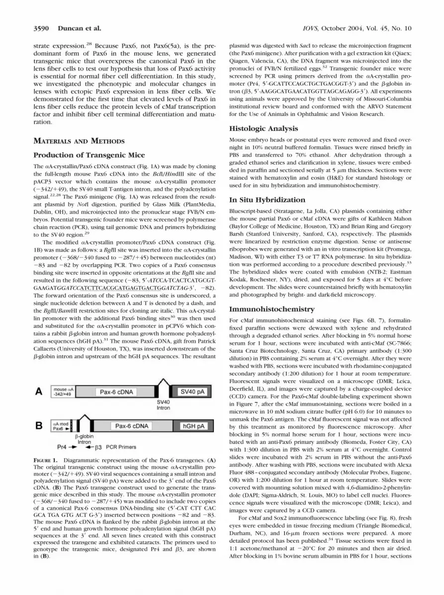
FIGURE 1. Diagrammatic representation of the Pax-6 transgenes. (A)The original transgenic construct using the mouse �A-crystallin pro-moter (�342/�49). SV40 viral sequences containing a small intron andpolyadenylation signal (SV40 pA) were added to the 3� end of the Pax6cDNA. (B) The Pax6 transgene construct used to generate the trans-genic mice described in this study. The mouse �A-crystallin promoter(�368/�340 fused to �287/�45) was modified to include two copiesof a canonical Pax-6 consensus DNA-binding site (5�-CAT CTT CACGCA TGA GTG ACT G-3�) inserted between positions �82 and �83.The mouse Pax6 cDNA is flanked by the rabbit �-globin intron at the5� end and human growth hormone polyadenylation signal (hGH pA)sequences at the 3� end. All seven lines created with this constructexpressed the transgene and exhibited cataracts. The primers used togenotype the transgenic mice, designated Pr4 and �3, are shownin (B).
3590 Duncan et al. IOVS, October 2004, Vol. 45, No. 10

were incubated with a primary antibody against cMaf or Sox2 (Chemi-con International, Temecula, CA) in PBS for 1 hour at room tempera-ture. After washing with PBS, sections were incubated with secondaryantibody conjugated with AlexaFluor 568 (Molecular Probes) at a 1:50dilution for 1 hour. Cell nuclei in Figures 8A and 8B were counter-stained with nuclear stain (1:1000 dilution, Syto13; Molecular Probes).Immunofluorescence signals were visualized on a confocal microscope(510 LSM; Carl Zeiss Meditec, Oberkochen, Germany).
Transmission Electron Microscopy
For ultrastructural analysis, 4-day-old lenses were dissected from theeye and fixed in 2% paraformaldehyde and 2% glutaraldehyde in 0.13 MNa-cacodylate and 0.13 mM CaCl2 (pH 7.4) for at least 2 hours at roomtemperature. Samples were washed with cacodylate buffer, postfixedin 1% osmium tetroxide in 0.1 M cacodylate buffer for 1 hour, dehy-drated through a graded ethanol series, and embedded in epoxy resin.Thin sagittal sections were cut through at least one third of the lensand stained with uranyl acetate and lead citrate. Sections were exam-ined and photographed with a transmission electron microscope(1200EX; JEOL Tokyo, Japan).
Two-Dimensional Gel Electrophoresis
One-week-old (P7) lenses were isolated from wild-type or LR2 trans-genic mice and frozen immediately in dry ice. Water-soluble lensproteins were prepared and protein concentration determined usingthe bicinchoninic acid (BCA) method. Lens proteins were first sepa-rated by isoelectric focusing on an immobilized-pH gradient gel strip(pH 5–9), and then by size on a 12% polyacrylamide gel. The gel wasstained with protein dye (Sypro Red; Molecular Probes), and theidentities of the protein spots were compared with standardized pro-teome maps of the FVB/N mouse lens.35 The relative amount of eachpolypeptide was quantitated on scanned gels (Delta2D software, ver.3.1; Decodon, Greifswald, Germany).
Quantitation of �B1-Crystallin Transcript Levels
Real-time RT-PCR analysis of �B1-crystallin transcript levels was com-pleted as described previously.36 Briefly, total RNA was isolated fromlenses using a commercial system (SV Total RNA Isolation System;Promega, Madison, WI). The RNA was reverse transcribed into cDNAusing mixed reverse transcriptases (Omniscript and Sensiscript; Qia-gen) and a reverse primer specific for the mouse �B1-crystallin gene(5�-CCT GCT CGA AGA CGA TCA GCC-3�). Then, the reverse transcrip-tase reaction was used for real-time PCR, by using a kit (QuantiTectSYBR Green PCR Kit; Qiagen) and the mouse �B1-crystallin forwardprimer (5�-CAG GGC CTG ATG GCA AGG GAA-3�) in a thermocycler(iCycler; Bio-Rad Laboratories, Hercules, CA).
RESULTS
Generation of Transgenic Mice that OverexpressPax6 in Lens Fiber Cells
In the normal lens, Pax6 expression was downregulated duringfiber cell differentiation (Fig. 2, and see Fig. 7A, WT). To testthe importance of this downregulation, we constructed a mini-gene using the mouse �A-crystallin promoter (�342/�49) todrive the mouse Pax6 cDNA (Fig. 1A). This promoter has beenused in numerous studies to target transgene expression spe-cifically in the lens fiber cells.22,28,29,31,37 We generated threeindependent founders, but none of them showed any lensdefects. RT-PCR analysis indicated that the transgene was notexpressed (data not shown). To enhance the �A-crystallinpromoter activity through a potential feedback mechanism, weused a modified �A-crystallin promoter containing two addi-tional copies of the Pax6 consensus binding site (Fig. 1B).30
Using this modified promoter, we generated seven transgenicfounders designated as OVE1076 to -1078 and LR1 to -4. In all
of them, cataracts developed with different degrees of severity.Seven independent lines were established and designated asOVE1076 to -1078 and LR1 to -4.
We analyzed transgene expression patterns by in situ hy-bridization (Fig. 2) and immunohistochemistry (e.g., Figs. 7A,7D, 7G) in embryonic day (E)18 eyes from lines OVE1076 to-1078. In the normal lens, Pax6 is highly expressed in the lensepithelial cells, and its expression is downregulated in the lensfiber cells (Fig. 2, WT). In transgenic lenses, Pax6 mRNA wasdetected in the lens epithelium at qualitatively normal levelswith additional ectopic expression in the fiber cells. Amongthe three lines tested in parallel (OVE1076 to -1078), OVE1078had the highest level of transgene expression, whereasOVE1077 had the lowest. We estimate, based on the severity ofthe lens defects, that LR2, a line used for many of our analyses,expressed the transgene at intermediate levels betweenOVE1078 and -1076. Immunohistochemical staining using ananti-Pax6 antibody suggested that the amount of Pax6 proteincorrelates with the level of Pax6 mRNA in the different lines(see Figs. 7A, 7D, 7G). Notably, the severity of the lenticulardefects in different transgenic lines correlated with the Pax6transgene expression levels. The severest cataracts developedin mice from lines OVE1078 and LR2, and they had smallerthan normal lenses with concomitant microphthalmia. Micefrom OVE1076 had obvious cataracts and only a slight reduc-tion in lens size, whereas OVE1077 mice exhibited very mildcataracts that were only transiently detectable at weaning�P21 (Table 1).
Lens Fiber Defects in Pax6 Transgenic Mice
We could not detect any lenticular defects in OVE1077 mice byhistology under light microscopy. Representative lens histol-ogy for other transgenic lines is shown in Figure 3. In thenormal lens, fiber cell differentiation and maturation is associ-ated with denucleation. As a result, the mature fibers at the lenscore do not contain DNA and therefore were free of hematox-
FIGURE 2. Pax-6 mRNA detected in E18 lenses by in situ hybridization.E18 embryos were isolated and processed for in situ hybridization witha mouse Pax-6 cDNA used as a probe. In the wild-type lens (WT), Pax-6was highly expressed in the lens epithelium (epi), and its expressionwas downregulated at the transitional zone (t, arrows) during fiber cell(fib) differentiation. In transgenic lenses from line OVE1076 to -1078,Pax-6 transcripts can be detected not only in the epithelium but also inthe fiber cells. Among the three lines tested, the order of transgeneexpression level is OVE1078�OVE1076�OVE1077, which correlateswith the severity of the fiber cell defects in the transgenic lenses (seeFig. 3).
IOVS, October 2004, Vol. 45, No. 10 Ectopic Pax6 in Lens Fiber Cell Differentiation 3591

ylin staining (Fig. 3A). Lenses from all transgenic lines (exceptOVE1077) retained hematoxylin-positive materials in the cen-tral fiber cells (white arrows in Fig. 3F), suggesting that de-nucleation is impaired in the transgenic lens. In all but one line(OVE1077), vacuoles were present in the cortical fiber cells,further suggesting that fiber cell differentiation is abnormal inthe transgenic lens. In the severely affected lines OVE1078 andLR2, fiber cell elongation was incomplete, and fiber cells weredisorganized. The apical surface (Fig. 3C, arrows) of the sec-ondary fiber cells failed to contact the anterior lens epithelium.To investigate further the lens defects in OVE1078, a develop-mental analysis was performed on lenses isolated from earlierdevelopmental stages (Fig. 4). At E15, the architecture of thetransgenic lens appeared normal with no obvious defects. AtE18, abnormalities in lens fiber cells became apparent. Thetransgenic lens exhibited vacuoles, fiber cell elongation wasincomplete (Figs. 4D, 4F, arrows), and denucleation was inhib-ited. These data suggest that high levels of Pax6 in lens fibercells interfere with normal fiber differentiation and maturation.
The structure of the lens fiber cells was further examined bytransmission electron microscopy (Fig. 5). In the normal P4
lens, fiber cells showed ball-and-socket junctions that formedlateral interdigitations with the neighboring cells (Figs. 5A, 5B,arrows). In contrast, the fiber cells in the OVE1078 transgeniclens either completely lacked such structures (Fig. 5C) or hadsignificantly fewer interdigitated joints than did the normallens (Figs. 5D, 5E). Similar defects were also observed intransgenic mice the expressed only the paired and homeodo-mains of Pax6,22 suggesting that the inhibitory effect is medi-ated through these two DNA-binding domains.
Reduction of cMaf but Not Sox2 Protein Levels inPax6 Transgenic Lenses
Pax6 is known to interact with other transcription factors tocontrol lens and eye development.8,38 To investigate the inhib-itory effect of Pax6 on fiber cell differentiation, we examinedthe expression of some transcription factors, including cMafand Sox2, which are known to be important for normal fibercell differentiation. Figure 6 shows the cMaf mRNA (Fig. 6A)and protein (Fig. 6B) localization in E14 and E18 lenses fromOVE1078 mice. In the normal lens, cMaf mRNA levels wereupregulated during fiber cell differentiation (Figs. 6A, WT), anda similar expression pattern was seen in the OVE1078 trans-genic lens (Fig. 6A, OVE1078). Quantitative RT-PCR also con-firmed that there was no significant difference in cMaf mRNAlevels between wild-type and OVE1078 transgenic lenses (datanot shown). Similarly, we detected high levels of cMaf proteinin the fiber nuclei of the wild-type lens by immunohistochem-istry (Fig. 6B, WT). However, very little cMaf immunoreactivitywas detected in the fiber cells of the OVE1078 transgenic lensat either age (Fig. 6B, OVE1078). Further, cMaf protein levelswere also significantly reduced in the LR2 transgenic lens (see
TABLE 1. Summary of Findings in Pax6 Transgenic Lenses
LineExpression Level
(mRNA)Expression Level
(Protein)Cataract
(Severity)
OVE1076 �� �� ��OVE1077 � � �/�OVE1078 ���� ���� ����LR2 ��� (estimated) ND ���
ND, not determined.
FIGURE 3. Histology of 1-week-old (P7) lenses from different transgenic lines. (A) Normal lens morphology with an anterior epithelial layer (epi)and uniformly stained fiber mass (fib). The transitional zone is indicated by t. (B–F) The lens epithelial layer appeared normal in all the transgeniclines. Cataracts developed with different severities because of lens fiber cell defects. Vacuoles were present in the cortical fibers (except in thelens of line OVE1077). In the severely affected line OVE1078 (C), the secondary fiber cells failed to elongate properly and the apical surface wasnot in contact with the anterior epithelium (arrows: anterior margin of the secondary fiber cells). Fiber cell denucleation was impaired in all thetransgenic lines (except OVE1077). Even in the mildly affected line LR3 (E, F), hematoxylin-staining-positive materials (F, arrows), probablychromosomal debris, can be seen in the central fiber cells. The boxed region in (E) is shown in a higher magnification in (F). (A) through (E) areof the same magnification. Scale bar, 100 �m.
3592 Duncan et al. IOVS, October 2004, Vol. 45, No. 10

Fig. 8B) suggesting that ectopic Pax6 expression in lens fibercells reduces cMaf protein levels but does not affect cMaf geneexpression.
To confirm that the reduction of cMaf protein in the trans-genic lens results from ectopic expression of Pax6 in the fibercells, we compared cMaf protein levels with Pax6 levels inlenses from OVE1076, -1077, and -1078 mice (Fig. 7; data notshown for OVE1077). In all lines, Pax6 protein levels did notappear altered in the lens epithelium compared with wild type.In the low-expressing OVE1077 mice, little ectopic Pax6 ex-pression was detectable in lens fibers, and cMaf protein levelswere similar to those in the wild-type lens (data not shown). Inthe intermediate-expressing line OVE1076, some fiber cell nu-clei had high Pax6 levels, whereas other fibers expressed Pax6at levels similar to or lower than the lens epithelium. Notably,double labeling for Pax6 and cMaf proteins revealed that nucleicontaining high levels of Pax6 protein had low levels of cMaf(Figs. 7D–F, representative nuclei, arrows), whereas cMaf pro-tein levels were close to normal in cells with low levels of Pax6(representative nuclei; arrowheads). In the high-expressingOVE1078 line, a significant number of fiber cell nuclei stainedpositively for Pax6 protein, and few to no cMaf-positive nucleiwere detected (Figs. 7G–I). This suggests that Pax6 negativelyregulates cMaf protein levels.
We then investigated whether Pax6 also inhibits the expres-sion of other transcription factors such as Sox2 in the LR2transgenic lens (Fig. 8). In contrast to the situation with cMaf
(Figs. 8A, 8B), Sox2 protein levels were unaffected in the LR2transgenic lens (Fig. 8D). We also examined the Sox2 mRNAand protein levels in E18 wild-type and OVE1078 lenses by insitu hybridization and immunostaining and again detected nodifferences in either expression pattern or levels (data notshown).
Reduction of �B1-Crystallin Expression Levels inTransgenic Lenses
Pax6, cMaf, and Sox proteins all have the ability to regulate theexpression of crystallins, the major structural proteins of thelens.8,39–43 Because the expression levels of Pax6 and cMaf arealtered in the transgenic lens, particularly in the high-express-ing lines OVE1078 and LR2, we decided to analyze the crystal-lin protein profile in the P7 wild-type and LR2 transgenic lensby 2D gel electrophoresis (Fig. 9). In the LR2 transgenic lens,the total amount of protein was approximately 60% less thanthat in the wild-type lens, which is consistent with the smallerlens (microphthalmia) phenotype. In the transgenic lens, therelative ratios of intact �A-, �B-, �A2-, �A4-, �A-, �B/C-, �D-,�E/F-, and �S-crystallins to the total amount of protein wereclose to normal, whereas the levels of intact �B2- and �B3-crystallin were slightly reduced. In addition, the levels of intact��1-crystallin were significantly reduced (approximately 40%to 50% less than normal) although approximately 40% of thisdecrease was due to the proteolytic processing of �B1-crystal-lin. Notably, the transgenic lens also contain elevated levels ofthe proteolytic products of �A-crystallin (Fig. 9, �A�) and�A3-crystallin (Fig. 9, �A3-11). These crystallin proteolyticproducts are usually seen only at appreciable levels in adultnormal lenses.35
Because �B1-crystallin protein levels were decreased in thetransgenic lens, we quantitated �B1-crystallin transcript levelsin P7 LR2 transgenic lenses by real-time RT-PCR. Notably,�B1-crystallin levels in the LR2 transgenic lens were reducedby more than 80% (87% � 13% P 0.003) compared withwild-type lenses.
FIGURE 5. Electron microscopy of the fiber cells at the cortical regionof P4 lenses. (A, B) Normal lens fiber cells formed interdigitationsbetween neighboring cells through ball-and-socket junctions (arrows).(C–E) The fiber cells in the transgenic lens have a significantly lessinterdigitated junction structure (D, E). In many areas, the ball-and-socket junctions were completely absent as shown (C). These defectswere consistently seen in two lenses from different animals at P4 aswell as from two additional animals at P7 (data not shown). Magnifi-cation: (A–C) 7500; (D, E) 15,000.
FIGURE 4. Histology of OVE1078 lenses from mice of different ages.Lens histology was compared between wild-type (A, C, E) andOVE1078 transgenic (B, D, F) mice. (A, B) At E15, the transgenic lensappeared normal. The apparent size difference is artificial, because thesection in (A) was from a more central region of the lens globe than thesection in (B). (C, D) Fiber cell (Fib) defects were first seen in the E18transgenic lens. The secondary fiber cells did not elongate completely(arrows, anterior margin of the fiber cells), leaving a lumen at thecortical region underneath the anterior epithelium (Epi) (D). (E, F) Thedefects in fiber cell elongation were still seen in the newborn trans-genic lens (F, arrows). More vacuoles were present in the fiber mass.Compared with the wild-type lens (E), fiber cells were not well orga-nized and aligned in the transgenic lens. Scale bar, 100 �m.
IOVS, October 2004, Vol. 45, No. 10 Ectopic Pax6 in Lens Fiber Cell Differentiation 3593

DISCUSSION
At early stages of lens development, Pax6 is expressed in headectoderm overlying the optic vesicle and then in the primitivelens as it progresses through the lens placode, cup, and vesiclestage.12 Previous studies have established that Pax6 expressionis necessary and, in some cases, sufficient for lens precursorcell specification and lens induction.6,11 As lens developmentproceeds, Pax6 expression is downregulated in differentiatedfiber cells and becomes restricted in the lens epithelium. Wehave shown that Pax6 negatively regulates �B1-crystallin ex-pression.17 To test our hypothesis that normal fiber cell differ-entiation and maturation requires downregulation of Pax6, wegenerated seven transgenic lines that overexpress Pax6 in thefiber cells. Lens defects, with different degrees of severity,developed in all the transgenic lines. In the severely affected
lines (OVE1078 and LR2), elongation of the secondary fibercells was incomplete, leaving a lumen beneath the anterior lensepithelium. Electron microscopy analysis showed that the spe-cialized membrane structures—the ball-and-socket interdigita-tions among the neighboring fiber cells—were underdevel-oped or attenuated in the transgenic lenses. Similar defectswere also observed in transgenic lenses that expressed only thepaired domain and the homeodomain of Pax6.22 Most interest-ing was the finding that elevated levels of Pax6 in the lens fibercells reduced the protein levels of cMaf, which is an importanttranscription factor in fiber cell differentiation. Our study im-plies that normal lens development depends on the properlevels of Pax6 in each lens cell type (epithelial and fiber), andloss of Pax6 expression is essential for fiber cell differentiationand maturation.
cMaf belongs to the family of basic-leucine zipper (bZip)transcription factors.44 Loss of cMaf results in defects in lensfiber cell differentiation.39,40,43 During early lens induction andspecification, Pax6 is essential for activating and maintainingthe expression of cMaf in the lens.45,46 After the lens is formed,cMaf is upregulated in fibers, whereas Pax6 levels decline. Thefunctional significance of this reciprocal expression profile isunclear. In our transgenic study, we demonstrate that higherlevels of Pax6 in the lens fiber cells sharply reduces the amountof cMaf protein. Therefore, although Pax6 activates the basalexpression of cMaf during early stages of normal lens develop-ment45 and Pax6 can directly activate the cMaf promoter incotransfections,46 we propose that downregulation of Pax6activity in lens fiber cells at later stages of lens development isessential for upregulating the cMaf protein levels during fibercell differentiation. Using the OVE1078 mice we generated,Goudreau et al.47 showed that ectopic expression of Pax6 canactivate Six3 expression. Taken together, our data imply thatduring late stages of lens development, Pax6 and Six3 expres-sion mutually regulate each other, whereas cMaf protein levelsare negatively regulated by Pax6. In our study, Sox2 levelswere not affected by Pax6. Because the lens expresses threeSox genes (Sox1, -2, and -3) with overlapping but differentexpression patterns,48 the effect of Pax6 overexpression onSox1 and -3 should be investigated.
How Pax6 reduces cMaf protein level is still unclear. Thereare two possibilities: Pax6 either inhibits the synthesis of cMafproteins or reduces its stability. Based on a recent publica-tion,49 it is more likely that overexpression of Pax6 reducescMaf protein stability in lens fiber cells. Previously Goudreau etal.47 found that active ERK levels are elevated in the fiber cellsof the OVE1078 lens, associated with abnormal expression ofPDGF�-R (platelet-derived growth factor receptor �). Further,Ochi et al.49 demonstrated that cMaf protein degradation isregulated by ERK phosphorylation. Thus, we speculate that thereduction of cMaf protein levels in the Pax6 transgenic lens,results in part from an increase in active ERK levels in the lensfiber cells, probably through abnormal expression and activa-tion of PDGF�-R in these cells. Whether PDGF�-R expression isdirectly regulated by Pax6 remains to be investigated.
The varied effects of ectopic Pax6 expression on lens fibercell biology are consistent with previously studies showingthat eye development and function is highly dependent on theprecise regulation of Pax6 levels. Transgenic mice harboring aYAC consisting of a large portion of the known Pax6 locusexhibit malformation of the iris and ciliary body with occa-sional animals exhibiting severe microphthalmia with associ-ated lens and retinal abnormalities.50 Although the lenses ofmost of the animals harboring the Pax6 YAC were normal, it isdifficult to assess the relevance of this result to the presentstudy, because the Pax6 YAC mice would presumably notexpress additional Pax6 in lens fiber cells since gene expres-sion was controlled by the endogenous Pax6 promoter. Mice
FIGURE 6. cMaf mRNA and protein localization in E14 and E18 lenses.(A) cMaf mRNA was detected by in situ hybridization, with a mousecMaf cDNA used as a probe. cMaf mRNA was detected in lens epithelialand fiber cells, and the expression level was upregulated in the differ-entiating fiber cells. The expression levels of cMaf were not signifi-cantly different between the nontransgenic (WT) and transgenic(OVE1078) lens at both ages. (B) Immunofluorescence labeling forcMaf protein in the lens. Rhodamine fluorescence appears white. Inthe wild-type lenses, cMaf proteins were found in lens epithelial andfiber cell nuclei, but the levels were much higher in the fiber cellnuclei (WT). Compared with the normal lens, significantly fewer nu-clei stained positively for cMaf in the transgenic lens (OVE1078). Scalebar, 100 �m.
3594 Duncan et al. IOVS, October 2004, Vol. 45, No. 10

heterozygous for mutations in the Pax6 gene have defects inthe iris51 and cornea52,53 similar to those in the human diseaseaniridia. Lenses from mice heterozygous for Pax6 mutationshave anterior subcapsular cataracts22 that appear to arise fromlocalized epithelial-to-mesenchymal transitions. It is intriguingto note that, in newborn mice chimeric for wild-type Pax6 and
heterozygous Pax6 loss-of-function cells, only the cells withwild-type Pax6 are found in the lens epithelium, because cellsheterozygous for the Pax6 mutant gene preferentially undergofiber cell differentiation between E12.5 and E16.5.18 In light ofthe present data, it is possible that the cMaf levels are abnor-mally upregulated in the cells heterozygous for a Pax6 muta-tion, and this Pax6 haploinsufficiency forces these cells todifferentiate preferentially into the fiber cells when exposed tothe appropriate environment. Thus, Pax6 expression in thelens epithelium may both maintain the commitment to anepithelial phenotype and prevent premature differentiationinto fiber cells during late-stage lens growth and development.
Pax6 overexpression in lens fiber cells results in multipledefects in lens morphology and structure. Some of the defectsare directly or indirectly related to the disruption of coordi-nated levels of Pax6 and cMaf proteins in the lens. (1) Second-ary fiber cell elongation is not complete in the Pax6 transgeniclens, leaving a lumen beneath the anterior lens epithelium. Thisresult is consistent with the observation that loss of cMafresults in the failure of fiber cell elongation in gene knockoutmice.39,40,43 (2) cMaf activity is essential for the expression ofthe major fiber cell proteins �- and �-crystallin, and to someextent, it is also important for �A-crystallin expression.39,40 Inthe severely affected Pax6 transgenic mice, the total amount ofproteins was approximately 60% lower than normal. Becausecrystallins make up approximately 90% of the total lens pro-tein, we assume the levels of crystallins are reduced to thesimilar extent. The decrease in total crystallin proteins coulddirectly result from the low levels of cMaf in the Pax6 trans-genic lens. Furthermore, elevated levels of Pax6 in the fibercells may inhibit �-crystallin expression as shown previously incotransfections.17 Indeed, the relative ratios of �B1-crystallinmRNA and protein are significantly reduced in the Pax6 trans-genic lens. This result coincides with the most recent findingby Cui et al.54 that Pax6 can block cMaf-mediated transactiva-tion of the chicken �B1-crystallin promoter in cotransfectionexperiments.
FIGURE 7. Double labeling for Pax6(A, D, G) and cMaf (B, E, H) proteinsin wild-type lenses (WT, A–C) andtransgenic lenses from line OVE1076(D–F) or OVE1078 (G–I). Superim-posed images are shown in (C, F, I).In the wild-type lenses, Pax6 protein(green) levels were reduced in fibercells and were hardly detectable (A).Under the same labeling conditions,Pax6 was found in the fiber cell nu-clei of transgenic lenses (D, G). Thehigher the expression level, the morePax6-positive nuclei were detectedamong the fiber cell nuclei (compareOVE1076 with OVE1078, D–G). Inthe wild-type lenses, cMaf proteinlevels were upregulated during fibercell differentiation. Many cMaf-posi-tive nuclei (red) were found in thelens fiber cells of wild-type mice (B).In the transgenic lenses, the numberof cMaf-positive nuclei correlatednegatively with the expression levelsof the Pax-6 transgene (E, H). Forexample, in OVE1076 mice (D–F),the nuclei containing high levelsof Pax6 protein had low levels ofcMaf (arrows, representative nu-clei), whereas fiber cells with normallevels of cMaf proteins showed low levels of Pax6 (arrowheads, representative nuclei). In the high-expressing OVE1078 mice (G–I), a significantnumber of fiber cell nuclei stained positive for Pax6 protein and few to no cMaf-positive nuclei were detected. Scale bar, 100 �m.
FIGURE 8. cMaf (A, B) and Sox2 (C, D) immunolocalization in P7normal (WT) (A, C) and LR2 (B, D) transgenic lenses. As shown intransgenic OVE1078 mice (Fig. 3), cMaf protein (red) was less abun-dant in the fiber cells of the LR2 transgenic lenses (B). Unlike cMafprotein, Sox2 protein levels (red) were unaffected in the LR2 trans-genic lenses. Cell nuclei in (A) and (B) were counterstained withnuclear stain (green).
IOVS, October 2004, Vol. 45, No. 10 Ectopic Pax6 in Lens Fiber Cell Differentiation 3595

One notable feature of the present study is that obviousphenotypic changes were not seen in transgenic lenses untilE18, although the modified �A-crystallin promoter used in thisstudy are active by E11.30 This is similar to mice that overex-press Pax6(5a) in lens fibers that do not develop major lensabnormalities until after birth although the transgene is activeby E12.5.28 Because it has been observed that appreciable Pax6expression is found in all lens cells shortly after the lens forms,whereas it is lost from lens fibers later in development,10 it isprobable that the late onset of these phenotypes reflects a latesusceptibility to Pax6 overexpression in lens fibers. This maybe at least part of the mechanism controlling developmentalchanges in crystallin gene expression in lens fiber cells.35,55
However, the onset of the morphologic alterations is also likelyto be influenced by the observation that not all lens fiber cellnuclei in transgenic lenses contain the same amount of Pax6protein, possibly due to a complex feedback loop between thepromoter used in these studies and Pax6. Further, althoughPax6 is a transcription factor whose effects should be cellautonomous, the need for lens fibers to form ball-and-socketjoints and extensive gap-junctional communication with theirneighbors can result in extensive abnormalities, even in lensescontaining only a few mutant cells.56,57
Microarray analysis on P7 wild-type and LR2 transgeniclenses has revealed more than 500 differentially expressedgenes.58 Because our present data suggest that Pax6 can regu-late gene activity at the protein level as well as the transcrip-tion level, the full spectrum of Pax6 effects on the lens arelikely to be extremely complex. Further, there are two Pax6isoforms, Pax6 and Pax6(5a), that result from alternative splic-ing of the 5a exon.23 Both isoforms are found in human lens atequal levels, whereas Pax6 predominates in mouse and bovinelenses. The two Pax6 isoforms have overlapping but differentDNA-binding specificities.24 For example, Pax6 and Pax6(5a)can both transactivate some Pax6 targets such as �B-crystal-lin14 but only Pax6 is able to bind to the PISCES element of theglucagon promoter59 and to repress the chicken �B1-crystallinpromoter.17 Mice without the 5a exon undergo eye formation,unlike complete Pax6-null animals; however, they have irishypoplasia.25 Transgenic mice that overexpress Pax6(5a) inlens fiber cells have been generated and exhibited lens defectsphenotypically similar to those in the Pax6 mice in this study.28
However, the population of genes with expression levels thatare altered by Pax6(5a) and Pax6 expression are quite differentas assayed by microarray analysis.58,60 Further, preliminarystudies have shown that, unlike the Pax6(5a) mice, �5 and�1-integrin protein levels are not changed in Pax6 transgeniclenses (data not shown). In contrast to the Pax6 mice, the cMafprotein levels are unaffected in the Pax6(5a) transgenic lenses
(data not shown). These observations support the hypothesisthat Pax6 and Pax6(5a) control the expression of overlappingyet distinct sets of genes in vivo.
In summary, we tested the hypothesis that ectopic expres-sion of Pax6 in lens fiber cells would disrupt normal celldifferentiation and maturation. We found that elevated levels ofPax6 in lens fiber cells inhibits fiber cell elongation, denucle-ation, and formation of specialized membrane structures. Mostinteresting was our demonstration, for the first time in vivo,that Pax6 negatively regulated the protein levels of cMaf andtranscript levels of �B1-crystallin in the lens, implying that therole of Pax6 in late-stage lens development is to maintain theundifferentiated phenotype of the lens epithelium.
Acknowledgments
The authors thank Kirk Czymmek (University of Delaware Core Mi-croscopy Facility), the Electron Microscopy Core Facility at the Uni-versity of Missouri-Columbia, Phillip Wilmarth, Li Xu, and Shanyu Hofor technical support; Paul Overbeek (Baylor College of Medicine,Houston, TX) for generation of OVE1076 to -1078 transgenic mice andfor a critique of the manuscript; the University of Missouri-ColumbiaTransgenic Animal Core, for generating the transgenic mice lines LR1-to -4, and Ales Cvekl (Albert Einstein College of Medicine, New York,NY) for valuable discussions.
References
1. Piatigorsky J. Lens differentiation in vertebrates: a review of cellu-lar and molecular features. Differentiation. 1981;19:134–153.
2. Grainger RM. Embryonic lens induction: shedding light on verte-brate tissue determination. Trends Genet. 1992;8:349–355.
3. Wride MA. Cellular and molecular features of lens differentiation:a review of recent advances. Differentiation. 1996;61:77–93.
4. Chow RL, Lang RA. Early eye development in vertebrates. AnnuRev Cell Dev Biol. 2001;17:255–296.
5. Jean D, Ewan K, Gruss P. Molecular regulators involved in verte-brate eye development. Mech Dev. 1998;76:3–18.
6. Ashery-Padan R, Gruss P. Pax6 lights-up the way for eye develop-ment. Curr Opin Cell Biol. 2001;13:706–714.
7. Callaerts P, Halder G, Gehring WJ. PAX-6 in development andevolution. Annu Rev Neurosci. 1997;20:483–532.
8. Cvekl A, Piatigorsky J. Lens development and crystallin geneexpression: many roles for Pax-6. Bioessays. 1996;18:621–630.
9. Hogan BL, Horsburgh G, Cohen J, Hetherington CM, Fisher G,Lyon MF. Small eyes (Sey): a homozygous lethal mutation onchromosome 2 which affects the differentiation of both lens andnasal placodes in the mouse. J Embryol Exp Morphol. 1986;97:95–110.
10. Grindley JC, Davidson DR, Hill RE. The role of Pax-6 in eye andnasal development. Development. 1995;121:1433–1442.
FIGURE 9. Lens crystallin proteinanalysis by 2-D gel electrophoresis.Equal amounts of water-soluble pro-teins from P7 normal (A) and LR2transgenic (B) lenses were used foranalysis. The �-crystallin protein lev-els (�A and �B) were not signifi-cantly affected in the transgenic lens.However, the fiber cell crystallins,including different isoforms of �- and�-crystallins, were reduced in thetransgenic lens. In addition, trans-genic lenses contained more proteo-lytic and modified crystallins (circledspots), which are usually seen only innormal aged lenses.35 These prod-ucts include proteolytic products of�A-crystallin (�A�) (box), a modified
form of �B-crystallin (�Bm), a modified form of �B1-crystallin (�B1m), and �A3-crystallin with 11 amino acids removed from the N terminus(�A3-11). Other proteins on the gel included fatty acid binding protein (fab) and �Ainsert-crystallin (�Ai).
3596 Duncan et al. IOVS, October 2004, Vol. 45, No. 10

11. Chow RL, Altmann CR, Lang RA, Hemmati-Brivanlou A. Pax6 in-duces ectopic eyes in a vertebrate. Development. 1999;126:4213–4222.
12. Walther C, Gruss P. Pax-6, a murine paired box gene, is expressedin the developing CNS. Development. 1991;113:1435–1449.
13. Cvekl A, Kashanchi F, Sax CM, Brady JN, Piatigorsky J. Transcrip-tional regulation of the mouse alpha A-crystallin gene: activationdependent on a cyclic AMP-responsive element (DE1/CRE) and aPax-6-binding site. Mol Cell Biol. 1995;15:653–660.
14. Gopal-Srivastava R, Cvekl A, Piatigorsky J. Pax-6 and alphaB-crys-tallin/small heat shock protein gene regulation in the murine lens:interaction with the lens-specific regions, LSR1 and LSR2. J BiolChem. 1996;271:23029–23036.
15. Cvekl A, Sax CM, Li X, McDermott JB, Piatigorsky J. Pax-6 andlens-specific transcription of the chicken delta 1-crystallin gene.Proc Natl Acad Sci USA. 1995;92:4681–4685.
16. Richardson J, Cvekl A, Wistow G. Pax-6 is essential for lens-specificexpression of zeta-crystallin. Proc Natl Acad Sci USA. 1995;92:4676–4680.
17. Duncan MK, Haynes JI II, Cvekl A, Piatigorsky J. Dual roles forPax-6: a transcriptional repressor of lens fiber cell-specific beta-crystallin genes. Mol Cell Biol. 1998;18:5579–5586.
18. Collinson JM, Quinn JC, Buchanan MA, et al. Primary defects in thelens underlie complex anterior segment abnormalities of the Pax6heterozygous eye. Proc Natl Acad Sci USA. 2001;98:9688–9693.
19. van Raamsdonk CD, Tilghman SM. Dosage requirement and allelicexpression of PAX6 during lens placode formation. Development.2000;127:5439–5448.
20. Glaser T, Jepeal L, Edwards JG, Young SR, Favor J, Maas RL. PAX6gene dosage effect in a family with congenital cataracts, aniridia,anophthalmia and central nervous system defects. Nat Genet.1994;7:463–471.
21. Hill RE, Favor J, Hogan BL, et al. Mouse small eye results frommutations in a paired-like homeobox-containing gene (publishedcorrection appears in Nature 1992;355:750). Nature. 1991;354:522–525.
22. Duncan MK, Cvekl A, Li X, Piatigorsky J. Truncated forms of Pax-6disrupt lens morphology in transgenic mice. Invest OphthalmolVis Sci. 2000;41:464–473.
23. Epstein JA, Glaser T, Cai J, Jepeal L, Walton DS, Maas RL. Twoindependent and interactive DNA-binding subdomains of the Pax6paired domain are regulated by alternative splicing. Genes Dev.1994;8:2022–2034.
24. Kozmik Z, Czerny T, Busslinger M. Alternatively spliced insertionsin the paired domain restrict the DNA sequence specificity of Pax6and Pax8. EMBO J. 1997;16:6793–6803.
25. Singh S, Mishra R, Arango NA, Deng JM, Behringer RR, SaundersGF. Iris hypoplasia in mice that lack the alternatively splicedPax6(5a) isoform. Proc Natl Acad Sci USA. 2002;99:6812–6815.
26. Zhang W, Cveklova K, Oppermann B, Kantorow M, Cvekl A.Quantitation of PAX6 and PAX6(5a) transcript levels in adult hu-man lens, cornea, and monkey retina. Mol Vis. 2001;7:1–5.
27. Jaworski C, Sperbeck S, Graham C, Wistow G. Alternative splicingof Pax6 in bovine eye and evolutionary conservation of intronsequences. Biochem Biophys Res Comm. 1997;240:196–202.
28. Duncan MK, Kozmik Z, Cveklova K, Piatigorsky J, Cvekl A. Over-expression of PAX6(5a) in lens fiber cells results in cataract andupregulation of �5�1 integrin expression. J Cell Sci. 2000;113:3173–3185.
29. Reneker LW, Silversides DW, Patel K, Overbeek PA. TGF alpha canact as a chemoattractant to perioptic mesenchymal cells in devel-oping mouse eyes. Development. 1995;121:1669–1680.
30. Zhao H, Yang Y, Rizo CM, Overbeek PA, Robinson ML. Insertion ofPax6 consensus binding site into the �A-crystallin promoter acts asa lens epithelial cell enhancer in transgenic mice. Invest Ophthal-mol Vis Sci. 2004;45:1930–1939
31. Robinson ML, MacMillan-Crow LA, Thompson JA, Overbeek PA.Expression of a truncated FGF receptor results in defective lensdevelopment in transgenic mice. Development. 1995;121:3959–3967.
32. Taketo M, Schroeder AC, Mobraaten LE, et al. FVB/N: an inbredmouse strain preferable for transgenic analyses. Proc Natl Acad SciUSA. 1991;88:2065–2069.
33. Xu L, Overbeek PA, Reneker LW. Systematic analysis of E-, N- andP-cadherin expression in mouse eye development. Exp Eye Res.2002;74:753–760.
34. Reed NA, Oh DJ, Czymmek KJ, Duncan MK. An immunohisto-chemical method for the detection of proteins in the vertebratelens. J Immunol Methods. 2001;253:243–252.
35. Ueda Y, Duncan MK, David LL. Lens proteomics: the accumulationof crystallin modifications in the mouse lens with age. InvestOphthalmol Vis Sci. 2002;43:205–215.
36. Taube JR, Gao CY, Ueda Y, Zelenka PS, David LL, Duncan MK.General utility of the chicken betaB1-crystallin promoter to driveprotein expression in lens fiber cells of transgenic mice. Trans-genic Res. 2002;11:397–410.
37. Chen Q, Hung FC, Fromm L, Overbeek PA. Induction of cell cycleentry and cell death in postmitotic lens fiber cells by overexpres-sion of E2F1 or E2F2. Invest Ophthalmol Vis Sci. 2000;41:4223–4231.
38. Kondoh H. Transcription factors for lens development assessed invivo. Curr Opin Genet Dev. 1999;9:301–308.
39. Kawauchi S, Takahashi S, Nakajima O, et al. Regulation of lens fibercell differentiation by transcription factor c-Maf. J Biol Chem.1999;274:19254–19260.
40. Ring BZ, Cordes SP, Overbeek PA, Barsh GS. Regulation of mouselens fiber cell development and differentiation by the Maf gene.Development. 2000;127:307–317.
41. Kamachi Y, Sockanathan S, Liu Q, Breitman M, Lovell-Badge R,Kondoh H. Involvement of SOX proteins in lens-specific activationof crystallin genes. EMBO J. 1995;14:3510–3519.
42. Muta M, Kamachi Y, Yoshimoto A, Higashi Y, Kondoh H. Distinctroles of SOX2, Pax6 and Maf transcription factors in the regulationof lens-specific delta1-crystallin enhancer. Genes Cells. 2002;7:791–805.
43. Wigle JT, Chowdhury K, Gruss P, Oliver G. Prox1 function iscrucial for mouse lens-fibre elongation. Nat Genet. 1999;21:318–322.
44. Blank V, Andrews NC. The Maf transcription factors: regulators ofdifferentiation. Trends Biochem Sci. 1997;22:437–441.
45. Ashery-Padan R, Marquardt T, Zhou X, Gruss P. Pax6 activity in thelens primordium is required for lens formation and for correctplacement of a single retina in the eye. Genes Dev. 2000;14:2701–2711.
46. Sakai M, Serria MS, Ikeda H, Yoshida K, Imaki J, Nishi S. Regulationof c-maf gene expression by Pax6 in cultured cells. Nucleic AcidsRes. 2001;29:1228–1237.
47. Goudreau G, Petrou P, Reneker LW, Graw J, Loster J, Gruss P.Mutually regulated expression of Pax6 and Six3 and its implica-tions for the Pax6 haploinsufficient lens phenotype. Proc NatlAcad Sci USA. 2002;99:8719–8724.
48. Nishiguchi S, Wood H, Kondoh H, Lovell-Badge R, Episkopou V.Sox1 directly regulates the gamma-crystallin genes and is essentialfor lens development in mice. Genes Dev. 1998;12:776–781.
49. Ochi H, Ogino H, Kageyama Y, Yasuda K. The stability of thelens-specific Maf protein is regulated by fibroblast growth factor(FGF)/ERK signaling in lens fiber differentiation. J Biol Chem.2003;278:537–544.
50. Schedl A, Ross A, Lee M, et al. Influence of PAX6 gene dosage ondevelopment: overexpression causes severe eye abnormalities.Cell. 1996;86:71–82.
51. Baulmann DC, Ohlmann A, Flugel-Koch C, Goswami S, Cvekl A,Tamm ER. Pax6 heterozygous eyes show defects in chamber angledifferentiation that are associated with a wide spectrum of otheranterior eye segment abnormalities. Mech Dev. 2002;118:3–17.
52. Ramaesh T, Collinson JM, Ramaesh K, Kaufman MH, West JD,Dhillon B. Corneal abnormalities in Pax6�/� small eye micemimic human aniridia-related keratopathy. Invest Ophthalmol VisSci. 2003;44:1871–1878.
53. Davis J, Duncan MK, Robison WG Jr, Piatigorsky J. Requirementfor Pax6 in corneal morphogenesis: a role in adhesion. J Cell Sci.2003;116:2157–2767.
54. Cui W, Tomarev SI, Piatigorsky J, Chepelinsky AB, Duncan MK.Mafs, Prox1 and Pax6 can regulate chicken beta B1-cystallin geneexpression. J Biol Chem. 2004;12:11088–11095.
IOVS, October 2004, Vol. 45, No. 10 Ectopic Pax6 in Lens Fiber Cell Differentiation 3597

55. Hejtmancik JF, Beebe DC, Ostrer H, Piatigorsky J. delta- and beta-Crystallin mRNA levels in the embryonic and posthatched chickenlens: temporal and spatial changes during development. Dev Biol.1985;109:72–81.
56. Yoshiki A, Hanazono M, Oda S, Wakasugi N, Sakakura T, KusakabeM. Developmental analysis of the eye lens obsolescence (Elo) genein the mouse: cell proliferation and Elo gene expression in theaggregation chimera. Development. 1991;113:1293–1304.
57. Muggleton-Harris AL, Hardy K, Higbee N. Rescue of developmentallens abnormalities in chimaeras of noncataractous and congenitalcataractous mice. Development. 1987;99:473–480.
58. Chauhan BK, Reed NA, Yang Y, et al. A comparative cDNA mi-croarray analysis reveals a spectrum of genes regulated by Pax6 inmouse lens. Genes Cells. 2002;7:1267–1283.
59. Bagby S, Harvey TS, Eagle SG, Inouye S, Ikura M. Structural simi-larity of a developmentally regulated bacterial spore coat proteinto beta gamma-crystallins of the vertebrate eye lens. Proc NatlAcad Sci USA. 1994;91:4308–4312.
60. Chauhan BK, Reed NA, Zhang W, Duncan MK, Kilimann MW,Cvekl A. Identification of genes downstream of Pax6 in the mouselens using cDNA microarrays. J Biol Chem. 2002;277:11539–11548.
3598 Duncan et al. IOVS, October 2004, Vol. 45, No. 10