regulation of transcription factor yy1 by acetylation and deacetylation
TRANSCRIPT

MOLECULAR AND CELLULAR BIOLOGY,0270-7306/01/$04.0010 DOI: 10.1128/MCB.21.17.5979–5991.2001
Sept. 2001, p. 5979–5991 Vol. 21, No. 17
Copyright © 2001, American Society for Microbiology. All Rights Reserved.
Regulation of Transcription Factor YY1 by Acetylationand Deacetylation
YA-LI YAO, WEN-MING YANG, AND EDWARD SETO*
Department of Medical Microbiology and Immunology, Interdisciplinary Oncology Program, H. Lee Moffitt CancerCenter and Research Institute, College of Medicine, University of South Florida, Tampa, Florida 33612
Received 30 January 2001/Returned for modification 12 March 2001/Accepted 30 May 2001
YY1 is a sequence-specific DNA-binding transcription factor that has many important biological roles. Itactivates or represses many genes during cell growth and differentiation and is also required for the normaldevelopment of mammalian embryos. Previous studies have established that YY1 interacts with histoneacetyltransferases p300 and CREB-binding protein (CBP) and histone deacetylase 1 (HDAC1), HDAC2, andHDAC3. Here, we present evidence that the activity of YY1 is regulated through acetylation by p300 and PCAFand through deacetylation by HDACs. YY1 was acetylated in two regions: both p300 and PCAF acetylated thecentral glycine-lysine-rich domain of residues 170 to 200, and PCAF also acetylated YY1 at the C-terminalDNA-binding zinc finger domain. Acetylation of the central region was required for the full transcriptionalrepressor activity of YY1 and targeted YY1 for active deacetylation by HDACs. However, the C-terminal regionof YY1 could not be deacetylated. Rather, the acetylated C-terminal region interacted with HDACs, whichresulted in stable HDAC activity associated with the YY1 protein. Finally, acetylation of the C-terminal zincfinger domain decreased the DNA-binding activity of YY1. Our findings suggest that in the natural context,YY1 activity is regulated through intricate mechanisms involving negative feedback loops, histone deacetyla-tion, and recognition of the cognate DNA sequence affected by acetylation and deacetylation of the YY1 protein.
YY1, also known as d, NF-E1, and UCRBP, is a multifunc-tional transcription factor. It binds, with its four C2H2 zincfingers, to a specific DNA sequence (CGCCATNTT) locatedin many different promoters and either activates or repressestranscription (for comprehensive reviews, see references 57and 65). YY1 regulates the expression of both cellular and viralgenes, including those encoding c-Myc, c-Fos, p53, a-actin,gamma interferon, P5 of adeno-associated virus, E6 and E7 ofhuman papillomavirus (HPV), and a number of other virallong terminal repeats. Many of these gene products have im-portant consequences for cell growth and differentiation(reviewed in reference 57). YY1 is highly conserved amonghuman, mouse, and Xenopus laevis cells, and a Drosophilamelanogaster homologue of YY1 exists (6, 14, 20, 47, 49, 58).Knockout studies show that deletion of YY1 results in peri-implantation lethality in mice (13), further demonstrating theimportance of YY1 in fundamental biological processes suchas development.
Various biochemical methods have been employed to dissectthe functional domains of YY1 in order to understand how theactivity of YY1 is regulated (1, 7, 8, 38–40, 58, 73). It can besummarized that YY1 contains two repression domains, oneembedded within residues 170 to 200 and the other overlap-ping with the C-terminal zinc finger DNA-binding domain.YY1 might also contain an independent activation domain atthe N terminus. This modular nature of YY1 supports the ideathat YY1 is bifunctional, capable of both activating and re-pressing transcription. It is generally believed that whetherYY1 behaves as a transcriptional activator or repressor de-
pends on its relative concentration (7), other cell type-specificfactors (4, 29, 36, 60, 74), and the promoter sequences sur-rounding the YY1 binding sites (59). However, the specificcues dictating when and how this decision is executed remaina mystery. It has been shown that YY1 is a stable phosphory-lated protein expressed ubiquitously regardless of cell cycleposition or the differentiation status of the cell (1), suggestingthat the activity of YY1 is regulated at the posttranslationallevel, possibly through interactions with other proteins.
In support of the hypothesis that the actions of YY1 arecontrolled by protein-protein interactions, a wide variety oftranscription factors have been shown to associate with YY1.These include proteins of the basal transcription machinery,such as TATA binding protein (1), TFIIB (67); sequence-specific DNA-binding transcriptional activators, such as SpI(37, 55), c-Myc (60), ATF/CREB (79), C/EBP (4); and varioustranscriptional coregulators, such as E1A (40), TAFII55 (11),p300, CREB-binding protein (CBP) (1, 36), and HDAC1,HDAC2, and HDAC3 (73, 75). The YY1-p300 and YY1-HDACs interactions are of particular interest. p300 and CBPare two closely related transcriptional coactivators (15) thathave been shown to be histone acetyltransferases (HATs) (2,46). Hyperacetylated histones have long been known to asso-ciate with activated genes (23, 54, 68), and recent studies fur-ther suggest a causal relationship between histone acetylationand gene activation (reviewed in references 30 and 33). It isthought that addition of acetyl groups to the lysine residues ofhistone tails facilitates access of transcription factors to DNAby disrupting higher-order packaging of the chromatin (34)and by neutralizing the positive charge of the histone proteins,which reduces the affinity of histones for DNA (24). In certaincircumstances, the transcriptional activator activity of YY1directly depends on its association with p300 (36). In contrast,HDACs have the opposite effects on transcription. HDAC1,
* Corresponding author. Mailing address: H. Lee Moffitt CancerCenter and Research Institute at USF, 12902 Magnolia Dr., MRC4072, Tampa, FL 33612. Phone: (813) 979-6754. Fax: (813) 979-7264.E-mail: [email protected].
5979

HDAC2, and HDAC3 are class I HDACs (reviewed in refer-ence 12) that have a high degree of homology to the Saccha-romyces cerevisiae global transcriptional regulator Rpd3p. It hasbeen shown that the transcriptional repression activity of YY1is mediated by association of HDAC2 with residues 170 to 200of YY1, which corresponds to one of the repression domains ofYY1 (73). It is conceivable that by selectively associating witheither HATs or HDACs, YY1 becomes an activator or a re-pressor. However, it has not been shown definitively that suchan active selection system exists for YY1.
Recently p300, CBP, and another HAT, PCAF (p300-CBPassociated factor) have been shown to acetylate transcriptionfactors in addition to their histone substrates (reviewed inreference 63). Importantly, acetylation was a key regulatorymechanism for the regulation of these transcription factors.Because YY1 interacts with both p300 and HDACs, we ex-plored the possibility that YY1 is also regulated by acetylationand deacetylation. Indeed, our results demonstrate that YY1was acetylated by both p300 and PCAF and was deacetylatedby HDAC1 and HDAC2. p300 acetylated the central region ofYY1, amino acids (aa) 170 to 200, while PCAF acetylated boththe central region and the C-terminal zinc finger domain ofYY1. The acetylated central region of YY1 was a target ofactive deacetylation by HDAC1 and HDAC2, whereas theC-terminal zinc finger domain was not deacetylated. Acetyla-tion and deacetylation, in turn, had intriguing influences on thesequence-specific DNA-binding and transcriptional activitiesof YY1.
MATERIALS AND METHODS
Plasmids. Glutathione S-transferase (GST) was expressed from pGSTag (51).GST-YY1 (aa 1 to 414) [or, simply, GST-YY1(1–414)] was expressed frompGST-YY1 (73), and deletion constructs of GST-YY1 were generated by re-striction enzyme digestion and religations of pGST-YY1. GST-p53 expressionplasmid has been described (26). GST-p300 and GST-PCAF were expressedfrom plasmids pGEX2T-p300 (aa 1195 to 1810) and pGEX5X-PCAF (aa 352 to832), respectively (9).
Gal4-YY1 was expressed from pM1-YY1, which was constructed by insertingthe full-length YY1 cDNA in frame with the Gal4 DNA-binding domain in pM1(52). pM1-YY1 (K170-R200) was prepared using adapter oligonucleotides con-taining AAG (lysine) to AGG (arginine) mutations within YY1 aa 170 to 200 ofpM1-YY1. pG5CAT-Control was constructed by inserting five Gal4 DNA-bind-ing sites into the BglII site of pCAT-Control (Promega), which contains thechloramphenicol acetyltransferase (CAT) gene downstream of the simian virus40 promoter and enhancer sequences. Flag-tagged YY1 (F-YY1) was expressedfrom pCEP4F-YY1, which was generated by inserting the full-length YY1 cDNAinto the pCEP4F vector (80). Serial Flag-YY1 deletion constructs were made byrestriction enzyme digestion and religations of pCEP4F-YY1. pET15b-YY1,which expressed nontagged full-length YY1 in Escherichia coli in an induciblesystem, was constructed by cloning the full-length YY1 cDNA into pET15b(Novagen). pGEM7Zf3X-HD1, which was used to generate in vitro-translatedHDAC1, was made by subcloning full-length HDAC1 into the pGEM7Zf3Xvector, which is derived from pGEM7Zf(1) (Promega).
The following plasmids have been previously described: pBJ5-HD1F (64),which expresses HDAC1 C-terminally tagged with a Flag epitope; pBJ5.1-HD1F(H199F), HDAC1 point mutant (22); and pME18S-FLAG-HDAC2, which ex-presses Flag-tagged HDAC2 (35).
Recombinant proteins. The GST fusion constructs of p300, PCAF, p53, andYY1 deletion mutants were expressed in E. coli DH5a, bound to glutathione-agarose beads (Sigma), washed extensively in phosphate-buffered saline (PBS),and eluted with 25 mM reduced glutathione. The eluate was then dialyzedagainst a buffer containing 20 mM Tris-HCl (pH 8), 0.5 mM EDTA, 100 mMKCl, 20% glycerol, 0.5 mM dithiothreitol (DTT), and 1 mM phenylmethylsulfo-nyl fluoride.
Non-tagged YY1 was expressed in E. coli BL21(DE3), induced with 0.2 mMisoprophyl-b-D-thiogalactopyranoside (IPTG), and captured by Ni21-immobi-
lized metal affinity chromatography (Invitrogen). Unbound bacterial proteinswere removed with 50 mM imidazole, and bound YY1 was eluted with 500 mMimidazole. F-YY1 used in electrophoretic mobility shift assays (EMSA) waspurified on an anti-Flag column (Sigma) under stringent conditions following themanufacturer’s suggestions.
In vitro acetylation reactions. Purified GST-YY1 and the serial deletion pro-teins were incubated at 30°C for 30 min with 0.25 mCi of [3H]acetyl coenzyme A([3H]acetyl-CoA) (Amersham) and either purified GST-p300 (0.2 mg) or GST-PCAF (1 mg) in 30 ml of acetylation buffer containing 50 mM Tris-HCl (pH 8.0),5% glycerol, 0.1 mM EDTA, 50 mM KCl, 1 mM DTT, 1 mM phenylmethylsul-fonyl fluoride and 10 mM sodium butyrate. Proteins were resolved by sodiumdodecyl sulfate-polyacrylamide gel electrophoresis (SDS-PAGE). The gels werefixed by Coomassie blue staining and subjected to signal amplification (Amplify;Amersham) prior to exposing to X-ray film.
For subsequent mass spectrometry analyses, 0.5 pmol of the YY1 peptide(GRVKKGGGKKSGKKSYLSGGAGAAGGRGADP) was acetylated in acet-ylation buffer for 2 h with CAT-assay grade acetyl-CoA (Amersham).
Cell culture, transfection, and CAT assays. HeLa cells were maintained inDulbecco’s modified Eagle’s medium with 10% fetal bovine serum and penicillin-streptomycin. 106 HeLa cells were seeded into 60-mm-diameter tissue culturedishes. Sixteen hours later, 10 mg of plasmids (for CAT assays, 5 mg of pG5CAT-Control plus 5 mg of pM1 effector plasmids) were transfected into cells using thecalcium phosphate coprecipitation method (17). Forty-eight hours after trans-fection, cells were harvested by scraping and lysed by repeated freeze-thawing,and extracts were assayed for CAT activity by thin-layer chromatography (16).
Immunoprecipitation and Western blot analysis. Immunoprecipitation ofFlag-YY1 deletion proteins was done using anti-Flag M2 affinity gel (Sigma)following the manufacturer’s suggestions.
Western blot analyses were performed using standard protocols (21). Immu-noprecipitated proteins were detected with diluted primary antibodies (1:1,000dilution of anti-acetyl-lysine [Upstate]; 1:5,000 dilution of anti-Flag M2 [Sigma];1:1,000 dilution of anti-HDAC1, HDAC2, or HDAC3 rabbit anti-serum [35, 66,72]) followed by 1:7,500-diluted alkaline phosphatase-conjugated secondary an-tibodies (Promega). The blots were subsequently developed with 5-bromo-4-chloro-3-indolyl phosphate and nitro blue tetrazolium (Promega).
In vitro protein-protein interaction assays. 35S-labeled HDAC1 was generatedfrom pGEM7Zf3X-HD1 using T7 RNA polymerase and the TNT ReticulocyteLysate System (Promega). GST-YY1 (aa 170 to 200) was either acetylated withcold acetyl-CoA or mock acetylated and was then captured onto glutathione-agarose beads. In vitro-translated HDAC1 (5 ml) was mixed with the beads in thepresence of PBS plus 0.2% NP-40 at 4°C for 1 h. Beads were washed extensivelyin PBS plus 0.2% NP-40. Bound proteins were eluted by boiling in Laemmlisample buffer, separated by SDS-PAGE, and detected by Coomassie blue stain-ing and autoradiography.
Chemical labeling of peptides with [3H]acetate. Peptides were labeled chem-ically according to the protocol described (64) with minor modifications. Thepeptides (0.4 mg) were labeled with 5 mCi of [3H]sodium acetate (2 to 5Ci/mmol; New England Nuclear) in the presence of 0.24 M benzotriazol-1-gloxytrif-(dimethylamino)-phosphonium hexafluorophosphate and 0.2 M trieth-ylamine (Aldrich). Labeled peptides were purified on a Microcon-SCX column(Millipore).
Histone deacetylation assays. HeLa cells were transfected with 10 mg ofpCEP4F-YY1 deletion plasmids by the calcium phosphate coprecipitationmethod (17). Forty-eight hours after transfection, cells were harvested, lysed inPBS plus 0.1% NP-40, and immunoprecipitated with anti-Flag M2 affinity gel(Sigma). HDAC activities of the immunoprecipitated Flag-YY1 were deter-mined using a peptide corresponding to residues 2 to 24 of histone H4 asdescribed (64) except that incubation was performed at room temperature over-night. Trichostatin A (TSA) (400 nM [final concentration]; Sigma) or a five-column volume of Flag peptide (Sigma) was added to the immunoprecipitate 30min prior to addition of the H4 substrate peptide, if appropriate.
Immunofluorescence analysis. HeLa cells were grown on chamber slides(Nalge Nunc International) for about 24 h and transfected with 10 mg of F-YY1deletion constructs. Two days later, cells were washed with ice-cold PBS, fixedwith 4% paraformaldehyde for 10 min, rinsed again with PBS, covered with 400ml of 1% bovine serum albumin in PBS for 1 h at room temperature, washedagain in PBS, and then treated with 1:200 dilution of anti-Flag fluoresceinisothiocyanate (FITC) conjugate antibody (Upstate) for 1 h at room tempera-ture. Subsequently, cells were subjected to extensive washing with PBS andcoverslips were applied with one drop of antifade mounting medium with DAPI(49,69-diamidino-2-phenylindole) (Vector) before analysis under a fluorescencemicroscope.
5980 YAO ET AL. MOL. CELL. BIOL.

EMSAs. Purified YY1 proteins and GST-p53 were first acetylated or mockacetylated. Single-stranded oligodeoxynucleotides corresponding to a consensusYY1-binding site (56) or a p53 cognate sequence (18) were labeled individuallywith [g32-P]ATP and T4 polynucleotide kinase, heated together at 65°C, andallowed to anneal by slow cooling to room temperature. Binding reactions wereperformed in a 12-ml reaction volume containing 12 mM HEPES (pH 7.9), 10%glycerol, 5 mM MgCl2, 60 mM KCl, 1 mM DTT, 0.5 mM EDTA, bovine serumalbumin (50 mg/ml), 0.05% NP-40, 0.1 mg of poly(dI-dC), approximately 1 ng ofproteins, and 5 fmol of radiolabeled DNA. Reaction mixtures were incubated for10 min at room temperature and separated on 4% nondenaturing polyacryl-amide gels. The gels were then dried and exposed to film.
RESULTS
YY1 is acetylated by p300 and PCAF. Acetylation has beenshown to regulate the activity of many transcription factors,including sequence-specific DNA-binding factors p53 (18),GATA-1 (5, 27), E2F (43, 44), and MyoD (53). Analysis of theYY1 amino acid sequence reveals that YY1 contains multiplelysine residues that are potential substrates for acetylation.Interestingly, within YY1’s HDAC interaction domain (resi-dues 170 to 200), there are six lysines arranged in pairs (Fig. 1).As described previously, YY1 interacts with the HAT p300 (1,36), which interacts with another HAT, PCAF (76). Remark-ably, both p300 and PCAF catalyze the acetylation of transcrip-tion factors (reviewed in reference 63). To determine if p300 orPCAF acetylated YY1, we performed in vitro acetylation re-actions using GST-tagged p300 and PCAF purified from E. colias enzymes. YY1 serial deletion constructs were similarly pu-rified from E. coli as GST fusion proteins and used as sub-strates. GST-PCAF acetylated various deletion constructs ofYY1 (Fig. 2A, lanes 1 to 5, 8 to 11, 13, 15, and 16) but not GSTalone (lanes 12 and 17). Unlike PCAF, GST-p300 efficientlyacetylated GST-YY1(170–200) as well as GST-YY1(1–200)(Fig. 2B, lanes 3, 12, and 15). GST alone was not acetylated byp300, showing that the in vitro acetylation was specific to YY1(Fig. 2B, lane 1). Moreover, acetylation of YY1 was dependenton p300 or PCAF and was not due to YY1 auto-acetylation(Fig. 2C).
The arrangement of the six lysines within the p300 acetyla-tion domain of YY1 closely resembles the sequence of thep300-acetylated region of histone H2B (63), suggesting thatYY1 residues 170 to 200 contain an authentic p300 acetylationdomain. Furthermore, the PCAF-interacting domain of p300overlaps with its YY1-interacting domain (1, 76), which sug-gests that acetylation of YY1 by PCAF versus by p300 might be
either cooperative or mutually exclusive in vivo, depending onwhether or not YY1 binding to p300 can be competed byPCAF. Our data that full-length GST-YY1 was acetylated byPCAF and not by p300 (Fig. 2A, lanes 1, 11, and 13, and Fig.2B, lanes 2, 7, and 19) further strengthen the suggestion thatthe conformation of YY1, perhaps affected by selective inter-action with p300 and PCAF, is important for YY1 acetylation.Taken together, we found that PCAF acetylated YY1 at resi-dues 170 to 200 and at its C terminus. p300, in contrast, effi-ciently acetylated YY1 only at residues 170 to 200 and onlywhen YY1 was in a C-terminal truncated form, implying thatp300-mediated acetylation of YY1 is dependent on the con-formation of YY1.
To determine which region of YY1 was acetylated in vivo,F-YY1 deletion constructs were transiently transfected intoHeLa cells and immunoprecipitated with anti-Flag antibody.Acetylated forms of F-YY1 were detected by Western blottingwith anti-acetyl lysine antibody (Fig. 2D, top panel). To ensurethat all F-YY1 deletions were expressed, blots were alsoprobed with anti-Flag antibody (Fig. 2D, bottom panel). Theresults of these experiments show that YY1 is acetylated invivo at residues 170 to 200 as well as in the C-terminal residues261 to 414. Taken together, our in vitro and in vivo acetylationresults suggest that there are two acetylation domains on YY1:residues 170 to 200, which are acetylated by both p300 andPCAF, and the C terminus, which is acetylated by PCAF only.
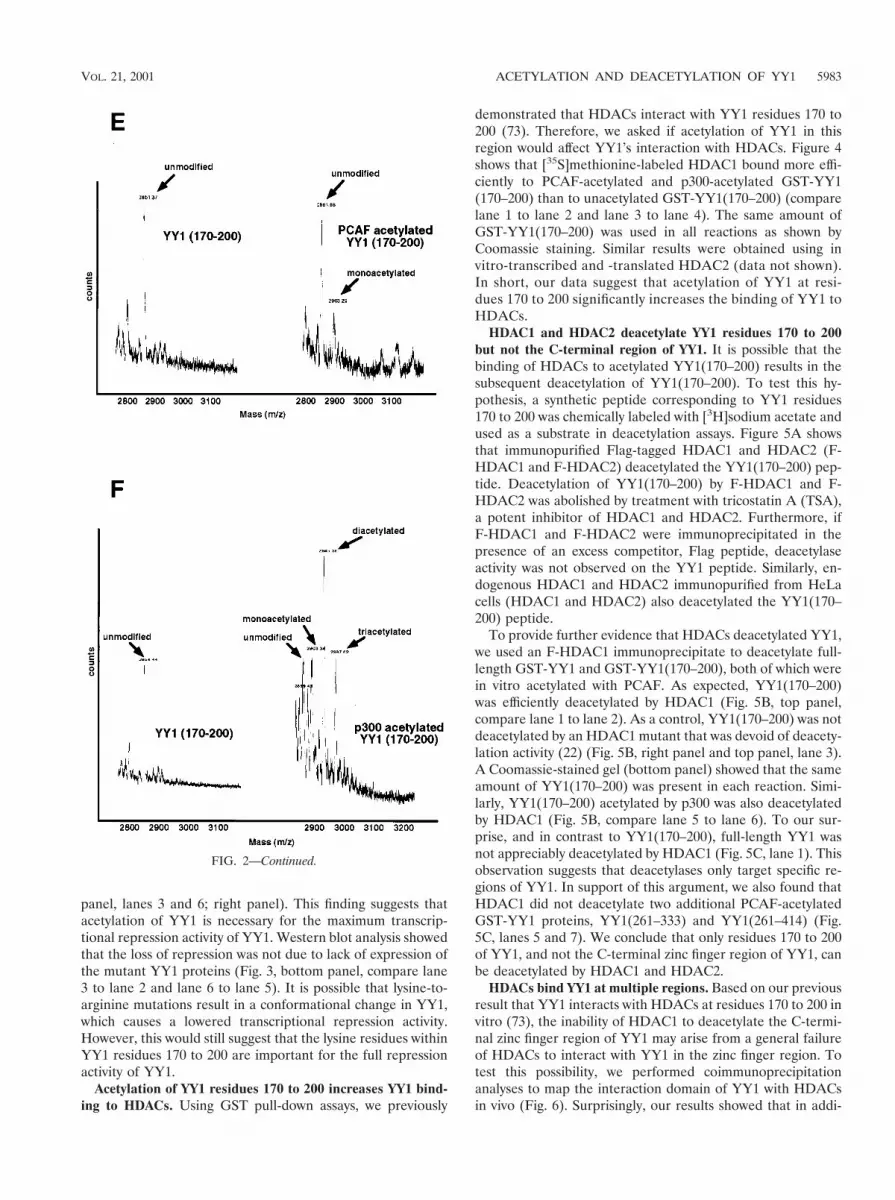
Because both p300 and PCAF acetylate YY1 at residues 170to 200, we were interested in determining the specificity ofacetylation by these two enzymes. Using GST-p300 or GST-PCAF, we in vitro acetylated a synthetic peptide correspondingto YY1 residues 170 to 200. We then compared the massspectra produced by mass spectrometry. Figure 2E shows thatmock-acetylated YY1 peptide had an Mr of 2,861 (left, 2861m/z). When this peptide was acetylated with PCAF, anotherpeak emerged with an Mr of 2,903 (right, 2903 m/z). Comparedto the mock-acetylated peptide, this additional peak had amass corresponding to one additional acetyl group (2903 22861 5 42), suggesting that the YY1 peptide was acetylatedonce by PCAF. Interestingly, when we compared the spectrumfrom the p300-acetylated peptide with that of the mock-acety-lated peptide, we found three additional peaks at 2903, 2945,and 2987 m/z, which contained one, two, and three additionalacetyl groups, respectively (Fig. 2F). This result strongly sug-gests that YY1 can be acetylated by p300 at three differentlysines between residues 170 and 200.
Lysine-to-arginine mutations within YY1 residues 170 to200 significantly reduce the transcriptional repression activityof YY1. To understand the effect of acetylation on the tran-scriptional activity of YY1, we mutated the six lysines withinYY1 residues 170 to 200 to arginines. Arginine substitutionspreserve the charges of the affected amino acid residues butprevent acetylation in vivo by histone acetyltransferases. Bothwild-type and mutant YY1 were fused to a Gal4 DNA-bindingdomain and transfected into HeLa cells in combination with aCAT reporter driven by the SV40 promoter containing fiveGal4 binding sites. Consistent with previous findings (58, 73),wild-type Gal4-YY1 was a potent transcriptional repressor(Fig. 3, top panel, lanes 2 and 5; right panel). However, whenthe six lysines were mutated and no longer able to be acety-lated, YY1 lost much of its repression activity (Fig. 3, top
FIG. 1. Multiple functional domains of transcription factor YY1.YY1 has one transcriptional activation domain at the N terminus andtwo repression domains, one encompassing residues 170 to 200 and theother one residing at the C terminus. The amino acid sequence of thecentral repression domain (residues 170 to 200) is given with lysineresidues underlined. His, histidine-rich domain; GA, glycine-alanine-rich domain; GK, glycine-lysine-rich domain.
VOL. 21, 2001 ACETYLATION AND DEACETYLATION OF YY1 5981

FIG. 2. Acetylation of YY1 in vitro and in vivo. (A) Acetylation of YY1 by PCAF. Serial GST-YY1 deletion proteins were incubated withGST-PCAF and separated by SDS-PAGE. The gels were then exposed to X-ray film to detect acetylated proteins. Open arrows indicate acetylatedYY1 proteins. Auto-acetylated forms of PCAF were detected as three bands of ;68 kDa. Solid arrows indicate GST-YY1 deletion proteins. (B)Acetylation of YY1 by p300. Serial GST-YY1 deletion proteins were incubated with GST-p300. Arrows indicate YY1 proteins acetylated by p300.(C) Acetylation of YY1 is dependent on p300 or PCAF. In vitro acetylation reactions were performed in the presence or absence of p300 or PCAF.Arrows indicate that YY1 was acetylated only in the presence of p300 or PCAF. (D) Identification of regions of YY1 acetylated in vivo. F-YY1serial deletion proteins were transiently expressed in HeLa cells, immunoprecipitated with anti-Flag antibody, and analyzed by Western blottingusing anti-acetyl lysine antibody and anti-Flag antibody. Arrows indicate immunoprecipitated YY1 proteins that were acetylated. (E and F) De-termination of the number of acetylated lysines by mass spectrometry. A YY1 peptide containing residues 170 to 200 was in vitro acetylated byPCAF or p300 and subjected to mass spectrometry analysis. Left panels are spectra from mock-acetylated peptides. Right panels contain spectra fromacetylated as well as unacetylated peptides. Small peaks around 3100 m/z in Fig. 2E represent degraded GST-PCAF or background chemical noises.
5982 YAO ET AL. MOL. CELL. BIOL.
panel, lanes 3 and 6; right panel). This finding suggests thatacetylation of YY1 is necessary for the maximum transcrip-tional repression activity of YY1. Western blot analysis showedthat the loss of repression was not due to lack of expression ofthe mutant YY1 proteins (Fig. 3, bottom panel, compare lane3 to lane 2 and lane 6 to lane 5). It is possible that lysine-to-arginine mutations result in a conformational change in YY1,which causes a lowered transcriptional repression activity.However, this would still suggest that the lysine residues withinYY1 residues 170 to 200 are important for the full repressionactivity of YY1.
Acetylation of YY1 residues 170 to 200 increases YY1 bind-ing to HDACs. Using GST pull-down assays, we previously
demonstrated that HDACs interact with YY1 residues 170 to200 (73). Therefore, we asked if acetylation of YY1 in thisregion would affect YY1’s interaction with HDACs. Figure 4shows that [35S]methionine-labeled HDAC1 bound more effi-ciently to PCAF-acetylated and p300-acetylated GST-YY1(170–200) than to unacetylated GST-YY1(170–200) (comparelane 1 to lane 2 and lane 3 to lane 4). The same amount ofGST-YY1(170–200) was used in all reactions as shown byCoomassie staining. Similar results were obtained using invitro-transcribed and -translated HDAC2 (data not shown).In short, our data suggest that acetylation of YY1 at resi-dues 170 to 200 significantly increases the binding of YY1 toHDACs.
HDAC1 and HDAC2 deacetylate YY1 residues 170 to 200but not the C-terminal region of YY1. It is possible that thebinding of HDACs to acetylated YY1(170–200) results in thesubsequent deacetylation of YY1(170–200). To test this hy-pothesis, a synthetic peptide corresponding to YY1 residues170 to 200 was chemically labeled with [3H]sodium acetate andused as a substrate in deacetylation assays. Figure 5A showsthat immunopurified Flag-tagged HDAC1 and HDAC2 (F-HDAC1 and F-HDAC2) deacetylated the YY1(170–200) pep-tide. Deacetylation of YY1(170–200) by F-HDAC1 and F-HDAC2 was abolished by treatment with tricostatin A (TSA),a potent inhibitor of HDAC1 and HDAC2. Furthermore, ifF-HDAC1 and F-HDAC2 were immunoprecipitated in thepresence of an excess competitor, Flag peptide, deacetylaseactivity was not observed on the YY1 peptide. Similarly, en-dogenous HDAC1 and HDAC2 immunopurified from HeLacells (HDAC1 and HDAC2) also deacetylated the YY1(170–200) peptide.
To provide further evidence that HDACs deacetylated YY1,we used an F-HDAC1 immunoprecipitate to deacetylate full-length GST-YY1 and GST-YY1(170–200), both of which werein vitro acetylated with PCAF. As expected, YY1(170–200)was efficiently deacetylated by HDAC1 (Fig. 5B, top panel,compare lane 1 to lane 2). As a control, YY1(170–200) was notdeacetylated by an HDAC1 mutant that was devoid of deacety-lation activity (22) (Fig. 5B, right panel and top panel, lane 3).A Coomassie-stained gel (bottom panel) showed that the sameamount of YY1(170–200) was present in each reaction. Simi-larly, YY1(170–200) acetylated by p300 was also deacetylatedby HDAC1 (Fig. 5B, compare lane 5 to lane 6). To our sur-prise, and in contrast to YY1(170–200), full-length YY1 wasnot appreciably deacetylated by HDAC1 (Fig. 5C, lane 1). Thisobservation suggests that deacetylases only target specific re-gions of YY1. In support of this argument, we also found thatHDAC1 did not deacetylate two additional PCAF-acetylatedGST-YY1 proteins, YY1(261–333) and YY1(261–414) (Fig.5C, lanes 5 and 7). We conclude that only residues 170 to 200of YY1, and not the C-terminal zinc finger region of YY1, canbe deacetylated by HDAC1 and HDAC2.
HDACs bind YY1 at multiple regions. Based on our previousresult that YY1 interacts with HDACs at residues 170 to 200 invitro (73), the inability of HDAC1 to deacetylate the C-termi-nal zinc finger region of YY1 may arise from a general failureof HDACs to interact with YY1 in the zinc finger region. Totest this possibility, we performed coimmunoprecipitationanalyses to map the interaction domain of YY1 with HDACsin vivo (Fig. 6). Surprisingly, our results showed that in addi-
FIG. 2—Continued.
VOL. 21, 2001 ACETYLATION AND DEACETYLATION OF YY1 5983

tion to residues 170–200, YY1 also interacted with HDACs atresidues 261 to 333 in vivo. These results, together with thoseof our previous in vitro acetylation deacetylation experiment,suggest that YY1 interacts with HDACs at two domains: res-idues 170 to 200, where bound HDACs deacetylate YY1, andthe C-terminal residues 261 to 333, where bound HDACs donot result in deacetylation of YY1.
YY1 contains associated HDAC activity, which localizes toC-terminal residues 261 to 333 of YY1. After we preciselymapped the HDAC-binding domains in YY1, we sought todetermine the functional effect of this interaction. We foundthat immunoprecipitated endogenous YY1 from HeLa cellsalso contained histone deacetylase activity, which was inhibitedby TSA (Fig. 7A). Using serial F-YY1 deletions, we deter-mined the histone deacetylase activity domain of YY1, whichlocalized to residues 261 to 333 (Fig. 7B and C). This region ofYY1 was necessary and sufficient for the HDAC activity asso-ciated with YY1 (Fig. 7B and C). Most strikingly, the YY1histone deacetylase activity domain completely overlappedwith one of the HDAC-interacting domains of YY1. Further-more, the HDAC activity associated with YY1 residues 261 to333 was highly specific, because the activity was sensitive toTSA (Fig. 7B) and competed by excess Flag peptide (Fig. 7B).A representative Western blot shows that different F-YY1deletion mutants expressed equally well (Fig. 7D). Immuno-fluorescence analysis confirmed that F-YY1 deletion proteinsthat did not exhibit HDAC activity localized to either thenucleus or both the nucleus and cytoplasm, ruling out thepossibility that the lack of histone deacetylase activity was dueto abnormal localization of the mutants (Fig. 7E). These re-sults strongly suggest that stable interaction between HDACsand YY1 contributes to YY1’s histone deacetylase activity.
Acetylation of YY1 at the C-terminal zinc finger domaindecreases the DNA-binding activity of YY1. Because the C-terminal acetylation domain (residues 261 to 333) of YY1
overlaps with the zinc finger DNA-binding domain (residues261 to 414) of YY1, we tested whether acetylation of YY1 atthe zinc finger domain would affect the DNA-binding activityof YY1. Indeed, when in vitro acetylated by PCAF, GST-YY1
FIG. 3. The effect of acetylation of YY1 residues 170 to 200 on the transcriptional repressor activity of YY1. HeLa cells were transfected withGa14 DNA-binding domain alone (Gal4), Gal4-YY1 fusion construct (Gal4-YY1), or Gal4-YY1 mutant with the six lysines mutated to arginines[Gal4-YY1 (K170–200R)]. Transcriptional activities were analyzed by CAT assays using a CAT reporter containing five Gal4 binding sequencesin tandem, and a representative autoradiogram is shown. Quantification of the relative CAT activities was performed using the PhosphoImagerStorm system (model 860) and ImageQuant software (Molecular Dynamics). Western blot analyses were performed to verify the expression levelsof the effector proteins.
FIG. 4. Increased HDAC binding to YY1 residues 170 to 200 byacetylation. A representative autoradiogram of in vitro-translatedHDAC1 captured by acetylated or mock-acetylated GST-YY1(170–200) is shown here. The input lane represents 1/10 the amount ofHDAC1 used in each binding reaction. Reaction mixtures were sepa-rated by SDS-PAGE, and the gels were stained with Coomassie blueprior to exposure to film to confirm that equal amounts of GST-YY1(170–200) were used in the binding reactions.
5984 YAO ET AL. MOL. CELL. BIOL.

FIG. 5. Identification of the region of YY1 deacetylated by HDACs. (A) YY1 peptide deacetylation by HDACs. Endogenous HDAC1 andHDAC2 and overexpressed F-HDAC1 and F-HDAC2 were immunoprecipitated from HeLa cells. A YY1 (residues 170 to 200) peptide waslabeled with [3H]acetate, and 20,000 cpm of the labeled peptide was used in deacetylation reactions. (B) YY1 protein deacetylation by HDACs.(Left panels) GST-YY1(170–200) was in vitro acetylated by p300 or PCAF. Half of the acetylated GST-YY1(170–200) was mixed with protein Gbeads alone, and the other half was mixed with immunoprecipitated wild-type or mutant (H199F) HDAC1. Samples were then separated bySDS-PAGE, and the gels were subsequently stained with Coomassic blue and exposed to film. Arrows indicate the position of GST-YY1(170–200).(Right panel) Transiently expressed wild-type and H199F mutant HDAC1 were immunoprecipitated from HeLa cells and tested for their HDACactivity against an H4 peptide. (C) Deacetylation of YY1 serial deletion proteins by HDAC1. Serial deletion proteins of GST-YY1 were in vitroacetylated by PCAF. Half of the GST-YY1 proteins were mixed with protein G beads, and the other half were mixed with HDAC1 immunopre-cipitate. Samples were then separated by SDS-PAGE, and the gels were subsequently stained with Coomassie blue and exposed to film. Arrowsindicate the positions of GST-YY1 deletion proteins.
VOL. 21, 2001 ACETYLATION AND DEACETYLATION OF YY1 5985

bound less avidly than did unacetylated GST-YY1 to the ini-tiator element of the adeno-associated virus P5 promoter,which contains a consensus YY1 binding site (Fig. 8A, com-pare lane 7 to lane 8). Similar results were obtained when aGST-YY1 deletion construct that contained only the zinc fin-ger domain of YY1 was tested for its DNA-binding properties(Fig. 8A, compare lane 5 to lane 6). In contrast, and consistentwith earlier reports, acetylated GST-p53 bound to its recogni-tion sequence better than unacetylated GST-p53 (Fig. 8A,compare lane 3 to lane 2). Thus, the decrease in DNA-bindingactivity caused by acetylation is specific to YY1. To rule out thepossibility that this decrease in DNA-binding activity was as-sociated with the GST fusion constructs, we tested nontaggedYY1 purified from E. coli, as well as F-YY1 purified fromHeLa cells. Both forms of YY1 exhibited decreased DNA-binding activity upon acetylation (Fig. 8B), proving that the
DNA-binding activity of YY1 decreases when the zinc fingerdomain of YY1 is acetylated.
DISCUSSION
In this report, we demonstrate that the activity of the mul-tifunctional transcription factor YY1 is regulated by acetyla-tion and deacetylation. Acetylation and deacetylation of YY1represent novel and complex means of regulating the activityof a DNA-binding transcription factor (Fig. 9). YY1 is acety-lated in two regions, one at the previously identified HDAC-interacting domain of residues 170 to 200, and the other at theC-terminal DNA-binding zinc finger domain. Residues 170 to200 of YY1 are acetylated by p300 and PCAF, while the C-terminal zinc finger domain is acetylated only by PCAF (Fig.
FIG. 6. Mapping of the HDAC interaction domains of YY1. Serial F-YY1 deletion constructs were transfected into HeLa cells and immu-noprecipitated with anti-Flag antibody. Immunoprecipitated proteins were removed from the resin by competitive elution with excess Flag peptideand analyzed by Western blotting for the presence of HDAC1, HDAC2, and HDAC3 (lanes 1 to 8) or HDAC2 and HDAC3 only (lanes 9 to 16).The bottom panel summarizes the HDAC-binding domains of YY1. 1, positive interactions between YY1 and HDACs; 2, absence of YY1-HDAC interactions.
5986 YAO ET AL. MOL. CELL. BIOL.

9A). Acetylation of these two regions results in dramaticallydifferent outcomes (Fig. 9). First, when residues 170 to 200 ofYY1 are acetylated, YY1 becomes a more effective transcrip-tional repressor and binds HDACs more efficiently. However,upon binding to acetylated YY1 residues 170 to 200, HDACsalso actively deacetylate this region, possibly resulting in anegative feedback loop. Second, YY1 possesses histonedeacetylase activity toward histone H4 by associating withHDACs using the C-terminal zinc finger region. This is mostlikely a result of active targeting of acetylated YY1 zinc fingerdomains by HDACs. However, our data do not indicate thatassociation of the YY1 C terminus with HDACs brings aboutdeacetylation of YY1 in this region. In contrast, the interactionbetween YY1 and HDACs at residues 170–200 is likely to be adynamic and highly regulated process and does not result inassociated histone deacetylase activity stable enough to bedetected in our experimental system. Finally, acetylation of
YY1 zinc fingers decreases YY1’s DNA-binding activity, whichwill have further impact on YY1’s activity as a transcriptionfactor.
This differential regulation of YY1 by acetylation anddeacetylation suggests a complex regulatory system unlike thatof any other transcription factor known to date. In cells whereYY1 functions primarily as a transcriptional repressor, acety-lation of YY1 at the central HDAC-binding region and theC-terminal DNA-binding region most likely will result in anintricate network of negative-feedback regulation (Fig. 9B):acetylation of YY1 at residues 170 to 200 augments YY1’srepressor activity, but acetylation of this region also targetsYY1 for deacetylation. In the meantime, acetylation of theC-terminal DNA-binding region of YY1 most likely stabilizesinteraction between YY1 and HDACs, but acetylation of thisregion in turn decreases YY1’s DNA-binding activity. Todate, we have not been able to determine the relative levels of
FIG. 7. Identification of the HDAC activity domain of YY1. (A) HDAC activity of endogenous YY1 in HeLa cells. Endogenous YY1 wasimmunoprecipitated from HeLa cells and assayed for deacetylase activity against the H4 peptide. Where indicated (1) TSA was added to 400 nMprior to addition of the peptide substrate. (B and C) Identification of aa 261 to 333 as the HDAC activity domain of YY1. F-YY1 deletionconstructs were transfected into HeLa cells, immunoprecipitated with anti-Flag antibody, and assayed for deacetylase activity against the H4peptide. Where indicated (1) TSA was added to 400 nM prior to addition of the peptide substrate or excess Flag peptides (competitor) were addedprior to addition of the peptide substrate. The experiments in panel C were performed up to three times, with standard deviations less than or equalto 5%. (D) Expression of F-YY1 deletion mutants. Overexpressed F-YY1 deletion constructs and Flag alone from the parental vector wereimmunoprecipitated from HeLa cells using anti-Flag antibody, separated by SDS-PAGE, and analyzed by Western blotting using anti-Flagantibody. Arrows indicate the positions of F-YY1 deletion proteins. (E) Subcellular localization of F-YY1 deletion constructs. Various F-YY1deletion constructs were transiently transfected into HeLa cells. Transfected HeLa cells were then fixed and probed with anti-Flag FITCconjugated antibody. The nuclei were stained with DAPI. Images were obtained from a fluorescence microscope. Merged images from FITC andDAPI stains indicate subcellular localization of F-YY1 deletion constructs.
VOL. 21, 2001 ACETYLATION AND DEACETYLATION OF YY1 5987

acetylation between the central region and the C terminus ofYY1 under physiological conditions; therefore, the relativecontribution of the acetylation of these two regions is un-known. Interestingly, we also found that TSA had little effect
on the acetylation status of YY1 (data not shown), suggestingthat regulation of YY1 by acetylation is more prominent thanby deacetylation. This finding is also in agreement with ourdiscovery that deacetylation of YY1 only occurs at residues 170to 200, while acetylation of YY1 can happen at the C-terminalDNA-binding domain as well.
In many experimental systems, YY1 can also activate tran-scription, and it is still uncertain how this is accomplished.Different models, including bending of DNA, the relative dis-tance between the YY1 binding site and the transcriptionalinitiation site, as well as protein-protein interactions, havebeen proposed (reviewed in references 57 and 65). Increas-ingly, more evidence shows that interactions with other pro-teins are probably the most important factors in YY1-medi-ated transcriptional activation. It has been suggested thatinteractions of YY1 with other cellular proteins or viral pro-teins can either disrupt the quenching activity of YY1 on othertranscriptional activators or stimulate transcription with asso-ciated enzymatic activities such as HATs (36, 58, 65). Ourfindings here provide an additional speculation that on pro-moters activated by YY1, YY1-associated p300 and PCAF canactivate transcription by both acetylating core histones andacetylating YY1 at the C-terminal zinc finger domain (PCAFonly), which in turn decreases the overall histone deacetylationactivity at the promoter.
In addition to histones and several nonhistone chromatinproteins, many transcription factors have been shown to beregulated by acetylation. These transcription factors includep53 (18), human immunodeficiency virus Tat (32), E1A (77),GATA-1 (5), EKLF (78), MyoD (50), E2F (43), TFIIE, TFIIF(28), CIITA (62), TCF (71), HNF-4 (61), UBF (48), TAL1(also known as SCL) (25), and nuclear receptor coactivatorsACTR, SRC-1, and TIF2 (10). Many of these factors are acety-lated by both p300 and PCAF; therefore, it is not surprisingthat YY1 is also acetylated by both p300 and PCAF. Thep300-interaction domain of YY1 has been mapped to the C-terminal 17 residues (36, 38), which were not acetylated byp300 in our study. This finding is reminiscent of acetylation ofp53 by p300, in which the N terminus of p53 interacts with CBP(19, 41) while the region acetylated by p300 is at the C termi-nus of p53 (18). Moreover, in our in vitro acetylation studies,full-length GST-YY1 was acetylated by PCAF and not by p300,which suggests that the conformation of YY1, perhaps affectedby selective interaction with p300 and PCAF, is important toYY1 acetylation. We also found that in HeLa cells, over-expression of PCAF, but not p300, partially alleviated thetranscriptional repressor activity of a Gal4 DNA-binding do-main-YY1 fusion protein. However, in NIH 3T3 cells, over-expression of p300, but not PCAF, relieved repression fromGal4-YY1 (data not shown). In this regard, it will be importantto identify the in vivo triggers directing PCAF versus p300acetylation. p53 is particularly interesting among acetylatedtranscription factors because it has been elegantly demon-strated that DNA damage (UV and ionizing radiation) causesp53 acetylation at two distinct lysines, one by p300 and theother by PCAF (42). To date, this is the only report linkingspecific environmental cues to acetylation of transcription fac-tors by HATs, and yet it is still uncertain why acetylation wouldrequire two HATs. It is interesting that while an increase inDNA-binding activity has been observed for most acetylated
FIG. 8. The effect of acetylation on YY1’s sequence-specific DNA-binding activity. (A) Representative EMSA of GST-tagged YY1. Pu-rified full-length GST-YY1(1–414), zinc finger domain of GST-YY1(261–414), and GST-p53 were in vitro acetylated with PCAF and mixedwith 32P-labeled probes containing either a YY1 or p53-binding se-quence. Mock-acetylated GST fusion proteins were treated identicallyas acetylated proteins with the exception that acetyl-CoA was omittedfrom the in vitro acetylation reactions. Protein-DNA complexes wereresolved on nondenaturing polyacrylamide gels. Black arrows indi-cate the positions of full-length YY1- and p53-DNA complexes. Theopen arrow indicates the position of the complex between DNA andthe zinc finger domain of YY1. (B) Representative EMSA of bac-terially expressed nontagged YY1 and HeLa cell-expressed Flag-tagged YY1. Black arrows indicate the positions of the YY1-DNAcomplexes.
5988 YAO ET AL. MOL. CELL. BIOL.

DNA-binding transcription factors, HMG I(Y) binds DNAless when it is acetylated (45). The most striking observation isthat only when HMG I(Y) is acetylated by CBP, not by PCAF,is there a decrease in its DNA-binding activity (45). This phe-nomenon is reminiscent of YY1 acetylation in its zinc fingerdomain by PCAF but not by p300 and the consequent reduc-tion in the DNA-binding activity of YY1.
We were also interested in finding out if acetylation of YY1also contributes to cellular events other than transcriptional
control. So far, we have no evidence to suggest that acetylationchanges the subcellular localization of YY1. YY1 has beenshown to be a rather stable protein expressed at comparablelevels in both growing and differentiating cells (1). Interest-ingly, acetylation affects the conformation of HNF-4 (61), thehalf-life of E2F (43), and promotes protein-protein interac-tions between Rch1 and importin-b (3). Therefore, it will beinformative to test whether acetylation of YY1 may have sim-ilar consequences.
FIG. 9. Summary of regulation of YY1 by acetylation and deacetylation. (A) Summary of different domains of YY1 affected by acetylation anddeacetylation. His, histidine-rich domain; GA, glycine-alanine-rich domain; GK, glycine-lysine-rich domain. (B) YY1 is acetylated in two regions:residues 170 to 200 and the C-terminal DNA-binding domain. Thin arrows represent YY1 protein modifications: arrows with solid heads representacetylation (Ac), while an arrow with an empty head represents deacetylation (DeAc). Acetylation of YY1 at residues 170 to 200 by p300 andPCAF augments YY1’s repressor activity (shown as a thick arrow leading to transcriptional repression), but acetylation of this region also targetsYY1 for deacetylation, resulting in negative feedback regulation. Acetylation of the C terminus of YY1 results in stable association of histonedeacetylase activity with YY1 as well as decreased DNA-binding activity. The in vivo association between YY1 and HDACs at the C-terminalregion is probably mediated through an unidentified protein (depicted as a question mark in a rounded rectangle).
VOL. 21, 2001 ACETYLATION AND DEACETYLATION OF YY1 5989

Our data demonstrating that YY1 interacts with HDACsat two different regions open the possibility that these twoHDAC-interacting regions have distinct effects on YY1’s rolein transcriptional control. Recently a Drosophila homolog ofYY1 was identified as PHO, which is encoded by pleio-homeotic, a member of the Polycomb group (PcG) genes (6). Ithas been proposed that the Drosophila YY1 homolog, PHO,binds to PcG response elements and interacts with other pro-teins to form a repressor complex with nucleosome remodelingactivity (6, 31, 69, 70). YY1, PHO, and the Xenopus YY1homolog FIII are almost completely identical in the zinc fingerregion in amino acid sequence, reinforcing the role of YY1 asa DNA-targeting factor in nucleating a repressor complex ca-pable of modulating chromatin structures. The stable associa-tion of YY1 with HDACs at the C-terminal zinc finger re-gion might represent an ancient mode of the functions ofYY1, which is to form a repressor complex associated withthe promoter. However, regulation of the affinity of the cen-tral region of YY1 for HDACs by acetylation might haveevolved as a more sophisticated means of control, whichdefines a novel functional consequence of nonhistone factoracetylation.
ACKNOWLEDGMENTS
We thank Shelly Berger, Nobuo Horikoshi, and Stuart Schreiber fortheir generous gifts of plasmids; Nancy Olashaw, Rosalind Jackson,and Peter Neame for critical reading of the manuscript; and the corefacilities at the Moffitt Cancer Center as well as the Protein ChemistryCore at the University of Florida for technical support. We also thankJim Davie and his laboratory for their help in setting up preliminaryexperiments involving HDAC activity associated with YY1.
Y.-L.Y. was supported by a fellowship from the American HeartAssociation. This work was supported by a grant from the NationalInstitutes of Health (GM58486) to E.S.
REFERENCES
1. Austen, M., B. Luscher, and F. J. Luscher. 1997. Characterization of thetranscriptional regulator YY1. The bipartite transactivation domain is inde-pendent of interaction with the TATA box-binding protein, transcriptionfactor IIB, TAFII55, or cAMP-responsive element-binding protein (CPB)-binding protein. J. Biol. Chem. 272:1709–1717.
2. Bannister, A. J., and T. Kouzarides. 1996. The CBP co-activator is a histoneacetyltransferase. Nature 384:641–643.
3. Bannister, A. J., E. A. Miska, D. Gorlich, and T. Kouzarides. 2000. Acety-lation of importin-alpha nuclear import factors by CBP/p300. Curr. Biol. 10:467–470.
4. Bauknecht, T., R. H. See, and Y. Shi. 1996. A novel C/EBP beta-YY1complex controls the cell-type-specific activity of the human papillomavirustype 18 upstream regulatory region. J. Virol. 70:7695–7705.
5. Boyes, J., P. Byfield, Y. Nakatani, and V. Ogryzko. 1998. Regulation ofactivity of the transcription factor GATA-1 by acetylation. Nature 396:594–598.
6. Brown, J. L., D. Mucci, M. Whiteley, M. L. Dirksen, and J. A. Kassis. 1998.The Drosophila Polycomb group gene pleiohomeotic encodes a DNA bind-ing protein with homology to the transcription factor YY1. Mol. Cell 1:1057–1064.
7. Bushmeyer, S., K. Park, and M. L. Atchison. 1995. Characterization offunctional domains within the multifunctional transcription factor, YY1.J. Biol. Chem. 270:30213–30220.
8. Bushmeyer, S. M., and M. L. Atchison. 1998. Identification of YY1 se-quences necessary for association with the nuclear matrix and for transcrip-tional repression functions. J. Cell. Biochem. 68:484–499.
9. Candau, R., J. X. Zhou, C. D. Allis, and S. L. Berger. 1997. Histone acetyl-transferase activity and interaction with ADA2 are critical for GCN5 func-tion in vivo. EMBO J. 16:555–565.
10. Chen, H., R. J. Lin, W. Xie, D. Wilpitz, and R. M. Evans. 1999. Regulationof hormone-induced histone hyperacetylation and gene activation via acet-ylation of an acetylase. Cell 98:675–686.
11. Chiang, C. M., and R. G. Roeder. 1995. Cloning of an intrinsic human TFIIDsubunit that interacts with multiple transcriptional activators. Science 267:531–536.
12. Cress, W. D., and E. Seto. 2000. Histone deacetylases, transcriptional con-trol, and cancer. J. Cell. Physiol. 184:1–16.
13. Donohoe, M. E., X. Zhang, L. McGinnis, J. Biggers, E. Li, and Y. Shi. 1999.Targeted disruption of mouse Yin Yang 1 transcription factor results inperi-implantation lethality. Mol. Cell. Biol. 19:7237–7244.
14. Flanagan, J. R., K. G. Becker, D. L. Ennist, S. L. Gleason, P. H. Driggers,B. Z. Levi, E. Appella, and K. Ozato. 1992. Cloning of a negative transcrip-tion factor that binds to the upstream conserved region of Moloney murineleukemia virus. Mol. Cell. Biol. 12:38–44.
15. Giordano, A., and M. L. Avantaggiati. 1999. p300 and CBP: partners for lifeand death. J. Cell. Physiol. 181:218–230.
16. Gorman, C. M., L. F. Moffat, and B. H. Howard. 1982. Recombinant ge-nomes which express chloramphenicol acetyltransferase in mammalian cells.Mol. Cell. Biol. 2:1044–1051.
17. Graham, F. L., and A. J. van der Eb. 1973. A new technique for the assay ofinfectivity of human adenovirus 5 DNA. Virology 52:456–467.
18. Gu, W., and R. G. Roeder. 1997. Activation of p53 sequence-specific DNAbinding by acetylation of the p53 C-terminal domain. Cell 90:595–606.
19. Gu, W., X. L. Shi, and R. G. Roeder. 1997. Synergistic activation of tran-scription by CBP and p53. Nature 387:819–823.
20. Hariharan, N., D. E. Kelley, and R. P. Perry. 1991. Delta, a transcriptionfactor that binds to downstream elements in several polymerase II promot-ers, is a functionally versatile zinc finger protein. Proc. Natl. Acad. Sci. USA88:9799–9803.
21. Harlow, E., and D. Lane 1999. Using antibodies: a laboratory manual, 2nded. Cold Spring Harbor Laboratory Press, Cold Spring Harbor, N.Y.
22. Hassig, C. A., J. K. Tong, T. C. Fleischer, T. Owa, P. G. Grable, D. E. Ayer,and S. L. Schreiber. 1998. A role for histone deacetylase activity in HDAC1-mediated transcriptional repression. Proc. Natl. Acad. Sci. USA 95:3519–3524.
23. Hebbes, T. R., A. W. Thorne, and C. Crane-Robinson. 1988. A direct linkbetween core histone acetylation and transcriptionally active chromatin.EMBO J. 7:1395–1402.
24. Hong, L., G. P. Schroth, H. R. Matthews, P. Yau, and E. M. Bradbury. 1993.Studies of the DNA binding properties of histone H4 amino terminus.Thermal denaturation studies reveal that acetylation markedly reduces thebinding constant of the H4 “tail” to DNA. J. Biol. Chem. 268:305–314.
25. Huang, S., Y. Qiu, Y. Shi, Z. Xu, and S. J. Brandt. 2000. P/CAF-mediatedacetylation regulates the function of the basic helix-loop-helix transcriptionfactor TAL1/SCL. EMBO J. 19:6792–6803.
26. Huibregtse, J. M., M. Scheffner, and P. M. Howley. 1991. A cellular proteinmediates association of p53 with the E6 oncoprotein of human papilloma-virus types 16 or 18. EMBO J. 10:4129–4135.
27. Hung, H. L., J. Lau, A. Y. Kim, M. J. Weiss, and G. A. Blobel. 1999. CREB-binding protein acetylates hematopoietic transcription factor GATA-1 atfunctionally important sites. Mol. Cell. Biol. 19:3496–3505.
28. Imhof, A., X. J. Yang, V. V. Ogryzko, Y. Nakatani, A. P. Wolffe, and H. Ge.1997. Acetylation of general transcription factors by histone acetyltrans-ferases. Curr. Biol. 7:689–692.
29. Inouye, C. J., and E. Seto. 1994. Relief of YY1-induced transcriptionalrepression by protein-protein interaction with the nucleolar phosphoproteinB23. J. Biol. Chem. 269:6506–6510.
30. Jones, K. A., and J. T. Kadonaga. 2000. Exploring the transcription-chro-matin interface. Genes Dev. 14:1992–1996.
31. Kehle, J., D. Beuchle, S. Treuheit, B. Christen, J. A. Kennison, M. Bienz, andJ. Muller. 1998. dMi-2, a hunchback-interacting protein that functions inpolycomb repression. Science 282:1897–1900.
32. Kiernan, R. E., C. Vanhulle, L. Schiltz, E. Adam, H. Xiao, F. Maudoux, C.Calomme, A. Burny, Y. Nakatani, K. T. Jeang, M. Benkirane, and C. VanLint. 1999. HIV-1 tat transcriptional activity is regulated by acetylation.EMBO J. 18:6106–6118.
33. Kingston, R. E., and G. J. Narlikar. 1999. ATP-dependent remodeling andacetylation as regulators of chromatin fluidity. Genes Dev. 13:2339–2352.
34. Kornberg, R. D., and Y. Lorch. 1999. Twenty-five years of the nucleosome,fundamental particle of the eukaryote chromosome. Cell 98:285–294.
35. Laherty, C. D., W. M. Yang, J. M. Sun, J. R. Davie, E. Seto, and R. N.Eisenman. 1997. Histone deacetylases associated with the mSin3 corepressormediate mad transcriptional repression. Cell 89:349–356.
36. Lee, J. S., K. M. Galvin, R. H. See, R. Eckner, D. Livingston, E. Moran, andY. Shi. 1995. Relief of YY1 transcriptional repression by adenovirus E1A ismediated by E1A-associated protein p300. Genes Dev. 9:1188–1198.
37. Lee, J. S., K. M. Galvin, and Y. Shi. 1993. Evidence for physical interactionbetween the zinc-finger transcription factors YY1 and Spl. Proc. Natl. Acad.Sci. USA 90:6145–6149.
38. Lee, J. S., R. H. See, K. M. Galvin, J. Wang, and Y. Shi. 1995. Functionalinteractions between YY1 and adenovirus E1A. Nucleic Acids Res. 23:925–931.
39. Lee, T. C., Y. Zhang, and R. J. Schwartz. 1994. Bifunctional transcriptionalproperties of YY1 in regulating muscle actin and c-myc gene expressionduring myogenesis. Oncogene 9:1047–1052.
40. Lewis, B. A., G. Tullis, E. Seto, N. Horikoshi, R. Weinmann, and T. Shenk.
5990 YAO ET AL. MOL. CELL. BIOL.

1995. Adenovirus E1A proteins interact with the cellular YY1 transcriptionfactor. J. Virol. 69:1628–1636.
41. Lill, N. L., S. R. Grossman, D. Ginsberg, J. DeCaprio, and D. M. Livingston.1997. Binding and modulation of p53 by p300/CBP coactivators. Nature 387:823–827.
42. Liu, L., D. M. Scolnick, R. C. Trievel, H. B. Zhang, R. Marmorstein, T. D.Halazonetis, and S. L. Berger. 1999. p53 sites acetylated in vitro by PCAFand p300 are acetylated in vivo in response to DNA damage. Mol. Cell. Biol.19:1202–1209.
43. Martinez-Balbas, M. A., U. M. Bauer, S. J. Nielsen, A. Brehm, and T.Kouzarides. 2000. Regulation of E2F1 activity by acetylation. EMBO J. 19:662–671.
44. Marzio, G., C. Wagener, M. I. Gutierrez, P. Cartwright, K. Helin, and M.Giacca. 2000. E2F family members are differentially regulated by reversibleacetylation. J. Biol. Chem. 275:10887–10892.
45. Munshi, N., M. Merika, J. Yie, K. Senger, G. Chen, and D. Thanos. 1998.Acetylation of HMG I(Y) by CBP turns off IFN beta expression by disrupt-ing the enhanceosome. Mol. Cell 2:457–467.
46. Ogryzko, V. V., R. L. Schiltz, V. Russanova, B. H. Howard, and Y. Nakatani.1996. The transcriptional coactivators p300 and CBP are histone acetyltrans-ferases. Cell 87:953–959.
47. Park, K., and M. L. Atchison. 1991. Isolation of a candidate repressor/activator, NF-E1 (YY-1, delta), that binds to the immunoglobulin kappa 39enhancer and the immunoglobulin heavy-chain mu E1 site. Proc. Natl. Acad.Sci. USA 88:9804–9808.
48. Pelletier, G., V. Y. Stefanovsky, M. Faubladier, I. Hirschler-Laszkiewicz, J.Savard, L. I. Rothblum, J. Cote, and T. Moss. 2000. Competitive recruitmentof CBP and Rb-HDAC regulates UBF acetylation and ribosomal transcrip-tion. Mol. Cell 6:1059–1066.
49. Pisaneschi, G., S. Ceccotti, M. L. Falchetti, S. Fiumicino, F. Carnevali, andE. Beccari. 1994. Characterization of FIII/YY1, a Xenopus laevis conservedzinc-finger protein binding to the first exon of L1 and L14 ribosomal proteingenes. Biochem. Biophys. Res. Commun. 205:1236–1242.
50. Polesskaya, A., A. Duquet, I. Naguibneva, C. Weise, A. Vervisch, E. Bengal,F. Hucho, P. Robin, and A. Harel-Bellan. 2000. CREB-binding protein/p300activates MyoD by acetylation. J. Biol. Chem. 275:34359–34364.
51. Ron, D., and H. Dressler. 1992. pGSTag—a versatile bacterial expressionplasmid for enzymatic labeling of recombinant proteins. BioTechniques 13:866–869.
52. Sadowski, I., B. Bell, P. Broad, and M. Hollis. 1992. GAL4 fusion vectors forexpression in yeast or mammalian cells. Gene 118:137–141.
53. Sartorelli, V., P. L. Puri, Y. Hamamori, V. Ogryzko, G. Chung, Y. Nakatani,J. Y. Wang, and L. Kedes. 1999. Acetylation of MyoD directed by PCAF isnecessary for the execution of the muscle program. Mol. Cell 4:725–734.
54. Sealy, L., and R. Chalkley. 1978. DNA associated with hyperacetylatedhistone is preferentially digested by DNase I. Nucleic Acids Res. 5:1863–1876.
55. Seto, E., B. Lewis, and T. Shenk. 1993. Interaction between transcriptionfactors Sp1 and YY1. Nature 365:462–464.
56. Seto, E., Y. Shi, and T. Shenk. 1991. YY1 is an initiator sequence-bindingprotein that directs and activates transcription in vitro. Nature 354:241–245.
57. Shi, Y., J. S. Lee, and K. M. Galvin. 1997. Everything you have ever wantedto know about Yin Yang 1. Biochim. Biophys. Acta 1332:F49–F66.
58. Shi, Y., E. Seto, L. S. Chang, and T. Shenk. 1991. Transcriptional repressionby YY1, a human GLI-Kruppel-related protein, and relief of repression byadenovirus E1A protein. Cell 67:377–388.
59. Shrivastava, A., and K. Calame. 1994. An analysis of genes regulated by themulti-functional transcriptional regulator Yin Yang-1. Nucleic Acids Res.22:5151–5155.
60. Shrivastava, A., S. Saleque, G. V. Kalpana, S. Artandi, S. P. Goff, and K.Calame. 1993. Inhibition of transcriptional regulator Yin-Yang-1 by associ-ation with c-Myc. Science 262:1889–1892.
61. Soutoglou, E., N. Katrakili, and I. Talianidis. 2000. Acetylation regulates
transcription factor activity at multiple levels. Mol. Cell 5:745–751.62. Spilianakis, C., J. Papamatheakis, and A. Kretsovali. 2000. Acetylation by
PCAF enhances CIITA nuclear accumulation and transactivation of majorhistocompatibility complex class II genes. Mol. Cell. Biol. 20:8489–8498.
63. Sterner, D. E., and S. L. Berger. 2000. Acetylation of histories and transcrip-tion-related factors. Microbiol. Mol. Biol. Rev. 64:435–459.
64. Taunton, J., C. A. Hassig, and S. L. Schreiber. 1996. A mammalian histonedeacetylase related to the yeast transcriptional regulator Rpd3p. Science272:408–411.
65. Thomas, M. J., and E. Seto. 1999. Unlocking the mechanisms of transcrip-tion factor YY1: are chromatin modifying enzymes the key? Gene 236:197–208.
66. Tsai, S. C., N. Valkov, W. M. Yang, J. Gump, D. Sullivan, and E. Seto. 2000.Histone deacetylase interacts directly with DNA topoisomerase II. Nat.Genet. 26:349–353.
67. Usheva, A., and T. Shenk. 1994. TATA-binding protein-independent initia-tion: YY1, TFIIB, and RNA polymerase II direct basal transcription onsupercoiled template DNA. Cell 76:1115–1121.
68. Vidali, G., L. C. Boffa, E. M. Bradbury, and V. G. Allfrey. 1978. Butyratesuppression of histone deacetylation leads to accumulation of multiacety-lated forms of histones H3 and H4 and increased DNase I sensitivity of theassociated DNA sequences. Proc. Natl. Acad. Sci. USA 75:2239–2243.
69. Wade, P. A., P. L. Jones, D. Vermaak, G. J. Veenstra, A. Imhof, T. Sera, C.Tse, H. Ge, Y. B. Shi, J. C. Hansen, and A. P. Wolffe. 1998. Histone deacety-lase directs the dominant silencing of transcription in chromatin: associationwith MeCP2 and the Mi-2 chromodomain SWI/SNF ATPase. Cold SpringHarbor Symp. Quant. Biol. 63:435–445.
70. Wade, P. A., P. L. Jones, D. Vermaak, and A. P. Wolffe. 1998. A multiplesubunit Mi-2 histone deacetylase from Xenopus laevis cofractionates with anassociated Snf2 superfamily ATPase. Curr. Biol. 8:843–846.
71. Waltzer, L., and M. Bienz. 1998. Drosophila CBP represses the transcriptionfactor TCF to antagonize Wingless signalling. Nature 395:521–525.
72. Wen, Y. D., V. Perissi, L. M. Staszewski, W. M. Yang, A. Krones, C. K. Glass,M. G. Rosenfeld, and E. Seto. 2000. The histone deacetylase-3 complexcontains nuclear receptor corepressors. Proc. Natl. Acad. Sci. USA 97:7202–7207.
73. Yang, W. M., C. Inouye, Y. Zeng, D. Bearss, and E. Seto. 1996. Transcrip-tional repression by YY1 is mediated by interaction with a mammalianhomolog of the yeast global regulator RPD3. Proc. Natl. Acad. Sci. USA 93:12845–12850.
74. Yang, W. M., C. J. Inouye, and E. Seto. 1995. Cyclophilin A and FKBP12interact with YY1 and alter its transcriptional activity. J. Biol. Chem. 270:15187–15193.
75. Yang, W. M., Y. L. Yao, J. M. Sun, J. R. Davie, and E. Seto. 1997. Isolationand characterization of cDNAs corresponding to an additional member ofthe human histone deacetylase gene family. J. Biol. Chem. 272:28001–28007.
76. Yang, X. J., V. V. Ogryzko, J. Nishikawa, B. H. Howard, and Y. Nakatani.1996. A p300/CBP-associated factor that competes with the adenoviral on-coprotein E1A. Nature 382:319–324.
77. Zhang, Q., H. Yao, N. Vo, and R. H. Goodman. 2000. Acetylation of adeno-virus E1A regulates binding of the transcriptional corepressor CtBP. Proc.Natl. Acad. Sci. USA 97:14323–14328.
78. Zhang, W., and J. J. Bieker. 1998. Acetylation and modulation of erythroidKruppel-like factor (EKLF) activity by interaction with histone acetyltrans-ferases. Proc. Natl. Acad. Sci. USA 95:9855–9860.
79. Zhou, Q., R. W. Gedrich, and D. A. Engel. 1995. Transcriptional repressionof the c-fos gene by YY1 is mediated by a direct interaction with ATF/CREB. J. Virol. 69:4323–4330.
80. Zhu, X., M. A. Mancini, K. H. Chang, C. Y. Liu, C. F. Chen, B. Shan, D.Jones, T. L. Yang-Feng, and W. H. Lee. 1995. Characterization of a novel350-kilodalton nuclear phosphoprotein that is specifically involved in mitot-ic-phase progression. Mol. Cell. Biol. 15:5017–5029.
VOL. 21, 2001 ACETYLATION AND DEACETYLATION OF YY1 5991