dynamic changes in histone acetylation regulate origins of dna replication
TRANSCRIPT
Dynamic changes in histone acetylation regulate origins of DNAreplication
Ashwin Unnikrishnan1,2, Philip R. Gafken3, and Toshio Tsukiyama1,*1Divison of Basic Sciences, Fred Hutchinson Cancer Research Center, 1100 Fairview AvenueNorth, Seattle, WA 98109, U.S.A.2Molecular and Cellular Biology Program, Fred Hutchinson Cancer Research Center andUniversity of Washington, Seattle, WA 98195, U.S.A.3Proteomics Facility, Fred Hutchinson Cancer Research Center 1100 Fairview Avenue North,Seattle, WA 98109, U.S.A.
AbstractWhile histone modifications have been implicated in many DNA-dependent processes, theirprecise role in DNA replication remains largely unknown. Here, we describe a very efficient,single-step method to specifically purify histones located around an origin of replication from S.cerevisiae. Using high-resolution mass spectrometry, we have obtained a comprehensive view ofthe histone modifications surrounding the origin of replication throughout the cell cycle. We havediscovered that histone H3 and H4 acetylation is dynamically regulated around an origin ofreplication, at the level of multiply-acetylated histones. Furthermore, we find that this acetylationis required for efficient origin activation during S-phase.
KeywordsDNA replication; histone modifications; mass spectrometry
Eukaryotic DNA replication occurs in the context of chromatin. The fundamental unit ofchromatin is the nucleosome, in which 147 base pairs of DNA tightly wrap around a histoneoctamer, consisting of the core histones H2A, H2B, H3 and H41. The packaging of DNAinto chromatin profoundly influences DNA-dependent processes such as transcription,replication, repair and recombination2,3. A large variety of covalent modifications are addedto histones post-translationally, including acetylation, methylation, phosphorylation andubiquitination, to influence DNA-dependent processes4.
DNA replication occurs in the S-phase of the cell cycle and initiates at discrete sites on thechromosome called origins of replication (henceforth “origins”). DNA replication has beenbest studied in the budding yeast, Saccharomyces cerevisiae, where origins were firstidentified as autonomous replication sequence (ARS) elements in plasmid maintenanceassays5. A number of protein complexes are assembled at an origin in a tightly regulated,temporally controlled manner over the cell cycle to initiate replication. Replication forksthen travel bi-directionally outwards from the origin until the entire genome is replicated6.
*Corresponding author: [email protected] CONTRIBUTIONS A.U. performed the experiments, with help by P.G. on MS analyses. T.T. supervised theexperiments. A.U. and T.T. wrote the manuscript. All authors discussed the results and approved the manuscript.
NIH Public AccessAuthor ManuscriptNat Struct Mol Biol. Author manuscript; available in PMC 2011 March 18.
Published in final edited form as:Nat Struct Mol Biol. 2010 April ; 17(4): 430–437. doi:10.1038/nsmb.1780.
NIH
-PA Author Manuscript
NIH
-PA Author Manuscript
NIH
-PA Author Manuscript
It is thought that the accessibility of DNA to replication factors can be influenced by localchromatin structure6,7,8. Additionally, chromatin structure modulates origin firing time andefficiency, although the exact mechanisms are unknown9,10. The acetylation of the ε-aminogroup of a lysine residue on a histone protein neutralizes its positive, which is believed tocreate a more accessible chromatin structure, facilitating DNA-dependent processes.Supporting a role for histone acetylation in DNA replication, deletions of the Rpd3 histonedeacetylase in S. cerevisiae advance the activation time of a subset of origins11,12.Conversely, artificial recruitment of the Gcn5/KAT2 histone acetyltransferase to the vicinityof a late-firing origin (ARS1412) advances the firing time of the origin12. Intriguingly, amammalian-specific MYST-family histone acetyltransferase Hbo1 is found associated withOrc113 and Mcm214 in vivo. The NAD(+)-dependent histone deacetylase, Sir2, has beenshown to repress the firing of origins in the ribosomal DNA cluster15 and of somechromosomal origins in S. cerevisiae16.
Despite these links between histone modifications and DNA replication, the exact histonemodifications involved in DNA replication remain largely unknown. There are two probablereasons: (i.) In most organisms apart from S. cerevisiae, it is hard to localize origins ofreplication to a small, well-defined region of DNA. In a number of organisms, origins haveonly been narrowed down to at most a few kilobases, which encompasses manynucleosomes. Even in S. cerevisiae, there are only 200–400 origins in the ~12.5Mbgenome17,18, meaning that on average, there is one origin every 30kb. Since no origin isused in every cell cycle, histone modifications specifically associated with active originswill be sparsely distributed throughout the genome. These modifications will therefore be oflow-abundance in a background of abundant modifications on genomic chromatin, andwould likely remain undetected in analyses on bulk histones. (ii.) Most studies of histonemodifications have been performed by chromatin immunoprecipitation (ChIP), which is avery useful tool in studying histone modifications. Yet, ChIP has some limitations as a toolfor discovering new histone modifications or in studying combinations of modifications.ChIP requires a priori knowledge of the existence of modifications, since antibodies againstthe modifications have to be generated. Many of the modified residues on histones are oftenclustered close together, especially at the amino-terminus, and may act in combination toregulate DNA-dependent processes. Given the large number of modifications, it is becomingincreasingly difficult to raise specific antibodies against all combinations of modifications.The binding of antibodies to their epitopes can be blocked by nearby modifications, andantibodies raised against single modifications can recognize different populations ofhistones to those raised against combinations of the modifications19.
Here, we describe the development of a method to very efficiently purify histonesspecifically from a region flanking an active origin, enabling the detection of histonemodifications by high-resolution mass spectrometry. Using this system, we havecomprehensively identified histone modifications, some of which were detected only aroundthe origin and/or in specific stages of the cell cycle. By quantitatively analyzing histoneacetylation, we uncover the dynamic regulation of histones H3 and H4 amino-terminalacetylation, uniquely around ARS1, operating specifically at the level of multiply-acetylatedhistones. Importantly, we find that that this histone acetylation facilitates firing ofendogenous origins at chromosomal locations.
ResultsTo elucidate how histone modifications affect DNA replication, we first sought to identifymodifications that are present around an origin throughout the cell cycle, in a comprehensiveand unbiased manner.
Unnikrishnan et al. Page 2
Nat Struct Mol Biol. Author manuscript; available in PMC 2011 March 18.
NIH
-PA Author Manuscript
NIH
-PA Author Manuscript
NIH
-PA Author Manuscript
We decided to use tandem mass spectrometry (MS) to identify histone modifications. MS isa powerful tool to identify new histone modifications20,21. MS does not require a prioriknowledge of the existence of the modification and can detect modifications of residuesburied within the nuclesome core22. Importantly, MS is also able to detect combinations ofmodifications present on the same peptide23. However, thus far, MS has only been appliedto study modifications on bulk histones purified from the entire genome, since there are nocurrently available techniques to purify histones, with enough quality and quantity, fromspecific chromosomal regions.
Development of the TRP1-ARS1-LacO minichromosomeHistones purified from a small minichromosome are suitable for studying how histonemodifications change around an origin throughout the cell cycle. As described above,replication-associated histone modifications may be in low-abundance in bulk histones. Thesmall number of nucleosomes per minichromosome means that each nucleosome is in closeproximity to the origin, increasing the chance of identifying modifications specificallyenriched in a region of active replication. The TRP1-ARS1-LacO minichromosome (Fig 1a)is derived from a 1.45 kb EcoRI fragment of the yeast genome that contains the TRP1 geneand an early firing, efficient origin, ARS124. Nucleosome mapping revealed that the TRP1-ARS1 minichromosome contains seven nucleosomes25. Simpson and colleagues isolatedminichromosomes away from yeast genomic DNA by utilizing a modified TRP1-ARS1containing a single copy of the prokaryotic lac operator (lacO) sequence inserted intoARS126,27. The minichromosome was affinity-purified from whole cell extracts usingimmobilized lac repressor26. A similar system utilizing tet-repressor has also beendescribed28.
To enable the comprehensive identification of histone modifications, a high coverage of allthe histones was essential, for which about 50–100 ng per core histone was needed (data notshown). However, the existing minichromosome purification methods do not providesufficient quantity or quality of samples. We therefore needed a substantial improvement inboth yield and purity of minichromosomes. We achieved this by optimizing multiple stepsof the minichromosome purification protocol as schematized in Fig 1b (see Methods fordetails).
These modifications dramatically improved the purity and the yield of the template,allowing us to obtain 1–2 μg of total histones from a 10 liter of early log phase culture(~4×10¹⁰ cells). If average 50 copies of TALO8 exist per cell, this means about 40% ofhistones were recovered from TALO8. Given the relative enrichment of histones and thepurity of the samples (Fig 1c), we were able to perform MS directly following affinity-purification of TALO8. This enabled us to attain 85–95% coverage of the four core histones(Supplementary Figure 1). The majority of the undetected peptides were too small owing tothe close proximity of Arginine-C (ArgC) cleavage sites, illustrating that we identify thevast majority of analyzable histone peptides.
To analyze MS data, we used an open-source program, X!Tandem29. X!Tandem was run ina refinement search mode, searching for eight modifications – lysine (K) acetylation, lysinemono-, di- or tri-methylation, lysine mono-ubiquitylation, arginine (R) mono- or di-methylation and serine (S) phosphorylation – in all possible combinations (see Methods fordetails). The top candidates were then manually verified (Supplementary Figure 2).
Identification of histone modifications around an originWe hypothesized that good candidates for histone modifications involved in DNAreplication would exhibit cell-cycle specific changes in abundance, and/or be specifically
Unnikrishnan et al. Page 3
Nat Struct Mol Biol. Author manuscript; available in PMC 2011 March 18.
NIH
-PA Author Manuscript
NIH
-PA Author Manuscript
NIH
-PA Author Manuscript
enriched on TALO8, as compared to bulk chromatin. We therefore affinity-purified TALO8from asynchronously growing cells, from cells arrested in G1-phase with α-factor, and fromcells synchronously released from α-factor arrest and harvested in either S-phase or G2/M(Supplementary Figure 1a, see Methods for details). The S-phase time point for harvestingcells was determined by 2-D agarose gel electrophoresis30, which revealed that the firing ofARS1 origin on TALO8 was detected 17 minutes after release (data not shown). G2/M wastaken at 60 minutes after release, at which point the entire genome had been replicated asdetermined by flow cytometry (Supplementary Figure 1). As a control, and to gauge thegeneral level of histone modifications in the entire genome, we purified bulk, chromatin-bound histones from isogenic cells lacking TALO8 harvested in each of the correspondingcell cycle stages.
We were able to detect a large number of histone modifications with a very high degree ofconfidence, including previously unknown modifications (Supplementary Figure 1c).Representative examples of the high-quality data obtained are shown for several modifiedhistone peptides that are relevant to our analyses (Supplementary Figure 3). The MS data forother peptides are available upon request. A number of histone modifications are detected onboth TALO8-purified and bulk histones, while mono-methylation of H2B K111 is only seenon bulk histones. Some modifications also show cell cycle variation. For example, H3 K37mono-methylation is observed in G1 and G2/M-phases but undetectable in S-phase, whileH4 K79 acetylation is detected in S- and G2/M-phases but not in G1.
Interestingly, a handful of modifications are exclusively identified on TALO8-purifiedhistones, such as H2A S15 phosphorylation, H4 K79 acetylation and H3 K37monomethylation (Supplementary Figure 1c). This suggests the possibility that thesemodifications specifically occur in regions close to origins.
Detection of combinations of histone modifications around an originA major advantage of MS is the ability to detect combinations of multiple histonemodifications on the same molecule, when the modifications are closely clustered. Theamino-terminal tails of histones H3 and H4 have a large number of modifiable residues,many of which are in close proximity. We therefore reasoned that our MS data might revealpreviously unidentified patterns in histone modification combinations.
A number of modification combinations are identified on histones on TALO8, including di-acetylations on the amino-terminal of H3 and all combinations of acetylations of the H4amino-terminal tail (Fig 2), as previously reported23,31. The combinations of H3 and H4amino-terminal acetylations were detected on both TALO8 and bulk histones in all cellcycle stages. The peptide containing H3 K27 and K36 (K27SAPSTGGVK36K37PHR)enabled an investigation of the interplay between H3 K27 acetylation and H3 K36methylation. H3 K27 is acetylated by the Gcn5/KAT2 lysine acetyltransferase and shows ageneral enrichment in transcriptionally active regions32. H3 K36 is methylated by the Set2/KMT3 lysine methyltransferase33, enriched throughout the coding region of open readingframes34 and is involved in suppressing cryptic transcription within ORFs35. Intriguingly,specifically on TALO8, H3 K27 acetylation is always uniquely found in combination withtri-methylated H3 K36 and not with any other states of K36 methylation (Fig 2). On theother hand, TALO8-associated histone H3 containing monomethylated or di-methylated H3K36 all lacked H3 K27 acetylation on the same histone.
Additionally, mono-methylated H3 K37, which is a TALO8 specific histone modification, isalways detected in combination with mono-methylated H3 K36 (Fig 2). In contrast, wedetect peptides where H3 K36 alone is mono-methylated, suggesting that H3 K37 mono-
Unnikrishnan et al. Page 4
Nat Struct Mol Biol. Author manuscript; available in PMC 2011 March 18.
NIH
-PA Author Manuscript
NIH
-PA Author Manuscript
NIH
-PA Author Manuscript
methylation always happens in the context of mono-methylated H3 K36, whereas H3 K36mono-methylation does not require a modified H3 K37.
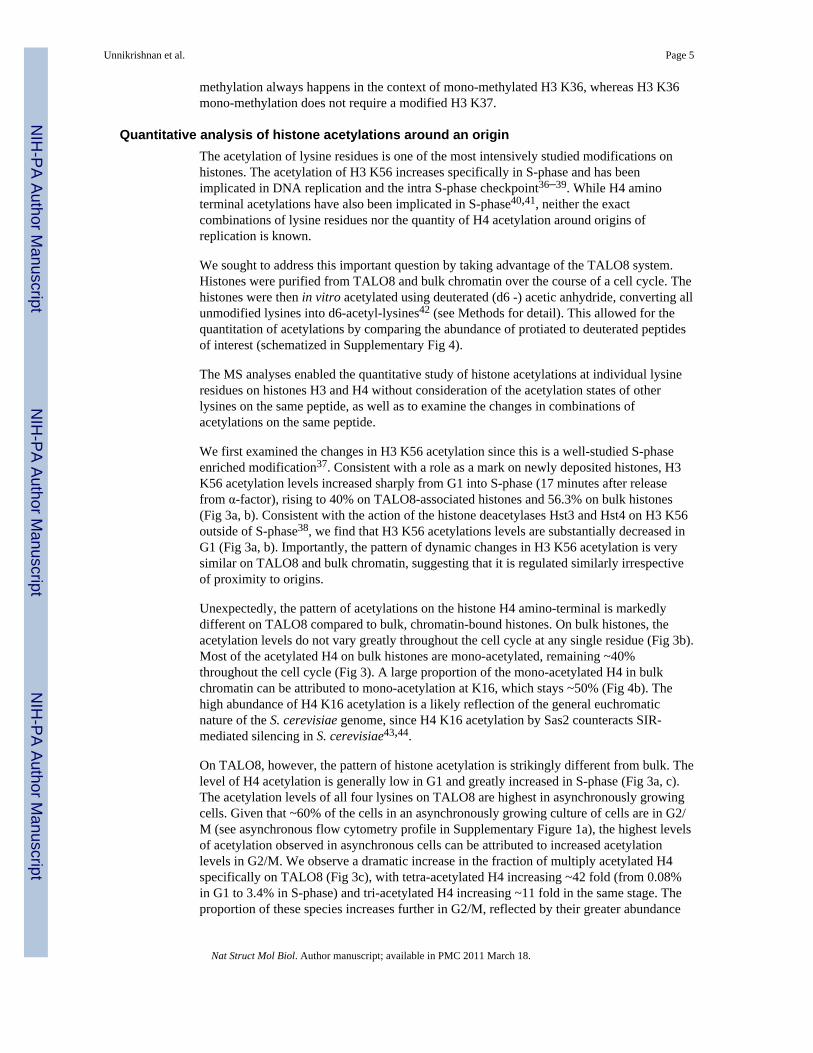
Quantitative analysis of histone acetylations around an originThe acetylation of lysine residues is one of the most intensively studied modifications onhistones. The acetylation of H3 K56 increases specifically in S-phase and has beenimplicated in DNA replication and the intra S-phase checkpoint36–39. While H4 aminoterminal acetylations have also been implicated in S-phase40,41, neither the exactcombinations of lysine residues nor the quantity of H4 acetylation around origins ofreplication is known.
We sought to address this important question by taking advantage of the TALO8 system.Histones were purified from TALO8 and bulk chromatin over the course of a cell cycle. Thehistones were then in vitro acetylated using deuterated (d6 -) acetic anhydride, converting allunmodified lysines into d6-acetyl-lysines42 (see Methods for detail). This allowed for thequantitation of acetylations by comparing the abundance of protiated to deuterated peptidesof interest (schematized in Supplementary Fig 4).
The MS analyses enabled the quantitative study of histone acetylations at individual lysineresidues on histones H3 and H4 without consideration of the acetylation states of otherlysines on the same peptide, as well as to examine the changes in combinations ofacetylations on the same peptide.
We first examined the changes in H3 K56 acetylation since this is a well-studied S-phaseenriched modification37. Consistent with a role as a mark on newly deposited histones, H3K56 acetylation levels increased sharply from G1 into S-phase (17 minutes after releasefrom α-factor), rising to 40% on TALO8-associated histones and 56.3% on bulk histones(Fig 3a, b). Consistent with the action of the histone deacetylases Hst3 and Hst4 on H3 K56outside of S-phase38, we find that H3 K56 acetylations levels are substantially decreased inG1 (Fig 3a, b). Importantly, the pattern of dynamic changes in H3 K56 acetylation is verysimilar on TALO8 and bulk chromatin, suggesting that it is regulated similarly irrespectiveof proximity to origins.
Unexpectedly, the pattern of acetylations on the histone H4 amino-terminal is markedlydifferent on TALO8 compared to bulk, chromatin-bound histones. On bulk histones, theacetylation levels do not vary greatly throughout the cell cycle at any single residue (Fig 3b).Most of the acetylated H4 on bulk histones are mono-acetylated, remaining ~40%throughout the cell cycle (Fig 3). A large proportion of the mono-acetylated H4 in bulkchromatin can be attributed to mono-acetylation at K16, which stays ~50% (Fig 4b). Thehigh abundance of H4 K16 acetylation is a likely reflection of the general euchromaticnature of the S. cerevisiae genome, since H4 K16 acetylation by Sas2 counteracts SIR-mediated silencing in S. cerevisiae43,44.
On TALO8, however, the pattern of histone acetylation is strikingly different from bulk. Thelevel of H4 acetylation is generally low in G1 and greatly increased in S-phase (Fig 3a, c).The acetylation levels of all four lysines on TALO8 are highest in asynchronously growingcells. Given that ~60% of the cells in an asynchronously growing culture of cells are in G2/M (see asynchronous flow cytometry profile in Supplementary Figure 1a), the highest levelsof acetylation observed in asynchronous cells can be attributed to increased acetylationlevels in G2/M. We observe a dramatic increase in the fraction of multiply acetylated H4specifically on TALO8 (Fig 3c), with tetra-acetylated H4 increasing ~42 fold (from 0.08%in G1 to 3.4% in S-phase) and tri-acetylated H4 increasing ~11 fold in the same stage. Theproportion of these species increases further in G2/M, reflected by their greater abundance
Unnikrishnan et al. Page 5
Nat Struct Mol Biol. Author manuscript; available in PMC 2011 March 18.
NIH
-PA Author Manuscript
NIH
-PA Author Manuscript
NIH
-PA Author Manuscript
in asynchronously growing cells. At the same time, the level of mono-acetylated H4 remainsrelatively constant throughout the cell cycle (Fig 3c). This therefore means that the largeincrease in H4 acetylation on TALO8 in S-phase and G2/M is primarily due to the increasein the fraction of H4 tail with multiple acetylations on the same peptide. At the same time,our data reveals a sharp wave of histone H4 deacetylation specifically around ARS1 (Fig 3a,c), as cells progress from G2/M into G1. Furthermore, given that the fraction of mono-acetylated H4 on TALO8 remains constant throughout the cell cycle, the main function ofthis wave of deacetylation is to decrease the amount of multiply acetylated histone H4specifically around the origin. The trends are similar for acetylations on the histone H3amino-terminal, although the changes are not as dramatic as H4.
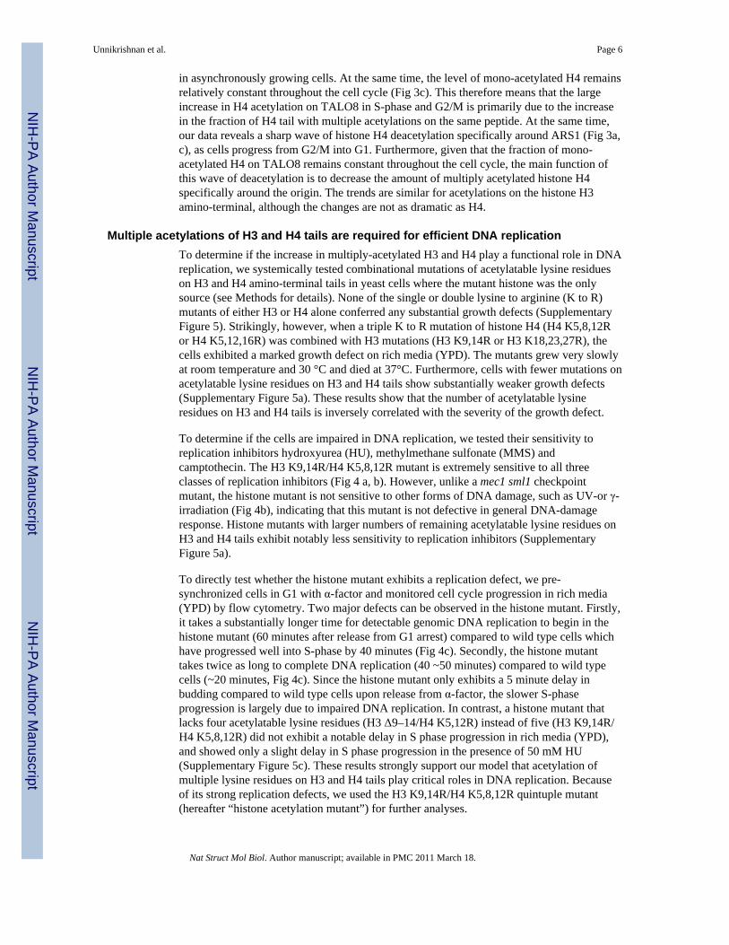
Multiple acetylations of H3 and H4 tails are required for efficient DNA replicationTo determine if the increase in multiply-acetylated H3 and H4 play a functional role in DNAreplication, we systemically tested combinational mutations of acetylatable lysine residueson H3 and H4 amino-terminal tails in yeast cells where the mutant histone was the onlysource (see Methods for details). None of the single or double lysine to arginine (K to R)mutants of either H3 or H4 alone conferred any substantial growth defects (SupplementaryFigure 5). Strikingly, however, when a triple K to R mutation of histone H4 (H4 K5,8,12Ror H4 K5,12,16R) was combined with H3 mutations (H3 K9,14R or H3 K18,23,27R), thecells exhibited a marked growth defect on rich media (YPD). The mutants grew very slowlyat room temperature and 30 °C and died at 37°C. Furthermore, cells with fewer mutations onacetylatable lysine residues on H3 and H4 tails show substantially weaker growth defects(Supplementary Figure 5a). These results show that the number of acetylatable lysineresidues on H3 and H4 tails is inversely correlated with the severity of the growth defect.
To determine if the cells are impaired in DNA replication, we tested their sensitivity toreplication inhibitors hydroxyurea (HU), methylmethane sulfonate (MMS) andcamptothecin. The H3 K9,14R/H4 K5,8,12R mutant is extremely sensitive to all threeclasses of replication inhibitors (Fig 4 a, b). However, unlike a mec1 sml1 checkpointmutant, the histone mutant is not sensitive to other forms of DNA damage, such as UV-or γ-irradiation (Fig 4b), indicating that this mutant is not defective in general DNA-damageresponse. Histone mutants with larger numbers of remaining acetylatable lysine residues onH3 and H4 tails exhibit notably less sensitivity to replication inhibitors (SupplementaryFigure 5a).
To directly test whether the histone mutant exhibits a replication defect, we pre-synchronized cells in G1 with α-factor and monitored cell cycle progression in rich media(YPD) by flow cytometry. Two major defects can be observed in the histone mutant. Firstly,it takes a substantially longer time for detectable genomic DNA replication to begin in thehistone mutant (60 minutes after release from G1 arrest) compared to wild type cells whichhave progressed well into S-phase by 40 minutes (Fig 4c). Secondly, the histone mutanttakes twice as long to complete DNA replication (40 ~50 minutes) compared to wild typecells (~20 minutes, Fig 4c). Since the histone mutant only exhibits a 5 minute delay inbudding compared to wild type cells upon release from α-factor, the slower S-phaseprogression is largely due to impaired DNA replication. In contrast, a histone mutant thatlacks four acetylatable lysine residues (H3 Δ9–14/H4 K5,12R) instead of five (H3 K9,14R/H4 K5,8,12R) did not exhibit a notable delay in S phase progression in rich media (YPD),and showed only a slight delay in S phase progression in the presence of 50 mM HU(Supplementary Figure 5c). These results strongly support our model that acetylation ofmultiple lysine residues on H3 and H4 tails play critical roles in DNA replication. Becauseof its strong replication defects, we used the H3 K9,14R/H4 K5,8,12R quintuple mutant(hereafter “histone acetylation mutant”) for further analyses.
Unnikrishnan et al. Page 6
Nat Struct Mol Biol. Author manuscript; available in PMC 2011 March 18.
NIH
-PA Author Manuscript
NIH
-PA Author Manuscript
NIH
-PA Author Manuscript
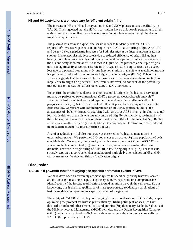
H3 and H4 acetylations are necessary for efficient origin firingThe increase in H3 and H4 tail acetylations in S and G2/M phases occurs specifically onTALO8. This suggested that the H3/H4 acetylations have a unique role pertaining to originactivity and that the replication defects observed in our histone mutant might be due toimpaired origin function.
The plasmid loss assay is a quick and sensitive means to identify defects in DNAreplication45. We tested plasmids harboring either ARS1 or a late-firing origin, ARS1413,and detected elevated plasmid loss rates for both plasmids in the histone mutant (data notshown). If elevated plasmid loss rate is due to reduced efficiency of origin firing, thenhaving multiple origins on a plasmid is expected to at least partially reduce the loss rate inthe histone acetylation mutant46. As shown in Figure 5a, the presence of multiple originsdoes not significantly affect the loss rate in wild type cells. In sharp contrast, an elevatedloss rate of a plasmid containing only one functional origin in the histone acetylation mutantis significantly reduced in the presence of eight functional origins (Fig 5a). This resultstrongly suggests that the elevated plasmid loss rates in the histone acetylation mutant arelargely due to origin firing defects. These results, however, do not exclude the possibilitythat H3 and H4 acetylation affects other steps in DNA replication.
To confirm the origin firing defects at chromosomal locations in the histone acetylationmutant, we performed two-dimensional (2-D) agarose gel electrophoresis analysis30.Because the histone mutant and wild type cells have dramatically different cell cycleprogression rates (Fig 4c), we first blocked cells in S-phase by releasing α-factor arrestedcells into HU. Consistent with our interpretation of the FACS profiles in Fig 4c, theappearance of “bubble” structures associated with an active ARS1 origin at its chromosomallocation is delayed in the histone mutant compared (Fig 5b). Furthermore, the intensity ofthe bubble arc is dramatically weaker than in wild type (~8-fold difference, Fig 5b). Bubblestructures at another early origin, ARS 607, at its chromosomal location were also reducedin the histone mutant (~5-fold difference, Fig 5c).
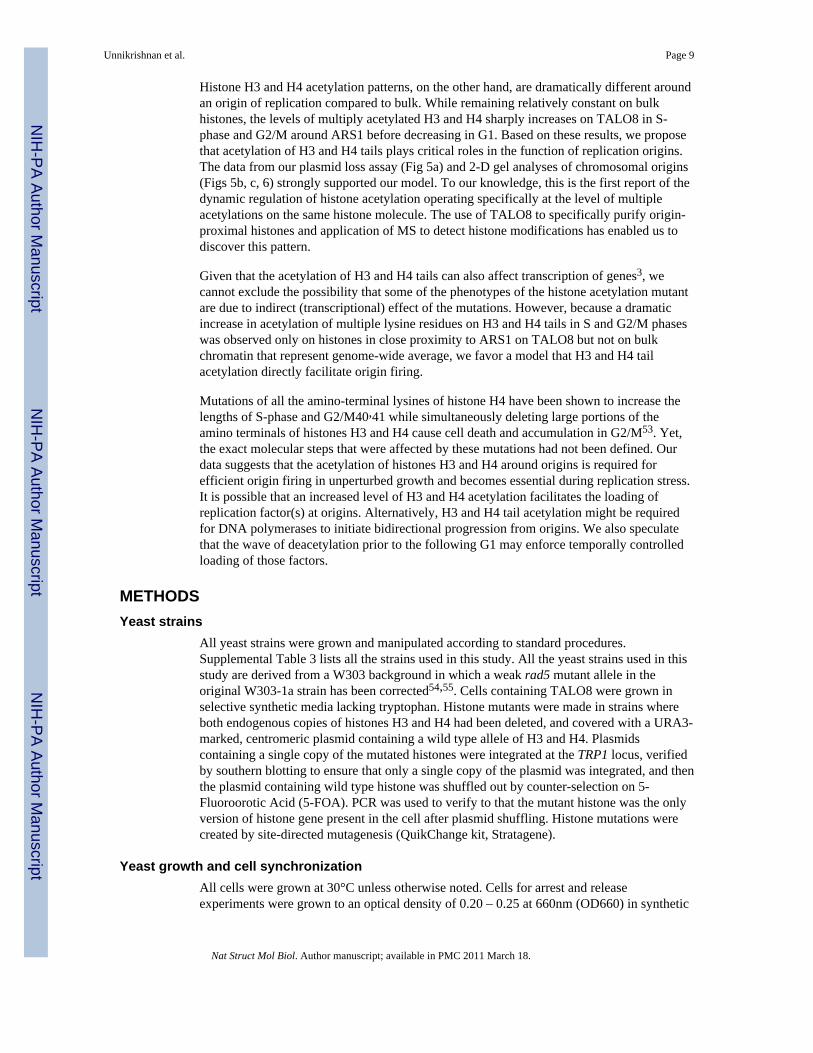
A similar reduction in bubble structures was observed in the histone mutant duringunperturbed growth. We performed 2-D gel analyses on pooled S-phase population of cells(see Methods). Once again, the intensity of bubble structures at ARS1 and ARS 607 areweaker in the histone mutant (Fig 6a). Furthermore, we observed similar, albeit lessdramatic, decrease in origin firing of ARS501, a late-firing origin (Fig 6b). These resultsstrongly support our conclusion that acetylation of multiple lysine residues on H3 and H4tails is necessary for efficient firing of replication origins.
DiscussionTALO8 is a powerful tool for studying site-specific chromatin events in vivo
We have developed an extremely efficient system to specifically purify histones locatedaround an origin in a single step. Using this system, we report the first comprehensiveidentification of the histone modifications around an origin through the cell cycle. To ourknowledge, this is the first application of mass spectrometry to identify combinations ofhistone modifications present in a specific region of the genome.
The utility of TALO8 extends beyond studying histone modifications. In this study, despiteoptimizing the protocol for histone purification by utilizing stringent washes, we havedetected a number of other chromatin-bound proteins (Supplementary Table 1). Subunits ofthe Minichomosome Maintenance (MCM) complex and the Origin Recognition Complex(ORC), which are involved in DNA replication were more abundant in S-phase cells onTALO8 (Supplementary Table 2).
Unnikrishnan et al. Page 7
Nat Struct Mol Biol. Author manuscript; available in PMC 2011 March 18.
NIH
-PA Author Manuscript
NIH
-PA Author Manuscript
NIH
-PA Author Manuscript

By optimizing the affinity-purification protocol, TALO8 or its derivatives can be used toselectively purify and study any DNA-dependent processes that take place on theminichromosome. For example, the scheme could be adapted to identify chromatin-bindingfactors around a region of double-strand break, and to determine the post-translationalmodifications of those proteins concurrently. Alternatively, as illustrated by Akiyoshi etal47, a centromere-containing TALO8 derivative can also be used to identify components ofthe kinetochore. Furthermore, the efficient purification of TALO8 and its associated proteinsmakes it an excellent substrate for in vitro biochemical assays. For example, TALO8 couldbe purified from mutants of transcription factors or chromatin modifying proteins to be usedas a template in transcription assays in vitro. Finally, the purity and yield of the TALO8 alsomakes it ideal for electron microscopic observations of specific chromatin structures, such asstalled or collapsed replication forks in TALO8-containing cells defective in the replicationcheckpoint, or to directly visualize defective nucleosome assembly by purifying TALO8from chromatin assembly mutants.
Apart from the dynamic pattern of H3 and H4 acetylations around an origin, we have alsodiscovered a few modifications previously uncharacterized in S. cerevisiae, including theH2A S15 phosphorylation (located within the amino-terminal tail) and H2B K111monomethylation (located on the surface of the nucleosome). We report the detection ofacetylation on H4 K79 (located on the surface of the nucleosome), previously reported incalf thymus histones21 and for which a role in telomeric and ribosomal DNA silencing hadbeen suggested from genetic analyses48. Furthermore, we detect a unique pattern of H2BK123 mono-ubiquitination on TALO8, which is implicated in nucleosome dynamics49,50.Another interesting modification we have detected is H3 K37 mono-methylation (located onthe amino-terminal tail), specifically on TALO8, which is absent during S-phase(Supplementary Figure 1c). Mono-methylated H3 K36, along with histone acetylation,facilitates the loading of Cdc45 onto early origins51. It is therefore possible that mono-methylation of an adjacent residue H3 K37, specifically around an origin and outside of S-phase, might serve to prevent improper recruitment of Cdc45.
The binding of the tetracyclin repressor on a 10kb tandem array of operator sequence on aplasmid can stall replication forks in E. coli52 and we cannot exclude the possibility that thebinding of LacI onto the eight copies of LacO on TALO8 in vivo might also affect itsreplication. Similarly, although we prepared whole cell extracts to isolate TALO8 and bulkchromatin from the same stages of the cell cycle in identical fashion, we cannot exclude thepossibility that the difference in the histone isolation methods made some contributions tothe differences in histone modifications found on TALO8 and bulk chromatin. However, wedo believe these factors have no major effects on histone modifications, as H3 K56acetylation dynamics on TALO8 and in bulk chromatin were identical and exactly asexpected from the nature of this modification (Fig 3), while acetylation on multiple lysineresidues on H3 and H4 tails that are detected on TALO8 was shown to play critical roles infunctions of chromosomal origins. In addition, we did not detect phosphorylation of H2Aserine 129, which takes place at the sites of DNA damage, on TALO8.
Acetylated histones H3 and H4 regulate origin activityBy utilizing the TALO8 system, we have identified a unique pattern of acetylationsspecifically on histones flanking an origin of DNA replication. Our analyses have revealedthat the two classes of histone acetylations implicated in DNA replication, one on H3 K56and the other on the amino terminals of histones H3 and H4, are dynamically regulated inmarkedly distinct ways throughout the cell cycle. H3 K56 acetylation patterns are similar onTALO8 and bulk histones, increasing from low levels in G1 to high levels in S-phase beforedropping again, as anticipated for a mark of histone deposition occurring behind areplication fork.
Unnikrishnan et al. Page 8
Nat Struct Mol Biol. Author manuscript; available in PMC 2011 March 18.
NIH
-PA Author Manuscript
NIH
-PA Author Manuscript
NIH
-PA Author Manuscript
Histone H3 and H4 acetylation patterns, on the other hand, are dramatically different aroundan origin of replication compared to bulk. While remaining relatively constant on bulkhistones, the levels of multiply acetylated H3 and H4 sharply increases on TALO8 in S-phase and G2/M around ARS1 before decreasing in G1. Based on these results, we proposethat acetylation of H3 and H4 tails plays critical roles in the function of replication origins.The data from our plasmid loss assay (Fig 5a) and 2-D gel analyses of chromosomal origins(Figs 5b, c, 6) strongly supported our model. To our knowledge, this is the first report of thedynamic regulation of histone acetylation operating specifically at the level of multipleacetylations on the same histone molecule. The use of TALO8 to specifically purify origin-proximal histones and application of MS to detect histone modifications has enabled us todiscover this pattern.
Given that the acetylation of H3 and H4 tails can also affect transcription of genes3, wecannot exclude the possibility that some of the phenotypes of the histone acetylation mutantare due to indirect (transcriptional) effect of the mutations. However, because a dramaticincrease in acetylation of multiple lysine residues on H3 and H4 tails in S and G2/M phaseswas observed only on histones in close proximity to ARS1 on TALO8 but not on bulkchromatin that represent genome-wide average, we favor a model that H3 and H4 tailacetylation directly facilitate origin firing.
Mutations of all the amino-terminal lysines of histone H4 have been shown to increase thelengths of S-phase and G2/M40,41 while simultaneously deleting large portions of theamino terminals of histones H3 and H4 cause cell death and accumulation in G2/M53. Yet,the exact molecular steps that were affected by these mutations had not been defined. Ourdata suggests that the acetylation of histones H3 and H4 around origins is required forefficient origin firing in unperturbed growth and becomes essential during replication stress.It is possible that an increased level of H3 and H4 acetylation facilitates the loading ofreplication factor(s) at origins. Alternatively, H3 and H4 tail acetylation might be requiredfor DNA polymerases to initiate bidirectional progression from origins. We also speculatethat the wave of deacetylation prior to the following G1 may enforce temporally controlledloading of those factors.
METHODSYeast strains
All yeast strains were grown and manipulated according to standard procedures.Supplemental Table 3 lists all the strains used in this study. All the yeast strains used in thisstudy are derived from a W303 background in which a weak rad5 mutant allele in theoriginal W303-1a strain has been corrected54,55. Cells containing TALO8 were grown inselective synthetic media lacking tryptophan. Histone mutants were made in strains whereboth endogenous copies of histones H3 and H4 had been deleted, and covered with a URA3-marked, centromeric plasmid containing a wild type allele of H3 and H4. Plasmidscontaining a single copy of the mutated histones were integrated at the TRP1 locus, verifiedby southern blotting to ensure that only a single copy of the plasmid was integrated, and thenthe plasmid containing wild type histone was shuffled out by counter-selection on 5-Fluoroorotic Acid (5-FOA). PCR was used to verify to that the mutant histone was the onlyversion of histone gene present in the cell after plasmid shuffling. Histone mutations werecreated by site-directed mutagenesis (QuikChange kit, Stratagene).
Yeast growth and cell synchronizationAll cells were grown at 30°C unless otherwise noted. Cells for arrest and releaseexperiments were grown to an optical density of 0.20 – 0.25 at 660nm (OD660) in synthetic
Unnikrishnan et al. Page 9
Nat Struct Mol Biol. Author manuscript; available in PMC 2011 March 18.
NIH
-PA Author Manuscript
NIH
-PA Author Manuscript
NIH
-PA Author Manuscript
media lacking tryptophan (YC - Trp) and arrested in G1-phase with 5 μg ml−1 α-factor for90 minutes. Cells were released from G1-arrest by filtration on 0.45 μm nitrocellulosemembranes (Millipore), washed and resuspended in fresh, pre-warmed media lacking α-factor. Cell synchrony was followed by flow cytometry56. Five-fold serial dilutions wereperformed on YPD agar plates (with or without appropriate drugs) and cells were grown for3 days. Plates containing drugs were used within 24 hours of preparation.
Affinity-purification of TALO8TALO8-containing cells were quickly killed at a specific cell cycle stage by vigorouslymixing cultures with a frozen solution of 0.1M EDTA, pH 8.0 (to a final concentration of17mM) and 0.1% Sodium Azide (Sigma). Cells were then spun down, washed once intwenty-fold cell pellet volume of water containing 2mM phenylmethanesulfonyl fluoride(PMSF) and then once in ten-fold cell pellet volume of Buffer H 150 [BH 150: 25mMHEPES-KOH pH 7.6, 2mM MgCl2, 0.5mM EGTA, 0.1mM EDTA, 10% (v/v) glycerol,150mM KCl, 0.02% (v/v) NP40] containing protease inhibitors [1mM PMSF, 2μMpepstatin, 600nM leupeptin, 2mM benzamidine, 2μg ml−1 chymostatin A (Sigma) ],phosphatase inhibitors [2mM imidazole, 1mM sodium fluoride, 1.15mM sodium molybdate,1mM sodium orthovanadate, 4mM sodium tartarate dihydrate, 2.5μM (−)-p-bromotetramisole oxalate, 0.5μM cantharidin, 0.5nM microcystin, (Sigma)] and histonedeacetylase inhibitors [0.5μM Trichostatin A (Sigma), 25μM Sirtinol (Calbiochem) ]. Allthe buffers in the following steps were kept on ice and supplemented with the inhibitorsdescribed above, unless otherwise specified. Whole cell extracts were made by bead beatingcells resuspended in an equal volume of BH 150. The whole cell extracts were thencentrifuged in a Beckman SW40Ti rotor at 27,000 rpm for 90 minutes at 4°C. The solubleextract was isolated and incubated with magnetic beads (Protein G Dynal beads, Invitrogen)crosslinked with anti-FLAG M2 antibody (Sigma) for 3 hours at 4°C. Typically, per ~ 4×10⁹
cell equivalents of extract, 25 μl of Protein G beads slurry and 11.5 μg of anti-FLAG M2antibodies were used. After the incubation, the magnetic beads were rinsed three times withBH 150 and then washed four times, 5 minutes per wash, in BH 300 (which was identical toBH 150, except containing 300mM KCl). The beads were then rinsed three times in RinseBuffer [25mM HEPES KOH pH 7.6, 2mM MgCl2, 10% (v/v) glycerol, 150mM KCl, noinhibitors] and then four sequential elutions were performed, for 30 minutes each, at roomtemperature with Elution Buffer [50mM ammonium bicarbonate, 0.1% (w/v) Rapigest(Waters), no inhibitors]. The eluates were pooled, digested overnight with 100 ng ofendoproteinase Arginine-C (Roche) at 37°C, trifluoroacetic acid was added to a finalconcentration 0.5% and incubated at 37°C for 1 hour to degrade Rapigest. The resultingsolution was dried in a SpeedVac, resuspended in 0.1% (v/v) formic acid and loaded ontothe mass spectrometer.
To prepare bulk, chromatin-bound histones, 2 ~ 4×10⁸ cells (at OD660 = 0.2–0.25) wereharvested from different cell cycle stages as described above. The cells were washed asabove and lysed by bead beating in BH 150 supplemented with inhibitors, as describedabove. Soluble chromatin was prepared by sonication and incubated with Source 15Q (GEHealthcare) anion exchange beads for 20 minutes at 4°C. The beads were then washed twicewith BH 250 (250mM KCl) and twice with BH 500 (500mM KCl), for 5 minutes per washat 4°C. Core histones were then eluted in 2M KCl, digested overnight with 100 ng of Arg-Cat 37°C, desalted using ZipTip C18 columns (Millipore) and analyzed on the massspectrometer. All the buffers were kept chilled on ice and supplemented with inhibitors,unless otherwise specified.
Unnikrishnan et al. Page 10
Nat Struct Mol Biol. Author manuscript; available in PMC 2011 March 18.
NIH
-PA Author Manuscript
NIH
-PA Author Manuscript
NIH
-PA Author Manuscript
Protein and Modification IdentificationAll the data were gathered by using liquid chromatography coupled in-line with electrosprayionization tandem mass spectrometry (LC-ESI MS/MS). This setup consisted of an EksigentTechnologies 2D nanoLC coupled to 75 μm × 20 cm analytical column made from a NewObjective PicoFrit packed with 5 μm, 100 Å particle MAGIC C18AQ packing material anddirectly spraying into a Thermo-Scientific LTQ Orbitrap XL hybrid mass spectrometer viathe mass spectrometer's nanospray ionization source. Chromatographic elution wasperformed at 500 nL/min using a 60 minute gradient from 2% solvent B to 50% solvent B,where solvent A was 0.1% (v/v) formic acid in water and solvent B was 0.1% (v/v) formicacid in acetonitrile, and a spray voltage of 2800 volts was applied to the electrospray tip.The mass spectrometer was operated in the “shotgun” mode for MS/MS acquisitions. Here,an MS survey scan was performed in the OrbiTrap portion of the instrument (AGC targetvalue 1e6, resolution 60K, and injection time 150 ms) and tandem MS (MS/MS)fragmentation scans on the top five abundant ions from the survey scan were performed inthe ion trap portion of the instrument (normalized collision energy of 35%; isolation widthof 2 m/z; target value of 1e4; injection time of 100 ms). Selected ions were dynamicallyexcluded for 45 seconds with a repeat count of 1. The precursor ion selection for MS/MSwas set to +/− 0.5 Da (parent mass width) and charge state screening was enabled allowingonly +2 and +3 charged peptides to be selected for MS/MS.
Protein and modification identification were performed by analyzing mass spectrometry datawith the protein database search algorithm X!Tandem used in the refinement mode. Thefollowing variable mass modifications were used: 14.015 Da and 28.031 Da for mono- anddimethylation on lysine respectively, 42.047 Da for trimethylation on lysine, 42.011 Da foracetylation on lysine, 114.043 for ubiquitination on lysine, 15.995 for oxidation onmethionine, and 79.966 for phosphorylation on serine. The Saccharomyces GenomeDatabase was used as the reference protein database with the addition of human keratins andLac repressor protein sequences to account for potential contaminating proteins. The scorefunction of native X!Tandem was replaced with a dot-product based scoring algorithm thatis compatible with Peptide Prophet57. Peptide identifications results were filtered and sortedin CPAS58 using parameters that result in a false discovery rate of 5% or less, before beingmanually verified. Detection of all the modifications, including quantitative analysis ofacetylation, is based on 20–50 peptides per modification. The MS-MS data of relevantmodified histone peptides are shown in Supplemental Figure 2. The MS-MS data for othermodified histone peptides are available upon request.
Acetylation QuantitationFor the quantitation of acetylation by mass spectrometry, TALO8 (or bulk histones) waseluted as per normal and the histones were precipitated using 20% (w/v) Trichloroacetic acid(Sigma). The dried eluates were then acetylated in vitro, digested overnight with Arg-C at37°C and the data analyzed as described in (Smith et al., 2003), but with the followingmodification: the in vitro acetylation was performed in a solution of 5% (v/v) triethylamine,5% (v/v) d6-acetic anhydride and acetonitrile for 1 hour at room temperature. Massspectrometry was performed in an identical fashion as described above for modificationidentification, except that the dynamic exclusion function was not utilized. The precursorion selection for MS/MS was set to +/− 4.5 Da, and an inclusion list of ions selected for MS/MS quantitation were 494.77, 530.31, 639.62 and 722.405. We performed the analysis induplicate, and the results were essentially identical.
Two-dimensional gel electrophoresis and hybridizationCells were arrested with EDTA and sodium azide, as previously described for TALO8. Totalgenomic DNA was prepared according to the “CTAB extraction” procedure59,60. To prepare
Unnikrishnan et al. Page 11
Nat Struct Mol Biol. Author manuscript; available in PMC 2011 March 18.
NIH
-PA Author Manuscript
NIH
-PA Author Manuscript
NIH
-PA Author Manuscript
pooled fraction of S-phase cells, equal volumes of cells were harvested at designatedintervals that spanned the entirety of S-phase for both wild type and histone mutant, arrestedwith EDTA and sodium azide, pooled and total genomic DNA was harvested. 5μg of DNAwas digested with either NcoI (ARS1), BspHI and ClaI (ARS607) or XbaI (ARS501) and 2-D gels were performed as originally described30. The DNA was blotted onto NylonGeneScreen Plus membranes (PerkinElmer) and separately probed with 32P-labelled DNAprobes spanning each respective ARS. Probes were amplified from yeast genomic DNA byPCR and gel purified (sequences available upon request). Signals were quantified on aPhosphorimager (GE Healthcare) and ImageQuant, as described64. The values are reportedas a percentage of the intensity of the 1N linear spot. All enzymes used in this paper werepurchased from New England Bio labs.
Supplementary MaterialRefer to Web version on PubMed Central for supplementary material.
AcknowledgmentsWe are especially grateful to Sue Biggins, Bungo Akiyoshi and Noelle Ebel for technical help and advice indeveloping the TALO8 purification scheme. We thank Lisa Nader Jones and Jason Hogan of the ProteomicsFacility at the FHCRC for help with MS, and Jimmy Eng and Brendan MacLean for advice with using X!Tandem.We thank Tracey Kwong (FHCRC), Doug Koshland (Carnegie Institute), Bonita Brewer and M. K. Raghuraman(University of Washington) for the plasmids used in the plasmid loss assay. We also thank members of theTsukiyama lab, Susan Parkhurst, Sue Biggins, Bungo Akiyoshi and Christian Nelson for helpful comments with themanuscript. This work is supported in part by NIH grant R01 GM078259 to TT. The FHCRC Proteomics Facility issupported by cancer center support grant P30 CA15704.
References1. Luger K, Mader AW, Richmond RK, Sargent DF, Richmond TJ. Crystal structure of the
nucleosome core particle at 2.8 A resolution. Nature. 1997; 389:251–60. see comments. [PubMed:9305837]
2. Groth A, Rocha W, Verreault A, Almouzni G. Chromatin challenges during DNA replication andrepair. Cell. 2007; 128:721–33. [PubMed: 17320509]
3. Li B, Carey M, Workman JL. The role of chromatin during transcription. Cell. 2007; 128:707–19.[PubMed: 17320508]
4. Kouzarides T. Chromatin modifications and their function. Cell. 2007; 128:693–705. [PubMed:17320507]
5. Stinchcomb DT, Struhl K, Davis RW. Isolation and characterisation of a yeast chromosomalreplicator. Nature. 1979; 282:39–43. [PubMed: 388229]
6. Bell SP, Dutta A. DNA replication in eukaryotic cells. Annu Rev Biochem. 2002; 71:333–74.[PubMed: 12045100]
7. Brown JA, Holmes SG, Smith MM. The chromatin structure of Saccharomyces cerevisiaeautonomously replicating sequences changes during the cell division cycle. Mol Cell Biol. 1991;11:5301–11. [PubMed: 1922046]
8. Simpson RT. Nucleosome positioning can affect the function of a cis-acting DNA element in vivo.Nature. 1990; 343:387–9. [PubMed: 2405281]
9. Ferguson BM, Fangman WL. A position effect on the time of replication origin activation in yeast.Cell. 1992; 68:333–9. [PubMed: 1733502]
10. Stevenson JB, Gottschling DE. Telomeric chromatin modulates replication timing nearchromosome ends. Genes Dev. 1999; 13:146–51. [PubMed: 9925638]
11. Aparicio JG, Viggiani CJ, Gibson DG, Aparicio OM. The Rpd3-Sin3 histone deacetylase regulatesreplication timing and enables intra-S origin control in Saccharomyces cerevisiae. Mol Cell Biol.2004; 24:4769–80. [PubMed: 15143171]
Unnikrishnan et al. Page 12
Nat Struct Mol Biol. Author manuscript; available in PMC 2011 March 18.
NIH
-PA Author Manuscript
NIH
-PA Author Manuscript
NIH
-PA Author Manuscript
12. Vogelauer M, Rubbi L, Lucas I, Brewer BJ, Grunstein M. Histone acetylation regulates the time ofreplication origin firing. Mol Cell. 2002; 10:1223–33. [PubMed: 12453428]
13. Iizuka M, Stillman B. Histone acetyltransferase HBO1 interacts with the ORC1 subunit of thehuman initiator protein. J Biol Chem. 1999; 274:23027–34. [PubMed: 10438470]
14. Burke TW, Cook JG, Asano M, Nevins JR. Replication factors MCM2 and ORC1 interact with thehistone acetyltransferase HBO1. J Biol Chem. 2001; 276:15397–408. [PubMed: 11278932]
15. Pasero P, Bensimon A, Schwob E. Single-molecule analysis reveals clustering and epigeneticregulation of replication origins at the yeast rDNA locus. Genes Dev. 2002; 16:2479–84.[PubMed: 12368258]
16. Pappas DL Jr. Frisch R, Weinreich M. The NAD(+)-dependent Sir2p histone deacetylase is anegative regulator of chromosomal DNA replication. Genes Dev. 2004; 18:769–81. [PubMed:15082529]
17. Raghuraman MK, et al. Replication dynamics of the yeast genome. Science. 2001; 294:115–21.[PubMed: 11588253]
18. Wyrick JJ, et al. Genome-wide distribution of ORC and MCM proteins in S. cerevisiae: high-resolution mapping of replication origins. Science. 2001; 294:2357–60. [PubMed: 11743203]
19. Cheung P, et al. Synergistic coupling of histone H3 phosphorylation and acetylation in response toepidermal growth factor stimulation. Mol Cell. 2000; 5:905–15. [PubMed: 10911985]
20. Strahl BD, et al. Methylation of histone H4 at arginine 3 occurs in vivo and is mediated by thenuclear receptor coactivator PRMT1. Curr Biol. 2001; 11:996–1000. [PubMed: 11448779]
21. Zhang L, Eugeni EE, Parthun MR, Freitas MA. Identification of novel histone post-translationalmodifications by peptide mass fingerprinting. Chromosoma. 2003; 112:77–86. [PubMed:12937907]
22. Ye J, et al. Histone H4 lysine 91 acetylation a core domain modification associated with chromatinassembly. Mol Cell. 2005; 18:123–30. [PubMed: 15808514]
23. Taverna SD, et al. Long-distance combinatorial linkage between methylation and acetylation onhistone H3 N termini. Proc Natl Acad Sci U S A. 2007; 104:2086–91. [PubMed: 17284592]
24. Zakian VA, Scott JF. Construction, replication, and chromatin structure of TRP1 RI circle, amultiple-copy synthetic plasmid derived from Saccharomyces cerevisiae chromosomal DNA. MolCell Biol. 1982; 2:221–32. [PubMed: 6287231]
25. Thoma F, Bergman LW, Simpson RT. Nuclease digestion of circular TRP1ARS1 chromatinreveals positioned nucleosomes separated by nuclease-sensitive regions. J Mol Biol. 1984;177:715–33. [PubMed: 6384525]
26. Ducker CE, Simpson RT. The organized chromatin domain of the repressed yeast a cell-specificgene STE6 contains two molecules of the corepressor Tup1p per nucleosome. Embo J. 2000;19:400–9. [PubMed: 10654939]
27. Dean A, Pederson DS, Simpson RT. Isolation of yeast plasmid chromatin. Methods Enzymol.1989; 170:26–41. [PubMed: 2671602]
28. Ivanov D, Nasmyth K. A topological interaction between cohesin rings and a circularminichromosome. Cell. 2005; 122:849–60. [PubMed: 16179255]
29. Craig R, Beavis RC. TANDEM: matching proteins with tandem mass spectra. Bioinformatics.2004; 20:1466–7. [PubMed: 14976030]
30. Brewer BJ, Fangman WL. The localization of replication origins on ARS plasmids in S. cerevisiae.Cell. 1987; 51:463–71. [PubMed: 2822257]
31. Garcia BA, et al. Organismal differences in post-translational modifications in histones H3 and H4.J Biol Chem. 2007; 282:7641–55. [PubMed: 17194708]
32. Kristjuhan A, et al. Transcriptional inhibition of genes with severe histone h3 hypoacetylation inthe coding region. Mol Cell. 2002; 10:925–33. [PubMed: 12419235]
33. Strahl BD, et al. Set2 is a nucleosomal histone H3-selective methyltransferase that mediatestranscriptional repression. Mol Cell Biol. 2002; 22:1298–306. [PubMed: 11839797]
34. Pokholok DK, et al. Genome-wide map of nucleosome acetylation and methylation in yeast. Cell.2005; 122:517–27. [PubMed: 16122420]
Unnikrishnan et al. Page 13
Nat Struct Mol Biol. Author manuscript; available in PMC 2011 March 18.
NIH
-PA Author Manuscript
NIH
-PA Author Manuscript
NIH
-PA Author Manuscript
35. Carrozza MJ, et al. Histone H3 methylation by Set2 directs deacetylation of coding regions byRpd3S to suppress spurious intragenic transcription. Cell. 2005; 123:581–92. [PubMed:16286007]
36. Masumoto H, Hawke D, Kobayashi R, Verreault A. A role for cell-cycle-regulated histone H3lysine 56 acetylation in the DNA damage response. Nature. 2005; 436:294–8. [PubMed:16015338]
37. Li Q, et al. Acetylation of histone H3 lysine 56 regulates replication-coupled nucleosomeassembly. Cell. 2008; 134:244–55. [PubMed: 18662540]
38. Maas NL, Miller KM, DeFazio LG, Toczyski DP. Cell cycle and checkpoint regulation of histoneH3 K56 acetylation by Hst3 and Hst4. Mol Cell. 2006; 23:109–19. [PubMed: 16818235]
39. Kaplan T, et al. Cell cycle- and chaperone-mediated regulation of H3K56ac incorporation in yeast.PLoS Genet. 2008; 4:e1000270. [PubMed: 19023413]
40. Megee PC, Morgan BA, Smith MM. Histone H4 and the maintenance of genome integrity. GenesDev. 1995; 9:1716–27. [PubMed: 7622036]
41. Megee PC, Morgan BA, Mittman BA, Smith MM. Genetic analysis of histone H4: essential role oflysines subject to reversible acetylation. Science. 1990; 247:841–5. [PubMed: 2106160]
42. Smith CM, et al. Mass spectrometric quantification of acetylation at specific lysines within theamino-terminal tail of histone H4. Anal Biochem. 2003; 316:23–33. [PubMed: 12694723]
43. Suka N, Luo K, Grunstein M. Sir2p and Sas2p opposingly regulate acetylation of yeast histone H4lysine16 and spreading of heterochromatin. Nat Genet. 2002; 32:378–83. [PubMed: 12379856]
44. Kimura A, Umehara T, Horikoshi M. Chromosomal gradient of histone acetylation established bySas2p and Sir2p functions as a shield against gene silencing. Nat Genet. 2002; 32:370–7.[PubMed: 12410229]
45. Tye BK. Minichromosome maintenance as a genetic assay for defects in DNA replication.Methods. 1999; 18:329–34. [PubMed: 10454994]
46. Hogan E, Koshland D. Addition of extra origins of replication to a minichromosome suppresses itsmitotic loss in cdc6 and cdc14 mutants of Saccharomyces cerevisiae. Proc Natl Acad Sci U S A.1992; 89:3098–102. [PubMed: 1557417]
47. Akiyoshi B, Nelson CR, Ranish JA, Biggins S. Quantitative proteomic analysis of purified yeastkinetochores identifies a PP1 regulatory subunit. Genes Dev. 2009
48. Hyland EM, et al. Insights into the role of histone H3 and histone H4 core modifiable residues inSaccharomyces cerevisiae. Mol Cell Biol. 2005; 25:10060–70. [PubMed: 16260619]
49. Chandrasekharan MB, Huang F, Sun ZW. Ubiquitination of histone H2B regulates chromatindynamics by enhancing nucleosome stability. Proc Natl Acad Sci U S A. 2009; 106:16686–91.[PubMed: 19805358]
50. Fleming AB, Kao CF, Hillyer C, Pikaart M, Osley MA. H2B ubiquitylation plays a role innucleosome dynamics during transcription elongation. Mol Cell. 2008; 31:57–66. [PubMed:18614047]
51. Pryde F, et al. H3 k36 methylation helps determine the timing of cdc45 association with replicationorigins. PLoS One. 2009; 4:e5882. [PubMed: 19521516]
52. Possoz C, Filipe SR, Grainge I, Sherratt DJ. Tracking of controlled Escherichia coli replicationfork stalling and restart at repressor-bound DNA in vivo. Embo J. 2006; 25:2596–604. [PubMed:16724111]
53. Ling X, Harkness TA, Schultz MC, Fisher-Adams G, Grunstein M. Yeast histone H3 and H4amino termini are important for nucleosome assembly in vivo and in vitro: redundant and position-independent functions in assembly but not in gene regulation. Genes Dev. 1996; 10:686–99.[PubMed: 8598296]
54. Thomas BJ, Rothstein R. The genetic control of direct-repeat recombination in Saccharomyces: theeffect of rad52 and rad1 on mitotic recombination at GAL10, a transcriptionally regulated gene.Genetics. 1989; 123:725–38. [PubMed: 2693208]
55. Zhao X, Muller EG, Rothstein R. A suppressor of two essential checkpoint genes identifies a novelprotein that negatively affects dNTP pools. Mol Cell. 1998; 2:329–40. [PubMed: 9774971]
56. Vincent JA, Kwong TJ, Tsukiyama T. ATP-dependent chromatin remodeling shapes the DNAreplication landscape. Nat Struct Mol Biol. 2008; 15:477–84. [PubMed: 18408730]
Unnikrishnan et al. Page 14
Nat Struct Mol Biol. Author manuscript; available in PMC 2011 March 18.
NIH
-PA Author Manuscript
NIH
-PA Author Manuscript
NIH
-PA Author Manuscript
57. Keller A, Nesvizhskii AI, Kolker E, Aebersold R. Empirical statistical model to estimate theaccuracy of peptide identifications made by MS/MS and database search. Anal Chem. 2002;74:5383–92. [PubMed: 12403597]
58. Rauch A, et al. Computational Proteomics Analysis System (CPAS): an extensible, open-sourceanalytic system for evaluating and publishing proteomic data and high throughput biologicalexperiments. J Proteome Res. 2006; 5:112–21. [PubMed: 16396501]
59. Liberi G, et al. Methods to study replication fork collapse in budding yeast. Methods Enzymol.2006; 409:442–62. [PubMed: 16793417]
60. Lopes M, Cotta-Ramusino C, Liberi G, Foiani M. Branch migrating sister chromatid junctionsform at replication origins through Rad51/Rad52-independent mechanisms. Mol Cell. 2003;12:1499–510. [PubMed: 14690603]
Unnikrishnan et al. Page 15
Nat Struct Mol Biol. Author manuscript; available in PMC 2011 March 18.
NIH
-PA Author Manuscript
NIH
-PA Author Manuscript
NIH
-PA Author Manuscript
Figure 1. The TRP1-ARS1-8xLacO (TALO8) minichromosome system(a) TALO8 is a ~1.8 kb minichromosome with an ARS1 origin of replication, seven well-mapped nucleosomes25 (depicted as blue circles), a tandem array of eight lac operatorsequences (8× Lac O) and a TRP1 selectable marker. (b) A schematic overview of theaffinity-purification of TALO8. (c) Representative agarose gel (ethidium bromide-stained)of purified TALO8 DNA linearized by NheI (left) and silver-stained polyacrylamide gel ofthe purified proteins associated with TALO8 (right). The amount of samples loaded on eachelution lane corresponds to 1.5 × 10⁵ and 7.5 × 10⁴ cell equivalents for the agarose andpolyacrylamide gels respectively. Typically, eluates 1 and 2 were pooled and prepared formass spectrometry.
Unnikrishnan et al. Page 16
Nat Struct Mol Biol. Author manuscript; available in PMC 2011 March 18.
NIH
-PA Author Manuscript
NIH
-PA Author Manuscript
NIH
-PA Author Manuscript
Figure 2. Identification of combinations of modifications occuring on the same peptide inTALO8 histonesCombinations of histone modifications detected on the same peptide from histones H3 andH4 on TALO8 are shown on the same horizontal row. For lysine methylations, the numberof squares indicates the level of methylation.
Unnikrishnan et al. Page 17
Nat Struct Mol Biol. Author manuscript; available in PMC 2011 March 18.
NIH
-PA Author Manuscript
NIH
-PA Author Manuscript
NIH
-PA Author Manuscript
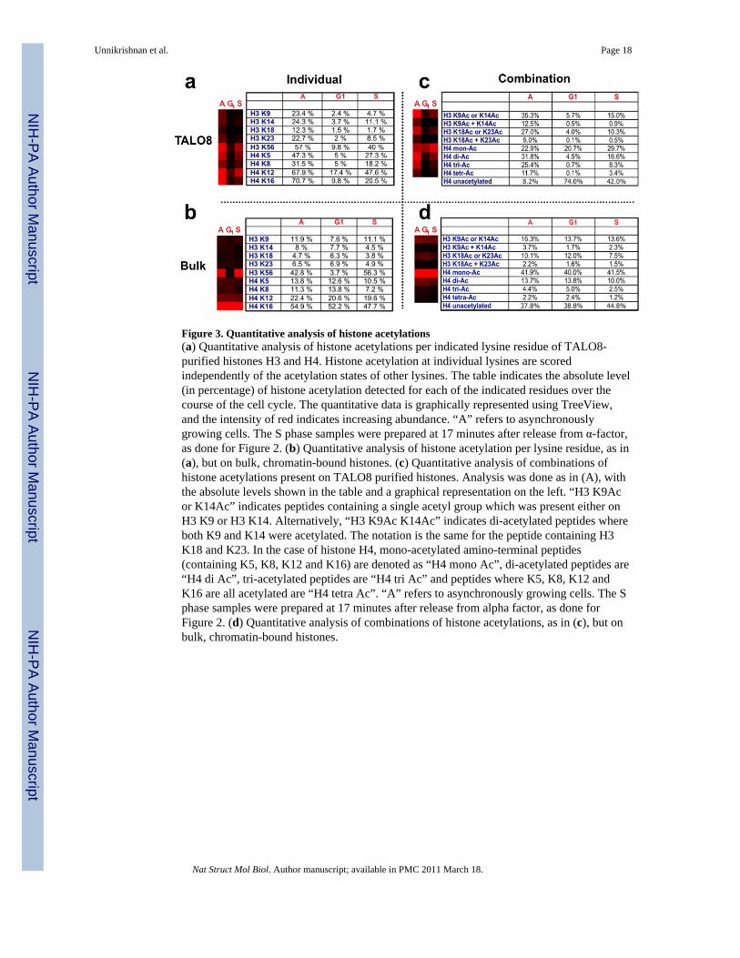
Figure 3. Quantitative analysis of histone acetylations(a) Quantitative analysis of histone acetylations per indicated lysine residue of TALO8-purified histones H3 and H4. Histone acetylation at individual lysines are scoredindependently of the acetylation states of other lysines. The table indicates the absolute level(in percentage) of histone acetylation detected for each of the indicated residues over thecourse of the cell cycle. The quantitative data is graphically represented using TreeView,and the intensity of red indicates increasing abundance. “A” refers to asynchronouslygrowing cells. The S phase samples were prepared at 17 minutes after release from α-factor,as done for Figure 2. (b) Quantitative analysis of histone acetylation per lysine residue, as in(a), but on bulk, chromatin-bound histones. (c) Quantitative analysis of combinations ofhistone acetylations present on TALO8 purified histones. Analysis was done as in (A), withthe absolute levels shown in the table and a graphical representation on the left. “H3 K9Acor K14Ac” indicates peptides containing a single acetyl group which was present either onH3 K9 or H3 K14. Alternatively, “H3 K9Ac K14Ac” indicates di-acetylated peptides whereboth K9 and K14 were acetylated. The notation is the same for the peptide containing H3K18 and K23. In the case of histone H4, mono-acetylated amino-terminal peptides(containing K5, K8, K12 and K16) are denoted as “H4 mono Ac”, di-acetylated peptides are“H4 di Ac”, tri-acetylated peptides are “H4 tri Ac” and peptides where K5, K8, K12 andK16 are all acetylated are “H4 tetra Ac”. “A” refers to asynchronously growing cells. The Sphase samples were prepared at 17 minutes after release from alpha factor, as done forFigure 2. (d) Quantitative analysis of combinations of histone acetylations, as in (c), but onbulk, chromatin-bound histones.
Unnikrishnan et al. Page 18
Nat Struct Mol Biol. Author manuscript; available in PMC 2011 March 18.
NIH
-PA Author Manuscript
NIH
-PA Author Manuscript
NIH
-PA Author Manuscript
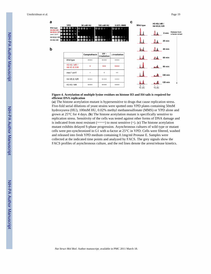
Figure 4. Acetylation of multiple lysine residues on histone H3 and H4 tails is required forefficient DNA replication(a) The histone acetylation mutant is hypersensitive to drugs that cause replication stress.Five-fold serial dilutions of yeast strains were spotted onto YPD plates containing 50mMhydroxyurea (HU), 100mM HU, 0.02% methyl methanesulfonate (MMS) or YPD alone andgrown at 25°C for 4 days. (b) The histone acetylation mutant is specifically sensitive toreplication stress. Sensitivity of the cells was tested against other forms of DNA damage andis indicated from most resistant (++++) to most sensitive (+). (c) The histone acetylationmutant exhibits delayed S-phase progression. Asynchronous cultures of wild type or mutantcells were pre-synchronized in G1 with α-factor at 25°C in YPD. Cells were filtered, washedand released into fresh YPD medium containing 0.1mg/ml Pronase E. Samples werecollected at the indicated time points and analyzed by FACS. The grey signals show theFACS profiles of asynchronous culture, and the red lines denote the arrest/release kinetics.
Unnikrishnan et al. Page 19
Nat Struct Mol Biol. Author manuscript; available in PMC 2011 March 18.
NIH
-PA Author Manuscript
NIH
-PA Author Manuscript
NIH
-PA Author Manuscript
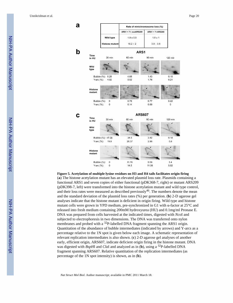
Figure 5. Acetylation of multiple lysine residues on H3 and H4 tails facilitates origin firing(a) The histone acetylation mutant has an elevated plasmid loss rate. Plasmids containing afunctional ARS1 and seven copies of either functional (pDK368-7, right) or mutant ARS209(pDK398-7, left) were transformed into the histone acetylation mutant and wild type control,and their loss rates were measured as described previously46. The numbers denote the meanand the standard deviation of the plasmid loss rates (%) per generation. (b) 2-D agarose gelanalyses indicate that the histone mutant is deficient in origin firing. Wild type and histonemutant cells were grown in YPD medium, pre-synchronized in G1 with α-factor at 25°C andreleased into fresh medium containing 200mM hydroxyurea (HU) and 0.1mg/ml Pronase E.DNA was prepared from cells harvested at the indicated times, digested with NcoI andsubjected to electrophoresis in two dimensions. The DNA was transferred onto nylonmembranes and probed with a 32P-labelled DNA fragment spanning the ARS1 origin.Quantitation of the abundance of bubble intermediates (indicated by arrows) and Y-arcs as apercentage relative to the 1N spot is given below each image. A schematic representation ofrelevant replication intermediates is also shown. (c) 2-D agarose gel analyses of anotherearly, efficient origin, ARS607, indicate deficient origin firing in the histone mutant. DNAwas digested with BspHI and ClaI and analyzed as in (b), using a 32P-labelled DNAfragment spanning ARS607. Relative quantitation of the replication intermediates (aspercentage of the 1N spot intensity) is shown, as in (b).
Unnikrishnan et al. Page 20
Nat Struct Mol Biol. Author manuscript; available in PMC 2011 March 18.
NIH
-PA Author Manuscript
NIH
-PA Author Manuscript
NIH
-PA Author Manuscript
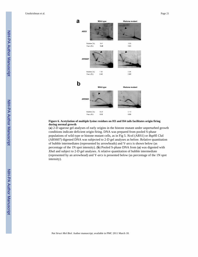
Figure 6. Acetylation of multiple lysine residues on H3 and H4 tails facilitates origin firingduring normal growth(a) 2-D agarose gel analyses of early origins in the histone mutant under unperturbed growthconditions indicate deficient origin firing. DNA was prepared from pooled S-phasepopulations of wild type or histone mutant cells, as in Fig 5. NcoI (ARS1) or BspHI ClaI(ARS607) digested DNA was subjected to 2-D gel analyses as before. Relative quantitationof bubble intermediates (represented by arrowheads) and Y-arcs is shown below (aspercentage of the 1N spot intensity). (b) Pooled S-phase DNA from (a) was digested withXbaI and subject to 2-D gel analyses. A relative quantitation of bubble intermediate(represented by an arrowhead) and Y-arcs is presented below (as percentage of the 1N spotintensity).
Unnikrishnan et al. Page 21
Nat Struct Mol Biol. Author manuscript; available in PMC 2011 March 18.
NIH
-PA Author Manuscript
NIH
-PA Author Manuscript
NIH
-PA Author Manuscript