histone crosstalk between h3s10ph and h4k16ac generates a histone code that mediates transcription...
TRANSCRIPT
Histone Crosstalk between H3S10phand H4K16ac Generates a Histone Codethat Mediates Transcription ElongationAlessio Zippo,1 Riccardo Serafini,1 Marina Rocchigiani,1 Susanna Pennacchini,1 Anna Krepelova,1
and Salvatore Oliviero1,*1Dipartimento di Biologia Molecolare Universita di Siena, Via Fiorentina 1, 53100 Siena, Italy
*Correspondence: [email protected]
DOI 10.1016/j.cell.2009.07.031
SUMMARY
The phosphorylation of the serine 10 at histone H3has been shown to be important for transcriptionalactivation. Here, we report the molecular mechanismthrough which H3S10ph triggers transcript elonga-tion of the FOSL1 gene. Serum stimulation inducesthe PIM1 kinase to phosphorylate the preacetylatedhistone H3 at the FOSL1 enhancer. The adaptorprotein 14-3-3 binds the phosphorylated nucleo-some and recruits the histone acetyltransferaseMOF, which triggers the acetylation of histone H4at lysine 16 (H4K16ac). This histone crosstalk gener-ates the nucleosomal recognition code composed ofH3K9acS10ph/H4K16ac determining a nucleosomeplatform for the bromodomain protein BRD4 binding.The recruitment of the positive transcription elonga-tion factor b (P-TEFb) via BRD4 induces the releaseof the promoter-proximal paused RNA polymeraseII and the increase of its processivity. Thus, the singlephosphorylation H3S10ph at the FOSL1 enhancertriggers a cascade of events which activate tran-scriptional elongation.
INTRODUCTION
Dynamic changes of chromatin obtained by covalent modifica-
tions of histones, including phosphorylation, acetylation, meth-
ylation and ubiquitination, play a key role in regulating gene
expression (Jenuwein and Allis, 2001; Strahl and Allis, 2000).
Two different mechanisms were proposed to explain how his-
tone modifications exert their influence on transcriptional activa-
tion. The first is based on the alteration of the DNA-nucleosome
contacts due to a cumulative effect of a large number of histone
modifications. The second predicts the generation of binding
platforms on histone tails recognized by regulatory proteins.
This model is based on the histone code hypothesis, where
specific histone modifications, acting alone or in combination,
can promote or inhibit the binding of a protein (the ‘‘reader’’) to
the nucleosome (Strahl and Allis, 2000). The reader can promote
a second histone modification, determining a histone crosstalk
1122 Cell 138, 1122–1136, September 18, 2009 ª2009 Elsevier Inc.
which generates a different binding platform for the further
recruitment of proteins that regulate gene expression.
In Drosophila it has been shown that H3S10ph is required for
the recruitment of the positive transcription elongation factor
b (P-TEFb) on the heat shock genes (Ivaldi et al., 2007) although
these results have been challenged (Cai et al., 2008).
In mammalian cells, nucleosome phosphorylation localized at
promoters has been directly linked with transcriptional activa-
tion. It has been shown that H3S10ph enhances the recruitment
of GCN5, which acetylates K14 on the same histone tail (Agalioti
et al., 2002; Cheung et al., 2000). Steroid hormone induces the
transcriptional activation of the MMLTV promoter by activating
MSK1 that phosphorylates H3S10, leading to HP1g displace-
ment and recruitment of the ATP-dependent remodeling com-
plex. MSK1/2- mediated phosphorylation of H3S10 in response
to serum at c-Jun and c-Fos genes induces the recruitment of
14-3-3 (Macdonald et al., 2005; Soloaga et al., 2003). Moreover,
it was elucidated that the 14-3-3 binding affinities increase if H3
phosphorylation occurs in an acetylated (K9 or K14 acetylation)
context both in mammals and yeast (Macdonald et al., 2005;
Walter et al., 2008; Winter et al., 2008). However, the mech-
anisms that link transcriptional activation with these events
remain elusive.
We recently demonstrated that transcriptional activation of
about 20% of MYC target genes is dependent on its cooperation
with the kinase PIM1, which phosphorylates H3S10 at the
FOSL1 enhancer (Zippo et al., 2007). H3S10 phosphorylation
at the FOSL1 enhancer induces the increase of RNA polymerase
II (RNAP) phosphorylation at serine 2 suggesting that H3S10ph
stimulates the elongation step of transcription.
The release of the pausing RNAP from the promoter is a tightly
regulated process modulated by negative elongation factors
such as DSIF and NELF and positive elongation factors like
transcription-elongation factor-b (P-TEFb) that phosphorylates
the C-terminal domain of RNA-polymerase (RNAP) at serine 2
(Mason and Struhl, 2005; Ni et al., 2004; Peterlin and Price,
2006; Saunders et al., 2006; Sims et al., 2004).
P-TEFb can be associated with an activating complex con-
taining the bromodomain protein BRD4 (Jang et al., 2005;
Yang et al., 2005). BRD4 belongs to the BET family of proteins
that are characterized by two tandem bromodomains and an
extra-terminal domain (ET) of unknown function. BRD4 and its
closely related BRD2 and BRD3 proteins are associated with
euchromatin regions by interacting with acetylated histones H3
and H4 (Dey et al., 2003, 2000).
We show here that H3S10ph triggers a histone crosstalk that
generates a histone code determining the transcription elonga-
tion of the FOSL1 gene. Serum treatment induces 14-3-3 binding
to the phosphorylated nucleosome and recruits MOF, which
acetylates histone H4 at lysine 16. Thus, at the FOSL1 enhancer,
PIM1-dependent H3S10 phosphorylation promotes H4K16
acetylation.
The histone crosstalk between H3S10ph and H4K16ac gener-
ates the combination of H3K9ac and H4K16ac modifications on
the nucleosome determining a binding platform for the double
bromodomain protein BRD4. The recruitment of P-TEFb, via
BRD4 binding to the nucleosome, allows the release of the
promoter-proximal paused RNAP stimulating a productive elon-
gation.
RESULTS
H3S10ph Modulates Transcription Elongationby Recruiting the P-TEFb/BRD4 ComplexHistone H3 phosphorylation is a key event in transcriptional
activation of stimuli-responsive genes including Hsp70, c-Jun,
c-Fos and FOSL1. To elucidate the mechanism through which
H3S10ph triggers an increase of RNA Polymerase II engaged
in transcript elongation we used the serum-inducible FOSL1
gene as a model. We first performed in vitro transcription assays
(run-on) to analyze the distribution of transcribing RNA polymer-
ases across the FOSL1 gene (Figure 1A). Radiolabeled RNAs
obtained from nuclei isolated from serum-stimulated cells
expressing either a scramble (control) or PIM1 short hairpin
RNA (shPIM1) were hybridized to different probes across the
FOSL1 gene. Serum treatment increased elongating polymerase
density across the entire gene compared to the 50UTR in control
cells while shPIM1 determined a general reduction of elongation
products that was more pronounced at the 30 end of the gene
(Figure 1B) suggesting that RNAP processivity was affected by
PIM1-dependent H3S10 phosphorylation. As we previously
observed that in uninduced cells the RNAP is already associated
with the promoter (Zippo et al., 2007), we asked whether the
RNAP that has not initiated transcription is associated with the
preinitiation complex or it is paused immediately downstream
of the transcription start site. To verify the presence of stalled
RNAP we analyzed by ChIP the RNAP distribution on the
FOSL1 gene (Figure 1C). Before serum treatment, we observed
high RNAP signal near the transcription start site (TSS) together
with low signal across the gene both in control and in PIM1-
silenced cells, indicative of stalled RNAP as previously described
(Muse et al., 2007; Zeitlinger et al., 2007). Following serum treat-
ment, RNAP redistributed across the gene in control cells, but
not in shPIM1 where the RNAP remained concentrated in the
proximity of the promoter.
We then performed time-course analyses of RNAP occupancy
on the promoter and across the coding region of the FOSL1 gene
(Figure 1D, and Figure S3A available with this article online) by
comparing the RNAP signal obtained by ChIP in samples
untreated or treated with high salt concentration. Salt-resistant
RNAP represents molecules engaged in transcription elongation
C
while RNAP in preinitiation complexes are dissociated in the
same conditions (Wang et al., 2005). Before serum treatment
the RNAP associated on the FOSL1 promoter is already engaged
and its level increases 60 min after the serum treatment. Shortly
after RNAP was released from the promoter and it was localized
in the coding region of the gene. This step of the transcription
cycle was affected by PIM1 knockdown as we did not observe
the increase of elongating RNAP in shPIM1 cells. To confirm
these observations, the salt-resistant RNAP was subjected to re-
immunoprecipitation (Re-ChIP) by using an antibody recognizing
the elongating isoform of the RNAP, thus allowing us to distin-
guish between stalled versus elongating RNAP. The normalized
data showed that at 60 min the RNAP loaded on the promoter
was engaged but not elongating. In fact the level of CTD phos-
phorylated at serine 2 (Ser2P) increased at 80 min in control cells
both in the promoter and in the coding region and this increase
was affected by PIM1 silencing (Figure 1D, lower panels). Taken
together these results suggest that PIM1-dependent phosphor-
ylation of H3 at the FOSL1 enhancer is necessary for productive
elongation.
As the release of the stalled polymerases depends on CTD
phosphorylation at serine 2 mediated by P-TEFb, we measured
by ChIP the recruitment of the active complex P-TEFb/BRD4 on
FOSL1 gene. P-TEFb/BRD4 complex was recruited to the pro-
moter starting from the 80 min time point in control but not in
PIM1 silenced cells (Figure 1E). Thus, the recruitment of the
P-TEFb/BRD4 complex is coincident with the release of the
stalled polymerase from the promoter (Figure 1E). To confirm
that H3S10ph is necessary for transcription elongation of
FOSL1, we tested the recruitment of the P-TEFb/BRD4 complex
and elongating RNAP in the presence of H3S10A mutant. We
performed Re-ChIP analysis on mononucleosomes obtained
from cells expressing either the wild-type H3 YFP fusion protein
(YFP-H3 WT) or the mutant in serine 10 (YFP-H3 S10A)
(Figure S1). These experiments showed that the recruitment of
P-TEFb/BRD4 is dependent on the level of H3S10ph (Figures
S1A and S1B) and this event is necessary for the phosphoryla-
tion of RNAP serine 2 on the CTD (Figures S1C and S1D). In
conclusion, serum treatment induces PIM1-dependent phos-
phorylation of H3S10 at the FOSL1 enhancer, which triggers
P-TEFb/BRD4 recruitment. This event releases the promoter-
proximal paused RNAP and enhances RNAP processivity along
the gene.
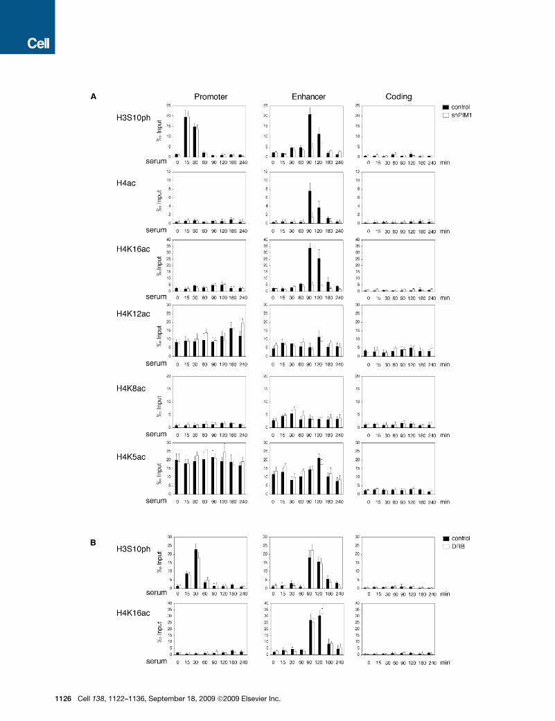
H3S10ph Modulates H4K16 Acetylation at the FOSL1EnhancerTo elucidate the key mechanism that links H3S10ph on the
enhancer to the release of the promoter-proximal stalled
RNAP, we first analyzed the dynamics of other histone modifica-
tions occurring at the FOSL1 gene in wild-type or PIM1 knock-
down cells (Figure 2). We previously observed that the FOSL1
enhancer is preacetylated on H3 and serum treatment does
not induce any change in the H3 acetylation levels (Zippo
et al., 2007). By analyzing the pattern of H4 acetylation across
the FOSL1 gene, we observed signal enrichment at the enhancer
from 90 min time point, thus concomitant with the increase of
H3S10ph (Figure 2A). Moreover, this increment was not
observed in PIM1-silenced cells suggesting that H3S10ph is
ell 138, 1122–1136, September 18, 2009 ª2009 Elsevier Inc. 1123
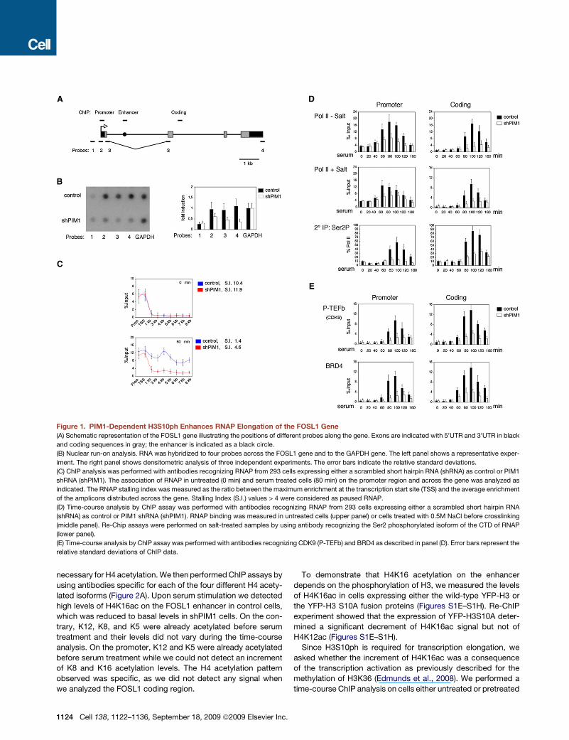
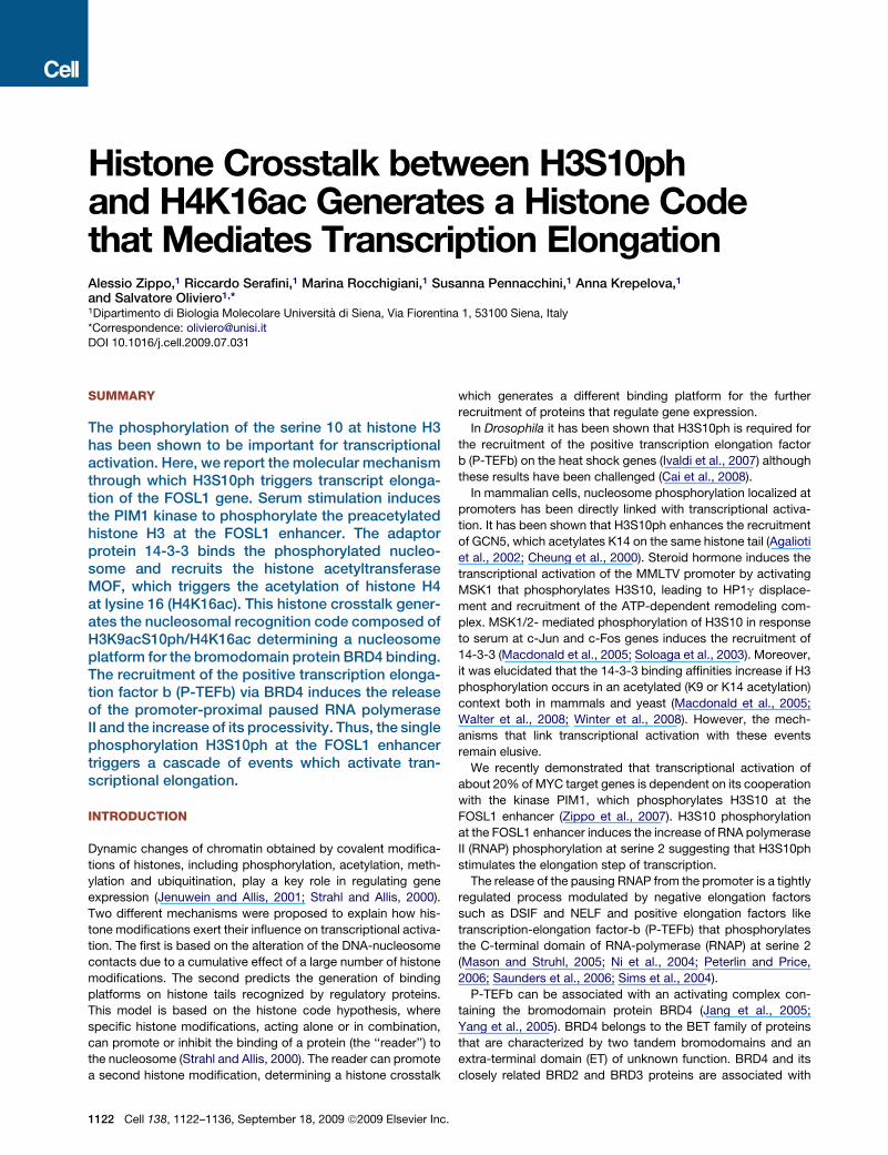
Figure 1. PIM1-Dependent H3S10ph Enhances RNAP Elongation of the FOSL1 Gene
(A) Schematic representation of the FOSL1 gene illustrating the positions of different probes along the gene. Exons are indicated with 50UTR and 30UTR in black
and coding sequences in gray; the enhancer is indicated as a black circle.
(B) Nuclear run-on analysis. RNA was hybridized to four probes across the FOSL1 gene and to the GAPDH gene. The left panel shows a representative exper-
iment. The right panel shows densitometric analysis of three independent experiments. The error bars indicate the relative standard deviations.
(C) ChIP analysis was performed with antibodies recognizing RNAP from 293 cells expressing either a scrambled short hairpin RNA (shRNA) as control or PIM1
shRNA (shPIM1). The association of RNAP in untreated (0 min) and serum treated cells (80 min) on the promoter region and across the gene was analyzed as
indicated. The RNAP stalling index was measured as the ratio between the maximum enrichment at the transcription start site (TSS) and the average enrichment
of the amplicons distributed across the gene. Stalling Index (S.I.) values > 4 were considered as paused RNAP.
(D) Time-course analysis by ChIP assay was performed with antibodies recognizing RNAP from 293 cells expressing either a scrambled short hairpin RNA
(shRNA) as control or PIM1 shRNA (shPIM1). RNAP binding was measured in untreated cells (upper panel) or cells treated with 0.5M NaCl before crosslinking
(middle panel). Re-Chip assays were performed on salt-treated samples by using antibody recognizing the Ser2 phosphorylated isoform of the CTD of RNAP
(lower panel).
(E) Time-course analysis by ChIP assay was performed with antibodies recognizing CDK9 (P-TEFb) and BRD4 as described in panel (D). Error bars represent the
relative standard deviations of ChIP data.
necessary for H4 acetylation. We then performed ChIP assays by
using antibodies specific for each of the four different H4 acety-
lated isoforms (Figure 2A). Upon serum stimulation we detected
high levels of H4K16ac on the FOSL1 enhancer in control cells,
which was reduced to basal levels in shPIM1 cells. On the con-
trary, K12, K8, and K5 were already acetylated before serum
treatment and their levels did not vary during the time-course
analysis. On the promoter, K12 and K5 were already acetylated
before serum treatment while we could not detect an increment
of K8 and K16 acetylation levels. The H4 acetylation pattern
observed was specific, as we did not detect any signal when
we analyzed the FOSL1 coding region.
1124 Cell 138, 1122–1136, September 18, 2009 ª2009 Elsevier Inc.
To demonstrate that H4K16 acetylation on the enhancer
depends on the phosphorylation of H3, we measured the levels
of H4K16ac in cells expressing either the wild-type YFP-H3 or
the YFP-H3 S10A fusion proteins (Figures S1E–S1H). Re-ChIP
experiment showed that the expression of YFP-H3S10A deter-
mined a significant decrement of H4K16ac signal but not of
H4K12ac (Figures S1E–S1H).
Since H3S10ph is required for transcription elongation, we
asked whether the increment of H4K16ac was a consequence
of the transcription activation as previously described for the
methylation of H3K36 (Edmunds et al., 2008). We performed a
time-course ChIP analysis on cells either untreated or pretreated
with DRB, a specific inhibitor of P-TEFb which blocks transcrip-
tion activation (Figure 2B). We detected a strong H3S10ph and
H4K16ac signal increment, on the FOSL1 enhancer, after serum
treatment also in DRB pretreated cells suggesting that both
modifications occur before transcriptional elongation. On the
contrary, the methylation levels of H3K4 and H3K36 decreased
in DRB pretreated cells (Figure S2). These results demonstrate
that H3S10ph is necessary for H4K16 acetylation at the FOSL1
enhancer, which precedes P-TEFb recruitment.
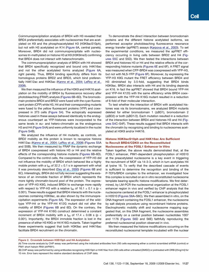
H3S10ph Enhances H4K16ac by Inducingthe Recruitment of MOFTo identify the acetyltransferase responsible for acetylation of
H4K16 we analyzed the binding of three members of the histone
acetyltransferases MYST family MOF, Tip60, and HBO1 that
were previously described to be able to acetylate H4. MOF
was recruited to the FOSL1 enhancer from 90 min after serum
treatment thus with the same kinetics as H4K16 acetylation
(Figure 3A) and its binding was dependent on H3S10ph (Fig-
ures 3A and S1I). Tip60 was binding both the promoter and the
enhancer already before serum treatment. Importantly, both
the dynamics and the extent of Tip60 binding was not affected
by PIM1 silencing. We could not detect any signal of HBO1
above the background noise (Figure 3A). We conclude that to
the FOSL1 enhancer H3S10ph is required for the recruitment
of MOF, which acetylates H4K16.
We next investigated whether the observed histone crosstalk
between H3S10ph and H4K16ac is specific to FOSL1 or is a
general event occurring after serum induction. To this end, we
analyzed the dynamics of global H4 acetylations by immunoblot-
ting in both control and PIM1 knockdown cells. As we previously
demonstrated, serum-induced H3S10ph follows a bimodal curve
with two peaks respectively at 15 and 90 min. Only the second
phosphorylation event isdependent onPIM1kinaseactivity (Zippo
et al., 2007) (Figures 3B and 3C). As quantified by densitometry
analysis, serum treatment increased the global H4K16 acetylation
levels in controls, but not in the PIM1-silenced cells (Figure 3C)
while H4K8 acetylation increased after serum treatment in both
control and shPIM1 cells. Moreover, the H4K12 and H4K5 were
highly acetylated before serum treatment and their levels did not
change either during the time frame analyzed or by PIM1 silencing.
These data suggest that after serum stimulation there is a global
histone crosstalk between H3S10ph and H4K16ac.
14-3-3 Binding to H3S10ph Induces the Recruitmentof MOFWe then analyzed whether H3S10ph per se facilitated the bind-
ing of MOF on the FOSL1 enhancer. To this aim we measured the
binding of the 3 and z isoforms of 14-3-3 phosphoserine adaptor
proteins to H3S10ph (Figure 4A). Time-course ChIP analysis
revealed the association of 14-3-3 shortly after serum treatment
with the promoter in both control and shPIM1 silenced cells.
From the 90 min time point, 14-3-3 proteins were binding to
the FOSL1 enhancer in control cells, but not in shPIM1 cells.
Thus, 14-3-3 proteins were recruited to the FOSL1 gene through
recognition and binding to phosphoserine signal induced by
serum treatment. These conclusions were supported by Re-
ChIP experiments on 293 cells expressing the YFP-H3 S10A
C
mutant in which 14-3-3 recruitment to the FOSL1 enhancer
was significantly affected (Figure S1K).
As 14-3-3 proteins mediate protein-protein interactions
(Dougherty and Morrison, 2004), we verified whether they were
able to interact with MOF. For this purpose we immunoprecipi-
tated 14-3-3 or MOF from chromatin extracts obtained from
serum-induced 293 cells and identified the interacting proteins
by immunoblotting. The results showed that 14-3-3 interacts
with MOF and also coimmunoprecipitated with nucleosomes
phosphorylated on H3S10 and acetylated on H4K16 while it
did not immunoprecipitate with Tip60 (Figure 4B).
To study the dynamics of the 14-3-3/MOF interactions we
performed time-course immunoprecipitation experiments that
showed the formation of the 14-3-3/MOF complex starting from
90 min after serum treatment and an increase of interaction with
nucleosomes containing H4K16ac (Figures 4C and 4D). These
data suggest that MOF is dynamically recruited to chromatin
via 14-3-3, which recognizes and binds the H3S10ph acetylating
H4K16.
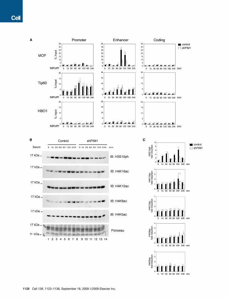
FOSL1 Transcription Elongation Is Dependent on MOFRecruitment via 14-3-3 BindingTo verify whether 14-3-3 recruits MOF on the FOSL1 enhancer we
knocked down both the 14-3-3z and 14-3-33 isoforms by shRNA
(Figure S3B) and performed ChIP assays on the FOSL1 enhancer
in unstimulated and serum-treated cells (Figure 5A). Compared to
the control cells, the 14-3-3 silencing induced a reduction of MOF
binding, which determined a lower level of H4K16ac (but not of
H4K12ac or H3S10ph). The 14-3-3 silencing determined a
decreased recruitment of P-TEFb/BRD4 complex, a reduction
of the release of the stalled RNAP after serum treatment (Fig-
ure S3D) and a reduction of the RNAP phosphorylated on serine
2 (Figure 5A). Moreover, 14-3-3 silencing affected the transcrip-
tion of FOSL1. Thus, the silencing of 14-3-3 determined
a cascade response that ultimately affected FOSL1 transcription
elongation.
To confirm that the effects of 14-3-3 silencing were dependent
only on its ability to recruit MOF on the FOSL1 enhancer, we
knocked down MOF (Figure S3C) and performed the same
ChIP analysis (Figure 5B). MOF silencing determined a reduction
of H4K16ac without affecting the levels of H4K12ac or H3S10ph.
MOF silencing also affected the recruitment of P-TEFb/BRD4
complex, the release of the stalled RNAP (Figure S3E), the phos-
phorylation of RNAP at serine 2, and transcription elongation.
Taken together these results demonstrated that 14-3-3 recruits
MOF to the FOSL1 enhancer resulting in the transcription activa-
tion.
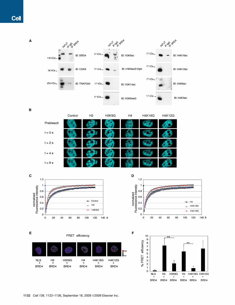
BRD4 Binds Nucleosomes Containing both H3K9acand H4K16acBRD4 is a double bromodomain protein that binds acetylated
histone H3 and H4 (Dey et al., 2003). For this reason we attemp-
ted to identify the histone acetylation pattern that is recognized
and bound by BRD4. We immunoprecipitated the endogenous
BRD4 protein from chromatin extracts obtained from serum-
treated 293 cells (Figure 6A). As expected BRD4 associated
with CDK9 and with TRAP220, a component of the mediator
(Yang et al., 2005; Jang et al., 2005) (Figure 6A, left panels).
ell 138, 1122–1136, September 18, 2009 ª2009 Elsevier Inc. 1125
1126 Cell 138, 1122–1136, September 18, 2009 ª2009 Elsevier Inc.
Coimmunoprecipitation analysis of BRD4 with H3 revealed that
BRD4 preferentially associates with nucleosomes that are acet-
ylated on K9 and the phosphoacetylated H3 (H3K9acS10ph),
but not with H3 acetylated on K14 (Figure 6A, central panels).
Moreover, BRD4 did not coimmunoprecipitate with nucleo-
somes tri-methylated on histone H3 at K9 (H3K9me3) confirming
that BRD4 does not interact with heterochromatin.
The coimmunopreciptation analysis of BRD4 with H4 showed
that BRD4 specifically recognized and bound only H4K16ac
and not the other acetylated forms analyzed (Figure 6A,
right panels). Thus, BRD4 binding specificity differs from its
homologous proteins BRD2 and BRD3, which bind preferen-
tially H4K12ac and H4K5ac (Kanno et al., 2004; LeRoy et al.,
2008).
We then measured the influence of the H3K9 and H4K16 acet-
ylation on the mobility of BRD4 by fluorescence recovery after
photobleaching (FRAP) analysis (Figures 6B–6D). The bromodo-
main proteins BRD4 and BRD2 were fused with the cyan fluores-
cent protein (CFP) while H3, H4 and their corresponding mutants
were fused to the yellow fluorescent proteins (YFP) and coex-
pressed in 3T3 cells (Figure S4). The ectopically expressed
histones used in these assays behaved identically to the endog-
enous counterpart as YFP-histones were incorporated to the
same levels in eu- and hetero-chromatin as the endogenous
H3 and H4 (Figure S4A) and were uniformly localized in the nuclei
(Figure S4B).
We analyzed the influence of H4 mutants, as controls, on
BRD2 mobility as this protein is known to recognize histone
H4K12ac (Kanno et al., 2004; LeRoy et al., 2008) (Figures S5A
and S5B). We then measured by FRAP the dynamic exchange
of BRD4 coexpressed with histone H3 or H4 and the relative
effects of the corresponding histone mutants (Figures 6B–6D).
Compared to the control cells, the coexpression of YFP-H3 did
not influence the mobility of BRD4 which behaved like a highly
mobile protein with a t80 of 32.2 ± 0.1 s in agreement with what
it was previously described (Dey et al., 2000) (Figures 6B and
6C). Interestingly, BRD4 did not fully recover suggesting the exis-
tence of an immobile fraction of BRD4 which represents the
more tightly chromatin-bound pool of the protein. The expres-
sion of YFP-H3 K9G, induced BRD4 to exchange more rapidly
with respect to YFP-H3 with a relative t80 of 16.1 ± 0.1 s (p <
0.001). These results suggest that BRD4 interacts with the nucle-
osomes containing H3K9ac, as also shown by the immunopre-
cipitation experiments (Figure 6A). The expression of the wild-
type YFP-H4 or the YFP-H4 K12G mutant did not alter the
mobility of BRD4 (Figures 6B and 6D). On the contrary, the
expression of YFP-H4 K16G construct determined a constant
increment of BRD4 mobility with a t80 of 17.4 ± 0.08 s (p <
0.001). Importantly, the BRD4 immobile fraction is lost in the
presence of either H3 K9G or H4 K16G mutants. Taken together
these experiments suggest that both H3K9ac and H4K16ac
facilitate BRD4 recruitment on the chromatin.
To demonstrate the direct interaction between bromodomain
proteins and the different histone acetylated isoforms, we
performed acceptor photobleaching fluorescence resonance
energy transfer (apFRET) assays (Karpova et al., 2003). To set
the experimental conditions, we measured the apFRET effi-
ciency occurring in living cells between BRD2 and H4 (Fig-
ures S5C and S5D). We then tested the interactions between
BRD4 and histones H3 or H4 and the relative effects of the cor-
responding histone mutants (Figures 6E and 6F). A FRET signal
was measured when CFP-BRD4 was coexpressed with YFP-H3,
but not with NLS-YFP (Figure 6F). Moreover, by expressing the
YFP-H3 K9G mutant the FRET efficiency between BRD4 and
H3 diminished by 3.5-fold, suggesting that BRD4 binds
H3K9ac. BRD4 also interacts with H4 and its binding depends
on K16. In fact the apFRET showed that BRD4 bound YFP-H4
and YFP-H4 K12G with the same efficiency while BRD4 coex-
pression with the YFP-H4 K16G mutant resulted in a reduction
of 6-fold of their molecular interaction.
To test whether the interaction of BRD4 with acetylated his-
tones was via its bromodomains, we analyzed BRD4 mutants
deleted for either bromodomain 1 (DBD1), bromodomain 2
(DBD2) or both (DBD1/2). Each mutation resulted in a reduction
of the interaction between BRD4 and histones H3 and H4 (Fig-
ures S4C–S4F). These results suggest that BRD4 interacts with
the chromatin by recognizing and binding to nucleosomes acet-
ylated at H3K9 and/or H4K16.
Histone H3K9acS10ph and H4K16ac Are Sufficientto Recruit BRD4/CDK9 on the ReconstitutedNucleosome at the FOSL1 Enhancer In VitroTaken together, the above results demonstrated that, at the
FOSL1 enhancer, PIM1-dependent phosphorylation of H3S10
at the preacetylated nucleosome is a key event in triggering
the recruitment of MOF via 14-3-3, which in turn acetylates H4
at lysine 16. To verify that the described cascade of events
is sufficient to determine the recognition and binding of the
P-TEFb/BRD4 complex to the enhancer, we investigated how
this complex is recruited on an in vitro reconstituted nucleosome
template bearing specific histone modifications. We first deter-
mined, by LM-PCR the nucleosomal organization at the FOSL1
enhancer region in vivo and verified by ChIP analysis that the
nucleosome centered at the FOSL1 enhancer is phosphorylated
in H3S10 (Figures S6A–S6C). We then assembled in vitro, on the
DNA fragment containing the FOSL1 enhancer, the nucleosome
by salt dialysis procedure using recombinant histone proteins.
Electrophoretic mobility shift and nucleosome mapping sug-
gested that, on this DNA fragment, the nucleosome assembles
preferentially on a central position between nucleotides 1007
and 1176 (Figures S6D and S6E) faithfully reproducing the
preferred nucleosomal position observed in vivo.
We then measured the histone modifications occurring on the
reconstituted nucleosomal template incubated with the nuclear
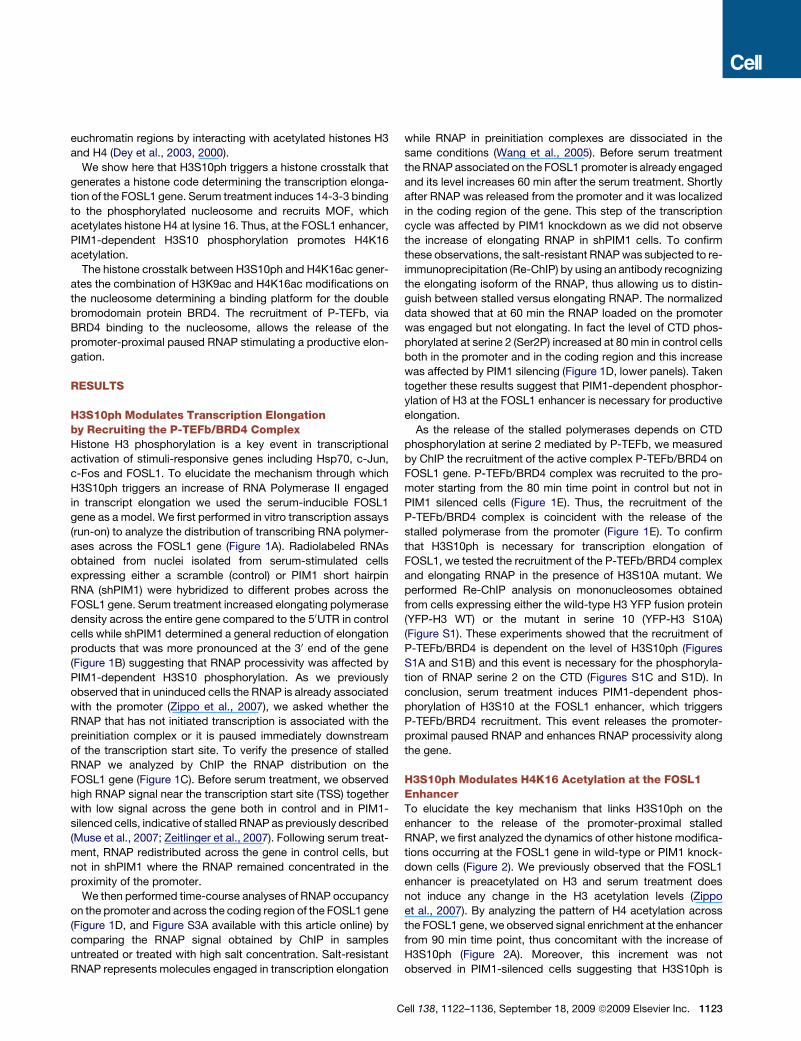
Figure 2. Crosstalk between H3S10ph and H4 Acetylation
(A) Time-course analysis by ChIP assay was performed using the indicated antibodies from 293 cells expressing either a control scrambled shRNA (control) or
PIM1 short hairpin RNA (shPIM1).
(B) ChIP assay was performed by using antibodies recognizing H3S10ph or H4K16ac from 293 cells either untreated (DMSO) or pretreated with DRB (25ng/ml) for
10 min. Error bars represent the relative standard deviations of ChIP data.
Cell 138, 1122–1136, September 18, 2009 ª2009 Elsevier Inc. 1127
1128 Cell 138, 1122–1136, September 18, 2009 ª2009 Elsevier Inc.

Figure 4. MOF Interacts with the 14-3-3 Proteins
(A) Time-course analysis by ChIP assay was performed by using antibodies recognizing 14-3-33 and 14-3-3z as specified from 293 cells expressing either
a control scrambled shRNA or PIM1 shRNA. The error bars indicate the relative standard deviations.
(B) Cell proteins extracted from serum-treated 293 cells were either immunostained (INPUT) or subjected to immunoprecipitation (IP) with anti-IgG, anti-14-3-33,
anti-14-3-3z, or anti-MOF antibodies as indicated. Immunostaining (IB) analysis was performed using the indicated antibodies. 10% of the total protein samples
were loaded as inputs. Nonspecific bands are indicated with an asterisk.
(C) Time-course analysis by immunostaining of input extracts, and IgG (-), or 14-3-33 (+) immunoprecipitates from serum-treated 293 cells as indicated. Interact-
ing proteins were revealed using indicated antibodies. 10% of the total protein samples were loaded as inputs.
(D) Densitometry analysis of signals obtained from 6 independent immunoblotting experiments using the indicated antibodies. The results were normalized
against the input for each protein analyzed and are represented as immunoprecitation enrichment. The error bars indicate the relative standard deviations.
extracts in the presence or absence of ATP and acetyl CoA
(AcCoA) (Figure 7A). At the same time we determined the conse-
quence of those histone modifications on the binding affinity of
the interacting proteins. Acetylation of histone H3 at K9 and
C
K14 occurred independently of the H3S10ph (Figure 7B, com-
pare lanes 4 and 5), confirming what previously observed by
ChIP assay (Zippo et al., 2007). In contrast, acetylation of H4K16
occurred only on nucleosomes that were phosphorylated on
Figure 3. MOF Recruitment Is Dependent on H3S10ph(A) Time-course analysis by ChIP assay was performed from 293 cells expressing either a control scrambled shRNA or PIM1 shRNA using the specified anti-
bodies. The error bars indicate the relative standard deviations.
(B) Time-course analysis of histone modifications was performed by immunoblotting protein extracts obtained from 293 cells expressing either a control scram-
bled shRNA or PIM1 shRNA with the indicated antibodies. Histones were enriched in nuclear extracts after micrococcal nuclease digestion. A representative
experiment is shown.
(C) Densitometry analysis of signals obtained from 6 independent immunoblotting experiments by using the indicated antibodies. The results were normalized
against total histones. The data are represented as fold induction with respect to the uninduced signal (0 min time point) for both control and shPIM1 samples. The
error bars indicate the relative standard deviations. The T-student test was applied to determine the probability (** = p < 0.001).
ell 138, 1122–1136, September 18, 2009 ª2009 Elsevier Inc. 1129
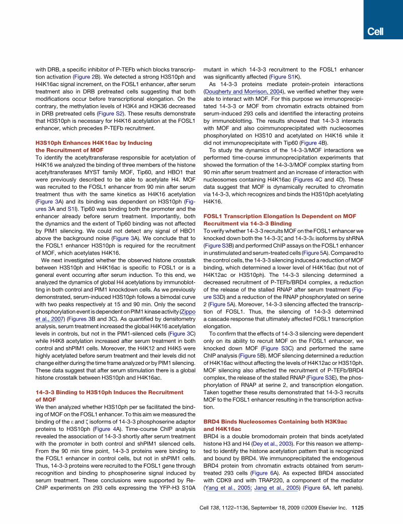
Figure 5. CDK9/BRD4 Recruitment on FOSL1 Enhancer Is Mediated by MOF via 14-3-3
(A) ChIP assay was performed using the indicated antibodies from 293 cells expressing either a control scrambled shRNA or 14-3-3 shRNA. Last panel represents
quantitative real time RT-PCR of FOSL1 mRNA obtained from the same samples. Gene specific RNA levels were measured and normalized on GAPDH expres-
sion and are represented as fold induction (vertical bars).
(B) ChiP assay was performed with the same antibodies as in (A) from 293 cells expressing either a control scrambled shRNA or MOF shRNA. Last panel repre-
sents quantitative real time RT-PCR of FOSL1 mRNA obtained from the same samples. Gene specific RNA levels were measured and normalized on GAPDH
expression and are represented as fold induction (vertical bars). The data represent triplicate real-time quantitative RT-PCR measurements. Error bars represent
the relative standard deviations of ChIP data.
H3S10 while H4K12 was always acetylated in the presence
of AcCoA, (Figure 7B, compare lanes 4 and 5). These results
were confirmed by using as a template a nucleosome reconsti-
tuted with the mutant H3S10A (Figure 7B, lane 6) suggesting
that acetylation of H4K16 is strictly dependent on the phosphor-
ylation of H3S10. Looking at the interacting proteins, we
observed that 14-3-3 was bound only on the phosphorylated
nucleosome, independently of the acetylation state (Figure 7B,
lanes 3, 5, and 6). We did not observe a significant increase of
14-3-3 binding affinity in the presence of phosphoacetylated
nucleosome as has been previously described (Macdonald
1130 Cell 138, 1122–1136, September 18, 2009 ª2009 Elsevier Inc.
et al., 2005; Walter et al., 2008; Winter et al., 2008) possibly
because of the different conditions of this assay with respect
to previous experiments. MOF binding was also dependent on
the level of H3 phosphorylation (Figure 7B, compare line 4, 5
and 6). The increment of H4K16ac corresponded to the binding
of MOF in the presence of AcCoA (Figure 7B, compare lanes 4
and 5). The combination of the phosphoacetylation on H3 at
K9 and S10 and acetylation of K16 on H4 induced the binding
of the P-TEFb/BRD4 complex on the nucleosome template reca-
pitulating in vitro what previously observed in serum-treated
cells.
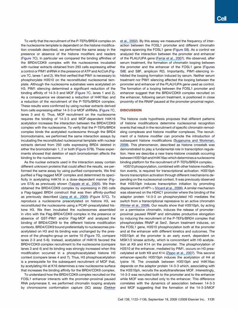
To verify that the recruitment of the P-TEFb/BRD4 complex on
the nucleosome template is dependent on the histone modifica-
tion crosstalk described, we performed the same assay in the
presence or absence of the histone modifiers and readers
(Figure 7C). In particular we compared the binding affinities of
the BRD4/CDK9 complex with the nucleosomes incubated
with nuclear extracts obtained from 293 cells expressing either
a control or PIM1 shRNA in the presence of ATP and AcCoA (Fig-
ure 7C, lanes 1 and 2). We first verified that PIM1 is necessary to
phosphorylate H3S10 on the reconstituted nucleosomal tem-
plate. Although the nucleosome substrates were acetylated on
H3, PIM1 silencing determined a significant reduction of the
binding affinity of 14-3-3 and MOF (Figure 7C, lanes 1 and 2).
As a consequence we observed a reduction of H4K16ac and
a reduction of the recruitment of the P-TEFb/BRD4 complex.
These results were confirmed by using nuclear extracts derived
from cells expressing either 14-3-3 or MOF shRNAs. (Figure 7C,
lanes 3 and 4). Thus, MOF recruitment on the nucleosome
requires the binding of 14-3-3 and MOF-dependent H4K16
acetylation increases the interaction between the BRD4/CDK9
complex with the nucleosome. To verify that the P-TEFb/BRD4
complex binds the acetylated nucleosome through the BRD4
bromodomains, we performed the same interaction assays by
incubating the reconstituted nucleosomal template with nuclear
extracts derived from 293 cells expressing BRD4 deleted in
either the bromodomain 1, 2 or both (Figure S7B). These exper-
iments showed that deletion of each bromodomain affects the
binding to the nucleosome.
As the nuclear extracts used in the interaction assay contain
different unknown proteins that could affect the results, we per-
formed the same assay by using purified components. We first
purified a Flag-tagged MOF complex and determined its speci-
ficity in acetylating H4K16 in a dose-dependent manner (Fig-
ure S7A) as previously shown (Taipale et al., 2005). We then
obtained the BRD4/CDK9 complex by expressing in 293 cells
a Flag-tagged BRD4 construct that was then affinity-purified
as previously described (Jang et al., 2005) (Figure S7C). To
reproduce a nucleosome preacetylated on histone H3, we
reconstituted the nucleosome using a PCAF-preacetylated his-
tone H3. We then incubated the nucleosomes assembled
in vitro with the Flag-BRD4/CDK9 complex in the presence or
absence of GST-PIM1 and/or Flag-MOF and analyzed the
binding of BRD4/CDK9 to the different histone modification
contexts. BRD4/CDK9 bound preferentially to nucleosomes pre-
acetylated on H3 and its binding was unchanged by the pres-
ence of the phospho-group on serine 10 (Figure 7D, compare
lanes 2-3 and 5-6). Instead, acetylation of H4K16 favored the
BRD4/CDK9 complex recruitment to the nucleosome (compare
lanes 3 and 4) and its binding was strongly increased when this
modification occurred in a phosphoacetylated histone H3
context (compare lanes 4 and 7). Thus, H3 phosphoacetylation
is a prerequisite for the subsequent recruitment of MOF that,
by acetylating H4 at K16 determines a new nucleosome surface
that increases the binding affinity for the BRD4/CDK9 complex.
To understand how the BRD4/CDK9 complex recruited on the
FOSL1 enhancer interacts with the promoter-proximal paused
RNA polymerase II, we performed chromatin looping analysis
by chromosome conformation capture (3C) assay (Dekker
et al., 2002). By this assay we measured the frequency of inter-
action between the FOSL1 promoter and different chromatin
regions spanning the FOSL1 gene (Figure S8). As a control we
analyzed the interaction between the promoter and enhancer
of the PLAU/UPA gene (Ferrai et al., 2007). We observed, after
serum treatment, the formation of chromatin looping between
the promoter and the enhancer of the FOSL1 gene (Figures
S8E and S8F, amplicon R2). Importantly, PIM1-silencing in-
hibited the looping formation induced by serum. Neither serum
treatment nor PIM1 silencing affected the looping between the
promoter and enhancer of the PLAU/UPA gene used as control.
The formation of a looping between the FOSL1 promoter and
enhancer suggest that the BRD4/CDK9 complex recruited on
the enhancer, following serum treatment, is positioned in close
proximity of the RNAP paused at the promoter-proximal region.
DISCUSSION
The histone code hypothesis proposes that different patterns
of histone modifications determine nucleosomal recognition
events that result in the recruitment of specific chromatin remod-
eling complexes and histone modifier complexes. The recruit-
ment of a histone modifier can promote the introduction of
subsequent histone modifications (Suganuma and Workman,
2008). This phenomenon, described as histone crosstalk was
demonstrated to play a fundamental role in transcription regula-
tion. Here we describe a new trans-histone crosstalk occurring
betweenH3S10phandH4K16acwhichdeterminesa nucleosome
binding platform for the recruitment of P-TEFb/BRD4 complex.
H3S10 phosporylation, combined with other histone modifica-
tion events, is required for transcriptional activation. H3S10ph
favors transcription activation through different mechanisms de-
pending on the nucleosomal context. It was previously observed
that H3S10ph induces transcription initiation by promoting
displacement of HP1-g (Vicent et al., 2006). A similar mechanism
was observed on the HDAC1 promoter where the binding of the
adaptor protein 14-3-3 on phosphorylated H3 mediates the
switch from a transcriptional repressive to an active chromatin
(Winter et al., 2008). Our results show that H3S10ph, by acting
on a permissive chromatin, induces the release of promoter-
proximal paused RNAP and stimulates productive elongation
by inducing the recruitment of the P-TEFb/BRD4 complex that
phosphorylates RNAP at Ser2. Serum treatment induces, on
the FOSL1 gene, H3S10 phosphorylation both at the promoter
and at the enhancer with different kinetics and outcomes. The
H3S10ph at the promoter is an early event, dependent on
MSK1/2 kinase activity, which is concomitant with H3 acetyla-
tion at K9 and K14 on the promoter. The phosphorylation of
H3S10 at the enhancer, mediated by PIM1, occurs on H3 prea-
cetylated at both K9 and K14 (Zippo et al., 2007). This second
enhancer-specific H3S10ph induces the acetylation of H4 at
lysine 16. The crosstalk between H3S10ph and H4K16ac
depends on the adaptor protein 14-3-3 which, associating with
the H3S10ph, recruits the acetyltransferase MOF. Interestingly,
14-3-3 was recruited both to the promoter and to the enhancer
while MOF was recruited only to the enhancer. This difference
correlates with the dynamics of association between 14-3-3
and MOF suggesting that the formation of the 14-3-3/MOF
Cell 138, 1122–1136, September 18, 2009 ª2009 Elsevier Inc. 1131
1132 Cell 138, 1122–1136, September 18, 2009 ª2009 Elsevier Inc.
complex requires other serum-induced proteins and/or post-
translational modifications.
The role of the 14-3-3/MOF complex in modulating H4K16
acetylation was further analyzed by using an in vitro reconsti-
tuted system. The results showed that once recruited to the
enhancer MOF acetylates histone H4 at lysine16, in agreement
with previous analysis of MOF specificity (Smith et al., 2005; Tai-
pale et al., 2005). We therefore propose that the acetylation of
H4K16 by MOF determines a different combination of histone
modifications, which stimulates transcription elongation.
Several studies identified long-range acetylation of H4K16
across the genes mediated by MOF suggesting that H4K16ac
induces chromatin decondensation and therefore indirectly influ-
ences transcription (Calestagne-Morelli and Ausio, 2006; Rea
et al., 2007). However, it was also demonstrated by genome-
wide analysis in Drosophila, that on autosomes MOF associates
preferentially with the 50 ends of genes through an MSL-indepen-
dent complex (Kind et al., 2008). Our data show that MOF-
dependent acetylation of H4K16 at FOSL1 is not spread across
the gene but is restricted to the enhancer and occurs over
a limited time frame corresponding to the transcription of the
gene supporting the hypothesis that H4K16ac generates a nucle-
osomal surface for the recruitment of transcription complexes
required for transcription activation.
Since recently, it was assumed that the recruitment of RNA
polymerases and preinitiation complex assembly during initia-
tion are the rate-limiting step in transcription. However, recent
genome-wide analysis in Drosophila (Muse et al., 2007; Zeitlinger
et al., 2007) and in mammalian cells (Core et al., 2008; Guenther
et al., 2007) suggested that post-recruitment regulation occurs
much more often than was previously described. The regulation
of the release of the promoter-proximal pausing RNAP is a
broadly used mechanism of gene regulation mediated by the
recruitment of P-TEFb (Core and Lis, 2008; Wade and Struhl,
2008). It has been previously demonstrated that transcription
factors such as NF-kB (Barboric et al., 2001), CIITA (Kanazawa
et al., 2000), Androgen receptor (Lee et al., 2001), MyoD (Simone
et al., 2002) and Myc (Eberhardy and Farnham, 2002) can recruit
P-TEFb to specific promoters. However, inhibitors of CDK9
affect up to 80% of RNAP-dependent transcription suggesting
that P-TEFb could be also recruited through a more general
mechanism (Peterlin and Price, 2006). In fact it was demon-
strated that P-TEFb forms a complex with BRD4, which associ-
ates with the mediator and binds acetylated histones. Our
results, in line with this mechanism of BRD4 recruiting P-TEFb
to transcription units, demonstrate that acetylation of H4K16 is
the key event for the generation of a binding surface for BRD4.
Thus, the function of H4K16ac in modulating transcription elon-
gation is dependent on its ability to recruit BRD4. Moreover, we
observed in vivo that BRD4 also binds to histone H3 acetylated
at K9 suggesting that the tandem bromodomain of BRD4 is able
to recognize two different acetylated residues on two different
histones. This hypothesis was verified by analyzing in vivo the
dynamics of BRD4 binding to the chromatin that contained
mutated histones at specific residues and in vitro by biochemical
analysis which showed that the single H3K9ac and H4K16ac
modifications each contribute to the binding of BRD4 to the
nucleosome, and the presence of both modifications on the
same nucleosome strongly increases BRD4 binding affinity.
Furthermore, analysis of global histone modification patterns
suggests that P-TEFb/BRD4 recruitment to H3K9ac / H4K16ac
is not limited to the FOSL1 enhancer but represents a general
event.
The P-TEFb/BRD4 complex recruited to the enhancer associ-
ates with the promoter by the formation of looping between the
promoter and enhancer and phosphorylates the promoter-
proximal paused RNAP, inducing its release. Our data show
that the formation of the physical association between the
promoter and the enhancer is strongly enhanced by a cascade
of events that initiate with the phosphorylation of H3S10 on the
enhancer.
We conclude that the occurrence of acetylation at H3K9 and at
H4K16 facilitates the binding of the P-TEFb/BRD4 complex to
the nucleosome. Thus, we have here identified and deciphered
a new histone code for the recruitment of the P-TEFb/BRD4
complex to the chromatin.
EXPERIMENTAL PROCEDURES
Protein Immunoprecipitation
Nuclear proteins were extracted by incubating isolated nuclei with F-buffer
(Zippo et al., 2007) and treated with 1U/ml Micrococcal nuclease (Sigma).
Protein extracts were incubated with the specific antibody and the immuno-
complexes were washed four times with F-buffer and two times with 0.15 M
NaCl F-Buffer. Interacting protein were eluted by incubating with 0.4M NaCl
TE buffer and analyzed by Western blotting.
Figure 6. BRD4 Binds the Nucleosome through Acetyl Groups on Histones H3 and H4
(A) Immunoprecipitation assay of chromatin proteins obtained from 293 serum-treated cells. Protein complexes were immunoprecipitated by using an antibody
recognizing either IgG or BRD4. The specific interacting proteins were revealed by immunoblotting the immunoprecipitated samples with the indicated anti-
bodies.
(B) Time-laps imaging of CFP-BRD4 before and after photobleaching in 3T3 cells coexpressing the indicated constructs. The white circles highlight the areas
were the bleaching and the following recovery occurred. The bleached area corresponds to a circle with a diameter of 2 mm.
(C) FRAP analysis of CFP-BRD4 in 3T3 cells coexpressing NLS-YFP (control, black line), YFP-H3 (blue line) and YFP-H3 K9G (red line). The normalized data are
represented as spots that correspond to the mean of 14 samples analyzed and the error bars indicate the relative standard deviations.
(D) FRAP analysis of CFP-BRD4 in 3T3 cells coexpressing YFP-H4 (black line), YFP-H4K12G (blue line) and YFP-H4K16G (red line). The normalized data are
represented as in panel C.
(E) Selected imagines showing the FRET efficiency occurring when CFP-BRD4 is coexpressed with the indicated YFP-constructs. The scale bar represents the
color range of FRET efficiency.
(F) FRET efficiency was calculated as described in the experimental procedure and the data are represented as the mean and the relative standard deviation
obtained by analyzing 16 samples in three independent biological replicas. The error bars indicate the relative standard deviations. The T-student test was applied
to determine the probability (** = p < 0.001).
Cell 138, 1122–1136, September 18, 2009 ª2009 Elsevier Inc. 1133
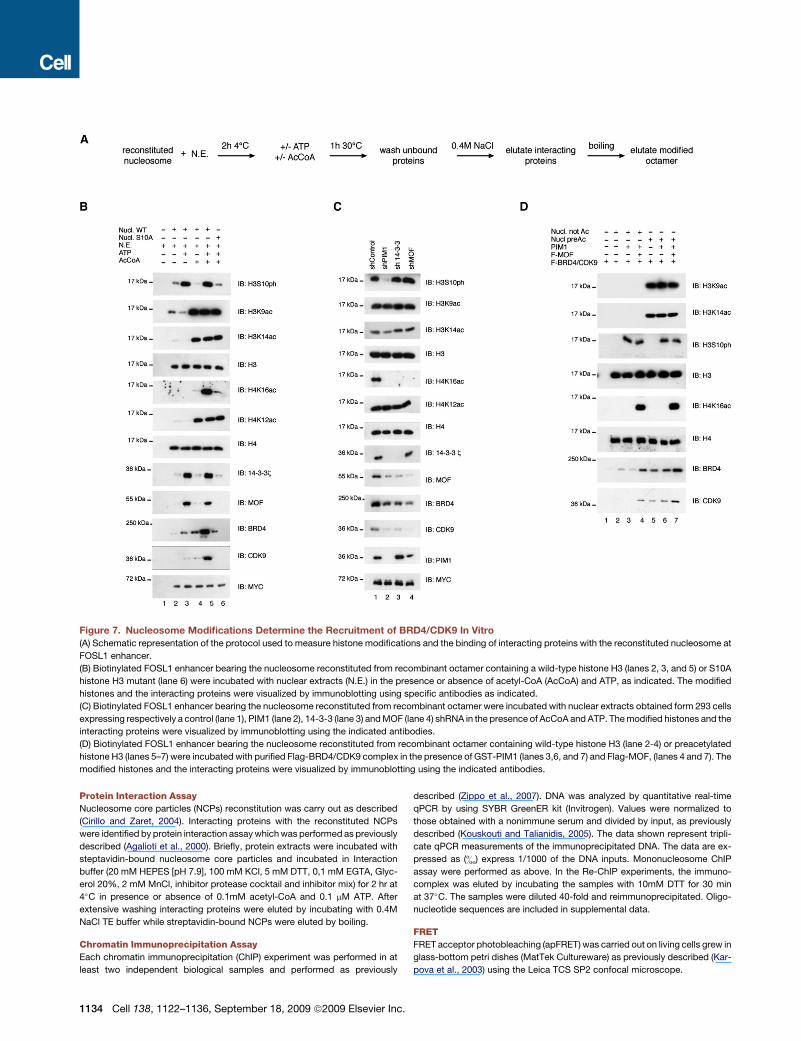
Figure 7. Nucleosome Modifications Determine the Recruitment of BRD4/CDK9 In Vitro
(A) Schematic representation of the protocol used to measure histone modifications and the binding of interacting proteins with the reconstituted nucleosome at
FOSL1 enhancer.
(B) Biotinylated FOSL1 enhancer bearing the nucleosome reconstituted from recombinant octamer containing a wild-type histone H3 (lanes 2, 3, and 5) or S10A
histone H3 mutant (lane 6) were incubated with nuclear extracts (N.E.) in the presence or absence of acetyl-CoA (AcCoA) and ATP, as indicated. The modified
histones and the interacting proteins were visualized by immunoblotting using specific antibodies as indicated.
(C) Biotinylated FOSL1 enhancer bearing the nucleosome reconstituted from recombinant octamer were incubated with nuclear extracts obtained form 293 cells
expressing respectively a control (lane 1), PIM1 (lane 2), 14-3-3 (lane 3) and MOF (lane 4) shRNA in the presence of AcCoA and ATP. The modified histones and the
interacting proteins were visualized by immunoblotting using the indicated antibodies.
(D) Biotinylated FOSL1 enhancer bearing the nucleosome reconstituted from recombinant octamer containing wild-type histone H3 (lane 2-4) or preacetylated
histone H3 (lanes 5–7) were incubated with purified Flag-BRD4/CDK9 complex in the presence of GST-PIM1 (lanes 3,6, and 7) and Flag-MOF, (lanes 4 and 7). The
modified histones and the interacting proteins were visualized by immunoblotting using the indicated antibodies.
Protein Interaction Assay
Nucleosome core particles (NCPs) reconstitution was carry out as described
(Cirillo and Zaret, 2004). Interacting proteins with the reconstituted NCPs
were identified by protein interaction assay which was performed as previously
described (Agalioti et al., 2000). Briefly, protein extracts were incubated with
steptavidin-bound nucleosome core particles and incubated in Interaction
buffer (20 mM HEPES [pH 7.9], 100 mM KCl, 5 mM DTT, 0,1 mM EGTA, Glyc-
erol 20%, 2 mM MnCl, inhibitor protease cocktail and inhibitor mix) for 2 hr at
4�C in presence or absence of 0.1mM acetyl-CoA and 0.1 mM ATP. After
extensive washing interacting proteins were eluted by incubating with 0.4M
NaCl TE buffer while streptavidin-bound NCPs were eluted by boiling.
Chromatin Immunoprecipitation Assay
Each chromatin immunoprecipitation (ChIP) experiment was performed in at
least two independent biological samples and performed as previously
1134 Cell 138, 1122–1136, September 18, 2009 ª2009 Elsevier Inc.
described (Zippo et al., 2007). DNA was analyzed by quantitative real-time
qPCR by using SYBR GreenER kit (Invitrogen). Values were normalized to
those obtained with a nonimmune serum and divided by input, as previously
described (Kouskouti and Talianidis, 2005). The data shown represent tripli-
cate qPCR measurements of the immunoprecipitated DNA. The data are ex-
pressed as (&) express 1/1000 of the DNA inputs. Mononucleosome ChIP
assay were performed as above. In the Re-ChIP experiments, the immuno-
complex was eluted by incubating the samples with 10mM DTT for 30 min
at 37�C. The samples were diluted 40-fold and reimmunoprecipitated. Oligo-
nucleotide sequences are included in supplemental data.
FRET
FRET acceptor photobleaching (apFRET) was carried out on living cells grew in
glass-bottom petri dishes (MatTek Cultureware) as previously described (Kar-
pova et al., 2003) using the Leica TCS SP2 confocal microscope.
FRET measurement was performed using the apFRET software (from Leica),
according to the manufacturer’s instructions.
FRAP
FRAP was carried out using the Leica TCS SP2 confocal microscope on living
cells grew in glass-bottom petri dishes (MatTek Cultureware). Bleaching was
performed with two 208 ms pulses using the 458 nm line of the Argon laser
at 95% power on a 2 mm area. Fluorescent recovery was monitored by collect-
ing 120 images at 1 s intervals with low laser intensity (4% power with 458 laser
line, detection 465-510). Data were collected from at least three independent
experiments and were normalized as previously described (Agresti et al.,
2005).
SUPPLEMENTAL DATA
Supplemental Data include eight figures, two tables, Supplemental Experi-
mental Procedures, and Supplemental References and can be found with this
article online at http://www.cell.com/supplemental/S0092-8674(09)00911-8.
ACKNOWLEDGMENTS
This work was supported by Associazione Italiana Ricerca sul cancro (AIRC),
Fondazione Monte dei Paschi di Siena, and Istituto Toscano Tumori (ITT). We
are in indebted to Isabel Delany, Marco Bianchi, Alessandra Agresti, and Enzo
Scarlato for helpful suggestions and critical reading of the manuscript.
Received: February 2, 2009
Revised: May 13, 2009
Accepted: July 16, 2009
Published: September 17, 2009
REFERENCES
Agalioti, T., Chen, G., and Thanos, D. (2002). Deciphering the transcriptional
histone acetylation code for a human gene. Cell 111, 381–392.
Agalioti, T., Lomvardas, S., Parekh, B., Yie, J., Maniatis, T., and Thanos, D.
(2000). Ordered recruitment of chromatin modifying and general transcription
factors to the IFN-beta promoter. Cell 103, 667–678.
Agresti, A., Scaffidi, P., Riva, A., Caiolfa, V.R., and Bianchi, M.E. (2005). GR
and HMGB1 interact only within chromatin and influence each other’s resi-
dence time. Mol. Cell 18, 109–121.
Barboric, M., Nissen, R.M., Kanazawa, S., Jabrane-Ferrat, N., and Peterlin,
B.M. (2001). NF-kappaB binds P-TEFb to stimulate transcriptional elongation
by RNA polymerase II. Mol. Cell 8, 327–337.
Cai, W., Bao, X., Deng, H., Jin, Y., Girton, J., Johansen, J., and Johansen, K.M.
(2008). RNA polymerase II-mediated transcription at active loci does not
require histone H3S10 phosphorylation in Drosophila. Development 135,
2917–2925.
Calestagne-Morelli, A., and Ausio, J. (2006). Long-range histone acetylation:
biological significance, structural implications, and mechanisms. Biochem.
Cell Biol. 84, 518–527.
Cheung, P., Tanner, K.G., Cheung, W.L., Sassone-Corsi, P., Denu, J.M., and
Allis, C.D. (2000). Synergistic coupling of histone H3 phosphorylation and acet-
ylation in response to epidermal growth factor stimulation. Mol. Cell 5,
905–915.
Cirillo, L.A., and Zaret, K.S. (2004). Preparation of defined mononucleosomes,
dinucleosomes, and nucleosome arrays in vitro and analysis of transcription
factor binding. Methods Enzymol. 375, 131–158.
Core, L.J., and Lis, J.T. (2008). Transcription regulation through promoter-
proximal pausing of RNA polymerase II. Science 319, 1791–1792.
Core, L.J., Waterfall, J.J., and Lis, J.T. (2008). Nascent RNA sequencing
reveals widespread pausing and divergent initiation at human promoters.
Science 322, 1845–1848.
C
Dekker, J., Rippe, K., Dekker, M., and Kleckner, N. (2002). Capturing chromo-
some conformation. Science 295, 1306–1311.
Dey, A., Chitsaz, F., Abbasi, A., Misteli, T., and Ozato, K. (2003). The double
bromodomain protein Brd4 binds to acetylated chromatin during interphase
and mitosis. Proc. Natl. Acad. Sci. USA 100, 8758–8763.
Dey, A., Ellenberg, J., Farina, A., Coleman, A.E., Maruyama, T., Sciortino, S.,
Lippincott-Schwartz, J., and Ozato, K. (2000). A bromodomain protein,
MCAP, associates with mitotic chromosomes and affects G(2)-to-M transition.
Mol. Cell. Biol. 20, 6537–6549.
Dougherty, M.K., and Morrison, D.K. (2004). Unlocking the code of 14–3-3.
J. Cell Sci. 117, 1875–1884.
Eberhardy, S.R., and Farnham, P.J. (2002). Myc recruits P-TEFb to mediate the
final step in the transcriptional activation of the cad promoter. J. Biol. Chem.
277, 40156–40162.
Edmunds, J.W., Mahadevan, L.C., and Clayton, A.L. (2008). Dynamic histone
H3 methylation during gene induction: HYPB/Setd2 mediates all H3K36 trime-
thylation. EMBO J. 27, 406–420.
Ferrai, C., Munari, D., Luraghi, P., Pecciarini, L., Cangi, M.G., Doglioni, C.,
Blasi, F., and Crippa, M.P. (2007). A transcription-dependent micrococcal
nuclease-resistant fragment of the urokinase-type plasminogen activator
promoter interacts with the enhancer. J. Biol. Chem. 282, 12537–12546.
Guenther, M.G., Levine, S.S., Boyer, L.A., Jaenisch, R., and Young, R.A.
(2007). A chromatin landmark and transcription initiation at most promoters
in human cells. Cell 130, 77–88.
Ivaldi, M.S., Karam, C.S., and Corces, V.G. (2007). Phosphorylation of histone
H3 at Ser10 facilitates RNA polymerase II release from promoter-proximal
pausing in Drosophila. Genes Dev. 21, 2818–2831.
Jang, M.K., Mochizuki, K., Zhou, M., Jeong, H.S., Brady, J.N., and Ozato, K.
(2005). The bromodomain protein Brd4 is a positive regulatory component of
P-TEFb and stimulates RNA polymerase II-dependent transcription. Mol.
Cell 19, 523–534.
Jenuwein, T., and Allis, C.D. (2001). Translating the histone code. Science 293,
1074–1080.
Kanazawa, S., Okamoto, T., and Peterlin, B.M. (2000). Tat competes with
CIITA for the binding to P-TEFb and blocks the expression of MHC class II
genes in HIV infection. Immunity 12, 61–70.
Kanno, T., Kanno, Y., Siegel, R.M., Jang, M.K., Lenardo, M.J., and Ozato, K.
(2004). Selective recognition of acetylated histones by bromodomain proteins
visualized in living cells. Mol. Cell 13, 33–43.
Karpova, T.S., Baumann, C.T., He, L., Wu, X., Grammer, A., Lipsky, P., Hager,
G.L., and McNally, J.G. (2003). Fluorescence resonance energy transfer from
cyan to yellow fluorescent protein detected by acceptor photobleaching using
confocal microscopy and a single laser. J. Microsc. 209, 56–70.
Kind, J., Vaquerizas, J.M., Gebhardt, P., Gentzel, M., Luscombe, N.M., Ber-
tone, P., and Akhtar, A. (2008). Genome-wide analysis reveals MOF as a key
regulator of dosage compensation and gene expression in Drosophila. Cell
133, 813–828.
Kouskouti, A., and Talianidis, I. (2005). Histone modifications defining active
genes persist after transcriptional and mitotic inactivation. EMBO J. 24,
347–357.
Lee, D.K., Duan, H.O., and Chang, C. (2001). Androgen receptor interacts with
the positive elongation factor P-TEFb and enhances the efficiency of transcrip-
tional elongation. J. Biol. Chem. 276, 9978–9984.
LeRoy, G., Rickards, B., and Flint, S.J. (2008). The double bromodomain
proteins Brd2 and Brd3 couple histone acetylation to transcription. Mol. Cell
30, 51–60.
Macdonald, N., Welburn, J.P., Noble, M.E., Nguyen, A., Yaffe, M.B., Clynes,
D., Moggs, J.G., Orphanides, G., Thomson, S., Edmunds, J.W., et al. (2005).
Molecular basis for the recognition of phosphorylated and phosphoacetylated
histone h3 by 14–3-3. Mol. Cell 20, 199–211.
Mason, P.B., and Struhl, K. (2005). Distinction and relationship between elon-
gation rate and processivity of RNA polymerase II in vivo. Mol. Cell 17,
831–840.
ell 138, 1122–1136, September 18, 2009 ª2009 Elsevier Inc. 1135

Muse, G.W., Gilchrist, D.A., Nechaev, S., Shah, R., Parker, J.S., Grissom, S.F.,
Zeitlinger, J., and Adelman, K. (2007). RNA polymerase is poised for activation
across the genome. Nat. Genet. 39, 1507–1511.
Ni, Z., Schwartz, B.E., Werner, J., Suarez, J.R., and Lis, J.T. (2004). Coordina-
tion of transcription, RNA processing, and surveillance by P-TEFb kinase on
heat shock genes. Mol. Cell 13, 55–65.
Peterlin, B.M., and Price, D.H. (2006). Controlling the elongation phase of tran-
scription with P-TEFb. Mol. Cell 23, 297–305.
Rea, S., Xouri, G., and Akhtar, A. (2007). Males absent on the first (MOF): from
flies to humans. Oncogene 26, 5385–5394.
Saunders, A., Core, L.J., and Lis, J.T. (2006). Breaking barriers to transcription
elongation. Nat. Rev. Mol. Cell Biol. 7, 557–567.
Simone, C., Stiegler, P., Bagella, L., Pucci, B., Bellan, C., De Falco, G., De
Luca, A., Guanti, G., Puri, P.L., and Giordano, A. (2002). Activation of MyoD-
dependent transcription by cdk9/cyclin T2. Oncogene 21, 4137–4148.
Sims, R.J., 3rd, Belotserkovskaya, R., and Reinberg, D. (2004). Elongation by
RNA polymerase II: the short and long of it. Genes Dev. 18, 2437–2468.
Smith, E.R., Cayrou, C., Huang, R., Lane, W.S., Cote, J., and Lucchesi, J.C.
(2005). A human protein complex homologous to the Drosophila MSL complex
is responsible for the majority of histone H4 acetylation at lysine 16. Mol. Cell.
Biol. 25, 9175–9188.
Soloaga, A., Thomson, S., Wiggin, G.R., Rampersaud, N., Dyson, M.H., Haz-
zalin, C.A., Mahadevan, L.C., and Arthur, J.S. (2003). MSK2 and MSK1
mediate the mitogen- and stress-induced phosphorylation of histone H3 and
HMG-14. EMBO J. 22, 2788–2797.
Strahl, B.D., and Allis, C.D. (2000). The language of covalent histone modifica-
tions. Nature 403, 41–45.
Suganuma, T., and Workman, J.L. (2008). Crosstalk among Histone Modifica-
tions. Cell 135, 604–607.
1136 Cell 138, 1122–1136, September 18, 2009 ª2009 Elsevier Inc.
Taipale, M., Rea, S., Richter, K., Vilar, A., Lichter, P., Imhof, A., and Akhtar, A.
(2005). hMOF histone acetyltransferase is required for histone H4 lysine 16
acetylation in mammalian cells. Mol. Cell. Biol. 25, 6798–6810.
Vicent, G.P., Ballare, C., Nacht, A.S., Clausell, J., Subtil-Rodriguez, A., Quiles,
I., Jordan, A., and Beato, M. (2006). Induction of progesterone target genes
requires activation of Erk and Msk kinases and phosphorylation of histone
H3. Mol. Cell 24, 367–381.
Wade, J.T., and Struhl, K. (2008). The transition from transcriptional initiation to
elongation. Curr. Opin. Genet. Dev. 18, 130–136.
Wang, G., Balamotis, M.A., Stevens, J.L., Yamaguchi, Y., Handa, H., and Berk,
A.J. (2005). Mediator requirement for both recruitment and postrecruitment
steps in transcription initiation. Mol. Cell 17, 683–694.
Walter, W., Clynes, D., Tang, Y., Marmorstein, R., Mellor, J., and Berger, S.L.
(2008). 14–3-3 interaction with histone H3 involves a dual modification pattern
of phosphoacetylation. Mol. Cell. Biol. 28, 2840–2849.
Winter, S., Simboeck, E., Fischle, W., Zupkovitz, G., Dohnal, I., Mechtler, K.,
Ammerer, G., and Seiser, C. (2008). 14–3-3 proteins recognize a histone
code at histone H3 and are required for transcriptional activation. EMBO J.
27, 88–99.
Yang, Z., Yik, J.H., Chen, R., He, N., Jang, M.K., Ozato, K., and Zhou, Q.
(2005). Recruitment of P-TEFb for stimulation of transcriptional elongation
by the bromodomain protein Brd4. Mol. Cell 19, 535–545.
Zeitlinger, J., Stark, A., Kellis, M., Hong, J.W., Nechaev, S., Adelman, K., Lev-
ine, M., and Young, R.A. (2007). RNA polymerase stalling at developmental
control genes in the Drosophila melanogaster embryo. Nat. Genet. 39,
1512–1516.
Zippo, A., De Robertis, A., Serafini, R., and Oliviero, S. (2007). PIM1-depen-
dent phosphorylation of histone H3 at serine 10 is required for MYC-depen-
dent transcriptional activation and oncogenic transformation. Nat. Cell Biol.
9, 932–944.