surgical sepsis and organ crosstalk: the role of the kidney
TRANSCRIPT

SURGICAL SEPSIS AND ORGAN CROSSTALK: THE ROLE OFTHE KIDNEY
Laura E. White, MD1,*, Rahul Chaudhary, MD1,*, Laura J. Moore, MD1, Frederick A. Moore,MD1, and Heitham T. Hassoun, MD1,21 Department of Surgery, The Methodist Hospital and Research Institute, Houston TX2 The Methodist DeBakey Heart and Vascular Center, Houston TX
AbstractAcute kidney injury (AKI) is a common complication of hospitalized patients, and clinicaloutcomes remain poor despite advances in renal replacement therapy. The acceptedpathophysiology of AKI in the setting of sepsis has evolved from one of simple decreased renalblood flow to one that involves a more complex interaction of intra-glomerular microcirculatoryvasodilation combined with the local release of inflammatory mediators and apoptosis. Evidencefrom pre-clinical AKI models suggests that crosstalk occurs between kidneys and other organsystems via soluble and cellular inflammatory mediators, and that this involves both the innate andadaptive immune systems. These interactions are reflected by genomic changes and abnormalrates of cellular apoptosis in distant organs including the lungs, heart, gut, liver, and centralnervous system. The purpose of this article is to review the influence of AKI, particularly sepsis-associated AKI, on inter-organ crosstalk in the context of systemic inflammation and multipleorgan failure (MOF).
KeywordsAcute kidney injury; sepsis; inflammation; apoptosis; immune response
INTRODUCTIONAcute Kidney Injury (AKI) is a common and often catastrophic complication amongsthospitalized patients. It affects 3-7% of patients admitted to the hospital and approximately25-30% of patients in the Intensive Care Unit (ICU) [1]. Mortality rates for ICU patientswith AKI have a reported range from 30-70% even with advances in renal replacementtherapy, and AKI is an independent risk factor for mortality even after adjustment fordemographics, severity of illness and other patient factors [2,3]. AKI has been summarizedby two consensus definitions: 1) The Risk-Injury-Failure-Loss-Endstage renal disease(RIFLE) classification, and 2) The Acute Kidney Injury Network (AKIN) criteria. TheRIFLE classification uses serum creatinine or glomerular filtration rate (GFR) and urine
© 2010 Elsevier Inc. All rights reserved.Correspondence to: Heitham T. Hassoun, M.D. Associate Professor of Cardiovascular Surgery The Methodist Hospital PhysicianOrganization 6550 Fannin Street, SM 1401 Houston, TX 77030 Telephone: 713/441-6201 Fax: 713/709-3058 [email protected].*Contributed equally to the authorship of this manuscriptPublisher's Disclaimer: This is a PDF file of an unedited manuscript that has been accepted for publication. As a service to ourcustomers we are providing this early version of the manuscript. The manuscript will undergo copyediting, typesetting, and review ofthe resulting proof before it is published in its final citable form. Please note that during the production process errors may bediscovered which could affect the content, and all legal disclaimers that apply to the journal pertain.
NIH Public AccessAuthor ManuscriptJ Surg Res. Author manuscript; available in PMC 2012 May 15.
Published in final edited form as:J Surg Res. 2011 May 15; 167(2): 306–315. doi:10.1016/j.jss.2010.11.923.
NIH
-PA Author Manuscript
NIH
-PA Author Manuscript
NIH
-PA Author Manuscript
flow per body weight over time to stratify renal injury by severity, with “risk” as the leastsevere category and “failure” as the most severe category. The AKIN classification modifiedthe RIFLE criteria in 2007 to exclude GFR and classify AKI into stages 1-3, with stage 3representing the requirement for renal replacement therapy [4].
Despite the advancement in renal replacement therapy, the mortality rates associated withAKI have remained unchanged over the past 2 decades [3]. Both clinical and translationallaboratory studies have demonstrated very complex mechanisms of interactions between theinjured kidney and distant organs such as the lung, heart, liver, gut, brain and hematologicalsystem. Recent studies on AKI-associated distant organ dysfunction have highlighted theimportance of both the innate and adaptive immune response, activation of pro-inflammatory cascades and an alteration in transcriptional events during ischemic AKI. Forexample, cell adhesion molecule and cytokine-chemokine expression, apoptosisdysregulation and leukocyte trafficking to distant organs all occur during ischemic AKI. Thegoal of this manuscript is to review emerging concepts regarding the clinical significance ofsepsis-associated AKI, the altered immune response that follows, and the mechanisms bywhich AKI contributes to distant organ injury. For a complete list of abbreviations used inthis manuscript, please see Table 1.
SURGICAL SEPSIS AND ITS ROLE IN AKISepsis is a well-established risk factor for AKI, and mortality rates in patients with both AKIand sepsis are much greater than the mortality rate in patients with either AKI or sepsisalone, particularly in the setting of MOF [5]. Thus, the combination of sepsis and AKI posesa particularly serious problem and the concept that sepsis-associated AKI may have adistinct pathophysiology from other etiologies of AKI is supported not only by experimentaldata and evidence from small clinical studies, but also by epidemiological data showing‘dose response’ trends in incidence rates and outcomes for septic AKI by severity of eithersepsis or AKI [5-11] (Figure 1).
While the etiology of AKI in critically ill patients is multi-factorial, sepsis has consistentlybeen a leading contributing factor for AKI in the ICU setting [12-16]. The Centers forDisease Control has listed sepsis as the 10th leading cause of death, and annual costs due tothis disease exceed $17 billion [17]. The National Surgical Quality Improvement Project(NSQIP) dataset from the American College of Surgeons defines sepsis as the presence ofsystemic inflammatory response syndrome (SIRS) with a source of infection, as documentedby positive blood cultures or purulence from any site thought to be causative [18]. Severesepsis is defined as sepsis associated with organ dysfunction, hypoperfusion abnormality, orsepsis-induced hypotension by the American College of Chest Physicians/Society of CriticalCare Medicine (ACCP/SCCM) Consensus Conference Guidelines [19]. Severe sepsis is notseparately identified by NSQIP data but is included in the definition of septic shock, sepsiswith organ and/or circulatory dysfunction (Table 2) [20,21].
Surgical sepsis, defined as sepsis requiring surgical intervention for source control or sepsiswithin 14 days of a surgical procedure, occurs more frequently than other post-operativecomplications such as pulmonary embolism and myocardial infarction [18,21]. Thepublished incidence rate of surgical sepsis is 3 cases per 1,000 patients and it carries a highmortality rate ranging from 26-50% [16]. The prevalence of AKI in patients with severesepsis or septic shock has been reported as high as 43% [14]. While the epidemiology ofsurgical sepsis-associated AKI is largely unknown, prior studies describing sepsis-associatedAKI have consistently concluded that it contributes to nearly two-fold higher mortality thaneither non-septic AKI or sepsis alone [12-16]. This is likely due to the fact that AKI rarely
White et al. Page 2
J Surg Res. Author manuscript; available in PMC 2012 May 15.
NIH
-PA Author Manuscript
NIH
-PA Author Manuscript
NIH
-PA Author Manuscript

occurs in isolation and is associated with distant organ dysfunction in the context of multipleorgan failure (MOF).
Surgical sepsis is considered different from medical sepsis for several reasons. First, surgicaltrauma and anesthetic agents used during surgery alter host local and systemic immunefunction. Surgical trauma systemically activates macrophages, neutrophils, natural killercells and endothelial cells of the innate immune response which then in turn synthesizemediators including tumor necrosis factor-α (TNF-α) and interleukin-6 (IL-6). The adaptiveimmune response is also activated, mediated by pro-inflammatory type-1 helper cells (Th1)and anti-inflammatory type-2 helper cells (Th2) [22]. Additionally, in contrast to themedical ICU population, patients who have surgical sepsis often require source control inthe form of urgent surgical intervention. The timing of source control must be coordinatedwith resuscitation efforts, but has the potential to dramatically reverse the cycle of septicshock. Patients with surgical sepsis who are at highest risk for AKI are those whose clinicalinstability requires aggressive volume resuscitation and hemodynamic support with broadspectrum antimicrobial coverage prior to operative intervention. The appropriate timing ofsource control is unknown, however expert consensus opinion recommends this should becompleted within six hours in order to break the cycle of persistent septic shock [23].
Traditional concepts of sepsis-associated AKIThe pathophysiology of sepsis-associated AKI is complex and multi-factorial. Traditionally,AKI in sepsis and septic shock was thought to result from renal ischemia secondary tovasoconstriction and inadequate renal blood flow (RBF). Some of the major mechanismsthat have also been linked either directly or indirectly to sepsis-associated AKI include: 1)alterations in hemodynamics with subsequent renal vasoconstriction leading to ischemia andtissue hypoxia, 2) hyperglycemia causing functional alterations in leukocytes andmacrophages leading to inflammation, and 3) activation of the coagulation and fibrinolyticcascades leading to disseminated intravascular coagulation (DIC) and micro-vascularthrombosis [11,24].
Changing paradigm in the pathophysiology of sepsis-associated AKIOur understanding of sepsis-associated AKI pathophysiology is shifting from renalvasoconstriction, ischemia and acute tubular necrosis to that of heterogeneous vasodilation,hyperemia and acute tubular apoptosis. The concept of renal vasoconstriction and kidneyischemia as a key pathogenic factor is certainly valid for all low-flow states (i.e. cardiogenicor hemorrhagic shock). However, during hyperdynamic states (i.e. sepsis and other acutesystemic inflammatory conditions), the hemodynamic alterations within the kidney appear tobe heterogeneous, with reduced perfusion to microvascular beds despite increased total RBF[25]. Human data on renal hemodynamics in sepsis is limited, but a recent systematic reviewof the available experimental evidence showed that in all three human studies and in 30% ofanimal studies, RBF remained unchanged or even increased. Cardiac output (CO) was foundto be the only statistically significant predictor of RBF. Furthermore, the majority of studiesreporting a reduction in renal blood flow were derived mostly from hypodynamic modelscharacterized by a reduced cardiac output. In multiple experimental models in whichhyperdynamic sepsis was produced, RBF increased proportionately to CO. Therefore, it islikely that the AKI associated with sepsis occurs in a setting of normal or even raised RBFresulting in a hyperemic kidney [25-27].
The loss of GFR can now be explained by the differences in the glomerular capillarypressure created by the afferent and efferent renal vessels. In this condition, there isdisproportionally increased renal vasodilation in the efferent blood vessels as compared tothe afferent vessels. This causes a reduction in glomerular capillary pressure, in turn, leading
White et al. Page 3
J Surg Res. Author manuscript; available in PMC 2012 May 15.
NIH
-PA Author Manuscript
NIH
-PA Author Manuscript
NIH
-PA Author Manuscript
to a reduced GFR and causing oliguria and reduced solute clearance. This hypothesis wasconfirmed with animal models of hyperdynamic sepsis in which angiotensin II, avasoconstrictor hormone which causes a preferential increase in efferent arteriole resistance,was administered by continuous infusion. The animals which received the infusiondemonstrated a restored arterial blood pressure and a significantly increased urine outputand creatinine clearance compared to placebo [28]. Thus, it seems that sepsis-associatedAKI does not result from ischemia secondary to decreased overall renal blood flow, butinstead from derangements inside the glomerulus leading to decreased GFR.
Though sepsis-associated AKI may result from heterogeneous hypoperfusion as opposed toglobal renal hypoperfusion, similar injury patterns are demonstrated in experimental modelsof both sepsis and IRI induced AKI. In addition to intra-glomerular vascular changes, non-hemodynamic effects of sepsis-associated AKI are locally mediated by the release ofinflammatory cytokines, particularly TNF-α, with subsequent tubular cell apoptosis [29].Studies have shown that endotoxin stimulates the release of TNF-α from glomerularmesangial cells [30]. Additional work has demonstrated attenuation of TNF-α mediatedacute renal failure in both TNF receptor-neutralized and in TNF receptor knockout mice[31,32]. Cellular apoptosis, which is an energy-requiring and genetically-directed process,has been demonstrated to occur alongside necrosis in experimental models of both ischemia-reperfusion and septic acute kidney injury. Apoptosis was elicited in cultured kidneyproximal tubular and glomerular cells by both TNF-α and lipopolysaccharide (LPS) [33,34].Caspases, enzymes responsible for carrying out apoptosis, are inhibited in animal models ofboth LPS-induced sepsis and kidney IRI, and in this setting, mice treated with caspaseinhibitors are protected from both sepsis and IRI associated AKI [35,36].
Hyperglycemia and DIC commonly occur in patients with sepsis and MOF, and therapiesincluding intensive insulin therapy (IIT) and activated protein C (APC) decrease both theonset of sepsis-associated AKI and mortality [11,37]. The benefits of IIT are most profoundin patients with surgical sepsis, and a number of physiologic mechanisms of renal protectionhave been proposed [38]. Despite a lack of influence on hemodynamics, IIT improves thelipid profile in patients with sepsis and MOF, and attenuation of both ischemic andendotoxemic AKI by high density lipoprotein has been demonstrated in experimentalmodels [39-41]. AKI and hyperglycemia are also associated with an increase in expressionof inducible nitric oxide synthase (iNOS), endothelial activation of ICAM-1, upregulation ofpro-inflammatory cytokines (TNFα, IL-6), release of oxidative stress mediators andinfiltration of inflammatory cells, which lead to renal tubular injury [38,42,43].Additionally, APC has anti-inflammatory, antithrombotic and profibrinolytic properties andreduces mortality in severe sepsis [37]. In a model of polymicrobial sepsis induced by cecalligation and puncture, APC administration decreased the expression of inflammatorycytokines, the incidence of apoptosis and the presence of acute tubular necrosis [44].
In summary, AKI associated with sepsis can be characterized by a vasomotor nephropathywhich includes disturbances in renal microcirculation, activation of pro-inflammatorymediators, and activation of renal cell apoptosis, thus resulting in kidney failure andimbalances in body water and electrolyte homeostasis. Due to the difficulties associated withmeasuring these effects simultaneously and the complexity of the condition, further studieswill be needed to elucidate the detailed mechanisms of sepsis-associated AKI and thepathway for recovery of kidney function after injury.
KIDNEY CROSSTALK WITH DISTANT ORGANSThe incidence of AKI continues to increase and is associated with a changing spectrum ofillnesses, significant co-morbid and extra-renal complications, and unsatisfactory preventive
White et al. Page 4
J Surg Res. Author manuscript; available in PMC 2012 May 15.
NIH
-PA Author Manuscript
NIH
-PA Author Manuscript
NIH
-PA Author Manuscript

or treatment strategies [45-47]. Despite advances in treatment such as renal replacementtherapy, mortality rates associated with AKI have not changed significantly since the 1950’s[2,3]. This is likely due to the fact that AKI rarely occurs in isolation and is usually acomponent of inter-organ crosstalk and MOF. It is apparent that much of the increased riskof death is derived from extra-renal complications related to remote organ damage anddysfunction. More recently, animal studies have shown a direct effect of AKI on distantorgans such as the lung, brain, liver and the gut [48-53]. These animal studies includemodels of IRI and sepsis, namely LPS endotoxin-induced sepsis, due to its reproducibility increating distant organ failure including AKI [54].
Kidney- Lung interactions during AKICurrently, the kidney and lung represent the two most commonly involved organs in MOF[55]. Acute lung injury (ALI) is defined by the American-European Consensus Conferenceon Acute Respiratory Distress Syndrome (ARDS) as a PaO2/FiO2 ratio of less than 300 andchest radiograph findings of acute bilateral infiltrates in the absence of elevated cardiacfilling pressures [56]. Acute hypoxia is a frequent presenting symptom of postoperativesepsis, and a decreased PaO2/FiO2 ratio results from increased pulmonary shunting inresponse to acidosis and carbon monoxide retention. ALI accounts for a significantcomponent of the mortality associated with AKI even in the absence of volume overloadcontributed by renal failure [57-59]. The mortality of combined AKI and ALI is extremelyhigh and may approach 80% [60]. Therefore, defining the mechanisms of kidney-lungcrosstalk in the critically ill is necessary for reducing overall mortality associated with AKI.
Mechanisms of Kidney-Lung CrosstalkThe mechanisms of AKI-associated lung injury remain incompletely understood. Severalstudies have demonstrated the involvement of pro-inflammatory and pro-apoptotic factorssuch as leukocyte trafficking, cytokines/chemokines, activation of caspases, oxidative stress,and uremic toxins. AKI and ALI act as a self propagating cycle (Figure 2). AKI leads tolung injury and inflammation and in turn, ALI, with its attendant hypoxemia andhypercapnia worsened by mechanical ventilation-associated high positive-end expiratorypressure (PEEP), causes a decline in renal hemodynamics and function. Experimentalevidence has demonstrated that lung injury in the setting of AKI is featured by markedpulmonary microvascular permeability, erythrocyte sludging in lung capillaries, interstitialedema, focal alveolar hemorrhage and inflammatory cell infiltration [49,52,59]. Kidneyischemia-reperfusion injury (IRI) has been shown not only to cause an increase inpulmonary vascular permeability, but it also results in down-regulation of the pulmonaryepithelial salt and water transporters (ENaC, Na, K-ATPase and aquaporins) in the rat lung,all of which contribute to decreased alveolar fluid clearance [49,59,61].
Role of inflammation, cytokines and chemokines—The upregulation of pro-inflammatory genes and inflammatory cytokines are important mediators connecting theeffects of AKI on distant organs [62]. Recent studies by Hassoun and colleaguesinvestigated the lung structural, functional and genomic response during kidney IRI orbilateral nephrectomy (BNx) [52,63]. A comprehensive genomic map and analysis ofinflammation-associated transcriptional changes in local and remote organs during ischemicAKI identified markedly similar transcriptomic changes occurring concomitantly in both thekidney and lung during AKI, including significant changes in the expression of 109prominent proinflammatory genes including Cd14, serum amyloid A 3 (Saa3), lipocalin 2,CXCL-2 and the IL-1 receptor (IL-1r2) [63]. Of note, the demonstrated changes in the lungfollowing ischemic AKI were distinguishable from those caused by uremia alone andinvolved early and persistent activation of proinflammatory and proapoptotic pathways [52].
White et al. Page 5
J Surg Res. Author manuscript; available in PMC 2012 May 15.
NIH
-PA Author Manuscript
NIH
-PA Author Manuscript
NIH
-PA Author Manuscript

Cytokines/chemokines have a key role in the initiation as well as progression of both AKIand ALI. Interleukin-6 (IL-6), inteleukin-10 (IL-10), and Saa3 are increased in the lungduring AKI [59,64]. Klein et al. demonstrated that IL-6 is elevated after both ischemic AKIand BNx in mice and in the serum of patients with AKI, and furthermore predicts mortality[61]. The same investigators also discovered that IL-6-deficiency reduced lung neutrophilinfiltration, myeloperoxidase activity, expression of the chemokines KC (CXCL-1) andMIP-2 (macrophage inflammatory protein-2) and capillary leak during AKI in mice.Another recent study has demonstrated cytokine-mediated pulmonary injury during AKI,including increased IL-6, IL-1, IL-12 (p40), granulocyte macrophage colony-stimulatingfactor (GM-CSF), whereby anti-inflammatory IL-10 inhibits both production and action ofnumerous pro-inflammatory cytokines and reduced lung injury during AKI [53,61]. Clearly,the mechanisms by which inflammation might initiate and affect AKI-induced ALI arecomplex and only partially understood, but may be influenced by regulation of cytokinesand chemokines.
Lung apoptosis—There is increasing evidence that pulmonary cell apoptosis may play animportant role in the pathophysiology of AKI induced ALI [65]. Both enhanced pulmonaryendothelial and epithelial cell apoptosis and delayed leukocyte apoptosis have beenassociated with ALI [66-68]. Investigations of vascular permeability have highlighted theimportance of the balance between complex tethering forces involved in cell-to-cell andcell-to-extracellular matrix interactions. These studies have also shown that endothelialapoptosis leads to the disruption of these complex interactions and a potential for loss ofendothelial barrier function [69]. Recent laboratory data demonstrated that kidney IRI in ratsinduces pulmonary endothelial apoptosis and lung injury, and these effects were abrogatedby the caspase inhibitor z-VAD-fmk, suggesting a direct role of caspase dependent apoptosisin ischemic AKI induced lung injury [70].
Role of innate and adaptive immunity—Kidney ischemia-reperfusion injury activatesboth the innate and adaptive immune responses [71]. The innate immune response includesneutrophils, macrophages and possibly natural killer cells. The adaptive immune system isalso activated after kidney IRI via CD4 + T cells, particularly of the Th1 phenotype [71].Pathogenesis of post-ischemic injury is thought to be mediated by interferon-γ (INF-γ)produced by CD4+ T cells [72]. Additionally, the inactivation of IL-16, a chemoattractantstrongly expressed on renal tubules during IRI, resulted in less IRI-induced CD4+
lymphocyte trafficking and subsequent kidney injury and dysfunction [73]. T lymphocytetrafficking occurs as early as one hour after kidney IRI and persists for up to 6 weeks postinjury [74,75]. In fact, these T cells may recognize antigens released during kidney IRI andsubsequently target the kidney in an autoimmune response, leading to long term progressionof renal dysfunction. This mechanism was demonstrated in a murine adoptive transfer modelin which naïve mice received T cells from mice who were 6 weeks post-kidney IRI andsubsequently developed increased albuminuria [74].
Leukocytes play a fundamental role in the development of ALI/ARDS and several recentstudies have documented lung leukocyte activation and trafficking during experimental AKI.In rat models of both bilateral kidney IRI and bilateral nephrectomy, studies have shownearly and sustained lung neutrophil sequestration [53,76]. While neutrophils are the keymediators in several extra-pulmonary models of ALI such as sepsis and mesenteric IRI, theirimportance in AKI-associated lung injury is less clear, and in fact, uremic neutrophils havebeen shown to attenuate ALI in mice [51].
Macrophage and lymphocyte infiltration and/or proliferation are other potential mediators ofthe distant organ effects of AKI. Macrophage activation inhibitor CNI-1493 has been shownto attenuate lung microvascular leak following bilateral kidney IRI in rats [49]. In addition,
White et al. Page 6
J Surg Res. Author manuscript; available in PMC 2012 May 15.
NIH
-PA Author Manuscript
NIH
-PA Author Manuscript
NIH
-PA Author Manuscript

unilateral kidney ischemia has resulted in increased macrophages in both the contralateralkidney as well as the cardiac interstitium [77]. Lie et al have recently reported on theinfiltration of activated CD3+ CD8+ cytotoxic T lymphocytes into mouse lungs duringkidney IRI and their potential role in mediating lung apoptosis in this setting [78].
Kidney interactions with other distant organsAcute kidney injury interacts with and affects many other major organ-systems including theheart, brain and central nervous system, the hematologic system, the liver and gut [79].Though the exact pathophysiology of these interactions are unclear, the general mechanismsby which AKI induces distant organ effects remain fairly universal and includeinflammation, activation of both soluble and cellular factors, as well as hemodynamic andneurohumoral alterations which lead to cellular apoptosis and organ damage (Figure 3).
Kidney-Heart Interactions—While cardiovascular collapse is one of the most commoncauses for death in the setting of AKI, the mechanisms involved are incompletelyunderstood [80]. It has been demonstrated by Kelly et al. that during kidney IRI in rats thereis left ventricular (LV) dilatation, increased left ventricular end diastolic and end systolicdiameter, increased relaxation time, and decreased fractional shortening [81]. It has alsobeen shown that cardiac ischemia, in a setting of AKI, causes a sustained ventricularfibrillation of longer duration than cardiac ischemia without AKI [82]. Conversely, somestudies have shown that ischemic preconditioning of the kidney can induce distant organprotection in certain circumstances, protecting the myocardium against irreversible damageproduced by prolonged coronary artery occlusion under hypothermic conditions [47].
Cardiac myocyte apoptosis and neutrophil infiltration are two of the most importantcontributors to the pathophysiology of myocardial infarction during AKI and transgenicmodels have demonstrated that even apoptosis alone can lead to lethal heart failure[5,31,77,78,83]. During AKI, there is an increased amount of both cardiac and systemicTNF-alpha and IL-1 along with increased expression of ICAM-1 mRNA, which results inmyocyte apoptosis and neutrophil infiltration of the heart [48,84]. Decreased renal ischemiatime attenuates cardiac apoptosis and IL-1 and ICAM-1 levels, as does administration ofanti-TNF-α antibodies [48].
Kidney-Brain Interactions—Effects of AKI on brain and nervous system include earlyclinical signs such as clumsiness, fatigue, impaired concentration and apathy which maylater progress to delirium, confusion and coma. Interestingly, dialysis improves, but fails tofully correct, central nervous system manifestations of renal failure in both the acute andchronic setting [85]. Much of the symptoms of encephalopathy are attributed to uremictoxins, however both soluble and cellular inflammatory mediators, similar to those seen inkidney-lung and kidney-heart interactions, have also been implicated.
It has been demonstrated that AKI causes an increase in the levels of soluble mediators suchas KC, G-CSF and GFAP in the cerebral cortex and hippocampus of the brain, which mayfunction to recruit neutrophils to sites of neuronal damage. This increased KC and G-CSF inthe brain potentially represent increased neuronal production of these pro-inflammatoryfactors or an accumulation of these proteins through an altered blood-brain barrier(increased micro-vascular permeability) arising from a systemic or renal source. AKI alsocauses a cell-mediated inflammatory response in the brain such as seen with activatedmicroglial cells (brain macrophages) [86].
Kidney-Liver Interactions—Liver injury often correlates with severity of kidney injury.Ischemic AKI has been shown to induce oxidative stress and promote inflammation,
White et al. Page 7
J Surg Res. Author manuscript; available in PMC 2012 May 15.
NIH
-PA Author Manuscript
NIH
-PA Author Manuscript
NIH
-PA Author Manuscript
apoptosis and tissue damage in hepatocytes. Hepatic stellate cells (HSCs) are known toregulate leukocyte trafficking and activation by secreting chemokines such as interleukin-8(IL-8) [87]. Crosstalk between CD40-expressing HSCs and immune effector cells likelyoccurs through activating nuclear factor κβ (NF-κβ) and c-Jun N-terminal kinase to up-regulate chemokine secretion [88]. These HSCs are LPS-inducible, and LPS endotoxemiacauses hepatic injury by enhancing neutrophil transmigration out of the hepatic sinusoid andinto the liver parenchyma [89].
During ischemic and non-ischemic AKI, oxidative stress causes hepatic malondialdehyde,an index of lipid peroxidation, to increase while total glutathione (a antioxidant) decreases[90]. Oxidative stress occurs as a component of the surgical stress response, particularlyafter IRI, sepsis, kidney and liver failure [91-93]. Oxidative stress also plays an importantrole in sepsis-associated AKI. In an animal model of sepsis induced by cecal ligation andpuncture, the administration of antioxidants ethyl pyruvate and methyl-2-acetamidoacrylate(M2AA) significantly reduced mortality, improved the pro- and anti-inflammatory cytokineresponse, and attenuated liver and kidney injury [94,95]. Kidney-liver crosstalk during AKItherefore likely occurs by a complex combination of soluble inflammatory mediators andcellular immunity.
Kidney-Gut Interactions—In the past, investigators and clinicians have labeled the gutas the “motor” of MOF due to its ability to amplify the systemic SIRS response in the settingof shock and gut hypoperfusion [96-98]. Mechanisms by which this crosstalk occurs includeincreased intestinal epithelial permeability, interactions between host and bacterialpathogens, and propagation of toxins to distant organs via the lymphatic system [97,99-101].These mechanisms could potentially play a role in the converse direction of interactionsbetween the kidney and the gut during AKI.
Our understanding of how AKI influences gut physiology is limited, however the gut hasbeen shown to mitigate some adverse effects of AKI, particularly in the handling of excesspotassium. Clinical studies have long demonstrated the increased secretion of potassium bythe colon and rectum [102,103]. Recent literature has linked channel-inducing factor(CHIF), a potassium channel regulator found in both the kidney and the colon, to ischemicIRI. In animals subjected to kidney IRI, CHIF was upregulated in the colon while a renalsecretory potassium channel, ROMK1, was downregulated in the kidney, possiblyexplaining why hyperkalemia does not universally occur in AKI [50,104]. Aldosteronesecreted from the kidney is associated with the upregulation of CHIF in the gut and mayserve a role in kidney-gut crosstalk, however much potential for research exists inelucidating the mechanisms of AKI induced gut injury [50].
CONCLUSIONAKI is a common complication in hospitalized patients, and mortality rates of AKI inconjunction with sepsis and MOF are unacceptably high. Our understanding of thepathophysiology of sepsis-associated AKI continues to evolve, as does our understanding ofthe mechanisms by which AKI induces distant organ failure. Further investigation of AKI-induced distant organ effects may lead to potential therapeutic targets and a future reductionin patient mortality.
AcknowledgmentsSupported by: NIH K08HL089181
White et al. Page 8
J Surg Res. Author manuscript; available in PMC 2012 May 15.
NIH
-PA Author Manuscript
NIH
-PA Author Manuscript
NIH
-PA Author Manuscript
REFERENCES1. Barry, M.; Brenner, FCR. Brenner and Rector’sThe Kidney. Saunders, An Imprint of Elsevier;
2007.2. Levy EM, Viscoli CM, Horwitz RI. The effect of acute renal failure on mortality. A cohort analysis.
Jama. 1996; 275:1489–1494. [PubMed: 8622223]3. Metnitz PG, Krenn CG, Steltzer H, Lang T, Ploder J, Lenz K, Le Gall JR, Druml W. Effect of acute
renal failure requiring renal replacement therapy on outcome in critically ill patients. Crit Care Med.2002; 30:2051–2058. [PubMed: 12352040]
4. Cruz DN, Ricci Z, Ronco C. Clinical review: RIFLE and AKIN--time for reappraisal. Crit Care.2009; 13:211. [PubMed: 19638179]
5. Edelstein, CL.; S., R. Disease of the Kidney and Urinary Tract. Lippincott Williams and Wilkins;Philadelphia: 2001.
6. Brenner M, Schaer GL, Mallory DL, Suffredini AF, Parrillo JE. Detection of renal blood flowabnormalities in septic and critically ill patients using a newly designed indwelling thermodilutionrenal vein catheter. Chest. 1990; 98:170–179. [PubMed: 2361386]
7. Langenberg C, Wan L, Bagshaw SM, Egi M, May CN, Bellomo R. Urinary biochemistry inexperimental septic acute renal failure. Nephrol Dial Transplant. 2006; 21:3389–3397. [PubMed:16998215]
8. Langenberg C, Wan L, Egi M, May CN, Bellomo R. Renal blood flow in experimental septic acuterenal failure. Kidney Int. 2006; 69:1996–2002. [PubMed: 16641923]
9. Lucas CE, Rector FE, Werner M, Rosenberg IK. Altered renal homeostasis with acute sepsis.Clinical significance. Arch Surg. 1973; 106:444–449. [PubMed: 4696717]
10. Rector F, Goyal SC, Rosenberg IK, Lucas CE. Renal hyperemia in association with clinical sepsis.Surg Forum. 1972; 23:51–53. [PubMed: 4671024]
11. Van den Berghe G, Wouters P, Weekers F, Verwaest C, Bruyninckx F, Schetz M, Vlasselaers D,Ferdinande P, Lauwers P, Bouillon R. Intensive insulin therapy in the critically ill patients. N EnglJ Med. 2001; 345:1359–1367. [PubMed: 11794168]
12. Bagshaw SM, Uchino S, Bellomo R, Morimatsu H, Morgera S, Schetz M, Tan I, Bouman C,Macedo E, Gibney N, Tolwani A, Oudemans-van Straaten HM, Ronco C, Kellum JA. Septic acutekidney injury in critically ill patients: clinical characteristics and outcomes. Clin J Am SocNephrol. 2007; 2:431–439. [PubMed: 17699448]
13. Lopes JA, Jorge S, Resina C, Santos C, Pereira A, Neves J, Antunes F, Prata MM. Acute renalfailure in patients with sepsis. Crit Care. 2007; 11:411. [PubMed: 17466080]
14. Oppert M, Engel C, Brunkhorst FM, Bogatsch H, Reinhart K, Frei U, Eckardt KU, Loeffler M,John S. Acute renal failure in patients with severe sepsis and septic shock--a significantindependent risk factor for mortality: results from the German Prevalence Study. Nephrol DialTransplant. 2008; 23:904–909. [PubMed: 18065435]
15. Yegenaga I, Hoste E, Van Biesen W, Vanholder R, Benoit D, Kantarci G, Dhondt A, Colardyn F,Lameire N. Clinical characteristics of patients developing ARF due to sepsis/systemicinflammatory response syndrome: results of a prospective study. Am J Kidney Dis. 2004; 43:817–824. [PubMed: 15112172]
16. Hoste EA, Lameire NH, Vanholder RC, Benoit DD, Decruyenaere JM, Colardyn FA. Acute renalfailure in patients with sepsis in a surgical ICU: predictive factors, incidence, comorbidity, andoutcome. J Am Soc Nephrol. 2003; 14:1022–1030. [PubMed: 12660337]
17. Angus DC, Linde-Zwirble WT, Lidicker J, Clermont G, Carcillo J, Pinsky MR. Epidemiology ofsevere sepsis in the United States: analysis of incidence, outcome, and associated costs of care.Crit Care Med. 2001; 29:1303–1310. [PubMed: 11445675]
18. Moore LJ, Moore FA, Todd SR, Jones SL, Turner KL, Bass BL. Sepsis in general surgery: the2005 2007 national surgical quality improvement program perspective. Arch Surg. 2010; 145:695–700. [PubMed: 20644134]
19. Bone RC, Balk RA, Cerra FB, Dellinger RP, Fein AM, Knaus WA, Schein RM, Sibbald WJ.Definitions for sepsis and organ failure and guidelines for the use of innovative therapies in sepsis.
White et al. Page 9
J Surg Res. Author manuscript; available in PMC 2012 May 15.
NIH
-PA Author Manuscript
NIH
-PA Author Manuscript
NIH
-PA Author Manuscript
The ACCP/SCCM Consensus Conference Committee. American College of Chest Physicians/Society of Critical Care Medicine. Chest. 1992; 101:1644–1655. [PubMed: 1303622]
20. Moore LJ, Moore FA, Jones SL, Xu J, Bass BL. Sepsis in general surgery: a deadly complication.Am J Surg. 2009; 198:868–874. [PubMed: 19969144]
21. Moore L, Turner K, Todd S, Sucher J, Mckinley B, Moore F. Computerized clinical decisionsupport improves mortality in intra abdominal surgical sepsis. American Journal of Surgery. 2010
22. Decker D, Tolba R, Springer W, Lauschke H, Hirner A, von Ruecker A. Abdominal surgicalinterventions: local and systemic consequences for the immune system--a prospective study onelective gastrointestinal surgery. J Surg Res. 2005; 126:12–18. [PubMed: 15916969]
23. Solomkin JS, Mazuski JE, Bradley JS, Rodvold KA, Goldstein EJ, Baron EJ, O’Neill PJ, ChowAW, Dellinger EP, Eachempati SR, Gorbach S, Hilfiker M, May AK, Nathens AB, Sawyer RG,Bartlett JG. Diagnosis and management of complicated intra-abdominal infection in adults andchildren: guidelines by the Surgical Infection Society and the Infectious Diseases Society ofAmerica. Surg Infect (Larchmt). 2010; 11:79–109. [PubMed: 20163262]
24. Shimamura K, Oka K, Nakazawa M, Kojima M. Distribution patterns of microthrombi indisseminated intravascular coagulation. Arch Pathol Lab Med. 1983; 107:543–547. [PubMed:6688518]
25. Molitoris BA. Renal blood flow in sepsis: a complex issue. Crit Care. 2005; 9:327–328. [PubMed:16137373]
26. Langenberg C, Bellomo R, May C, Wan L, Egi M, Morgera S. Renal blood flow in sepsis. CritCare. 2005; 9:R363–374. [PubMed: 16137349]
27. Bellomo R, Wan L, Langenberg C, May C. Septic acute kidney injury: new concepts. Nephron ExpNephrol. 2008; 109:e95–100. [PubMed: 18802375]
28. Wan L, Langenberg C, Bellomo R, May CN. Angiotensin II in experimental hyperdynamic sepsis.Crit Care. 2009; 13:R190. [PubMed: 19948019]
29. Lerolle N, Nochy D, Guerot E, Bruneval P, Fagon JY, Diehl JL, Hill G. Histopathology of septicshock induced acute kidney injury: apoptosis and leukocytic infiltration. Intensive Care Med.2010; 36:471–478. [PubMed: 19924395]
30. Baud L, Oudinet JP, Bens M, Noe L, Peraldi MN, Rondeau E, Etienne J, Ardaillou R. Productionof tumor necrosis factor by rat mesangial cells in response to bacterial lipopolysaccharide. KidneyInt. 1989; 35:1111–1118. [PubMed: 2549293]
31. Cunningham PN, Dyanov HM, Park P, Wang J, Newell KA, Quigg RJ. Acute renal failure inendotoxemia is caused by TNF acting directly on TNF receptor-1 in kidney. J Immunol. 2002;168:5817–5823. [PubMed: 12023385]
32. Knotek M, Rogachev B, Wang W, Ecder T, Melnikov V, Gengaro PE, Esson M, Edelstein CL,Dinarello CA, Schrier RW. Endotoxemic renal failure in mice: Role of tumor necrosis factorindependent of inducible nitric oxide synthase. Kidney Int. 2001; 59:2243–2249. [PubMed:11380827]
33. Jo SK, Cha DR, Cho WY, Kim HK, Chang KH, Yun SY, Won NH. Inflammatory cytokines andlipopolysaccharide induce Fas-mediated apoptosis in renal tubular cells. Nephron. 2002; 91:406–415. [PubMed: 12119470]
34. Schumer M, Colombel MC, Sawczuk IS, Gobe G, Connor J, O’Toole KM, Olsson CA, Wise GJ,Buttyan R. Morphologic, biochemical, and molecular evidence of apoptosis during the reperfusionphase after brief periods of renal ischemia. Am J Pathol. 1992; 140:831–838. [PubMed: 1562048]
35. Guo R, Wang Y, Minto AW, Quigg RJ, Cunningham PN. Acute renal failure in endotoxemia isdependent on caspase activation. J Am Soc Nephrol. 2004; 15:3093–3102. [PubMed: 15579512]
36. Zhang X, Zheng X, Sun H, Feng B, Chen G, Vladau C, Li M, Chen D, Suzuki M, Min L, Liu W,Garcia B, Zhong R, Min WP. Prevention of renal ischemic injury by silencing the expression ofrenal caspase 3 and caspase 8. Transplantation. 2006; 82:1728–1732. [PubMed: 17198267]
37. Bernard GR, Vincent JL, Laterre PF, LaRosa SP, Dhainaut JF, Lopez-Rodriguez A, Steingrub JS,Garber GE, Helterbrand JD, Ely EW, Fisher CJ Jr. Efficacy and safety of recombinant humanactivated protein C for severe sepsis. N Engl J Med. 2001; 344:699–709. [PubMed: 11236773]
White et al. Page 10
J Surg Res. Author manuscript; available in PMC 2012 May 15.
NIH
-PA Author Manuscript
NIH
-PA Author Manuscript
NIH
-PA Author Manuscript

38. Schetz M, Vanhorebeek I, Wouters PJ, Wilmer A, Van den Berghe G. Tight blood glucose controlis renoprotective in critically ill patients. J Am Soc Nephrol. 2008; 19:571–578. [PubMed:18235100]
39. Mesotten D, Swinnen JV, Vanderhoydonc F, Wouters PJ, Van den Berghe G. Contribution ofcirculating lipids to the improved outcome of critical illness by glycemic control with intensiveinsulin therapy. J Clin Endocrinol Metab. 2004; 89:219–226. [PubMed: 14715853]
40. Thiemermann C, Patel NS, Kvale EO, Cockerill GW, Brown PA, Stewart KN, Cuzzocrea S, BrittiD, Mota-Filipe H, Chatterjee PK. High density lipoprotein (HDL) reduces renal ischemia/reperfusion injury. J Am Soc Nephrol. 2003; 14:1833–1843. [PubMed: 12819243]
41. Zager RA, Johnson AC, Hanson SY. Renal tubular triglyercide accumulation following endotoxic,toxic, and ischemic injury. Kidney Int. 2005; 67:111–121. [PubMed: 15610234]
42. Melin J, Hellberg O, Larsson E, Zezina L, Fellstrom BC. Protective effect of insulin on ischemicrenal injury in diabetes mellitus. Kidney Int. 2002; 61:1383–1392. [PubMed: 11918745]
43. Mehta RL. Glycemic control and critical illness: is the kidney involved? J Am Soc Nephrol. 2007;18:2623–2627. [PubMed: 17656475]
44. Gupta A, Berg DT, Gerlitz B, Sharma GR, Syed S, Richardson MA, Sandusky G, Heuer JG,Galbreath EJ, Grinnell BW. Role of protein C in renal dysfunction after polymicrobial sepsis. JAm Soc Nephrol. 2007; 18:860–867. [PubMed: 17301189]
45. Jo SK, Rosner MH, Okusa MD. Pharmacologic treatment of acute kidney injury: why drugshaven’t worked and what is on the horizon. Clin J Am Soc Nephrol. 2007; 2:356–365. [PubMed:17699435]
46. Mehta RL, Pascual MT, Soroko S, Savage BR, Himmelfarb J, Ikizler TA, Paganini EP, ChertowGM. Spectrum of acute renal failure in the intensive care unit: the PICARD experience. KidneyInt. 2004; 66:1613–1621. [PubMed: 15458458]
47. Xue JL, D. F, Star RA, Kimmel PL, Eggers PW, Molitoris BA, Himmelfarb J, Collins AJ.Incidence and mortality of acute renal failure in Medicare beneficiaries, 1992 to 2001. J Am SocNephrol. 2006; 17:1135–1142. [PubMed: 16495381]
48. Kelly KJ. Distant effects of experimental renal ischemia/reperfusion injury. J Am Soc Nephrol.2003; 14:1549–1558. [PubMed: 12761255]
49. Kramer AA, Postler G, Salhab KF, Mendez C, Carey LC, Rabb H. Renal ischemia/reperfusionleads to macrophage-mediated increase in pulmonary vascular permeability. Kidney Int. 1999;55:2362–2367. [PubMed: 10354283]
50. Rabb H, Wang Z, Postler G, Soleimani M. Possible molecular basis for changes in potassiumhandling in acute renal failure. Am J Kidney Dis. 2000; 35:871–877. [PubMed: 10793021]
51. Zarbock A, Schmolke M, Spieker T, Jurk K, Van Aken H, Singbartl K. Acute uremia but not renalinflammation attenuates aseptic acute lung injury: a critical role for uremic neutrophils. J Am SocNephrol. 2006; 17:3124–3131. [PubMed: 17035612]
52. Hassoun HT, Grigoryev DN, Lie ML, Liu M, Cheadle C, Tuder RM, Rabb H. Ischemic acutekidney injury induces a distant organ functional and genomic response distinguishable frombilateral nephrectomy. Am J Physiol Renal Physiol. 2007; 293:F30–40. [PubMed: 17327501]
53. Hoke TS, Douglas IS, Klein CL, He Z, Fang W, Thurman JM, Tao Y, Dursun B, Voelkel NF,Edelstein CL, Faubel S. Acute renal failure after bilateral nephrectomy is associated withcytokine-mediated pulmonary injury. J Am Soc Nephrol. 2007; 18:155–164. [PubMed: 17167117]
54. Doi K, Leelahavanichkul A, Yuen PS, Star RA. Animal models of sepsis and sepsis-inducedkidney injury. J Clin Invest. 2009; 119:2868–2878. [PubMed: 19805915]
55. Ricci Z, Ronco C. Pulmonary/renal interaction. Curr Opin Crit Care. 2010; 16:13–18. [PubMed:19935063]
56. Bernard GR, Artigas A, Brigham KL, Carlet J, Falke K, Hudson L, Lamy M, Legall JR, Morris A,Spragg R. The American-European Consensus Conference on ARDS. Definitions, mechanisms,relevant outcomes, and clinical trial coordination. Am J Respir Crit Care Med. 1994; 149:818–824. [PubMed: 7509706]
57. Groeneveld AB, Tran DD, van der Meulen J, Nauta JJ, Thijs LG. Acute renal failure in the medicalintensive care unit: predisposing, complicating factors and outcome. Nephron. 1991; 59:602–610.[PubMed: 1766500]
White et al. Page 11
J Surg Res. Author manuscript; available in PMC 2012 May 15.
NIH
-PA Author Manuscript
NIH
-PA Author Manuscript
NIH
-PA Author Manuscript
58. Rabb H, Chamoun F, Hotchkiss J. Molecular mechanisms underlying combined kidney-lungdysfunction during acute renal failure. Contrib Nephrol. 2001:41–52. [PubMed: 11395909]
59. Rabb H, Wang Z, Nemoto T, Hotchkiss J, Yokota N, Soleimani M. Acute renal failure leads todysregulation of lung salt and water channels. Kidney Int. 2003; 63:600–606. [PubMed:12631124]
60. Mehta RL, Pascual MT, Gruta CG, Zhuang S, Chertow GM. Refining predictive models incritically ill patients with acute renal failure. J Am Soc Nephrol. 2002; 13:1350–1357. [PubMed:11961023]
61. Klein CL, Hoke TS, Fang WF, Altmann CJ, Douglas IS, Faubel S. Interleukin-6 mediates lunginjury following ischemic acute kidney injury or bilateral nephrectomy. Kidney Int. 2008; 74:901–909. [PubMed: 18596724]
62. Lemay S, Rabb H, Postler G, Singh AK. Prominent and sustained up-regulation of gp130-signalingcytokines and the chemokine MIP-2 in murine renal ischemia-reperfusion injury. Transplantation.2000; 69:959–963. [PubMed: 10755557]
63. Grigoryev DN, Liu M, Hassoun HT, Cheadle C, Barnes KC, Rabb H. The local and systemicinflammatory transcriptome after acute kidney injury. J Am Soc Nephrol. 2008; 19:547–558.[PubMed: 18235097]
64. Feltes CM, Van Eyk J, Rabb H. Distant-organ changes after acute kidney injury. Nephron Physiol.2008; 109:p80–84. [PubMed: 18802379]
65. Fine A, Janssen-Heininger Y, Soultanakis RP, Swisher SG, Uhal BD. Apoptosis in lungpathophysiology. Am J Physiol Lung Cell Mol Physiol. 2000; 279:L423–427. [PubMed:10956615]
66. Matute-Bello G, Liles WC, Radella F 2nd, Steinberg KP, Ruzinski JT, Jonas M, Chi EY, HudsonLD, Martin TR. Neutrophil apoptosis in the acute respiratory distress syndrome. Am J Respir CritCare Med. 1997; 156:1969–1977. [PubMed: 9412582]
67. Matute-Bello G, Liles WC, Steinberg KP, Kiener PA, Mongovin S, Chi EY, Jonas M, Martin TR.Soluble Fas ligand induces epithelial cell apoptosis in humans with acute lung injury (ARDS). JImmunol. 1999; 163:2217–2225. [PubMed: 10438964]
68. Rafi AQ, Zeytun A, Bradley MJ, Sponenberg DP, Grayson RL, Nagarkatti M, Nagarkatti PS.Evidence for the involvement of Fas ligand and perforin in the induction of vascular leaksyndrome. J Immunol. 1998; 161:3077–3086. [PubMed: 9743374]
69. Dudek SM, Garcia JG. Cytoskeletal regulation of pulmonary vascular permeability. J ApplPhysiol. 2001; 91:1487–1500. [PubMed: 11568129]
70. Hassoun HT, Lie ML, Grigoryev DN, Liu M, Tuder RM, Rabb H. Kidney ischemia-reperfusioninjury induces caspase-dependent pulmonary apoptosis. Am J Physiol Renal Physiol. 2009;297:F125–137. [PubMed: 19403643]
71. Jang HR, Ko GJ, Wasowska BA, Rabb H. The interaction between ischemia-reperfusion andimmune responses in the kidney. J Mol Med. 2009; 87:859–864. [PubMed: 19562316]
72. Burne MJ, Daniels F, El Ghandour A, Mauiyyedi S, Colvin RB, O’Donnell MP, Rabb H.Identification of the CD4(+) T cell as a major pathogenic factor in ischemic acute renal failure. JClin Invest. 2001; 108:1283–1290. [PubMed: 11696572]
73. Wang S, Diao H, Guan Q, Cruikshank WW, Delovitch TL, Jevnikar AM, Du C. Decreased renalischemia-reperfusion injury by IL-16 inactivation. Kidney Int. 2008; 73:318–326. [PubMed:18004294]
74. Burne-Taney MJ, Liu M, Ascon D, Molls RR, Racusen L, Rabb H. Transfer of lymphocytes frommice with renal ischemia can induce albuminuria in naive mice: a possible mechanism linkingearly injury and progressive renal disease? Am J Physiol Renal Physiol. 2006; 291:F981–986.[PubMed: 16757731]
75. Liu M, Chien CC, Grigoryev DN, Gandolfo MT, Colvin RB, Rabb H. Effect of T cells on vascularpermeability in early ischemic acute kidney injury in mice. Microvasc Res. 2009; 77:340–347.[PubMed: 19323971]
76. Kim do J, Park SH, Sheen MR, Jeon US, Kim SW, Koh ES, Woo SK. Comparison of experimentallung injury from acute renal failure with injury due to sepsis. Respiration. 2006; 73:815–824.[PubMed: 16960438]
White et al. Page 12
J Surg Res. Author manuscript; available in PMC 2012 May 15.
NIH
-PA Author Manuscript
NIH
-PA Author Manuscript
NIH
-PA Author Manuscript

77. Tokuyama H, Kelly DJ, Zhang Y, Gow RM, Gilbert RE. Macrophage infiltration and cellularproliferation in the non-ischemic kidney and heart following prolonged unilateral renal ischemia.Nephron Physiol. 2007; 106:p54–62. [PubMed: 17570949]
78. Lie, ML.; L., M.; Grigoryev, DN.; Rabb, H.; Hassoun, HT. Academic Surgical Congress. FortMeyers, FL: 2009. Cytotoxic T cell mediated distant organ dysfunction during ischemic acutekidney injury.
79. Li X, Hassoun HT, Santora R, Rabb H. Organ crosstalk: the role of the kidney. Curr Opin CritCare. 2009; 15:481–487. [PubMed: 19851101]
80. Blake P, Hasegawa Y, Khosla MC, Fouad-Tarazi F, Sakura N, Paganini EP. Isolation of“myocardial depressant factor(s)” from the ultrafiltrate of heart failure patients with acute renalfailure. Asaio J. 1996; 42:M911–915. [PubMed: 8945020]
81. Waikar SS, Curhan GC, Wald R, McCarthy EP, Chertow GM. Declining mortality in patients withacute renal failure, 1988 to 2002. J Am Soc Nephrol. 2006; 17:1143–1150. [PubMed: 16495376]
82. Kuhar, C. Grasic; Budihna, MV.; Pleskovic, RZ. Mibefradil is more effective than verapamil forrestoring post-ischemic function of isolated hearts of guinea pigs with acute renal failure. Eur JPharmacol. 2004; 488:137–146. [PubMed: 15044045]
83. Langenberg C, Bellomo R, May CN, Egi M, Wan L, Morgera S. Renal vascular resistance insepsis. Nephron Physiol. 2006; 104:p1–11. [PubMed: 16691034]
84. Bryant D, Becker L, Richardson J, Shelton J, Franco F, Peshock R, Thompson M, Giroir B.Cardiac failure in transgenic mice with myocardial expression of tumor necrosis factor-alpha.Circulation. 1998; 97:1375–1381. [PubMed: 9577949]
85. Brouns R, De Deyn PP. Neurological complications in renal failure: a review. Clin NeurolNeurosurg. 2004; 107:1–16. [PubMed: 15567546]
86. Liu M, Liang Y, Chigurupati S, Lathia JD, Pletnikov M, Sun Z, Crow M, Ross CA, Mattson MP,Rabb H. Acute kidney injury leads to inflammation and functional changes in the brain. J Am SocNephrol. 2008; 19:1360–1370. [PubMed: 18385426]
87. Masumoto T, Ohkubo K, Yamamoto K, Ninomiya T, Abe M, Akbar SM, Michitaka K, Horiike N,Onji M. Serum IL-8 levels and localization of IL-8 in liver from patients with chronic viralhepatitis. Hepatogastroenterology. 1998; 45:1630–1634. [PubMed: 9840119]
88. Schwabe RF, Schnabl B, Kweon YO, Brenner DA. CD40 activates NF-kappa B and c-Jun N-terminal kinase and enhances chemokine secretion on activated human hepatic stellate cells. JImmunol. 2001; 166:6812–6819. [PubMed: 11359840]
89. Paik YH, Schwabe RF, Bataller R, Russo MP, Jobin C, Brenner DA. Toll-like receptor 4 mediatesinflammatory signaling by bacterial lipopolysaccharide in human hepatic stellate cells.Hepatology. 2003; 37:1043–1055. [PubMed: 12717385]
90. Golab F, Kadkhodaee M, Zahmatkesh M, Hedayati M, Arab H, Schuster R, Zahedi K, Lentsch AB,Soleimani M. Ischemic and non-ischemic acute kidney injury cause hepatic damage. Kidney Int.2009; 75:783–792. [PubMed: 19177157]
91. Cornu-Labat G, Serra M, Smith A, McGregor WE, Kasirajan K, Hirko MK, Turner JJ, Rubin JR.Systemic consequences of oxidative stress following aortic surgery correlate with the degree ofantioxidant defenses. Ann Vasc Surg. 2000; 14:31–36. [PubMed: 10629261]
92. Misthos P, Katsaragakis S, Theodorou D, Milingos N, Skottis I. The degree of oxidative stress isassociated with major adverse effects after lung resection: a prospective study. Eur J CardiothoracSurg. 2006; 29:591–595. [PubMed: 16476542]
93. Ruzic B, Tomaskovic I, Trnski D, Kraus O, Bekavac-Beslin M, Vrkic N. Systemic stress responsesin patients undergoing surgery for benign prostatic hyperplasia. BJU Int. 2005; 95:77–80.[PubMed: 15638899]
94. Leelahavanichkul A, Yasuda H, Doi K, Hu X, Zhou H, Yuen PS, Star RA. Methyl-2-acetamidoacrylate, an ethyl pyruvate analog, decreases sepsis-induced acute kidney injury in mice.Am J Physiol Renal Physiol. 2008; 295:F1825–1835. [PubMed: 18922884]
95. Sappington PL, Cruz RJ Jr. Harada T, Yang R, Han Y, Englert JA, Ajami AA, Killeen ME, DeludeRL, Fink MP. The ethyl pyruvate analogues, diethyl oxaloproprionate, 2-acetamidoacrylate, andmethyl-2-acetamidoacrylate, exhibit anti-inflammatory properties in vivo and/or in vitro. BiochemPharmacol. 2005; 70:1579–1592. [PubMed: 16226725]
White et al. Page 13
J Surg Res. Author manuscript; available in PMC 2012 May 15.
NIH
-PA Author Manuscript
NIH
-PA Author Manuscript
NIH
-PA Author Manuscript
96. Carrico CJ, Meakins JL, Marshall JC, Fry D, Maier RV. Multiple-organ-failure syndrome. ArchSurg. 1986; 121:196–208. [PubMed: 3484944]
97. Clark JA, Coopersmith CM. Intestinal crosstalk: a new paradigm for understanding the gut as the“motor” of critical illness. Shock. 2007; 28:384–393. [PubMed: 17577136]
98. Hassoun HT, Kone BC, Mercer DW, Moody FG, Weisbrodt NW, Moore FA. Post-injury multipleorgan failure: the role of the gut. Shock. 2001; 15:1–10. [PubMed: 11198350]
99. Fink MP, Delude RL. Epithelial barrier dysfunction: a unifying theme to explain the pathogenesisof multiple organ dysfunction at the cellular level. Crit Care Clin. 2005; 21:177–196. [PubMed:15781156]
100. Alverdy JC, Laughlin RS, Wu L. Influence of the critically ill state on host-pathogen interactionswithin the intestine: gut-derived sepsis redefined. Crit Care Med. 2003; 31:598–607. [PubMed:12576972]
101. Senthil M, Brown M, Xu DZ, Lu Q, Feketeova E, Deitch EA. Gut-lymph hypothesis of systemicinflammatory response syndrome/multiple-organ dysfunction syndrome: validating studies in aporcine model. J Trauma. 2006; 60:958–965. discussion 965-957. [PubMed: 16688055]
102. Hayes CP Jr. McLeod ME, Robinson RR. An extravenal mechanism for the maintenance ofpotassium balance in severe chronic renal failure. Trans Assoc Am Physicians. 1967; 80:207–216. [PubMed: 6082243]
103. Martin RS, Panese S, Virginillo M, Gimenez M, Litardo M, Arrizurieta E, Hayslett JP. Increasedsecretion of potassium in the rectum of humans with chronic renal failure. Am J Kidney Dis.1986; 8:105–110. [PubMed: 3740056]
104. Gimelreich D, Popovtzer MM, Wald H, Pizov G, Berlatzky Y, Rubinger D. Regulation of ROMKand channel-inducing factor (CHIF) in acute renal failure due to ischemic reperfusion injury.Kidney Int. 2001; 59:1812–1820. [PubMed: 11318952]
White et al. Page 14
J Surg Res. Author manuscript; available in PMC 2012 May 15.
NIH
-PA Author Manuscript
NIH
-PA Author Manuscript
NIH
-PA Author Manuscript

Figure 1. Surgical Sepsis and Multiple Organ Failure- The Role of the KidneySurgical sepsis causes AKI, which in turn contributes to early SIRS and MOF, then lateCompensatory Anti-inflammatory Response Syndrome (CARS) and MOF through analtered innate and adaptive immune response.
White et al. Page 15
J Surg Res. Author manuscript; available in PMC 2012 May 15.
NIH
-PA Author Manuscript
NIH
-PA Author Manuscript
NIH
-PA Author Manuscript
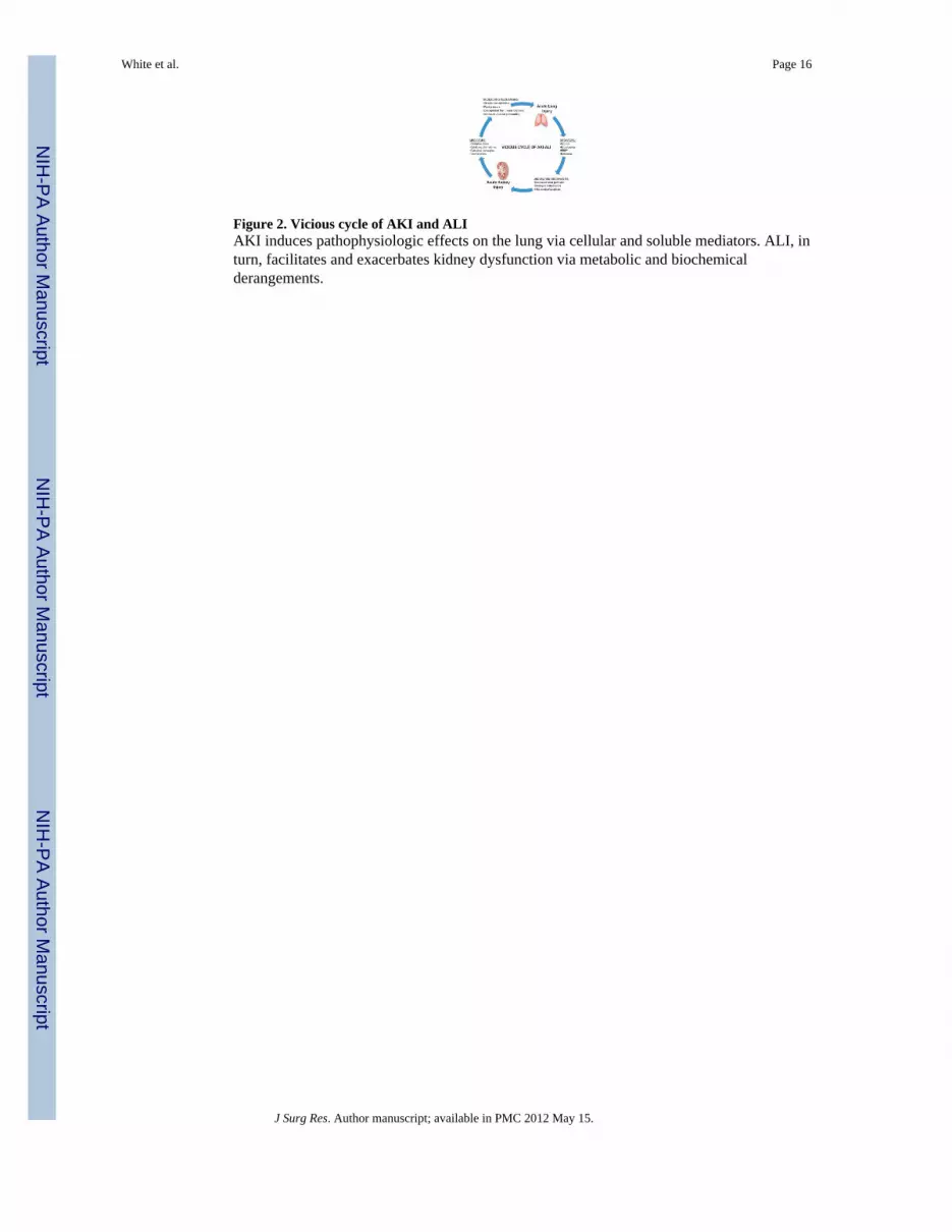
Figure 2. Vicious cycle of AKI and ALIAKI induces pathophysiologic effects on the lung via cellular and soluble mediators. ALI, inturn, facilitates and exacerbates kidney dysfunction via metabolic and biochemicalderangements.
White et al. Page 16
J Surg Res. Author manuscript; available in PMC 2012 May 15.
NIH
-PA Author Manuscript
NIH
-PA Author Manuscript
NIH
-PA Author Manuscript
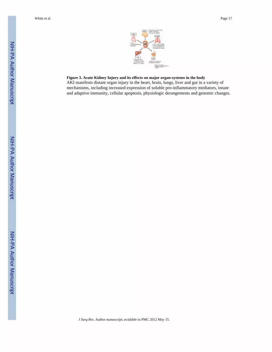
Figure 3. Acute Kidney Injury and its effects on major organ-systems in the bodyAKI manifests distant organ injury in the heart, brain, lungs, liver and gut in a variety ofmechanisms, including increased expression of soluble pro-inflammatory mediators, innateand adaptive immunity, cellular apoptosis, physiologic derangements and genomic changes.
White et al. Page 17
J Surg Res. Author manuscript; available in PMC 2012 May 15.
NIH
-PA Author Manuscript
NIH
-PA Author Manuscript
NIH
-PA Author Manuscript

NIH
-PA Author Manuscript
NIH
-PA Author Manuscript
NIH
-PA Author Manuscript
White et al. Page 18
Table 1
Abbreviations
ACCP/SCCM American College of Chest Physicians/Society of Critical Care Medicine
AKI Acute Kidney Injury
AKIN Acute Kidney Injury Network
ALI Acute Lung Injury
APC Activated Protein C
ARDS Acute Respiratory Distress Syndrome
ARDSNet Acute Respiratory Syndrome Clinical Trials Network
BNx Bilateral Nephrectomy
CHIF Channel Inducing Factor
CO Cardiac Output
DIC Disseminated Intravascular Coagulation
GFAP Glial Fibrillary Acidic Protein
GFR Glomerular Filtration Rate
GM-CSF Granulocyte Macrophage Colony-Stimulating Factor
HSC Hepatic Stellate Cells
ICAM Inter-Cellular Adhesion Molecule
ICU Intensive Care Unit
IIT Intensive Insulin Therapy
IL Interleukin
INF-γ Interferon-gamma
iNOS Inducible Nitric Oxide Synthase
IRI Ischemia Reperfusion Injury
KC Keratinocyte Chemoattractant
LPS Lipopolysaccharide
LV Left Ventricle
M2AA Methyl-2-Acetamidoacrylate
MIP-2 Macrophage Inflammatory Protein-2
MOF Multiple Organ Failure
NF-κβ Nuclear Factor Kappa Beta
NSQIP National Surgical Quality Improvement Project
PEEP Positive End-Expiratory Pressure
PICARD Program to Improve Care in Acute Renal Disease
RBF Renal Blood Flow
RIFLE Risk Injury Failure Loss Endstage Renal Disease
RRT Renal Replacement Therapy
Saa3 Serum Amyloid A 3
SIRS Systemic Inflammatory Response Syndrome
SOAP Sepsis Occurrence in Acutely Ill Patients
J Surg Res. Author manuscript; available in PMC 2012 May 15.

NIH
-PA Author Manuscript
NIH
-PA Author Manuscript
NIH
-PA Author Manuscript
White et al. Page 19
Th T Helper Cell
TNF-α Tumor Necrosis Factor Alpha
J Surg Res. Author manuscript; available in PMC 2012 May 15.

NIH
-PA Author Manuscript
NIH
-PA Author Manuscript
NIH
-PA Author Manuscript
White et al. Page 20
Table 2
Sepsis and Related Definitions
SystemicInflammatoryResponseSyndrome (SIRS)
The systemic inflammatory response to a variety of severe clinicalinsults. Response includes two or more of the following clinicalconditions:
• Temperature >38°C or <36°C
• Heart rate >90 beats per minute
• Respiratory rate > 20 breaths per minute or PaCO2 < 32 mmHg
• White blood cell count >12,000/cu mm, < 4,000/cu mm or >10% immature (band) forms.
Sepsis The systemic response to infection, manifested by two or more ofthe above clinical criteria listed under SIRS
Severe Sepsis Sepsis associated with an acute organ dysfunction:
• Neurologic: GCS1 <13 upon recognition of sepsis or deteriorating to <13 during the first 24 hours of sepsis
• Pulmonary: PaO2/FiO2 ratio <250 (<200 if lung is primary source of infection) and PCWP2 (if available) notsuggestive of fluid overload
• Renal- One of the following: 1) UOP3 < 0.5 ml/kg for ≥ 1hour despite adequate volume resuscitation, 2)Increase in serum creatinine ≥ 0.5 mg/dl from baseline (measured within 24 hours of starting sepsisresuscitation) despite adequate volume resuscitation, 3) increase in serum creatinine ≥ 0.5 mg/dl during thefirst 24 hours despite adequate volume resuscitation4
• Coagulation- One of the following: 1) INR > 1.5, 2) Platelet count <80,000 or ≥ 50% decrease platelet countin 24 hour period in the absence of chronic liver disease
• Hypoperfusion: Lactate level> 4 mmol/L
Septic Shock Sepsis-induced hypotension despite adequate volume resuscitationalong with the presence of perfusion abnormalities (as above);patients requiring inotropic or vasopressor medications may not behypotensive at the time when perfusion abnormalities aremeasured.
Sepsis-inducedHypotension
Systolic blood pressure <90 mmHg or a reduction of ≥ 40 mmHgfrom baseline in the absence of other causes of hypotension.
1GCS = Glasgow Coma Score
2PCWP = Pulmonary Capillary Wedge Pressure
3UOP = Urine Output
4Adequate volume resuscitation defined by a minimum intravenous fluid infusion of 20 ml/kg/ideal body weight or central venous pressure ≥ 8
mm Hg or PCWP ≥ 12 mm Hg
J Surg Res. Author manuscript; available in PMC 2012 May 15.