the first naupliar stage of pennella balaenopterae koren and danielssen, 1877 (copepoda:...
TRANSCRIPT
The first naupliar stage of Pennella balaenopterae Koren and Danielssen, 1877(Copepoda: Siphonostomatoida, Pennellidae)
Nina L. Arroyo, Pablo Abaunza & Izaskun Preciado
Arroyo NL, Abaunza P, Preciado I. 2002. The first naupliar stage of Pennella balaenopterae Koren andDanielssen, 1877 (Copepoda: Siphonostomatoida, Pennellidae). Sarsia 87:333–337.
SARSIA
The first nauplius of Pennella balaenopterae is described and illustrated. This is the first description ofthe nauplius in this species and genus. Antennae and mandibles resemble those previously described inpennelid nauplii. The first antenna is uniramous and one-segmented, while the second antenna and themandible show a two-segmented endopod and a four-segmented exopod. No trace of any otherappendage or of the gut is found. Some comments on the life cycle of Pennella balaenopterae are made.
N. L. Arroyo, Departamento de Biologıa Animal I (Zoologıa), Facultad de Biologıa, UniversidadComplutense de Madrid, ES-28040 Madrid, Spain. Present address: Abo Academy University,Department of Biology Environmental and Marine Biology, FIN-20500 Abo, Finland.P. Abaunza & I. Preciado, Instituto Espanol de Oceanografıa, Centro Oceanografico de Santander,Apdo. 240, ES-39080 Santander, Spain.E-mail: [email protected]; [email protected]; [email protected]
Keywords: Pennella balaenopterae; Pennellidae; first nauplius; morphological description; life cycle.
INTRODUCTION
The significance of nauplii to the crustaceans is nowwidely accepted, its morphological organization beingso fundamental that its presence has been proposed aspart of the definition of the Crustacea (Walossek &Muller 1990), and several theories have been suppliedassessing its usage in phylogenetic studies (Dahms2000).
Within the siphonostome family Pennellidae, usuallyone or two nauplius stages have been reported(Sproston 1942; Heegaard 1947; Kabata 1976; Schram1979). Two nauplius stages were found in Lernaeenicussprattae (Sowerby, 1806) (cf. Schram 1979), and onenauplius stage was found in Lernaeocera branchialis(Linnaeus, 1767) (cf. Sproston 1942; Heegaard 1947)and Haemobaphes diceraus Wilson, 1917 (cf. Kabata1976). In Lernaeenicus longiventris Wilson, 1917, thenewly hatched nauplius was described and illustrated,although the exact number of nauplius stages was notascertained (Wilson 1917). Some pennelids lack a free-swimming nauplius stage whatsoever, and the eggshatch directly as copepodids, the naupliar stage beingpassed inside the egg sac (Bennet 1961; Perkins 1983;Izawa 1997).
The larval stages of pennelids belonging to the genusPennella have been described for the copepodid andchalimus stages of Pennella varians Steenstrup &Lutken, 1861 (Rose & Hamon 1953). Also, Pascual
(1996) described the copepodid and chalimus stages ofa pennelid infecting squids whose identity he could notascertain but which he related to the larval stages ofPennella varians described by Rose & Hamon (1953).
Relatively few studies have been conducted onPennella balaenopterae, its life cycle is almostunknown and only the adult female has been identifiedwith certainty to date (Turner 1905; Hogans 1987). In1997, a fin whale (Balaenoptera physalus Linnaeus,1758), stranded on the coast of Cantabria, was carrying12 adult females of Pennella balaenopterae (Abaunza& al. 2001). In the present paper, we describe andillustrate a naupliar stage of Pennella balaenopterae,providing the first description of a naupliar stage for thespecies and the genus.
MATERIAL AND METHODS
The fin whale stranded on the coast of Cantabria(northern Spain) in November 1997, was a 19 m longmale, with no apparent malformations, either externalor internal. It died on the coast, and the parasites werecollected 3 days later.
The parasites without egg strings were found with thechephalothorax and anterior part of the thoracic regionembedded in the whale’s blubber, and were locatedmainly on the anterior half of its body. The specimenswere placed in a plastic bag, and once in the laboratory,all of them were found to bear long, string-like ovisacs,
� 2002 Taylor & Francis
Published in collaboration with the University of Bergen and the Institute of Marine Research, Norway
protruding from the genital pores: clearly these stringshad been extruded by the animal during transport.
The parasites, together with the ovisacs, were storedin 70% alcohol for fixation and conservation. Deadembryos and nauplius larvae (without egg membrane)were found in the preservation fluid. Ovisacs containedyet more unreleased eggs.
The nauplii were mounted whole for examination,broken glass fibres being added to prevent them frombeing compressed and to facilitate rolling to allowviewing from all sides. Some specimens were mountedon aluminium cobble slides for the same purpose.Drawings were made from single specimens with theaid of a camera lucida.
Body lengths were measured from the anterior to theposterior end of the nauplius; body width is given as thewidest part of the nauplius. Appendages were measuredfrom the point of insertion in the body to their distal-most part. Setae were measured from the point ofinsertion in the appendage to the tip of the setae. ALeica light microscope, an interference 40� lensmagnification, and an image analyser (Visilog 5.2)were used.
RESULTS
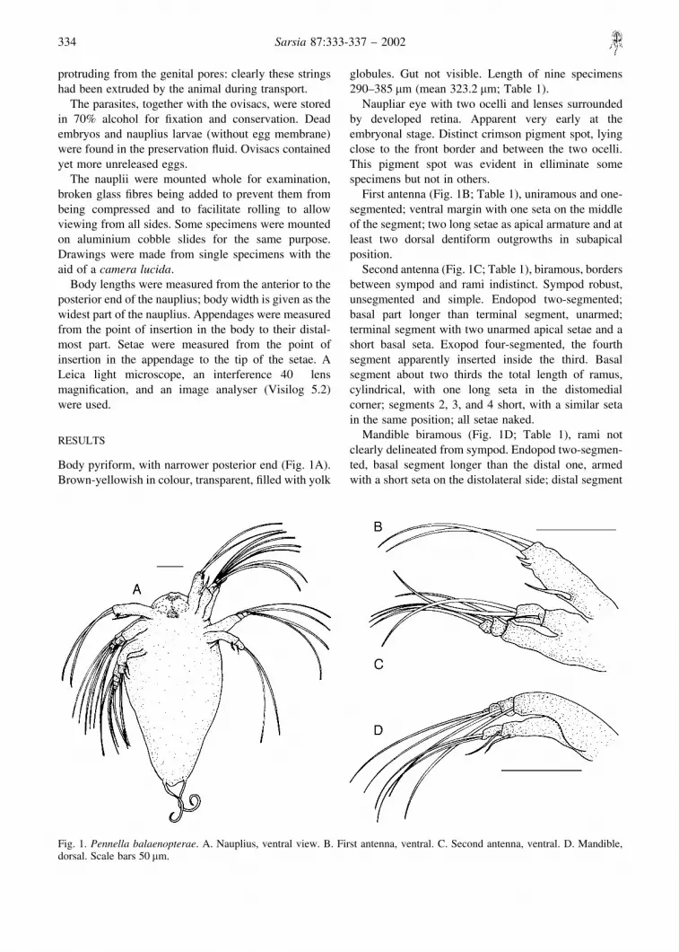
Body pyriform, with narrower posterior end (Fig. 1A).Brown-yellowish in colour, transparent, filled with yolk
globules. Gut not visible. Length of nine specimens290–385 �m (mean 323.2 �m; Table 1).
Naupliar eye with two ocelli and lenses surroundedby developed retina. Apparent very early at theembryonal stage. Distinct crimson pigment spot, lyingclose to the front border and between the two ocelli.This pigment spot was evident in elliminate somespecimens but not in others.
First antenna (Fig. 1B; Table 1), uniramous and one-segmented; ventral margin with one seta on the middleof the segment; two long setae as apical armature and atleast two dorsal dentiform outgrowths in subapicalposition.
Second antenna (Fig. 1C; Table 1), biramous, bordersbetween sympod and rami indistinct. Sympod robust,unsegmented and simple. Endopod two-segmented;basal part longer than terminal segment, unarmed;terminal segment with two unarmed apical setae and ashort basal seta. Exopod four-segmented, the fourthsegment apparently inserted inside the third. Basalsegment about two thirds the total length of ramus,cylindrical, with one long seta in the distomedialcorner; segments 2, 3, and 4 short, with a similar setain the same position; all setae naked.
Mandible biramous (Fig. 1D; Table 1), rami notclearly delineated from sympod. Endopod two-segmen-ted, basal segment longer than the distal one, armedwith a short seta on the distolateral side; distal segment
Fig. 1. Pennella balaenopterae. A. Nauplius, ventral view. B. First antenna, ventral. C. Second antenna, ventral. D. Mandible,dorsal. Scale bars 50 �m.
334 Sarsia 87:333-337 – 2002
with two long apical setae and a short seta on its medialmargin. Exopod four-segmented, basal segment muchlonger than the other three. Each segment with a seta inthe distomedial corner but with no other armature.
A first trace of the labrum was distinct in somespecimens, but no sign of the maxillae or the swimmingappendages could be detected in any of them.
Balancers protrude from a slight swelling, usuallypositioned downwards and often appearing coiled dueto preservation (Fig. 1A; Table 1).
DISCUSSION
The first nauplius of Pennella balaenopterae resemblesthose of other pennelid species described previously,both in its general appearance and its appendages(Sproston 1942; Kabata 1976; Schram 1979). The lackof rudiments of further stages in the specimens foundpoints to them being a “newly hatched first nauplius”stage. However, differences in some aspects of theirmorphology suggest that they were in different devel-opmental phases when they died.
The appendages differ only slightly from thepreviously described species of the family Pennellidaein the setation of the different segments and in that setaeof Pennella balaenopterae are naked while otherspecies present plumose or semi-pinnate setae (Kabata1976; Schram 1979).
Lernaeenicus sprattae also has a single segmentedfirst antenna, armed with three setae but with up to fourspines (Schram 1979), instead of the two spines in thepresent specimens. The first antenna of Haemobaphesdiceraus Wilson, 1917 shows basically the samesimplified structure, resembling also that of Haemo-baphes cyclopterina Fabricius 1780, as described by
Heegaard (1947). The characteristic feature of the firstnauplius of the former species is a previously unknownclaw-like spine arising from a papilliform outgrowth atthe base of the first antenna, which has not been foundon any other nauplius (Kabata 1976).
Both Lernaeocera branchialis and Haemobaphesdiceraus (Sproston 1942; Kabata 1976) show the samenumber of segments in the second antenna as Pennellabalaenopterae, with an indistinctly segmented exopod,and the same disposition of apical setation. Lernaeeni-cus sprattae has a clearly four-segmented exopod and atwo-jointed endopod, each with a very similar setationpattern (Schram 1979). However, in other Siphonosto-matoid families, species such as Eudactylina similis T.Scott, 1902 (Eudactylinidae), Caligus elongatus vonNordmann, 1832 (Caligidae) and Pseudocharopinusdentatus (Wilson, 1912) (Lernaeopodidae), present anexopodite with five segments, as is the case ofpoecilostomatoid nauplii (Kabata 1976; Piasecki1996). A three- to four-segmented exopod thereforeseems characteristic of pennelid nauplii.
Heegaard (1947) used the naupliar mandible as oneof the main features distinguishing his Fistulata andPectinata, taxa corresponding approximately to thepoecilostome and siphonostome copepods, respec-tively. According to him, the nauplii of the formerhave mandibles with two-segmented endopods,whereas the corresponding ramus in the mandible ofthe latter consists of three segments. Sproston (1942)found a three-segmented endopod on Lernaeocerabranchialis, but Kabata (1976) failed to corroborateHeegaard’s findings. He proved that the mandibles ofthe poecilostomes Ergasilus turgidus Fraser, 1920,Bomolochus cuneatus Fraser, 1920, and Chondra-canthus gracilis Fraser, 1920 indeed had two-segmen-
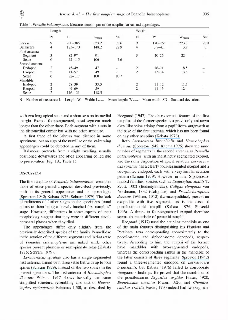
Table 1. Pennella balaenopterae. Measurements in �m of the nauplius larvae and appendages.
Length Width
N L Lmean SD N W Wmean SD
Larvae 9 290–385 323.2 32.6 9 190–263 223.8 26.8Balancers 4 123–170 148.2 22.9 4 3.9–4.1 3.9 0.1First antenna
Segment 3 82–97 91 – 3 20–25 22 –Setae 6 92–115 106 7.6
Second antennaEndopod 2 45–49 47 – 2 16–21 18.5 –Exopod 2 41–57 49 – 2 13–14 13.5 –Setae 6 92–117 100 10.7
MandibleEndopod 2 28–39 33.5 – 2 11–12 11.5 –Exopod 2 49–69 59 – 2 11–13 12 –Setae 2 116–121 118.5
N – Number of measures; L – Length; W – Width; Lmean – Mean length; Wmean – Mean width; SD – Standard deviation.
Arroyo & al. – The first naupliar stage of Pennella balaenopterae 335

ted endopods, but that the same was true of the twonaupliar mandibles of the siphonostomes Eudactylinasimilis and Haemobaphes diceraus. A two-segmentedendopod was also observed in the mandibles of ourspecimens of Pennella balaenopterae, and in the firstand second nauplii of Lernaeenicus sprattae (cf.Schram 1979), corroborating further Kabata’s findings.Moreover, a one-segmented endopod was found byPiasecki (1996) in the mandibles of both the first andsecond nauplii of the siphonostome caligid, Caliguselongates von Nordmann, 1832.
At the time of collection no parasites had ovisacs, buton arrival at the laboratory new egg strings had beenformed; extruded during their transfer to the laboratory.The embryos may have been released from these eggstrings during transport, perhaps aided by the increasein temperature experienced by the parasites in thiscontainer. It has been proved that an increase intemperature enhances and accelerates the ovipositionprocess (Nakai 1927; Gnanamuthu 1951; Schram1979), allowing the nauplii to hatch in a much shortertime (Schram & Anstenstud 1985). Embryos maydevelop even once the adult female is dead, the naupliihatching several days later (Schram 1979).
The presence of both nauplii and eggs in the alcohol-fixed material, plus additional eggs inside the ovisacs,suggests that perhaps fixation interrupted the oviposi-tion and hatching processes. However, some authorsdescribe nauplii hatching directly from the egg strings(Sproston 1942; Heegaard 1947; Schram 1979), whichcould also have been the case here. Then, the embryos(eggs and fully formed nauplii) could have been forcedout of the egg string in response to fixation.
The nauplius herein described could be the first of aseries of moults, or the only free-swimming naupliuspreceding the copepodid stages, as described previouslyfor Pennelids (Sproston 1942; Heegaard 1947; Kabata
1976). The second nauplius described by Schram(1979) for Lernaeenicus sprattae could be distin-guished from the first one by its size, shape and pigmenttint, but most easily by the presence of rudiments of thecopepodid seen inside the nauplius. Schram noted thedifficulty in detecting these differences and suggestedthat a second naupliar stage could have been ignored oroverlooked in previous studies.
In our case, no rudiments of maxillae or otherappendages were observed. Features such as differencesin eye pigmentation indicate that the specimens belongto different developmental phases, but always withinthe first newly hatched nauplius, as described bySchram (1979).
With this finding we can conclude that Pennellabalaenopterae hatches in the form of a nauplius fromadult females attached to a cetacean definitive host.Whether cephalopod species are the intermediate hoston which infective copepodids develop through thechalimus stages to adults, and on which mating takesplace, as suggested for other members of the genus(Rose & Hamon 1953; Pascual 1996; Carbonell & al.1999), needs further studies which presumably wouldalso provide better descriptions of these stages and theyet unknown male.
ACKNOWLEDGEMENTS
Thanks are due to Hans-Uwe Dahms for his help during theinterpretation and drawing of the larvae and for revision of anearlier version of the manuscript, and to the Zoosystematischund Morfologie Arbeitsgruppe, University of Oldenburg,where the drawings were made. The authors thank G. GarcıaCastrillo and C. Rodriguez, who were responsible for thewhale’s autopsy, for their help in collecting the specimens ofPennella balaenopterae, and to three anonymous referees fortheir valuable comments. T. Schram and S. Pascual alsoprovided useful references.
REFERENCES
Abaunza P, Arroyo NL, Preciado I. 2001. A contribution to theknowledge on the morphometry and the anatomicalcharacters of Pennella balaenopterae (Copepoda,Siphonostomatoida, Pennellidae), with special refer-ence to the buccal complex. Crustaceana 74:193–210.
Bennet PS. 1961. Peroderma cylindricum Heller, a copepodparasite of Sardinella albella. Journal of the MarineBiological Association of India 3:70–74.
Carbonell E, Massutı E, Castro JJ, Garcıa RM. 1999.Parasitism of dolphinfishes, Coryphaena hippurus andCoryphaena equiselis, in the western Mediterranean(Balearic Islands) and central-eastern Atlantic (CanaryIslands). Scientia Marina 63:343–354.
Dahms HU. 2000. Phylogenetic implications of the Crustaceannauplius. Hydrobiologia 417:91–99.
Gnanamuthu CP. 1951. Notes on the life history of a parasiticcopepod, Lernaea chackoensis. Parasitology 41:148–155.
Heegaard P. 1947. Contribution to the phylogeny of thearthropods. Copepoda. Spolia Zoologica Musei Hau-niensis 8:1–227.
Hogans WE. 1987. Morphological variation in Pennellabalaenoptera and P. filosa (Copepoda:Pennellidae) witha review of the genus Pennella Oken, 1816 parasitic onCetacea. Bulletin of Marine Science 40:442–453.
Izawa K. 1997. The copepodid of Peniculisa shiinoi Izawa,
336 Sarsia 87:333-337 – 2002
1965 (Copepoda, Siphonostomatoida, Pennelidae), asingle free-swimming larval stage of the species.Crustaceana 70:911–919.
Kabata Z. 1976. Early stages of some copepods (Crustacea)parasitic on marine fishes of British Columbia. Journalof the Fisheries Research Board of Canada 33:2507–2525.
Nakai N. 1927. On the development of a parasitic copepod,Lernaea elegans Leigh-Sharpe, infesting on Cyprinuscarpio L. Journal of the Imperial Fisheries Institute(Japan) 23(3):39–57.
Pascual S. 1996. Los sistemas hospedador-parasito en lapesquerıa de ommastrefidos de Galicia. PhD thesis.Universidad de Vigo, Spain. 165 p.
Perkins PS. 1983. The life history of Cardiodectes medusaeus(Wilson), a copepod parasite of lanternfishes (Mycto-phidae). Journal of Crustacean Biology 3:70–87.
Piasecki W. 1996. The developmental stages of Caliguselongatus von Nordmann, 1832 (Copepoda: Caligidae).Canadian Journal of Zoology 74:1459–1478.
Rose M, Hamon M. 1953. A propos de Pennella variansSteenstrup et Lutken, 1861, parasite des branchies deCephalopodes. Bulletin Societe Histoire Naturelle del’Afrique du Nord 44:172–183.
Schram TA. 1979. The life history of the eye-maggot of thesprat, Lernaeenicus sprattae (Sowerby) (Copepoda,Lernaeoceridae). Sarsia 64:279–316.
Schram TA, Anstensrud M. 1985. Lernaeenicus sprattae(Sowerby) larvae in the Oslofjord plankton and somelaboratory experiments with the nauplius and copepodid(Copepoda, Pennellidae). Sarsia 70:127–134.
Sproston NG. 1942. The developmental stages of Lernaeocerabranchialis (Linn.). Journal of the Marine BiologicalAssociation of the United Kingdom 25:441–466.
Turner W. 1905. On Pennella balaenoptera: a crustacean,parasitic on a finner whale, Balaenoptera musculus.Transactions of the Royal Society of Edinburgh 2:409–434.
Walossek D, Muller KJ. 1990. Upper Cambrian stem-lineagecrustaceans and their bearing upon the monophyleticorigin of Crustacea and the position of Agnostus.Lethaia 23:409–427.
Wilson CB. 1917. North American parasitic copepods belong-ing to the Lernaeidae with a revision of the entirefamily. Proceedings of the United States NationalMuseum 53:1–50.
Accepted 20 July 2001 – Printed 19 December 2002Editorial responsibility: Tore Høisæter
Arroyo & al. – The first naupliar stage of Pennella balaenopterae 337
