revision of the spider genus caloctenus keyserling, 1877 (araneae, ctenidae) revisión del género...
TRANSCRIPT
5
Revisión: The spiders of genus Caloctenus
Rev. peru. biol. 11 (1): 5 - 26 (2004)
http://sisbib.unmsm
.edu.pe/BV
Revistas/biologia/biologiaN
EW.htm
Rev. peru. biol. 11 (1): 5 - 26 (2004)© Facultad de Ciencias Biológicas UNMSM
Presentado: 02/07/2004Aceptado: 20/07/2004
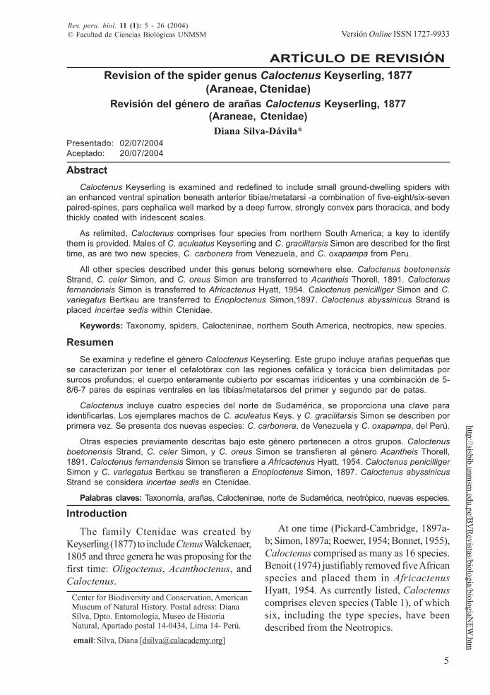
Revision of the spider genus Caloctenus Keyserling, 1877(Araneae, Ctenidae)
Revisión del género de arañas Caloctenus Keyserling, 1877(Araneae, Ctenidae)Diana Silva-Dávila*
Center for Biodiversity and Conservation, AmericanMuseum of Natural History. Postal adress: DianaSilva, Dpto. Entomología, Museo de HistoriaNatural, Apartado postal 14-0434, Lima 14- Perú.
email: Silva, Diana [[email protected]]
AbstractCaloctenus Keyserling is examined and redefined to include small ground-dwelling spiders with
an enhanced ventral spination beneath anterior tibiae/metatarsi -a combination of five-eight/six-sevenpaired-spines, pars cephalica well marked by a deep furrow, strongly convex pars thoracica, and bodythickly coated with iridescent scales.
As relimited, Caloctenus comprises four species from northern South America; a key to identifythem is provided. Males of C. aculeatus Keyserling and C. gracilitarsis Simon are described for the firsttime, as are two new species, C. carbonera from Venezuela, and C. oxapampa from Peru.
All other species described under this genus belong somewhere else. Caloctenus boetonensisStrand, C. celer Simon, and C. oreus Simon are transferred to Acantheis Thorell, 1891. Caloctenusfernandensis Simon is transferred to Africactenus Hyatt, 1954. Caloctenus penicilliger Simon and C.variegatus Bertkau are transferred to Enoploctenus Simon,1897. Caloctenus abyssinicus Strand isplaced incertae sedis within Ctenidae.
Keywords: Taxonomy, spiders, Calocteninae, northern South America, neotropics, new species.
ResumenSe examina y redefine el género Caloctenus Keyserling. Este grupo incluye arañas pequeñas que
se caracterizan por tener el cefalotórax con las regiones cefálica y torácica bien delimitadas porsurcos profundos; el cuerpo enteramente cubierto por escamas iridicentes y una combinación de 5-8/6-7 pares de espinas ventrales en las tibias/metatarsos del primer y segundo par de patas.
Caloctenus incluye cuatro especies del norte de Sudamérica, se proporciona una clave paraidentificarlas. Los ejemplares machos de C. aculeatus Keys. y C. gracilitarsis Simon se describen porprimera vez. Se presenta dos nuevas especies: C. carbonera, de Venezuela y C. oxapampa, del Perú.
Otras especies previamente descritas bajo este género pertenecen a otros grupos. Caloctenusboetonensis Strand, C. celer Simon, y C. oreus Simon se transfieren al género Acantheis Thorell,1891. Caloctenus fernandensis Simon se transfiere a Africactenus Hyatt, 1954. Caloctenus penicilligerSimon y C. variegatus Bertkau se transfieren a Enoploctenus Simon, 1897. Caloctenus abyssinicusStrand se considera incertae sedis en Ctenidae.
Palabras claves: Taxonomía, arañas, Calocteninae, norte de Sudamérica, neotrópico, nuevas especies.
Introduction
The family Ctenidae was created byKeyserling (1877) to include Ctenus Walckenaer,1805 and three genera he was proposing for thefirst time: Oligoctenus, Acanthoctenus, andCaloctenus.
At one time (Pickard-Cambridge, 1897a-b; Simon, 1897a; Roewer, 1954; Bonnet, 1955),Caloctenus comprised as many as 16 species.Benoit (1974) justifiably removed five Africanspecies and placed them in AfricactenusHyatt, 1954. As currently listed, Caloctenuscomprises eleven species (Table 1), of whichsix, including the type species, have beendescribed from the Neotropics.
ARTÍCULO DE REVISIÓN
Versión Online ISSN 1727-9933
6
Silva
Rev. peru. biol. 11 (1): 5 - 26 (2004)
http
://sis
bib.
unm
sm.e
du.p
e/B
VR
evist
as/b
iolo
gia/
biol
ogia
NEW
.htm
Since early in the history of ctenids(Keyserling, 1877; Simon, 1897a; Simon, 1909;Mello-Leitão, 1936), Caloctenus was placedin its own group and easily separated fromother genera by its labium wider than long andabout one third the length of the endites, avaulted cephalothorax, seven pairs of spinesbeneath tibiae I-II, and anterior spinnerets notlonger than the posterior ones.
Although none of these characters wereunique to Caloctenus, at least the labium shapeappeared to hold together a natural group;initially as the section Calocteneae (Simon,1897a) and later recognized as the subfamilyCalocteninae by various authors.
The monophyly of caloctenines was firsttested (Silva Dávila, 1994) to examine therelationships among species of Caloctenus.Based on genitalic similarities, this hypothesissupported a sister-group relationship betweenC. aculeatus and C. gracilitarsis, and in turn,this clade sister to C. oxapampa plus C. car-bonera. More recently (Silva Dávila, 2003),caloctenines were compared to a morecomprehensive sample of the various ctenidgroups. Both analyses (Silva Dávila, 1994,2003) showed that caloctenines, as delimitedin the literature, were polyphyletic; however,
both sets of data strongly supported onedistinctive clade, which was recognized asCalocteninae sensu stricto.
Caloctenines comprise five genera fromSouth America, Sri Lanka, the Seychelles Islands,and Madagascar. Although this group is stronglysupported by seven synapomorphies, all but one(long and thick anal setae) are subject to somedegree of homoplasy. The phylogeneticrelationships within this group are being re-examined with the description of several newspecies from Sri Lanka and Madagascar.
Both hypotheses (Silva Dávila, 1994, 2003)show a sister group relationship betweenCaloctenus and Gephyroctenus Mello-Leitão,1936. The latter is a more speciose taxonrestricted to the Amazonian lowland forests;a taxonomical revision of Gephyroctenus isalso in progress (Brescovit, pers. comm.).
Interestingly, a new genus from Madagascarappears to be the sister group to the SouthAmerican caloctenines. Luckly, exhaustive fieldwork in recent years is providing a more com-plete set of data to re-examine the phylogeneticrelationships among caloctenines and help toclarify their distribution patterns; which are sofar, characterized by narrow endemisms atgeneric and species level.
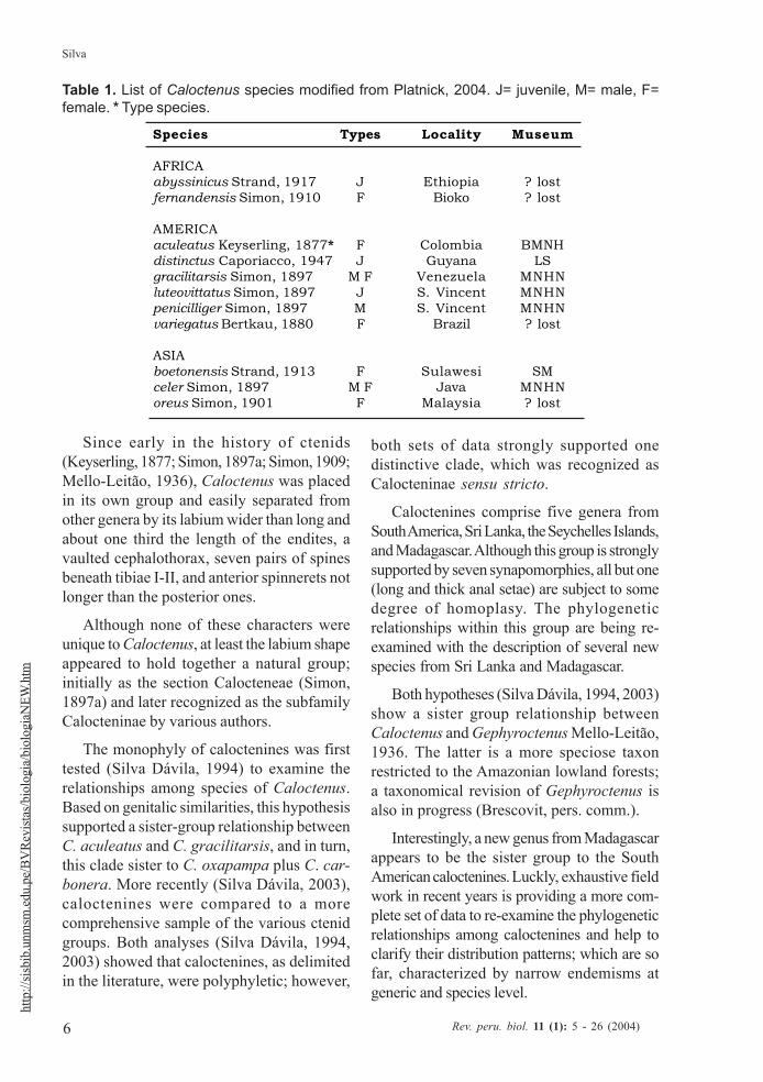
Table 1. List of Caloctenus species modified from Platnick, 2004. J= juvenile, M= male, F=female. * Type species.
Species Types Locality Museum
AFRICAabyssinicus Strand, 1917 J Ethiopia ? lostfernandensis Simon, 1910 F Bioko ? lost
AMERICAaculeatus Keyserling, 1877* F Colombia BMNHdistinctus Caporiacco, 1947 J Guyana LSgracilitarsis Simon, 1897 M F Venezuela MNHNluteovittatus Simon, 1897 J S. Vincent MNHNpenicilliger Simon, 1897 M S. Vincent MNHNvariegatus Bertkau, 1880 F Brazil ? lost
ASIAboetonensis Strand, 1913 F Sulawesi SMceler Simon, 1897 M F Java MNHNoreus Simon, 1901 F Malaysia ? lost
7
Revisión: The spiders of genus Caloctenus
Rev. peru. biol. 11 (1): 5 - 26 (2004)
http://sisbib.unmsm
.edu.pe/BV
Revistas/biologia/biologiaN
EW.htm
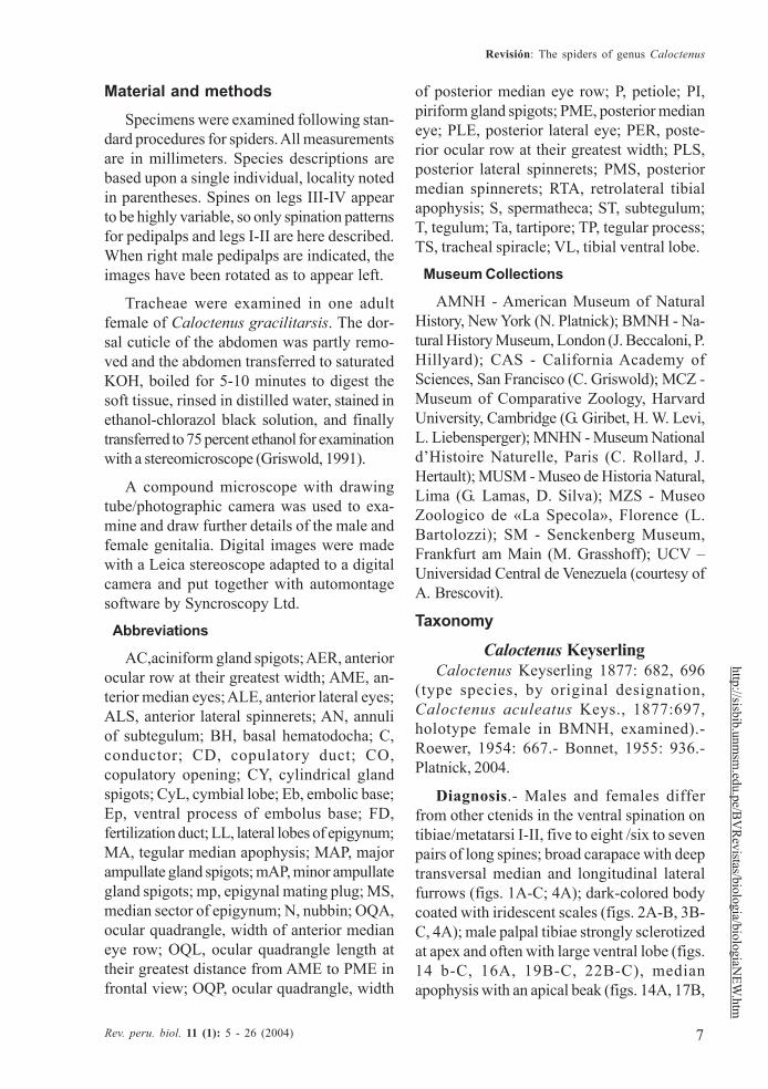
Material and methods
Specimens were examined following stan-dard procedures for spiders. All measurementsare in millimeters. Species descriptions arebased upon a single individual, locality notedin parentheses. Spines on legs III-IV appearto be highly variable, so only spination patternsfor pedipalps and legs I-II are here described.When right male pedipalps are indicated, theimages have been rotated as to appear left.
Tracheae were examined in one adultfemale of Caloctenus gracilitarsis. The dor-sal cuticle of the abdomen was partly remo-ved and the abdomen transferred to saturatedKOH, boiled for 5-10 minutes to digest thesoft tissue, rinsed in distilled water, stained inethanol-chlorazol black solution, and finallytransferred to 75 percent ethanol for examinationwith a stereomicroscope (Griswold, 1991).
A compound microscope with drawingtube/photographic camera was used to exa-mine and draw further details of the male andfemale genitalia. Digital images were madewith a Leica stereoscope adapted to a digitalcamera and put together with automontagesoftware by Syncroscopy Ltd.
Abbreviations
AC,aciniform gland spigots; AER, anteriorocular row at their greatest width; AME, an-terior median eyes; ALE, anterior lateral eyes;ALS, anterior lateral spinnerets; AN, annuliof subtegulum; BH, basal hematodocha; C,conductor; CD, copulatory duct; CO,copulatory opening; CY, cylindrical glandspigots; CyL, cymbial lobe; Eb, embolic base;Ep, ventral process of embolus base; FD,fertilization duct; LL, lateral lobes of epigynum;MA, tegular median apophysis; MAP, majorampullate gland spigots; mAP, minor ampullategland spigots; mp, epigynal mating plug; MS,median sector of epigynum; N, nubbin; OQA,ocular quadrangle, width of anterior medianeye row; OQL, ocular quadrangle length attheir greatest distance from AME to PME infrontal view; OQP, ocular quadrangle, width
of posterior median eye row; P, petiole; PI,piriform gland spigots; PME, posterior medianeye; PLE, posterior lateral eye; PER, poste-rior ocular row at their greatest width; PLS,posterior lateral spinnerets; PMS, posteriormedian spinnerets; RTA, retrolateral tibialapophysis; S, spermatheca; ST, subtegulum;T, tegulum; Ta, tartipore; TP, tegular process;TS, tracheal spiracle; VL, tibial ventral lobe.
Museum Collections
AMNH - American Museum of NaturalHistory, New York (N. Platnick); BMNH - Na-tural History Museum, London (J. Beccaloni, P.Hillyard); CAS - California Academy ofSciences, San Francisco (C. Griswold); MCZ -Museum of Comparative Zoology, HarvardUniversity, Cambridge (G. Giribet, H. W. Levi,L. Liebensperger); MNHN - Museum Nationald’Histoire Naturelle, Paris (C. Rollard, J.Hertault); MUSM - Museo de Historia Natural,Lima (G. Lamas, D. Silva); MZS - MuseoZoologico de «La Specola», Florence (L.Bartolozzi); SM - Senckenberg Museum,Frankfurt am Main (M. Grasshoff); UCV –Universidad Central de Venezuela (courtesy ofA. Brescovit).
Taxonomy
Caloctenus KeyserlingCaloctenus Keyserling 1877: 682, 696
(type species, by original designation,Caloctenus aculeatus Keys., 1877:697,holotype female in BMNH, examined).-Roewer, 1954: 667.- Bonnet, 1955: 936.-Platnick, 2004.
Diagnosis.- Males and females differfrom other ctenids in the ventral spination ontibiae/metatarsi I-II, five to eight /six to sevenpairs of long spines; broad carapace with deeptransversal median and longitudinal lateralfurrows (figs. 1A-C; 4A); dark-colored bodycoated with iridescent scales (figs. 2A-B, 3B-C, 4A); male palpal tibiae strongly sclerotizedat apex and often with large ventral lobe (figs.14 b-C, 16A, 19B-C, 22B-C), medianapophysis with an apical beak (figs. 14A, 17B,
8
Silva
Rev. peru. biol. 11 (1): 5 - 26 (2004)
http
://sis
bib.
unm
sm.e
du.p
e/B
VR
evist
as/b
iolo
gia/
biol
ogia
NEW
.htm
20B, 22D); and epigynal folds fused to variousdegrees (figs. 15A, 18A, 21A, 24A).
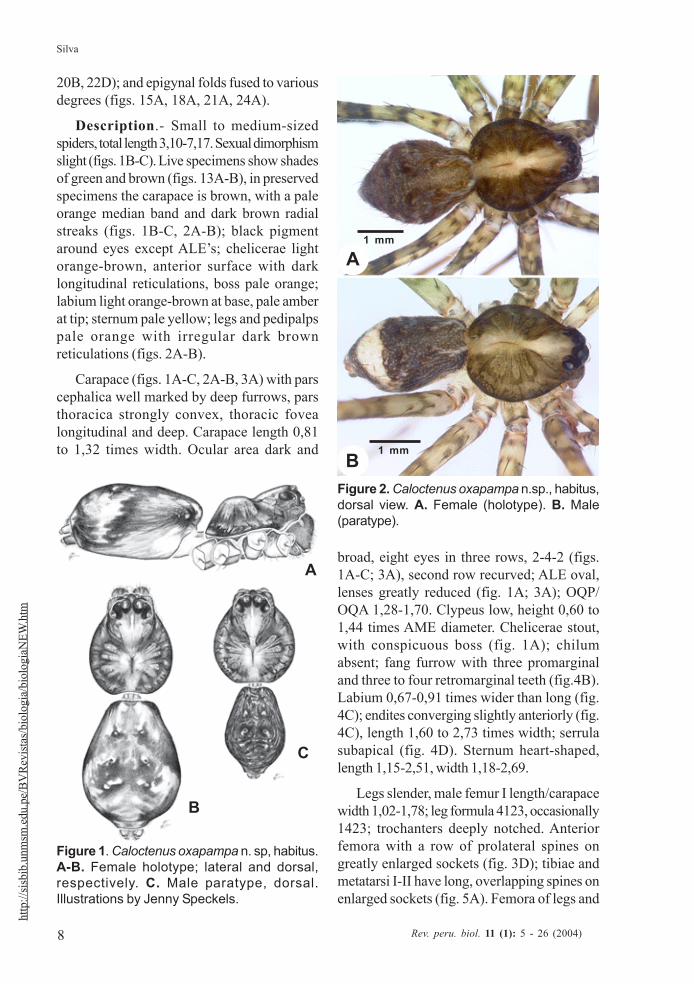
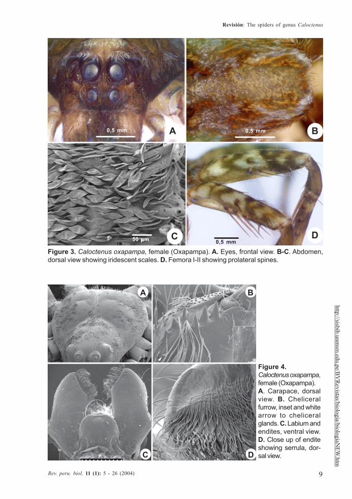
Description.- Small to medium-sizedspiders, total length 3,10-7,17. Sexual dimorphismslight (figs. 1B-C). Live specimens show shadesof green and brown (figs. 13A-B), in preservedspecimens the carapace is brown, with a paleorange median band and dark brown radialstreaks (figs. 1B-C, 2A-B); black pigmentaround eyes except ALE’s; chelicerae lightorange-brown, anterior surface with darklongitudinal reticulations, boss pale orange;labium light orange-brown at base, pale amberat tip; sternum pale yellow; legs and pedipalpspale orange with irregular dark brownreticulations (figs. 2A-B).
Carapace (figs. 1A-C, 2A-B, 3A) with parscephalica well marked by deep furrows, parsthoracica strongly convex, thoracic fovealongitudinal and deep. Carapace length 0,81to 1,32 times width. Ocular area dark and
broad, eight eyes in three rows, 2-4-2 (figs.1A-C; 3A), second row recurved; ALE oval,lenses greatly reduced (fig. 1A; 3A); OQP/OQA 1,28-1,70. Clypeus low, height 0,60 to1,44 times AME diameter. Chelicerae stout,with conspicuous boss (fig. 1A); chilumabsent; fang furrow with three promarginaland three to four retromarginal teeth (fig.4B).Labium 0,67-0,91 times wider than long (fig.4C); endites converging slightly anteriorly (fig.4C), length 1,60 to 2,73 times width; serrulasubapical (fig. 4D). Sternum heart-shaped,length 1,15-2,51, width 1,18-2,69.
Legs slender, male femur I length/carapacewidth 1,02-1,78; leg formula 4123, occasionally1423; trochanters deeply notched. Anteriorfemora with a row of prolateral spines ongreatly enlarged sockets (fig. 3D); tibiae andmetatarsi I-II have long, overlapping spines onenlarged sockets (fig. 5A). Femora of legs and
Figure 2. Caloctenus oxapampa n.sp., habitus,dorsal view. A. Female (holotype). B. Male(paratype).
A
B
C
Figure 1. Caloctenus oxapampa n. sp, habitus.A-B. Female holotype; lateral and dorsal,respectively. C. Male paratype, dorsal.Illustrations by Jenny Speckels.
B
A1 mm
1 mm
9
Revisión: The spiders of genus Caloctenus
Rev. peru. biol. 11 (1): 5 - 26 (2004)
http://sisbib.unmsm
.edu.pe/BV
Revistas/biologia/biologiaN
EW.htm
Figure 3. Caloctenus oxapampa, female (Oxapampa). A. Eyes, frontal view. B-C. Abdomen,dorsal view showing iridescent scales. D. Femora I-II showing prolateral spines.
Figure 4.Caloctenus oxapampa,female (Oxapampa).A. Carapace, dorsalview. B. Cheliceralfurrow, inset and whitearrow to cheliceralglands. C. Labium andendites, ventral view.D. Close up of enditeshowing serrula, dor-sal view.
A B
C D
0,5 mm 0,5 mm
0,5 mm50 µµµµµm
A B
C D

10
Silva
Rev. peru. biol. 11 (1): 5 - 26 (2004)
http
://sis
bib.
unm
sm.e
du.p
e/B
VR
evist
as/b
iolo
gia/
biol
ogia
NEW
.htm
pedipalps with dorsal and lateral spines, ven-tral spinules may be present; palpal patella withdorsal and lateral spines, patellae I-IVfrequently with one dorsal spine; tibiae I-II with5-8 ventral-paired spines, rarely with one totwo pairs of lateral spines; metatarsi I-II with6-7 paired-ventral spines, rarely with one totwo pairs of lateral spines; tibiae III-IV withvariable number of dorsal, lateral, and ventralspines. Tarsal scopula sparse (fig. 5B). Twoor three tarsal claws (fig. 5C), with ridgedsurface; superior tarsal claws pectinate with6 to 9 teeth, inferior tarsal claw smooth (fig.5C). Claw tuft hairs sparse (figs. 5B-C). Tarsalorgan capsulate and seemingly sexuallydimorphic (fig. 6A-D). Tarsi with two to threeirregular rows of trichobothria; trichobothrialhood with two to four transverse ridges(fig.6B).
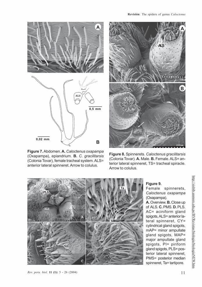
Abdomen widest posteriorly (figs. 1B-C),coated with iridescent scales, sparse whiteplumose hairs, club-shaped red hairs, andmacrosetae (figs. 2A-B, 3B-C). Epiandriumlacking spigots (fig. 7A) Tracheal system (fig.7B); consisting of a pair of slender tubes nearlyas long as abdomen, and a lateral shorterbranch arising from each tube, tracheal spiraclecloser to colulus (fig. 7B, 8B); colulus a short,
Figure 5. Caloctenus oxapampa n. sp., female.A. Close up of spines on metatarsus I. B. Clawtufts and scopula, I left tarsus. C. Close up oftarsus showing inferior claw.
Figure 6. Left tarsi I. A-B.Caloctenus gracilitarsis female(Colonia Tovar).A. Inset of TO. B. Inset of Tb.C. Tarsal organ, C. aculeatusfemale (Páramo de Montserrate).D. C. carbonera male (El Valle);inset of TO, juvenile male (Ecua-dor). TO= tarsal organ, Tb=trichobotrium.
A
B
C
TOTb
A20 µµµµµm
B20 µµµµµm
DC 100 µµµµµm
90 µµµµµm
30 µµµµµm
30 µµµµµm
11
Revisión: The spiders of genus Caloctenus
Rev. peru. biol. 11 (1): 5 - 26 (2004)
http://sisbib.unmsm
.edu.pe/BV
Revistas/biologia/biologiaN
EW.htm
Figure 7. Abdomen. A. Caloctenus oxapampa(Oxapampa), epiandrium. B. C. gracilitarsis(Colonia Tovar), female tracheal system. ALS=anterior lateral spinneret. Arrow to colulus.
Figure 8. Spinnerets. Caloctenus gracilitarsis(Colonia Tovar). A. Male. B. Female. ALS= an-terior lateral spinneret, TS= tracheal spiracle.Arrow to colulus.
Figure 9.Female spinnerets,Caloctenus oxapampa(Oxapampa).A. Overview. B. Close upof ALS. C. PMS. D. PLS.AC= aciniform glandspigots, ALS= anterior la-teral spinneret, CY=cylindrical gland spigots,mAP= minor ampullategland spigots, MAP=major ampullate glandspigots, PI= piriformgland spigots, PLS= pos-terior lateral spinneret,PMS= posterior medianspinneret, Ta= tartipore.
A
B
A
B
0,5 mm
0,02 mm
TS
ALS
PLSTa
12
Silva
Rev. peru. biol. 11 (1): 5 - 26 (2004)
http
://sis
bib.
unm
sm.e
du.p
e/B
VR
evist
as/b
iolo
gia/
biol
ogia
NEW
.htm
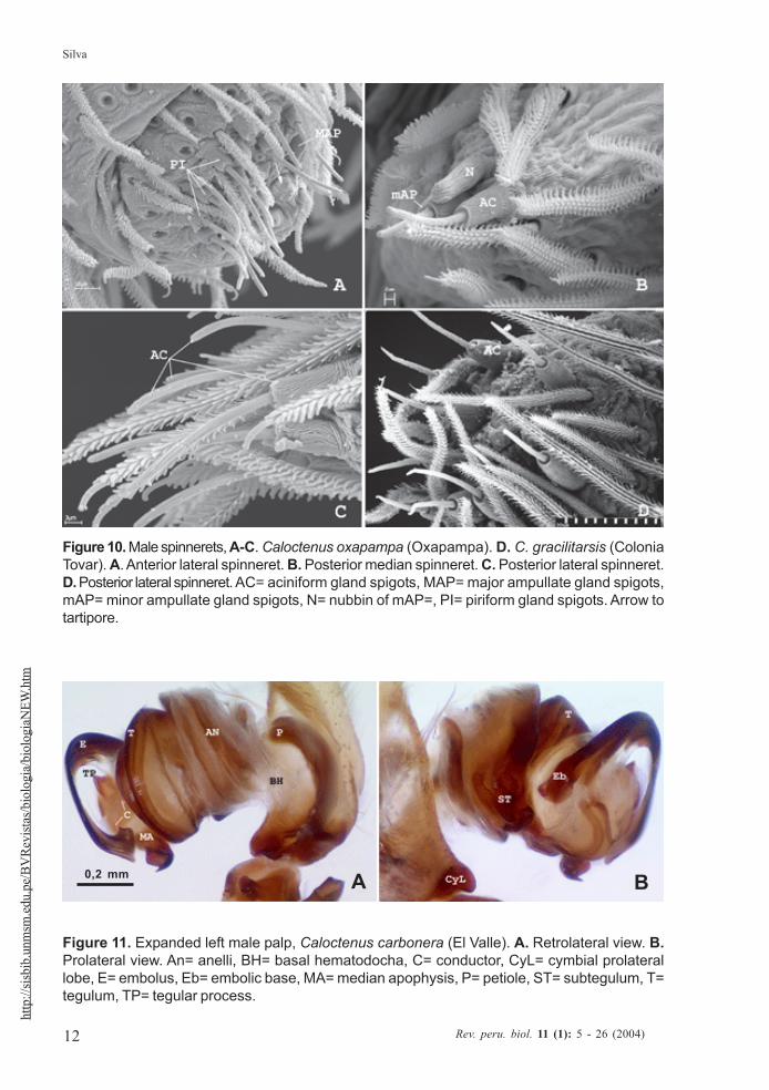
Figure 10. Male spinnerets, A-C. Caloctenus oxapampa (Oxapampa). D. C. gracilitarsis (ColoniaTovar). A. Anterior lateral spinneret. B. Posterior median spinneret. C. Posterior lateral spinneret.D. Posterior lateral spinneret. AC= aciniform gland spigots, MAP= major ampullate gland spigots,mAP= minor ampullate gland spigots, N= nubbin of mAP=, PI= piriform gland spigots. Arrow totartipore.
0,2 mm
Figure 11. Expanded left male palp, Caloctenus carbonera (El Valle). A. Retrolateral view. B.Prolateral view. An= anelli, BH= basal hematodocha, C= conductor, CyL= cymbial prolaterallobe, E= embolus, Eb= embolic base, MA= median apophysis, P= petiole, ST= subtegulum, T=tegulum, TP= tegular process.
A B
13
Revisión: The spiders of genus Caloctenus
Rev. peru. biol. 11 (1): 5 - 26 (2004)
http://sisbib.unmsm
.edu.pe/BV
Revistas/biologia/biologiaN
EW.htm
Copulatory duct openings either antero-mesal(figs. 15A, 18A), antero-lateral (fig. 21A), orposterior (fig. 24A, 25A); spermathecal headdefined by few large pores (figs. 18B, 25B);fertilization ducts posterior (figs. 15B, 18B,25B), or antero-mesal (fig. 21B).
Composition.- Four species, two of themare new.
Misplaced species.- An examination ofthe type specimens of Caloctenus celer Simonand Caloctenus penicilliger Simon showsthey belong somewhere else. Caloctenuspenicilliger belongs in Enoploctenus Simon,while C. celer is a member of AcantheisThorell. The type specimens of Caloctenusdistinctus Caporiacco and C. luteovittatusSimon, are both immatures of Enoploctenus.The type specimen of Caloctenus variegatusBertkau is lost and, according to Brescovit (inlitt.), is a penultimate stage of a femaleEnoploctenus cyclothorax Bertkau. I havenot been able to locate the type specimen ofCaloctenus fernandensis Simon, an adultfemale; however, Simon’s remarks (1910: 362)indicate that this species belongs inAfricactenus Hyatt; no other African ctenidgenus has an epigynum projecting anteriorlyin two lobes and six pairs of ventral spinesbeneath tibiae I-II.
The type specimen of Caloctenus abyssinicusStrand is lost. The original description (Strand 1917:41) is based on an immature female and does notallow a positive identification. Also, a cheliceralfurrow with a combination of two prolateral andfour retromarginal teeth is unusual for the family.Caloctenus abyssinicus is placed incertaesedis, within Ctenidae.
Natural history. Based on collectionrecords of C. gracilitarsis and C. aculeatus,Simon (1897a:120) remarks that Caloctenusinhabited the highest mountains and cloudforests of South America. Labels of recentcollections indicate these spiders are found atelevations over 1800 m, running about on theground surface of cloud forests or adjacenthabitats (roadsides).
hairy lobe (figs. 7B, 8A-B); anal tubercle withlong, thick ventral setae (fig. 9A).
Six spinnerets (fig. 9A). Female ALS (fig.9B) with 2 major ampullate gland spigots, atartipore, and more than 20 piriform glandspigots; PMS (fig. 9C) with two minorampullate gland spigots, three-four cylindricalgland spigots, and more than 20 aciniform glandspigots; PLS (fig. 9D) with three-fourcylindrical gland spigots and more than 20aciniform gland spigots. Male ALS (fig. 10A)with single major ampullate gland spigot,nubbin, tartipore, and more than 20 piriform glandspigots; PMS (fig. 10B) with single minorampullate gland spigot, nubbin, and reduced fieldof aciniform gland spigots; PLS (figs. 10C-D)with reduced field of aciniform gland spigots.
Male palpal tibia with large retroapicalapophysis and ventral lobe (figs. 14 B-C, 16A,19B-C, 22B-C). Cymbium projectingprolaterally into a small basal lobe (fig. 11B),variously produced retrolaterally (figs. 14A-B, 17A, 19B, 20A, 22A), petiole nearly as largeas alveolus (fig. 11A); subtegulum cup-shaped,with 4-5 annuli (figs. 11A-B), prolaterally witha basal process (fig. 11B); tegulum oftenvariously produced at embolus and medianapophysis base (figs. 14B, 17A-B, 19B); ST-T locking mechanism conspicuous when partlyor totally expanded (figs. 11B, 22D); embolusarising on prolateral side of tegulum, coniform(fig. 17A) or spiral-like (fig. 20A), embolic baseprojecting prolaterally into a basal process (figs.11B, 22D); median apophysis with an apicalbeak, often thin and translucent (figs. 17B,22D); small, hyaline conductor, arising frommesoapex of tegulum (fig. 17A, 20B, 23A);sperm duct simple, encircles tegulum (figs. 11A,14B, 19A, 22A-B).
Epigynum either divided into median sec-tor and lateral lobes (fig. 15A) or median sec-tor partly (fig. 18A) or entirely fused to laterallobes (figs. 21A, 24A); lateral lobes lackingteeth; vulva with copulatory ducts somewhatelongated (figs. 15B, 18B), slightly convoluted(fig. 21B), or very short (figs. 24B, 25B).
14
Silva
Rev. peru. biol. 11 (1): 5 - 26 (2004)
http
://sis
bib.
unm
sm.e
du.p
e/B
VR
evist
as/b
iolo
gia/
biol
ogia
NEW
.htm
Peck and Peck (collection labels), by usingflight interception traps along altitudinal transectsin Venezuela, found males of C. carbonera inrain forests at 1200m as well as in cloud forestsfrom 2250 to 2400m. Brignoli (1972:81) recordsone female of C. gracilitarsis from a cave inMiranda, near Caracas (Venezuela); this recordsuggests a preference for dimly lit and humidenvironments.
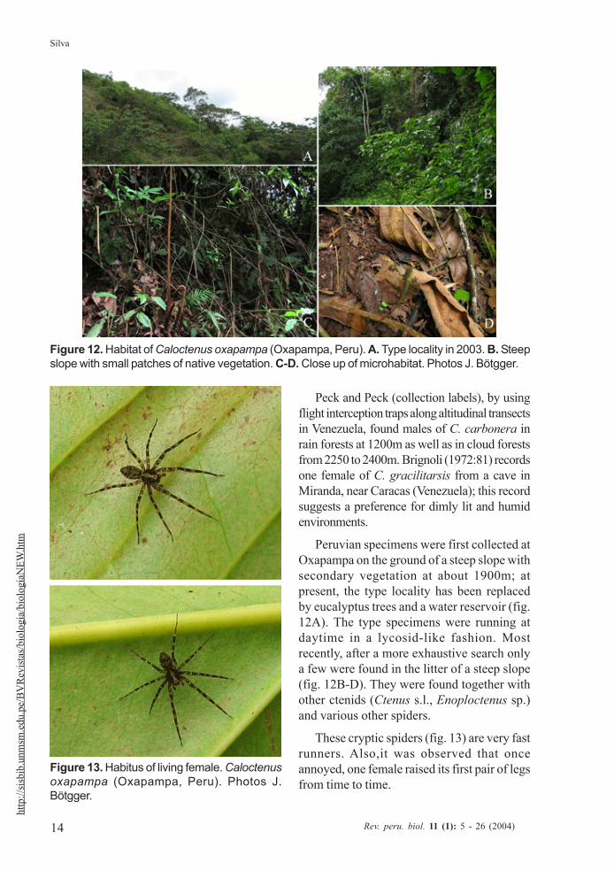
Peruvian specimens were first collected atOxapampa on the ground of a steep slope withsecondary vegetation at about 1900m; atpresent, the type locality has been replacedby eucalyptus trees and a water reservoir (fig.12A). The type specimens were running atdaytime in a lycosid-like fashion. Mostrecently, after a more exhaustive search onlya few were found in the litter of a steep slope(fig. 12B-D). They were found together withother ctenids (Ctenus s.l., Enoploctenus sp.)and various other spiders.
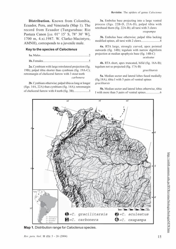
These cryptic spiders (fig. 13) are very fastrunners. Also,it was observed that onceannoyed, one female raised its first pair of legsfrom time to time.
Figure 12. Habitat of Caloctenus oxapampa (Oxapampa, Peru). A. Type locality in 2003. B. Steepslope with small patches of native vegetation. C-D. Close up of microhabitat. Photos J. Bötgger.
Figure 13. Habitus of living female. Caloctenusoxapampa (Oxapampa, Peru). Photos J.Bötgger.
15
Revisión: The spiders of genus Caloctenus
Rev. peru. biol. 11 (1): 5 - 26 (2004)
http://sisbib.unmsm
.edu.pe/BV
Revistas/biologia/biologiaN
EW.htm
Distribution. Known from Colombia,Ecuador, Peru, and Venezuela (Map 1). Therecord from Ecuador (Tungurahua: RioPastaza Canon [ca. 01° 15’ S, 78° 30’ W],1700 m, 4.xi.1987. W. Clarke-Macintyre,AMNH), corresponds to a juvenile male.
Key to the species of Caloctenus1a. Males.................................................................2
1b. Females...............................................................5
2a. Cymbium with large retrolateral projection (fig.19B); palpal tibia shorter than cymbium (fig. 19A-C);retromargin of cheliceral furrow with 3 stout teeth
carbonera
2b. Cymbium otherwise; palpal tibia as long or longer(figs. 14A, 22A) than cymbium (fig. 14A); retromarginof cheliceral furrow with 4 teeth (fig. 3B)....................3
3a. Embolus base projecting into a large ventralprocess (figs. 22B-D, 23A-D), palpal tibia withretrobasal thorn (fig. 22A-B); all tarsi with 3 claws
oxapampa
3b. Embolus base otherwise; palpal tibia lackingmodified spines, all tarsi with 2 claws.........................4
4a. RTA large, strongly curved, apex pointedoutwards (fig. 14B); tegulum with narrow digitiformprojection at median apophysis base (fig. 14B-C)
aculeatus
4b. RTA short, apex truncated, bifid (fig. 16A-B);tegulum not so projected (fig. 17A-B)
gracilitarsis
5a. Median sector and lateral lobes fused medially(fig.18A), tibia I with 5 pairs of ventral spinesgracilitarsis
5b. Median sector and lateral lobes otherwise, tibiaI with more than 5 pairs of ventral spines...................6
Map 1. Distribution range for Caloctenus species.
16
Silva
Rev. peru. biol. 11 (1): 5 - 26 (2004)
http
://sis
bib.
unm
sm.e
du.p
e/B
VR
evist
as/b
iolo
gia/
biol
ogia
NEW
.htm
6a. Epigynal median sector arrowhead-shaped, innermargin of lateral lobes heavily sclerotized (fig. 15A)
aculeatus
6b. Epigynal median sector and lateral lobesotherwise; epigynum lightly sclerotized......................7
7a. Median sector and lateral lobes fused anteriorly(fig. 21A); fertilization ducts anterior (fig. 21B); all tarsiwith two claws .......................................carbonera
7b. Median sector and lateral lobes fused posteriorly(fig. 24A); fertilization ducts posterior (figs. 24B, 25B);all tarsi with three claws..........................oxapampa
Caloctenus aculeatus KeyserlingFigures 14-15; Map 1
Caloctenus aculeatus Keys., 1877: 697(female holotype from Bogota, Colombia, inBMNH, no. 2921, examined).- Roewer, 1954:667.- Bonnet, 1955: 936.- Platnick, 2004.
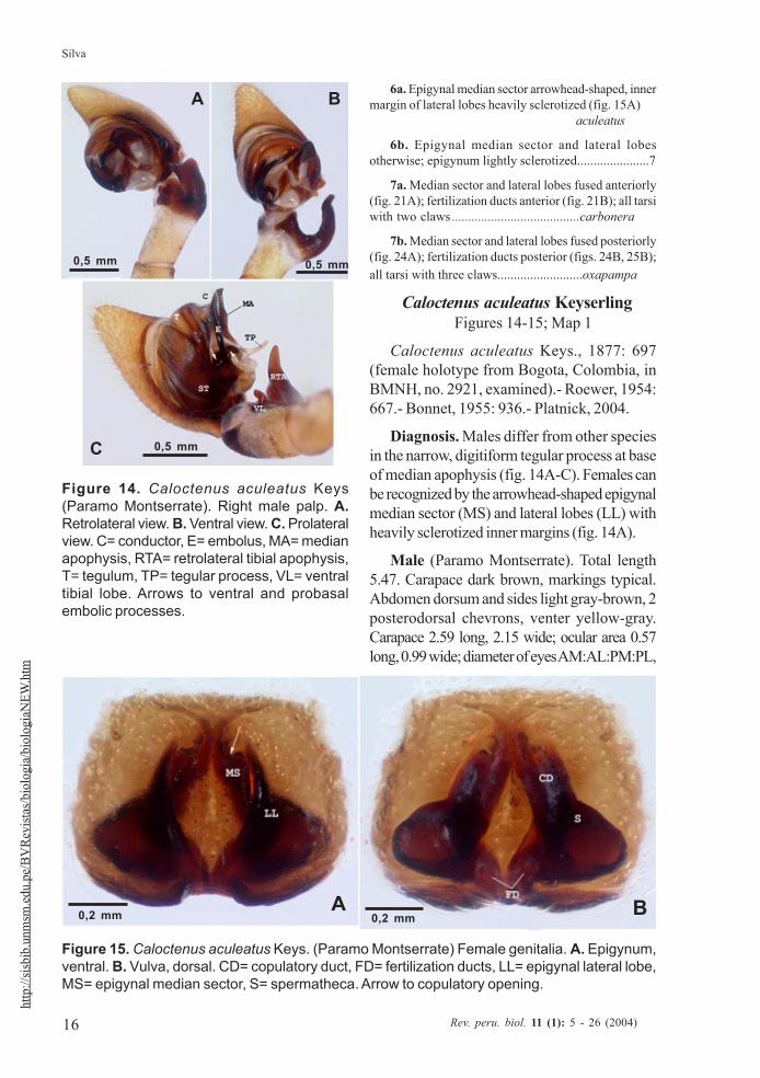
Diagnosis. Males differ from other speciesin the narrow, digitiform tegular process at baseof median apophysis (fig. 14A-C). Females canbe recognized by the arrowhead-shaped epigynalmedian sector (MS) and lateral lobes (LL) withheavily sclerotized inner margins (fig. 14A).
Male (Paramo Montserrate). Total length5.47. Carapace dark brown, markings typical.Abdomen dorsum and sides light gray-brown, 2posterodorsal chevrons, venter yellow-gray.Carapace 2.59 long, 2.15 wide; ocular area 0.57long, 0.99 wide; diameter of eyes AM:AL:PM:PL,
Figure 14. Caloctenus aculeatus Keys(Paramo Montserrate). Right male palp. A.Retrolateral view. B. Ventral view. C. Prolateralview. C= conductor, E= embolus, MA= medianapophysis, RTA= retrolateral tibial apophysis,T= tegulum, TP= tegular process, VL= ventraltibial lobe. Arrows to ventral and probasalembolic processes.
Figure 15. Caloctenus aculeatus Keys. (Paramo Montserrate) Female genitalia. A. Epigynum,ventral. B. Vulva, dorsal. CD= copulatory duct, FD= fertilization ducts, LL= epigynal lateral lobe,MS= epigynal median sector, S= spermatheca. Arrow to copulatory opening.
0,2 mm 0,2 mmA B
A B
C
0,5 mm 0,5 mm
0,5 mm
17
Revisión: The spiders of genus Caloctenus
Rev. peru. biol. 11 (1): 5 - 26 (2004)
http://sisbib.unmsm
.edu.pe/BV
Revistas/biologia/biologiaN
EW.htm
0.16:0.07:0.26:0.24; AME-AME 0.4 times AMEdiameter; PME separation 0.44 times PMEdiameter; clypeal height 0.20. Sternum 2.48long, 2.52 wide; labium 0.33 long, 0.42 wide;endites 0.82 long, 0.40 wide. Femur I length1.39 times carapace width. Spination: palpus -femur d0-1-1, p0-0-1, r0-0-1, patella d0-0-1,p1-0-0, tibia p1-1-0, r0-1-0; leg I -femur d0-1-1-0, p0-2-1-1, r0-1-0-1, v0-0-1-0, patella d0-0-1, tibia v2-2-2-2-2-2-1, metatarsus v2-2-2-2-2-1; leg II -femur d0-1-1-1, p1-1-1-0, r1-1-1-0, patella d0-0-1, tibia v2-2-2-2-2-2-2,metatarsus v2-2-2-2-2; leg III -femur d0-1-1-1, p1-1-1-0,r1-1-1-0, patella d0-0-1, tibia d0-0-1-0, p0-1-1-0, r0-1-1-0, v2-2-0-2, metatarsusd0-0-1-0, p1-1-0-1, r1-1-0-1, v0-1-1-1; leg IV-femur d1-1-0-1, p1-1-0-1, r1-0-1-1, patella d0-0-1, tibia d0-0-1-0, p0-1-1-0, r0-1-1-0, v2-2-0-0, metatarsus d0-1-0-0, p1-1-0-1, r1-1-0-1, v1-1-1-2. Leg measurements:
I II III IV PalpFemur 2,99 2,81 2,59 3,32 1,20Patella 1,06 1,02 0,91 0,95 0,62Tibia 3,25 2,73 2,30 3,07 0,51Metatarsus 3,23 2,74 2,63 3,87 —Tarsus 1,06 0,91 0,91 1,17 1,28
Total 11,59 10,21 9,34 12,38 3,61
Palpal tibia (figs. 14A-C) stout, with largeventral lobe, length 0,17 times femur I; RTA(figs. 14A-C) large, broad, and curved, directedoutwards. Cymbium retrolaterally projectinginto a small subbasal lobe (figs. 14A-B);tegulum with a slender hyaline process atmedian apophysis base (figs. 14B-C); MAorigin retro-mesal (fig. 14B); embolus coniform,arising from a large sclerotized process onprolateral side of tegulum (fig. 14C); conduc-tor (figs. 14A-C) hyaline, origin mesal.
Female (holotype).- Total length 4,71.Markings as in male except paler. Carapace2,15 long, 1,79 wide, 0,68 high; ocular area 0,55long, 0,91 wide, diameter of eyesAM:AL:PM:PL, 0,11:0,06:0,22:0,22; AME-AME nearly AME diameter; PME separation0,68 times PME diameter; clypeal height 0,13;
sternum 1,02 long, 1,02 wide; endites 0,68 long,0,34 wide. Femur I 1,08 times carapace width.Leg III missing. Spination: palpus -femur d0-0 -2-1, p0-0-2-0, r0-0-1-0, patella d1-0-0-1, p1-0-0,tibia d1-0-0-1, p2-0-0-0, tarsus p2-1-0-0, r1-1-0-0; leg I -femur d0-1-1-1, p0-2-1-1, r1-1-1-1,patella d-0-0-1, tibia p0-0-1-1-0, r0-0-1-1-0, v2-2-2-2-2-2, metatarsus p0-0-1-1-0, r0-0-1-1-0, v2-2-2-2-2-2; leg II -femur d0-1-0-1-0-1, p2-1-1-1-1-1, r1-0-0-0-1-0, tibia v2-2-2-2-2-2-2, metatarsusv2-2-2-2-2-2-2. Leg measurements:
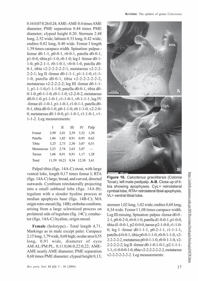
Figure 16. Caloctenus gracilitarsis (ColoniaTovar), left male pedipalp. A-B. Close up of ti-bia showing apophyses. CyL= retrolateralcymbial lobe, RTA= retrolateral tibial apophysis,VL= ventral tibial lobe.
18
Silva
Rev. peru. biol. 11 (1): 5 - 26 (2004)
http
://sis
bib.
unm
sm.e
du.p
e/B
VR
evist
as/b
iolo
gia/
biol
ogia
NEW
.htm
I II III IV Palp
Femur 1,93 1,86 — 2,23 0,77Patella 0,73 0,77 — 0,66 0,40Tibia 1,86 1,61 — 1,86 0,49Metatarsus 1,72 1,57 — 2,48 —Tarsus 0,53 0,51 — 0,75 1,31Total 6,77 6,32 — 7,98 2,97
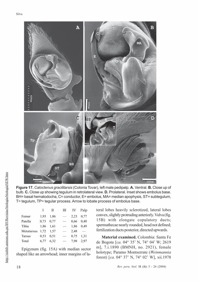
Epigynum (fig. 15A) with median sectorshaped like an arrowhead; inner margins of la-
teral lobes heavily sclerotized, lateral lobesconvex, slightly protruding anteriorly. Vulva (fig.15B) with elongate copulatory ducts;spermathecae nearly rounded, head not defined;fertilization ducts posterior, directed upwards.
Material examined. Colombia: Santa Fede Bogota [ca. 04° 35’ N, 74° 04’ W; 2619m], 7.i.1890 (BMNH, no. 2921), femaleholotype; Paramo Montserrate (Weinmanniaforest) [ca. 04° 37’ N, 74° 02’ W], xii.1978
Figure 17. Caloctenus gracilitarsis (Colonia Tovar), left male pedipalp. A. Ventral. B. Close up ofbulb. C. Close up showing tegulum in retrolateral view. D. Prolateral. Inset shows embolus base.BH= basal hematodocha, C= conductor, E= embolus, MA= median apophysis, ST= subtegulum,T= tegulum, TP= tegular process. Arrow to lobate process of embolus base.
19
Revisión: The spiders of genus Caloctenus
Rev. peru. biol. 11 (1): 5 - 26 (2004)
http://sisbib.unmsm
.edu.pe/BV
Revistas/biologia/biologiaN
EW.htm
(A. Bernal, MCZ 33292), 1 female; ParamoMontserrate, iv.1979 (A. Bernal, MCZ 30753),1 male.
Distribution.- Known from Bogota, Co-lombia (Map 1).
Caloctenus gracilitarsis SimonFigures 16-18; Map 1
Caloctenus gracilitarsis Simon, 1897a:496 (2 syntype females from Colonia Tovar(MNHN 11025) plus 2 syntype females and ajuvenile male (11118) from Caracas, Venezue-la.- Roewer, 1954: 667.- Bonnet, 1955: 936.-Caporiacco, 1955: 397 (male palp andepigynum illustrated, fig. 55).- Brignoli, 1972:
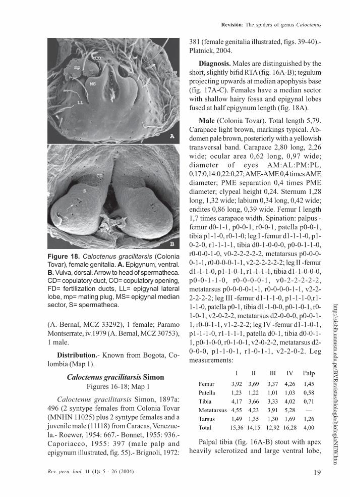
381 (female genitalia illustrated, figs. 39-40).-Platnick, 2004.
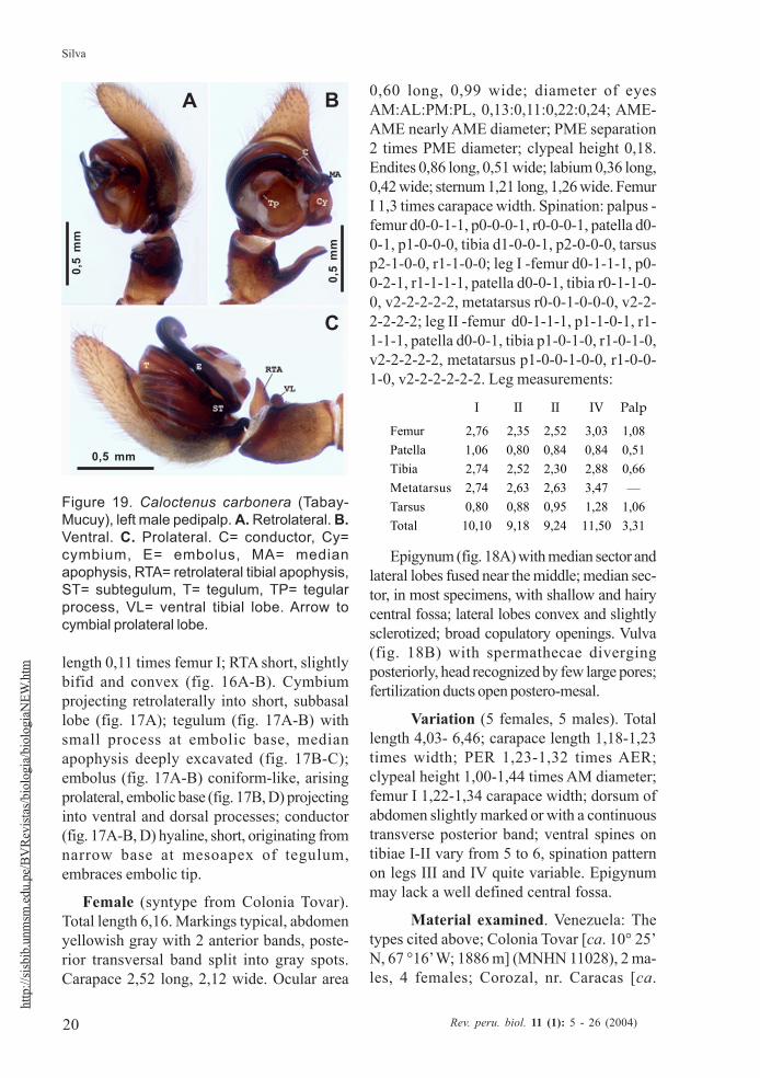
Diagnosis. Males are distinguished by theshort, slightly bifid RTA (fig. 16A-B); tegulumprojecting upwards at median apophysis base(fig. 17A-C). Females have a median sectorwith shallow hairy fossa and epigynal lobesfused at half epigynum length (fig. 18A).
Male (Colonia Tovar). Total length 5,79.Carapace light brown, markings typical. Ab-domen pale brown, posteriorly with a yellowishtransversal band. Carapace 2,80 long, 2,26wide; ocular area 0,62 long, 0,97 wide;diameter of eyes AM:AL:PM:PL,0,17:0,14:0,22:0,27; AME-AME 0,4 times AMEdiameter; PME separation 0,4 times PMEdiameter; clypeal height 0,24. Sternum 1,28long, 1,32 wide; labium 0,34 long, 0,42 wide;endites 0,86 long, 0,39 wide. Femur I length1,7 times carapace width. Spination: palpus -femur d0-1-1, p0-0-1, r0-0-1, patella p0-0-1,tibia p1-1-0, r0-1-0; leg I -femur d1-1-1-0, p1-0-2-0, r1-1-1-1, tibia d0-1-0-0-0, p0-0-1-1-0,r0-0-0-1-0, v0-2-2-2-2-2, metatarsus p0-0-0-0-1-1, r0-0-0-0-1-1, v2-2-2-2-2-2; leg II -femurd1-1-1-0, p1-1-0-1, r1-1-1-1, tibia d1-1-0-0-0,p0-0-1-1-0, r0-0-0-0-1, v0-2-2-2-2-2,metatarsus p0-0-0-0-1-1, r0-0-0-0-1-1, v2-2-2-2-2-2; leg III -femur d1-1-1-0, p1-1-1-0,r1-1-1-0, patella p0-1, tibia d1-1-0-0, p0-1-0-1, r0-1-0-1, v2-0-2-2, metatarsus d2-0-0-0, p0-0-1-1, r0-0-1-1, v1-2-2-2; leg IV -femur d1-1-0-1,p1-1-1-0, r1-1-1-1, patella d0-1, tibia d0-0-1-1, p0-1-0-0, r0-1-0-1, v2-0-2-2, metatarsus d2-0-0-0, p1-1-0-1, r1-0-1-1, v2-2-0-2. Legmeasurements:
I II III IV Palp
Femur 3,92 3,69 3,37 4,26 1,45Patella 1,23 1,22 1,01 1,03 0,58Tibia 4,17 3,66 3,33 4,02 0,71Metatarsus 4,55 4,23 3,91 5,28 —Tarsus 1,49 1,35 1,30 1,69 1,26Total 15,36 14,15 12,92 16,28 4,00
Palpal tibia (fig. 16A-B) stout with apexheavily sclerotized and large ventral lobe,
Figure 18. Caloctenus gracilitarsis (ColoniaTovar), female genitalia. A. Epigynum, ventral.B. Vulva, dorsal. Arrow to head of spermatheca.CD= copulatory duct, CO= copulatory opening,FD= fertilization ducts, LL= epigynal laterallobe, mp= mating plug, MS= epigynal mediansector, S= spermatheca.
20
Silva
Rev. peru. biol. 11 (1): 5 - 26 (2004)
http
://sis
bib.
unm
sm.e
du.p
e/B
VR
evist
as/b
iolo
gia/
biol
ogia
NEW
.htm length 0,11 times femur I; RTA short, slightly
bifid and convex (fig. 16A-B). Cymbiumprojecting retrolaterally into short, subbasallobe (fig. 17A); tegulum (fig. 17A-B) withsmall process at embolic base, medianapophysis deeply excavated (fig. 17B-C);embolus (fig. 17A-B) coniform-like, arisingprolateral, embolic base (fig. 17B, D) projectinginto ventral and dorsal processes; conductor(fig. 17A-B, D) hyaline, short, originating fromnarrow base at mesoapex of tegulum,embraces embolic tip.
Female (syntype from Colonia Tovar).Total length 6,16. Markings typical, abdomenyellowish gray with 2 anterior bands, poste-rior transversal band split into gray spots.Carapace 2,52 long, 2,12 wide. Ocular area
0,60 long, 0,99 wide; diameter of eyesAM:AL:PM:PL, 0,13:0,11:0,22:0,24; AME-AME nearly AME diameter; PME separation2 times PME diameter; clypeal height 0,18.Endites 0,86 long, 0,51 wide; labium 0,36 long,0,42 wide; sternum 1,21 long, 1,26 wide. FemurI 1,3 times carapace width. Spination: palpus -femur d0-0-1-1, p0-0-0-1, r0-0-0-1, patella d0-0-1, p1-0-0-0, tibia d1-0-0-1, p2-0-0-0, tarsusp2-1-0-0, r1-1-0-0; leg I -femur d0-1-1-1, p0-0-2-1, r1-1-1-1, patella d0-0-1, tibia r0-1-1-0-0, v2-2-2-2-2, metatarsus r0-0-1-0-0-0, v2-2-2-2-2-2; leg II -femur d0-1-1-1, p1-1-0-1, r1-1-1-1, patella d0-0-1, tibia p1-0-1-0, r1-0-1-0,v2-2-2-2-2, metatarsus p1-0-0-1-0-0, r1-0-0-1-0, v2-2-2-2-2-2. Leg measurements:
I II II IV Palp
Femur 2,76 2,35 2,52 3,03 1,08Patella 1,06 0,80 0,84 0,84 0,51Tibia 2,74 2,52 2,30 2,88 0,66Metatarsus 2,74 2,63 2,63 3,47 —Tarsus 0,80 0,88 0,95 1,28 1,06Total 10,10 9,18 9,24 11,50 3,31
Epigynum (fig. 18A) with median sector andlateral lobes fused near the middle; median sec-tor, in most specimens, with shallow and hairycentral fossa; lateral lobes convex and slightlysclerotized; broad copulatory openings. Vulva(fig. 18B) with spermathecae divergingposteriorly, head recognized by few large pores;fertilization ducts open postero-mesal.
Variation (5 females, 5 males). Totallength 4,03- 6,46; carapace length 1,18-1,23times width; PER 1,23-1,32 times AER;clypeal height 1,00-1,44 times AM diameter;femur I 1,22-1,34 carapace width; dorsum ofabdomen slightly marked or with a continuoustransverse posterior band; ventral spines ontibiae I-II vary from 5 to 6, spination patternon legs III and IV quite variable. Epigynummay lack a well defined central fossa.
Material examined. Venezuela: Thetypes cited above; Colonia Tovar [ca. 10° 25’N, 67 °16’ W; 1886 m] (MNHN 11028), 2 ma-les, 4 females; Corozal, nr. Caracas [ca.
Figure 19. Caloctenus carbonera (Tabay-Mucuy), left male pedipalp. A. Retrolateral. B.Ventral. C. Prolateral. C= conductor, Cy=cymbium, E= embolus, MA= medianapophysis, RTA= retrolateral tibial apophysis,ST= subtegulum, T= tegulum, TP= tegularprocess, VL= ventral tibial lobe. Arrow tocymbial prolateral lobe.
0,5
mm
0,5
mm
0,5 mm
C
A B
21
Revisión: The spiders of genus Caloctenus
Rev. peru. biol. 11 (1): 5 - 26 (2004)
http://sisbib.unmsm
.edu.pe/BV
Revistas/biologia/biologiaN
EW.htm
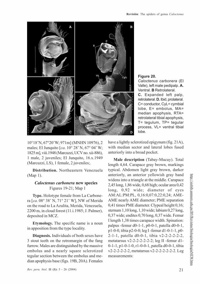
Figure 20.Caloctenus carbonera (ElValle), left male pedipalp. A.Ventral. B Retrolateral.C. Expanded left palp,retrolateral. D. Ibid, prolateral.C= conductor, CyL= cymbiallobe, E= embolus, MA=median apophysis, RTA=retrolateral tibial apophysis,T= tegulum, TP= tegularprocess, VL= ventral tibiallobe.
10°18’N, 67°20’W; 971m] (MNHN 10976), 2males; El Junquito [ca. 10° 28’ N, 67° 04’ W;1825 m], viii.1948 (Marcuzzi, UCV no. xii-886),1 male, 2 juveniles; El Junquito, 16.x.1949(Marcuzzi, LS), 1 female, 2 juveniles;.
Distribution. Northeastern Venezuela(Map 1).
Caloctenus carbonera new speciesFigures 19-21; Map 1
Type. Holotype female from La Carbone-ra [ca. 08° 38’ N, 71° 21’ W], NW of Meridaon the road to La Azulita, Merida, Venezuela,2200 m, in cloud forest (11.i.1985; J. Palmer),deposited in MCZ.
Etymology. The specific name is a nounin apposition from the type locality.
Diagnosis. Individuals of both sexes have3 stout teeth on the retromargin of the fangfurrow. Males are distinguished by the massiveembolus and a nearly square sclerotizedtegular section between the embolus and me-dian apophysis base (figs. 19B, 20A). Females
have a lightly sclerotized epigynum (fig. 21A),with median sector and lateral lobes fusedanteriorly into a broad pocket.
Male description (Tabay-Mucuy). Totallength 4,64. Carapace gray brown, markingstypical. Abdomen light gray brown, darkeranteriorly, an anterior yellowish gray bandwidens into a triangle at the middle. Carapace2,45 long, 1,86 wide, 0,68 high; ocular area 0,42long, 0,92 wide; diameter of eyesAM:AL:PM:PL, 0,16:0,07:0,22:0,24; AME-AME nearly AME diameter; PME separation0,41 times PME diameter. Clypeal height 0,16;sternum 1,10 long, 1,10 wide; labium 0,27 long,0,37 wide; endites 0,70 long, 0,37 wide. FemurI length 1,38 times carapace width. Spination:palpus -femur d0-1-1, p0-0-1, patella d0-0-1,p1-0-0, tibia p2-0-0; leg I -femur d1-0-1-1, p0-2-1-1, patella d0-0-1, tibia v2-2-2-2-2-2,metatarsus v2-2-2-2-2-2-2; leg II -femur d1-0-1-1, p1-0-1-0, r1-0-0-1, patella d0-0-1, tibiav2-2-2-2-2-2, metatarsus v2-2-2-2-2-2-2. Legmeasurements:
22
Silva
Rev. peru. biol. 11 (1): 5 - 26 (2004)
http
://sis
bib.
unm
sm.e
du.p
e/B
VR
evist
as/b
iolo
gia/
biol
ogia
NEW
.htm
I II II IV PalpFemur 3,03 2,66 2,56 3,18 0,84Patella 0,88 0,84 0,69 0,73 0,44Tibia 3,54 3,07 2,37 2,96 0,62Metatarsus 3,54 3,03 2,74 3,76 —Tarsus 1,31 1,13 1,02 1,28 0,84Total 12,30 10,73 9,38 11,91 2,74
Palpal tibia shorter than cymbium (figs.19A-C), length 0,21 times femur I; RTA,almost as long as tibia length (figs. 19B-C,20A), blunt-tipped (fig. 20B-C). Cymbium withlarge retrolateral projection (figs. 19B, 20A),small probasal projection (fig. 19C); tegulumwith small process at embolus base (figs. 19A,20A); median apophysis (figs. 19A-B, 20B,D) small, infolding margins meeting apicallybecoming an acute tip; embolus stout,apex bifid(fig. 20C), conductor does not embraceembolic tip (figs. 20A-B, D).
Female (holotype).- Total length 4,10.Carapace gray brown, markings typical; ab-domen with stripes joining into a trapezoidpattern. Carapace 2,01 long, 1,62 wide, 0,66high; ocular area 0,51 long, 0,85 wide, diameterof eyes AM:AL:PM:PL, 0,13:0,04:0,22:0,22;AM-AM nearly AM diameter; PMEseparation 0,41 times PME diameter; clypealheight 0,13. Sternum 0,93 long, 0,95 wide;endites 0,48 long, 0,29 wide. Femur I 1,10 ti-mes carapace width. Spination: palpus -femur
d0-1-1, p0-0-0-1, patella d0-0-1, p1-0-0, tibiap2-0-0-0, r1-0-0-1, tarsus p2-1-0-0, r1-1-0-0;leg I -femur d0-1-1-0, p0-2-1-0, r1-0-0-0. v0-1-2-2, patella d-0-0-1, tibia v2-2-2-2-2-2,metatarsus v2-2-2-2-2-2-2-1; leg II -femur d0-1-1-1, p1-1-1-0, r1-0-1-0, patella d0-0-1, tibiav2-2-2-2-2-2, metatarsus v2-2-2-2-2-2-2. Legmeasurements:
I II III IV Palp
Femur 1,79 1,70 1,64 2,04 0,69Patella 0,69 0,66 0,58 0,62 0,37Tibia 1,86 1,64 1,35 1,83 0,42Metatarsus 1,75 1,61 1,61 2,19 —-Tarsus 0,47 0,47 0,58 0,73 0,58Total 6,56 6,08 5,76 7,41 2,06
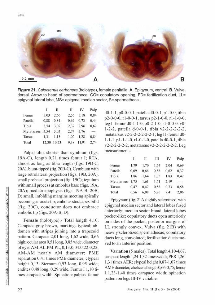
Epigynum (fig. 21A) lightly sclerotized, withepigynal median sector and lateral lobes fusedanteriorly; median sector broad, lateral lobespocket-like; copulatory ducts open anteriorlyon sides of the pocket, posterior margins ofLL strongly convex. Vulva (fig. 21B) withheavily sclerotized spermathecae, copulatoryducts long, convoluted; fertilization ducts mo-ved to an anterior position.
Variation (5 males). Total length 4,10-4,67,carapace length 1,24-1,32 times width; PER 1,26-1,31 times AER; clypeal height 0,87-1,07 timesAME diameter; cheliceral length 0,66-0,75; femurI 1,23-1,40 times carapace width; spinationpattern on legs III-IV variable.
Figure 21. Caloctenus carbonera (holotype), female genitalia. A. Epigynum, ventral. B. Vulva,dorsal. Arrow to head of spermatheca. CO= copulatory opening, FD= fertilization duct, LL=epigynal lateral lobe, MS= epigynal median sector, S= spermatheca.
A B0,2 mm
23
Revisión: The spiders of genus Caloctenus
Rev. peru. biol. 11 (1): 5 - 26 (2004)
http://sisbib.unmsm
.edu.pe/BV
Revistas/biologia/biologiaN
EW.htm
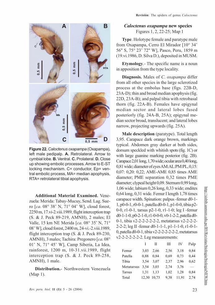
Figure 22. Caloctenus oxapampa (Oxapampa),left male pedipalp. A. Retrolateral. Arrow tocymbial lobe. B. Ventral. C. Prolateral. D. Closeup showing embolic processes. Arrow to E-STlocking mechanism. C= conductor, Ep= ven-tral embolic process, MA= median apophysis,RTA= retrolateral tibial apophysis.
Additional Material Examined. Vene-zuela: Merida: Tabay-Mucuy, Send. Lag. Sue-ro [ca. 08° 38’ N, 71° 04’ W], cloud forest,2250 m, 17.vi-2.viii.1989, flight interception trap(S. & J. Peck 89-219, AMNH), 2 males; ElValle, 15 km NE Merida [ca. 08° 35’ N, 71°08’ W], cloud forest, 2400 m, 24-vi.-2.viii.1989,flight interception trap (S. & J. Peck 89-230,AMNH), 3 males; Tachira: Pregonero [ca. 08°01’ N, 71° 45’ W], Camp Siberia, La Idea,rainforest, 1200 m, 10-31.vii.1989, flightinterception trap (S. & J. Peck 89-258,AMNH), 1 male.
Distribution.- Northwestern Venezuela(Map 1).
Caloctenus oxapampa new speciesFigures 1, 2, 22-25; Map 1
Type. Holotype female and paratype malefrom Oxapampa, Cerro El Mirador [10° 34’56" S, 75° 23’ 72" W], Pasco, Peru, 1859 m(19.vi.1986, D. Silva D.), deposited in MUSM.
Etymology.- The specific name is a nounin apposition from the type locality.
Diagnosis. Males of C. oxapampa differfrom all other species in the large sclerotizedprocess at the embolus base (figs. 22B-D,23A-D); thin and broad median apophysis (fig.22D, 23A-B); and palpal tibia with retrobasalthorn (fig. 22A-B). Females have epigynalmedian sector and lateral lobes fusedposteriorly (fig. 24A-B, 25A); epigynal me-dian sector broad, translucent; and lateral lobesnarrow, projecting upwards (fig. 25A).
Male description (paratype). Total length3,95. Carapace dark orange brown, markingstypical. Abdomen gray darker at both sides,dorsum speckled with whitish spots (fig. 1C) orwith large guanine marking posterior (fig. 2B).Carapace 2,01 long, 1,70 wide; ocular area 0,40 long,0,81 wide; diameter of eyes AM:AL:PM:PL, 0,15:0,07: 0,20: 0,22; AME-AME 0,85 times AMEdiameter; PME separation 0,32 times PMEdiameter; clypeal height 0,09. Sternum 0,99 long,1,06 wide; labium 0,26 long, 0,33 wide; endites0,64 long, 0,31 wide. Femur I length 1,78 timescarapace width. Spination: palpus -femur d0-1-1, p0-0-1, r0-0-1, patella d0-0-1, p1-0-0, tibia p2-0-0, r1-0-1, tarsus p2-1-0, r1-1-0; leg I -femurd0-1-1-0, p0-2-1-0, r1-0-0-0, v0-1-2-2, patella d0-0-1, tibia v2-2-2-2-2-2-2, metatarsus v2-2-2-2-2-2-2; leg II -femur d0-1-1-1, p1-1-1-0, r1-0-1-0, patella d0-0-1, tibia v2-2-2-2-2-2-2, metatarsusv2-2-2-2-2-2-2. Leg measurements:
I II III IV Palp
Femur 3,03 2,66 2,56 3,18 0,84Patella 0,88 0,84 0,69 0,73 0,44Tibia 3,54 3,07 2,37 2,96 0,62Metatarsus 3,54 3,03 2,74 3,76 —Tarsus 1,31 1,13 1,02 1,28 0,84Total 12,30 10,73 9,38 11,91 2,74
0,5
mm
0,5
mm
0,5
mm
0,5 mmC D
A B
24
Silva
Rev. peru. biol. 11 (1): 5 - 26 (2004)
http
://sis
bib.
unm
sm.e
du.p
e/B
VR
evist
as/b
iolo
gia/
biol
ogia
NEW
.htm Palpal tibia (fig. 22A-C) relatively long,
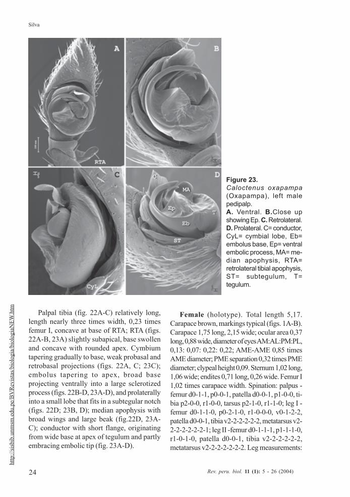
length nearly three times width, 0,23 timesfemur I, concave at base of RTA; RTA (figs.22A-B, 23A) slightly subapical, base swollenand concave with rounded apex. Cymbiumtapering gradually to base, weak probasal andretrobasal projections (figs. 22A, C; 23C);embolus tapering to apex, broad baseprojecting ventrally into a large sclerotizedprocess (figs. 22B-D, 23A-D), and prolaterallyinto a small lobe that fits in a subtegular notch(figs. 22D; 23B, D); median apophysis withbroad wings and large beak (fig.22D, 23A-C); conductor with short flange, originatingfrom wide base at apex of tegulum and partlyembracing embolic tip (fig. 23A-D).
Female (holotype). Total length 5,17.Carapace brown, markings typical (figs. 1A-B).Carapace 1,75 long, 2,15 wide; ocular area 0,37long, 0,88 wide, diameter of eyes AM:AL:PM:PL,0,13: 0,07: 0,22: 0,22; AME-AME 0,85 timesAME diameter; PME separation 0,32 times PMEdiameter; clypeal height 0,09. Sternum 1,02 long,1,06 wide; endites 0,71 long, 0,26 wide. Femur I1,02 times carapace width. Spination: palpus -femur d0-1-1, p0-0-1, patella d0-0-1, p1-0-0, ti-bia p2-0-0, r1-0-0, tarsus p2-1-0, r1-1-0; leg I -femur d0-1-1-0, p0-2-1-0, r1-0-0-0, v0-1-2-2,patella d0-0-1, tibia v2-2-2-2-2-2, metatarsus v2-2-2-2-2-2-2-1; leg II -femur d0-1-1-1, p1-1-1-0,r1-0-1-0, patella d0-0-1, tibia v2-2-2-2-2-2,metatarsus v2-2-2-2-2-2-2. Leg measurements:
Figure 23.Caloctenus oxapampa(Oxapampa), left malepedipalp.A. Ventral. B.Close upshowing Ep. C. Retrolateral.D. Prolateral. C= conductor,CyL= cymbial lobe, Eb=embolus base, Ep= ventralembolic process, MA= me-dian apophysis, RTA=retrolateral tibial apophysis,ST= subtegulum, T=tegulum.
25
Revisión: The spiders of genus Caloctenus
Rev. peru. biol. 11 (1): 5 - 26 (2004)
http://sisbib.unmsm
.edu.pe/BV
Revistas/biologia/biologiaN
EW.htm
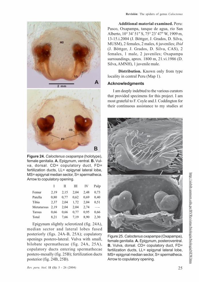
Figure 25. Caloctenus oxapampa (Oxapampa),female genitalia. A. Epigynum, posteroventral.B. Vulva, dorsal. CD= copulatory duct, FD=fertilization ducts, LL= epigynal lateral lobe,MS= epigynal median sector, S= spermatheca.Arrow to copulatory opening.
I II III IV Palp
Femur 2,19 2,15 2,04 2,48 0,73Patella 0,80 0,77 0,62 0,69 0,40Tibia 2,37 2,04 1,72 2,04 0,51Metatarsus 2,19 2,04 2,04 2,74 —-Tarsus 0,66 0,66 0,77 0,95 0,66Total 8,21 7,66 7,19 8,90 2,30
Epigynum slightly sclerotized (fig. 24A),median sector and lateral lobes fusedposteriorly (figs. 24A-B, 25A); copulatoryopenings postero-lateral. Vulva with small,bilobate spermathecae (fig. 24A, 25A),copulatory ducts entering spermathecaepostero-mesally (fig. 25B); fertilization ductsposterior (fig. 24B, 25B).
Additional material examined. Peru:Pasco, Oxapampa, tanque de agua, rio SanAlberto, 10° 34’ 51" S, 75° 23’ 47" W, 1909 m,13-15.i.2004 (J. Böttger, J. Grados, D. Silva,MUSM), 2 females, 2 males, 6 juveniles; Ibid(J. Böttger, J. Grados, D. Silva, CAS), 2females, 1 male, 2 juveniles; Oxapampasurroundings, aprox. 1800 m, 21.vi.1986 (D.Silva, AMNH), 1 juvenile male.
Distribution. Known only from typelocality in central Peru (Map 1).
Acknowledgments
I am deeply indebted to the various curatorsthat provided specimens for this project. I ammost grateful to F. Coyle and J. Coddington fortheir continuous assistance to my studies at
Figure 24. Caloctenus oxapampa (holotype),female genitalia. A. Epigynum, ventral. B. Vul-va, dorsal. CD= copulatory duct, FD=fertilization ducts, LL= epigynal lateral lobe,MS= epigynal median sector, S= spermatheca.Arrow to copulatory opening.
2 mmA
B
26
Silva
Rev. peru. biol. 11 (1): 5 - 26 (2004)
http
://sis
bib.
unm
sm.e
du.p
e/B
VR
evist
as/b
iolo
gia/
biol
ogia
NEW
.htm
Western Carolina University, where this projectstarted as a MS thesis. C. Griswold is muchacknowledged for his continuous support to myresearch work and time for reviewing drafts ofthis paper. Thanks to R. Jocqué for his commentson the last draft.
Undetermined ctenid spiders in variousmuseums were examined through financial aidfrom the joint AMNH-Cornell UniversityProgram and the Ernst Mayr Grant of theMCZ at Harvard University. A postdoctoralfellowship from the California Academy ofSciences and the Schlinger Foundation greatlycontributed to complete this work.
Illustrations are by J. Speckels. J. Bötggerkindly provided photos of the habitat and livingspiders. Fieldwork in Peru was made possiblethrough a Research Permit from The Instituto Na-cional de Recursos Naturales (INRENA). I amvery grateful to B. León, J. Grados and J. Bötggerfor making my fieldwork in Peru possible.
Literature citedBertkau, P. 1880. Verzeichniss der von Prof. Ed. van
Beneden auf seiner im Auftrage der BelgischenRegierung unternommen wissenschaftlichenReise nach Brasilien und La Plata in Jahren1872—73 gesammelten Arachniden. Mém.cour. Acad. Belg 43: 1—120.
Benoit, P.L.G. 1974. Contribution à l’étude du genreAfricactenus Hyatt avec une clé des espèces(Araneae - Ctenidae). Revue Zool. afr. 88:131—142.
Bonnet, P. 1955. Bibliographia Araneorum. Toulouse, 2(2): 919—1925.
Brignoli, P. M. 1972. Sur quelques araignées cavernicolesd’Argentine, Uruguay, Brésil et Venezuelarécoltées par le Dr. P. Strinati (Arachnida,Araneae). Revue suisse Zool. 79: 361—385.
Caporiacco, L. di. 1947. Diagnosi preliminari de specienuove di aracnidi della Guiana Brittanicaraccolte dai professori Beccari e Romiti.Monitore zool. ital. 56: 20—34.
Caporiacco, L. di. 1955. Estudios sobre los aracnidos deVenezuela. 2a parte: Araneae. Acta biol. venez.1: 265—448.
Griswold, C. E. 1991. A revision and phylogeneticanalysis of the African spider genusMachadonia Lehtinen (Araneae: Lycosoidea).Ent. Scand. 22: 305—351.
Hyatt, K. H. 1954. The African spiders of the familyCtenidae in the collections of the British
Museum (Natural History). Ann. Mag. nat.Hist. (12) 7: 877—894.
Keyserling, E. 1877. Uber amerikanische Spinnenartender Unterordnung Citigradae. Verh. zool-botGesell. Wien 26: 609—708.
Mello-Leitão, C. F. 1936. Contribution a l’étude desCténides du Brésil. Festschr. Strand 1: 1-31,598-601.
Pickard-Cambridge, F. O. 1897a. On cteniform spidersfrom the lower Amazons and other regions ofNorth and South America, with a list of allknown species of these groups hithertorecorded from the New World. Ann. Mag. nat.Hist. 19: 52—106.
Pickard-Cambridge, F. O. 1897b. On the cteniformspiders of Ceylon, Burmah, and the IndianArchipelago, west and north Wallace’s line:with bibliography and list of those fromAustralasia, south and east Wallace’s line. Ann.Mag. nat. Hist. 20: 329—356.
Platnick, N. I. 2004. The world spider catalog, version4.5. American Museum of Natural History,online at http://research.amnh.org/entomology/spiders/catalog/index.html
Roewer, C. F. 1954. Katalog der Araneae von 1758 bis1940. Brussels, 2a: 1-923.
Silva Dávila, D. 1994. Taxonomic revision of theNeotropical spider genus Caloctenus Keyserling(Araneae, Ctenidae). MS Thesis, Western Ca-rolina University, North Carolina, USA.
Silva Dávila, D. 2003. Higher-level relationships of thespider family Ctenidae (Araneae: Ctenoidea).Bull. Am. Mus. nat. Hist. 274: 1—86.
Simon, E. 1897a. Histoire naturelle des araignées. Paris,2 (1): 1—192.
Simon, E. 1897b. On the spiders of the island of St.Vincent. Part III. Proc. Zool. Soc. London 1897:860—890.
Simon, E. 1901. On the Arachnida collected during theSkeat expedition to the Malay Peninsula.Proc. zool. Soc. Lond. 1901(2): 45-84.
Simon, E. 1909. Araneae. 2e partie. In Michaelsen &Hartmeyer, Die Fauna Sudwest-Australiens.Jena, 2(13): 152—212.
Simon, E. 1910. Arachnides recueillis par L. Fea sur lacôte occidentale d’Afrique. 2e partie. Ann. Mus.civ. stor. nat. Genova 44: 335—449.
Strand, E. 1913. Neue indoaustralische und polynesischeSpinnen des Senckenbergischen Museums.Arch. Naturg. 79(A6): 113—123.
Strand, E. 1917. Arachnologica varia XXI-XXIV. Arch.Naturg. 82(A3): 39—44.
Walckenaer, C. A. 1805. Tableau des aranéides oucaractères essentiels des tribus, genres, familleset races que renferme le genre Aranea de Linné,avec la désignation des espèces comprises danschacune de ces divisions. Paris: 1—88.