subliminal instrumental conditioning demonstrated in the human brain
TRANSCRIPT
Subliminal Instrumental Conditioning Demonstrated in the HumanBrain
Mathias Pessiglione1,2∗, Predrag Petrovic1, Jean Daunizeau1, Stefano Palminteri2,Raymond J. Dolan1, and Chris D. Frith11Wellcome Trust Centre for NeuroImaging, Institute of Neurology, University College London, 12 QueenSquare, London WC1N 3BG, UK.
2Laboratoire INSERM U610, Centre de NeuroImagerie de Recherche (CENIR), Institut Fédératif deRecherche en Neurosciences, Hôpital Pitié-Salpêtrière, Université Pierre et Marie Curie (Paris 6), 47Boulevard de l'Hôpital 75013 Paris, France.
SummaryHow the brain uses success and failure to optimize future decisions is a long-standing question inneuroscience. One computational solution involves updating the values of context-actionassociations in proportion to a reward prediction error. Previous evidence suggests that suchcomputations are expressed in the striatum and, as they are cognitively impenetrable, represent anunconscious learning mechanism. Here, we formally test this by studying instrumental conditioningin a situation where we masked contextual cues, such that they were not consciously perceived.Behavioral data showed that subjects nonetheless developed a significant propensity to choose cuesassociated with monetary rewards relative to punishments. Functional neuroimaging revealed thatduring conditioning cue values and prediction errors, generated from a computational model, bothcorrelated with activity in ventral striatum. We conclude that, even without conscious processing ofcontextual cues, our brain can learn their reward value and use them to provide a bias on decisionmaking.
KeywordsSYSNEURO
IntroductionHumans frequently invoke an argument that their intuition can result in a better decision thanconscious reasoning. Such assertions may rely on subconscious associative learning betweensubliminal signals present in a given situation and choice outcomes. For instance, cliniciansmay improve their therapeutic decisions through learned associations between treatmentoutcomes and subliminal signs presented by their patients. Likewise, poker players can improvetheir gambles through a learned association between monetary outcomes and subliminalbehavioral manifestations of their opponents (the so-called “gamblers' tell”).
© 2008 ELL & Excerpta Medica∗Corresponding author [email protected] document was posted here by permission of the publisher. At the time of deposit, it included all changes made during peer review,copyediting, and publishing. The U.S. National Library of Medicine is responsible for all links within the document and for incorporatingany publisher-supplied amendments or retractions issued subsequently. The published journal article, guaranteed to be such by Elsevier,is available for free, on ScienceDirect.
Sponsored document fromNeuron
Published as: Neuron. 2008 August 28; 59(4): 561–567.
Sponsored Docum
ent Sponsored D
ocument
Sponsored Docum
ent
The idea that such instrumental conditioning can occur subconsciously has been around foralmost a century (Thorndike, 1911). This assumption originally rested on observations thatrewards and punishments shape behavioral responses in species allegedly lacking consciousawareness. However, subliminal conditioning studies in humans have so far been restricted toPavlovian paradigms such as fear conditioning (Clark and Squire, 1998; Knight et al., 2003;Morris et al., 1998; Olsson and Phelps, 2004), where subliminal stimuli (like unseen faces) arepaired with unpleasant events (like white noise) to increase automatic responses (like skinconductance). To our knowledge, subliminal instrumental conditioning, where decisionmaking would be biased by unperceived cues predicting rewards or punishments, has neverbeen previously demonstrated.
Our subliminal conditioning task was adapted from a published supraliminal task, whereinsubjects selected between visual cues so as to learn choices that maximized monetary outcomes(Pessiglione et al., 2006). In our previous study, we modeled subjects' behavior by optimizingthe free parameters of a standard machine learning algorithm (termed Q learning), to getmaximal likelihoods for the observed decisions. When we regressed key output variables ofthe optimized model against simultaneously acquired functional neuroimaging data, weshowed that prediction errors were expressed in the striatum. Postexperimental debriefingindicated that some subjects managed to understand the statistical structure of the task, whileothers appeared to rely on what they referred to as their intuition. These latter reports suggestthat subjects can improve their decisions without consciously following the incremental stepsof the Q-learning procedure.
The motivating assumption of the current experiment was that processes associated with striatallearning are not consciously accessible but, nonetheless, influence choice decision making.Indeed, if contextual cues reach awareness, other brain systems are likely to play a role, asexpressed in conscious reasoning or strategic control, which allows one to develop explicitknowledge of statistical contingencies. However, if the cues remain unseen, learning wouldsolely depend on a subconscious processing that involves the striatum, with an algorithmicstructure similar to a Q learning, which does not embody explicit information about statisticalcontingencies. Under these assumptions, we predicted that, if in our task visual cues weremasked, both striatal activity and behavioral choices would still reflect Q-learning outputs.
ResultsA prerequisite for the present study was to demonstrate efficient masking of the visual cues.These cues were novel abstract symbols, which were scrambled and mixed to create maskimages. To assess visual awareness, we successively displayed two masked cues on a computerscreen and asked subjects whether they perceived a difference or not. We reasoned that ifsubjects are unable to correctly perceive any difference between the masked cues, then theyare also unable to build conscious representations of cue-outcome associations. The procedurehas the advantage of not showing the cues unmasked, so that, by the end of the experiment,subjects had no idea what the cues look like.
The perceptual discrimination task was performed outside the scanner at the beginning of theexperiment, in order to adapt duration of cue display to each individual, and in the scanner atthe end of the experiment, to check for any effect of learning or change in visual conditions.For all subjects, cue duration was set at either 33 or 50 ms and was kept fixed through the entireexperiment. In every individual, correct guessing on the final assessment did not differ fromchance (p > 0.05, chi-square test). At group level, average percentage of correct responses forthe 20 subjects was 48% ± 3%, which again was not different from chance (p > 0.5, two-tailedpaired t test). Average d′ was 0.08 ± 0.20, showing that, even when correcting for responsebias, signal detection was not different from zero (p > 0.5, two-tailed paired t test). Thus,
Pessiglione et al. Page 2
Published as: Neuron. 2008 August 28; 59(4): 561–567.
Sponsored Docum
ent Sponsored D
ocument
Sponsored Docum
ent

subjects remained unable to discriminate between the different masked cues, from thebeginning to the end of conditioning sessions.
We employed the same masking procedure in the subliminal conditioning task, in which cueswere paired with monetary outcomes (Figure 1). From these outcomes (−£1, £0, +£1), subjectshad to learn when to take the risky response (either “Go” or “NoGo,” depending on subjects).Subjects were also told that, for the risky response, the outcome would depend on the cuehidden behind the masking image (see instructions in Supplemental Data available online). Asthey would not see the cues, we encouraged them to follow their intuition, taking the riskyresponse if they had a feeling they were in a winning trial and choosing a safe response if theyfelt it was a losing trial. Note that if subjects always made the same response, or if theyperformed at chance, their final payoff would be zero.
As a dependent variable to assess for conditioning effects, we used monetary payoff, whichcorresponds to the area below the reward and above the punishment learning curves(Figure 2A). Overall subjects won money in this task, on average £7.5 ± £1.8 (p < 0.001, one-tailed paired t test), indicating that the risky response was more frequently chosen followingreward predictive relative to punishment predictive cues. Both reward and punishmentconditions also differed significantly from the neutral condition (p < 0.05, one-tailed paired ttest). There was no significant difference (p > 0.5, two-tailed paired t test) between the rewardand punishment condition: on average subjects won £24.3 ± £1.9 and avoid losing £23.2 ±£2.1. Learning curves showed that responses improved symmetrically for rewards andpunishments, ending with 63% ± 5% of correct responses on average. Surprisingly, this plateauwas reached at around the halfway point of the learning session. The effects of subliminalconditioning were subsequently assessed with a preference judgment task, in which cues wereuncovered and rated by the subjects from the most to least liked (Figure 2B). Ratings weresignificantly higher for reward compared to punishment cues (p < 0.01, one-tailed paired t test),consistent with subjects having learned the affective values of subliminal cues, such that thesevalues were able to bias their preferences. When uncovering the cues, subjects were also askedto signal any cue that they may have seen during conditioning sessions; none was reported aspreviously seen.
To model instrumental conditioning, we implemented a standard Q-learning algorithm(Pessiglione et al., 2006), with inputs from individual histories of cues, choices, and outcomes.On every trial, the model estimates the likelihood of the risky response from the value of thedisplayed cue. If the risky response was actually taken, the model then updates the value of thedisplayed cue in proportion to the prediction error. The parameters of the model were optimizedsuch that likelihoods of risky responses provided the best fit of subjects' actual responses acrossconditioning sessions (Figure 2A). The Q values and prediction errors generated by thisoptimized algorithm were then used as regressors for analysis of brain imaging data (seeFigure S1).
We recorded brain activity while subjects performed the subliminal conditioning task, usingfunctional magnetic resonance imaging (fMRI). We first examined brain regions reflecting Qvalue at the time of cue onset, increasing their response to reward-predicting cues anddecreasing their response to punishment-predicting cues, across learning sessions. Aftercorrection for multiple comparisons (family-wise error, p < 0.05), we noted significantcorrelated activity in ventral striatum bilaterally (Figures 3A and 3B, left). The same regionwas also significantly activated at the time of outcome in keeping with prediction errors beingexpressed at this time point (Figure 3C, left). In a second analysis, we computed regressioncoefficients for the different conditions at the time of cue and outcome onsets, separately forthe first and second half of conditioning sessions. Contrasts with the neutral condition werethen averaged over all ventral striatal voxels showing significant activation at the most
Pessiglione et al. Page 3
Published as: Neuron. 2008 August 28; 59(4): 561–567.
Sponsored Docum
ent Sponsored D
ocument
Sponsored Docum
ent

conservative threshold in the first analysis. This confirmed that from the first to the second halfof conditioning sessions, ventral striatal responses increased for reward cues and decreased forpunishment cues (Figure 4A, left). At the time of outcome onset, the same ventral striatal regionreflected positive prediction errors in the reward condition and negative prediction errors inthe punishment condition. In keeping with the Q-learning model, both positive and negativeprediction errors decreased from the first to the second half of conditioning sessions. Thus,across subliminal conditioning, the ventral striatal response was consistent with the expressionof Q values (for unseen cues) and prediction errors (based on visible outcomes).
We further examined variability in individual performance to explain why some subjects wonmore money than others. More precisely, we searched for brain regions where coefficients ofQ-value regressors correlated with individual payoffs. These regions were confined toextrastriate visual cortex (Figure 3A, right) at the most conservative threshold (familywiseerror, p < 0.05), spreading into the ventral occipitotemporal stream (Figure 3B, right) with amore liberal threshold (uncorrected, p < 0.001). Contrast estimates confirmed that extrastriatevoxels progressively differentiated the reward and punishment cues from the first to the secondhalf of conditioning sessions (Figure 4A, right). At the time of outcome onset, these extrastriateregions responded positively for both rewards and punishments, showing no evidence forencoding of prediction errors. Thus, during the subliminal conditioning task, the extrastriatevisual cortex learned to discriminate between unseen cues according to their reward value butdid not express outcome-related prediction errors (Figure 3C, right).
To further assess whether the ventral striatum and visual cortex were able to discriminatebetween the subliminal cues, we extracted time courses of BOLD response. These time courseswere averaged over trials, sessions, and subjects, separately for the reward and punishmentconditions (Figure 4B). We found that BOLD responses to reward and punishment cuessignificantly differed after two acquisition volumes (3.9 s) in the ventral striatum (one-tailedpaired t test, p < 0.01) and after three (5.85 s) in the visual cortex (one-tailed paired t test, p <0.01).
Finally, we ascertained whether neuroimaging and behavioral effects of subliminalconditioning were driven by subjects scoring at the high end in perceptual discriminationperformance. We tested for correlations between correct guessing assessed in the finalperceptual discrimination test and coefficients of Q-value regressors in both the ventral andextrastriate cortex. None was significant; Pearson's correlation coefficients were respectively−0.25 and −0.18. We also tested correlation of correct guessing with monetary payoffs fromconditioning sessions and differential ratings in the preference judgment task. Again, none wassignificant; Pearson's correlation coefficients were respectively 0.26 and 0.29.
DiscussionWe provide evidence that instrumental learning can occur without conscious processing ofcontextual cues. This finding might relate to previous evidence for implicit or procedurallearning, where behavioral responses can be adapted to the statistical structure of stimuli thatfails to be reported explicitly (Bayley et al., 2005; Berns et al., 1997; Destrebecqz andCleeremans, 2001; Seitz and Watanabe, 2003). Interestingly, implicit/procedural learning hasbeen suggested to involve the basal ganglia, in contrast with explicit/declarative memory whichwould involve the medial temporal lobe (Cohen et al., 1985; Milner et al., 1998; Poldrack andPackard, 2003; Squire, 1992). In implicit learning tasks, such as serial reaction time orprobabilistic classification, authors have claimed that subjects can achieve good acquisitionwithout explicit knowledge of the task structure. However, methods for assessing awarenessof statistical contingencies have been criticized, principally on the issue that questions weretoo demanding in terms of memory (Lagnado et al., 2006; Lovibond and Shanks, 2002;
Pessiglione et al. Page 4
Published as: Neuron. 2008 August 28; 59(4): 561–567.
Sponsored Docum
ent Sponsored D
ocument
Sponsored Docum
ent
Wilkinson and Shanks, 2004). Thus, to formally test whether instrumental conditioning canoccur without awareness, we took a more stringent approach: masking the cues, so that theyremained unperceived.
We believe our methodology avoids most previous problems related to assessing awareness,by demonstrating that subjects were not able to discriminate between masked cues (withoutthe help of rewards and punishments), rather than retrospectively assessing awareness ofcontingencies. Moreover, postconditioning recognition tests would not be sufficient in ourcase, since subjects would not need to identify cues for associative learning to be conscious.Indeed, they could learn associations between risky response outcomes and tiny fragments ofthe visual dynamic pattern formed by the mask/cue/mask flashing. However, postconditioningdebriefing questions might be informative in explaining why subjects could not discriminatebetween masked cues. Thus, when we explicitly asked subjects to state what the cues lookedlike, they reported in majority of cases that they had no idea. When the subjects were presentedwith the cues, now unmasked, they reported surprise at seeing the symbols while asserting thatthey had never seen them before. This suggests that during conditioning, subjects had no apriori representational knowledge to guide a visual search for cues hidden behind the masks.We believe that absence of an a priori representation is a crucial feature of our design, which,in addition to visual masking, prevented subjects from consciously seeing the cues.
Using this methodology, we show that pairing rewards and punishments can guide behavioralresponses and even condition preferences for abstract cues that subjects could not consciouslysee. Note that if cues were visible, learning curves would have been optimized in one trial ortwo; hence we are not claiming that conscious awareness is unhelpful in supraliminalinstrumental conditioning. However, in our subliminal conditioning task, conscious strategies(such as win-stay/lose-switch) might have been detrimental, which would explain why learningcurves were limited well below the optimum.
We also identified brain circuitry associated with subliminal instrumental conditioning. Theventral striatum responded to subliminal cues and to visible outcomes in a manner that closelyapproximates Q-learning algorithm, expressing reward expected values and prediction errors,just as was reported in supraliminal instrumental conditioning studies (O'Doherty et al., 2004;Pagnoni et al., 2002; Pessiglione et al., 2006; Yacubian et al., 2006). Interestingly, there is noneed for representing the statistical structure of the task in Q learning, which is an incrementalprocedure updating the expected values of chosen actions according to the subject's history ofreward and punishment outcomes. This accords well with views that the striatum is a majorplayer in implicit/procedural learning (Graybiel, 2005; Hikosaka et al., 1999; Packard andKnowlton, 2002) and with evidence that ventral striatum encodes reward-related information(Delgado, 2007; Knutson and Cooper, 2005; Pecina et al., 2006).
For the sake of simplicity, we have described ventral striatum activity as directly reflectingkey outputs of Q-learning algorithm: Q value at the time of cue onset and prediction error atthe time of outcome. There are nonetheless other variables in machine learning literature thatwould also correlate with ventral striatum activity and which could provide an alternativeinterpretational framework for our study. In particular, it is important to note that average Qvalues (over the reward, neutral, and punishment conditions) remain around zero during ourconditioning paradigm. Hence, Q value is approximately equal to Q value minus average Qvalue, which can be seen as equivalent to a cue prediction error (actual Q value minus predictedQ value). Our data are therefore equally compatible with the notion that the ventral striatumencodes prediction errors at the time of both cue and outcome onsets. However, becauseprediction errors represent a function of Q values, the brain has to learn about Q values in orderto signal prediction errors. Thus, whether we consider the ventral striatum as encoding a Q
Pessiglione et al. Page 5
Published as: Neuron. 2008 August 28; 59(4): 561–567.
Sponsored Docum
ent Sponsored D
ocument
Sponsored Docum
ent
value or a prediction error does not alter our central conclusion: namely, the human brain canlearn the reward value of subliminal cues, so as to later influence behavioral choices.
It is of interest that extrastriate visual cortex also reflected the reward value of subliminal cues,but not outcome-related prediction errors. Modulation of visual cortex activity by monetaryincentives has already been reported in neuroimaging studies of supraliminal processes, suchas visuomotor transformation, attentional control, and working memory (Krawczyk et al.,2007; Ramnani and Miall, 2003; Small et al., 2005). In our case, the modulation suggests thatconditioning involves an interaction between the extrastriate cortex (which would discriminatecues according to their visual properties) and the ventral striatum (which would tag cues withaffective values depending on reward prediction errors). However, we acknowledge that wedo not as yet have a complete account of how the brain produces behavioral effects ofsubliminal conditioning. Notably, we failed to identify the brain regions mapping affectivevalues onto motor commands, which would complete the circuit from visual cues to behavioralresponses. Further experiments will be necessary to fill in these explanatory gaps.
More generally, our approach, combining perceptual masking and computational modeling,can be extended over the field of functional neuroimaging. Computational reinforcementlearning theory has proven useful to model both brain activity and behavioral choices in humanand nonhuman primates (Daw and Doya, 2006; McClure et al., 2004; O'Doherty et al., 2007).Brain activity reflecting sophisticated computations are unlikely to be accessed by theconscious mind, which takes minutes to solve even simple equations. This brain activity wouldtherefore represent unconscious processes, which we formally demonstrated here in the caseof instrumental conditioning. Combining masking and modeling can, in principle, make moretractable the identification of basic neuronal mechanisms shared within other species,eliminating the use of reportable knowledge that might be unique to humans. It might also helpassess the integrity of these same basic mechanisms in patients with neurological or psychiatricconditions, avoiding confounds generated by conscious strategic compensations.
Experimental ProceduresSubjects
The study was approved by the National Hospital for Neurology and Neurosurgery and theInstitute of Neurology joint Ethics Committee. Subjects were recruited via Gumtree websiteand screened for exclusion criteria: left handedness, age below 18 or above 39, regular takingof drug or medication, history of psychiatric or neurological illnesses and contra-indicationsto MRI scanning (pregnancy, claustrophobia, metallic implants). All subjects gave informedconsent prior to taking part. We scanned 20 subjects: 11 males (mean age 26.8 ± 6.3 years)and 9 females (mean age 23.8 ± 3.3 years). Two more subjects were scanned but discardedfrom the analysis, because they eventually could describe parts of the visual cues, were abovechance level in the perception task, and won unusually large amounts of money in theconditioning task. Subjects were told that they would play for real money, but at the end of theexperiment their winnings were rounded up to a fixed amount.
Behavioral Task and AnalysisSubjects first read the instructions (see Supplemental Data) about the different tasks, whichwere later explained again step by step. Before scanning, subjects were trained to perform theconditioning task and the perception task on practice versions. In the scanner, they had toperform three sessions of the conditioning task, each containing 120 trials and lasting 13 min,and one session of the perception task, containing 120 trials and lasting about 7 min. Theabstract cues were letters taken from the Agathodaimon font. The 12 cues shown in the scannerwere randomly assigned to the four task sessions, each session hence employing 3 new cues.
Pessiglione et al. Page 6
Published as: Neuron. 2008 August 28; 59(4): 561–567.
Sponsored Docum
ent Sponsored D
ocument
Sponsored Docum
ent
The same two masking patterns (see Figure 1), one displayed before and the other after thecue, were used in all task sessions. The sequence of display and the cue-outcome associationswere also randomized for every subject.
The perceptual discrimination task was used to select the appropriate duration for cue display,which was then kept to either 33 or 50 ms for the entire experiment. In this task, subjects wereflashed two masked cues, 3 s apart, displayed on the center of a computer screen, each followinga fixation cross. They had to report whether or not they perceived any difference between thetwo visual stimulations. The response was given manually, by pressing one of two buttonsassigned to “same” and “different” choices. The perceptual discrimination task was thenemployed as a control for awareness at the end of conditioning sessions. We checked with achi-square test that in all included subjects performance was not significantly different fromchance level (50% of correct responses). We also calculated d′ measure, which is the differencebetween normalized rates of hits (correct “different” response) and false alarms (incorrect“different” responses). We ensured that this measure was not significantly different from zero,at group level, using one-tailed paired t test.
The instrumental conditioning task involved choosing between pressing or not pressing abutton, in response to masked cues. After showing the fixation cross and the masked cue, theresponse interval was indicated on the computer screen by a question mark. The interval wasfixed to 3 s and the response was taken at the end: “Go” if the button was being pressed, “No”if the button was released. The response was written on the screen as soon as the delay hadelapsed. Subjects were told that one response was safe (you do not win or lose anything) whilethe other was risky (you can win £1, lose £1, or get nothing). The risky response was assignedto Go for half of the subjects, and to NoGo for the other half, such that motor aspects werecounterbalanced between reward and punishment conditions. Subjects were also told that theoutcome of the risky response would depend on the cue that was displayed between the maskimages. In fact, three cues were used, one was rewarding (+£1), one was punishing (−£1), andthe last was neutral (£0). Because subjects were not informed about the associations, they couldonly learn them by observing the outcome, which was displayed at the end of the trial. Thiswas either a circled coin image (meaning +£1), a barred coin image (meaning −£1), or a graysquare (meaning £0).
Subjects were then debriefed about their visual perceptions and their response strategies. Theyreported responding either at chance, following their intuition, or following logical rules. Noneof them had the slightest idea of what the cues looked like. For the preference judgment task,the cues were then shown unmasked on a computer screen. The three cues used for a givensession were displayed side by side, the position being randomized. Subjects were asked torate them in order of preferences: 3 for the most liked, 2 for the intermediate, and 1 for the leastone.
To assess for instrumental conditioning, we used one-tailed paired t tests comparing individualearnings with chance level (which is £0). Similarly, to assess for preference conditioning, weused one-tailed paired t tests comparing differential rating of winning and losing cues withchance level (which is 0).
Computational ModelWe used a standard Q-learning algorithm (Sutton and Barto, 1998), which has been shownpreviously to offer a good account of instrumental choice in both humans and monkeys (Dawand Doya, 2006; McClure et al., 2004; O'Doherty et al., 2007). For each cue, the modelestimates the expected value of the risky response, on the basis of individual sequences ofchoices and outcomes. This value, termed a Q value, is essentially the amount of rewardexpected from choosing the risky response given the contextual cue. These Q values were set
Pessiglione et al. Page 7
Published as: Neuron. 2008 August 28; 59(4): 561–567.
Sponsored Docum
ent Sponsored D
ocument
Sponsored Docum
ent
at 0.1 before learning, and after every risky response the value of the cue was updated accordingto the Rescorla-Wagner rule: Q(t + 1) = Q(t) + α∗δ(t). Following this rule, values are increasedif the outcome is better than expected, and decreased in the opposite case. The prediction errorwas δ(t) = R(t) − Q(t), with R(t) defined as the reinforcement obtained from choosing the riskyresponse at trial t. In other words, the prediction error δ(t) is the difference between the expectedoutcome, i.e., Q(t), and the actual outcome, i.e., R(t). The reinforcement magnitude R was +1and −1 for winning and losing £1, and 0 for neutral outcomes. Given the Q value, the associatedprobability of choosing the risky response was estimated by implementing the softmax rule: P(t) = 1/(1 + exp(−Q(t)/β)). This rule ensures that likelihood will be superior to 0.5 for positivevalues and inferior to 0.5 for negative values. The learning rate α concerns the amplitude ofvalue changes from one trial to the next. The temperature β concerns the randomness of decisionmaking. These two free parameters, α and β, were adjusted to maximize the probability (orlikelihood) of the actual choices under the model. With the constraint that the parameters shouldbe identical for reward and punishment cues we found: α = 0.1 and β = 0.9. The model wasthen used to create statistical regressors corresponding to the Q values and prediction errors,for analysis of brain images.
Images Acquisition and AnalysisT2∗-weighted echo planar images (EPI) were acquired with blood oxygen-level dependent(BOLD) contrast on a 3.0 Tesla magnetic resonance scanner. We employed a tilted planeacquisition sequence designed to optimize functional sensitivity in the orbitofrontal cortex andmedial temporal lobes (Deichmann et al., 2003). To cover the whole brain with a short TR(1.95 s), we used the following parameters: 30 slices, 2 mm slice thickness, 2 mm interslicegap. T1-weighted structural images were also acquired, coregistered with the mean EPI,normalized to a standard T1 template, and averaged across subjects to allow group levelanatomical localization. EPI images were analyzed in an event-related manner, within a generallinear model, using the statistical parametric mapping software SPM5 (Wellcome Trust centerfor NeuroImaging, London, UK). The first 5 volumes of each session were discarded to allowfor T1 equilibration effects. Preprocessing consisted of spatial realignment, normalizationusing the same transformation as structural images, and spatial smoothing using a Gaussiankernel with a full-width at half-maximum of 6 mm.
We used two different statistical linear regression models for our analyses. In both every trialwas modeled as having two time points, corresponding to cue and outcome onsets. In the firstmodel, two separate regressors were created for cues and outcomes, respectively modulatedby the Q values and prediction errors computed by our optimized algorithm. In the secondmodel, 12 separate regressors were created corresponding to the two time points (cues andoutcomes) times the two conditioning phases (first and second half of each session) times thethree conditions (reward, neutral, and punishment). In all cases, the regressors of interest wereconvolved with a canonical hemodynamic response function (HRF). To correct for motionartifact, subject-specific realignment parameters were modeled as covariates of no interest.Linear contrasts of regression coefficients were computed at the individual subject level andthen taken to a group level random-effects analysis. At group level, we performed two statisticalanalyses: first a one-sample t test to find brain regions where regression coefficients weresignificant across subjects, and second a correlation with individual payoffs to find brainregions where regression coefficients increased with higher conditioning effect. A thresholdof p < 0.05 after familywise error (FWE) correction for multiple comparisons was applied toavoid any a priori on brain localization. A more liberal threshold (p < 0.001, uncorrected) wasalso used to observe the extension of significant activations. To further illustrate activations,time courses were estimated by fitting a flexible basis set of finite impulse responses (FIRs),separated from the next by one scan (1.95 s). Both regression coefficients and time courses
Pessiglione et al. Page 8
Published as: Neuron. 2008 August 28; 59(4): 561–567.
Sponsored Docum
ent Sponsored D
ocument
Sponsored Docum
ent
were then averaged across subjects, pooling together the voxels that passed the conservativethreshold in statistical parametric maps (SPMs).
Supplemental DataRefer to Web version on PubMed Central for supplementary material.
Supplemental DataRefer to Web version on PubMed Central for supplementary material.
AcknowledgmentsThis work was funded by the Wellcome Trust research program grants to C.D.F. and R.J.D. M.P. received a YoungResearcher Prize from the Bettencourt-Schuller Foundation, P.P. was supported by the Swedish Research Council,and J.D. is a beneficiary of the Marie Curie Intra-European research fellowship program. We wish to thank HelenBates and Maël Lebreton, who independently replicated the behavioral findings outside the scanner.
ReferencesBayley et al, 2005. Bayley P.J. Frascino J.C. Squire L.R. Robust habit learning in the absence of awareness
and independent of the medial temporal lobe. Nature 2005;436:550–553. [PubMed: 16049487]Berns et al, 1997. Berns G.S. Cohen J.D. Mintun M.A. Brain regions responsive to novelty in the absence
of awareness. Science 1997;276:1272–1275. [PubMed: 9157889]Clark and Squire, 1998. Clark R.E. Squire L.R. Classical conditioning and brain systems: the role of
awareness. Science 1998;280:77–81. [PubMed: 9525860]Cohen et al, 1985. Cohen N.J. Eichenbaum H. Deacedo B.S. Corkin S. Different memory systems
underlying acquisition of procedural and declarative knowledge. Ann. N Y Acad. Sci. 1985;444:54–71. [PubMed: 3860122]
Daw and Doya, 2006. Daw N.D. Doya K. The computational neurobiology of learning and reward. Curr.Opin. Neurobiol. 2006;16:199–204. [PubMed: 16563737]
Deichmann et al, 2003. Deichmann R. Gottfried J.A. Hutton C. Turner R. Optimized EPI for fMRI studiesof the orbitofrontal cortex. Neuroimage 2003;19:430–441. [PubMed: 12814592]
Delgado, 2007. Delgado M.R. Reward-related responses in the human striatum. Ann. N Y Acad. Sci.2007;1104:70–88. [PubMed: 17344522]
Destrebecqz and Cleeremans, 2001. Destrebecqz A. Cleeremans A. Can sequence learning be implicit?New evidence with the process dissociation procedure. Psychon. Bull. Rev. 2001;8:343–350.[PubMed: 11495124]
Graybiel, 2005. Graybiel A.M. The basal ganglia: learning new tricks and loving it. Curr. Opin. Neurobiol.2005;15:638–644. [PubMed: 16271465]
Hikosaka et al, 1999. Hikosaka O. Nakahara H. Rand M.K. Sakai K. Lu X. Nakamura K. Miyachi S.Doya K. Parallel neural networks for learning sequential procedures. Trends Neurosci.1999;22:464–471. [PubMed: 10481194]
Knight et al, 2003. Knight D.C. Nguyen H.T. Bandettini P.A. Expression of conditional fear with andwithout awareness. Proc. Natl. Acad. Sci. USA 2003;100:15280–15283. [PubMed: 14657356]
Knutson and Cooper, 2005. Knutson B. Cooper J.C. Functional magnetic resonance imaging of rewardprediction. Curr. Opin. Neurol. 2005;18:411–417. [PubMed: 16003117]
Krawczyk et al, 2007. Krawczyk D.C. Gazzaley A. D'Esposito M. Reward modulation of prefrontal andvisual association cortex during an incentive working memory task. Brain Res. 2007;1141:168–177. [PubMed: 17320835]
Lagnado et al, 2006. Lagnado D.A. Newell B.R. Kahan S. Shanks D.R. Insight and strategy in multiple-cue learning. J. Exp. Psychol. Gen. 2006;135:162–183. [PubMed: 16719649]
Pessiglione et al. Page 9
Published as: Neuron. 2008 August 28; 59(4): 561–567.
Sponsored Docum
ent Sponsored D
ocument
Sponsored Docum
ent
Lovibond and Shanks, 2002. Lovibond P.F. Shanks D.R. The role of awareness in Pavlovian conditioning:empirical evidence and theoretical implications. J. Exp. Psychol. Anim. Behav. Process. 2002;28:3–26. [PubMed: 11868231]
McClure et al, 2004. McClure S.M. York M.K. Montague P.R. The neural substrates of reward processingin humans: the modern role of FMRI. Neuroscientist 2004;10:260–268. [PubMed: 15155064]
Milner et al, 1998. Milner B. Squire L.R. Kandel E.R. Cognitive neuroscience and the study of memory.Neuron 1998;20:445–468. [PubMed: 9539121]
Morris et al, 1998. Morris J.S. Öhman A. Dolan R.J. Conscious and unconscious emotional learning inthe human amygdala. Nature 1998;393:467–470. [PubMed: 9624001]
O'Doherty et al, 2004. O'Doherty J. Dayan P. Schultz J. Deichmann R. Friston K. Dolan R.J. Dissociableroles of ventral and dorsal striatum in instrumental conditioning. Science 2004;304:452–454.[PubMed: 15087550]
O'Doherty et al, 2007. O'Doherty J.P. Hampton A. Kim H. Model-based fMRI and its application toreward learning and decision making. Ann. N Y Acad. Sci. 2007;1104:35–53. [PubMed: 17416921]
Olsson and Phelps, 2004. Olsson A. Phelps E.A. Learned fear of “unseen” faces after Pavlovian,observational, and instructed fear. Psychol. Sci. 2004;15:822–828. [PubMed: 15563327]
Packard and Knowlton, 2002. Packard M.G. Knowlton B.J. Learning and memory functions of the BasalGanglia. Annu. Rev. Neurosci. 2002;25:563–593. [PubMed: 12052921]
Pagnoni et al, 2002. Pagnoni G. Zink C.F. Montague P.R. Berns G.S. Activity in human ventral striatumlocked to errors of reward prediction. Nat. Neurosci. 2002;5:97–98. [PubMed: 11802175]
Pecina et al, 2006. Pecina S. Smith K.S. Berridge K.C. Hedonic hot spots in the brain. Neuroscientist2006;12:500–511. [PubMed: 17079516]
Pessiglione et al, 2006. Pessiglione M. Seymour B. Flandin G. Dolan R.J. Frith C.D. Dopamine-dependentprediction errors underpin reward-seeking behaviour in humans. Nature 2006;442:1042–1045.[PubMed: 16929307]
Poldrack and Packard, 2003. Poldrack R.A. Packard M.G. Competition among multiple memory systems:converging evidence from animal and human brain studies. Neuropsychologia 2003;41:245–251.[PubMed: 12457750]
Ramnani and Miall, 2003. Ramnani N. Miall R.C. Instructed delay activity in the human prefrontal cortexis modulated by monetary reward expectation. Cereb. Cortex 2003;13:318–327. [PubMed:12571121]
Seitz and Watanabe, 2003. Seitz A.R. Watanabe T. Psychophysics: Is subliminal learning really passive?Nature 2003;422:36. [PubMed: 12621425]
Small et al, 2005. Small D.M. Gitelman D. Simmons K. Bloise S.M. Parrish T. Mesulam M.M. Monetaryincentives enhance processing in brain regions mediating top-down control of attention. Cereb.Cortex 2005;15:1855–1865. [PubMed: 15746002]
Squire, 1992. Squire L.R. Memory and the hippocampus: a synthesis from findings with rats, monkeys,and humans. Psychol. Rev. 1992;99:195–231. [PubMed: 1594723]
Sutton and Barto, 1998. Sutton, R.S.; Barto, A.G. MIT Press; Cambridge, MA: 1998. ReinforcementLearning.
Thorndike, 1911. Thorndike, E.L. Macmillan; New York: 1911. Animal Intelligence: ExperimentalStudies.
Wilkinson and Shanks, 2004. Wilkinson L. Shanks D.R. Intentional control and implicit sequencelearning. J. Exp. Psychol. Learn. Mem. Cogn. 2004;30:354–369. [PubMed: 14979810]
Yacubian et al, 2006. Yacubian J. Glascher J. Schroeder K. Sommer T. Braus D.F. Buchel C. Dissociablesystems for gain- and loss-related value predictions and errors of prediction in the human brain. J.Neurosci. 2006;26:9530–9537. [PubMed: 16971537]
Pessiglione et al. Page 10
Published as: Neuron. 2008 August 28; 59(4): 561–567.
Sponsored Docum
ent Sponsored D
ocument
Sponsored Docum
ent
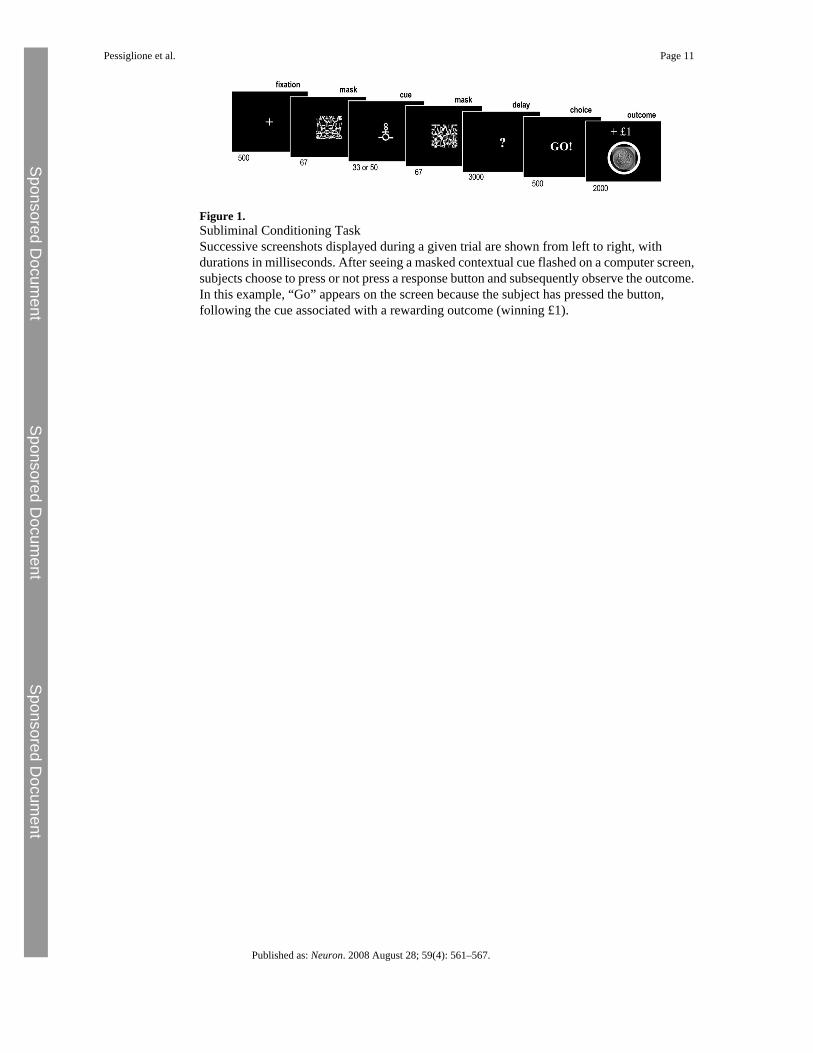
Figure 1.Subliminal Conditioning TaskSuccessive screenshots displayed during a given trial are shown from left to right, withdurations in milliseconds. After seeing a masked contextual cue flashed on a computer screen,subjects choose to press or not press a response button and subsequently observe the outcome.In this example, “Go” appears on the screen because the subject has pressed the button,following the cue associated with a rewarding outcome (winning £1).
Pessiglione et al. Page 11
Published as: Neuron. 2008 August 28; 59(4): 561–567.
Sponsored Docum
ent Sponsored D
ocument
Sponsored Docum
ent

Figure 2.Behavioral Data(A) Learning curves. Colors indicate cues for which button presses are rewarded (green),neutral (blue), or punished (red). Diamonds represent, across trials, percentages of subjectsthat pressed the button. Left: continuous lines join the diamonds to illustrate actual choicesmade by subjects. Right: continuous lines represent the probabilities of button press estimatedby an optimized Q-learning model.(B) Preferences. After the conditioning phase, cues were unmasked and subjects rated them,from the most (3) to the least liked (1). The graph shows the average rating for reward (green),neutral (blue), and punishment (red) cues. Bars are ± intersubjects standard errors of the mean.
Pessiglione et al. Page 12
Published as: Neuron. 2008 August 28; 59(4): 561–567.
Sponsored Docum
ent Sponsored D
ocument
Sponsored Docum
ent
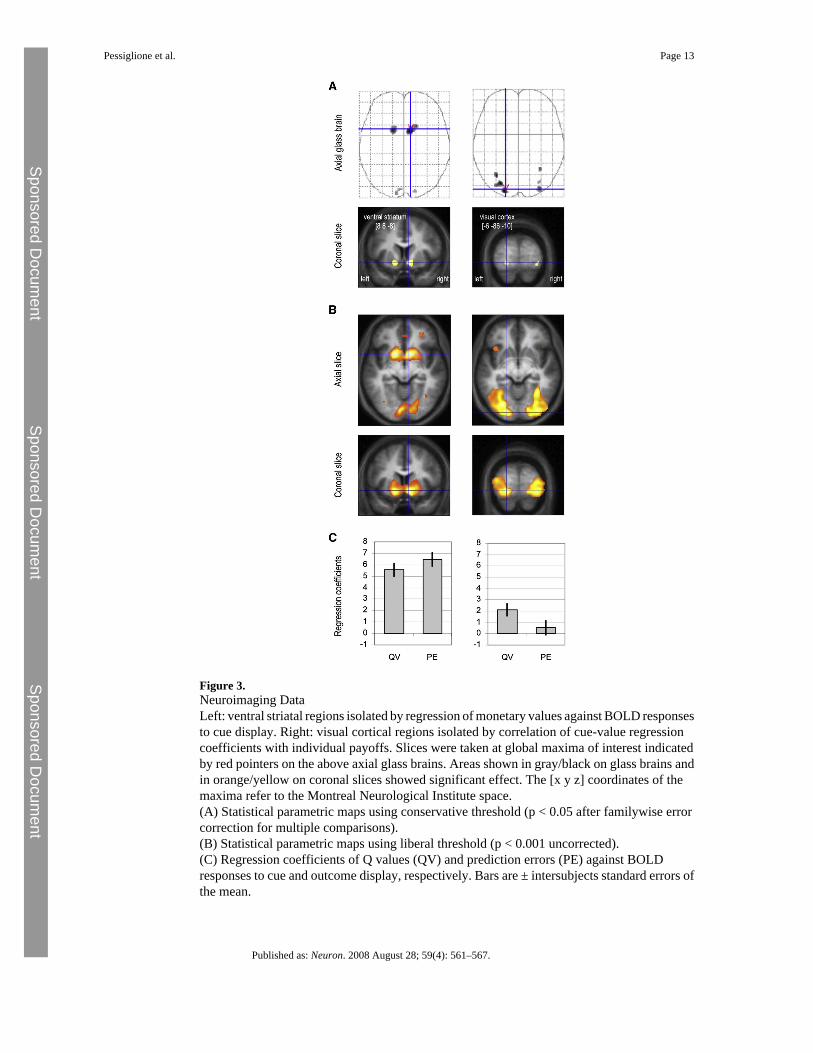
Figure 3.Neuroimaging DataLeft: ventral striatal regions isolated by regression of monetary values against BOLD responsesto cue display. Right: visual cortical regions isolated by correlation of cue-value regressioncoefficients with individual payoffs. Slices were taken at global maxima of interest indicatedby red pointers on the above axial glass brains. Areas shown in gray/black on glass brains andin orange/yellow on coronal slices showed significant effect. The [x y z] coordinates of themaxima refer to the Montreal Neurological Institute space.(A) Statistical parametric maps using conservative threshold (p < 0.05 after familywise errorcorrection for multiple comparisons).(B) Statistical parametric maps using liberal threshold (p < 0.001 uncorrected).(C) Regression coefficients of Q values (QV) and prediction errors (PE) against BOLDresponses to cue and outcome display, respectively. Bars are ± intersubjects standard errors ofthe mean.
Pessiglione et al. Page 13
Published as: Neuron. 2008 August 28; 59(4): 561–567.
Sponsored Docum
ent Sponsored D
ocument
Sponsored Docum
ent
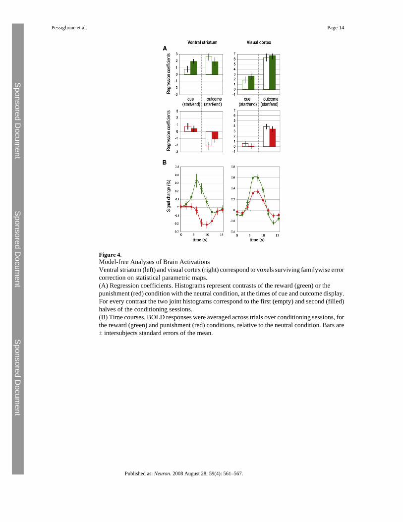
Figure 4.Model-free Analyses of Brain ActivationsVentral striatum (left) and visual cortex (right) correspond to voxels surviving familywise errorcorrection on statistical parametric maps.(A) Regression coefficients. Histograms represent contrasts of the reward (green) or thepunishment (red) condition with the neutral condition, at the times of cue and outcome display.For every contrast the two joint histograms correspond to the first (empty) and second (filled)halves of the conditioning sessions.(B) Time courses. BOLD responses were averaged across trials over conditioning sessions, forthe reward (green) and punishment (red) conditions, relative to the neutral condition. Bars are± intersubjects standard errors of the mean.
Pessiglione et al. Page 14
Published as: Neuron. 2008 August 28; 59(4): 561–567.
Sponsored Docum
ent Sponsored D
ocument
Sponsored Docum
ent