sera from patients with active systemic lupus erythematosus patients enhance the toll-like receptor...
TRANSCRIPT

Carvalheiro et al. Journal of Inflammation (2015) 12:38 DOI 10.1186/s12950-015-0083-2
RESEARCH Open Access
Sera from patients with active systemic lupuserythematosus patients enhance the toll-likereceptor 4 response in monocyte subsetsTiago Carvalheiro1, Diane Gomes2, Ligia A. Pinto3, Luis Inês4,5,6, Ana Lopes1, Ana Henriques5, Susana Pedreiro1,António Martinho1, Hélder Trindade1, Howard A. Young7, José António Pereira da Silva4,5 and Artur Paiva1,2*
Abstract
Background: Systemic Lupus Erythematosus (SLE) is an auto-immune disease whose complex pathogenesisremains unraveled. Here we aim to explore the inflammatory ability of SLE patients’ sera upon peripheral blood (PB)monocyte subsets and myeloid dendritic cells (mDCs) obtained from healthy donors.
Methods: In this study we included 11 SLE patients with active disease (ASLE), 11 with inactive disease (ISLE) and 10healthy controls (HC). PB from healthy donors was stimulated with patients’ sera, toll-like receptor (TLR) 4 ligand –lipopolysaccharide or both. The intracellular production of TNF-α was evaluated in classical, non-classical monocytesand mDCs, using flow cytometry. TNF-α mRNA expression was assessed in all these purified cells, after sera treatment.
Results: We found that sera of SLE patients did not change spontaneous TNF-α production by monocytes ordendritic cells. However, upon stimulation of TLR4, the presence of sera from ASLE patients, but not ISLE, significantlyincreased the intracellular expression of TNF-α in classical and non-classical monocytes. This ability was related to titersanti-double stranded DNA antibodies in the serum. High levels of anti-TNF-α in the patients’ sera were associated withincreased TNF-α expression by co-cultured mDCs. No relationship was found with the levels of a wide variety of otherpro-inflammatory cytokines. A slight increase of TNF-α mRNA expression was observed in these purified cells when theywere cultured only in the presence of SLE serum.
Conclusions: Our data suggest that SLE sera induce an abnormal in vitro TLR4 response in classical and non-classicalmonocytes, reflected by a higher TNF-α intracellular expression. These effects may be operative in the pathogenesisof SLE.
Keywords: Systemic lupus erythematosus, Serum, Cytokines, Toll like receptor 4, Classical monocytes, Non-classicalmonocytes, Myeloid dendritic cells
BackgroundSystemic lupus erythematosus (SLE) is a systemic auto-immune disease characterized by heterogeneous clinicalmanifestations with varying degrees of severity and alter-nating phases of remission and flare. The primary patho-logical findings in SLE patients are inflammation,vasculitis, immune complex deposition, and vasculopathy
* Correspondence: [email protected] and Transplantation Center of Coimbra, Portuguese Institute of Bloodand Transplantation, Quinta da Vinha Moura, São Martinho do Bispo,3041-861 Coimbra, Portugal2College of Health Technology of Coimbra, Rua 5 de Outubro, São Martinhodo Bispo, 3046-854 Coimbra, PortugalFull list of author information is available at the end of the article
© 2015 Carvalheiro et al.
[1–4]. The disease is characterized by the presence of a var-iety of autoantibodies against cell components and circulat-ing proteins, which are associated with differing diseasemanifestations [5].Monocytes are key players in both innate and adaptive
immune responses since they can produce large amountsof soluble cytokines and are equipped with a large array ofreceptors proving them with the ability of recognizinglipids, dying cells, microorganisms and their derivates.Given the plasticity of monocytes and their distinct role ininflammation, repair and healing processes [6, 7], accord-ing to the recommendations of Ziegler-Heitbrock et al. itis possible to subdivide human monocytes into three

Table 1 Clinical findings in 22 patients with systemic lupuserythematosus (SLE)
ASLE (n = 11) ISLE (n = 11)
Mean SLEDAI scores 10.4 ± 4.1 1.5 ± 1.2
Mean time since diagnosis 6.6 ± 6.0 8.3 ± 4.9
Lupus nephritis 45.5 % 54.5 %
Neurolupus 0 % 18.2 %
Lupus arthritis 72.7 % 63.6 %
Hematological involvement 100 % 90.9 %
Lupus cutaneous involvement 72.7 % 72.7 %
Severe Lupusa 45.5 % 54.5 %
Anti-dsDNA antibodiesb
Negative 0 % 27.2 %
Low positive 9.1 % 36.4 %
Moderate positive 18.2 % 27.3 %
High positive 72.7 % 9.1 %
Treatment
Hydroxychloroquine 90.9 % 90.9 %
Immunossupressantsc 54.5 % 36.4 %
Steroidsd 90.9 % 18.2 %
Low dose 40.0 % 100 %
Moderate dose 30.0 % 0 %
High dose 30.0 % 0 %aLupus severity in accordance with cumulative major organ involvementbAnti-dsDNA antibodies: low positive (<20 IU); moderate positive (20–50 IU);high positive (>50 IU)cazathioprine, mycophenolate mofetil, cyclosporine, tacrolimus, methotrexate,cyclophosphamide or rituximabdLow dose, up to 10 mg/day; moderate dose, 10–30 mg/day; high dose, morethan 30 mg/day
Carvalheiro et al. Journal of Inflammation (2015) 12:38 Page 2 of 9
subsets on the basis of the expression of CD14 and theCD16 receptors. The classical monocytes show high CD14expression but no CD16 (CD14++CD16−), the intermedi-ate monocytes show a high level of CD14 together withlow CD16 (CD14++CD16+), and the nonclassicalmonocytes express a low level of CD14 together withhigh CD16 (CD14 + CD16++) [8]. Functionally, clas-sical monocytes are professional phagocytes that ingestnative low-density lipoprotein, generate reactive oxygenspecies and secret cytokines in response to lipopolysac-charide (LPS) [9]. In contrast, non-classical monocytesdo not generate reactive oxygen species and are weakphagocytes, taking-up preferentially oxidized low-densitylipoprotein. They secrete substantial amounts of pro-inflammatory cytokines (tumor necrosis factor [TNF]-α, interleukin [IL]-1β and CCL3) after toll-like receptor(TLR) - dependent activation by viruses and nucleicacids [9–11].It is also well accepted that dendritic cells (DCs) repre-
sent a heterogeneous population of potent lineage-negativeHLA-DR+ antigen-presenting cells [12]. It is possible toidentify at least two subsets of circulating DCs on theblood [8]; the myeloid DC (mDC) subpopulation, stronglyexpressing HLA-DR and the myeloid-associated anti-gens CD11c and CD33 and the plasmacytoid DC(pDCs) subset, expressing high levels of both HLA-DRand CD123, without expressing myeloid associated an-tigens [13].Abnormalities in monocytes and DCs from SLE pa-
tients have already been described [14–16] and animportant role of DCs in SLE pathogenesis been advo-cated [17, 18]. It has been reported that sera of SLE pa-tients can induce the production of IFN-α in normalplasmacytoid DCs [19, 20]. SLE sera have also beenshown to induce the differentiation of normal mono-cytes into DCs [21], and to promote a B cell response[22]. This pro-inflammatory activity apparently requiresthe presence of circulating immune complexes in thesera [20]. However the ability of the soluble mediatorspresents in SLE patients’ serum to activate normal per-ipheral blood monocytes subsets and mDCs is not wellestablished.In this context, we have evaluated the ability of sera
from SLE patients to induce the intracellular productionof TNF-α by classical, non-classical monocytes andmDCs. We examined the effect of SLE sera in the pres-ence and absence of TLR4 stimulation by LPS. Interest-ingly, SLE sera did not change the intracellularproduction of TNF-α in the absence of TLR4 stimula-tion. However, the stimulation with TLR4 ligand in thepresence of sera from ALSE resulted in a higher intracel-lular production of TNF-α by classical and non-classicalmonocytes, than in the presence of sera obtained fromhealthy controls and ISLE.
Materials and methodsPatientsTwenty two patients with SLE, according to the 1997American College of Rheumatology classification criteria[23] followed at the Lupus Clinic, Rheumatology Depart-ment of the University Hospital Center of Coimbra, wererecruited and asked to provide a blood sample for thisstudy. Disease activity was assessed at the time of blood col-lection, according to the SLE Disease Activity Index 2000(SLEDAI 2 k) [23, 24]. SLE patients were divided into twogroups, one with clinically active disease (ASLE) (SLEDAI2 k ≥ 5; n = 11, 100 % female, median age 27: 19–52 year)and the other with clinically inactive disease (ISLE) (SLEDAI2 k < 5; n = 11, 64 % female, median age 28: 19–45 year)[25]. Current medication details and additional routine la-boratory parameters were collected from the patients’ files(Table 1). Sera samples were stored at −20 °C until analysis.
ControlsThe healthy control group (HC) comprised 10 healthyindividuals (90.0 % female, median age 28.5: 25 – 35 year)
Carvalheiro et al. Journal of Inflammation (2015) 12:38 Page 3 of 9
who provided blood samples for determination of serumcytokine levels. Five different healthy donors providedan additional blood samples (90 % female; median age 29:25 – 33 years) collected in heparin anticoagulant tubes(6 mL of blood). These participants were required tocomplete a brief questionnaire regarding previous orcurrent medical conditions and therapies. All were freefrom autoimmune disease, other active inflammatory con-ditions and medication with immunomodulatory drugs.
Ethical aspectsThe study protocol was approved by the ethics commit-tee of the University Hospital Center of Coimbra. Allparticipants provided a signed informed consent prior toany study procedures and the principles of the HelsinkiDeclaration were fully respected.
Peripheral blood cultures with sera from SLE patients andHC with and without co-stimulation with TLR4 ligand - LPSTo assess the effect of SLE sera upon TNF-α productionby normal peripheral blood monocyte subsets and mDCs,sera from 11 ASLE patients, 11 ISLE patients and 10healthy individuals were used. The heparinized wholeblood samples obtained from healthy donors were washedthrice with NaCl 0.9 % saline solution and resuspended in0.25 mL; then samples were diluted (1:1) in RPMI-1640medium (Life Technologies – Thermo Fisher Scientific;Carlsbad, C.A., USA), supplemented with 2 mM L-glu-tamine and antibiotic–antimycotic agent (Life Technolo-gies – Thermo Fisher Scientific) in a total of 0.5 mL. Fourdifferent conditions were created: 1) adding 0.05 mL ofsera from patients with SLE or HC; 2) adding 100 ng/mLof LPS from Escherichia coli (serotype 055:B5; Sigma) plus0.05 mL of sera from patients with SLE or HC; 3) adding100 ng/mL of LPS from Escherichia coli (as a positive con-trol) and 4) an unstimulated condition (as the negativecontrol). All experiments included the presence of 10 μg/mL of Brefeldin A (ref: B7651; Sigma, St. Louis, MO,USA) to prevent the release of cytokines from the cells.Samples were incubated for 6 h at 37 °C in a sterile envir-onment with a 5 % CO2 humid atmosphere.
Immunofluorescent stainingAfter the 6 h incubation period, samples were aliquoted intwo different tubes (0.250 mL/tube) in order to analysethe intracellular production of TNF-α in classical andnon-classical monocytes as well as in mDCs. For the iden-tification of these populations, cells were stained with thefollowing monoclonal antibody combination: anti-CD45krome orange (clone: J.33; Beckman Coulter – Immuno-tech, Marseille, France), anti-CD33 phycoerythrin cyanine 7tandem (clone: D3HL60.251; Beckman Coulter – Immuno-tech) anti-CD14 allophycocyanin (clone: RM052; BeckmanCoulter – Immunotech) and anti-HLA-DR peridinin
chlorophyll protein cyanine 5 (clone: L243; Becton andDickinson (BD) Biosciences, San Jose, CA, USA). Aftergentle mixing, cells were incubated for 15 min at roomtemperature in the dark followed by an intracytoplasmaticpermeabilization protocol with IntraPrep PermeabilizationReagent (Beckman Coulter – Immunotech). Cells werefixed and permeabilized according to the manufacturer’sinstructions. Thereafter, anti-TNF-α antibody (cloneMAb11; BD Pharmingen, San Diego, CA, USA) was addedand incubated for 15 min at room temperature in thedark. The cells were then washed twice with phosphate-buffered saline (Gibco BRL-life Technologies) and resus-pended in 0.250 mL of this buffer.
Flow cytometry data acquisition and analysisData acquisition was performed in a FACSCanto II flowcytometer (BD Biosciences) with the FACSDiva software(BD Biosciences) using the EuroFlow instrument setupdata acquisition standard operating procedures [26]. Foreach sample at least 250.000 events were acquired.Data analysis for each variable was performed using
the flow cytometry software Infinicyt 1.6 (Cytognos,Salamanca, Spain). The evaluation of TNF-α productionwas based on the frequency (%) of positive cells withineach cell subset and their corresponding expression asdetermined by the mean fluorescence intensity (MFI),expressed as a relative logical scale.Since CD16 expression is lost shortly after LPS stimu-
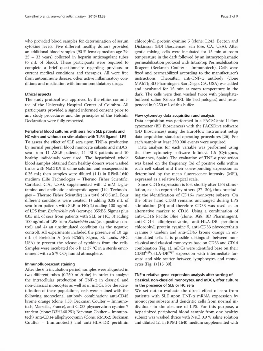
lation, as also reported by others [27–30], thus preclud-ing the identification of CD16+ monocyte subsets. Onthe other hand CD33 remains unchanged during LPSstimulation [30] and therefore CD33 was used as analternative marker to CD16. Using a combination ofanti-CD16 Pacific Blue (clone: 3G8; BD Pharmingen),anti-CD14 allophycocyanin, anti-HLA-DR peridininchlorophyll protein cyanine 5, anti-CD33 phycoerythrincyanine 7 tandem and anti-CD45 krome orange in un-stimulated cells it is possible distinguish between non-classical and classical monocytes base on CD33 and CD14combination (Fig. 1). mDCs were identified base on theirCD33high/HLA-DRhigh expression with intermediate for-ward and side scatter between lymphocytes and mono-cytes (Fig. 1) [15, 30].
TNF-α relative gene expression analysis after sorting ofclassical, non-classical monocytes, and mDCs, after culturein the presence of SLE or HC seraWe set out to evaluate the direct effect of sera frompatients with SLE upon TNF-α mRNA expression bymonocytes subsets and dendritic cells from normal in-dividuals in the absence of LPS. For this purpose, aheparinized peripheral blood sample from one healthysubject was washed thrice with NaCl 0.9 % saline solutionand diluted 1:1 in RPMI-1640 medium supplemented with

A
D
B
E
C
F
Fig. 1 Flow cytometry gate-strategy to identify non-classical and classical monocyte subsets and myeloid dendritic cells (mDCs). In a the conventionalgating strategy is shown, representing classical, intermediate and non-classical monocyte populations based on CD14 and CD16 expression. Since afterLPS stimulation CD16 is downregulated, CD33 was used combined with CD14 to distinguish the classical and non-classical monocytes: R1 classicalmonocytes (CD14++CD33++) is equivalent to CD14++CD16−; R2 non-classical monocytes (CD14+/−CD33+/dim) correspond to CD14+CD16++ (b–c). mDCs(R3) were identified based on the following phenotype: CD14−CD33++HLA-DR++ (a and d). Monocytes and mDCs characteristics of forward scatter(FSC), side scatter (SSC) and CD45 expression and therefore lymphocytes populations are excluded from the analyses (e–f)
Carvalheiro et al. Journal of Inflammation (2015) 12:38 Page 4 of 9
2 mM L-glutamine and antibiotic–antimycotic agent.0.05 mL of serum was added in a final volume of 0.5 mL:3 from ASLE, 3 from ISLE and 3 from HC. Each samplewas incubated in quadruplicate for 6 h, at 37 °C in a sterileenvironment under 5 % CO2.For the cell sorting of classical monocytes, non-
classical monocytes and mDCs, cells from each samplewere resuspended in a final volume of 1 mL per sample,and lysed with 10 mL of NH4Cl solution (Sigma) to re-move the red blood cells. After 20 min of incubation,samples were centrifuged (5 min, at 540 × g) and thecell pellet was stained with the following monoclonalantibodies combination: anti-CD45 Krome Orange,anti-HLA-DR fluorescein isothiocyanate (clone: Immu-357; Beckman Coulter – Immunotech), anti-CD14 peri-din chlorophyll protein – Cyanin 5.5 (clone:M5E2; BDPharmingen), anti-CD33 phycoerythrin (clone:P67.6;BD Biosciences) and anti-CD123 allophycocyanin(clone: 9 F5; BD Pharmingen). Next, the cells were in-cubated for 20 min at room temperature in the dark,
washed and resuspended in phosphate-buffered saline(Gibco BRL-life Technologies).Cell-sorting and purification were performed in FAC-
SAria II cell sorter (BD Biosciences) using the FACSDivasoftware (BD Biosciences). Classical monocytes wereidentified and sorted by HLA-DR+/CD14high/CD33high/CD45high phenotype, non-classical monocytes were HLA-DRinter/CD33inter/CD123negCD45high, and mDCs were de-fined as HLA-DRhigh/CD33high/CD14neg/CD123neg.Sorted cell populations were then centrifuged for
5 min at 300 × g and the pellets were resuspended in350 μL of RLT Lysis Buffer (Qiagen, Hilden, Germany).Total RNA extraction was performed with the RNeasyMicro kit (Qiagen) according to the supplier’s instruc-tions. Total RNA was eluted in a 14 μl volume ofRNase-free water. In order to quantify the amount oftotal RNA extracted and verify RNA integrity, sampleswere analyzed using a 6000 Nano Chip kit, in an Agilent2100 bioanalyzer (Agilent Technologies, Walbronn,Germany) and 2100 expert software, according to the
Carvalheiro et al. Journal of Inflammation (2015) 12:38 Page 5 of 9
manufacturer’s instructions. RNA was reverse tran-scribed with iScriptTM Reverse Transcription Supermixfor RTqPCR (Bio-Rad, Hercules, Calif., USA), accordingto the manufacturer’s instructions. Relative quantifica-tion of gene expression using real-time PCR was per-formed in the LightCycler 480 II (Roche Diagnostics,Rotkreuz, Switzerland). Real-time PCR reactions werecarried out using 1 × QuantiTect SYBR Green PCR Mas-ter Mix (Qiagen), 1 × QuantiTect Primer Assay TRAF1(QT00095732) (Qiagen) and 20 ng of cDNA sample, in atotal volume of 10 μL. The reactions were performedusing the following thermal profile: 15 min at 95 °C,and 50 cycles of 15 s at 94 °C, 30 s at 55 °C and 30 s at72 °C. Melting point analysis was done to ensure ampli-fication of the specific product. Real-time PCR resultswere analyzed with the LightCycler software (RocheDiagnostics). GeNorm Reference Gene Selection kit(PrimerDesign Ltd., Southampton, England) in con-junction with the geNorm software (PrimerDesign Ltd.)were used to select the reference genes to normalizedata. The reference genes used for gene expression ana-lysis of classical, non-classical monocytes and mDCwere the beta-2-microglobulin (B2M) (QT00088935)and the ubiquitin-c (UBC) (QT00234430). The normal-ized TNF-α gene expression were calculated using thedelta-Ct method [31].
Serum cytokine quantitationMeasurements of IL-17F, IL-17A, IL1-7E, IL-10, IL-12p70, IL-13, IL-15, IL-22, IL-21, IL-23, IFN-γ andTNF-α were performed in all serum samples by LuminexxMAP using the MILLIPLEX MAP Human TH17 Mag-netic Bead Panel (EMD Millipore, Billerica, MA, USA).The serum cytokine levels were determined by compari-son with a standard curve obtained using the corre-sponding recombinant human cytokines
Statistical analysesResults were expressed as median/mean and range/inter-quartile range. Statistical evaluation of data was per-formed through non-parametric tests: The χ2 andFisher’s exact tests were used to evaluate the significanceof associations between categorical variables. Continuousvariables were compared by Kruskal-Wallis test andMann–Whitney U test. A Spearman’s rank correlationwas applied to assess the association between differentparameters. The statistical analyses were performedusing Statistical Package for Social Sciences IBM SPSS20 (IBM, Armonk, NY. USA) and Graphpad Prism ver-sion 5 (GraphPad Software, San Diego, CA, USA). Dif-ferences were considered statistically significant whenthe p value was less than 0.05.
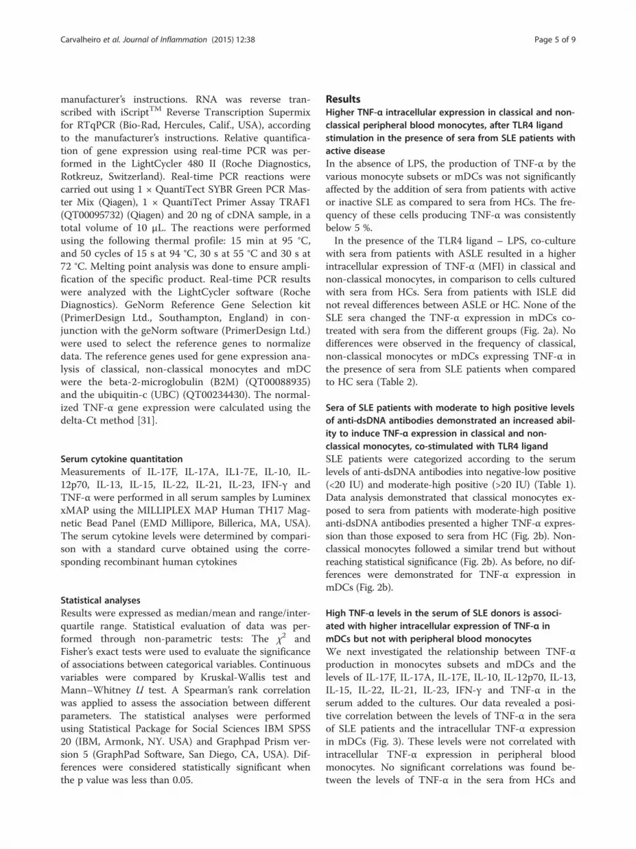
ResultsHigher TNF-α intracellular expression in classical and non-classical peripheral blood monocytes, after TLR4 ligandstimulation in the presence of sera from SLE patients withactive diseaseIn the absence of LPS, the production of TNF-α by thevarious monocyte subsets or mDCs was not significantlyaffected by the addition of sera from patients with activeor inactive SLE as compared to sera from HCs. The fre-quency of these cells producing TNF-α was consistentlybelow 5 %.In the presence of the TLR4 ligand – LPS, co-culture
with sera from patients with ASLE resulted in a higherintracellular expression of TNF-α (MFI) in classical andnon-classical monocytes, in comparison to cells culturedwith sera from HCs. Sera from patients with ISLE didnot reveal differences between ASLE or HC. None of theSLE sera changed the TNF-α expression in mDCs co-treated with sera from the different groups (Fig. 2a). Nodifferences were observed in the frequency of classical,non-classical monocytes or mDCs expressing TNF-α inthe presence of sera from SLE patients when comparedto HC sera (Table 2).
Sera of SLE patients with moderate to high positive levelsof anti-dsDNA antibodies demonstrated an increased abil-ity to induce TNF-α expression in classical and non-classical monocytes, co-stimulated with TLR4 ligandSLE patients were categorized according to the serumlevels of anti-dsDNA antibodies into negative-low positive(<20 IU) and moderate-high positive (>20 IU) (Table 1).Data analysis demonstrated that classical monocytes ex-posed to sera from patients with moderate-high positiveanti-dsDNA antibodies presented a higher TNF-α expres-sion than those exposed to sera from HC (Fig. 2b). Non-classical monocytes followed a similar trend but withoutreaching statistical significance (Fig. 2b). As before, no dif-ferences were demonstrated for TNF-α expression inmDCs (Fig. 2b).
High TNF-α levels in the serum of SLE donors is associ-ated with higher intracellular expression of TNF-α inmDCs but not with peripheral blood monocytesWe next investigated the relationship between TNF-αproduction in monocytes subsets and mDCs and thelevels of IL-17F, IL-17A, IL-17E, IL-10, IL-12p70, IL-13,IL-15, IL-22, IL-21, IL-23, IFN-γ and TNF-α in theserum added to the cultures. Our data revealed a posi-tive correlation between the levels of TNF-α in the seraof SLE patients and the intracellular TNF-α expressionin mDCs (Fig. 3). These levels were not correlated withintracellular TNF-α expression in peripheral bloodmonocytes. No significant correlations was found be-tween the levels of TNF-α in the sera from HCs and
A B
Fig. 2 TNF-α expression after TLR4 co-stimulation with different sera. TNF-α intracellular expression in classical, non-classical monocytes and mDCsaccording to the disease activity a and the amount of anti-dsDNA antibodies b. ***Statistically significant differences were considered when p <0.05 (Mann–Whitney U test)
Carvalheiro et al. Journal of Inflammation (2015) 12:38 Page 6 of 9
intracellular TNF-α expression in either mDCs (p = 0.535)or monocytes subsets.The others cytokines levels did not correlate with the
intracellular expression of TNF-α either in culturedmDCs or monocyte subsets. Additionally, no differenceswere found in the cytokines levels between the threegroups of participants (Table 3).
TNF-α mRNA gene expression in sorted monocytessubsets and mDCs in the presence of SLE patient´s serumwithout LPS co-stimulationSince the presence of SLE sera alone did not demon-strated the ability to shift the TNF-α intracellular expres-sion in monocytes subsets and mDCs as measured byflow cytometry, we next used qRT-PCR to measure theTNF-α mRNA expression in these purified cells aftertreatment with SLE or HC sera, in the absence of LPS.For this analysis, we selected 6 SLE sera (3 ASLE pa-tients and 3 ISLE patients) and 3 HC sera. Only a trendfor a higher TNF-α mRNA expression in the presence ofSLE sera versus HC sera in both classical and non-classical monocytes was observed (Fig. 4), although nostatically significant differences were observed.
DiscussionSLE is a chronic autoimmune disorder associated with alarge number of immunological abnormalities, which
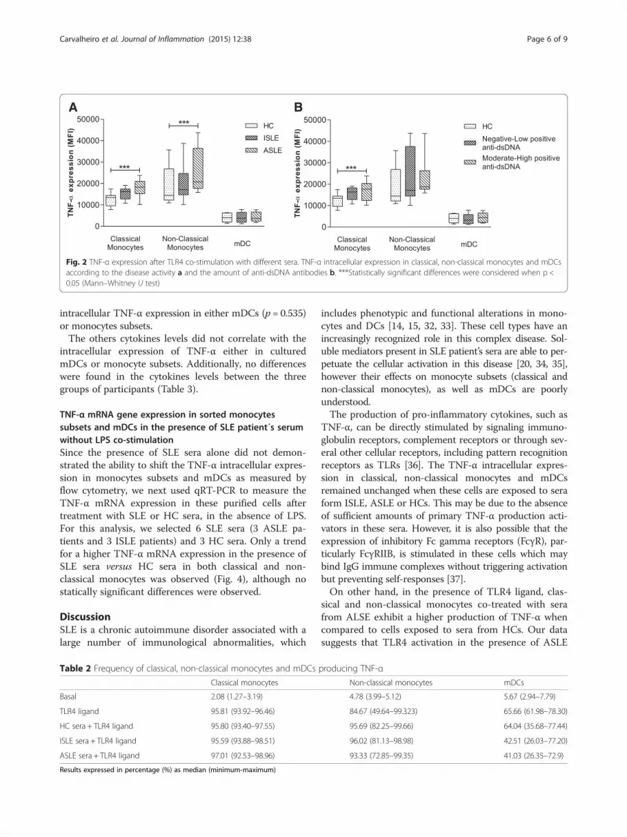
Table 2 Frequency of classical, non-classical monocytes and mDCs
Classical monocytes
Basal 2.08 (1.27–3.19)
TLR4 ligand 95.81 (93.92–96.46)
HC sera + TLR4 ligand 95.80 (93.40–97.55)
ISLE sera + TLR4 ligand 95.59 (93.88–98.51)
ASLE sera + TLR4 ligand 97.01 (92.53–98.96)
Results expressed in percentage (%) as median (minimum-maximum)
includes phenotypic and functional alterations in mono-cytes and DCs [14, 15, 32, 33]. These cell types have anincreasingly recognized role in this complex disease. Sol-uble mediators present in SLE patient’s sera are able to per-petuate the cellular activation in this disease [20, 34, 35],however their effects on monocyte subsets (classical andnon-classical monocytes), as well as mDCs are poorlyunderstood.The production of pro-inflammatory cytokines, such as
TNF-α, can be directly stimulated by signaling immuno-globulin receptors, complement receptors or through sev-eral other cellular receptors, including pattern recognitionreceptors as TLRs [36]. The TNF-α intracellular expres-sion in classical, non-classical monocytes and mDCsremained unchanged when these cells are exposed to seraform ISLE, ASLE or HCs. This may be due to the absenceof sufficient amounts of primary TNF-α production acti-vators in these sera. However, it is also possible that theexpression of inhibitory Fc gamma receptors (FcγR), par-ticularly FcγRIIB, is stimulated in these cells which maybind IgG immune complexes without triggering activationbut preventing self-responses [37].On other hand, in the presence of TLR4 ligand, clas-
sical and non-classical monocytes co-treated with serafrom ALSE exhibit a higher production of TNF-α whencompared to cells exposed to sera from HCs. Our datasuggests that TLR4 activation in the presence of ASLE
producing TNF-αNon-classical monocytes mDCs
4.78 (3.99–5.12) 5.67 (2.94–7.79)
84.67 (49.64–99.323) 65.66 (61.98–78.30)
95.69 (82.25–99.66) 64.04 (35.68–77.44)
96.02 (81.13–98.98) 42.51 (26.03–77.20)
93.33 (72.85–99.35) 41.03 (26.35–72.9)

0 10 20 300
2000
4000
6000
8000
10000mDCsrho=0.6091p=0.0034
Level of TNF- (pg/mL)
TNF-
expr
essi
on(M
FI)
0 10 20 300
5000
10000
15000
20000
25000Classical Monocytes
rho=0.2247p=0.3275
Level of TNF- (pg/mL)
TNF-
expr
essi
on(M
FI)
0 10 20 300
10000
20000
30000
40000
50000Non-Classical Monocytes
rho=-0.1351p=0.5594
Level of TNF- (pg/mL)
TNF-
expr
e ssi
on(M
FI)
Fig. 3 Correlation between SLE sera TNF-α levels and TNF-α intracellular expression after TLR4 co-stimulation with different sera. Statistical significantdifferences were considered when p < 0.05. The correlations were assessed by the Spearman’s rank correlation
Carvalheiro et al. Journal of Inflammation (2015) 12:38 Page 7 of 9
sera results in an aberrant response by classical andnon-classical monocytes. These results are in line withthe findings of Leadbetter et al. that reported an aber-rant B-cell response mediated by IgG complexes andTLR4 activation in their mice experiments [38].In order to better define which serum components
could be implicated in the activation of monocytes, we
Table 3 Cytokine levels obtained in the 22 SLE patients and 10 hea
HC n = 10 ISLE n
IL-17A Cytokine level (pg/mL) 0.0 (0.0–0.0): 0.00 0.0 (0
% of samples detected (n) 0 % (n = 0) 18.2
IL-17E Cytokine level (pg/mL) 0.0 (0.0–286.21): 81.27 0.0 (0
% of samples detected (n) 30 % (n = 3) 18.2
IL-17F Cytokine level (pg/mL) 0.0 (0.0–14.49): 3.93 0.0 (0
% of samples detected (n) 30 % (n = 3) 27.3
IL-12p70 Cytokine level (pg/mL) 0.0 (0.0–46.87): 6.98 0.96
% of samples detected (n) 30 % (n = 3) 54.5
IL-23 Cytokine level (pg/mL) 0.0 (0.0–1134.54): 276.46 0.0 (0
% of samples detected (n) 30 % (n = 3) 18.2
TNF-α Cytokine level (pg/mL) 7.68 (6.25–33.50): 12.38 13.57
% of samples detected (n) 100 % (n = 10) 100 %
IFN-γ Cytokine level (pg/mL) 0.0 (0.0–8.96): 2.95 0.0 (0
% of samples detected (n) 40 % (n = 4) 27.3
IL-15 Cytokine level (pg/mL) 0.0 (0.0–0.0): 0.0 0.0 (0
% of samples detected (n) 0 % (n = 0) 18.2
IL-10 Cytokine level (pg/mL) 0.0 (0.0–0.0): 0.0 0.0 (0
% of samples detected (n) 0 % (n = 0) 100 %
IL-22 Cytokine level (pg/mL) 0.0 (0.0–100.8): 18.77 0.0 (0
% of samples detected (n) 30 % (n = 3) 18.2
IL-21 Cytokine level (pg/mL) 0.0 (0.0–5.61): 0.80 0.0 (0
% of samples detected (n) 10 % (n = 1) 18.2
IL-13 Cytokine level (pg/mL) 0.0 (0.0–0.0): 0.0 0.0 (0
% of samples detected (n) 0 % (n = 0) 18.8
Results expressed in pg/mL as median (minimum-maximum): mean% and (n =) represent the percentage (number of cases) of samples detectedp values were calculated by Kruskal-Wallis test and Mann–Whitney U test for continuoubetween categorical variables. ns: non-significant
explored the relationship between this activations andthe serum levels of anti-dsDNA antibodies and cyto-kines. Remarkably, moderate-high levels of anti-dsDNAwere associated with more intense activation of TNF-αproduction by classical and non-classical monocytes.This finding enhances the hypothesis that the presenceof auto-antibodies and immune-complexes, with the
lthy individuals
= 11 ASLE n = 11 P value significance
.0–113.40): 12.69 0.0 (0.0–62.04): 9.28 ns
% (n = 2) 27.3 % (n = 3) ns
.0–1364.68): 159.38 0.0 (0.0–57.10): 5.19 ns
% (n = 2) 9.1 % (n = 1) ns
.00–158.44): 19.10 0.0 (0.0–57.10): 5.19 ns
% (n = 3) 9.1 % (n = 1) ns
(0.0–60.76): 8.05 0.0 (0.0–85.52): 8.85 ns
% (n = 6) 36.4 % (n = 4) ns
.0–11221.86): 1216.17 0.0 (0.0–2547.96): 258.94 ns
% (n = 2) 18.2 % (n = 2) ns
(4.30–26.73): 13.66 14.51 (1.01–27.90): 16.47 ns
(n = 11) 100 % (n = 11) ns
.0–167.11): 21.90 2.77 (0.0–79.40): 12.36 ns
% (n = 3) 63.6 % (n = 7) ns
.0–7.45): 1.41 0.0 (0.0–0.0): 0.0 ns
% (n = 2) 0 % (n = 0) ns
.0–6.58): 0.99 0.0 (0.0–5.36): 0.89 ns
(n = 11) 100 % (n = 11) ns
.0–598.67): 64.06 0.0 (0.0–159.85): 14.53 ns
% (n = 2) 9.1 % (n = 1) ns
.0–105.58): 12.13 0.0 (0.0–24.07): 2.68 ns
% (n = 2) 18.2 % (n = 2) ns
.0–56.44): 6.76 0.0 (0.0–0.0): 0.0 ns
% (n = 2) 0 % (n = 0) ns
s variables, and χ2 and Fisher’s exact tests were used to measure associations

Classical Monocytes
HC SLE0
1
2
3
4
5
exp
ress
ion
Non-classical Monocytes
HC SLE0
20
40
60
exp
ress
ion
mDC
HC SLE0
1
2
3
TN
F-
rela
tive
gen
e
TN
F-
rela
tive
gen
e
TN
F-
rela
tive
gen
eex
pre
ssio
n
Fig. 4 TNF-α relative gene expression in sorted classical, non-classical monocytes and mDCs stimulated with sera. Results expressed as mean andstandard deviation
Carvalheiro et al. Journal of Inflammation (2015) 12:38 Page 8 of 9
ability to activate FcγR and TLRs, leads to increased pro-duction of TNF-α [20, 39]. This is supported by the evi-dence that cross-linking between IgGs to FcγRs triggersa wide variety of cellular functions, including release ofinflammatory mediators, like cytokines, chemokines andreactive oxygen species [40, 41].mDCs was not sensitive to the effects of sera of these
diverse origins upon TNF-α expression, both in the ab-sence and in the presence of LPS. This suggests thatthis subpopulation of DCs is less sensitive to a periph-eral inflammatory environment, probably due to thefact that the majority of PB mDCs have an immaturephenotype [14, 15]. In fact, the different pattern ofTNF-α expression observed in classical monocytes,non-classical monocytes and mDCs can also be relatedwith the differential expression of FcγRs. Classicalmonocytes constitutively express CD64 (FcγRI), a highaffinity receptor, while non-classical monocytes ex-presses CD16 (FcγRIII), a low affinity receptor, andmDCs express low levels of CD32 (FcγRII) an inter-mediate affinity receptor [42–44].We also explored the relationship between TNF-α ex-
pression and the levels of a large array of cytokines in theserum added to the cultures. Interestingly, we observed apositive correlation between TNF-α sera levels and TNF-αintracellular expression in mDC co-stimulated with TLR4ligand in the presence of SLE sera. This finding is in linewith the ability of soluble TNF-α induces mDCs matur-ation as well as TNF-α production [45, 46].Finally, we analyzed whether SLE sera in the absence
of TLR4 ligand, could result in alterations of TNF-αmRNA expression in classical, non-classical monocytesand mDCs. A slight, non-significant increase in theTNF-α mRNA expression was observed in both mono-cyte subsets in the presence of SLE sera.
ConclusionIn summary, our data demonstrated that sera from pa-tients with active SLE increase TNF-α production byclassical and non-classical monocytes, in the presence of
LPS. This effect may be partially explained by circulatingauto-antibodies since the high levels of anti-dsDNA anti-bodies are associated with an enhanced TNF-α intracel-lular expression. This supports the concept that thepresence of the immune-complexes is an important fac-tor in cell activation and in the maintenance of chronicinflammation in SLE.
AbbreviationsASLE: Systemic lupus erythematosus patients in active disease; DC: Dendriticcell; dsDNA: Double stranded DNA; FcγR: Fc gamma receptor; IL: Interleukin;IFN: Interferon; ISLE: Systemic lupus erythematosus patients in inactivedisease; LPS: Lipopolysaccharide; mDC: Myeloid dendritic cell; SLE: Systemiclupus erythematosus; SLEDAI: Systemic lupus erythematosus disease activityindex 2000; TLR: Toll-like receptor; TNF-α: Tumor necrosis factor α.
Competing interestsThe authors declare that they have no competing interests.
Authors’ contributionsTC carried the analysis and interpretation of data, the statistical analysis anddrafted the manuscript; DM performed cell cultures and flow cytometryassays; LAP and HAY determined the sera cytokines levels; LI and JAPSprovided patients samples and their clinical information; AL and AMparticipated in the molecular studies; AH has been involved in manuscriptrevising; SP performed the cell sorting; HT, JAPS and AP contributed toconception and designed the study protocol and given final approval of theversion to be published. All authors read and approved the final manuscript.
AcknowledgmentsThis work was partially supported by research grant from the PortugueseSociety of Rheumatology (SPR): “Bolsa de Investigação na área das DoençasReumáticas Inflamatórias – Bolsa SPR/MSD 2014” and by the IntramuralResearch Program of the NIH, National Cancer Institute.
Author details1Blood and Transplantation Center of Coimbra, Portuguese Institute of Bloodand Transplantation, Quinta da Vinha Moura, São Martinho do Bispo,3041-861 Coimbra, Portugal. 2College of Health Technology of Coimbra, Rua5 de Outubro, São Martinho do Bispo, 3046-854 Coimbra, Portugal. 3HPVImmunology Laboratory, Frederick National Laboratory for Cancer Research,Building 469, 21702 Frederick, MD, USA. 4Rheumatology Department, CentroHospitalar e Universitário de Coimbra, Praceta Prof. Mota Pinto, 3000-075Coimbra, Portugal. 5Faculty of Medicine, University of Coimbra, Azinhaga deSanta Comba, Celas, 3000-548 Coimbra, Portugal. 6School of Health Sciences,University of Beira Interior, Avenida Infante D. Henrique, 6200-506 Covilhã,Portugal. 7Laboratory of Experimental Immunology, Cancer and InflammationProgram, National Cancer Institute at Frederick, Building 560, 21702-1201Frederick, MD, USA.

Carvalheiro et al. Journal of Inflammation (2015) 12:38 Page 9 of 9
Received: 11 January 2015 Accepted: 18 May 2015
References1. Agmon-Levin N, Mosca M, Petri M, Shoenfeld Y. Systemic lupus
erythematosus one disease or many? Autoimmun Rev. 2012;11:593–5.2. Fu SM, Deshmukh US, Gaskin F. Pathogenesis of systemic lupus
erythematosus revisited 2011: end organ resistance to damage,autoantibody initiation and diversification, and HLA-DR. J Autoimmun.2011;37:104–12.
3. Mok CC, Lau CS. Pathogenesis of systemic lupus erythematosus. J ClinPathol. 2003;56:481–90.
4. O’Neill S, Cervera R. Systemic lupus erythematosus. Best Pract Res ClinRheumatol. 2010;24:841–55.
5. Marks SD, Tullus K. Autoantibodies in systemic lupus erythematosus. PediatrNephrol. 2012;27:1855–68.
6. Wong KL, Yeap WH, Tai JJ, Ong SM, Dang TM, Wong SC. The three humanmonocyte subsets: implications for health and disease. Immunol Res.2012;53:41–57.
7. Auffray C, Sieweke MH, Geissmann F. Blood monocytes: development,heterogeneity, and relationship with dendritic cells. Annu Rev Immunol.2009;27:669–92.
8. Ziegler-Heitbrock L, Ancuta P, Crowe S, Dalod M, Grau V, Hart DN, et al.Nomenclature of monocytes and dendritic cells in blood. Blood.2010;116:e74–80.
9. Mosig S, Rennert K, Krause S, Kzhyshkowska J, Neunubel K, Heller R, et al.Different functions of monocyte subsets in familial hypercholesterolemia:potential function of CD14+ CD16+ monocytes in detoxification of oxidizedLDL. FASEB J. 2009;23:866–74.
10. Soehnlein O, Lindbom L. Phagocyte partnership during the onset andresolution of inflammation. Nat Rev Immunol. 2010;10:427–39.
11. Cros J, Cagnard N, Woollard K, Patey N, Zhang SY, Senechal B, et al. HumanCD14dim monocytes patrol and sense nucleic acids and viruses via TLR7and TLR8 receptors. Immunity. 2010;33:375–86.
12. Pereira MI, Paiva A. Dendritic cells in cord blood transplantation: a review.Stem Cells Int. 2011;2011:539896.
13. Crespo I, Paiva A, Couceiro A, Pimentel P, Orfao A, Regateiro F.Immunophenotypic and functional characterization of cord blood dendriticcells. Stem Cells Dev. 2004;13:63–70.
14. Carvalheiro T, Rodrigues A, Lopes A, Ines L, Velada I, Ribeiro A, et al.Tolerogenic versus inflammatory activity of peripheral blood monocytesand dendritic cells subpopulations in systemic lupus erythematosus. ClinDev Immunol. 2012;2012:934161.
15. Henriques A, Ines L, Carvalheiro T, Couto M, Andrade A, Pedreiro S, et al.Functional characterization of peripheral blood dendritic cells andmonocytes in systemic lupus erythematosus. Rheumatol Int. 2012;32:863–9.
16. Sule S, Rosen A, Petri M, Akhter E, Andrade F. Abnormal production of pro- andanti-inflammatory cytokines by lupus monocytes in response to apoptotic cells.PLoS One. 2011;6:e17495.
17. Crispin JC, Alcocer-Varela J. The role myeloid dendritic cells play in thepathogenesis of systemic lupus erythematosus. Autoimmun Rev.2007;6:450–6.
18. Fransen JH, van der Vlag J, Ruben J, Adema GJ, Berden JH, Hilbrands LB.The role of dendritic cells in the pathogenesis of systemic lupuserythematosus. Arthritis Res Ther. 2010;12:207.
19. Chiang EY, Yu X, Grogan JL. Immune complex-mediated cell activation fromsystemic lupus erythematosus and rheumatoid arthritis patients elaboratedifferent requirements for IRAK1/4 kinase activity across human cell types. JImmunol. 2011;186:1279–88.
20. Means TK, Latz E, Hayashi F, Murali MR, Golenbock DT, Luster AD. Humanlupus autoantibody-DNA complexes activate DCs through cooperation ofCD32 and TLR9. J Clin Invest. 2005;115:407–17.
21. Blanco P, Palucka AK, Gill M, Pascual V, Banchereau J. Induction of dendriticcell differentiation by IFN-alpha in systemic lupus erythematosus. Science.2001;294:1540–3.
22. Joo H, Coquery C, Xue Y, Gayet I, Dillon SR, Punaro M, et al. Serum frompatients with SLE instructs monocytes to promote IgG and IgA plasmablastdifferentiation. J Exp Med. 2012;209:1335–48.
23. Bombardier C, Gladman DD, Urowitz MB, Caron D, Chang CH. Derivation ofthe SLEDAI. A disease activity index for lupus patients. The Committee onprognosis studies in SLE. Arthritis Rheum. 1992;35:630–40.
24. Gladman DD, Ibanez D, Urowitz MB. Systemic lupus erythematosus diseaseactivity index 2000. J Rheumatol. 2002;29:288–91.
25. Griffiths B, Mosca M, Gordon C. Assessment of patients with systemic lupuserythematosus and the use of lupus disease activity indices. Best Pract ResClin Rheumatol. 2005;19:685–708.
26. Kalina T, Flores-Montero J, van der Velden VH, Martin-Ayuso M, Bottcher S,Ritgen M, et al. EuroFlow standardization of flow cytometer instrument settingsand immunophenotyping protocols. Leukemia. 2012;26:1986–2010.
27. Belge KU, Dayyani F, Horelt A, Siedlar M, Frankenberger M, Frankenberger B,et al. The proinflammatory CD14 (+) CD16 (+) DR (++) monocytes are amajor source of TNF. J Immunol. 2002;168:3536–42.
28. Dimitrov S, Shaikh F, Pruitt C, Green M, Wilson K, Beg N, et al. DifferentialTNF production by monocyte subsets under physical stress: bluntedmobilization of proinflammatory monocytes in prehypertensive individuals.Brain Behav Immun. 2013;27:101–8.
29. Picozza M, Battistini L, Borsellino G. Mononuclear phagocytes and markermodulation: when CD16 disappears, CD38 takes the stage. Blood.2013;122:456–7.
30. Poehlmann H, Schefold JC, Zuckermann-Becker H, Volk HD, Meisel C. Phenotypechanges and impaired function of dendritic cell subsets in patients with sepsis: aprospective observational analysis. Crit Care. 2009;13:R119.
31. Vandesompele J, De Preter K, Pattyn F, Poppe B, Van Roy N, De Paepe A,et al. Accurate normalization of real-time quantitative RT-PCR data by geometricaveraging of multiple internal control genes. Genome Biol.2002;3:RESEARCH0034.
32. Gerl V, Lischka A, Panne D, Grossmann P, Berthold R, Hoyer BF, et al. Blooddendritic cells in systemic lupus erythematosus exhibit altered activationstate and chemokine receptor function. Ann Rheum Dis. 2010;69:1370–7.
33. Li Y, Lee PY, Reeves WH. Monocyte and macrophage abnormalities in systemiclupus erythematosus. Arch Immunol Ther Exp (Warsz). 2010;58:355–64.
34. Sato Y, Miyata M, Sato Y, Nishimaki T, Kochi H, Kasukawa R. CpG motif-containingDNA fragments from sera of patients with systemic lupus erythematosusproliferate mononuclear cells in vitro. J Rheumatol. 1999;26:294–301.
35. Ronnelid J, Tejde A, Mathsson L, Nilsson-Ekdahl K, Nilsson B. Immune complexesfrom SLE sera induce IL10 production from normal peripheral bloodmononuclear cells by an FcgammaRII dependent mechanism: implications fora possible vicious cycle maintaining B cell hyperactivity in SLE. Ann Rheum Dis.2003;62:37–42.
36. Lacy P, Stow JL. Cytokine release from innate immune cells: association withdiverse membrane trafficking pathways. Blood. 2011;118:9–18.
37. Ravetch JV, Bolland S. IgG Fc receptors. Annu Rev Immunol. 2001;19:275–90.38. Leadbetter EA, Rifkin IR, Hohlbaum AM, Beaudette BC, Shlomchik MJ, Marshak-
Rothstein A. Chromatin-IgG complexes activate B cells by dual engagement ofIgM and Toll-like receptors. Nature. 2002;416:603–7.
39. Nielsen CT, Ostergaard O, Johnsen C, Jacobsen S, Heegaard NH. Distinctfeatures of circulating microparticles and their relationship to clinicalmanifestations in systemic lupus erythematosus. Arthritis Rheum.2011;63:3067–77.
40. Gessner JE, Heiken H, Tamm A, Schmidt RE. The IgG Fc receptor family. AnnHematol. 1998;76:231–48.
41. Nielsen CT, Ostergaard O, Stener L, Iversen LV, Truedsson L, Gullstrand B,et al. Increased IgG on cell-derived plasma microparticles in systemic lupuserythematosus is associated with autoantibodies and complement activation.Arthritis Rheum. 2012;64:1227–36.
42. Bruhns P. Properties of mouse and human IgG receptors and theircontribution to disease models. Blood. 2012;119:5640–9.
43. Almeida J, Bueno C, Alguero MC, Sanchez ML, Canizo MC, Fernandez ME,et al. Extensive characterization of the immunophenotype and pattern ofcytokine production by distinct subpopulations of normal human peripheralblood MHC II+/lineage- cells. Clin Exp Immunol. 1999;118:392–401.
44. Almeida J, Bueno C, Alguero MC, Sanchez ML, de Santiago M, Escribano L,et al. Comparative analysis of the morphological, cytochemical,immunophenotypical, and functional characteristics of normal humanperipheral blood lineage (−)/CD16(+)/HLA-DR(+)/CD14(−/lo) cells, CD14 (+)monocytes, and CD16 (−) dendritic cells. Clin Immunol. 2001;100:325–38.
45. Banchereau J, Steinman RM. Dendritic cells and the control of immunity.Nature. 1998;392:245–52.
46. Nelson EL, Strobl S, Subleski J, Prieto D, Kopp WC, Nelson PJ. Cycling of humandendritic cell effector phenotypes in response to TNF-alpha: modification of thecurrent ‘maturation’ paradigm and implications for in vivo immunoregulation.FASEB J. 1999;13:2021–30.